Evaluation of the Effect of Alogliptin on Tissue ... Gosho. Hirotaka Watada. ... study evaluated the...
13
ORIGINAL RESEARCH Evaluation of the Effect of Alogliptin on Tissue Characteristics of the Carotid Wall: Subanalysis of the SPEAD-A Trial Yoko Irie . Naoto Katakami . Tomoya Mita . Mitsuyoshi Takahara . Taka-aki Matsuoka . Masahiko Gosho . Hirotaka Watada . Iichiro Shimomura . Study of Preventive Effects of Alogliptin on Diabetic Atherosclerosis (SPEAD-A) Collaborators Received: November 14, 2017 / Published online: January 12, 2018 Ó The Author(s) 2018. This article is an open access publication ABSTRACT Introduction: Ultrasonic tissue characteriza- tion of the carotid wall using gray-scale median (GSM) reflects its composition and low-GSM plaque is considered to be unstable. The present study evaluated the effect of alogliptin, a dipeptidyl peptidase-4 inhibitor, on the longi- tudinal change in GSM, an index of the tissue characteristics of the carotid wall, in patients with type 2 diabetes (T2DM). Methods: This is a post hoc subanalysis using data obtained from the SPEAD-A trial, a ran- domized controlled trial that demonstrated the beneficial effect of alogliptin treatment on the progression of carotid intima-media thickness in patients with T2DM with no past history of apparent cardiovascular disease. A total of 322 subjects (161 in the alogliptin treatment group and 161 in the conventional treatment group) were enrolled. The primary outcome was the change from baseline in mean GSM-CCA (common carotid artery) during the 104-week observation period. Results: Both alogliptin treatment and con- ventional treatment significantly increased the mean GSM-CCA (from 60.7 ± 12.3 to Enhanced content To view enhanced content for this article go to http://www.medengine.com/Redeem/ 402DF06027669E70. Y. Irie Department of Endocrinology and Diabetes, Osaka Police Hospital, 10-31 Kitayamacho, Tennoji-ku, Osaka 543-0035, Japan N. Katakami (&) Á M. Takahara Á T. Matsuoka Á I. Shimomura Department of Metabolic Medicine, Osaka University Graduate School of Medicine, 2-2, Yamadaoka, Suita, Osaka 565-0871, Japan e-mail: [email protected] N. Katakami Department of Metabolism and Atherosclerosis, Osaka University Graduate School of Medicine, 2-2, Yamadaoka, Suita, Osaka 565-0871, Japan T. Mita Á H. Watada Department of Metabolism and Endocrinology, Juntendo University Graduate School of Medicine, Hongo 2-1-1, Bunkyo-ku, Tokyo 113-8421, Japan T. Mita Á H. Watada Center for Molecular Diabetology, Juntendo University Graduate School of Medicine, Hongo 2-1- 1, Bunkyo-ku, Tokyo 113-8421, Japan M. Takahara Department of Diabetes Care Medicine, Osaka University Graduate School of Medicine, 2-2, Yamadaoka, Suita, Osaka 565-0871, Japan M. Gosho Department of Clinical Trial and Clinical Epidemiology, Faculty of Medicine, University of Tsukuba, 1-1-1, Tennodai, Tsukuba, Ibaraki 305- 8575, Japan H. Watada Center for Therapeutic Innovations in Diabetes, Juntendo University Graduate School of Medicine, Hongo 2-1-1, Bunkyo-ku, Tokyo 113-8421, Japan Diabetes Ther (2018) 9:317–329 https://doi.org/10.1007/s13300-018-0367-7
Transcript of Evaluation of the Effect of Alogliptin on Tissue ... Gosho. Hirotaka Watada. ... study evaluated the...

ORIGINAL RESEARCH
Evaluation of the Effect of Alogliptin on TissueCharacteristics of the Carotid Wall: Subanalysisof the SPEAD-A Trial
Yoko Irie . Naoto Katakami . Tomoya Mita . Mitsuyoshi Takahara .
Taka-aki Matsuoka . Masahiko Gosho . Hirotaka Watada . Iichiro Shimomura .
Study of Preventive Effects of Alogliptin on Diabetic Atherosclerosis (SPEAD-A) Collaborators
Received: November 14, 2017 / Published online: January 12, 2018� The Author(s) 2018. This article is an open access publication
ABSTRACT
Introduction: Ultrasonic tissue characteriza-tion of the carotid wall using gray-scale median(GSM) reflects its composition and low-GSMplaque is considered to be unstable. The presentstudy evaluated the effect of alogliptin, adipeptidyl peptidase-4 inhibitor, on the longi-tudinal change in GSM, an index of the tissuecharacteristics of the carotid wall, in patientswith type 2 diabetes (T2DM).
Methods: This is a post hoc subanalysis usingdata obtained from the SPEAD-A trial, a ran-domized controlled trial that demonstrated thebeneficial effect of alogliptin treatment on theprogression of carotid intima-media thicknessin patients with T2DM with no past history ofapparent cardiovascular disease. A total of 322subjects (161 in the alogliptin treatment groupand 161 in the conventional treatment group)were enrolled. The primary outcome was thechange from baseline in mean GSM-CCA(common carotid artery) during the 104-weekobservation period.Results: Both alogliptin treatment and con-ventional treatment significantly increased themean GSM-CCA (from 60.7 ± 12.3 to
Enhanced content To view enhanced content for thisarticle go to http://www.medengine.com/Redeem/402DF06027669E70.
Y. IrieDepartment of Endocrinology and Diabetes, OsakaPolice Hospital, 10-31 Kitayamacho, Tennoji-ku,Osaka 543-0035, Japan
N. Katakami (&) � M. Takahara � T. Matsuoka �I. ShimomuraDepartment of Metabolic Medicine, OsakaUniversity Graduate School of Medicine, 2-2,Yamadaoka, Suita, Osaka 565-0871, Japane-mail: [email protected]
N. KatakamiDepartment of Metabolism and Atherosclerosis,Osaka University Graduate School of Medicine, 2-2,Yamadaoka, Suita, Osaka 565-0871, Japan
T. Mita � H. WatadaDepartment of Metabolism and Endocrinology,Juntendo University Graduate School of Medicine,Hongo 2-1-1, Bunkyo-ku, Tokyo 113-8421, Japan
T. Mita � H. WatadaCenter for Molecular Diabetology, JuntendoUniversity Graduate School of Medicine, Hongo 2-1-1, Bunkyo-ku, Tokyo 113-8421, Japan
M. TakaharaDepartment of Diabetes Care Medicine, OsakaUniversity Graduate School of Medicine, 2-2,Yamadaoka, Suita, Osaka 565-0871, Japan
M. GoshoDepartment of Clinical Trial and ClinicalEpidemiology, Faculty of Medicine, University ofTsukuba, 1-1-1, Tennodai, Tsukuba, Ibaraki 305-8575, Japan
H. WatadaCenter for Therapeutic Innovations in Diabetes,Juntendo University Graduate School of Medicine,Hongo 2-1-1, Bunkyo-ku, Tokyo 113-8421, Japan
Diabetes Ther (2018) 9:317–329
https://doi.org/10.1007/s13300-018-0367-7

65.9 ± 10.1, p\0.001 and 58.8 ± 14.4–65.2 ±
12.2, p\0.001, respectively) and there was nosignificant difference in changes in mean GSM-CCA between the treatment groups (p = 0.95).Additionally, there were no differences in thechanges in the left and right GSM-CCA betweenthe groups.Conclusions: A post hoc subanalysis revealedan improvement of tissue characteristics in thecarotid arterial wall in both the alogliptintreatment group and the conventional treat-ment group during the 104-week treatmentperiod and that there was no significant differ-ence between the treatment groups.Clinical Trial Registration: UMIN000019951.
Keywords: DPP-4 inhibitor; Carotid artery;Diabetes mellitus; Tissue characteristics;Carotid ultrasound
INTRODUCTION
Cardiovascular disease (CVD) is the main causeof death and impairment of quality of life inpatients with diabetes mellitus (DM). Therefore,prevention and management of cardiovascularrisk are critical in these patients.
It is well known that disruption of vulnerableatherosclerotic plaque plays a crucial role in thepathogenesis of CVD events and that plaquedisruption is dependent on the tissue charac-teristics of the plaque lesion: the lipid content,the presence of neovascular vessels, inflamma-tory cells in the atheroma, and the thickness ofthe fibrous cap [1–4]. Since diabetes is related toincreased vulnerability to plaque disruption andhigher incidence of clinical CVD [5], earlyidentification of vulnerable plaque and subse-quent prompt intervention are important toreducing the incidence of CVD events in themanagement of diabetes.
A recent study indicates that noninvasiveultrasonic tissue characterization of carotidplaque using gray-scale median (GSM) reflectsplaque composition and that low-GSM plaque,which consists mainly of neovascular vessels,has a high lipid content, and is characterized byinflammatory infiltration, is considered to beparticularly unstable [6]. Furthermore, the GSM
value in the carotid wall can serve as a predictorof future CVD events in subjects with andwithout diabetes, even after adjustment fortraditional risk factors [7–9].
Dipeptidyl peptidase-4 (DPP-4) inhibitors,which inhibit the degradation of active incre-tins including glucagon-like polypeptide-1(GLP-1) by DPP-4, increase the concentration ofactive incretins and thereby enhance their glu-coregulatory effects. In addition to the antidia-betic properties mentioned above, theseincretin mimetics potentially have anti-atherosclerotic properties [10]. First, GLP-1agonists can significantly decrease body weightand DPP-4 inhibitors are considered weightneutral, both of which are advantageous prop-erties given that weight gain is often seen withother antidiabetic agents [11, 12]. Second, GLP-1 acts directly on endothelial cells, vascularsmooth muscle cells, monocytes, macrophages,and lymphocytes, and GLP-1 and GLP-1 recep-tor agonists have been shown to inhibit foamcell formation and atherosclerosis by suppress-ing inflammation and oxidative stress [13–20].In addition, several in vitro and in vivo experi-ments have shown that DPP-4 inhibitors canalso inhibit foam cell formation andatherosclerosis in both a GLP-1-dependent andGLP-1-independent manner [21–26]. Vittoneet al. demonstrated that treatment ofapolipoprotein E-deficient mice with a DPP-4inhibitor reduced plaque inflammation andincreased plaque stability, potentially by GLP-1-mediated inhibition of chemokine-inducedmonocyte migration and macrophage MMP-9release [26]. Such anti-inflammatory and anti-atherosclerotic effects of DPP-4 inhibitors havealso been confirmed by studies conducted inclinical settings [25, 27, 28]. Interestingly,Balestrieri et al. evaluated the effect of incretin-based therapies in carotid plaques of asymp-tomatic patients undergoing carotidendarterectomy. They found that in compar-ison with non-diabetic plaques, plaques inpatients with DM had increased inflammationand oxidative stress along with reduced colla-gen content. Compared with non-incretin-treated plaques, incretin therapy-treated pla-ques presented with higher collagen content,
318 Diabetes Ther (2018) 9:317–329
and less inflammation and oxidative stress,indicating a more stable plaque phenotype [25].
We recently reported that treatment withalogliptin, a DPP-4 inhibitor, attenuated theprogression of carotid intima-media thickness(IMT) compared with conventional treatmentin patients with type 2 diabetes mellitus (T2DM)[29]. The aim of the Study of Preventive Effectsof Alogliptin on Diabetic Atherosclerosis(SPEAD-A), a prospective, randomized, open-label, multicenter, blinded end point study, wasto evaluate the efficacy of alogliptin in pre-venting the progression of atherosclerosis inT2DM patients with no past history of apparentCVD. The primary outcome was the change inIMT during the 2-year intervention period. Thistrial demonstrated that alogliptin potentlyinhibited increase in carotid IMT comparedwith conventional treatment and that there wassubstantial regression of IMT at the end of thealogliptin treatment period [29]. Although theexact mechanism is not well understood atpresent, it has been hypothesized that theregression of carotid IMT induced by alogliptintreatment could be related to stabilization ofatheromatous lipid-rich lesions via suppressionof inflammation and foam cell formation.However, little is known about the effect ofalogliptin on the tissue characteristics ofatherosclerotic lesions in the carotid wall.
To address this point, we evaluated the effectof alogliptin on the longitudinal change in theGSM value, an index of the ultrasonic tissuecharacteristics of the carotid wall, in patientswith T2DM, using data obtained from theSPEAD-A trial.
METHODS
Subjects
We performed a post hoc analysis based on dataobtained from the SPEAD-A trial [29]. Althoughthe study design, inclusion and exclusion cri-teria, study schedule, and measurements of theSPEAD-A trial have been described in detailpreviously [29], the outline is as follows. Par-ticipants eligible for the study were those whohad T2DM, were aged 30 years or older at the
time of enrollment, had been treated withagents other than DPP-4 inhibitors adminis-tered for 3 months or longer, and whose HbA1cwas below 9.4%. Exclusion criteria were (1) type1 diabetes mellitus or secondary diabetes, (2)severe infections before or after surgery or sev-ere trauma, (3) myocardial infarction, anginapectoris, cerebral stroke, or cerebral infarction,(4) moderate or severe renal dysfunction (serumcreatinine: men, C 1.4 mg/dL; women,C 1.2 mg/dL), (5) severe liver dysfunction (as-partate aminotransferase C 100 IU/L), (6) mod-erate or severe heart failure (New York HeartAssociation stage III or higher), (7) under treat-ment with an incretin preparation, such asother DPP-4 inhibitors, at the start of the study,(8) receiving insulin treatment, (9) receivingtherapeutic drugs not concomitantly adminis-trable with incretin preparations with regard tothe National Health Insurance program, such asDPP-4 inhibitors, at the start of the study, (10)pregnant, lactating, or possibly pregnantwomen or those planning to become pregnant,(11) medical history of hypersensitivity toinvestigational drugs, or (12) judged as ineligi-ble by clinical investigators.
Japanese T2DM patients with no past historyof apparent CVD who periodically attended theOutpatient Diabetes Clinics at 11 centers acrossJapan were asked to participate in this study andall patients who agreed to participate wereenrolled in the study. Originally, a total of 341patients were enrolled and randomly allocatedinto either the alogliptin group (n = 172) or theconventional treatment group (using drugsother than the DPP-4 inhibitor) (n = 169). Afterexcluding 19 patients from analyses (theywithdrew from the study and/or objected to theinclusion of their data in any analysis), 161subjects in the alogliptin treatment group and161 in the conventional treatment group wereincluded in the full analysis set.
The protocol was approved by the institu-tional review board of each participating insti-tution in compliance with the Declaration ofHelsinki and current legal regulations in Japan.Written informed consent was obtained fromall the participants after full explanation of thestudy. This study has been registered on theUniversity Hospital Medical Information
Diabetes Ther (2018) 9:317–329 319

Network Clinical Trials Registry, which is a non-profit organization in Japan and meets therequirements of the International Committee ofMedical Journal Editors (UMIN000007396,UMIN000019951).
Ultrasound Examination
B-mode ultrasonography of the carotid arterywas performed using an ultrasound machinewith a high-frequency ([7.5 MHz) lineartransducer. In accordance with the guidelines ofthe Japan Society of Ultrasonics [30], scanningof the extracranial common carotid artery(CCA), the carotid bulb, and the internal carotidartery in the neck was performed bilaterally inthree different longitudinal projections as wellas transverse projections. The IMT was mea-sured as the distance between two parallelechogenic lines corresponding to the blood-in-tima and media-adventitia interface on theposterior wall of the artery. The measurementsof mean IMT of the CCA (mean IMT-CCA) wereperformed using automated digital edge-detec-tion software (IntimaScope; MEDIA CROSS,Tokyo, Japan) [31]. The software system aver-aged 200 points of IMT values in the segment2 cm proximal to the dilation of the carotidbulb. The measurements of maximum IMT ofthe CCA (max IMT-CCA) were performed at thesite of the thickest point in the CCA. Themethod for determining IMT has been descri-bed in detail in previous reports [29].
The echogenicity of the arterial wall wasevaluated on the basis of the GSM method in agray-scale range of 0–255 (0 as the darkest and255 as the brightest tone). Adobe Photoshopsoftware (Adobe Systems, version 7.0, San Jose,CA, USA) was used for image standardizationand calculation of gray-scale values. In accor-dance with the previous report, the standard-ization of the B-mode image was performedusing a curve option, so that the GSM for theblood ranged from 0 to 5, and for the adventitiafrom 185 to 195 [32]. Then, the mean IMT-CCAarea (the segment 2 cm proximal to the dilationof the carotid bulb) was delineated with a free-hand tool, and the GSM of the selected area wasread from the entire delineated area. Similarly,
if there was an atherosclerotic plaque lesion(i.e., focal elevated lesion with max IMT-CCA[1.0 mm), the GSM of the plaque lesion wasalso measured using the same method. In theevent multiple plaque lesions were found in oneindividual, the plaque with the greatest thick-ness was subject to GSM measurement and theGSM value was used as the subject’s represen-tative value. To avoid inter-reader variability, allscans were electronically stored and read inrandom order by a single reader (Y.I.) who wasunaware of the clinical characteristics and thetreatment group of the subjects.
Outcome Measures
The primary outcome of this study was thechange of GSM value in the mean IMT-CCAarea (in particular, the arithmetic average of theright and the left GSM values in one individual)during the 104-week observation period. Incases where atherosclerotic plaque lesions werefound, the changes of the GSM value in theplaques were also evaluated. The definitions ofthe GSM measures used in this study are asfollows:• Right GSM-CCA: the GSM value of the
intima-media complex measured in themean IMT-CCA area (the segment 2 cmproximal to the dilation of the carotid bulb)of the right common carotid artery.
• Left GSM-CCA: the GSM value of the intima-media complex measured in the mean IMT-CCA area (the segment 2 cm proximal to thedilation of the carotid bulb) of the leftcommon carotid artery.
• Mean GSM-CCA: the arithmetic average ofthe right and left GSM-CCA values. (If eitherthe right or left GSM-CCA value was notobtained, the value of the other side wasused as the mean GSM-CCA value.)
• Right GSM-plaque: the GSM value of theplaque lesion (max IMT-CCA [1.0 mm)with the greatest thickness measured in theright common carotid artery.
• Left GSM-plaque: the GSM value of theplaque lesion (max IMT-CCA [1.0 mm)with the greatest thickness measured in theleft common carotid artery.
320 Diabetes Ther (2018) 9:317–329

Statistical Analyses
All values are reported as mean ± SD, median(range), or actual number of subjects with thepercentage in parentheses.
The primary end point was the change inGSM of the carotid wall from baseline to104 weeks. The primary analysis was performedusing the mixed-effects model for repeatedmeasures (MMRM) including treatment group,time (week), baseline GSM, and interactionbetween treatment group and time with anunstructured covariance structure to modelwithin-subject variability. Baseline and follow-up group comparisons were assessed with theStudent t test or Wilcoxon rank sum test forcontinuous variables and Fisher’s exact test forcategorical variables.
To evaluate the associations between changein the mean GSM-CCA and other clinicalparameters such as age, gender, body massindex (BMI), HbA1c, serum lipid levels (e.g., TC,HDL-C, TG), blood pressure, smoking status,and administration of antidiabetic, antihyper-tensive, antihyperlipidemic, and antiplateletdrugs, and mean IMT-CCA, regression analysesincluding treatment group as a covariate wereperformed.
All statistical tests were two-sided with a 5%significance level. Analyses were performedusing SAS 9.4 software (SAS Institute Inc., Cary,NC).
Compliance with Ethics Guidelines
This article is based on previously conductedstudies and does not involve any new studies ofhuman or animal subjects performed by any ofthe authors.
RESULTS
Baseline Characteristics of Study Subjects
The baseline demographic and clinical charac-teristics of the 322 study participants have beenpreviously reported (161 subjects in the alo-gliptin group and 161 in the conventional
treatment group) [29]. At baseline, there wereno significant differences between the aloglip-tin group and the conventional treatmentgroup in terms of the clinical parameters: agewas 64.4 ± 9.8 and 64.8 ± 9.1 (p = 0.72), thepercentage of males was 63% and 61%(p = 0.82), HbA1c was 7.3 ± 0.8% and7.2 ± 0.8% (p = 0.54), BMI was 24.6 ± 4.3 and24.9 ± 3.7 (p = 0.49), the prevalence of hyper-tension was 56% and 57% (p = 1.00), theprevalence of dyslipidemia was 53% and 58%(p = 0.43), and the percentage of statin use was38% and 46% (p = 0.18), respectively.
Alogliptin treatment had a more potentglucose-lowering effect than the conventionaltreatment (- 0.3 ± 0.7% vs. - 0.1 ± 0.8%,p = 0.004) without an increase in hypoglycemia[29]. However, the increase in BMI at 104 weekswas greater in the alogliptin group than in theconventional treatment group (0.3 ± 1.9 vs.- 0.3 ± 1.7 kg/m2, p = 0.003). The increase inserum VCAM-1 levels at 104 weeks was greaterin the alogliptin group than in the conven-tional treatment group (20 vs. - 8 ng/mL,p = 0.030). Regarding other markers of inflam-mation and endothelial injury such as hs-CRP,interleukin-6, and ICAM-1, however, there wereno significant differences between the treat-ment groups in differences in parameters frombaseline to 104 weeks (data not shown).
Effect of Alogliptin on the Carotid Wall
According to the results of the SPEAD-A trial,reductions in the mean common and the rightand left maximum IMT of the carotid arteries at104 weeks were significantly greater after alo-gliptin treatment than after conventionaltreatment [- 0.026 (SE 0.009) vs. 0.005 mm (SE0.009), p = 0.022; - 0.045 (SE 0.018) vs.0.011 mm (SE 0.017), p = 0.025, and - 0.079 (SE0.018) vs. - 0.015 mm (SE 0.018), p = 0.013,respectively] [29].
At baseline, mean GSM-CCA and left GSM-CCA values were measurable in all the studysubjects but right GSM-CCA values were notmeasurable in two subjects in the alogliptintreatment group. Atherosclerotic plaque lesionswere observed in the right CCA in 61 subjects
Diabetes Ther (2018) 9:317–329 321
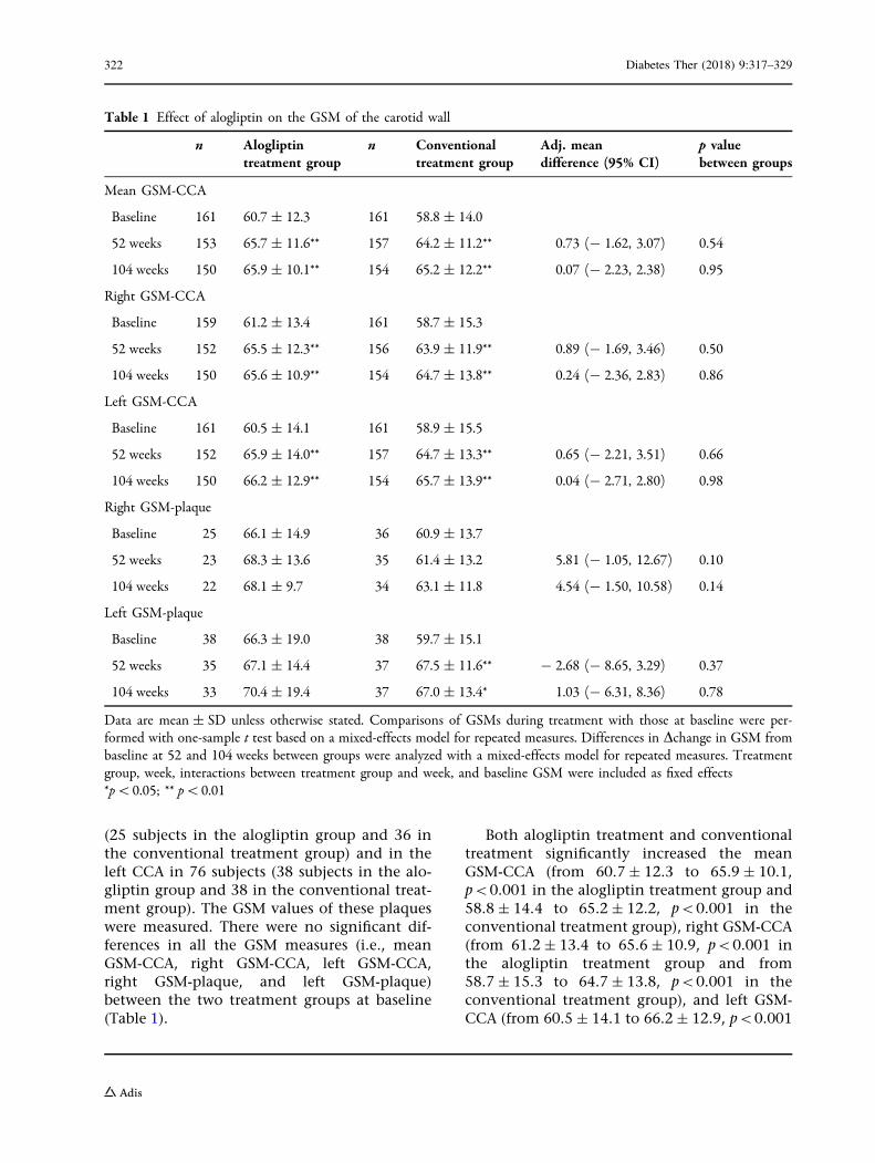
(25 subjects in the alogliptin group and 36 inthe conventional treatment group) and in theleft CCA in 76 subjects (38 subjects in the alo-gliptin group and 38 in the conventional treat-ment group). The GSM values of these plaqueswere measured. There were no significant dif-ferences in all the GSM measures (i.e., meanGSM-CCA, right GSM-CCA, left GSM-CCA,right GSM-plaque, and left GSM-plaque)between the two treatment groups at baseline(Table 1).
Both alogliptin treatment and conventionaltreatment significantly increased the meanGSM-CCA (from 60.7 ± 12.3 to 65.9 ± 10.1,p\0.001 in the alogliptin treatment group and58.8 ± 14.4 to 65.2 ± 12.2, p\0.001 in theconventional treatment group), right GSM-CCA(from 61.2 ± 13.4 to 65.6 ± 10.9, p\0.001 inthe alogliptin treatment group and from58.7 ± 15.3 to 64.7 ± 13.8, p\0.001 in theconventional treatment group), and left GSM-CCA (from 60.5 ± 14.1 to 66.2 ± 12.9, p\0.001
Table 1 Effect of alogliptin on the GSM of the carotid wall
n Alogliptintreatment group
n Conventionaltreatment group
Adj. meandifference (95% CI)
p valuebetween groups
Mean GSM-CCA
Baseline 161 60.7 ± 12.3 161 58.8 ± 14.0
52 weeks 153 65.7 ± 11.6** 157 64.2 ± 11.2** 0.73 (- 1.62, 3.07) 0.54
104 weeks 150 65.9 ± 10.1** 154 65.2 ± 12.2** 0.07 (- 2.23, 2.38) 0.95
Right GSM-CCA
Baseline 159 61.2 ± 13.4 161 58.7 ± 15.3
52 weeks 152 65.5 ± 12.3** 156 63.9 ± 11.9** 0.89 (- 1.69, 3.46) 0.50
104 weeks 150 65.6 ± 10.9** 154 64.7 ± 13.8** 0.24 (- 2.36, 2.83) 0.86
Left GSM-CCA
Baseline 161 60.5 ± 14.1 161 58.9 ± 15.5
52 weeks 152 65.9 ± 14.0** 157 64.7 ± 13.3** 0.65 (- 2.21, 3.51) 0.66
104 weeks 150 66.2 ± 12.9** 154 65.7 ± 13.9** 0.04 (- 2.71, 2.80) 0.98
Right GSM-plaque
Baseline 25 66.1 ± 14.9 36 60.9 ± 13.7
52 weeks 23 68.3 ± 13.6 35 61.4 ± 13.2 5.81 (- 1.05, 12.67) 0.10
104 weeks 22 68.1 ± 9.7 34 63.1 ± 11.8 4.54 (- 1.50, 10.58) 0.14
Left GSM-plaque
Baseline 38 66.3 ± 19.0 38 59.7 ± 15.1
52 weeks 35 67.1 ± 14.4 37 67.5 ± 11.6** - 2.68 (- 8.65, 3.29) 0.37
104 weeks 33 70.4 ± 19.4 37 67.0 ± 13.4* 1.03 (- 6.31, 8.36) 0.78
Data are mean ± SD unless otherwise stated. Comparisons of GSMs during treatment with those at baseline were per-formed with one-sample t test based on a mixed-effects model for repeated measures. Differences in Dchange in GSM frombaseline at 52 and 104 weeks between groups were analyzed with a mixed-effects model for repeated measures. Treatmentgroup, week, interactions between treatment group and week, and baseline GSM were included as fixed effects*p\0.05; ** p\0.01
322 Diabetes Ther (2018) 9:317–329

in the alogliptin treatment group and from58.9 ± 15.5 to 65.7 ± 13.9, p\0.001 in theconventional treatment group) during the104-week observation period. There was also asignificant increase in left GSM-plaque in theconventional treatment group (from59.7 ± 15.1 to 67.0 ± 13.4, p = 0.01).
The magnitude of the change in GSM valuesduring the treatment period between the twotreatment groups was compared using theMMRM (Table 1). These analyses demonstratedthat both alogliptin treatment and conven-tional treatment significantly increased all theGSM measures except for right GSM-plaque (inthe conventional treatment group). However,there was no significant difference in thechange in GSM measures from baseline at 52and 104 weeks between the two groups. Fur-thermore, similar findings were shown evenafter adjustment for possible confounding fac-tors such as age, gender, BMI, HbA1c, serumlipid levels (e.g., TC, HDL-C, TG), blood pres-sure, smoking status, and administration ofantidiabetic, antihypertensive, antihyperlipi-demic, and antiplatelet drugs (data not shown).Finally, we divided the subjects into the tertilesaccording to the level of GSM at baseline, andinvestigated the effect of alogliptin on low-GSM(echolucent) and high-GSM (echorich) plaqueseparately (Table 2). The change in right-GSM-plaque in the first tertile (lowest GSM group)was significantly greater in the alogliptin groupthan in the conventional treatment group [26.4(SE 4.6) vs. 10.9 (SE 2.9), p = 0.01]. However,there was no significant difference in thechanges in right GSM-plaque in the second andthird tertiles, and left GSM-plaque in any ter-tiles, between the two treatment groups.
Regression analyses revealed that gender andage at baseline (regression coefficient ± SE;3.93 ± 1.55, p = 0.012 and 0.17 ± 0.08,p = 0.04, respectively) were positively related tochanges in mean GSM-CCA and diastolic bloodpressure at baseline (- 0.17 ± 0.07, p = 0.01)was negatively related to changes in mean GSM-CCA. However, there was no statistically sig-nificant association between the other clinicalparameters including baseline mean IMT-CCAand mean GSM-CCA.
We also evaluated the relationship betweenthe changes in GSM during 104 weeks and thosein IMT/plaque thickness in the same site. Thechanges in mean GSM-CCA, right GSM-CCA,and left GSM-plaque were significantly associ-ated with those in IMT/plaque thickness in thesame site (r = - 0.14, p = 0.02; r = - 0.13,p = 0.02; r = - 0.28, p = 0.02, respectively),while the changes in left GSM-CCA and leftGSM-plaque were not.
DISCUSSION
We previously demonstrated that alogliptin, aDPP-4 inhibitor, more potently inhibited theprogression of carotid IMT than conventionaltreatment in patients with T2DM [29]. How-ever, few studies have evaluated the effect ofDPP-4 inhibitors on the tissue characteristics ofthe arterial wall.
The present study, a post hoc subanalysisusing data obtained from a randomized con-trolled trial that evaluated the efficacy of alo-gliptin treatment on the progression of carotidIMT in patients with T2DM, showed that alo-gliptin treatment significantly increased theGSM value, an index of ultrasonic tissue char-acteristics, of the carotid arterial wall over a104-week observation period. However, inter-estingly, conventional treatment also increasedGSM of the carotid arterial wall during this104-week period and there were no significantdifferences in the changes of GSM measuresbetween the two treatment groups.
Although the precise mechanism of the for-mation of vulnerable plaque with a lipid-richcore is unclear, it has been hypothesized thathypercholesterolemia, oxidative stress, inflam-mation, and insulin resistance are associatedwith its formation [33]. Clinical studies havealso shown that the composition of carotidplaque is related to serum lipid profiles, BMI,and inflammation markers. Our previous studyrevealed that the presence of echolucent low-GSM plaques in carotid arteries was related toserum lipid profiles and BMI [34]. Interestingly,in the present study, total cholesterol levels atthe 52-, 78-, and 104-week observation pointswere significantly decreased from the baseline
Diabetes Ther (2018) 9:317–329 323

in the conventional treatment group [29]. Sim-ilarly, total cholesterol levels at 52 and 78 weekswere significantly decreased from the baselinein the alogliptin treatment group [29]. There-fore, in both treatment groups, reduction inserum total cholesterol levels during the treat-ment period may have led to an increase inGSM of the carotid arterial wall.
This post hoc subanalysis of the SPEAD-Atrial showed that the tissue characteristics of the
arterial wall were improved in both treatmentgroups, although the original study had clearlydemonstrated that alogliptin treatment morepotently inhibited the progression of carotidIMT than conventional treatment in patientswith T2DM [29]. In addition, there was a weakbut statistical significant association betweenchanges in GSM and those in IMT or plaquethickness, suggesting that the improvement oftissue characteristics of the carotid wall
Table 2 Effect of alogliptin on the carotid plaque according to the level of baseline GSM
n Mean change (SE)of alogliptintreatment group
n Mean change (SE)of conventionaltreatment group
Adj. mean difference(95% CI)
p value betweengroups
Right GSM-plaque
First tertile
52 weeks 5 20.6 (5.2)** 14 6.6 (3.1)* 14.1 (1.3, 26.8) 0.03
104 weeks 26.4 (4.6)** 10.9 (2.9)** 15.6 (3.9, 27.2) 0.01
Second tertile
52 weeks 10 0.1 (4.2) 11 - 0.1 (4.0) 0.1 (- 12.1, 12.3) 0.99
104 weeks 0.3 (3.1) - 3.1 (3.0) 3.4 (- 5.6, 12.4) 0.44
Third tertile
52 weeks 8 - 5.2 (4.7) 10 - 8.8 (4.2) 3.6 (- 9.8, 17.0) 0.57
104 weeks - 9.2 (4.2)* - 6.6 (3.5) - 2.6 (- 14.4, 9.2) 0.65
Left GSM-plaque
First tertile
52 weeks 11 18.0 (4.0)** 14 21.2 (3.6)** - 3.2 (- 14.5, 8.0) 0.56
104 weeks 17.1 (4.6)** 19.1 (4.1)** - 2.0 (- 14.7, 10.8) 0.75
Second tertile
52 weeks 11 - 0.4 (3.5) 12 1.5 (3.3) - 1.9 (- 11.9, 8.1) 0.69
104 weeks 0.9 (4.0) 3.5 (3.7) - 2.7 (- 14.0, 8.7) 0.63
Third tertile
52 weeks 13 - 10.6 (4.1)* 11 - 6.4 (4.5) - 4.2 (- 18.0, 9.6) 0.53
104 weeks - 2.1 (5.3) - 7.6 (5.7) 5.5 (- 11.5, 22.4) 0.51
Data are mean (SE) unless otherwise stated. Comparisons of GSMs during treatment with those at baseline were performedwith one-sample t test based on a mixed-effects model for repeated measures. Differences in Dchange in GSM from baselineat 52 and 104 weeks between groups were analyzed with a mixed-effects model for repeated measures. Treatment group,week, interactions between treatment group and week, and baseline GSM were included as fixed effects*p\0.05; ** p\0.01
324 Diabetes Ther (2018) 9:317–329

contributed to the regression of the carotid wallthickness. However, the determinants of thetissue characteristics of the carotid wall andthose of the carotid IMT are not the same.Although regression of carotid IMT is supposedto be subsequent to pathological changes suchas reduction of cholesterol accumulation in thelocal site, the risk factors for the progression ofcarotid IMT are reported to include severalparameters including average HbA1c levelsduring the observation period [35]. In ourstudy, although a reduction in serum totalcholesterol levels, one of the most importantdeterminants for tissue characteristics of arterialwall, was observed in both treatment groups,reduction in HbA1c was observed only in thealogliptin treatment group [29]. For regressionof carotid IMT, therefore, improvement inhyperglycemia as well as a reduction in serumtotal cholesterol levels may be necessary inpatients with DM. Direct anti-atheroscleroticeffects of alogliptin on vascular cells is anotherpossible explanation for its beneficial effect oncarotid IMT.
There is also a possibility that the beneficialeffect of alogliptin on the tissue characteristicsof the carotid wall was masked by some con-founding factors. For example, the improve-ments in the determinants of plaqueechogenicity such as serum lipid profiles andBMI were relatively greater in the conventionaltreatment group as compared to the alogliptintreatment group in this study, althoughbetween-group differences did not reach statis-tical significance. Furthermore, the administra-tion rate of statins during the treatment periodwas also relatively higher in the conventionaltreatment group than in the alogliptin treat-ment group: the percentage of statin users was38% in the alogliptin treatment group and 46%in the conventional treatment group (p = 0.18)at baseline, and 39% and 50% (p = 0.06) at104 weeks. Since statins have a potent anti-atherogenic effect and have been reported todecrease IMT and improve the tissue character-istics of plaques in the carotid artery [36–38],such an uncontrolled imbalance in the admin-istration of statins might have masked thepotential beneficial effect of alogliptin.
There is also a possibility that the beneficialeffect of alogliptin on the carotid wall wasdependent on its characteristics at baseline.Therefore, we evaluated effect of alogliptin onlow-GSM (echolucent) and high-GSM (echor-ich) plaque separately. Interestingly, change inthe right GSM-plaque in the first tertile (lowestGSM group) was significantly greater in thealogliptin group than in the conventionaltreatment group but there was no significanttreatment-group difference in the changes ofright GSM-plaque in the second and third ter-tiles. Although these findings suggested a pos-sible beneficial effect of alogliptin on low-GSM(echolucent) plaque in the right carotid, thiswas not the case with the left carotid. This dis-crepancy could be due to lack of statisticalpower rather than bilateral difference, since thenumber of the subjects in each subgroup wasvery small. To evaluate whether the beneficialeffect of alogliptin on the carotid wall wasdependent on its characteristics at baseline,another study with larger scale is necessary.
Several limitations of our study should bediscussed. First, the present study is a post hocsubanalysis using data obtained from theSPEAD-A trial. Second, the ultrasound settingsfor each image were not always standardized.However, the blood was used as the referencefor black and the adventitia as the reference forwhite, and gain settings for measurementswithin an individual were similar throughoutthe study. Therefore, the impact of gain ofultrasound beam on the GSM value would bequite small, if any. Third, although adminis-tration of antidiabetic, antihyperlipidemic, andantihypertensive drugs may affect the plaquecomponents, the baseline medical prescriptionswere not matched completely. In addition, itwas not possible to adjust for the effect ofchanges in therapeutic regimen during theobservation period. These points should beconfirmed in further studies. Fourth, relativelylow-risk patients were enrolled in the SPEAD-Atrial: patients with a history of CVD or insulintherapy were not eligible for inclusion in thisstudy. Indeed, there were relatively few patientswho had low GSM lesions in the carotid wall.Although a previous study reported that carotidGSM values in the range of 30–40 are considered
Diabetes Ther (2018) 9:317–329 325

to be an adequate cutoff to detect individuals athigh risk for CVD [39, 40], the subjects who hadlow GSM lesions (GSM-CCA\40) in their car-otid walls at baseline represented only 10–20%of all subjects in this study. This may have led tothe beneficial effects of alogliptin on the tissuecharacteristics of the carotid wall being under-estimated. Finally, the subjects included in thisstudy were Asian T2DM patients without thesevere obesity that is often observed in non-Asian T2DM patients. It would thus be prema-ture to generalize our findings to non-Asianpopulations.
CONCLUSIONS
A post hoc subanalysis suggests that the tissuecharacteristics of the carotid arterial wall wereimproved in both the alogliptin treatmentgroup and the conventional treatment groupduring the 104-week treatment period and thatthere was no significant difference between thetreatment groups. Prespecified studies withlarge sample size would be necessary to confirmour findings.
ACKNOWLEDGEMENTS
Funding. This research was supported byGrants-in-Aid for Scientific Research from theJapanese Ministry of Education, Science, Sports,Culture and Technology (KAKENHI 16K09747).The journal’s article processing charges werealso funded from the KAKENHI 16K09747.
Medical Writing Assistance. The authorsgratefully acknowledge the assistance of H.Yamada and D. Takayama (Soiken Holdings Inc,Tokyo Japan) in performing statistical analysisand R. Kondo (WILL Medical Communications)in proofreading the manuscript. All authors hadfull access to all of the data in this study andtake complete responsibility for the integrity ofthe data and accuracy of the data analysis.
Authorship. All named authors meet theInternational Committee of Medical Journal
Editors (ICMJE) criteria for authorship for thismanuscript, take responsibility for the integrityof the work as a whole, and have given finalapproval for the version to be published.
Disclosures. Yoko Irie has nothing to dis-close. Naoto Katakami holds an endowed chair(Department of Metabolism and Atherosclero-sis) established by funds from Kowa Pharma-ceutical Co., has received research funds fromMSD and lecture fees from Arkray Co. Ltd.,Astellas Pharma Inc., Boehringer Ingelheim,Daiichi Sankyo Inc., Dainippon SumitomoPharma Co., Eli Lilly, Kowa Pharmaceutical Co.,Kyowa Hakko Kirin Co. Ltd., Mitsubishi TanabePharma Co., Novo Nordisk Pharma, Ono Phar-maceutical Co., Takeda Pharmaceutical Co.,Sanofi-Aventis, and Shionogi & Co. TomoyaMita received research funds from MSD andTakeda Pharma K.K. and has received lecturefees from AstraZeneca K.K., Boehringer Ingel-heim, Eli Lilly, Kowa Pharmaceutical Co., Mit-subishi Tanabe Pharma Co., MSD, OnoPharmaceutical Co., and Takeda PharmaceuticalCo. Mitsuyoshi Takahara holds an endowedchair (Department of Diabetes Care Medicine)established by AstraZeneca K.K., BoehringerIngelheim, Mitsubishi Tanabe Pharma Co.,MSD, Novo Nordisk Pharma, Ono Pharmaceu-tical Co., Taisho Toyama Pharmaceutical Co.Taka-aki Matsuoka is an associate professor(Department of Metabolic Medicine) andreceived research funds from Daiichi SankyoInc., Ono Pharmaceutical Co, Eli Lilly, NovoNordisk Pharma, Takeda Pharmaceutical Co.and lecture fees from MSD, Astellas PharmaInc., Boehringer Ingelheim, Daiichi Sankyo Inc.,Dainippon Sumitomo Pharma Co., Eli Lilly,Kowa Pharmaceutical Co., Kyowa Hakko KirinCo. Ltd., Mitsubishi Tanabe Pharma Co., NovoNordisk Pharma, Ono Pharmaceutical Co., Kis-sei Pharmaceutical Co., Taisho Toyama Phar-maceutical Co., Ltd., Sanwa Kagaku KenkyushoCo., Sanofi Co. Masahiko Gosho received lec-ture and/or consultant fees from Daiichi SankyoCompany, Ltd., Novartis, Taiho Pharma, andFerring Pharma, received travel fees fromTakeda Pharmaceutical Co., and receivedmanuscript fees from Kowa Co. Ltd. HirotakaWatada received lecture fees from Novo
326 Diabetes Ther (2018) 9:317–329
Nordisk, Inc., Eli Lilly and Company, Sanofi,Dainippon Sumitomo Pharma Co., Fujifilm,Bayer Health Care, Kissei Pharmaceutical Com-pany, Mochida Pharmaceutical Company, MSD,Takeda Pharmaceutical Company, BoehringerIngelheim Pharmaceuticals Inc., Daiichi-San-kyo, Ono Pharmaceutical Co. Ltd., NovartisPharmaceuticals Corporation, Mitsubishi Tan-abe Pharma Corporation, AstraZeneca LP,Kyowa Hakko Kirin Company Ltd., SanwaKagaku Kenkyusho Co. Ltd., Kowa CompanyLtd., Astellas Pharma Inc.; advisory fees fromNovo Nordisk, Inc., Mochida Pharma Com-pany, AstraZeneca LP, Kowa Company, AstellasPharma Inc., Sanofi, Boehringer IngelheimPharmaceuticals Inc., MSD, Mitsubishi TanabePharma Corporation, Novartis PharmaceuticalsCorporation, Dainippon Sumitomo PharmaCo., Takeda Pharmaceutical Company, OnoPharmaceutical Co., Pfizer Inc., and KowaCompany; and research funds from BoehringerIngelheim, Pfizer, Mochida Pharmaceutical Co.,Sanofi-Aventis, Novo Nordisk Pharma, NovartisPharmaceuticals, Sanwakagaku Kenkyusho,Terumo Corp. Eli Lilly, Mitsubishi TanabePharma, Daiichi Sankyo Inc., Takeda Pharma-ceutical Co., MSD, Shionogi, Pharma, Dainip-pon Sumitomo Pharma, Kissei Pharma, andAstrazeneca K.K. Iichiro Shimomura receivedlecture fees from Astellas Pharma Inc., AstraZe-neca K.K., MSD K.K., Ono Pharmaceutical Co.,Kyowa Hakko Kirin Co., Kowa PharmaceuticalCo., Sanofi K.K., Sanwa Kagaku Kenkyusho Co.,Daiichi Sankyo Co., Takeda Pharma K.K., Mit-subishi Tanabe Pharma Co., Teijin Pharma, EliLilly Japan K.K., Nippon Boehringer IngelheimCo., Novartis Pharma K.K., Novo NordiskPharma, Bayer Yakuhin, Pfizer Japan Inc., Bris-tol-Myers K.K., Mochida Pharmaceutical Co.,Shionogi & Co., and Taisho Toyama Pharma-ceutical Co., and research funds from AstellasPharma Inc., AstraZeneca K.K., Eisai Co., MSDK.K, Otsuka Pharmaceutical Co., Ono Pharma-ceutical Co., Kaken Pharmaceutical Co., KisseiPharmaceutical Co., Kyowa Hakko Kirin Co.,Sanofi K.K., Shionogi & Co., Daiichi Sankyo Co.,Dainippon Sumitomo Pharma Co., TakedaPharma K.K., Mitsubishi Tanabe Pharma Co.,Teijin Pharma, Nippon Boehringer IngelheimCo., Novartis Pharma K.K., Novo Nordisk
Pharma, Pfizer Japan Inc., Bristol-Myers K.K.,Mochida Pharmaceutical Co., Eli Lilly JapanK.K, Kowa Co., Ltd., Kowa Pharmaceutical Co.,and Taisho Toyama Pharmaceutical Co.
Compliance with Ethics Guidelines. Thisarticle is based on previously conducted studiesand does not involve any new studies of humanor animal subjects performed by any of theauthors.
Open Access. This article is distributedunder the terms of the Creative CommonsAttribution-NonCommercial 4.0 InternationalLicense (http://creativecommons.org/licenses/by-nc/4.0/), which permits any noncommer-cial use, distribution, and reproduction in anymedium, provided you give appropriate creditto the original author(s) and the source, providea link to the Creative Commons license, andindicate if changes were made.
REFERENCES
1. Fuster V, Badimon L, Badimon JJ, Chesebro JH. Thepathogenesis of coronary artery disease and theacute coronary syndromes. N Engl J Med.1992;326:242–50.
2. Lee RT, Grodzinsky AJ, Frank EH, Kamm RD,Schoen FJ. Structure-dependent dynamic mechani-cal behavior of fibrous caps from humanatherosclerotic plaques. Circulation.1991;83:1764–70.
3. Davies MJ, Richardson PD, Woolf N, Katz DR, MannJ. Risk of thrombosis in human atheroscleroticplaques: role of extracellular lipid, macrophage, andsmooth muscle cell content. Br Heart J.1993;69:377–81.
4. Falk E. Pathogenesis of atherosclerosis. J Am CollCardiol. 2006;47:C7–12.
5. Creager MA, Luscher TF, Cosentino F, Beckman JA.Diabetes and vascular disease: pathophysiology,clinical consequences, and medical therapy: part I.Circulation. 2003;108:1527–32.
6. Grønholdt ML, Wiebe BM, Laursen H, Nielsen TG,Schroeder TV, Sillesen H. Lipid-rich carotid arteryplaques appear echolucent on ultrasound B-modeimages and may be associated with intraplaque
Diabetes Ther (2018) 9:317–329 327
haemorrhage. Eur J Vasc Endovasc Surg.1997;14:439–45.
7. Nicolaides AN, Kakkos SK, Kyriacou E, et al.Asymptomatic internal carotid artery stenosis andcerebrovascular risk stratification. J Vasc Surg.2010;52:1486–96.
8. Irie Y, Katakami N, Kaneto H, et al. The utility ofultrasonic tissue characterization of carotid plaquein the prediction of cardiovascular events in dia-betic patients. Atherosclerosis. 2013;230:399–405.
9. Grønholdt ML, Nordestgaard BG, Schroeder TV,Vorstrup S, Sillesen H. Ultrasonic echolucent car-otid plaques predict future strokes. Circulation.2001;104:68–73.
10. Fadini GP, Avogaro A. Cardiovascular effects ofDPP-4 inhibition: beyond GLP-1. Vasc Pharmacol.2011;55:10–6.
11. Barrera JG, Sandoval DA, D’Alessio DA, Seeley RJ.GLP-1 and energy balance: an integrated model ofshort-term and long-term control. Nat Rev Endo-crinol. 2011;7:507–16.
12. Lovshin JA, Drucker DJ. Incretin-based therapies fortype 2 diabetes mellitus. Nat Rev Endocrinol.2009;5:262–9.
13. Ban K, Noyan-Ashraf MH, Hoefer J, Bolz SS, DruckerDJ, Husain M. Cardioprotective and vasodilatoryactions of glucagon-like peptide 1 receptor aremediated through both glucagon-like peptide 1receptor dependent and independent pathways.Circulation. 2008;117:2340–50.
14. Liu H, Dear AE, Knudsen LB, Simpson RW. A long-acting glucagon-like peptide-1 analogue attenuatesinduction of plasminogen activator inhibitor type-1and vascular adhesion molecules. J Endocrinol.2009;201:59–66.
15. Hattori Y, Jojima T, Tomizawa A, et al. A glucagon-like peptide-1 (GLP-1) analogue, liraglutide, upreg-ulates nitric oxide production and exerts anti-in-flammatory action in endothelial cells.Diabetologia. 2010;53:2256–63.
16. Arakawa M, Mita T, Azuma K, et al. Inhibition ofmonocyte adhesion to endothelial cells and atten-uation of atherosclerotic lesion by a glucagon-likepeptide-1 receptor agonist, exendin-4. Diabetes.2010;59:1030–7.
17. Gaspari T, Liu H, Welungoda I, et al. A GLP-1receptor agonist liraglutide inhibits endothelial celldysfunction and vascular adhesion moleculeexpression in an ApoE-/- mouse model. DiabetesVasc Dis Res. 2011;8:117–24.
18. Nagashima M,Watanabe T, Terasaki M, et al. Nativeincretins prevent the development of atheroscle-rotic lesions in apolipoprotein E knockout mice.Diabetologia. 2011;54:2649–59.
19. Goto H, Nomiyama T, Mita T, et al. Exendin-4, aglucagonlike peptide-1 receptor agonist, reducesintimal thickening after vascular injury. BiochemBiophys Res Commun. 2011;405:79–84.
20. Tanaka M, Matsuo Y, Yamakage H, et al. Differen-tial effects of GLP-1 receptor agonist on foam cellformation in monocytes between non-obese andobese subjects. Metabolism. 2016;65:1–11.
21. Terasaki M, Nagashima M, Nohtomi K, et al.Preventive effect of dipeptidyl peptidase-4 inhibitoron atherosclerosis is mainly attributable to incre-tin’s actions in nondiabetic and diabeticapolipoprotein E-null mice. PLoS One.2013;8:e70933.
22. Terasaki Y, Nomiyama T, Kawanami T, et al.Dipeptidyl peptidase-4 inhibitor linagliptin atten-uates neointima formation after vascular injury.Cardiovasc Diabetol. 2014;13:154.
23. Nader MA. Sitagliptin ameliorates lipid profilechanges and endothelium dysfunction induced byatherogenic diet in rabbits. Naunyn SchmiedebergsArch Pharmacol. 2014;387:433–44.
24. Hirano T, Mori Y. Anti-atherogenic and anti-in-flammatory properties of glucagon-like peptide-1,glucose-dependent insulinotropic polypepide, anddipeptidyl peptidase-4 inhibitors in experimentalanimals. J Diabetes Investig. 2016;7:80–6.
25. Balestrieri ML, Rizzo MR, Barbieri M, et al. Sirtuin 6expression and inflammatory activity in diabeticatherosclerotic plaques: effects of incretin treat-ment. Diabetes. 2015;64:1395–406.
26. Vittone F, Liberman A, Vasic D, et al. Sitagliptinreduces plaque macrophage content and stabilisesarteriosclerotic lesions in Apoe (-/-) mice. Dia-betologia. 2012;55:2267–75.
27. Satoh-Asahara N, Sasaki Y, Wada H, et al. A dipep-tidyl peptidase-4 inhibitor, sitagliptin, exerts anti-inflammatory effects in type 2 diabetic patients.Metabolism. 2013;62:347–51.
28. Tremblay AJ, Lamarche B, Deacon CF, Weisnagel SJ,Couture P. Effects of sitagliptin therapy on markersof low-grade inflammation and cell adhesionmolecules in patients with type 2 diabetes. Meta-bolism. 2014;63:1141–8.
29. Mita T, Katakami N, Yoshii H, et al. Alogliptin, adipeptidyl peptidase 4 inhibitor, prevents the pro-gression of carotid atherosclerosis in patients with
328 Diabetes Ther (2018) 9:317–329
type 2 diabetes: the study of preventive effects ofalogliptin on diabetic atherosclerosis (SPEAD-A).Diabetes Care. 2016;39:139–48.
30. Terminology and Diagnostic Criteria Committee,Japan Society of Ultrasonics in Medicine, Subcom-mittee for Preparing Guidelines for UltrasoundDiagnosis of Carotid Artery. Standard method forultrasound evaluation of carotid artery lesions. JpnJ Med Ultrason. 2009;36:501–18.
31. Yanase T, Nasu S, Mukuta Y, et al. Evaluation of anew carotid intima-media thickness measurementby B-mode ultrasonography using an innovativemeasurement software, intimascope. Am J Hyper-tens. 2006;19:1206–12.
32. Sabetai MM, Tegos TJ, Nicolaides AN, Dhanjil S,Pare GJ, Stevens JM. Reproducibility of computer-quantified carotid plaque echogenicity: can weovercome the subjectivity? Stroke.2000;31:2189–96.
33. Libby P, Aikawa M. Stabilization of atheroscleroticplaques: new mechanisms and clinical targets. NatMed. 2002;8:1257–62.
34. Irie Y, Katakami N, Kaneto H, et al. The risk factorsassociated with ultrasonic tissue characterization ofcarotid plaque in type 2 diabetic patients. J DiabetesComplicat. 2014;28:523–7.
35. Yamasaki Y, Kodama M, Nishizawa H, et al. Carotidintima-media thickness in Japanese type 2 diabeticsubjects: predictors of progression and relationshipwith incident coronary heart disease. Diabetes Care.2000;9:1310–5.
36. Katakami N, Sakamoto K, Kaneto H, et al. Lipid-lowering with atorvastatin improves tissue charac-teristics of carotid plaque. Atherosclerosis.2005;183:369–71.
37. Crisby M, Nordin-Fredriksson G, Shah PK, Yano J,Zhu J, Nilsson J. Pravastatin treatment increasescollagen content and decreases lipid content,inflammation, metalloproteinases, and cell death inhuman carotid plaques: implications for plaquestabilization. Circulation. 2001;103:926–33.
38. Makris GC, Lavida A, Nicolaides AN, Geroulakos G.The effect of statins on carotid plaque morphology:an LDL-associated action or one more pleiotropiceffect of statins? Atherosclerosis. 2010;213:8–20.
39. Falkowski A, Kaczmarczyk M, Cieszanowski A,Goracy I, Poncyliusz W, Wilk G. Computer-assistedcharacterisation of a carotid plaque. Med Sci Monit.2004;10:67–70.
40. Nicolaides AN, Kakkos SK, Kyriacou E, et al.Asymptomatic internal carotid artery stenosis andcerebrovascular risk stratification. J Vasc Surg.2010;52:1486–96.
Diabetes Ther (2018) 9:317–329 329