EVALUATION OF SURGICAL SITE INFECTION IN ABDOMINAL ...
149
A Dissertation on EVALUATION OF SURGICAL SITE INFECTION IN ABDOMINAL SURGERIES IN ADULTS Dissertation submitted In partial fulfilment of the regulations For the award of the degree of M.S.DEGREE BRANCH-I GENERAL SURGERY Of THE TAMILNADU DR.M.G.R. MEDICAL UNIVERSITY E.S.I.C. MEDICAL COLLEGE & PGIMSR, K.K. NAGAR, CHENNAI. APRIL 2020
Transcript of EVALUATION OF SURGICAL SITE INFECTION IN ABDOMINAL ...
A Dissertation on
EVALUATION OF SURGICAL SITE INFECTION
IN ABDOMINAL SURGERIES IN ADULTS
Dissertation submitted
In partial fulfilment of the regulations
For the award of the degree of
M.S.DEGREE BRANCH-I
GENERAL SURGERY
Of
THE TAMILNADU DR.M.G.R. MEDICAL UNIVERSITY
E.S.I.C. MEDICAL COLLEGE & PGIMSR,
K.K. NAGAR, CHENNAI.
APRIL 2020
ii
DECLARATION BY THE CANDIDATE
I solemnly declare that this dissertation entitled “EVALUATION OF
SURGICAL SITE INFECTION IN ABDOMINAL SURGERIES IN
ADULTS” is a bonafide and genuine research work carried out by me under the
guidance of Dr.P.N.SHANMUGA SUNDARAM,M.S., Department of General
Surgery, ESIC-Medical College & PGIMSR, K.K.Nagar, Chennai-78.
This dissertation is being submitted to The TamilNadu Dr.M.G.R Medical
University, Chennai, towards partial fulfilment of requirements of the degree of
M.S.[General Surgery] examination to be held in April 2020.
SIGNATUE OF THE CANDIDATE
Dr.P. DHANASEKARAN
Post Graduate student,
M.S General Surgery,
Dept. of General Surgery,
ESIC Medical College &PGIMSR,
Date: K.K Nagar,Chennai – 600078.
Place:
iii
DECLARATION BY THE CANDIDATE
I hereby declare that The Tamilnadu Dr. M.G.R. Medical University,
Chennai, shall have the rights to preserve, use and disseminate this
dissertation/thesis in print/electronic format for academic/ research purpose.
SIGNATURE OF THE CANDIDATE
Dr.P.DHANASEKARAN
Date:
Place:
THE TAMILNADU Dr. M.G.R. MEDICAL UNIVERSITY, CHENNAI.
iv
ENDORSEMENT BY THE DEAN
This is to certify that the dissertation entitled “EVALUATION OF
SURGICAL SITE INFECTION IN ABDOMINAL SURGERIES IN
ADULTS” is a bonafide research work done by Dr.P. DHANASEKARAN, Post
graduate resident in M.S.(General Surgery),ESIC Medical College &
PGIMSR,K.K.Nagar, Chennai-78 under direct guidance and supervision of
Dr. P.N. SHANMUGA SUNDARAM M.S, PROFESSOR & THE HEAD OF
THE DEPARTMENT, Dept of General Surgery, ESIC MEDICAL COLLEGE
&PGIMSR, K.K. NAGAR, CHENNAI-78 in partial fulfilment of the requirements
for the degree of M.S. General Surgery of The Tamilnadu Dr. M.G.R. Medical
University, Chennai. I forward this to The Tamilnadu Dr. M.G.R. Medical
University, Chennai, Tamilnadu.
DEAN
Dr.SOWMYA SAMPATH,
ESIC MEDICAL COLLEGE & PGIMSR,
K.K.NAGAR, CHENNAI-78
v
CERTIFICATE OF THE HEAD OF DEPARTMENT
This is to certify that the dissertation titled “EVALUATION OF
SURGICAL SITE INFECTION IN ABDOMINAL SURGERIES IN
ADULTS” is a bonafide research work done by Dr. P. DHANASEKARAN, in
partial fulfilment of the regulations for the degree of M.S. in General Surgery.
Dr.P.N.Shanmugasundaram. M.S,
Professor & HOD,
Department of General Surgery,
ESIC MEDICAL COLLEGE &PGIMSR,
K.K.NAGAR, CHENNAI.
Date:
Place:
vi
CERTIFICATE OF THE GUIDE
This is to certify that this dissertation entitled “EVALUATION OF SURGICAL SITE
INFECTION IN ABDOMINAL SURGERIES IN ADULTS” submitted by Dr. P.
DHANASEKARAN , appearing for M.S. Degree Branch- I General Surgery examination in
April 2020 is a bonafide research work done by him under my direct guidance and supervision in
partial fulfilment of the regulations of the Tamilnadu Dr.M.G.R. Medical University, Chennai. I
forward this to the Tamilnadu Dr.M.G.R. Medical University, Chennai, Tamilnadu, India.
Dr.P.N.Shanmugasundaram. M.S,
Professor & HOD,
Department of General Surgery,
ESIC MEDICAL COLLEGE &PGIMSR,
K.K.NAGAR, CHENNAI.
Date:
Place:
vii
CERTIFICATE OF THE CO-GUIDE
This is to certify that the dissertation titled “EVALUATION OF
SURGICAL SITE INFECTION IN ABDOMINAL SURGERIES IN
ADULTS” is a bonafide research work done by Dr.P.DHANASEKARAN, under
guidance of Dr.P.N.Shanmugasundaram Professor and HOD of Department of
General Surgery ESIC Medical College and PGIMSR, K.K.Nagar, Chennai-78 in
partial fulfilment of the requirement for the degree of M.S. in General Surgery.
Dr.Bhanumati Giridharan. M.S,
Associate professor
Department of General Surgery,
ESIC MEDICAL COLLEGE &PGIMSR,
K.K. NAGAR, CHENNAI-78
Date:
Place:
viii
ACKNOWLEDGEMENT
I am grateful to the Dean Dr. Sowmya Sampath , for permitting me to
conduct the study and utilize the resources of the college.
I avail this opportunity to express my gratitude to my beloved teacher, Guide
and Mentor, Prof. Dr.P.N. Shanmuga Sundaram,M.S, Professor & Head of the
Department, Dept. of General Surgery for his constant guidance ,support and
interest in my academic progress. The work and time spent in this study has given
me a clear vision in approaching a clinical study and documentation of
observations made. Throughout the study he has been my driving force in pursuing
and completing the study in meticulous manner.
I would also like to thank Dr.Bhanumathi Giridharan M.S,Associate
Professor, Co-guide for her blessings and moral support in all my professional
duties I carry out.
I express my deepest sense of thankfulness to my other Associate Professors
Dr. Uday S Kumbhar, Dr. Muthuraj, Dr.Murugesan , Dr. Gajendraraj,
Assistant Professors, Dr.NapaMadhusudhan, Dr.M.Vishwanathan,
Dr.R.Prabhakar, Dr.A.E. Poornima, Dr.Pankaj Surana, Dr. Byomakesh
Patro, Senior Residents , Dr.G.Vijayalakshmi, Dr.A.Balasubramaniam,
Dr.M.Vasanth, Dr.Arunraj, Dr.K.Lohitsai, Dr. Sarath Kumar, Dr. Sivaranjit
ix
kumar for their valuable inputs and constant encouragement without which this
dissertation could not have been completed.
Many thanks in particular to the Chairman and Members of the Institutional
Ethical committee for approving our study and for their valuable suggestions. I
thank our Biostatistician Dr. Aruna B.Patil MSc.Phd(Statistics), Asst.Professor in
Dept of Community Medicine , ESIC Medical College and PGIMSR, Chennai -78,
for her passionate guidance and enlightening knowledge with which we were able
to commute sample size and Data analysis.
I express my sincere thanks to my fellow post graduates Dr. Dinesh &
Dr. Amudhan and junior colleagues for their support and help in completing this
dissertation. It is my earnest duty to thank my family without whom accomplishing
this task would have been impossible.
My heartfelt thanks go to each and every patient who agreed to be a part of
this study and also my apologies to them in case of any inconvenience caused.
x
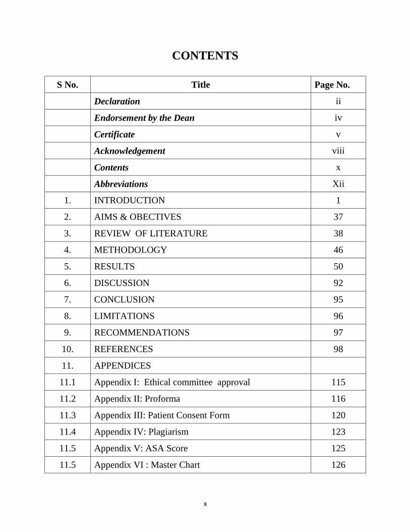
CONTENTS
S No. Title Page No.
Declaration ii
Endorsement by the Dean iv
Certificate v
Acknowledgement viii
Contents x
Abbreviations Xii
1. INTRODUCTION 1
2. AIMS & OBECTIVES 37
3. REVIEW OF LITERATURE 38
4. METHODOLOGY 46
5. RESULTS 50
6. DISCUSSION 92
7. CONCLUSION 95
8. LIMITATIONS 96
9. RECOMMENDATIONS 97
10. REFERENCES 98
11. APPENDICES
11.1 Appendix I: Ethical committee approval 115
11.2 Appendix II: Proforma 116
11.3 Appendix III: Patient Consent Form 120
11.4 Appendix IV: Plagiarism 123
11.5 Appendix V: ASA Score 125
11.5 Appendix VI : Master Chart 126
xi
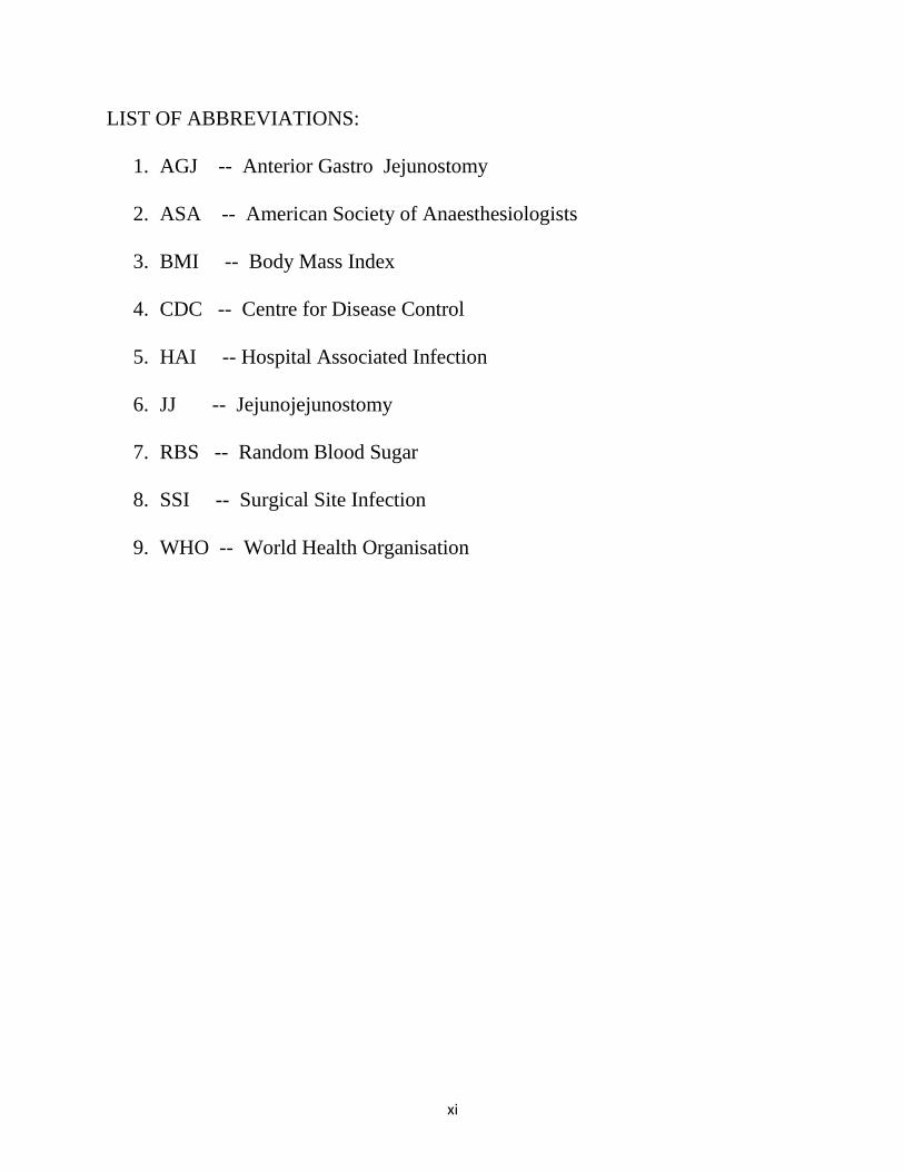
LIST OF ABBREVIATIONS:
1. AGJ -- Anterior Gastro Jejunostomy
2. ASA -- American Society of Anaesthesiologists
3. BMI -- Body Mass Index
4. CDC -- Centre for Disease Control
5. HAI -- Hospital Associated Infection
6. JJ -- Jejunojejunostomy
7. RBS -- Random Blood Sugar
8. SSI -- Surgical Site Infection
9. WHO -- World Health Organisation
1
INTRODUCTION
Surgical site infections (SSIs) are infections of the incision or organ or space
that occur after surgery 1. The term ‘surgical site infection’ (SSI) was introduced in
1992 to replace the previous term ‘surgical wound infection’ 2. Surgical site
infection (SSI) has always been a major complication of surgery and trauma and
has been documented for 4000–5000 years3.
SSI is both the most frequently
studied and the leading HAI reported hospital-wide in LMICs 4, 5.
World Health
Organization (WHO) Clean Care is Safer Care programme shows that surgical site
infection (SSI) affects up to one third of patients who have undergone a surgical
procedure in LMICs and the pooled incidence of SSI was 11.8 per 100 surgical
patients undergoing the procedure (range 1.2 to 23.6) 4, 5
.
Although SSI incidence is much lower in high-income countries, it remains
the second most frequent type of HAI in Europe and the United States of America
(USA). In some European countries, it even represents the most frequent type of
HAI.
SSIs are among the most preventable HAIs 6, 7
, but they still represent a
significant burden in terms of patient morbidity and mortality and additional costs
to health systems and service payers worldwide. Each SSI is associated with
approximately 7-10 additional postoperative hospital days and patients with an SSI
2
have a 2-11 times higher risk of death, compared with operative patients without
an SSI 8, 9.
Surgical patients initially seen with more complex co morbidities and the
emergence of antimicrobial-resistant pathogens increase the cost and challenge of
treating SSIs10, 11, 12.
For these reasons, the prevention of SSI has received
considerable attention from surgeons and infection control professionals, health
care authorities, the media and the public.
DEFINITION OF SSI:
Surgical site infection refers to an infection that occurs after surgery in the
part of the body where the surgery took place. Surgical site infections can
sometimes be superficial infections involving the skin only. Other surgical site
infections are more serious and can involve tissues under the skin, organs, or
implanted material.
(Source: United States Centers for Disease Control and Prevention.
https://www.cdc.gov/HAI/ssi/ssi.html, accessed 11July 2016.).
Surgical site infection is also defined as an infection that occurs within 30
days after the operation and involves the skin and subcutaneous tissue of the
incision (superficial incisional) and/or the deep soft tissue (for example, fascia,
muscle) of the incision (deep incisional) and/or any part of the anatomy (for
3
example, organs and spaces) other than the incision that was opened or
manipulated during an operation (organ/space).
(Source: European Centre for Disease Prevention and Control.
http://ecdc.europa.eu/en/publications/Publications/120215_TED_SSI_protocol.pdf
, accessed 16 August 2016).
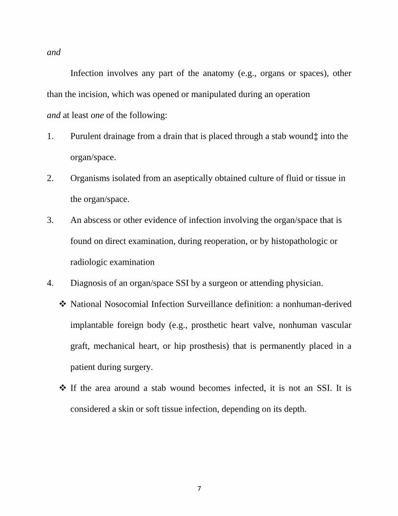
CRITERIA FOR DEFINING SURGICAL SITE INFECTION2:
The CDC’s NNIS system has developed standardized surveillance criteria
for defining SSIs .By these criteria, SSIs are classified as being either incisional or
organ/space. Incisional SSIs are further divided into those involving only skin and
subcutaneous tissue (superficial incisional SSI) and those involving deeper soft
tissues of the incision (deep incisional SSI). Organ/space SSIs involve any part of
the anatomy (e.g., organ or space) other than incised body wall layers, that was
Opened or manipulated during an operation.
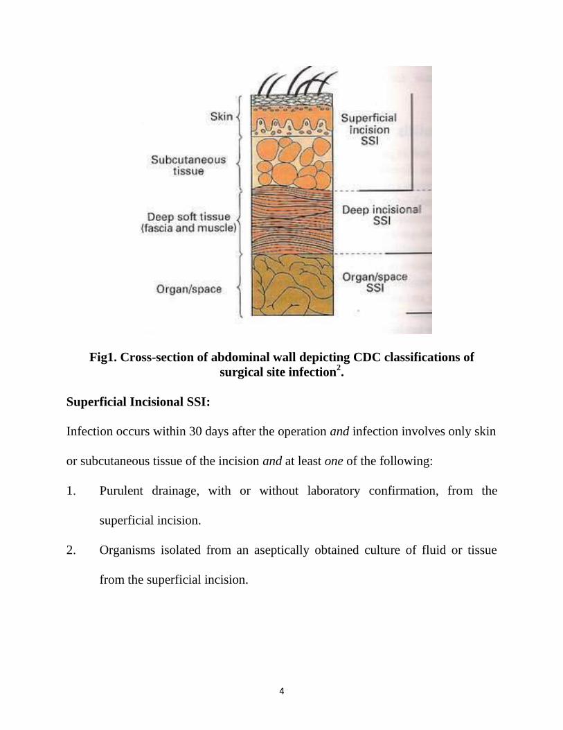
4
Fig1. Cross-section of abdominal wall depicting CDC classifications of
surgical site infection2.
Superficial Incisional SSI:
Infection occurs within 30 days after the operation and infection involves only skin
or subcutaneous tissue of the incision and at least one of the following:
1. Purulent drainage, with or without laboratory confirmation, from the
superficial incision.
2. Organisms isolated from an aseptically obtained culture of fluid or tissue
from the superficial incision.
5
3. At least one of the following signs or symptoms of infection: pain or
tenderness, localized swelling, redness, or heat and superficial incision is
deliberately opened by surgeon, unless incision is culture-negative.
4. Diagnosis of superficial incisional SSI by the surgeon or attending
physician.
Do not report the following conditions as SSI:
1. Stitch abscess (minimal inflammation and discharge confined to the points
of suture penetration).
2. Infection of an episiotomy or newborn circumcision site.
3. Infected burn wound.
4. Incisional SSI that extends into the fascial and muscle layers (see deep
incisional SSI).
Note: Specific criteria are used for identifying infected episiotomy and
circumcision sites and burn wounds.
Deep Incisional SSI:
Infection occurs within 30 days after the operation if no implant† is left in
place or within 1 year if implant is in place and the infection appears to be related
to the operation
and
infection involves deep soft tissues (e.g., fascial and muscle layers) of the incision
6
and at least one of the following:
1. Purulent drainage from the deep incision but not from the organ/space
component of the surgical site.
2. A deep incision spontaneously dehisces or is deliberately opened by a
surgeon when the patient has at least one of the following signs or
symptoms: fever (>38ºC), localized pain, or tenderness, unless site is
culture-negative.
3. An abscess or other evidence of infection involving the deep incision is
found on direct examination, during reoperation, or by histopathologic or
radiologic examination.
4. Diagnosis of a deep incisional SSI by a surgeon or attending physician.
Notes:
1. Report infection that involves both superficial and deep incision sites as
deep incisional SSI.
2. Report an organ/space SSI that drains through the incision as a deep
incisional SSI.
Organ/Space SSI:
Infection occurs within 30 days after the operation if no implant† is left in
place or within 1 year if implant is in place and the infection appears to be related
to the operation
7
and
Infection involves any part of the anatomy (e.g., organs or spaces), other
than the incision, which was opened or manipulated during an operation
and at least one of the following:
1. Purulent drainage from a drain that is placed through a stab wound‡ into the
organ/space.
2. Organisms isolated from an aseptically obtained culture of fluid or tissue in
the organ/space.
3. An abscess or other evidence of infection involving the organ/space that is
found on direct examination, during reoperation, or by histopathologic or
radiologic examination
4. Diagnosis of an organ/space SSI by a surgeon or attending physician.
National Nosocomial Infection Surveillance definition: a nonhuman-derived
implantable foreign body (e.g., prosthetic heart valve, nonhuman vascular
graft, mechanical heart, or hip prosthesis) that is permanently placed in a
patient during surgery.
If the area around a stab wound becomes infected, it is not an SSI. It is
considered a skin or soft tissue infection, depending on its depth.
8
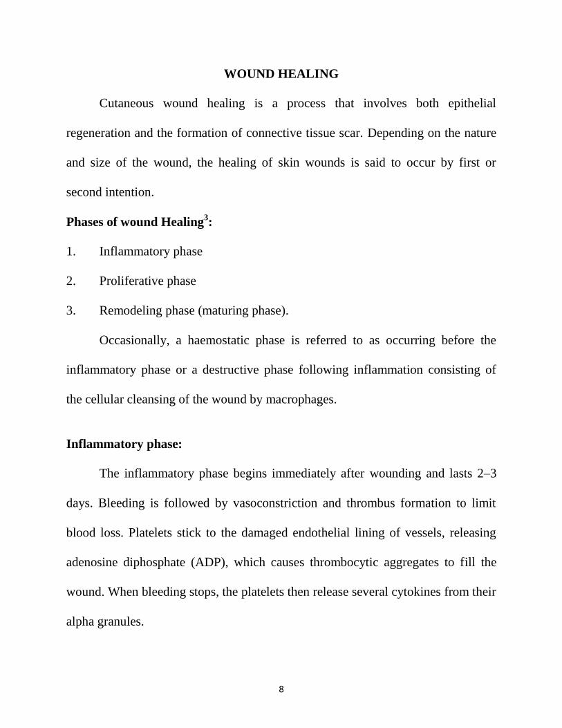
WOUND HEALING
Cutaneous wound healing is a process that involves both epithelial
regeneration and the formation of connective tissue scar. Depending on the nature
and size of the wound, the healing of skin wounds is said to occur by first or
second intention.
Phases of wound Healing3:
1. Inflammatory phase
2. Proliferative phase
3. Remodeling phase (maturing phase).
Occasionally, a haemostatic phase is referred to as occurring before the
inflammatory phase or a destructive phase following inflammation consisting of
the cellular cleansing of the wound by macrophages.
Inflammatory phase:
The inflammatory phase begins immediately after wounding and lasts 2–3
days. Bleeding is followed by vasoconstriction and thrombus formation to limit
blood loss. Platelets stick to the damaged endothelial lining of vessels, releasing
adenosine diphosphate (ADP), which causes thrombocytic aggregates to fill the
wound. When bleeding stops, the platelets then release several cytokines from their
alpha granules.
9
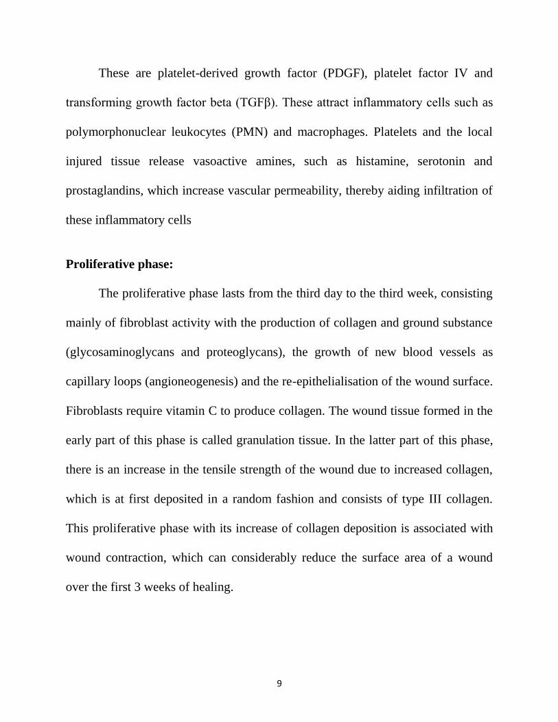
These are platelet-derived growth factor (PDGF), platelet factor IV and
transforming growth factor beta (TGFβ). These attract inflammatory cells such as
polymorphonuclear leukocytes (PMN) and macrophages. Platelets and the local
injured tissue release vasoactive amines, such as histamine, serotonin and
prostaglandins, which increase vascular permeability, thereby aiding infiltration of
these inflammatory cells
Proliferative phase:
The proliferative phase lasts from the third day to the third week, consisting
mainly of fibroblast activity with the production of collagen and ground substance
(glycosaminoglycans and proteoglycans), the growth of new blood vessels as
capillary loops (angioneogenesis) and the re-epithelialisation of the wound surface.
Fibroblasts require vitamin C to produce collagen. The wound tissue formed in the
early part of this phase is called granulation tissue. In the latter part of this phase,
there is an increase in the tensile strength of the wound due to increased collagen,
which is at first deposited in a random fashion and consists of type III collagen.
This proliferative phase with its increase of collagen deposition is associated with
wound contraction, which can considerably reduce the surface area of a wound
over the first 3 weeks of healing.
10
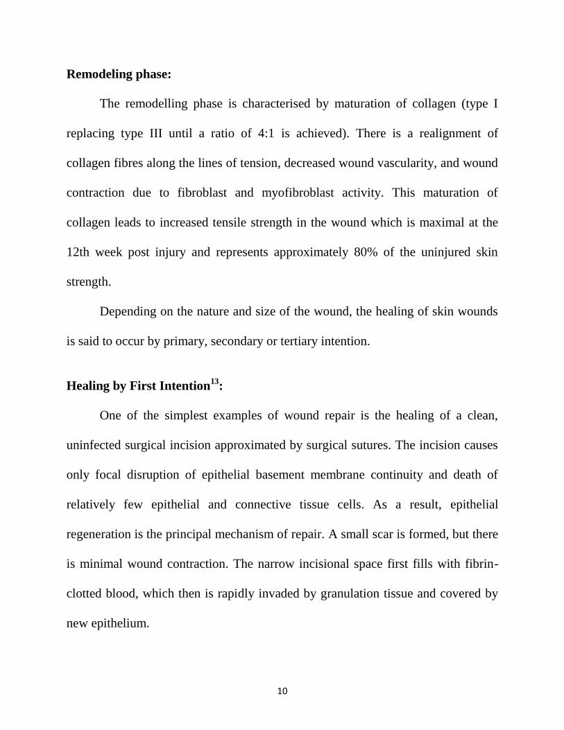
Remodeling phase:
The remodelling phase is characterised by maturation of collagen (type I
replacing type III until a ratio of 4:1 is achieved). There is a realignment of
collagen fibres along the lines of tension, decreased wound vascularity, and wound
contraction due to fibroblast and myofibroblast activity. This maturation of
collagen leads to increased tensile strength in the wound which is maximal at the
12th week post injury and represents approximately 80% of the uninjured skin
strength.
Depending on the nature and size of the wound, the healing of skin wounds
is said to occur by primary, secondary or tertiary intention.
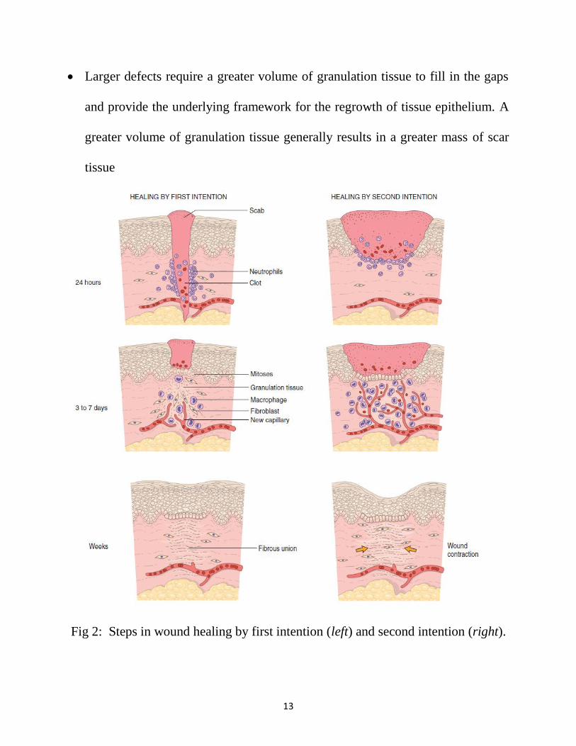
Healing by First Intention13
:
One of the simplest examples of wound repair is the healing of a clean,
uninfected surgical incision approximated by surgical sutures. The incision causes
only focal disruption of epithelial basement membrane continuity and death of
relatively few epithelial and connective tissue cells. As a result, epithelial
regeneration is the principal mechanism of repair. A small scar is formed, but there
is minimal wound contraction. The narrow incisional space first fills with fibrin-
clotted blood, which then is rapidly invaded by granulation tissue and covered by
new epithelium.
11
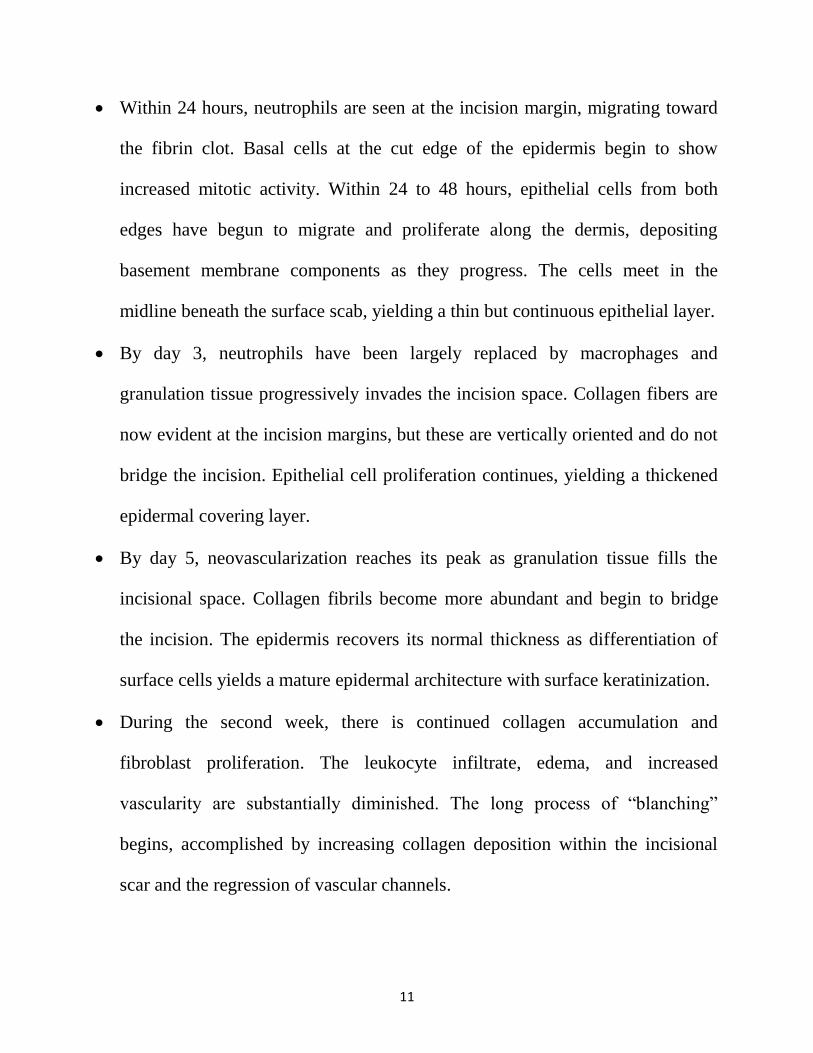
Within 24 hours, neutrophils are seen at the incision margin, migrating toward
the fibrin clot. Basal cells at the cut edge of the epidermis begin to show
increased mitotic activity. Within 24 to 48 hours, epithelial cells from both
edges have begun to migrate and proliferate along the dermis, depositing
basement membrane components as they progress. The cells meet in the
midline beneath the surface scab, yielding a thin but continuous epithelial layer.
By day 3, neutrophils have been largely replaced by macrophages and
granulation tissue progressively invades the incision space. Collagen fibers are
now evident at the incision margins, but these are vertically oriented and do not
bridge the incision. Epithelial cell proliferation continues, yielding a thickened
epidermal covering layer.
By day 5, neovascularization reaches its peak as granulation tissue fills the
incisional space. Collagen fibrils become more abundant and begin to bridge
the incision. The epidermis recovers its normal thickness as differentiation of
surface cells yields a mature epidermal architecture with surface keratinization.
During the second week, there is continued collagen accumulation and
fibroblast proliferation. The leukocyte infiltrate, edema, and increased
vascularity are substantially diminished. The long process of “blanching”
begins, accomplished by increasing collagen deposition within the incisional
scar and the regression of vascular channels.
12
By the end of the first month, the scar consists of a cellular connective tissue,
largely devoid of inflammatory cells, covered by an essentially normal
epidermis.
However, the dermal appendages destroyed in the line of the incision are
permanently lost. The tensile strength of the wound increases with time.
Healing by Second Intention13
:
When cell or tissue loss is more extensive, such as in large wounds, at sites
of abscess formation, ulceration, and ischemic necrosis (infarction) in parenchymal
organs, the repair process is more complex and involves a combination of
regeneration and scarring. In second intention healing of skin wounds, also known
as healing by secondary union the inflammatory reaction is more intense, and there
is development of abundant granulation tissue, with accumulation of ECM and
formation of a large scar, followed by wound contraction mediated by the action of
myofibroblasts. Secondary healing differs from primary healing in several
respects:
A larger clot or scab rich in fibrin and fibronectin forms at the surface of the
wound.
Inflammation is more intense because large tissue defects have a greater volume
of necrotic debris, exudates and fibrin that must be removed. Consequently,
large defects have a greater potential for secondary, inflammation-mediated,
injury.
13
Larger defects require a greater volume of granulation tissue to fill in the gaps
and provide the underlying framework for the regrowth of tissue epithelium. A
greater volume of granulation tissue generally results in a greater mass of scar
tissue
Fig 2: Steps in wound healing by first intention (left) and second intention (right).
14
Secondary healing involves wound contraction. Within 6 weeks, for example,
large skin defects may be reduced to 5% to 10% of their original size, largely
by contraction. This process has been ascribed to the presence of
myofibroblasts, which are modified fibroblasts exhibiting many of the
ultrastructural and functional features of contractile smooth muscle cells.
Healing by Tertiary intention3:
Also called as Delayed primary intention. Here healing occurs when the
wound edges are not opposed immediately, which may be necessary in
contaminated or untidy wounds. The inflammatory and proliferative phases of
healing are well established when delayed closure of the wound is carried out. This
will result in a less satisfactory scar than would result after healing by primary
intention.
Factors influencing healing of a wound:
Site of the wound
Structures involved
Mechanism of wounding:
Incision, Crush, Crush avulsion
Contamination (foreign bodies/bacteria)
Loss of tissue
15
Other local factors
Vascular insufficiency (arterial or venous), Previous radiation, Pressure
Systemic factors:
Malnutrition or vitamin and mineral deficiencies, Disease (e.g. diabetes
mellitus), Medications (e.g. steroids), Immune deficiencies (e.g. chemotherapy,
acquired immunodeficiency syndrome [AIDS], Smoking.
Factors that determine whether a wound will become infected3:
Host response
Virulence and inoculum of infective agent
Vascularity and health of tissue being invaded (including local ischaemia as
well as systemic shock)
Presence of dead or foreign tissue
Presence of antibiotics during the ‘decisive period’
Dose of bacterial contamination *virulence = Risk of surgical site infection
Resistance of the host patient
Quantitatively, it has been shown that if a surgical site is contaminated with
>105 microorganisms per gram of tissue, the risk of SSI is markedly increased.
However, the dose of contaminating microorganisms required to produce infection
may be much lower when foreign material is present at the site
16
THE DECISIVE PERIOD:
There is up to a 4-hour interval before bacterial growth becomes established
enough to cause an infection after a breach in the tissues, whether caused by
trauma or surgery. This interval is called the ‘decisive period’ and strategies aimed
at preventing infection from taking a hold become ineffective after this time
period. It is therefore logical that prophylactic antibiotics should be given to cover
this period and that they could be decisive in preventing an infection from
developing, before bacterial growth takes a hold.
Microbiology14
:
The microbiology of SSI depends on the nature of the procedure, location of
the incision, and whether a body cavity or hollow viscous is entered during
surgery. Most SSIs are caused by skin flora that are inoculated into the incision
during surgery, therefore, the most common SSI pathogens are all gram-
positivecocci—Staphylococcus epidermidis, S. aureus, and Enterococcus spp. For
infrainguinal incisions and intracavitary surgery, gramnegative bacilli such as
Escherichia coli and Klebsiella spp. are potential pathogens. When surgery is
performed on the pharynx, lower gastrointestinal tract, or female genital tract,
anaerobic bacteria become potential SSI pathogens.
Outbreaks or clusters of SSIs have also been caused by unusual pathogens,
such as Rhizopus oryzae, Clostridium perfringens, Rhodococcus bronchialis,
17
Nocardia farcinica,Legionella pneumophila and Legionella dumoffii, and
Pseudomonas multivorans. These rare outbreaks have been traced to contaminated
adhesive dressings, elastic bandages, colonized surgical personnel, tap water, or
contaminated disinfectant solutions.
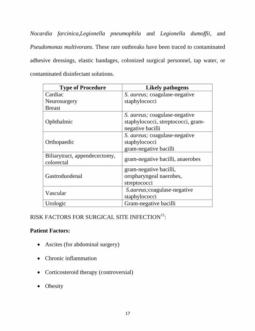
Type of Procedure Likely pathogens
Cardiac
Neurosurgery
Breast
S. aureus; coagulase-negative
staphylococci
Ophthalmic
S. aureus; coagulase-negative
staphylococci, streptococci, gram-
negative bacilli
Orthopaedic
S. aureus; coagulase-negative
staphylococci
gram-negative bacilli
Biliarytract, appendecectomy,
colorectal gram-negative bacilli, anaerobes
Gastroduodenal
gram-negative bacilli,
oropharyngeal naerobes,
streptococci
Vascular S.aureus;coagulase-negative
staphylococci
Urologic Gram-negative bacilli
RISK FACTORS FOR SURGICAL SITE INFECTION15
:
Patient Factors:
Ascites (for abdominal surgery)
Chronic inflammation
Corticosteroid therapy (controversial)
Obesity
18
Diabetes
Extremes of age
Hypoxemia
Peripheral vascular disease (for lower extremity surgery)
Postoperative anemia
Prior site irradiation
Recent operation
Remote infection
Skin or nasal carriage of staphylococci
Skin disease in the area of infection (e.g., psoriasis)
Undernutrition
Environmental Factors:
Contaminated medications
Inadequate disinfection/sterilization
Inadequate skin antisepsis
Inadequate ventilation
Treatment Factors:
Drains
Emergency procedure
Hypothermia
19
Inadequate antibiotic prophylaxis
Oxygenation (controversial)
Prolonged preoperative hospitalization
Prolonged operative time
1. Ascites :
Ascites increases intra-abdominal pressure and increase the risk of wound
dehiscence and thus delays wound healing which is predisposed to pathogens16
.
2. Chronic inflammation:
Preoperative inflammatory activity is related to a higher risk of SSI17, 18
. It is
well known that local inflammation impairs the healing process and systemic
inflammation impairs the immune response19
. Also Hypoalbuminemia occurs due
to the accelerated catabolism induced by systemic inflammation which in turn
contributes to impaired healing.
3. Corticosteroid therapy:
Immunosuppressive agents are drugs that inhibit or prevent activation of the
immune system. They are commonly prescribed to prevent rejection of
transplanted organs or for the treatment of inflammatory diseases, such as
rheumatoid arthritis or inflammatory bowel disease. Some observational studies
indicate that the immunosuppressive effect of the drugs could lead to impaired
wound healing and increased risk of SSI in patients treated with these agents20, 21
.
20
Conversely, WHO has not recommended the discontinuation of
immunosuppressive treatment because it could induce flares of disease activity and
long term interruptions of therapy might induce the formation of anti-drug
antibodies and subsequently decrease the effect of the immunosuppressive. Thus
discontinuation of immunosuppressive agents remains controversial.
4. Obesity:
Incidence of surgical site infection increases with an increase of BMI. The
mechanism by which obesity increases the risk of SSI is likely to be
multi factorial 22
. Obese surgical patients have been shown to have reduced
subcutaneous tissue oxygenation and to require a greater fraction of inspired
oxygen to achieve the same arterial oxygen tension as normal-weight patients, thus
predisposing them to SSI23
. Wound hypoxia impairs healing by a number of
potential mechanisms; healing wounds have high metabolic demands, and
insufficient oxygen will slow the healing process. Immune cells also have high
oxygen demands, requiring oxygen for the formation of microbicidal reactive
oxygen species24
.
In addition to poor tissue oxygenation, adequate tissue levels of prophylactic
antibiotics may be harder to achieve in obese patients25
. Antimicrobials show
different pharmacokinetics when administered to obese patients, with both
hydrophilic and hydrophobic compounds generally having a higher volume of
21
distribution, requiring a higher dose to reach the same plasma drug concentrations
as for non-obese patients25
. Hepatic clearance may also be increased in obese
patients26
. Also increase in operation time for the obese and a longer operation time
has been described as a significant predictor of postoperative wound infections 27,
28. Furthermore impaired immunity, elevated blood glucose levels and too much
tension on the surgical incision are also contributory factors to impaired wound
healing29, 30
5. Diabetes Mellitus:
Blood glucose levels rise during and after surgery due to surgical stress.
Surgery causes a stress response that result in a release of catabolic hormones and
the inhibition of insulin. Moreover, surgical stress influences pancreatic beta-cell
function, which results in lower plasma insulin levels. Taken together, this relative
hypoinsulinaemia, insulin resistance and excessive catabolism from the action of
counter-regulatory hormones make surgical patients at high risk for
hyperglycaemia, even non-diabetic individuals31
There is no significant relationship between increasing levels of HbA1c and
SSI rates32
. Also, increased glucose levels (>200 mg/dL) in the immediate
Postoperative period (<48 hours) were associated with increased SSI risk33, 34
.
Hyperglycemia related impairment in immune response, sensory peripheral
22
neuropathy, autonomic neuropathy and vascular insufficiency are the reasons
diabetic patients have an increased risk.
6. Age:
Increasing age is associated with an increased risk of development of SSI.
Increased prevalence of co morbid conditions, an increased severity of acute illness
and a decreased host response to bacterial invasion— in older patients are the
reasons older patients appear to have an increased risk of SSI 35, 36, 37
7. Hypoxia:
Tissue hypoxia appears to predispose to SSI38
. But it is controversial
whether perioperative oxygen administration is beneficial for the prevention of
infection39.
The ischemic milieu of the fresh surgical incision is vulnerable to
bacterial invasion. Moreover, oxygen has been postulated to have a direct
antibacterial effect.40, 41.
wound healing process involves numerous functions, many
of which depend on the presence of oxygen. Collagen production and development
influence the strength of the wound is directly correlated with the partial pressure
of oxygen (PO2) of the tissue. Synthesis of collagen, cross-linking and the
resulting wound strength depend on the normal function of specific enzymes. The
functions of these enzymes are directly related to the amount of oxygen present,
e.g. hydroxylation of proline and lysine by hydroxylase enzymes42
. Although
clinical trials have had conflicting results, one recent meta analysis has suggested a
23
benefit of supplemental oxygen administration specifically to reduce the incidence
of SSIs43
. WHO recommends that adult patients who are having anesthesia with
endotracheal intubation should receive an 80% fraction of inspired oxygen (FiO2)
both intraoperatively and in the immediate postoperative phase for 2–6 hours to
reduce the risk of SSI. The 80% FIO2 is associated with a decrease in SSI
compared to an FIO2 of 30%–35%31
7. Anaemia:
The World Health Organisation defines anaemia as an insufficient
circulating red cell mass, with a haemoglobin concentration of < 13.0 g/dL for men
and < 12.0 g/dL for women ; even mild anaemia adversely effects surgical
outcome and is independently associated with increased postoperative mortality,
complications, and length of hospital stay44, 45
.
Anaemia causes Suppression of immunity and decreased oxygenation in the
wound causing increased susceptibility to infection & impaired healing
8. Prior site irradiation:
Prior irradiation at the surgical site increases the risk of SSI, likely due to
tissue damage and wound ischemia46
.
9. Coexistent infections at a remote body site:
A pre-existing infection may be the source for hematogenous spread,
causing late infections to implant or be a contiguous site for bacterial transfer 47-49.
24
These infections at a site remote from the wound have been linked to increasing
SSI rates three- to five fold50
. It was observed in a study that among 383 patients
who had cultures taken from SSIs and remote sites, 55% of the wound infections
were preceded by urinary tract or lower respiratory tract infections with the same
microorganisms found in the surgical site and causing the SSI 50
.
10. Colonization with microorganisms:
S aureus colonization, found in the nares of 20% to 30% of healthy humans,
has been strongly implicated as a predictor of SSI involving this organism51, 52.
A
multivariate analysis demonstrated that such carriage is the most powerful
independent risk factor for SSI.
11. Malnutrition:
Nutritional status can have a profound impact on the immune system as
documented by some studies 53-55
. These alterations in host immunity may make
patients more susceptible to postoperative infections and malnutrition was reported
as a threat to surgical outcome, such as delayed recovery, higher rates of morbidity
and mortality, prolonged hospital stay, increased health care costs and a higher
early readmission rate 53-58
.
Given the role of nutrition in the host response to surgery, many researchers
believe that nutritional interventions would reduce SSI and the related morbidity.
However, an epidemiological association between SSI and malnutrition has been
25
difficult to demonstrate consistently for all surgical subspecialties. There is very
little consensus on the optimal timing and dosage of multiple nutrient-enhanced
nutrition, especially for the prevention of SSI.
12. Smoking:
Nicotine, nitric oxide and carbon monoxide use delays primary wound
healing and may increase the risk of SSI2, 59, 60
. Smoking causes endothelial
dysfunction, inflammation, and progression of atherothrombotic disease.
Moreover, smokers have evidence of an impaired systemic immune response with
suppressed immunoglobulin levels, an altered CD4 to CD8 cell ratio, and reduced
phagocyte activity59, 60
.
13. Altered immune response61
:
The altered host defenses can play a significant role in the development of
infection in surgical patients. Many factors associated with the patient have been
clearly identified as responsible for a decreased immune response: old age,
concomitant diseases (diabetes, renal and liver failure, solid and hematologic
neoplasias, malnutrition, autoimmune diseases, AIDS) and concomitant therapies
(corticosteroid, cytotoxic agents). Old age can affect both humoral and cell-
mediated immune responses. Chronic diseases can be responsible for a reduced
primary response or depression of delayed hypersensitivity reactions (renal failure,
neoplasias) or changes in leukocyte function (diabetes, leukemia, lymphomas).
26
Malnutrition frequently accompanies diseases such as cancer, chronic and
acute pancreatitis, and inflammatory bowel diseases. Deficiencies of vitamins and
minerals (B6, A, folate, biotin, riboflavin…) can alter significantly the leukocyte
function and immune response. Finally, there appears to be innate immune-
suppression following any form of injury which is correlated with its magnitude
and can affect any aspect of immunity. Surgical stress can include some reduction
of cell mediated immunity.
14. Low Albumin62, 63
:
Serum albumin is an indicator of the patient’s nutritional status. Malnutrition
is a well-documented risk factor for SSI. Malnourished patients are at risk of
impaired systemic and intestinal immune function, as well as decreased digestive
and absorptive capacity due to the altered architecture of the gut barrier. A
deficiency of protein can impair capillary formation, fibroblast proliferation,
proteoglycan synthesis, collagen synthesis, and wound remodeling. A deficiency of
protein also affects the immune system, with resultant decreased leukocyte
phagocytosis and increased susceptibility to infection.
15. Duration of surgical scrub:
Surgical hand preparation is probably the most important SSI prevention
strategy, although there is no strict randomized study comparing surgery with and
without previous hand antisepsis preparation. Bacterial growth is slowed after
27
preoperative scrubbing with an antiseptic agent64.
The surgical hand scrub helps to
eliminate transient microorganisms, reduce resident microorganisms, and maintain
the resident organisms at reduced levels until the end of the surgical procedure.
The scrub is usually performed with an alcohol-based hand rub with persistent
activity or an antimicrobial soap/product65, 66
. Hands and forearms are scrubbed
with antimicrobial soap for the length of time recommended by the manufacturer,
usually 2–5 minutes. When the quality of water is not assured in the Operating
Room, surgical hand antisepsis using Alcohol based hand rub can be used. A
sufficient amount of ABHR is applied to dry hands and forearms for the length of
time recommended by the manufacturer, typically 1.5 minutes, and hands and
forearms allowed to dry before donning sterile gloves31.
16. Skin antisepsis
Preoperative bathing is considered a good clinical process to clean and
reduce the bacterial load on the skin (skin decolonization). Preoperative bathing is
generally recommended for patients, usually with an antimicrobial soap such as
chlorhexidine gluconate (CHG 4% combined with a detergent) if affordable and
available. Other options are a triclosan preparation and—if no other options are
available—regular soap31.
Studies have concluded that preoperative antiseptic
bathing reduces the risk of SSI67.
28
17. Operative shaving:
Pre operative Shaving has been associated with increased risk of SSIs1, 14, 68
.
Hair removal with razor can cause microscopic trauma to the skin that later serve
as foci for bacterial multiplication31, 69.
Razors are preferred for preoperative hair
removal on only two body sites, the scalp and male genitalia, as clippers have been
shown to cause more skin damage in these areas. On all other body sites, if it is
necessary to remove hair prior to a surgical procedure, personnel should consider
clipping the hair1, 14, 68, and 69.
18. Preoperative skin prep:
It reduces the microbial load on the patient’s skin as much as possible before
incision of the skin barrier. Alcohol based solutions are generally recommended. If
alcohol cannot be included in the preparation, chlorhexidine is preferred over
iodine unless contraindications exist, chlorhexidine gluconate causes greater
reductions in skin microflora than povidone-iodine also had greater residual
activity after a single application70.
Further, chlorhexidine gluconate is not
inactivated by blood or serum proteins71, 72
Iodophors may be inactivated by blood
or serum proteins, but exert a bacteriostatic effect as long as they are present on the
skin71
.
19. Operating room ventilation:
Operating room air may contain microbial-laden dust, lint, skin squames, or
respiratory droplets. The microbial level in operating room air is directly
29
proportional to the number of people moving about in the room73.
The ventilation
system in the operating room is designed to provide certain functions, primarily to
create thermal comfort for the patient and staff and to maintain constant air quality
by eliminating aerosols and particles within the room Outbreaks of SSIs caused by
group A beta-hemolytic streptococci have been traced to airborne transmission of
the organism from colonized operating room personnel to patients74, 75
.The strain
causing the outbreak was recovered from the air in the operating room. Ideally,
around 20 air changes per hour are necessary to dilute microorganisms generated
in the operating room and to exclude ingress from surrounding areas 76
.
20. Inadequate sterilization of instruments:
Infection risk is certainly increased when non-sterile instruments are used
for surgery. This can occur due to inadequate supervision, lack of training and/or
short staffing facilitated poor handling practices during and after retrieval of
surgical sets from the autoclave77, 78
21. Length of preoperative stay:
It increases the risk of exposure to nosocomial pathogens thus increasing
risk of SSI79
. Length of preoperative stay is also likely a surrogate for severity of
illness and co-morbid conditions requiring inpatient work-up and/or therapy before
the operation14
.
30
22. Duration of operation:
Prolonged duration of operation results in increased exposure of operation
site to air, increased desiccation of tissue, decreased antibiotic level in tissues80
,
stress of prolonged anaesthesia and sometimes blood loss81
.
23. Antimicrobial prophylaxis:
The objective of surgical antibiotic prophylaxis is to achieve a sufficient
tissue level of the antibiotic before tissues are manipulated. Antibiotic levels
should be maintained through the entire procedure. The antibiotic is selected based
on the procedure being performed and the most likely pathogens that will be
encountered during the surgery. The amount of antibiotic administered should be
determined according to the patient’s weight68, 82
. It is optimal to administer the
drug intravenously 60 minutes before skin incision68
and it has been documented
that administration more than 60 minutes preoperatively is associated with higher
risk of surgical infection83
, with the exception of a few specific drugs (vancomycin
& fluroquinolones).
24. Surgical drains:
The use of drains has contributed significantly as a risk factor in causing
SSI. Epithelialization of the wound is prevented and the drain becomes a conduit,
holding open a portal for invasion by pathogens colonizing the skin. Bacterial
colonization of initially sterile drain tracts increases with the duration of time the
31
drain is left in place84
. Moreover drains are more likely to be used in contaminated
or dirty wounds and in emergency and prolonged operations which increases the
probability of the wound getting infected. Several studies of drains placed into
clean or clean-contaminated incisions also have shown that the rate of SSI rate is
increased15
.
25. Surgical technique14
:
Excellent surgical technique is widely believed to reduce the risk of SSI71, 85
.
Such techniques include maintaining effective hemostasis while preserving
adequate blood supply, preventing hypothermia, gently handling tissues, avoiding
inadvertent entries into a hollow viscus, removing devitalized (e.g., necrotic or
charred) tissues, using drains and suture material appropriately, eradicating dead
space, and appropriately managing the postoperative incision.
26. Hypothermia15
:
Hypothermia is defined as a core temperature below 35°C and has been
associated with an increased risk of SSI. It is common for patients to become
hypothermic during and after major surgical procedures that last more than two
hours. Hypothermia may occur as the result of exposure, large-volume infusion of
un warmed fluids or blood products, or evaporative losses during intracavitary
surgery, especially if the chest and abdomen are opened. Peripheral and cutaneous
vasoconstriction occurs to preserve core heat, but vasoconstriction decreases
32
microcirculatory blood flow leading to reduced levels of oxygen in the tissues,
which impairs the ability of neutrophils to kill organisms and therefore decreases
the wound’s ability to heal. Mild intraoperative hypothermia is associated with an
increased incidence of SSIs following elective colon surgery and diverse
Operations86.
27. Emergency procedures:
Surgical site infection occurs with greater frequency in emergency than
elective surgery because of factors such as inadequate preoperative preparation,
higher frequency of contaminated or dirty wounds in emergency surgeries87, 88
.
Lack of proper control of other medical comorbidities (such as uncontrolled
diabetes) & lack of timely antibiotic prophylaxis could also be a contributing
factor.
28. Blood transfusion89
:
Transfusion-related immunomodulation has been considered to be one of the
major mechanisms of these blood transfusion–induced SSI developments. Both
proinflammatory and immunosuppressive effects were reported to be
simultaneously induced by ABT, and they were mediated by allogeneic
mononuclear cells
28. Type of wound 90
:
Surgical management of the wound also is a critical determinant of the
propensity to develop a SSI. In healthy individuals, class I and II wounds may be
33
closed primarily, while skin closure of class III and IV wounds is associated with
high rates of incisional SSIs (~25% to 50%)
Surgical Wound Classification14
:
Class I/Clean: An uninfected operative wound in which no inflammation is
encountered and the respiratory, alimentary, genital, or uninfected urinary tract is
not entered. In addition, clean wounds are primarily closed and, if necessary,
drained with closed drainage. Operative incisional wounds that follow
nonpenetrating (blunt) trauma should be included in this category if they meet the
criteria.
Class II/Clean-Contaminated: An operative wound in which the respiratory,
alimentary, genital, or urinary tracts are entered under controlled conditions and
without unusual contamination. Specifically, operations involving the biliary tract,
appendix, vagina, and oropharynx are included in this category, provided no
evidence of infection or major break in technique is encountered.
Class III/Contaminated: Open, fresh, accidental wounds. In addition,
operations with major breaks in sterile technique (e.g., open cardiac massage) or
gross spillage from the gastrointestinal tract, and incisions in which acute,
nonpurulent inflammation is encountered are included in this category.
Class IV/Dirty-Infected: Old traumatic wounds with retained devitalized
tissue and those that involve existing clinical infection or perforated viscera. This
34
definition suggests that the organisms causing postoperative infection were present
in the operative field before the operation
CLINICAL FEATURES OF SSI15
Surgical site infection remains a clinical diagnosis. Presenting signs and
symptoms depend on the depth of infection, typically as early as postoperative day
4 or 5, although rare necrotizing SSIs caused by Streptococcus pyogenes or
Clostridium perfringens may develop within 24 hours after surgery. Clinical signs
range from local induration only to the hallmarks of infection (e.g.,erythema,
edema, tenderness, warmth, pain-related immobility),which may manifest before
wound drainage. In cases of deep incisional SSIs, tenderness may extend beyond
the margin of erythema, and crepitus, cutaneous vesicles, or bullae may be present.
With ongoing infection, signs of systemic inflammatory response syndrome (SIRS;
two or more of fever, leukocytosis, tachycardia, or tachypnea) herald the
development of sepsis. In intracavitary (organ, space) SSIs, there will be Purulent
drainage from a drain that is placed through a stab wound into the organ/space or
symptoms specific to the involved organ system will usually predominate, such as
ileus, respiratory distress or failure, or altered sensorium. These deep infections
may sometime remain occult or present with few symptoms, mimicking incisional
SSIs and leading to inadequate initial treatment; they become apparent only when a
major complication ensues.
35
Management of SSI15
:
Cultures are not mandatory for the management of superficial incisional
SSIs, particularly if drainage and wound care alone will suffice without antibiotics
and if superficial swab cultures are collected, which are susceptible to
contamination by nearby skin colonists. In cases of deeper infection or hospital
acquired infection, exudates or drainage specimens should be sent for analysis
from the surgically opened wound—as opposed to the already opened wound,
which becomes colonized. Blood culture is collected if evidence of systemic
involvement present. Ultrasonography can be applied to the infected wound area to
assess whether there is a collection for which drainage is required.
The first steps in the treatment of SSIs are to open and examine the
suspicious portion of the incision and decide about further surgical treatment. If the
infection is confined to the skin and superficial underlying subcutaneous tissue,
opening the incision and providing local wound care may be all the treatment that
is necessary. Antibiotic therapy of superficial incisional SSIs is indicated only for
erythema extending beyond the wound margin or for systemic signs of infection.
Deeper SSIs may require formal surgical exploration and débridement to obtain
local control of the infection. Surgical site infection must also be considered as a
cause of delayed or failed wound healing and prompt the same decisions as
described earlier.
36
Organ or space SSIs occur within a body cavity (e.g., intraabdominal,
intrapleural, intracranial) and are directly related to a surgical procedure. The
diagnosis of organ or space SSIs usually requires some form of imaging to confirm
the site and extent of infection. Adequate source control requires a drainage
procedure, whether open or percutaneous. Give the patient an antibiotic that covers
the likely causative organisms. Consider local resistance patterns and the results of
microbiological tests in choosing an antibiotic.
37
AIMS & OBJECTIVES
1. To evaluate the incidence of Surgical Site Infection (SSI) among the patients
undergoing abdominal surgeries in the dept. of General Surgery
2. To assess the risk factors of Surgical Site Infection.
3. To find out the types of surgical site infection.

38
REVIEW OF LITERATURE
Various studies have been conducted regarding surgical site infection in
India since 1972. These studies revealed that surgical site infection rates in India
were found to be between 4 to 30% (Agarwal91
, 1972; Rao and Harsha92
, 1975;
Kowli93
et al., 1985; Anvikar94
et al., 1999). Agarwal et al , Rao et al & Anvikar et
al reported the ineffectiveness of penicillin against staphylococcus aureu
Kowli et al found an infection rate of 17.4% when preoperative stay Was 0-
7 days, and an infection rate of 71.4% with a preoperative stay of more than 21
days. In Anvikar et al study the SSI rate was 6.1%. His study demonstrated that
preoperative hospital stay predisposed an individual to 1.76% risk of acquiring an
infection. With an increase in preoperative stay, the risk increased proportionally.
A preoperative stay of one week increased the risk rate to 5% .SSI was higher in
emergency than elective surgery & increased duration of surgery increased the risk
of SSI. All these studies also indicated gradual increase in the emergence of
antibiotic resistant microorganisms in surgical patients Preoperative antibiotic
decreased SSI
Hemant et al95
conducted a prospective clinical trial in 100 patients who
underwent abdominal surgeries. SSI rate was 14%. The SSI rate was the highest in
dirty surgeries (40%). Male patients were affected more (18.2%) than the female
patients (5.9%). The SSI rate increased with increasing age and it also increased
39
significantly with the increasing duration of pre-operative hospitalization. The SSI
rate was higher in emergency surgeries as compared to the elective. With increase
in the time of surgery, the risk of infection increased. The most commonly isolated
organism from SSIs was Pseudomonas (42.85%), followed by Klebsiella sp.
(28.5%) and other bacteria. Among the organisms that were isolated, the most of
them were multidrug resistant.
Amit agarwal et al96
conducted a prospective study on 375 patients who
underwent abdominal surgeries. But they excluded organ space SSI and duration of
surgery >2.5 hours from the study. SSI incidence was 15.7 % (59/375). SSI rate
was higher in emergency surgeries( 28.6%) than elective surgeries (5.7%). In
elective surgeries group maximum SSI was found in colonic surgery – 14.3%,
while minimum in cholecystectomy 2.2%. In Emergency surgery group maximum
incidence of SSI was observed in hepato biliary surgeries 44.4% while minimum
with appendicular pathology 19.4%. It was found that SSI increased with
increasing age linearly. Other significant factors involved were increasing class of
wound (dirty > clean wound class), increased preoperative stay, presence of remote
site infection, increased duration of surgery and use of drains. E. coli was found to
be the most common organism.
Mekla et al97
conducted a cohort study on 100 patients who underwent
abdominal surgeries. But they excluded those underwent laparoscopic surgery,
40
received antibiotics for duration of >1 week before surgery, reoperative surgery
from the study. The incidence rate of superficial SSI was 39% with 95% CI
(29.4%–49.2%). They found 12 variables significantly associated with superficial
SSI: middle or elderly age, male gender, diabetes mellitus, preoperative anemia,
preoperative hypoalbuminemia, tobacco smoking (RR 1.88, CI 1.18–2.9), higher
ASA score(RR 4.05, CI 2.65–6.33), perioperative blood transfusion, drain
placement, surgery duration >2 h(RR 3.24, CI 1.98–5.31), contaminated/dirty
wound class(RR 2.57, CI 1.52–4.31) and emergency surgery(RR 1.8, CI 1.1–3.0).
Adeyinka Ayodele Adejumo et al98
conducted a prospective study on 223
patients who underwent laporotomy. Incisional SSI was clinically diagnosed in 85
patients giving an incidence rate of 38.1%. Sixty-three (74.1%) were superficial
SSI while 22 (25.9%) were deep SSI. The risk factors for SSI were anaemia,
contaminated and dirty wounds, retroviral disease status, physiological status
(ASA scores IV and V), prolonged surgery time, cadre of surgeon, emergency
surgeries and use of drains. The high incidence of SSI observed in this study was
found more in patients that presented with septic abdomen and those that had large
bowel procedure. Staphlyococcuss aureus & klebseilla were the common
organism isolated.
Emil aga et al99
conducted a prospective cohort study which included 302
patients who underwent abdominal surgeries in the Western Galilee Medical
41
Center in Nahariya, Israel. The SSI incidence rate was 22.2%. The univariate
analysis defined 13 variables significantly associated with SSI: age > 60 years,
lower functional status, diabetes mellitus, congestive heart failure,
immunocompromising underlying disease, treatment with chemotherapy and other
immunosuppressive medications, impaired immune system open cholecystectomy,
laparotomy, an American Society of Anesthesiologists (ASA) score > 2, drain
insertion, and ‘dirty wound’ classification. In multivariate regression analysis,
treatment with immunosuppressive medications (OR = 2.5, 95% confidence
interval (CI) = 1.099–143.443), open cholecystectomy (OR = 2.25, 95% CI =
2.242–40.109), and dirty wound classification (OR = 2.179, 95% CI = 3.80–
20.551) were significantly associated with SSI
Lul raka et al100
conducted a prospective study in which a total of 253
surgical interventions in 225 patients were evaluated. The overall incidence rate of
SSI was 12%.Superficial incisional SSI was most common (55%). Clinical
infections were culture positive in 40.7% of cases. Duration of operation, duration
of preoperative stay, wound class, ASA score >2, use of antibiotic prophylaxis and
NNIS class of >2 were all found to be risk factors associated s (p < .001).
The International Nosocomial Infection Control Consortium (INICC)
conducted a cohort prospective surveillance study on surgical site infections in 10
hospitals in 6 Indian cities from January 2005 to December 2011101
. They
42
documented 1189 surgical site infections, associated with 28 340 surgical
procedures (4.2%; 95% CI 4.0–4.4).11 types of surgical procedures were included
for study of which the incidence of SSI was 6.0% for exploratory abdominal
surgery .
Ashish pathak et al102
conducted a study in a teaching hospital in ujjain in
720 patients admitted for surgery. SSI rate was 5%. Risk factors for SSI identified
were as follows: severity of disease (P = .001), presence of drains (P =.020),
history of previous hospitalization (P = .003), preoperative stay (P = .005), wound
classification (P < .001), and surgical duration (P < .001). Independent risk factors
identified included wound classification (odds ratio = 4.525; P < .001) and surgical
duration (odds ratio = 2.554; P = .015). Most patients (99%) were prescribed
antibiotics.Metronidazole (24.5%), ciprofloxacin (11%), and amikacin (9%) were
the most commonly prescribed antibiotics. Most commonly isolated bacteria were
Staphylococcus aureus (n = 14), of which 34% were methicillin-resistant
Staphylococcus aureus, and Pseudomonas aeruginosa (n = 6), which showed
resistance to ceftazidime (70%), ciprofloxacin (63%), and gentamicin (57%).
Anand saxena et al103
conducted a prospective study in a teaching hospital in
Bhopal on 300 patients admitted for various surgeries. Out of 300 patients
observed, 43 patients developed surgical site infections (14.33%). Out of 43
infected cases, 37 cases were culture positive (86.04%, 37/43), while 6 cases were
43
culture negative (13.96%, 6/43). Surgical site infection was found to be higher in
males and patients above 50 years of age & in emergency surgery than elective
surgery. SSI was increased with increased length of preoperative stay duration.
Obesity, Diabetes and Anemia were additional risk factors in surgical site
infection. Staphylococcus aureus (37.83%) was most commonly identified
organism in culture.
Suchitra et al104
conducted a prospective study on 1125 surgeries for the
incidence of surgical site infections. The results indicated that 12% ofpatients
undergoing surgery developed SSI. Staphylococcus aureus (33%) and
Enterococcus spp. (33%) were the commonest etiologic agents. Patients with SSIs
had a significantly extended ICU and ward stay (p<0.001), and incurred higher
hospital costs (p<0.001) when compared to those who did not develop SSIs. The
risk factors associated with SSIs were age above 45 years (p=0.012), female
(p=0.070), diabetic status (p<0.001)
Rajanikanth et al105
conducted a prospective study on 248 patients who
underwent various surgeries in the General Surgery department. Abdominal
surgeries contributed 47% of total surgeries in their study. Among 248 patients, 45
developed surgical site infection(18.14%). SSIs were most commonly found
among males, aged, diabetics, anaemic, underweight and overweight, hypertensive,
blood transfusion and patients with longer hospital stay. Surgical Site Infections
44
were higher in emergency cases than elective surgeries. Staphylococcus aureus
was the most common organism isolated from surgical site infections. Multidrug
resistance organisms were predominant in surgical site infections.
Pinakin et al106
conducted a prospective longitudinal study at a tertiary care
centre of Ahmadabad city. Total 480 patients operated for general surgical
procedures were included. The SSI rate was 9.4%.The risk factors associated with
SSI were age (18.3% versus 7.1%), diabetes (25.5% versus 7.6%), type of
anaesthesia (general = 13.6% versus regional=7.1%), type of surgery (emergency =
21.7% versus elective = 7.3%), duration of surgery (17.9% versus 7.2%), type of
wound (dirty = 28.4% versus clean = 2.99%), pre-operative hospital stay (27.3%
versus 3.3%) and presence of drain (15.2% versus 7.2%).
Satyanarayana et al107
conducted a retrospective observational study which
included patients who had undergone surgeries (abdominal) in the Department of
General Surgery and Department of Obstetrics and Gynacology.1000 cases were
included in the study. The overall surgical wound infection rate was 13.7%.The
infection rate was more with emergency surgery (25.2%) when compared to
elective surgery (7.6%). The surgical site infection rate increased as the risk index
score increased from 0 to 3. SSI was more with early operative and post operative
Prophylaxis. They found a definite correlation between the wound infection rate
and the timing of antimicrobial prophylaxis.
45
Lilani et al108
conducted a prospective study on 190 patients admitted for
surgery clean and clean-contaminated elective cases were included in the study.
Normal microbial flora was studied within 24 to 48 hours of admission in the
ward.Infected wounds were studied bacteriologically and clinically. The overall
infection rate was 8.95%.Surgical site infection rate was 3.03% in clean surgeries
and 22.41% in clean-contaminated surgeries. Significant increase was seen in
surgical site infection rate with an increase in preoperative stay and the increase in
duration of surgery. Surgical site infection rate was much higher (22.41%) in cases
where a drain was used than in non-drained wounds (3.03%). The most common
isolate was Staphylococcus aureus followed by Pseudomonas aeruginosa.
46
METHODOLOGY
Type of study: Prospective study
Place of study: ESIC MEDICAL COLLEGE & PGIMSR, K.KNAGAR, Chennai
Period of study: 18 months from April 2018 to September 2019
Study population:
100 adult patients undergoing abdominal surgeries (elective and emergency)
whomever satisfy the inclusion criteria.
INCLUSION CRITERIA:
Consenting patients undergoing elective & emergency abdominal surgeries
EXCLUSION CRITERIA:
1. Patients with HIV, HBV or HCV infection.
2. Patients on chemotherapy & radiotherapy
3. Patients on oral steroids & other immunosuppressant drugs.
4. Patients with features of hepatic, cardiac & renal failure.
5. ASA (American Society of Anaesthesiologists) score IV or V
Study procedure:
Patients who satisfy the inclusion criteria were included in the study.
Informed written consent was obtained. Appropriate history was taken; clinical
examination & relevant investigations were carried out. Patients were admitted.
Intravenous antibiotic was given 30 –60 minutes before the commencement of
47
procedure. Appropriate surgical management was carried out under strict aseptic
precautions. Immediate Post operative period of the patients was followed up.
Wound was examined on day 2, then every day till the day of discharge. Signs of
SSI were looked for. If the patient developed SSI in this period, then Type of SSI
was classified and swab culture was taken to identify the micro organism &
antibiotic sensitivity pattern. CDC (Centre for disease Prevention & Control)
criterion was used for diagnosis & classification of SSI. Patient was treated
accordingly. Then the patients were discharged. All the details were recorded in
the proforma. The patients were followed up every week till 30 days of post
operative period for SSI in the outpatient dept. If the patient developed any
features of SSI during follow up period after discharge, then Patient was treated
accordingly as described above. All details were recorded in the Proforma.
Ethical consideration:
The ethical standards for human experimentation were followed during the
study and permission from the institutional ethical committee was taken.
Data analysis:
Data analysis was done both manually and by using computer. Calculated
data were arranged in systemic manner, presented in various table and figures and
statistical analysis was made to evaluate the objectives of this study with the help
of Statistical Package for Social Science (SPSS) version 22.0.
48
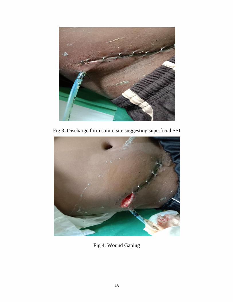
Fig 3. Discharge form suture site suggesting superficial SSI
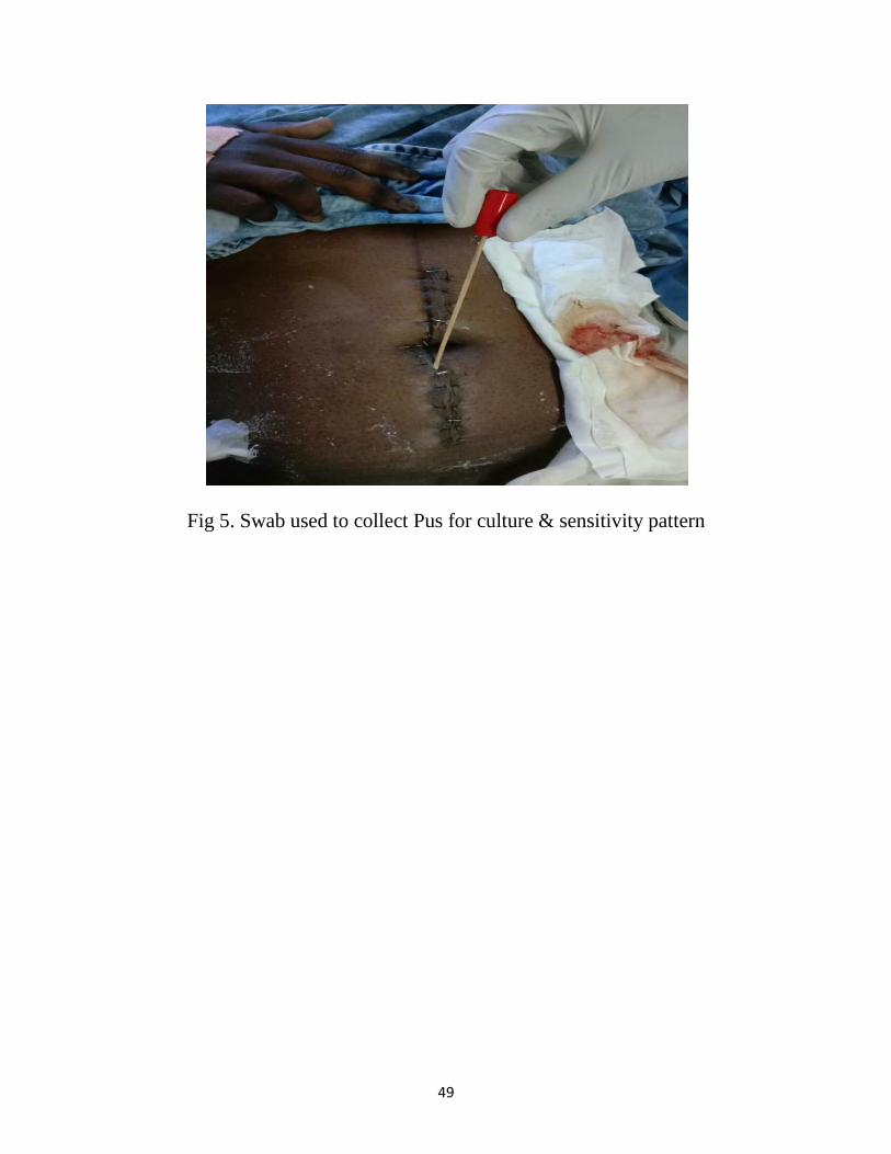
Fig 4. Wound Gaping
49
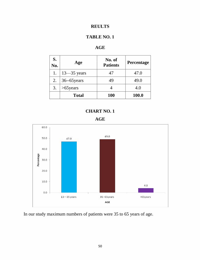
Fig 5. Swab used to collect Pus for culture & sensitivity pattern
50
REULTS
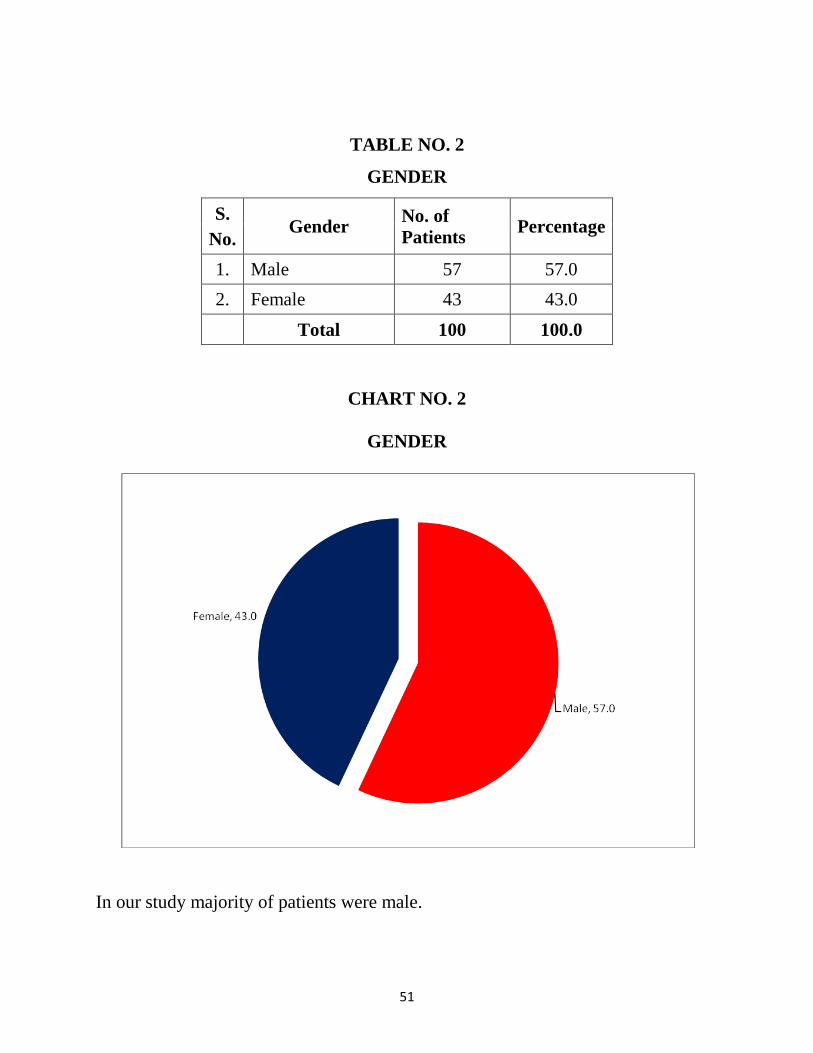
TABLE NO. 1
AGE
S.
No. Age
No. of
Patients Percentage
1. 13—35 years 47 47.0
2. 36--65years 49 49.0
3. >65years 4 4.0
Total 100 100.0
CHART NO. 1
AGE
In our study maximum numbers of patients were 35 to 65 years of age.
51
TABLE NO. 2
GENDER
S.
No. Gender
No. of
Patients Percentage
1. Male 57 57.0
2. Female 43 43.0
Total 100 100.0
CHART NO. 2
GENDER
In our study majority of patients were male.
52
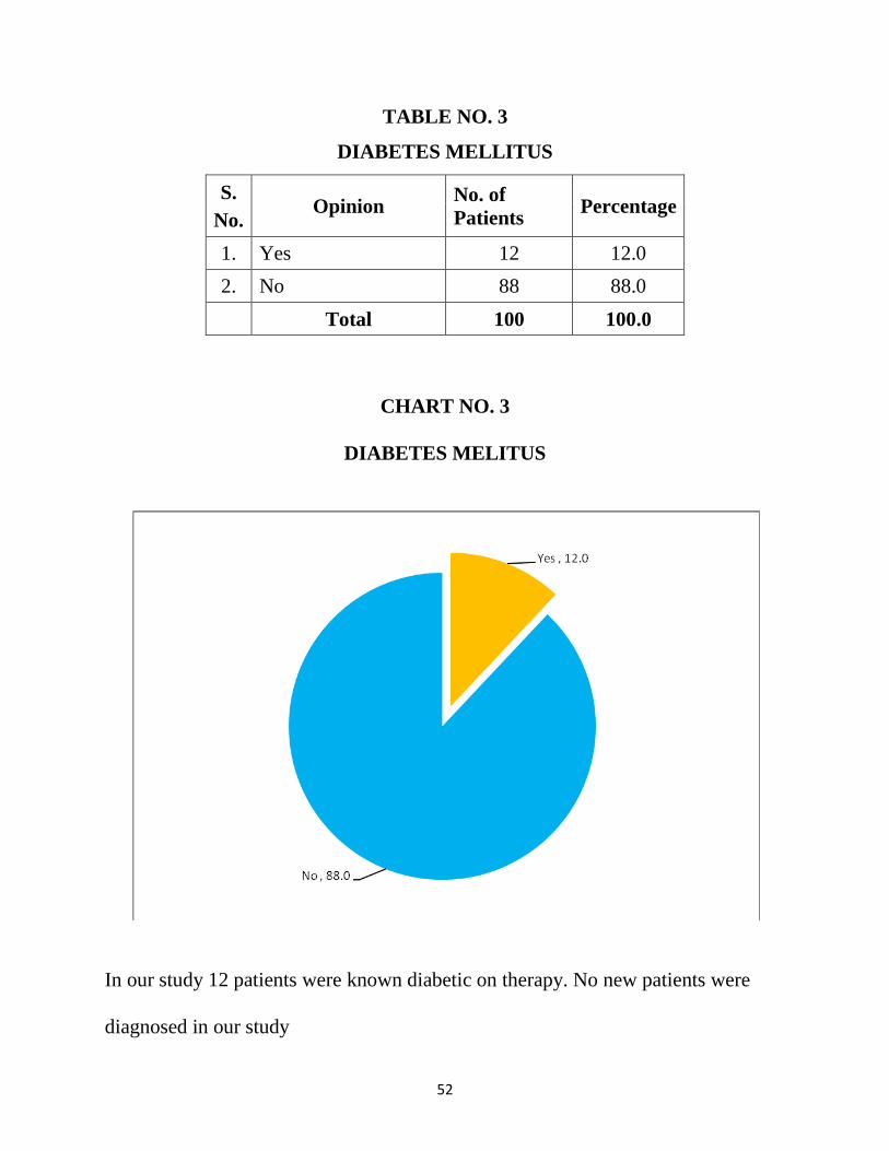
TABLE NO. 3
DIABETES MELLITUS
S.
No. Opinion
No. of
Patients Percentage
1. Yes 12 12.0
2. No 88 88.0
Total 100 100.0
CHART NO. 3
DIABETES MELITUS
In our study 12 patients were known diabetic on therapy. No new patients were
diagnosed in our study
53
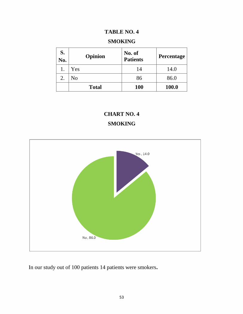
TABLE NO. 4
SMOKING
S.
No. Opinion
No. of
Patients Percentage
1. Yes 14 14.0
2. No 86 86.0
Total 100 100.0
CHART NO. 4
SMOKING
In our study out of 100 patients 14 patients were smokers.
54
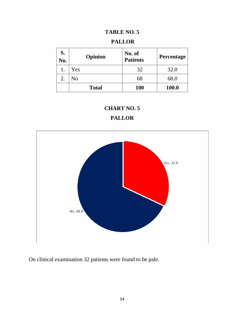
TABLE NO. 5
PALLOR
S.
No. Opinion
No. of
Patients Percentage
1. Yes 32 32.0
2. No 68 68.0
Total 100 100.0
CHART NO. 5
PALLOR
On clinical examination 32 patients were found to be pale.
55
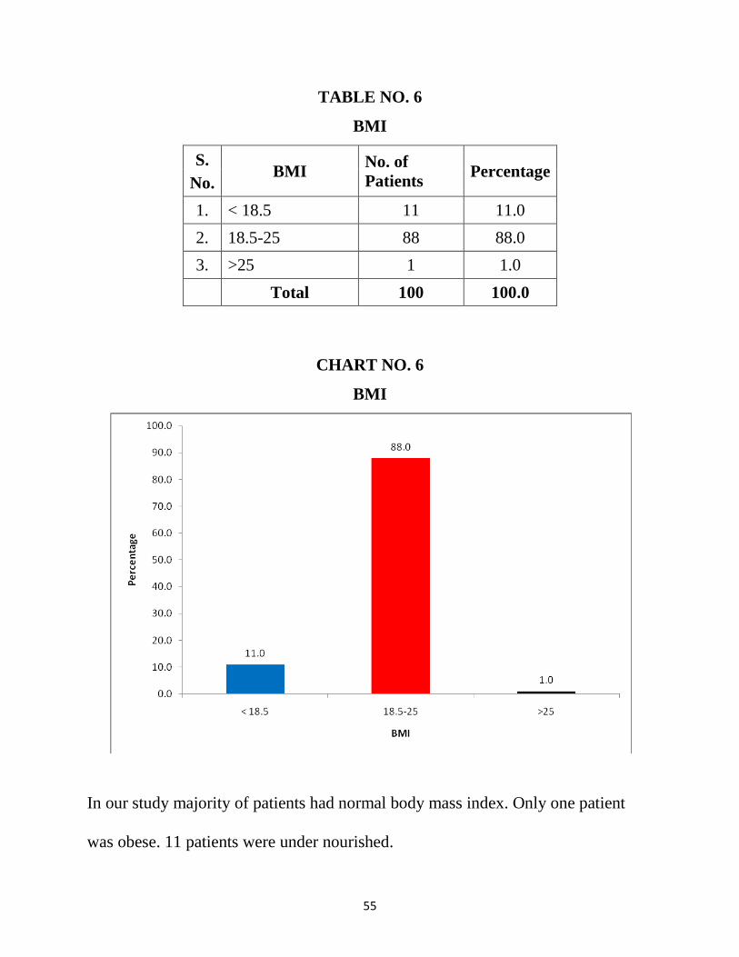
TABLE NO. 6
BMI
S.
No. BMI
No. of
Patients Percentage
1. < 18.5 11 11.0
2. 18.5-25 88 88.0
3. >25 1 1.0
Total 100 100.0
CHART NO. 6
BMI
In our study majority of patients had normal body mass index. Only one patient
was obese. 11 patients were under nourished.
56
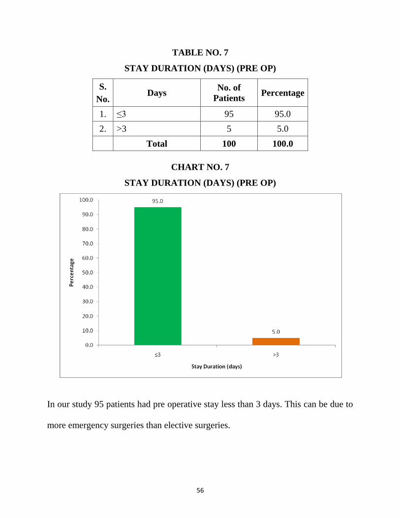
TABLE NO. 7
STAY DURATION (DAYS) (PRE OP)
S.
No. Days
No. of
Patients Percentage
1. ≤3 95 95.0
2. >3 5 5.0
Total 100 100.0
CHART NO. 7
STAY DURATION (DAYS) (PRE OP)
In our study 95 patients had pre operative stay less than 3 days. This can be due to
more emergency surgeries than elective surgeries.
57
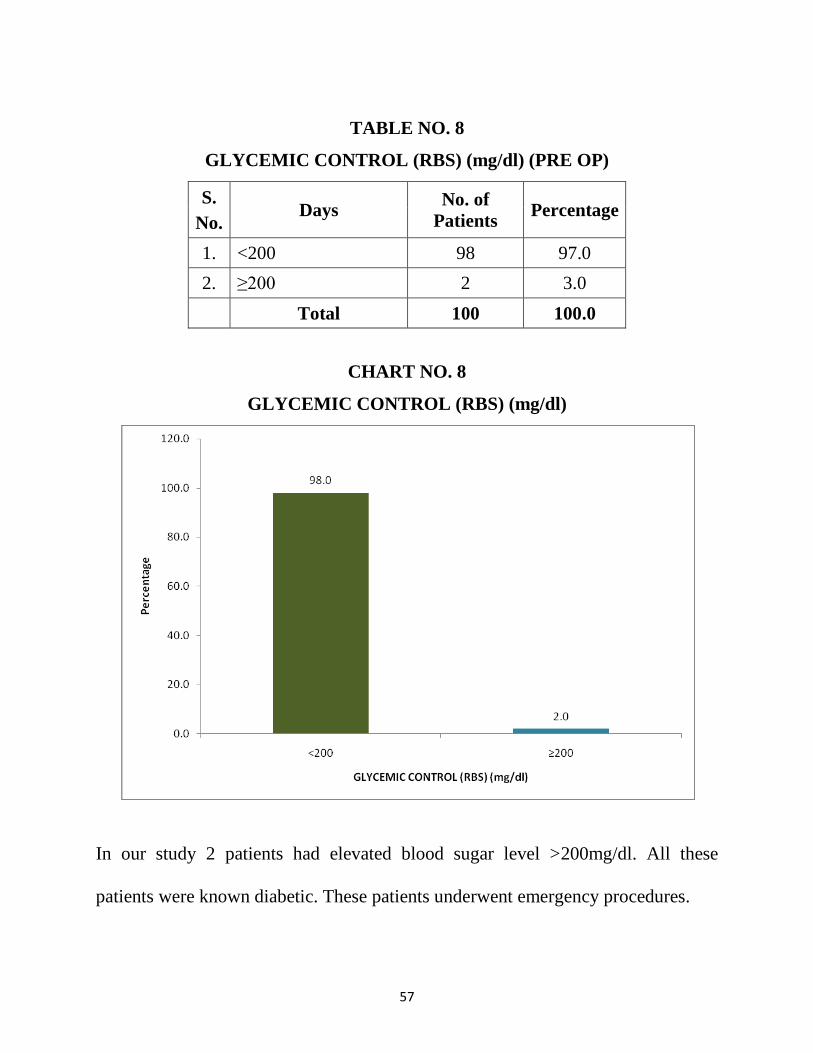
TABLE NO. 8
GLYCEMIC CONTROL (RBS) (mg/dl) (PRE OP)
S.
No. Days
No. of
Patients Percentage
1. <200 98 97.0
2. ≥200 2 3.0
Total 100 100.0
CHART NO. 8
GLYCEMIC CONTROL (RBS) (mg/dl)
In our study 2 patients had elevated blood sugar level >200mg/dl. All these
patients were known diabetic. These patients underwent emergency procedures.

58
TABLE NO. 9
HAEMOGLOBIN (g/dl) (Male)
S.
No. Count
No. of
Patients Percentage
1. <13 26 44.8
2. ≥13 32 55.2
Total 58 100.0
CHART NO. 9
HAEMOGLOBIN (g/dl) (Male)
Out of 58 male patients 26 were found to have anaemia. None of the patients
required blood transfusion. Majority of patients were of the age group 13 to 35
years. All the male patients above 65 years of age were found to have low
hemoglobin.
59
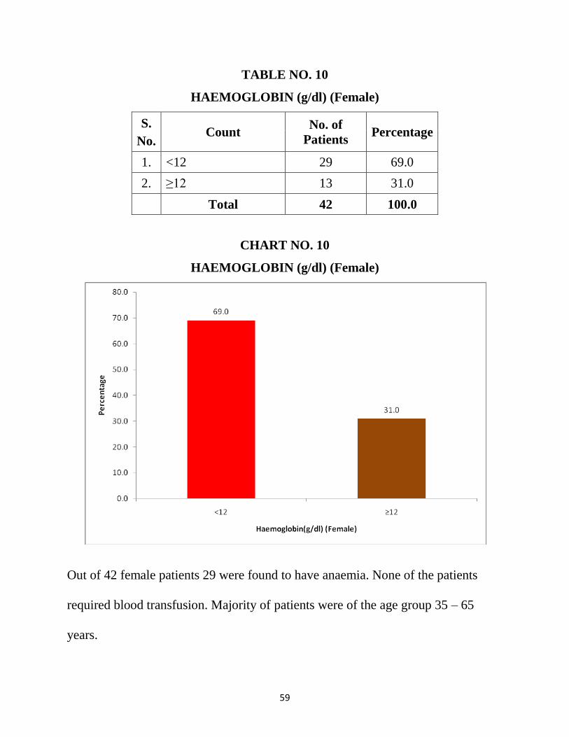
TABLE NO. 10
HAEMOGLOBIN (g/dl) (Female)
S.
No. Count
No. of
Patients Percentage
1. <12 29 69.0
2. ≥12 13 31.0
Total 42 100.0
CHART NO. 10
HAEMOGLOBIN (g/dl) (Female)
Out of 42 female patients 29 were found to have anaemia. None of the patients
required blood transfusion. Majority of patients were of the age group 35 – 65
years.
60
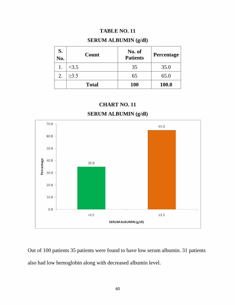
TABLE NO. 11
SERUM ALBUMIN (g/dl)
S.
No. Count
No. of
Patients Percentage
1. <3.5 35 35.0
2. ≥3.5 65 65.0
Total 100 100.0
CHART NO. 11
SERUM ALBUMIN (g/dl)
Out of 100 patients 35 patients were found to have low serum albumin. 31 patients
also had low hemoglobin along with decreased albumin level.
61
TABLE NO. 12
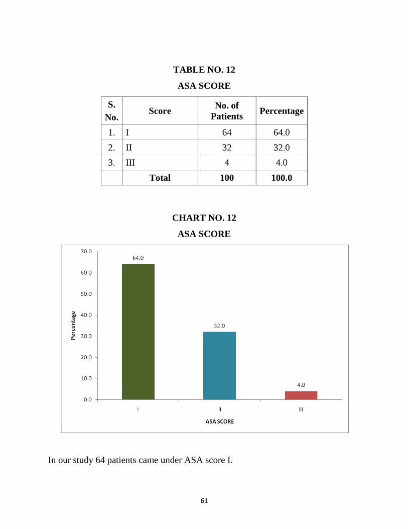
ASA SCORE
S.
No. Score
No. of
Patients Percentage
1. I 64 64.0
2. II 32 32.0
3. III 4 4.0
Total 100 100.0
CHART NO. 12
ASA SCORE
In our study 64 patients came under ASA score I.
62
TABLE NO. 13
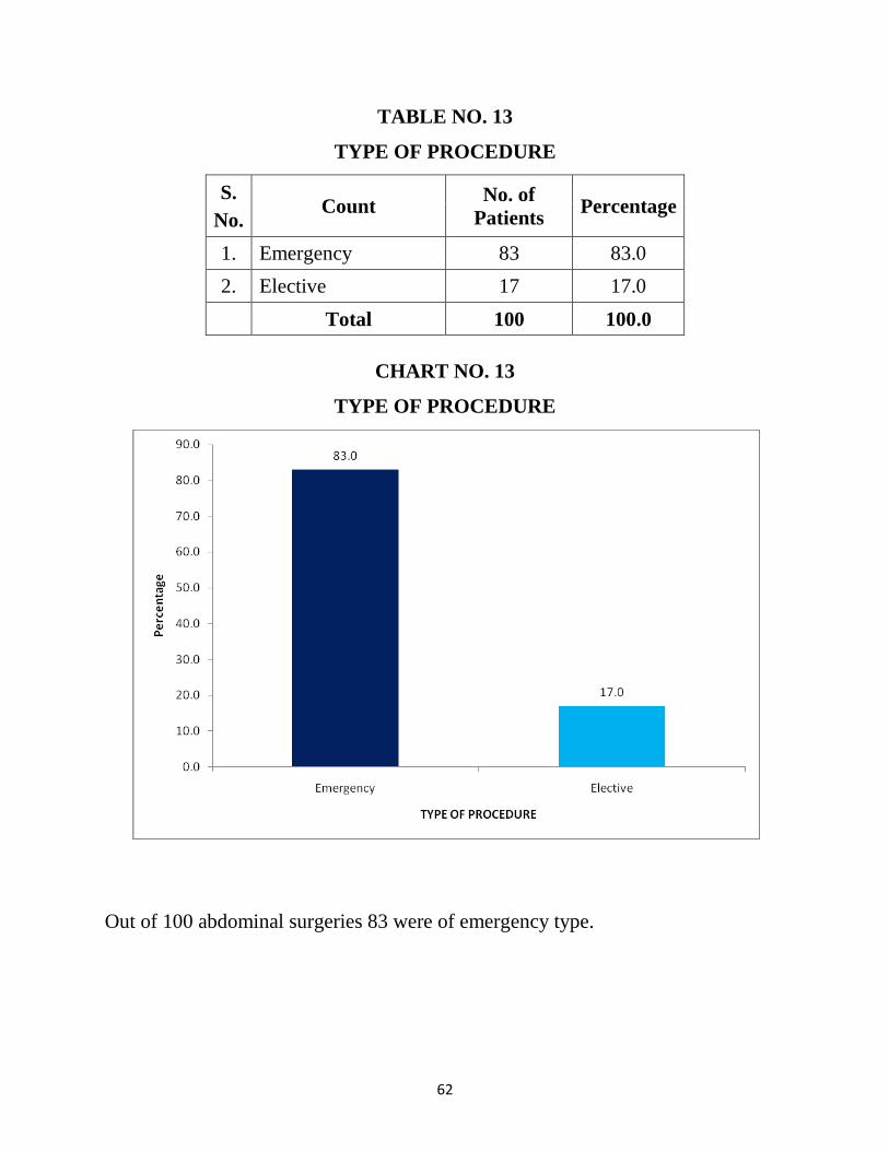
TYPE OF PROCEDURE
S.
No. Count
No. of
Patients Percentage
1. Emergency 83 83.0
2. Elective 17 17.0
Total 100 100.0
CHART NO. 13
TYPE OF PROCEDURE
Out of 100 abdominal surgeries 83 were of emergency type.
63
TABLE NO. 14
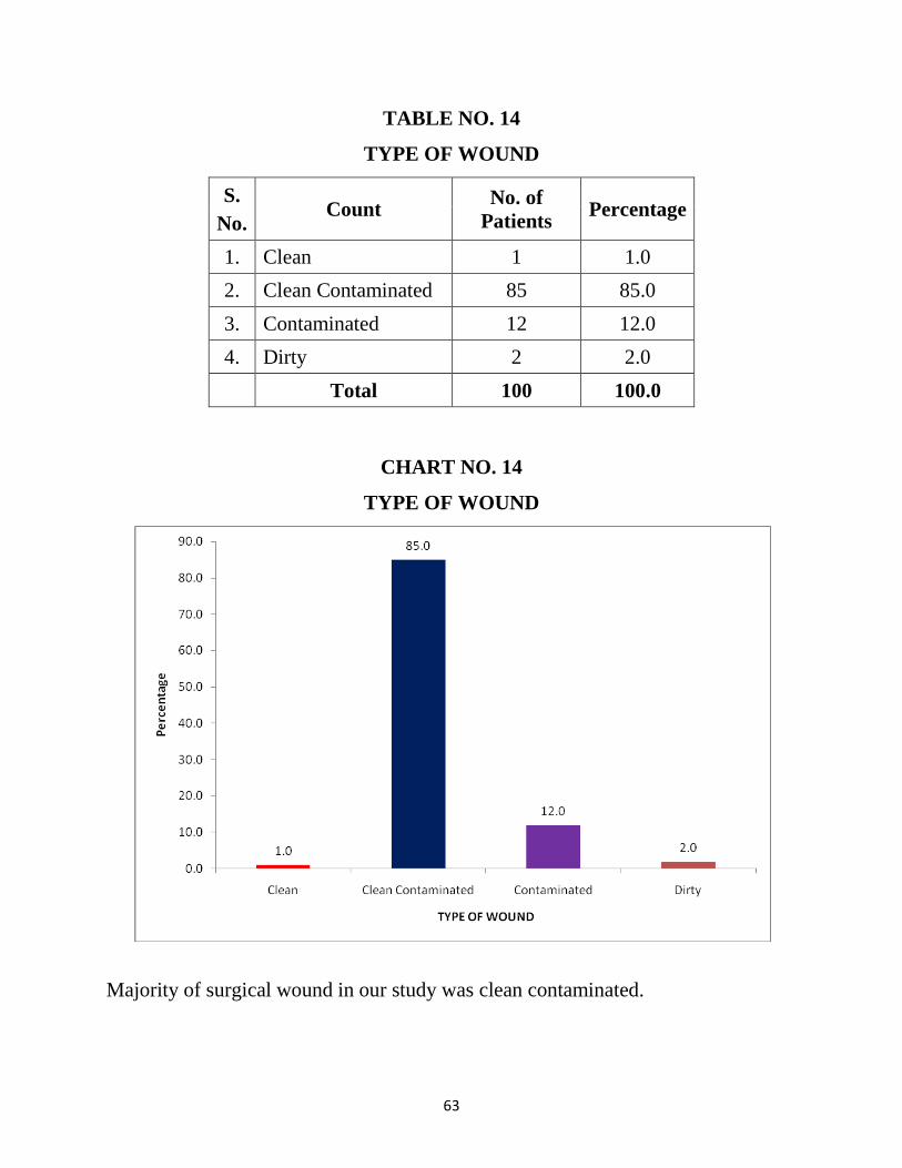
TYPE OF WOUND
S.
No. Count
No. of
Patients Percentage
1. Clean 1 1.0
2. Clean Contaminated 85 85.0
3. Contaminated 12 12.0
4. Dirty 2 2.0
Total 100 100.0
CHART NO. 14
TYPE OF WOUND
Majority of surgical wound in our study was clean contaminated.
64
TABLE NO. 15
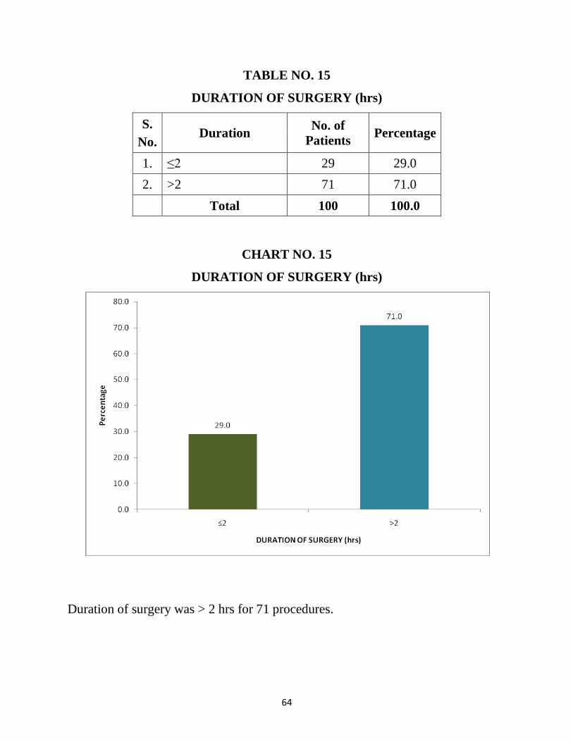
DURATION OF SURGERY (hrs)
S.
No. Duration
No. of
Patients Percentage
1. ≤2 29 29.0
2. >2 71 71.0
Total 100 100.0
CHART NO. 15
DURATION OF SURGERY (hrs)
Duration of surgery was > 2 hrs for 71 procedures.
65
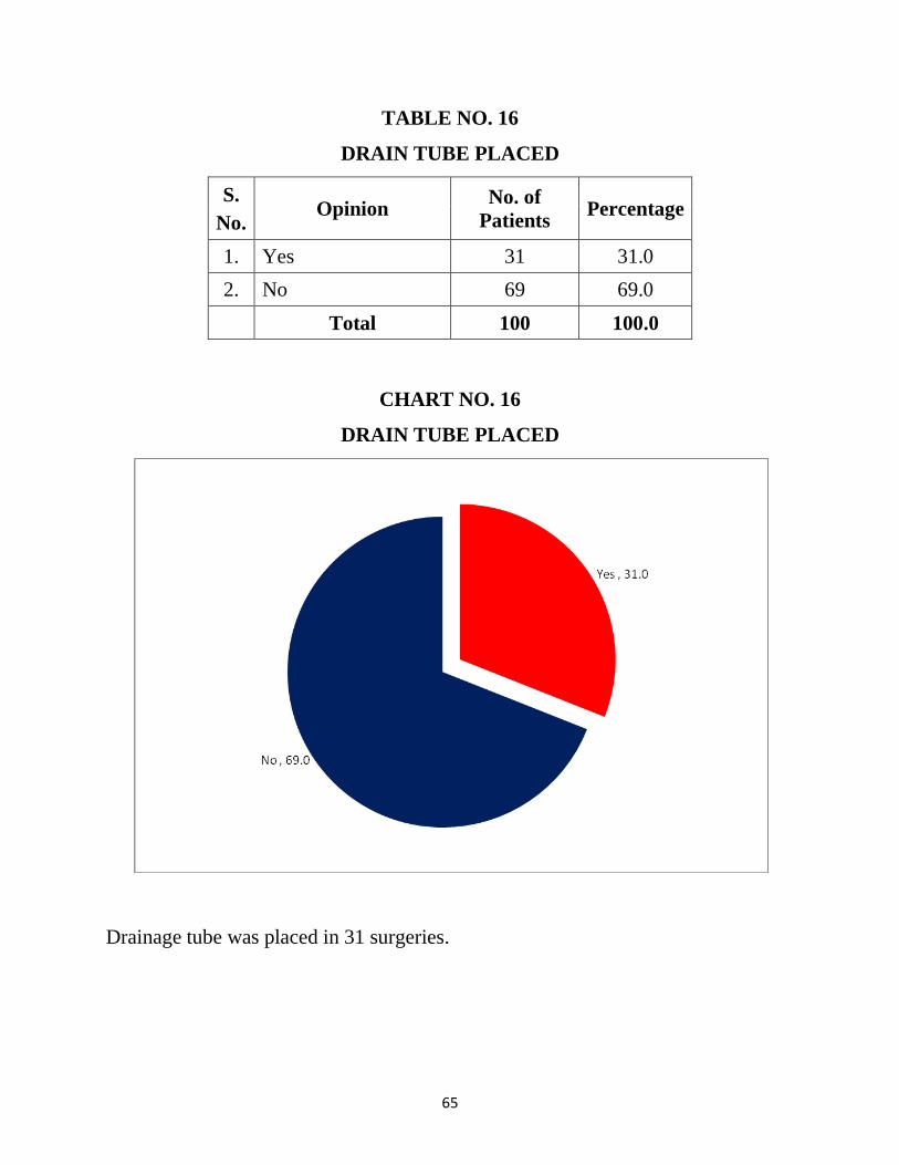
TABLE NO. 16
DRAIN TUBE PLACED
S.
No. Opinion
No. of
Patients Percentage
1. Yes 31 31.0
2. No 69 69.0
Total 100 100.0
CHART NO. 16
DRAIN TUBE PLACED
Drainage tube was placed in 31 surgeries.
66
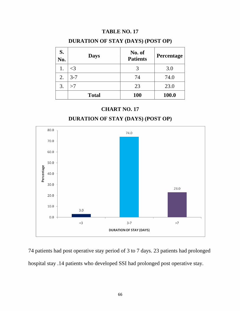
TABLE NO. 17
DURATION OF STAY (DAYS) (POST OP)
S.
No. Days
No. of
Patients Percentage
1. <3 3 3.0
2. 3-7 74 74.0
3. >7 23 23.0
Total 100 100.0
CHART NO. 17
DURATION OF STAY (DAYS) (POST OP)
74 patients had post operative stay period of 3 to 7 days. 23 patients had prolonged
hospital stay .14 patients who developed SSI had prolonged post operative stay.
67
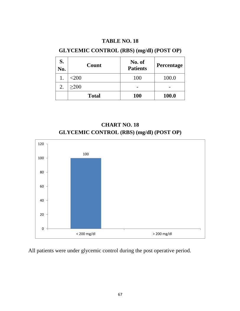
TABLE NO. 18
GLYCEMIC CONTROL (RBS) (mg/dl) (POST OP)
S.
No. Count
No. of
Patients Percentage
1. <200 100 100.0
2. ≥200 - -
Total 100 100.0
CHART NO. 18
GLYCEMIC CONTROL (RBS) (mg/dl) (POST OP)
All patients were under glycemic control during the post operative period.
100
0
20
40
60
80
100
120
< 200 mg/dl > 200 mg/dl
68
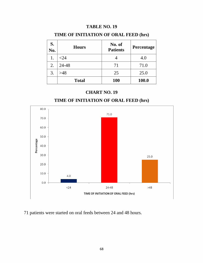
TABLE NO. 19
TIME OF INITIATION OF ORAL FEED (hrs)
S.
No. Hours
No. of
Patients Percentage
1. <24 4 4.0
2. 24-48 71 71.0
3. >48 25 25.0
Total 100 100.0
CHART NO. 19
TIME OF INITIATION OF ORAL FEED (hrs)
71 patients were started on oral feeds between 24 and 48 hours.
69
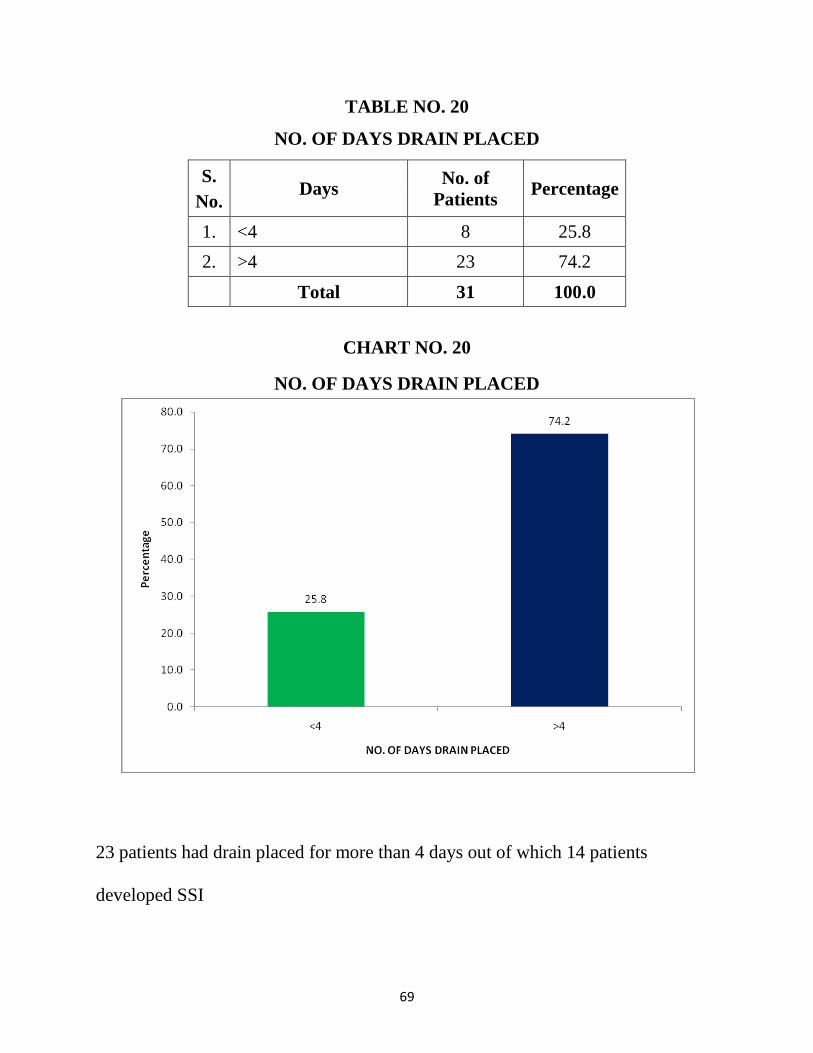
TABLE NO. 20
NO. OF DAYS DRAIN PLACED
S.
No. Days
No. of
Patients Percentage
1. <4 8 25.8
2. >4 23 74.2
Total 31 100.0
CHART NO. 20
NO. OF DAYS DRAIN PLACED
23 patients had drain placed for more than 4 days out of which 14 patients
developed SSI
70
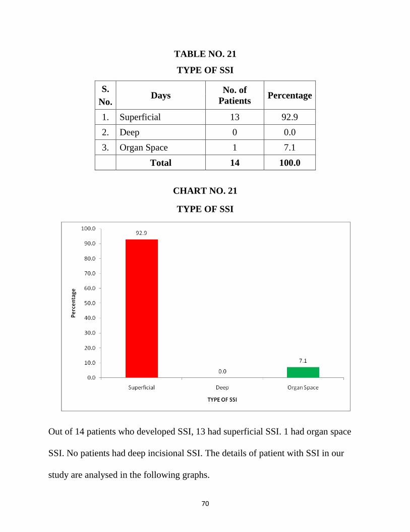
TABLE NO. 21
TYPE OF SSI
S.
No. Days
No. of
Patients Percentage
1. Superficial 13 92.9
2. Deep 0 0.0
3. Organ Space 1 7.1
Total 14 100.0
CHART NO. 21
TYPE OF SSI
Out of 14 patients who developed SSI, 13 had superficial SSI. 1 had organ space
SSI. No patients had deep incisional SSI. The details of patient with SSI in our
study are analysed in the following graphs.
71
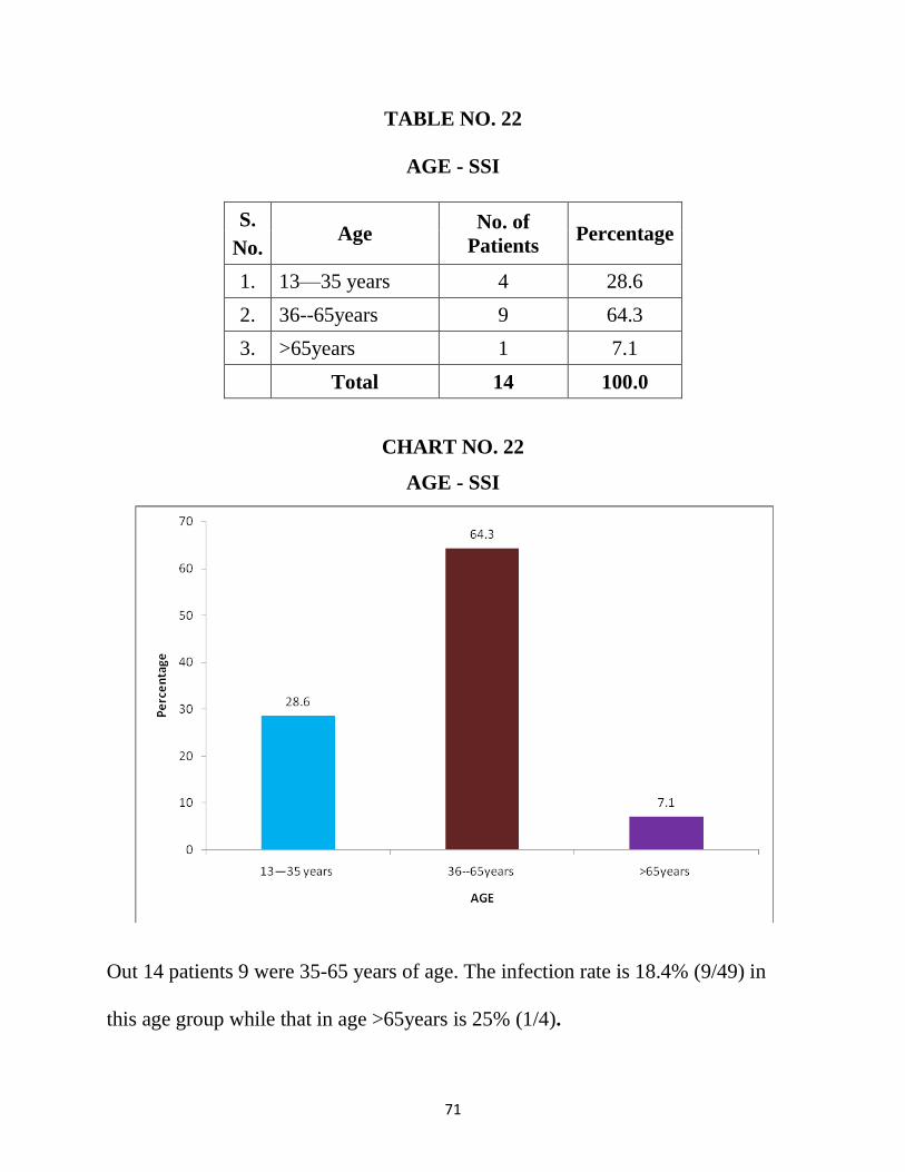
TABLE NO. 22
AGE - SSI
S.
No. Age
No. of
Patients Percentage
1. 13—35 years 4 28.6
2. 36--65years 9 64.3
3. >65years 1 7.1
Total 14 100.0
CHART NO. 22
AGE - SSI
Out 14 patients 9 were 35-65 years of age. The infection rate is 18.4% (9/49) in
this age group while that in age >65years is 25% (1/4).
72
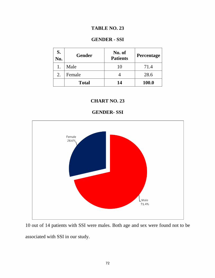
TABLE NO. 23
GENDER - SSI
S.
No. Gender
No. of
Patients Percentage
1. Male 10 71.4
2. Female 4 28.6
Total 14 100.0
CHART NO. 23
GENDER- SSI
10 out of 14 patients with SSI were males. Both age and sex were found not to be
associated with SSI in our study.
73
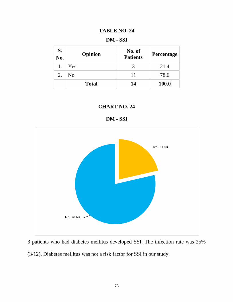
TABLE NO. 24
DM - SSI
S.
No. Opinion
No. of
Patients Percentage
1. Yes 3 21.4
2. No 11 78.6
Total 14 100.0
CHART NO. 24
DM - SSI
3 patients who had diabetes mellitus developed SSI. The infection rate was 25%
(3/12). Diabetes mellitus was not a risk factor for SSI in our study.
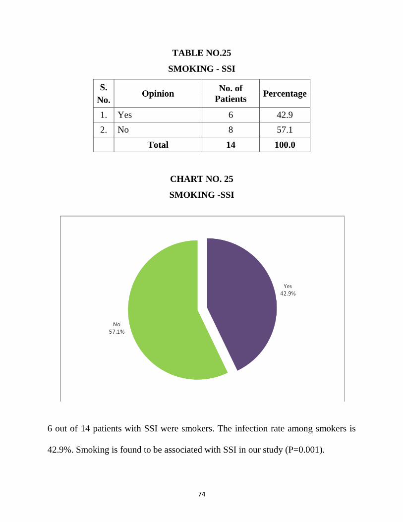
74
TABLE NO.25
SMOKING - SSI
S.
No. Opinion
No. of
Patients Percentage
1. Yes 6 42.9
2. No 8 57.1
Total 14 100.0
CHART NO. 25
SMOKING -SSI
6 out of 14 patients with SSI were smokers. The infection rate among smokers is
42.9%. Smoking is found to be associated with SSI in our study (P=0.001).
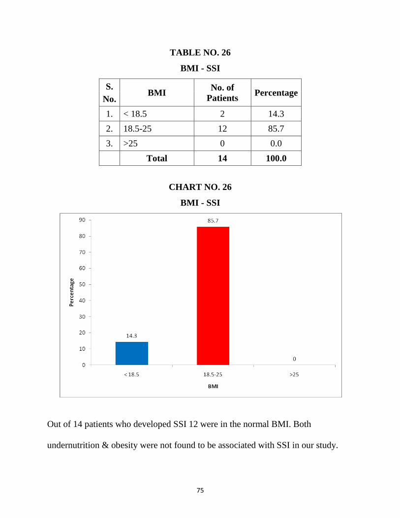
75
TABLE NO. 26
BMI - SSI
S.
No. BMI
No. of
Patients Percentage
1. < 18.5 2 14.3
2. 18.5-25 12 85.7
3. >25 0 0.0
Total 14 100.0
CHART NO. 26
BMI - SSI
Out of 14 patients who developed SSI 12 were in the normal BMI. Both
undernutrition & obesity were not found to be associated with SSI in our study.
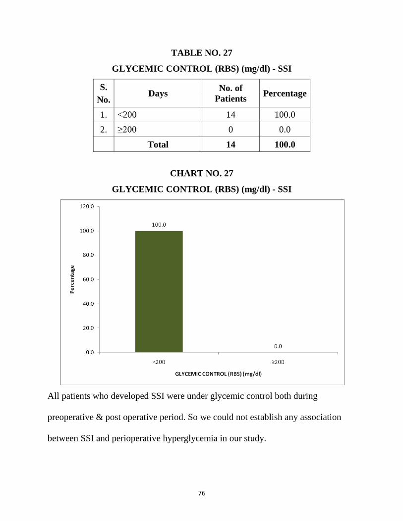
76
TABLE NO. 27
GLYCEMIC CONTROL (RBS) (mg/dl) - SSI
S.
No. Days
No. of
Patients Percentage
1. <200 14 100.0
2. ≥200 0 0.0
Total 14 100.0
CHART NO. 27
GLYCEMIC CONTROL (RBS) (mg/dl) - SSI
All patients who developed SSI were under glycemic control both during
preoperative & post operative period. So we could not establish any association
between SSI and perioperative hyperglycemia in our study.
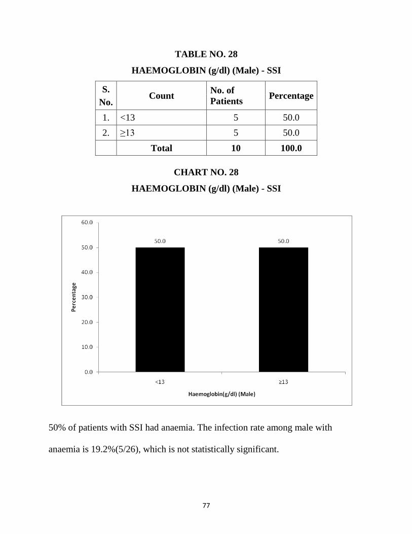
77
TABLE NO. 28
HAEMOGLOBIN (g/dl) (Male) - SSI
S.
No. Count
No. of
Patients Percentage
1. <13 5 50.0
2. ≥13 5 50.0
Total 10 100.0
CHART NO. 28
HAEMOGLOBIN (g/dl) (Male) - SSI
50% of patients with SSI had anaemia. The infection rate among male with
anaemia is 19.2%(5/26), which is not statistically significant.

78
TABLE NO. 29
HAEMOGLOBIN (g/dl) (Female) - SSI
S.
No. Count
No. of
Patients Percentage
1. <12 3 75.0
2. ≥12 1 25.0
Total 4 100.0
CHART NO. 29
HAEMOGLOBIN (g/dl) (Female) - SSI
Out of 4 female patients who developed SSI, 3 were found to have anaemia. But
the infection rate is 10.3% (3/26) in female patients with anemia. Anaemia in both
females and males were not associated with SSI in our study.
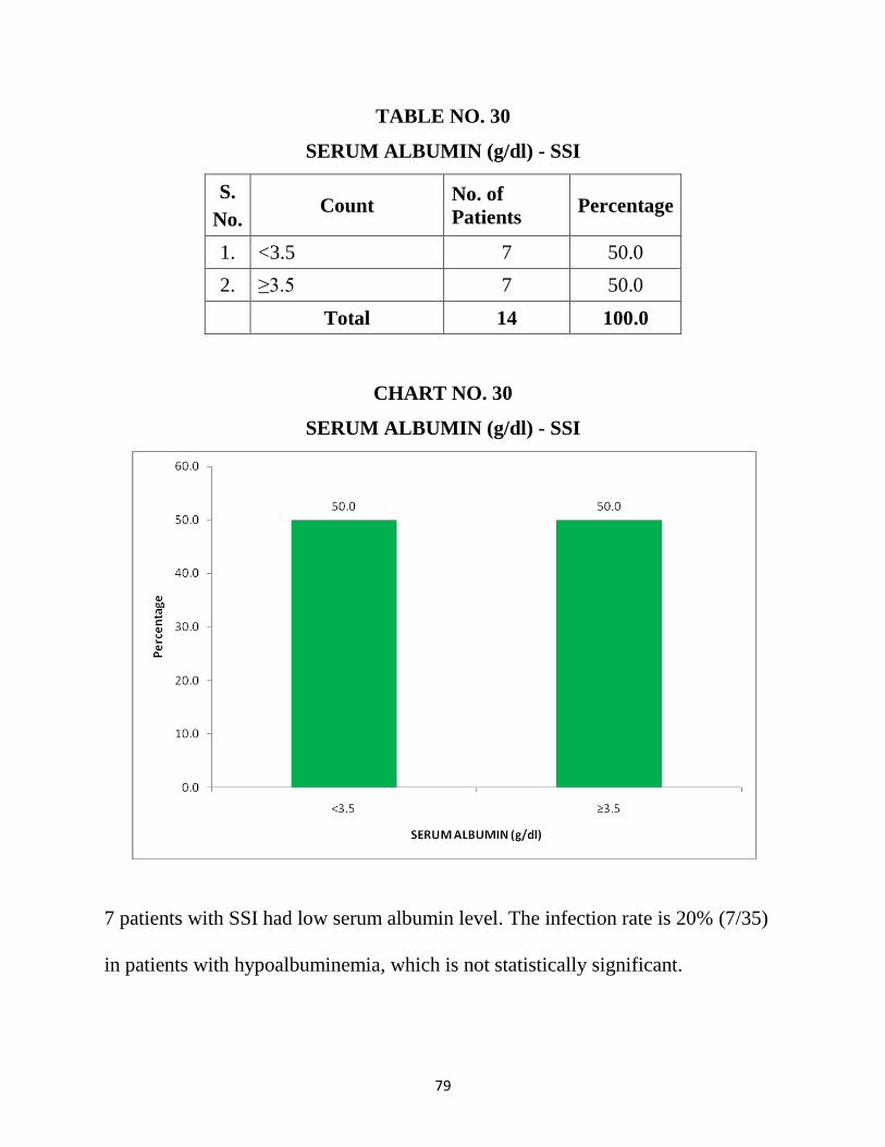
79
TABLE NO. 30
SERUM ALBUMIN (g/dl) - SSI
S.
No. Count
No. of
Patients Percentage
1. <3.5 7 50.0
2. ≥3.5 7 50.0
Total 14 100.0
CHART NO. 30
SERUM ALBUMIN (g/dl) - SSI
7 patients with SSI had low serum albumin level. The infection rate is 20% (7/35)
in patients with hypoalbuminemia, which is not statistically significant.
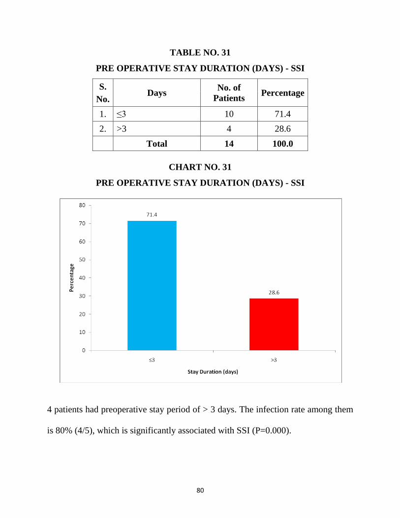
80
TABLE NO. 31
PRE OPERATIVE STAY DURATION (DAYS) - SSI
S.
No. Days
No. of
Patients Percentage
1. ≤3 10 71.4
2. >3 4 28.6
Total 14 100.0
CHART NO. 31
PRE OPERATIVE STAY DURATION (DAYS) - SSI
4 patients had preoperative stay period of > 3 days. The infection rate among them
is 80% (4/5), which is significantly associated with SSI (P=0.000).
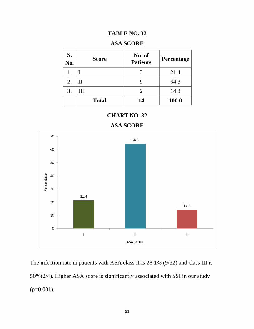
81
TABLE NO. 32
ASA SCORE
S.
No. Score
No. of
Patients Percentage
1. I 3 21.4
2. II 9 64.3
3. III 2 14.3
Total 14 100.0
CHART NO. 32
ASA SCORE
The infection rate in patients with ASA class II is 28.1% (9/32) and class III is
50%(2/4). Higher ASA score is significantly associated with SSI in our study
(p=0.001).
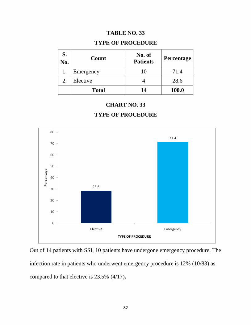
82
TABLE NO. 33
TYPE OF PROCEDURE
S.
No. Count
No. of
Patients Percentage
1. Emergency 10 71.4
2. Elective 4 28.6
Total 14 100.0
CHART NO. 33
TYPE OF PROCEDURE
Out of 14 patients with SSI, 10 patients have undergone emergency procedure. The
infection rate in patients who underwent emergency procedure is 12% (10/83) as
compared to that elective is 23.5% (4/17).
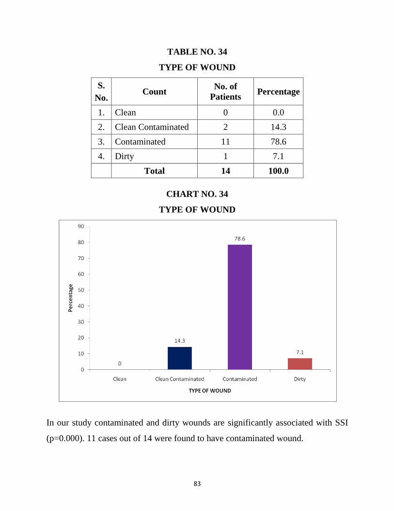
83
TABLE NO. 34
TYPE OF WOUND
S.
No. Count
No. of
Patients Percentage
1. Clean 0 0.0
2. Clean Contaminated 2 14.3
3. Contaminated 11 78.6
4. Dirty 1 7.1
Total 14 100.0
CHART NO. 34
TYPE OF WOUND
In our study contaminated and dirty wounds are significantly associated with SSI
(p=0.000). 11 cases out of 14 were found to have contaminated wound.
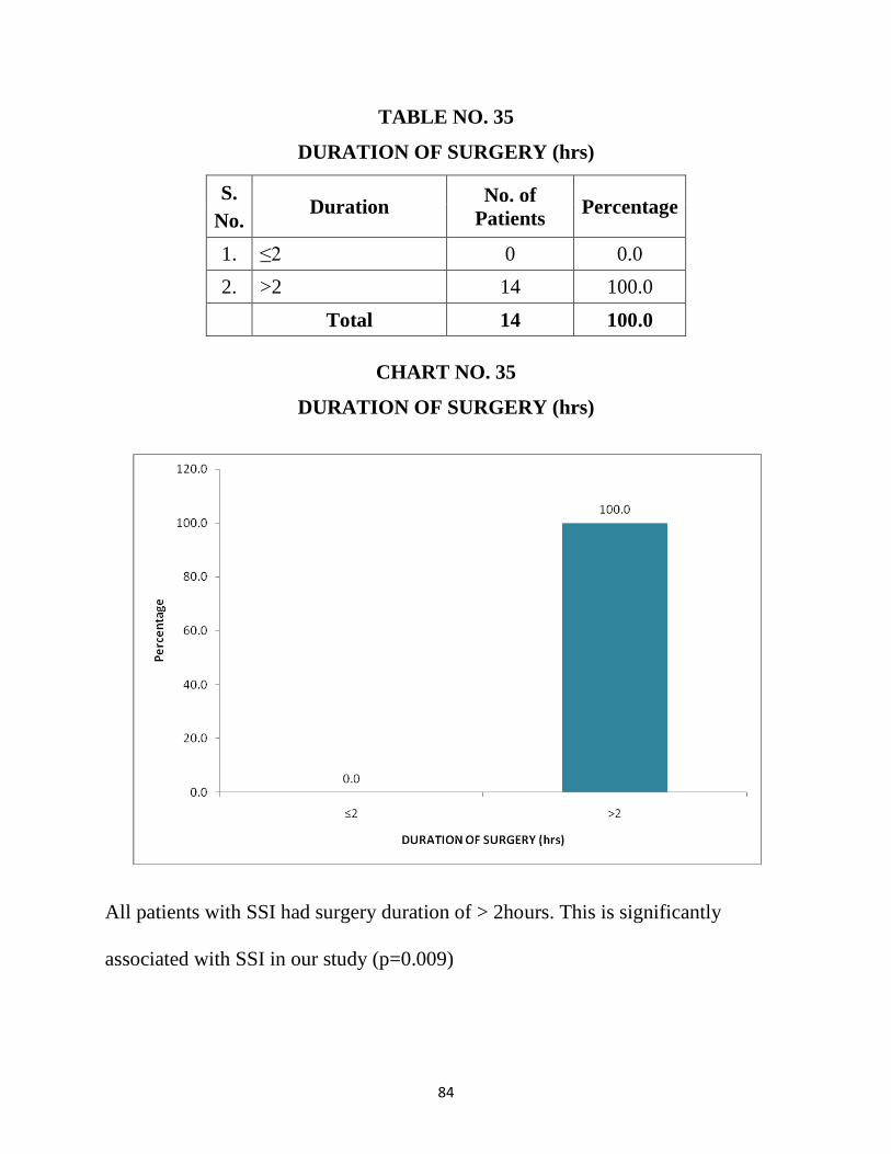
84
TABLE NO. 35
DURATION OF SURGERY (hrs)
S.
No. Duration
No. of
Patients Percentage
1. ≤2 0 0.0
2. >2 14 100.0
Total 14 100.0
CHART NO. 35
DURATION OF SURGERY (hrs)
All patients with SSI had surgery duration of > 2hours. This is significantly
associated with SSI in our study (p=0.009)
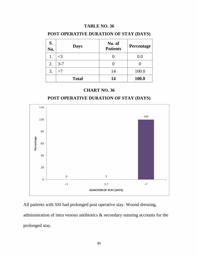
85
TABLE NO. 36
POST OPERATIVE DURATION OF STAY (DAYS)
S.
No. Days
No. of
Patients Percentage
1. <3 0 0.0
2. 3-7 0 0
3. >7 14 100.0
Total 14 100.0
CHART NO. 36
POST OPERATIVE DURATION OF STAY (DAYS)
All patients with SSI had prolonged post operative stay. Wound dressing,
administration of intra venous antibiotics & secondary suturing accounts for the
prolonged stay.
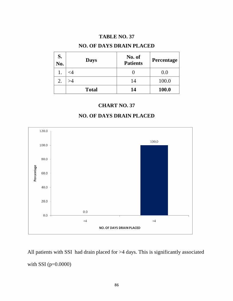
86
TABLE NO. 37
NO. OF DAYS DRAIN PLACED
S.
No. Days
No. of
Patients Percentage
1. <4 0 0.0
2. >4 14 100.0
Total 14 100.0
CHART NO. 37
NO. OF DAYS DRAIN PLACED
All patients with SSI had drain placed for >4 days. This is significantly associated
with SSI (p=0.0000)
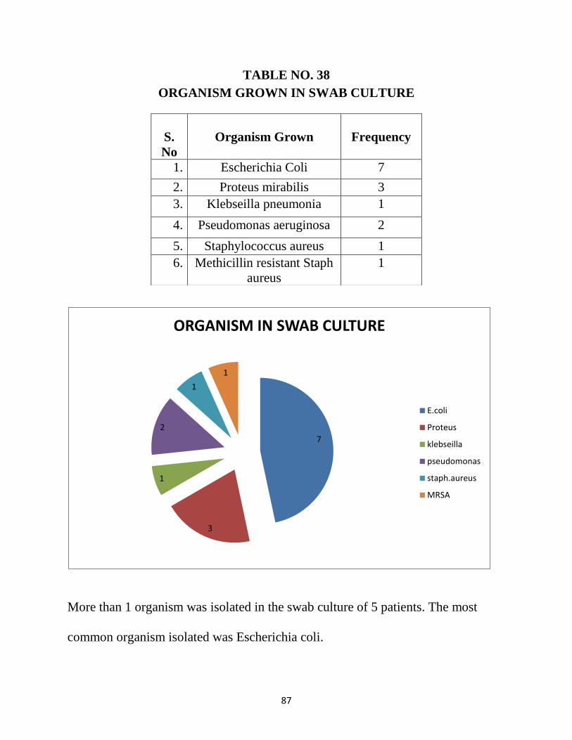
87
TABLE NO. 38
ORGANISM GROWN IN SWAB CULTURE
More than 1 organism was isolated in the swab culture of 5 patients. The most
common organism isolated was Escherichia coli.
S.
No
Organism Grown
Frequency
1. Escherichia Coli 7
2. Proteus mirabilis 3
3. Klebseilla pneumonia 1
4. Pseudomonas aeruginosa 2
5. Staphylococcus aureus 1
6. Methicillin resistant Staph
aureus
1
7
3
1
2
1
1
ORGANISM IN SWAB CULTURE
E.coli
Proteus
klebseilla
pseudomonas
staph.aureus
MRSA
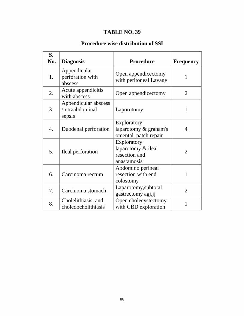
88
TABLE NO. 39
Procedure wise distribution of SSI
S.
No.
Diagnosis
Procedure
Frequency
1.
Appendicular
perforation with
abscess
Open appendicectomy
with peritoneal Lavage 1
2. Acute appendicitis
with abscess Open appendicectomy 2
3.
Appendicular abscess
/intraabdominal
sepsis
Laporotomy 1
4. Duodenal perforation
Exploratory
laparotomy & graham's
omental patch repair
4
5. Ileal perforation
Exploratory
laparotomy & ileal
resection and
anastamosis
2
6. Carcinoma rectum
Abdomino perineal
resection with end
colostomy
1
7. Carcinoma stomach Laparotomy,subtotal
gastrectomy agj,jj 2
8. Cholelithiasis and
choledocholithiasis
Open cholecystectomy
with CBD exploration 1
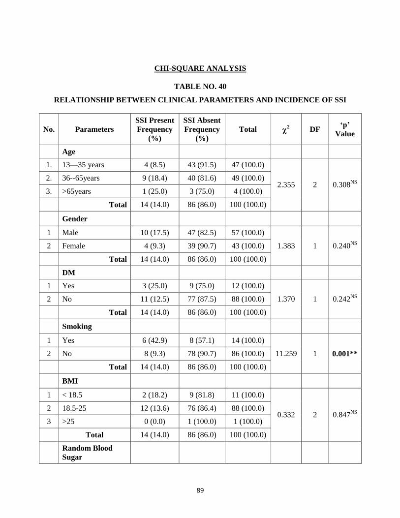
89
CHI-SQUARE ANALYSIS
TABLE NO. 40
RELATIONSHIP BETWEEN CLINICAL PARAMETERS AND INCIDENCE OF SSI
No. Parameters
SSI Present
Frequency
(%)
SSI Absent
Frequency
(%)
Total 2 DF
‘p’
Value
Age
1. 13—35 years 4 (8.5) 43 (91.5) 47 (100.0)
2.355 2 0.308NS
2. 36--65years 9 (18.4) 40 (81.6) 49 (100.0)
3. >65years 1 (25.0) 3 (75.0) 4 (100.0)
Total 14 (14.0) 86 (86.0) 100 (100.0)
Gender
1 Male 10 (17.5) 47 (82.5) 57 (100.0)
1.383 1 0.240NS
2 Female 4 (9.3) 39 (90.7) 43 (100.0)
Total 14 (14.0) 86 (86.0) 100 (100.0)
DM
1 Yes 3 (25.0) 9 (75.0) 12 (100.0)
1.370 1 0.242NS
2 No 11 (12.5) 77 (87.5) 88 (100.0)
Total 14 (14.0) 86 (86.0) 100 (100.0)
Smoking
1 Yes 6 (42.9) 8 (57.1) 14 (100.0)
11.259 1 0.001** 2 No 8 (9.3) 78 (90.7) 86 (100.0)
Total 14 (14.0) 86 (86.0) 100 (100.0)
BMI
1 < 18.5 2 (18.2) 9 (81.8) 11 (100.0)
0.332 2 0.847NS
2 18.5-25 12 (13.6) 76 (86.4) 88 (100.0)
3 >25 0 (0.0) 1 (100.0) 1 (100.0)
Total 14 (14.0) 86 (86.0) 100 (100.0)
Random Blood
Sugar
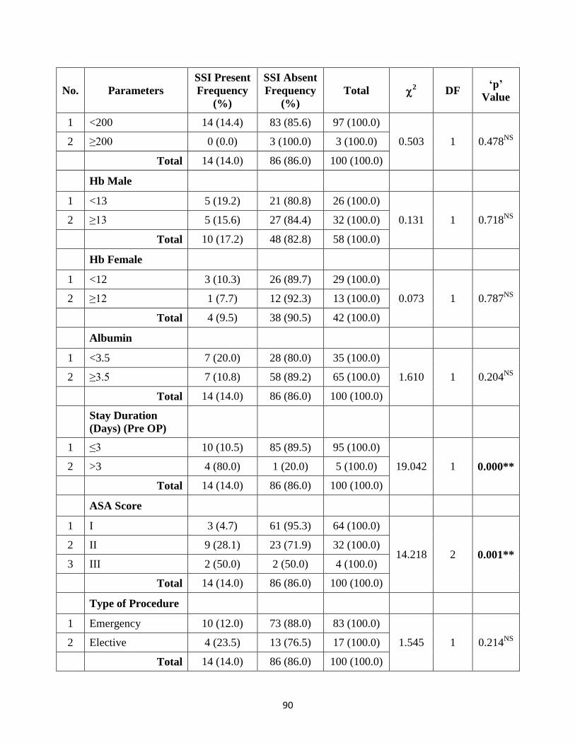
90
No. Parameters
SSI Present
Frequency
(%)
SSI Absent
Frequency
(%)
Total 2 DF
‘p’
Value
1 <200 14 (14.4) 83 (85.6) 97 (100.0)
0.503 1 0.478NS
2 ≥200 0 (0.0) 3 (100.0) 3 (100.0)
Total 14 (14.0) 86 (86.0) 100 (100.0)
Hb Male
1 <13 5 (19.2) 21 (80.8) 26 (100.0)
0.131 1 0.718NS
2 ≥13 5 (15.6) 27 (84.4) 32 (100.0)
Total 10 (17.2) 48 (82.8) 58 (100.0)
Hb Female
1 <12 3 (10.3) 26 (89.7) 29 (100.0)
0.073 1 0.787NS
2 ≥12 1 (7.7) 12 (92.3) 13 (100.0)
Total 4 (9.5) 38 (90.5) 42 (100.0)
Albumin
1 <3.5 7 (20.0) 28 (80.0) 35 (100.0)
1.610 1 0.204NS
2 ≥3.5 7 (10.8) 58 (89.2) 65 (100.0)
Total 14 (14.0) 86 (86.0) 100 (100.0)
Stay Duration
(Days) (Pre OP)
1 ≤3 10 (10.5) 85 (89.5) 95 (100.0)
19.042 1 0.000** 2 >3 4 (80.0) 1 (20.0) 5 (100.0)
Total 14 (14.0) 86 (86.0) 100 (100.0)
ASA Score
1 I 3 (4.7) 61 (95.3) 64 (100.0)
14.218 2 0.001** 2 II 9 (28.1) 23 (71.9) 32 (100.0)
3 III 2 (50.0) 2 (50.0) 4 (100.0)
Total 14 (14.0) 86 (86.0) 100 (100.0)
Type of Procedure
1 Emergency 10 (12.0) 73 (88.0) 83 (100.0)
1.545 1 0.214NS
2 Elective 4 (23.5) 13 (76.5) 17 (100.0)
Total 14 (14.0) 86 (86.0) 100 (100.0)
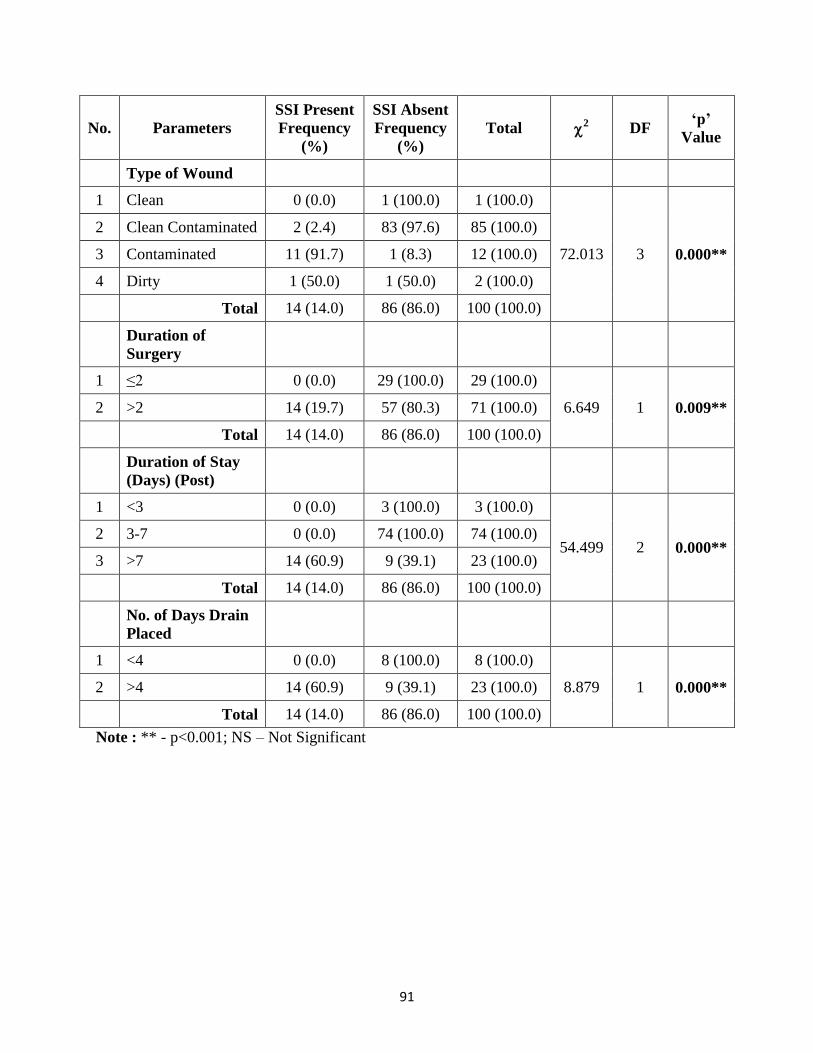
91
No. Parameters
SSI Present
Frequency
(%)
SSI Absent
Frequency
(%)
Total 2 DF
‘p’
Value
Type of Wound
1 Clean 0 (0.0) 1 (100.0) 1 (100.0)
72.013 3 0.000**
2 Clean Contaminated 2 (2.4) 83 (97.6) 85 (100.0)
3 Contaminated 11 (91.7) 1 (8.3) 12 (100.0)
4 Dirty 1 (50.0) 1 (50.0) 2 (100.0)
Total 14 (14.0) 86 (86.0) 100 (100.0)
Duration of
Surgery
1 ≤2 0 (0.0) 29 (100.0) 29 (100.0)
6.649 1 0.009** 2 >2 14 (19.7) 57 (80.3) 71 (100.0)
Total 14 (14.0) 86 (86.0) 100 (100.0)
Duration of Stay
(Days) (Post)
1 <3 0 (0.0) 3 (100.0) 3 (100.0)
54.499 2 0.000** 2 3-7 0 (0.0) 74 (100.0) 74 (100.0)
3 >7 14 (60.9) 9 (39.1) 23 (100.0)
Total 14 (14.0) 86 (86.0) 100 (100.0)
No. of Days Drain
Placed
1 <4 0 (0.0) 8 (100.0) 8 (100.0)
8.879 1 0.000** 2 >4 14 (60.9) 9 (39.1) 23 (100.0)
Total 14 (14.0) 86 (86.0) 100 (100.0)
Note : ** - p<0.001; NS – Not Significant
92
DISCUSSION
Large number of studies reported surgical site infection in abdominal
surgeries between 3.4% and 36.1% 96
. In our study out of 100 patients who
underwent abdominal surgeries, 14 patients developed SSI. The rate of SSI I in our
study is 14%. This is comparable to many studies in India95, 96, 107
and is higher
compared to developed countries and less as compared to few Indian studies97.
This
is due to the fact that in developed countries they have a systematic feedback of
SSI rate and surveillance bodies such as hospitals in Europe Link for Infection
Control through Surveillance (HELICS) in Europe and National Nosocomial
Infection Surveillance System (NNIS) in United States of America whereas in our
country we rely mainly on sporadic surveys. In our study most of patients are of
middle age group (35-65years) and there is male preponderance. The risk factors
associated with SSI in our study are smoking (p=0.001), pre operative stay of >
3days (p=0.000), ASA score (p=0.001), contaminated & dirty wound (p=0.000),
duration of surgery (p= 0.010) & duration drain placement (p=0.000). Our study
did not find association between SSI and BMI grading, anaemia,
hypoalbuminemia. In our study Smoking was found to be associated with SSI like
previous studies97.
The infection rate among smokers is 42.9% (6/14) while that in
non smokers is 9.3% (8/86). Pre operative stay duration of > 3 days is significantly
associated with SSI. The infection rate is 80% in patients with pre operative stay of
93
> 3 days as compared to 10.5% in patients with < 3 days duration. Similar finding
is observed in many studies95, 96, 100, 102, 103, 106, 108.
Patients with ASA class of 2 & 3 are associated with SSI. This is
comparable to previous studies97, 98, 99, 100, 107.
The infection rate in class II patients is
28.1% and in class III are 50%. In our study Contaminated & dirty wound were
associated with SSI as observed in previous studies95, 98, 99, 100, 102.
The infection rate
in contaminated wound is 91.7% (11/12) while in dirty wound it is 50% (1/2).
Duration of surgery > 2 hours duration is significantly associated with SSI. Reports
from other studies are in agreement with our findings95, 96, 97, 98, 100, and 102.
The
infection rate is 19.7% (14/71) in patients when the duration of surgery was >2
hours. No patient with surgery duration < 2 hours developed SSI in our study
(0/29).
Duration of drain placement for > 4 days is associated with SSI in our study.
Similar finding was observed in many studies96, 97, 98, 99, 102, 106, 107.
The infection rate
is 60.9% in patients with drain placed for > 4 days.
The most common disease condition encountered in our study is acute
appendicitis with or without abscess & surgical procedure observed is emergency
open Appendicectomy. SSI was most commonly observed in appendicular abscess
& duodenal perforation. SSI was noted on 4th post operat
ive day for 9 patients and
5th post operative day for 5 patients. None of the patients developed SSI after
94
discharge from hospital. The endogenous flora is responsible for infection in most
cases. The opening of the gastrointestinal tract increases the likelihood of Gram-
negative bacilli that was our finding in this study. The most common organism
isolated was E.coli. It was isolated in 50% of swab culture. This is similar to the
finding observed by Amit Agarwal et al & Lul raka et al96, 100.
Pseudomonas &
proteus mirabilis were next most common organisms isolated. More than 1
organism was isolated in the swab culture of 5 patients. E.coli was found sensitive
to piperacillin & Tazobactum, Imipenam , Colistin. The other organisms observed
in swab culture were klebseilla, staph aureus, MRSA. Swab culture was sterile in 3
patients in our study.
In patients who developed SSI, 13 patients had superficial SSI .1 patient had
organ space SSI. None of the patients developed deep incisional SSI. Secondary
wound closure was done for 9 (64.3%) patients who had SSI with residual wound
dehiscence with healthy granulation tissue in whom spontaneous closure did not
occur. All patients with SSI had prolonged post operative stay duration of more
than 7 days.
95
CONCLUSION
Surgical site infection is increasingly recognized as a measure of the quality
of patient care by surgeons, infection control practitioners, health planners and
public. The incidence of SSI in our environment is still high when compared to the
developed world. The SSI rate in our study is 14% and risk factors associated with
SSI in our study are smoking (p=0.001), pre operative stay of > 3days (p=0.000),
ASA score (p=0.001), contaminated & dirty wound (p=0.000), duration of surgery
(p= 0.010) & duration of drain placement (p=0.000).
Our study prompts us to look at the gaps in our surgical and infection control
protocols which will enable policy formulation that will foster a reduction in
wound infection rate. SSI can be reduced by decreasing the preoperative hospital
stay, appropriate antibiotic administration policies, adequate preoperative patient
preparation, reducing the duration of surgery to minimum, judicious use of drains
and intraoperative maintenance of asepsis and following operation theatre
discipline properly.
Although surgical site infections cannot be completely eliminated, a
reduction in the infection rate to a minimal level could have significant benefits, by
reducing postoperative morbidity and mortality, and wastage of health care
resources.
96
LIMITATIONS
Majority of surgical procedures in our study are of Emergency surgeries .so
the incidence of SSI is high in our study. Moreover we were not able to analyse the
rate of SSI in Elective surgeries.
We covered only abdominal surgeries. Other cases in surgery department
were not included. The scenario can change if other surgical cases are
incorporated. This may change the SSI surveillance system & infection control
policies.
97
RECOMMENDATIONS
A dedicated system of infection surveillance has to be established to identify
the gaps in our infection control protocols and therefore identify areas of focus to
reduce the burden of SSIs. It will also help to individualize policies regarding
infection control in different setups.
Appropriate precautionary measure has to be taken to reduce the incidences
of SSI that originate primarily from the care procedures provided during
hospitalization. A sound antibiotic policy, reduction of length of procedures
through adequate training of the staff on proper surgical techniques, proper intra-
operative infection control measures and feedback of appropriate data to surgeons
regarding SSIs would be desirable to reduce the surgical site infection.
98
REFERENCES
1. National Healthcare Safety Network, Centers for Disease Control and
Prevention. Surgical site infection (SSI) event. http://www.cdc.gov/nhsn
/pdfs/pscmanual/9pscssicurrent.pdf. Published January 2017. Accessed
January 25, 2017.
2. Horan TC, Gaynes RP, Martone WJ, Jarvis WR, Emori TG. CDC definitions
of nosocomial surgical site infections, 1992: a modification of CDC
definitions of surgical wound infections. Infect Control Hosp Epidemiol 1992;
13:606–608).
3. Bailey & love short practice of surgery, 27th edition.
4. Allegranzi B, Bagheri Nejad S, Combescure C,Graafmans W, Attar H,
Donaldson L, et al. Burden of endemic health-care-associated infection in
developing countries: systematic review and meta-analysis. Lancet. 2011; 377
(9761) :228-41.
5. Report on the burden of endemic health care-associated infection worldwide.
A systematic review of the literature. Geneva: World Health Organization;
2011.
6. Haley RW, Culver DH, White JW, Morgan WM,Emori TG, Munn VP, et al.
The efficacy of infection surveillance and control programs in preventing
99
nosocomial infections in US hospitals. Am J Epidemiol. 1985;121(2): 182-
205
7. Harbarth S, Sax H, Gastmeier P. The preventable proportion of nosocomial
infections: an overview of published reports. J Hosp Infect. 2003;54(4):258-
66; quiz 321.
8. Anderson DJ, Kaye KS, Classen D, et al. Strategies to prevent surgical site
infections in acute care hospitals. Infect Control Hosp Epidemiology. 2008;
29:S51–S61.
9. Engemann JJ, Carmeli Y, Cosgrove SE, et al. Adverse clinical and economic
outcomes attributable to methicillin resistance among patients with
Staphylococcus aureus surgical site infection. Clin Infect Dis. 2003;36:592–
598
10. Anderson DJ, Kirkland KB, Kaye KS, et al. Under resourced hospital
infection control and prevention programs: penny wise, pound foolish? Infect
Control Hosp Epidemiol. 2007;28(7):767-773
11. Centers for Disease Control and Prevention. The direct medical costs of
healthcare-associated infections in U.S. hospitals and the benefits of
prevention. http://www.cdc.gov/hai/pdfs/hai/scott_costpaper.pdf. Published
2009. Accessed January 25, 2017.
100
12. Stone PW, Braccia D, Larson E. Systematic review of economic analyses of
health care–associated infections. Am J Infect Control. 2005;33(9):501-509
13. Robbins basic pathology, 9th
Edition
14. Guideline for prevention of surgical site Infection,1999, Centre for Disease
Control. Alicia j. Mangram, md; teresa c. Horan, mph, cic; michele l. Pearson,
md; leah christine silver, bs; william r. Jarvis, md; Infection control &hospital
Epidemiology vol No. 20 No.4
15. Sabiston Textbook of Surgery 19th
edition
16. Risk factors for development of surgical site infections among liver
transplantation recipients: An integrative literature review vRamon Antônio
Oliveira, MSc, RN American journal of infection control, January 2018,
vol.46, issue 1 pg 88-93
17. Moyes LH, Leitch EF, McKee RF, Anderson JH, Horgan PG, McMillan DC.
Preoperative systemic inflammation predicts postoperative infectious
complications in patients undergoing curative resections for colorectal
cancer. Br J Cancer. 2009;100: 1236–1239.
18. Kubo T, Ono S, Ueno H, Shinto E, Yamamoto J, Hase K. Elevated
preoperative C-reactive protein levels are a risk factor for the development of
postoperative infectious complications following elective colorectal
surgery. Langenbecks Arch Surg. 2013;398: 965–971.
101
19. Nozoe T, Matsumata T, Sugimachi K. Preoperative elevation of serum C-
reactive protein is related to impaired immunity in patients with colorectal
cancer. Am J Clin Oncol. 2000;23: 263–266.
20. Cruse PJ. Polk HC, Stone HH. Some factors determining wound infection: a
prospective study of 30,000 wounds. Hospital-acquired infectious surgery..
Baltimore: University Park Press; 1997. 75–105.
21. Cruse PJ, Foord R. A five year prospective study of 23469 surgical wounds.
Arch Surg 1973;107: 206–12.
22. Falagas ME, Kompoti M. Obesity and infection. Lancet Infect
Dis2006;6:438–46.
23. Kabon B, Nagele A, Reddy D, Eagon C, Fleshman JW, Sessler DI, et al.
Obesity decreases perioperative tissue oxygenation.
Anesthesiology2004;100:274–80.
24. Sen CK. Wound healing essentials: let there be oxygen. Wound Repair
Regeneration 2009;17:1–18.
25. Falagas ME, Karageorgopoulos DE. Adjustment of dosing of antimicrobial
agents for bodyweight in adults. Lancet 2010;375:248–51.
26. Martin JH, Saleem M, Looke D. Therapeutic drug monitoring to adjust
dosing in morbid obesity—a new use for an old methodology. Br J
ClinPharmacol 2012;73:685–90
102
27. Mullen JT, Davenport DL, Hutter MM, Hosokawa PW, Henderson WG,
Khuri SF, et al. Impact of body mass index on perioperative outcomes in
patients undergoing major intra-abdominal cancer surgery. Ann Surg Oncol.
2008; 15(8): 2164–72.
28. Kurmann A, Vorburger SA, Candinas D, Beldi G. Operation time and body
Mass index are significant risk factors for surgical site infection in
laparoscopic sigmoid resection: a multicenter study. Surg Endosc.
2011;25(11):3531–4.
29. Tanaka S, Inoue S, Isoda F, Waseda M, Ishihara M, Yamakawa T, et al.
Impaired immunity in obesity: suppressed but reversible lymphocyte
responsiveness. Int J Obes Relat Metab Disord. 1993;17(11):631–6.
30. Stryker LS, Abdel MP, Morrey ME, Morrow MM, Kor DJ, Morrey BF.
Elevated postoperative blood glucose and preoperative hemoglobin A1C are
associated with increased wound complications following total joint
arthroplasty. J Bone Joint Surg Am. 2013;95(9):808–14, S801–802.
31. World Health Organization (WHO). Global Guidelines for the Prevention of
Surgical Site Infection. Geneva: WHO, 2016. Accessed February 18, 2018.
http://apps.who.int/iris/bitstream/10665/250680/1/9789241549882
103
32. Maradit Kremers H, et al. Diabetes mellitus, hyperglycemia, hemoglobin
A1C and the risk of prosthetic joint infections in total hip and knee
arthroplasty. J Arthroplasty. 2015 Mar;30(3):439–443.
33. Zerr KJ, Furnary AP, Grunkemeier GL, Bookin S, Kanhere V, Starr A.
Glucose control lowers the risk of wound infection in diabetics after open
heart operations. Ann Thorac Surg 1997;63(2):356-61.
34. Terranova A. The effects of diabetes mellitus on wound healing. Plast Surg
Nurs 1991;11(1):20-5
35. Pessaux P, Msika S, Atalla D, Hay JM, Flamant Y. Risk factors for
postoperative infectious complications in non colorectal abdominal surgery:
a multivariate analysis based on a prospective multicenter study of 4718
patients. Arch Surg 2003; 138:314–24.
36. Polly SM, Sanders WE Jr. Surgical infections in the elderly:
prevention,diagnosis, and treatment. Geriatrics 1977; 32:88–90, 95–87.
37. Raymond D, Pelletier S, Crabtree T, Schulman A, Pruett T, Sawyer R
Surgical infection and the aging population. Am Surg 2001; 67:827–32.
38. Ives CL, Harrison DK, Stansby GS: Tissue oxygen saturation, measured by
near-infrared spectroscopy, and its relationship to surgical-site infections. Br J
Surg 94:87–91, 2007.
104
39. Brar MS, Brar SS, Dixon E: Perioperative supplemental oxygen in colorectal
patients: A meta-analysis. J Surg Res 166:227–235, 2011.
40. Knighton DR, Halliday B, Hunt TK: Oxygen as an antibiotic. A comparison
of the effects of inspired oxygen concentration and antibiotic administration
on in vivo bacterial clearance. Arch Surg 121:191–195, 1986.
41. Gottrup F: Oxygen in wound healing and infection. World J Surg 28:312–
315, 2004.
42. Jonsson K, Hunt TK, Mathes SJ. Oxygen as an isolated variable influences
resistance to infection. Ann Surg. 1988;208 783-7
43. Dickinson A, Qadan M, Polk HC, Jr: Optimizing surgical care: A
contemporary assessment of temperature, oxygen, and glucose. Am Surg
76:571–577, 2010.
44. Perioperative anemia: an independent risk factor for infection, mortality, and
resource utilization in surgery. Dunne JR1, Malone D, Tracy JK, Gannon
C, Napolitano LM. Department of Surgery, VA Maryland Healthcare System,
Baltimore, Maryland, USA. Journal of surgical research,feb102(2), 237-44
45. Surgical Site Infections: Reanalysis of Risk Factors. Debra L. Malone,
M.D.,Thomas Genuit, M.D.,J.Kathleen Tracy, M.A., Christopher Gannon,
M.D.,Lena M. journal of surgical research volume 103,issue 1, march
2002,pg85-95
105
46. Olsen MA, Lefta M, Dietz JR, et al. Risk factors for surgical site infection
after major breast operation. J Am Coll Surg 2008; 207(3):326–33
47. Valentine RJ, Weigelt JA, Dryer D, Rodgers C. Effect of remote infections
on clean wound infection rates. Am J Infect Control 1986; 14(2):64-67.
48. Stuesse DC, Robinson JH, Durzinsky DS. A late sternal wound infection
caused by hematogenous spread of bacteria. Chest 1995; 108(6): 1742-1743.
49. Hunter JG, Padilla M, Cooper-Vastola S. Late Clostridium perfringens breast
implant infection after dental treatment. Ann Plast Surg 1996; 36(3):309-312.
50. Edwards LD. The epidemiology of 2056 remote site infections and 1966
surgical wound infections occurring in 1865 patients: a four year study of 40,923
operations at Rush-Presbyterian-St. Luke’s Hospital, Chicago. Ann Surg
1976; 184(4(6):758-766.
51. Perl TM, Golub JE. New approaches to reduce Staphylococcus aureus
nosocomial infection rates: treating S. aureus nasal carriage. Ann
Pharmacother. 1998;32:S7-S16.
52. Kluytmans JA, Mouton JW, Ijzerman EP, et al. Nasal carriage of
Staphylococcus aureus as a major risk factor for wound infections after
cardiac surgery. J Infect Dis. 1995;171:216-219.
53. Culebras JM. Malnutrition in the twenty-first century: an epidemic affecting
surgical outcome. Surg Infect (Larchmt). 2013;14(3):237-43.
106
54. Mainous MR, Deitch EA. Nutrition and infection. Surg Clin North Am.
1994;74(3):659-76.
55. Waitzberg DL, Ravacci GR, Raslan M. [Hospital hyponutrition]. Nutr Hosp.
2011;26(2):254-64.
56. Studley HO. Percentage of weight loss: a basic indicator of surgical risk in
patients with chronic peptic ulcer. 1936. Nutr Hosp.2001;16(4):141-3
57. Culebras-Fernandez JM, de Paz-Arias R, Jorquera- Plaza F, Garcia de
Lorenzo A. Nutrition in the surgical patient: immunonutrition. Nutr
Hosp.2001;16(3):67-77.
58. Fry DE. Fifty ways to cause surgical site infections. Surg Infect
(Larchmt).2011;12(6):497-500.
59. Chen Y, Guo Q, Pan X, et al. Smoking and impaired bone healing: will
activation of cholinergic anti-inflammatory pathway be the bridge? Int
Orthop. 2011;35:1267–1270. doi: 10.1007/s00264-011-1243-5
60. Rahman MM, Laher I. Structural and functional alteration of blood vessels
caused by cigarette smoking: an overview of molecular mechanisms. Curr
Vasc Pharmacol. 2007;5:276–279. doi: 10.2174/157016107782023406.
61. Ref: J Chemother. 2001 Nov;13 Spec No 1(1):12-6.Immune system and
surgical site infection. Esposito S.
107
62. Incidence and Etiology of Surgical Site Infections among Emergency
Postoperative Patients in Mbarara Regional Referral Hospital, South Western
Uganda. Abubaker Lubega,1 Bazira Joel,2 and Najjuka Justina Lucy1 1
Surgery Research and Practice Volume 2017, Article ID 6365172
63. M. Hedstrom, L. Gr ¨ ondal, ¨ A. ˚ Ortquist, N. Dal ¨ en, and T. Ahl, “Serum
albumin and deep infection in femoral neck fractures,” International Orthopaedics,
vol. 22, no. 3, pp. 182–184, 1998.
64. CDC Morbidity and Mortality Weekly Report October 25, 2002 / Vol. 51 /
No. RR-16
65. Larson EL, et al. Alcohol for surgical scrubbing? Infect Control Hosp
Epidemiol. 1990 Mar;11(3):139–143.
66. Mulberry G, et al. Evaluation of a waterless, scrubless chlorhexidine
gluconate/ethanol surgical scrub for antimicrobial efficacy. Am J Infect Control.
2001 Dec:29(6):377–382
67. Wisconsin Department of Health Services. Wisconsin Division of Public
Health Supplemental Guidance for the Prevention of Surgical Site Infections:
An Evidence-Based Perspective. Jan 2017. (Updated: Jul 2017.) Accessed
February 16, 2018. https://www.dhs.wisconsin.gov/publications/p01715.pdf.
108
68. Ban KA, et al. American College of Surgeons and Surgical Infection Society:
Surgical site infection guidelines, 2016 update. J Am Coll Surg. 2017
Jan;224(1):59–74.
69. Anderson DJ, et al. Strategies to prevent surgical site infections in acute care
hospitals: 2014 update. Infect Control Hosp Epidemiol. 2014 J un;35(6):605–
627.
70. Aly R, Maibach HI. Comparative antibacterial efficacy of a 2-minute surgical
scrub with chlorhexidine gluconate, povidone-iodine, and chloroxylenol
sponge-brushes. Am J Infect Control 1988;16:173-7.
71. Mayhall CG. Surgical infections including burns. In: Wenzel RP, ed.
Prevention and Control of Nosocomial Infections. 2nd ed. Baltimore:
Williams & Wilkins; 1993. p. 614-64.
72. Brown TR, Ehrlich CE, Stehman FB, Golichowski AM, Madura JA,Eitzen
HE. A clinical evaluation of chlorhexidine gluconate spray as compared with
iodophore scrub for preoperative skin preparation. Surg Gynecol Obstet
1984;158:363-6.
73. Ayliffe GA. Role of the environment of the operating suite in surgical
wound infection. Rev Infect Dis 1991;13(Suppl 10):S800-4.
74. Stamm WE, Feeley JC, Facklam RR. Wound infection due to group A
streptococcus traced to a vaginal carrier. J Infect Dis 1978;138:287-92.
109
75. Gryska PF, O’Dea AE. Postoperative streptococcal wound infection. The
anatomy of an epidemic. JAMA 1970;213:1189-91.
76. Sehulster L, Chinn RY. Guidelines for environmental infection control in
health-care facilities. Recommendations of CDC and the Healthcare Infection
Control Practices Advisory Committee (HICPAC). Morbid Mortal Wkly Rep.
2003;52(Rr-10):1-42.
77. N. Petrosillo, C. M. J. Drapeau, E. Nicastri et al., “Surgical site infections in
Italian Hospitals: a prospective multicenter study,” BMC Infectious Diseases,
vol. 8, article 34, 2008.
78. K.-S. Cha, O.-H. Cho, and S.-Y. Yoo, “Risk factors for surgical site
infections in patients undergoing craniotomy,” Journal of Korean Academy of
Nursing, vol. 40, no. 2, pp. 298–305, 2010
79. L. Pulido, E. Ghanem, A. Joshi, J. J. Purtill, and J. Parvizi, “Periprosthetic
joint infection: the incidence, timing, and predisposing factors,” Clinical
Orthopaedics and Related Research, vol. 466, no. 7, pp. 1710–1715, 2008.
80. Cheng H, Chen BP, Soleas IM, Ferko NC, Cameron CG, Hinoul P. Prolonged
operative duration increases risk of surgical site infections: A systematic
review. Surg Infect (Larchmt) 2017;18:722‑35.
110
81. Berard F, Gandon J. Factors influencing the incidence of wound
infection. Ann Surg. 1964;160:32–81.
82. Bratzler DW, et al. Clinical practice guidelines for antimicrobial prophylaxis
in surgery. Am J Health Syst Pharm. 2013 Feb 1;70:195–283.
83. Galandiuk S, et al. Re-emphasis of priorities in surgical antibiotic prophylaxis.
Surg Gynecol Obstet. 1989 Sep;169(3):219–222.
84. Drinkwater CJ, Neil MJ. Optimal timing of wound drain removal following
total joint arthroplasty. J Arthroplasty 1995;10(2):185-9.
85. Committee on Control of Surgical Infections of the Committee on Pre and
Postoperative Care, American College of Surgeons. Manual on Control of
Infection in Surgical Patients. Philadelphia: J.B. Lippincott Co;1984
86. Flores-Maldonado A, Medina-Escobedo CE, Rios-Rodriguez HM, et al: Mild
perioperative hypothermia and the risk of wound infection. Arch Med Res
32:227–231, 2001.
87. Cohn SM, Giannotti G, Ong AW, et al. Prospective randomized trial of two
wound management strategies for dirty abdominal wounds. Ann Surg
2001;233:409–413.
88. Watanabe A, Kohnoe S, Shimabukuro R, et al. Risk factors associated with
surgical site infection in upper and lower gastrointestinal surgery. Surg Today
2008;38:404–412.
111
89. Perioperative Allogeneic Blood Transfusion Is Associated With Surgical Site
Infection After Abdominoperineal Resection—a Space for the Implementation
of Patient Blood Management Strategies Kensuke Kaneko Kazushige Kawai ,
Nelson H. Tsuno, Soichiro Ishihara , Hironori Yamaguchi , Eiji Sunami ,
Toshiaki Watanabe Department of Surgical Oncology and Department of
Transfusion Medicine, Faculty of Medicine, University of Tokyo, Tokyo,
Japan
90. Schwartz principles of Surgery, 10th
edition.
91. Agarwal SL (1972). Study of postoperative wound infection. Indian J.
Surg. 34: 314-20.
92. Rao AS, Harsha M (1975). Postoperative wound infections. J Indian
Med. Assoc. 64 :90-3.
93. Kowli SS, Nayak MH, Mehta AP, Bhalerao RA (1985). Hospital infection.
Indian J. Surg. 48: 475-86.
94. Anvikar AR, Deshmukh AB, Karyakarte RP, Damle AS, Patwardhan NS,
Malik AK, Bichile LK, Bajaj JK, Baradkar VP, Kulkarni JD, Sachdeo
SM (1999). A one year prospective study of 3,280 surgical wounds. Indian J.
Med. Microbiol. 17: 129-32.
95. Abdominal Surgical Site Infection Occurrence and Risk Factors in Krishna
Institute of Medical Science, Karad. Hemant B Janugade, Basavaraj K Nagur,
112
Kruthika R Sajjan, Sangeeta B Biradar, Jignesh K Savsaviya, Mahesh Reddy.
International Journal of Scientific Study | February 2016 | Vol 3 | Issue 11
96. Surgical site infection in abdominal surgeries: a clinical study amit agrawal, r.
P. Singhjournal of evolution of med and dent sci/ eissn- 2278-4802, pissn-
278-4748/ vol. 3/ issue 40/sep 01, 2014 page 10188
97. Determinants of superficial surgical site infections in abdominal surgeries at a
Rural Teaching Hospital in Central India: A prospective study, Mekhla, Firoz
Rajiv Borle , Journal of Family Medicine and Primary Care Wednesday,
September 11, 2019.
98. Incidence of and risk factors for abdominal surgical site infection in a
Nigerian tertiary care centre, Adeyinka Ayodele Adejumo, Mshelia Nuhu,
Tolulope Afolaranmi, International Journal of Infection Control 2015, v11:i4
99. Surgical site infections after abdominal surgery: incidence and risk factors. A
prospective cohort study. Journal : infectious diseases Volume 47, 2015 -
Issue 11
100. Surgical site infections in an abdominal surgical ward at Kosovo Teaching
Hospital Lul Raka,Avdyl Krasniqi, Faton Hoxha, Ruustem Musa, Gjyle
Mulliqi, Selvete Krasniqi, Arsim Kurti, Antigona Dervishaj, Beqir
Nuhiu,Baton Kelmendi,2 Dalip Limani,2,3 Ilir Tolaj.3 J Infect Developing
Countries 2007; 1(3):337-341.
113
101. Surgical site infection rates in 6 cities of India: findings of the International
Nosocomial Infection Control Consortium (INICC). International Health
Advance Access published December 8, 2014
102. Incidence and factors associated with surgical site infections in a teaching
hospital in Ujjain, India. Ashish Pathak , Erika A. Saliba RN, Shailendra
Sharma MS,Vijay Kumar Mahadik , Harshada Shah , Cecilia Stålsby
Lundborg American Journal of Infection Control 42 (2014) e11-e15
103. Surgical site Infection among postoperative patients of tertiary care centre in
Central India - A prospective study Dr. Anand Saxena, Dr. Mahendra Pratap
Singh, Dr. Swagata Brahmchari, Dr. Malay Banerjee. Asian Journal of
Biomedical and Pharmaceutical Sciences 3(17) 2013, 41-44
104. Surgical site infections: Assessing risk factors, outcomes and antimicrobial
sensitivity patterns ,Suchitra Joyce B. and Lakshmidevi N. African Journal of
Microbiology Research Vol. 3 (4) pp. 175-179 April, 2009
105. A prospective study of surgical site infections and related risk factors in a
teaching hospital Rajanikanth Amrutham, Madhu Mohan Reddy B., Nagababu
Pyadala . International Surgery Journal 2017 Jan;4(1):237-241
106. Incidence and determinants of the surgical site infection: a hospital based
longitudinal study Pinakin K. Sutariya, Mallika V. Chavada . International
Surgery Journal 2016 Nov;3(4):2202-2206

114
107. Study of Surgical Site Infections in Abdominal Surgeries Satyanarayana V.,
Prashanth H.V., Basavaraj Bhandare, Kavyashree A. Journal of Clinical and
Diagnostic Research. 2011 October, Vol-5(5): 935-939 935
108. Surgical site infection in clean and clean-contaminated Cases SP lilani,
N jangale, A chowdhary, GB daver indian journal of medical microbiology,
(2005) 23 (4):249-52
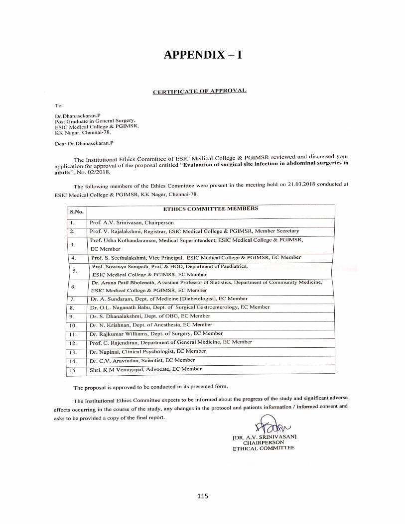
115
APPENDIX – I
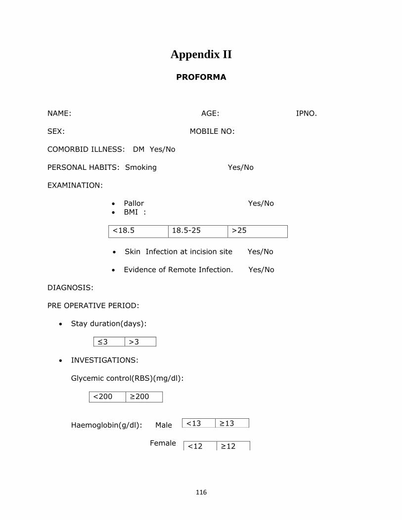
116
Appendix II
PROFORMA
NAME: AGE: IPNO.
SEX: MOBILE NO:
COMORBID ILLNESS: DM Yes/No
PERSONAL HABITS: Smoking Yes/No
EXAMINATION:
Pallor Yes/No BMI :
<18.5 18.5-25 >25
Skin Infection at incision site Yes/No
Evidence of Remote Infection. Yes/No
DIAGNOSIS:
PRE OPERATIVE PERIOD:
Stay duration(days):
≤3 >3
INVESTIGATIONS:
Glycemic control(RBS)(mg/dl):
<200 ≥200
Haemoglobin(g/dl): Male
Female
<13 ≥13
<12 ≥12
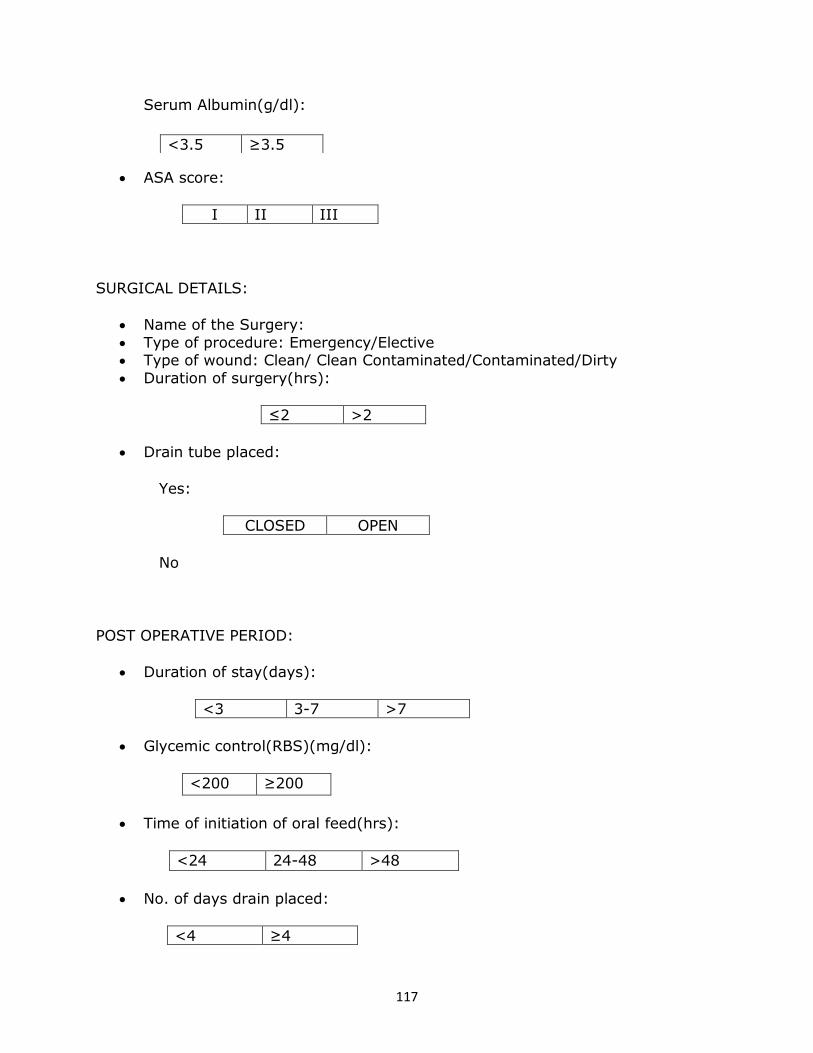
117
Serum Albumin(g/dl):
ASA score:
I II III
SURGICAL DETAILS:
Name of the Surgery:
Type of procedure: Emergency/Elective Type of wound: Clean/ Clean Contaminated/Contaminated/Dirty
Duration of surgery(hrs):
≤2 >2
Drain tube placed:
Yes:
CLOSED OPEN
No
POST OPERATIVE PERIOD:
Duration of stay(days):
<3 3-7 >7
Glycemic control(RBS)(mg/dl):
<200 ≥200
Time of initiation of oral feed(hrs):
<24 24-48 >48
No. of days drain placed:
<4 ≥4
<3.5 ≥3.5
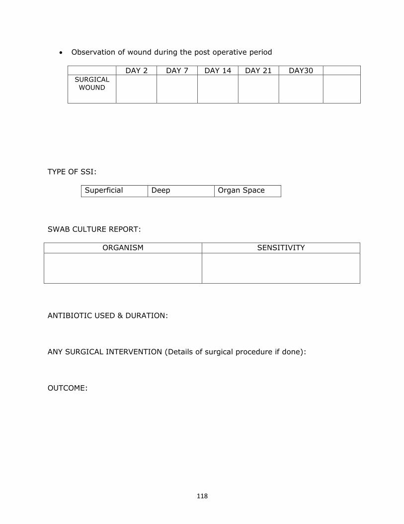
118
Observation of wound during the post operative period
DAY 2 DAY 7 DAY 14 DAY 21 DAY30 SURGICAL
WOUND
TYPE OF SSI:
Superficial Deep Organ Space
SWAB CULTURE REPORT:
ORGANISM SENSITIVITY
ANTIBIOTIC USED & DURATION:
ANY SURGICAL INTERVENTION (Details of surgical procedure if done):
OUTCOME:
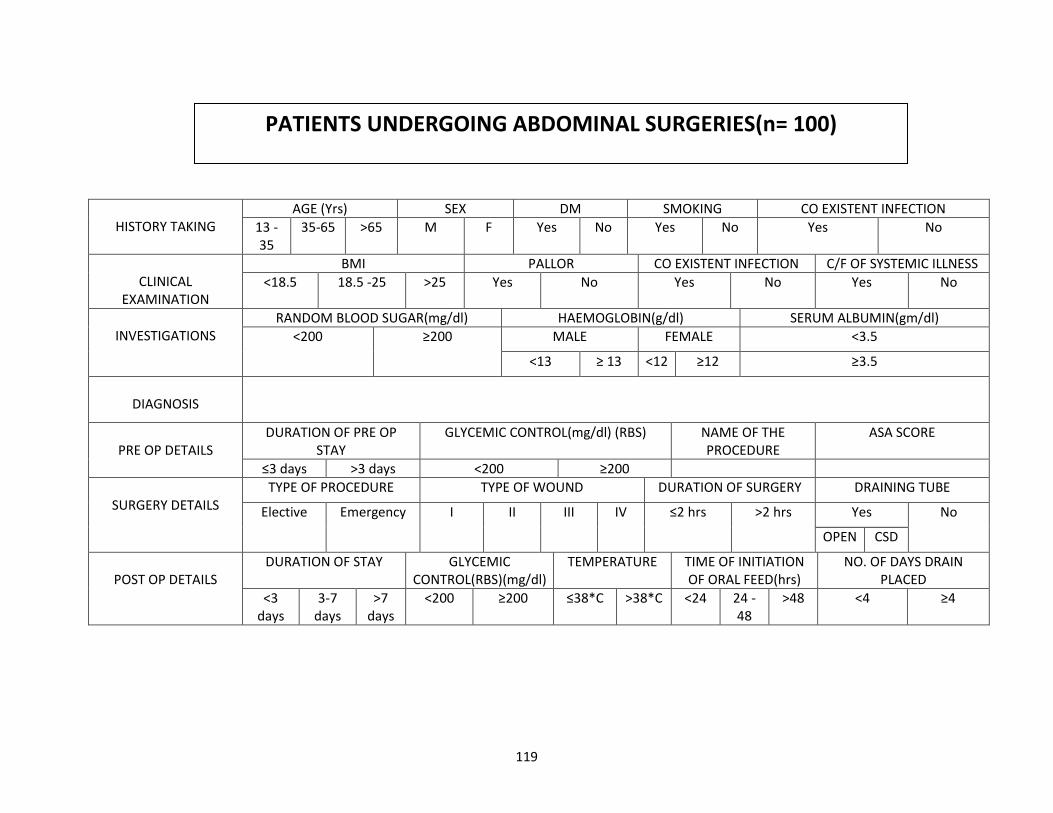
119
HISTORY TAKING
AGE (Yrs) SEX DM SMOKING CO EXISTENT INFECTION
13 -35
35-65 >65 M F Yes No Yes No Yes No
CLINICAL
EXAMINATION
BMI PALLOR CO EXISTENT INFECTION C/F OF SYSTEMIC ILLNESS
<18.5 18.5 -25 >25 Yes No Yes No Yes No
INVESTIGATIONS
RANDOM BLOOD SUGAR(mg/dl) HAEMOGLOBIN(g/dl) SERUM ALBUMIN(gm/dl)
<200 ≥200 MALE FEMALE <3.5
<13 ≥ 13 <12 ≥12 ≥3.5
DIAGNOSIS
PRE OP DETAILS
DURATION OF PRE OP STAY
GLYCEMIC CONTROL(mg/dl) (RBS) NAME OF THE PROCEDURE
ASA SCORE
≤3 days >3 days <200 ≥200
SURGERY DETAILS
TYPE OF PROCEDURE TYPE OF WOUND DURATION OF SURGERY DRAINING TUBE
Elective Emergency I II III IV ≤2 hrs >2 hrs Yes No
OPEN CSD
POST OP DETAILS
DURATION OF STAY GLYCEMIC CONTROL(RBS)(mg/dl)
TEMPERATURE TIME OF INITIATION OF ORAL FEED(hrs)
NO. OF DAYS DRAIN PLACED
<3 days
3-7 days
>7 days
<200 ≥200 ≤38*C >38*C <24 24 -48
>48 <4 ≥4
PATIENTS UNDERGOING ABDOMINAL SURGERIES(n= 100)
120
Appendix III
INFORMED CONSENT
Informed consent for patients who are attending surgical OPD or casualty in ESIC MEDICAL
COLLEGE &PGIMSR hospital, and whom we are inviting to participate in the research titled
“Evaluation of surgical site infections in abdominal surgeries”AT ESIC MEDICAL
COLLEGE & PGIMSR, KK Nagar, Chennai, 2014-15". Dr.Dhanasekaran.P, M.S. (General
surgery) post graduate is the principal investigator of this research under ESI-PGIMSR, Chennai.
Part I: Information Sheet
Introduction
We,Dr.Dhanasekaran. P, 1st year General Surgery PG, guided by
PROF.Dr. P.N. Shanmugasundaram, M.S., Professor and HOD Of General Surgery, are going to give
you information and invite you to be a part of this research. Before you decide, you can talk to
anyone of us you feel comfortable with about the research. This consent form may contain words that
you do not understand. Please ask us to stop as we go through the information and we will take time
to explain. If you have questions later, you can ask us.
Purpose of the research
We will treat you by performing appropriate surgery for your disease. You will be called for scheduled
follow up after discharge. All the details will be recorded in a proforma sheet and will be used to find out
the cause for infection at the surgery site.
Type of Research
This research will involve your participation in a non-experimental manner, with assured privacy and
confidentiality.
Right to Refuse or Withdraw
Your participation is strictly voluntary. Refusal to participate will not affect subsequent services to you
Confidentiality
All information you provide will be kept confidential. Your name will not be used in any way.
Whom to Contact
If you have any questions, you can ask them now or later. If you wish to ask questions later, you may
contact: Dr.P.Dhanasekaran
121
This proposal has been reviewed and approved by Institute Ethical Committee, which is a committee
whose task is to make sure that research participants are protected from any harm. If you have any
questions regarding any part of the study, feel free to ask.
Part II: CERTIFICATE OF CONSENT
I have read the information in the consent form (or it has been read to me.) I was free to ask any questions
and they have been answered. I understand what is being requested of me as a participant in this study. I
have been given satisfactory answers to my questions. I freely consent to participate in the study called
“Evaluation of surgical site infections in abdominal surgeries in adults” AT ESIC MEDICAL
COLLEGE &PGIMSR, K.K.Nagar, Chennai, 2018 -19".
I have read and understood this consent form and the information provided to me.
I have been explained about the nature of the study.
My rights and responsibilities have been explained by the investigator
I agree to cooperate with the investigator.
Currently I am not participating in any research study.
I hereby give permission to the investigators to release the information obtained from me as a result of
participation in the study to the regulatory authorities, government agency and ethical committee. I
understand that they may inspect my original records.
My records will be kept confidential
I have decided to participate in the study.
As I was not able to read, the consent form has been read out to me by the investigator and all my
questions have been answered and I give my consent with my free will.
______________________________________
Name of Participant
______________________________________ ______________
Sign of Participant Name of Investigator (Signed) Date
122
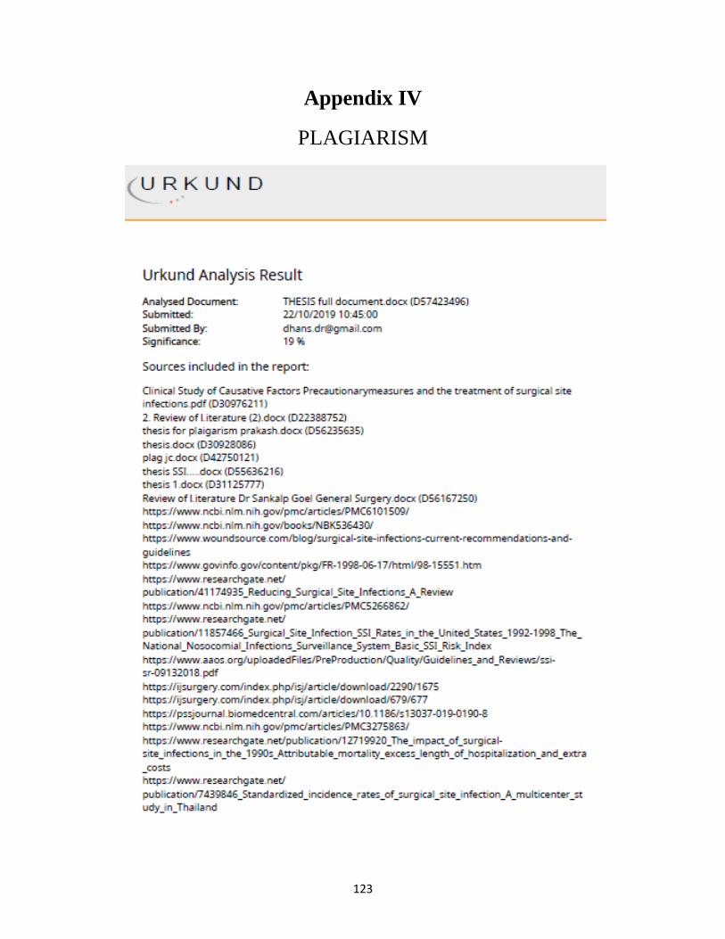
123
Appendix IV
PLAGIARISM
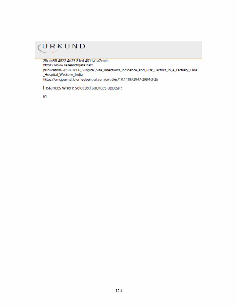
124
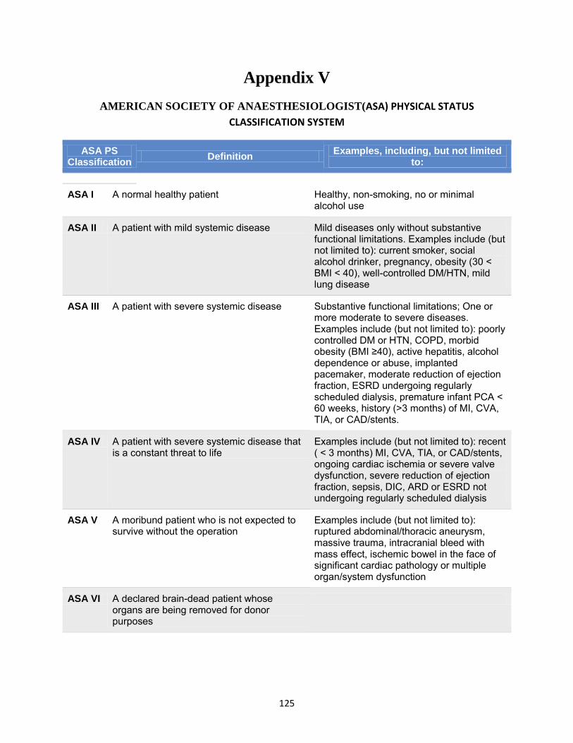
125
Appendix V
AMERICAN SOCIETY OF ANAESTHESIOLOGIST(ASA) PHYSICAL STATUS
CLASSIFICATION SYSTEM
ASA PS Classification Definition Examples, including, but not limited
to:
ASA I A normal healthy patient Healthy, non-smoking, no or minimal alcohol use
ASA II A patient with mild systemic disease Mild diseases only without substantive functional limitations. Examples include (but not limited to): current smoker, social alcohol drinker, pregnancy, obesity (30 < BMI < 40), well-controlled DM/HTN, mild lung disease
ASA III A patient with severe systemic disease Substantive functional limitations; One or more moderate to severe diseases. Examples include (but not limited to): poorly controlled DM or HTN, COPD, morbid obesity (BMI ≥40), active hepatitis, alcohol dependence or abuse, implanted pacemaker, moderate reduction of ejection fraction, ESRD undergoing regularly scheduled dialysis, premature infant PCA < 60 weeks, history (>3 months) of MI, CVA, TIA, or CAD/stents.
ASA IV A patient with severe systemic disease that is a constant threat to life
Examples include (but not limited to): recent ( < 3 months) MI, CVA, TIA, or CAD/stents, ongoing cardiac ischemia or severe valve dysfunction, severe reduction of ejection fraction, sepsis, DIC, ARD or ESRD not undergoing regularly scheduled dialysis
ASA V A moribund patient who is not expected to survive without the operation
Examples include (but not limited to): ruptured abdominal/thoracic aneurysm, massive trauma, intracranial bleed with mass effect, ischemic bowel in the face of significant cardiac pathology or multiple organ/system dysfunction
ASA VI A declared brain-dead patient whose organs are being removed for donor purposes
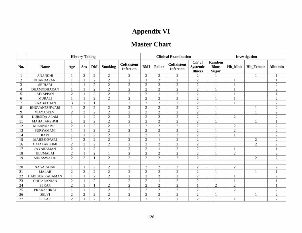
126
Appendix VI
Master Chart
History Taking Clinical Examination Investigation
No. Name Age Sex DM Smoking CoExistent
Infection BMI Pallor
CoExistent
Infection
C/F of
Systemic
Illness
Random
Bloos
Sugar
Hb_Male Hb_Female Albumin
1 ANANDHI 1 2 2 2 2 2 2 2 2 1
1 1
2 DHANDAPANI 1 1 2 2 2 1 2 2 2 1 1
1
3 SRIHARI 1 1 2 2 2 2 2 2 2 1 1
2
4 DHAMODHARAN 1 1 2 2 2 2 2 2 2 1 1
2
5 AIYAPPAN 2 1 2 2 2 2 1 2 2 1 1
1
6 MURALI 1 1 2 2 2 2 2 2 2 1 1
2
7 RAABATHAN 3 1 1 1 2 2 2 2 2 1 1
2
8 BHUVANESHWARI 1 2 2 2 2 2 2 2 2 1
1 2
9 VIAYASELVI 2 2 2 2 2 2 2 2 2 1
1 2
10 KURSIDA ALAM 1 1 2 2 2 2 2 2 2 1 2
2
11 MAHALAKSHMI 1 2 2 2 2 2 2 2 2 1
1 1
12 KULANDAIVEL 2 1 2 2 2 2 2 2 2 1 2
2
13 SURYAMANI 1 1 2 2 2 2 2 2 2 1 2
2
14 RAVI 1 1 2 2 2 2 1 2 2 1 1
2
15 MAHESHWARI 1 2 2 2 2 2 1 2 2 1
2 2
16 GAJALAKSHMI 2 2 2 2 2 2 2 2 2 1
2 2
17 JAYARAMAN 2 1 2 1 2 2 1 2 2 1 1
1
18 ELUMALAI 2 1 2 1 2 2 2 2 2 1 2
2
19 SARASWATHI 2 2 1 2 2 2 2 2 2 1
2 2
20 NAGARAJAN 1 1 2 2 2 2 2 2 2 1 2 2
21 MALAR 2 2 2 2 2 2 2 2 2 1 1 1
22 HABIBUR RAHAMAN 1 1 2 2 2 2 2 2 2 1 1 2
23 CHITARANJAN 2 1 2 1 2 2 1 2 2 1 1 1
24 SEKAR 2 1 1 2 2 2 2 2 1 2 2 1
25 PRAKASHRAJ 1 1 2 2 2 2 2 2 2 1 2 2
26 SELVI 2 2 2 2 2 2 2 2 2 1 1 2
27 SEKAR 2 1 2 2 2 2 1 2 2 1 1 2
127
History Taking Clinical Examination Investigation
No. Name Age Sex DM Smoking CoExistent
Infection BMI Pallor
CoExistent
Infection
C/F of
Systemic
Illness
Random
Bloos
Sugar
Hb_Male Hb_Female Albumin
28 RADHA 2 2 2 2 2 2 1 2 2 1 1 2
29 AZHAGU MEENA 2 2 1 2 2 2 2 2 2 2 2 2
30 LATHA 2 2 2 2 2 2 1 2 2 1 1 1
31 PRAVEEN 1 1 2 2 2 2 2 2 2 1 2 2
32 VISALATCHI 2 2 2 2 2 2 1 2 2 1 1 1
33 RAMAN 1 1 2 2 2 2 2 2 2 1 2 2
34 MANIKANDAN 1 1 2 2 2 2 2 2 2 1 2 2
35 SRINIVASAN 2 1 2 2 2 2 2 2 2 1 2 2
36 VALARMATHI 2 2 2 2 2 1 1 2 2 1 1 2
37 ETTIYAPPAN 1 1 2 2 2 2 2 2 2 1 2 2
38 MUFEES AHAMED 1 1 2 2 2 1 2 2 2 1 1 1
39 SATHYAVANI 2 2 2 2 2 2 1 2 2 1 1 1
40 ABINAYA 1 2 2 2 2 1 1 2 2 1 1 1
41 INDIRANI 3 2 1 2 2 3 1 2 2 2 1 1
42 LATHA 1 2 2 2 2 2 1 2 2 1 1 1
43 SARAN 1 1 2 2 2 1 2 2 2 1 2 2
44 ROSY 2 2 2 2 2 2 1 2 2 1 1 2
45 murali 2 2 2 2 2 2 1 2 2 1 2 2
46 SRINIVASAN 1 1 2 2 2 1 2 2 2 1 2 2
47 RAMESH 2 1 2 2 2 2 2 2 2 1 1 2
48 NANCY MARY 1 2 2 2 2 2 2 2 2 1 2 2
49 RAJESH 2 1 1 2 2 2 2 2 2 1 1 2
50 SIVASHANKAR 1 1 2 2 2 1 1 2 2 1 1 1
51 KAMATCHI 2 2 2 2 2 2 1 2 2 1 1 1
52 GUNASEKARAN 2 1 2 2 2 2 2 2 2 1 2 2
53 CHINNADURAI 2 1 2 1 2 2 2 2 2 1 1 2
54 VAMSI KRISHNAN 1 1 2 2 2 1 2 2 2 1 2 1
55 SAROJA 2 2 2 2 2 2 1 2 2 1 1 1
56 VARALAKSHMI 1 2 2 2 2 1 2 2 2 1 1 1
57 MALATHY 2 2 2 2 2 2 2 2 2 1 2 2
58 RAVISHANKAR 2 1 2 1 2 2 2 2 2 1 2 2
59 BAVITHA DEVI 1 2 2 2 2 2 1 2 2 1 1 1
60 KAVITHA 1 2 2 2 2 2 2 2 2 1 2 1
61 KANNIYAMMAL 2 2 2 2 2 2 1 2 2 1 1 1
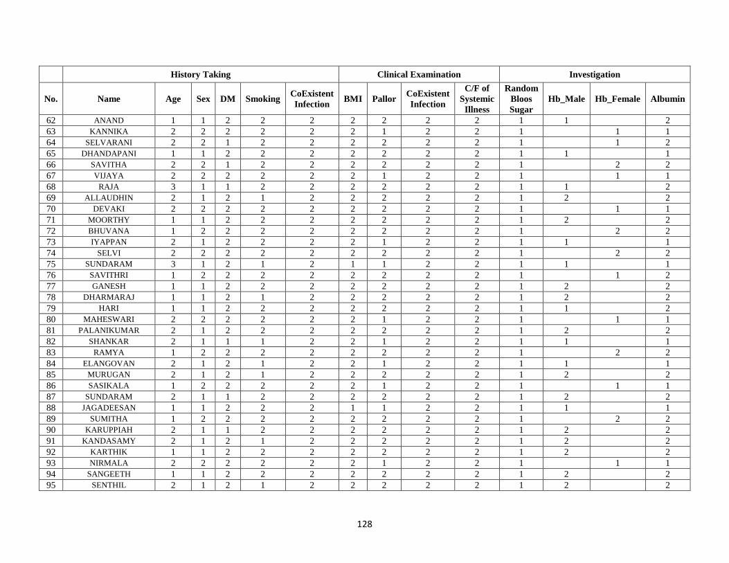
128
History Taking Clinical Examination Investigation
No. Name Age Sex DM Smoking CoExistent
Infection BMI Pallor
CoExistent
Infection
C/F of
Systemic
Illness
Random
Bloos
Sugar
Hb_Male Hb_Female Albumin
62 ANAND 1 1 2 2 2 2 2 2 2 1 1 2
63 KANNIKA 2 2 2 2 2 2 1 2 2 1 1 1
64 SELVARANI 2 2 1 2 2 2 2 2 2 1 1 2
65 DHANDAPANI 1 1 2 2 2 2 2 2 2 1 1 1
66 SAVITHA 2 2 1 2 2 2 2 2 2 1 2 2
67 VIJAYA 2 2 2 2 2 2 1 2 2 1 1 1
68 RAJA 3 1 1 2 2 2 2 2 2 1 1 2
69 ALLAUDHIN 2 1 2 1 2 2 2 2 2 1 2 2
70 DEVAKI 2 2 2 2 2 2 2 2 2 1 1 1
71 MOORTHY 1 1 2 2 2 2 2 2 2 1 2 2
72 BHUVANA 1 2 2 2 2 2 2 2 2 1 2 2
73 IYAPPAN 2 1 2 2 2 2 1 2 2 1 1 1
74 SELVI 2 2 2 2 2 2 2 2 2 1 2 2
75 SUNDARAM 3 1 2 1 2 1 1 2 2 1 1 1
76 SAVITHRI 1 2 2 2 2 2 2 2 2 1 1 2
77 GANESH 1 1 2 2 2 2 2 2 2 1 2 2
78 DHARMARAJ 1 1 2 1 2 2 2 2 2 1 2 2
79 HARI 1 1 2 2 2 2 2 2 2 1 1 2
80 MAHESWARI 2 2 2 2 2 2 1 2 2 1 1 1
81 PALANIKUMAR 2 1 2 2 2 2 2 2 2 1 2 2
82 SHANKAR 2 1 1 1 2 2 1 2 2 1 1 1
83 RAMYA 1 2 2 2 2 2 2 2 2 1 2 2
84 ELANGOVAN 2 1 2 1 2 2 1 2 2 1 1 1
85 MURUGAN 2 1 2 1 2 2 2 2 2 1 2 2
86 SASIKALA 1 2 2 2 2 2 1 2 2 1 1 1
87 SUNDARAM 2 1 1 2 2 2 2 2 2 1 2 2
88 JAGADEESAN 1 1 2 2 2 1 1 2 2 1 1 1
89 SUMITHA 1 2 2 2 2 2 2 2 2 1 2 2
90 KARUPPIAH 2 1 1 2 2 2 2 2 2 1 2 2
91 KANDASAMY 2 1 2 1 2 2 2 2 2 1 2 2
92 KARTHIK 1 1 2 2 2 2 2 2 2 1 2 2
93 NIRMALA 2 2 2 2 2 2 1 2 2 1 1 1
94 SANGEETH 1 1 2 2 2 2 2 2 2 1 2 2
95 SENTHIL 2 1 2 1 2 2 2 2 2 1 2 2
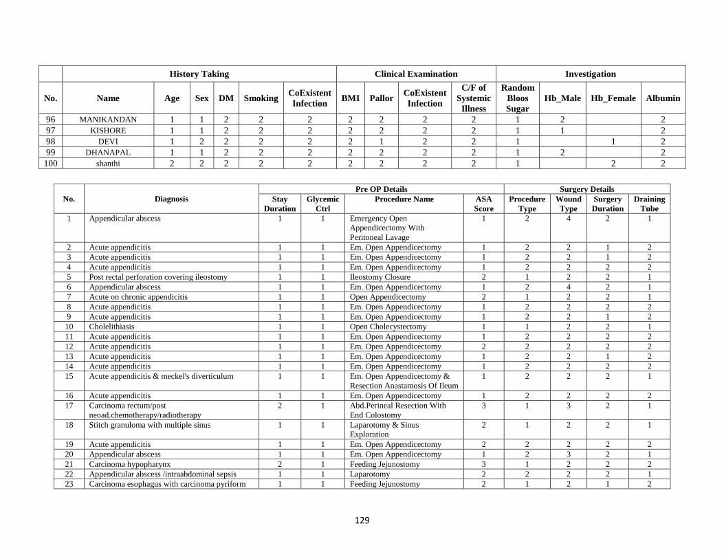
129
History Taking Clinical Examination Investigation
No. Name Age Sex DM Smoking CoExistent
Infection BMI Pallor
CoExistent
Infection
C/F of
Systemic
Illness
Random
Bloos
Sugar
Hb_Male Hb_Female Albumin
96 MANIKANDAN 1 1 2 2 2 2 2 2 2 1 2 2
97 KISHORE 1 1 2 2 2 2 2 2 2 1 1 2
98 DEVI 1 2 2 2 2 2 1 2 2 1 1 2
99 DHANAPAL 1 1 2 2 2 2 2 2 2 1 2 2
100 shanthi 2 2 2 2 2 2 2 2 2 1 2 2
No. Diagnosis
Pre OP Details Surgery Details
Stay
Duration
Glycemic
Ctrl
Procedure Name ASA
Score
Procedure
Type
Wound
Type
Surgery
Duration
Draining
Tube
1 Appendicular abscess 1 1 Emergency Open
Appendicectomy With
Peritoneal Lavage
1 2 4 2 1
2 Acute appendicitis 1 1 Em. Open Appendicectomy 1 2 2 1 2
3 Acute appendicitis 1 1 Em. Open Appendicectomy 1 2 2 1 2
4 Acute appendicitis 1 1 Em. Open Appendicectomy 1 2 2 2 2
5 Post rectal perforation covering ileostomy 1 1 Ileostomy Closure 2 1 2 2 1
6 Appendicular abscess 1 1 Em. Open Appendicectomy 1 2 4 2 1
7 Acute on chronic appendicitis 1 1 Open Appendicectomy 2 1 2 2 1
8 Acute appendicitis 1 1 Em. Open Appendicectomy 1 2 2 2 2
9 Acute appendicitis 1 1 Em. Open Appendicectomy 1 2 2 1 2
10 Cholelithiasis 1 1 Open Cholecystectomy 1 1 2 2 1
11 Acute appendicitis 1 1 Em. Open Appendicectomy 1 2 2 2 2
12 Acute appendicitis 1 1 Em. Open Appendicectomy 2 2 2 2 2
13 Acute appendicitis 1 1 Em. Open Appendicectomy 1 2 2 1 2
14 Acute appendicitis 1 1 Em. Open Appendicectomy 1 2 2 2 2
15 Acute appendicitis & meckel's diverticulum 1 1 Em. Open Appendicectomy &
Resection Anastamosis Of Ileum
1 2 2 2 1
16 Acute appendicitis 1 1 Em. Open Appendicectomy 1 2 2 2 2
17 Carcinoma rectum/post
neoad.chemotherapy/radiotherapy
2 1 Abd.Perineal Resection With
End Colostomy
3 1 3 2 1
18 Stitch granuloma with multiple sinus 1 1 Laparotomy & Sinus
Exploration
2 1 2 2 1
19 Acute appendicitis 1 1 Em. Open Appendicectomy 2 2 2 2 2
20 Appendicular abscess 1 1 Em. Open Appendicectomy 1 2 3 2 1
21 Carcinoma hypopharynx 2 1 Feeding Jejunostomy 3 1 2 2 2
22 Appendicular abscess /intraabdominal sepsis 1 1 Laparotomy 2 2 2 2 1
23 Carcinoma esophagus with carcinoma pyriform 1 1 Feeding Jejunostomy 2 1 2 1 2
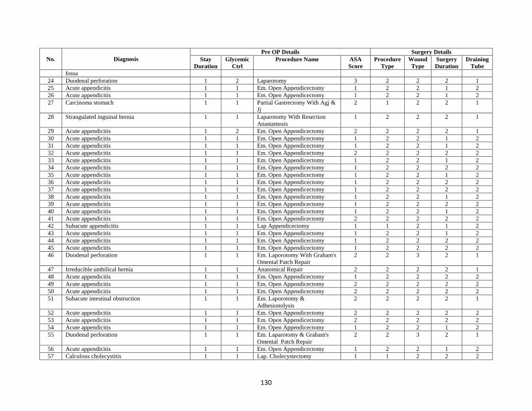
130
No. Diagnosis
Pre OP Details Surgery Details
Stay
Duration
Glycemic
Ctrl
Procedure Name ASA
Score
Procedure
Type
Wound
Type
Surgery
Duration
Draining
Tube
fossa
24 Duodenal perforation 1 2 Laparotomy 3 2 2 2 1
25 Acute appendicitis 1 1 Em. Open Appendicectomy 1 2 2 1 2
26 Acute appendicitis 1 1 Em. Open Appendicectomy 1 2 2 1 2
27 Carcinoma stomach 1 1 Partial Gastrectomy With Agj &
Jj
2 1 2 2 1
28 Strangulated inguinal hernia 1 1 Laparotomy With Resection
Anastamosis
1 2 2 2 1
29 Acute appendicitis 1 2 Em. Open Appendicectomy 2 2 2 2 1
30 Acute appendicitis 1 1 Em. Open Appendicectomy 1 2 2 1 2
31 Acute appendicitis 1 1 Em. Open Appendicectomy 1 2 2 1 2
32 Acute appendicitis 1 1 Em. Open Appendicectomy 2 2 2 2 2
33 Acute appendicitis 1 1 Em. Open Appendicectomy 1 2 2 1 2
34 Acute appendicitis 1 1 Em. Open Appendicectomy 1 2 2 2 2
35 Acute appendicitis 1 1 Em. Open Appendicectomy 1 2 2 1 2
36 Acute appendicitis 1 1 Em. Open Appendicectomy 1 2 2 2 2
37 Acute appendicitis 1 1 Em. Open Appendicectomy 1 2 2 2 2
38 Acute appendicitis 1 1 Em. Open Appendicectomy 1 2 2 1 2
39 Acute appendicitis 1 1 Em. Open Appendicectomy 1 2 2 2 2
40 Acute appendicitis 1 1 Em. Open Appendicectomy 1 2 2 1 2
41 Acute appendicitis 1 1 Em. Open Appendicectomy 2 2 2 2 2
42 Subacute appendicitis 1 1 Lap Appendicectomy 1 1 2 1 2
43 Acute appendicitis 1 1 Em. Open Appendicectomy 1 2 2 1 2
44 Acute appendicitis 1 1 Em. Open Appendicectomy 1 2 2 2 2
45 Acute appendicitis 1 1 Em. Open Appendicectomy 1 2 2 2 2
46 Duodenal perforation 1 1 Em. Laporotomy With Graham's
Omental Patch Repair
2 2 3 2 1
47 Irreducible umbilical hernia 1 1 Anatomical Repair 2 2 2 2 1
48 Acute appendicitis 1 1 Em. Open Appendicectomy 1 2 2 2 2
49 Acute appendicitis 1 1 Em. Open Appendicectomy 2 2 2 2 2
50 Acute appendicitis 1 1 Em. Open Appendicectomy 2 2 2 2 2
51 Subacute intestinal obstruction 1 1 Em. Laporotomy &
Adhesionolysis
2 2 2 2 1
52 Acute appendicitis 1 1 Em. Open Appendicectomy 2 2 2 2 2
53 Acute appendicitis 1 1 Em. Open Appendicectomy 2 2 2 2 2
54 Acute appendicitis 1 1 Em. Open Appendicectomy 1 2 2 1 2
55 Duodenal perforation 1 1 Em. Laparotomy & Graham's
Omental Patch Repair
2 2 3 2 1
56 Acute appendicitis 1 1 Em. Open Appendicectomy 1 2 2 1 2
57 Calculous cholecystitis 1 1 Lap. Cholecystectomy 1 1 2 2 2
131
No. Diagnosis
Pre OP Details Surgery Details
Stay
Duration
Glycemic
Ctrl
Procedure Name ASA
Score
Procedure
Type
Wound
Type
Surgery
Duration
Draining
Tube
58 Duodenal perforation 1 1 Em.Laporotomy & Graham's
Omental Patch Repair
2 2 3 2 1
59 Acute appendicitis 1 1 Em. Open Appendicectomy 1 2 2 2 2
60 Acute appendicitis 1 1 Em. Open Appendicectomy 1 2 2 1 2
61 Acute appendicitis 1 1 Em. Open Appendicectomy 1 2 2 1 2
62 Acute appendicitis 1 1 Em. Open Appendicectomy 1 2 2 1 2
63 Acute appendicitis 1 1 Em. Open Appendicectomy 1 2 2 2 2
64 Adhesive intestinal obstruction 1 1 Em.Laparotomy And
Adhesinolysis
2 2 2 2 1
65 Acute appendicitis 1 1 Em. Open Appendicectomy 1 2 2 1 2
66 Duodenal perforation 1 1 Exploratory Laparotomy &
Graham's Omental Patch Repair
2 2 3 2 1
67 Adhesive intestinal obstruction 1 1 Emergency Laparotomy And
Adhesinolysis
2 2 2 2 1
68 Acute appendicitis 1 1 Em. Open Appendicectomy 2 2 2 2 2
69 Cholelithiasis 1 1 Opencholecystectomy 1 1 2 2 2
70 Appendicular abscess 1 1 Em. Open Appendicectomy 1 2 3 2 1
71 Acute appendicitis 1 1 Em. Open Appendicectomy 1 2 2 1 2
72 Acute appendicitis 1 1 Em. Open Appendicectomy 1 2 2 2 2
73 Acute appendicitis 1 1 Em. Open Appendicectomy 2 2 2 2 2
74 Abdominal wall lipoma 1 1 Excision Biopsy 1 1 1 1 2
75 Carcinoma stomach 2 1 Laparotomy,Subtotal
Gastrectomy Agj,Jj
2 1 2 2 1
76 Acute appendicitis 1 1 Em. Open Appendicectomy 1 2 2 2 2
77 Acute appendicitis 1 1 Em. Open Appendicectomy 1 2 2 1 2
78 Acute appendicitis 1 1 Em. Open Appendicectomy 1 2 2 2 2
79 Acute appendicitis 1 1 Em. Open Appendicectomy 1 2 2 2 2
80 Acute appendicitis 1 1 Em. Open Appendicectomy 1 2 2 2 2
81 Acute appendicitis 1 1 Em. Open Appendicectomy 1 2 2 2 2
82 Ileal perforation 1 1 Emergency Laparotomy & Ileal
Resection And Anastamosis
2 2 3 2 1
83 Post lscs sinus tract 1 1 Sinus Tract Excision With Flap
Cover
1 1 2 2 1
84 Carcinoma stomach 2 1 Laparotomy,Subtotal
Gastrectomy Agj,Jj
3 1 3 2 1
85 Irreducible paraumbilical hernia 1 1 Emergency Anatomical Repair
With Omentectomy
2 2 2 2 1
86 Acute appendicitis 1 1 Em. Open Appendicectomy 1 2 2 2 2
87 Chronic appendicitis 1 1 Lap Appendicectomy 2 1 2 2 2
88 Acute appendicitis 1 1 Em. Open Appendicectomy 1 2 2 1 2
132
No. Diagnosis
Pre OP Details Surgery Details
Stay
Duration
Glycemic
Ctrl
Procedure Name ASA
Score
Procedure
Type
Wound
Type
Surgery
Duration
Draining
Tube
89 Acute appendicitis 1 1 Em. Open Appendicectomy 1 2 2 2 2
90 Cholelithiasis and choledocholithiasis 2 1 Open Cholecystectomy With
Cbd Exploration
2 1 3 2 1
91 Acute appendicitis 1 1 Em. Open Appendicectomy 1 2 2 1 2
92 Acute appendicitis 1 1 Em. Open Appendicectomy 1 2 2 1 2
93 Duodenal perforation 1 1 Exploratory Laparotomy &
Graham's Omental Patch Repair
2 2 3 2 1
94 Acute appendicitis 1 1 Em. Open Appendicectomy 1 2 2 2 2
95 Ileal perforation 1 1 Emergency Laparotomy With
Ileal Resection And
Anastamosis
2 2 3 2 1
96 Acute appendicitis 1 1 Em. Open Appendicectomy 1 2 2 2 2
97 Acute appendicitis 1 1 Em. Open Appendicectomy 1 2 2 2 2
98 Acute appendicitis 1 1 Em. Open Appendicectomy 1 2 2 2 2
99 Acute appendicitis 1 1 Em. Open Appendicectomy 1 2 2 1 2
100 Acute appendicitis 1 1 Em. Open Appendicectomy 1 2 2 1 2
No. Post OP Details
Stay
Duration
Glycemic
Control
Temp Time of
Initiation
of Oral
Feed
No. of
Days
Drain
Placed
Surgical
Would_2
Surgical Would_7 Surgical Would_14
1 3 1 1 3 2 Healthy Purulent Discharge & Wound Gaping
Present
Healed
2 2 1 1 2 Healthy Healthy Healthy
3 2 1 1 2 Healthy Healthy Healthy
4 2 1 1 2 Healthy Healthy Healthy
5 3 1 1 3 2 Healthy Healthy Healthy
6 3 1 1 2 2 Healthy Healthy Healthy
7 2 1 1 2 1 Healthy Healthy Healthy
8 2 1 1 2 Healthy Healthy Healthy
9 2 1 1 2 Healthy Healthy Healthy
10 2 1 1 2 1 Healthy Healthy Healthy
11 2 1 1 2 Healthy Healthy Healthy
12 2 1 1 2 Healthy Healthy Healthy
13 2 1 1 2 Healthy Healthy Healthy
14 2 1 1 2 Healthy Healthy Healthy
15 3 1 1 3 2 Healthy Healthy Healthy
16 2 1 1 2 Healthy Healthy Healthy
133
No. Post OP Details
Stay
Duration
Glycemic
Control
Temp Time of
Initiation
of Oral
Feed
No. of
Days
Drain
Placed
Surgical
Would_2
Surgical Would_7 Surgical Would_14
17 3 1 1 3 2 Healthy Serous Discharge Present Healthy
18 2 1 1 1 1 Healthy Healthy Healthy
19 2 1 1 2 Healthy Healthy Healthy
20 3 1 1 3 2 Healthy Serous Discharge Present Healthy
21 3 1 1 3 Healthy Healthy Healthy
22 3 1 1 3 2 Healthy Serous Discharge Present Healing With Wound Gaping
23 2 1 1 3 Healthy Healthy Healthy
24 3 1 1 3 2 Healthy Healthy Healthy
25 2 1 1 2 Healthy Healthy Healthy
26 2 1 1 2 Healthy Healthy Healthy
27 3 1 1 3 2 Healthy Healthy Healthy
28 3 1 1 3 2 Healthy Healthy Healthy
29 2 1 1 3 1 Healthy Healthy Healthy
30 2 1 1 2 Healthy Healthy Healthy
31 2 1 1 2 Healthy Healthy Healthy
32 2 1 1 2 Healthy Healthy Healthy
33 2 1 1 2 Healthy Healthy Healthy
34 2 1 1 2 Healthy Healthy Healthy
35 2 1 1 2 Healthy Healthy Healthy
36 2 1 1 2 Healthy Healthy Healthy
37 2 1 1 2 Healthy Healthy Healthy
38 2 1 1 2 Healthy Healthy Healthy
39 2 1 1 2 Healthy Healthy Healthy
40 2 1 1 2 Healthy Healthy Healthy
41 2 1 1 2 Healthy Healthy Healthy
42 2 1 1 2 Healthy Healthy Healthy
43 2 1 1 2 Healthy Healthy Healthy
44 2 1 1 2 Healthy Healthy Healthy
45 2 1 1 2 Healthy Healthy Healthy
46 3 1 1 3 2 Healthy Serous Discharge Present Healing
47 2 1 1 3 2 Healthy Healthy Healthy
48 2 1 1 2 Healthy Healthy Healthy
49 2 1 1 2 Healthy Healthy Healthy
50 2 1 1 2 Healthy Healthy Healthy
51 3 1 1 3 2 Healthy Healthy Healthy
52 2 1 1 2 Healthy Healthy Healthy
53 2 1 1 2 Healthy Healthy Healthy
54 2 1 1 2 Healthy Healthy Healthy
134
No. Post OP Details
Stay
Duration
Glycemic
Control
Temp Time of
Initiation
of Oral
Feed
No. of
Days
Drain
Placed
Surgical
Would_2
Surgical Would_7 Surgical Would_14
55 3 1 1 3 2 Healthy Healthy Healthy
56 2 1 1 2 Healthy Healthy Healthy
57 2 1 1 2 Healthy Healthy Healthy
58 3 1 1 3 2 Healthy Serous Discharge & Wound Gaping Present Healed By Secondary Intention
59 2 1 1 2 Healthy Healthy Healthy
60 2 1 1 1 Healthy Healthy Healthy
61 2 1 1 2 Healthy Healthy Healthy
62 2 1 1 2 Healthy Healthy Healthy
63 2 1 1 2 Healthy Healthy Healthy
64 2 1 1 2 1 Healthy Healthy Healthy
65 1 1 1 1 Healthy Healthy Healthy
66 3 1 2 3 2 Healthy Discharge Wound Gaping
67 2 1 1 2 1 Healthy Healthy Healthy
68 2 1 1 2 Healthy Healthy Healthy
69 2 1 1 2 Healthy Healthy Healthy
70 3 1 2 3 2 Healthy Pelvic Abcess ,Wound Gaping Wound Gaping
71 2 1 1 2 Healthy Healthy Healthy
72 2 1 1 2 Healthy Healthy Healthy
73 2 1 1 2 Healthy Healthy Healthy
74 1 1 1 1 Healthy Healthy Healthy
75 3 1 1 3 2 Healthy Wound Gaping Sec.Suturing Done
76 2 1 1 2 Healthy Healthy Healthy
77 2 1 1 2 Healthy Healthy Healthy
78 2 1 1 2 Healthy Healthy Healthy
79 2 1 1 2 Healthy Healthy Healthy
80 2 1 1 2 Healthy Healthy Healthy
81 2 1 1 2 Healthy Healthy Healthy
82 3 1 1 3 2 Healthy Wound Gaping Sec Suturing Done Onpod 11,Healthy
83 2 1 1 2 1 Healthy Healthy Healthy
84 3 1 1 3 2 Seroma Wound Gaping Healthy
85 2 1 1 2 1 Healthy Healthy Healthy
86 2 1 1 2 Healthy Healthy Healthy
87 1 1 1 2 Healthy Healthy Healthy
88 2 1 1 2 Healthy Healthy Healthy
89 2 1 1 2 Healthy Healthy Healthy
90 3 1 2 3 2 Healthy Pus Discharge Sec Suturing Done Onpod 11,Healthy
91 2 1 1 2 Healthy Healthy Healthy
92 2 1 1 2 Healthy Healthy Healthy
135
No. Post OP Details
Stay
Duration
Glycemic
Control
Temp Time of
Initiation
of Oral
Feed
No. of
Days
Drain
Placed
Surgical
Would_2
Surgical Would_7 Surgical Would_14
93 3 1 2 3 2 Healthy Seroma,Wound Gaping Sec Suturing Done Onpod 14,Healthy
94 2 1 1 2 Healthy Healthy Healthy
95 3 1 2 3 2 Healthy Pus Discharge,Wound Gaping Granulating Well
96 2 1 1 2 Healthy Healthy Healthy
97 2 1 1 2 Healthy Healthy Healthy
98 2 1 1 2 Healthy Healthy Healthy
99 2 1 1 2 Healthy Healthy Healthy
100 2 1 1 2 Healthy Healthy Healthy
No.
Post OP Details SSI
Surgical
Would_21
Surgical
Would_3
0
Name of Antibiotic
Duration
of
Antibioti
c
Typ
e of
SSI
SWAB Culture
Report_Organism
SWAB Culture
Report_Sensitivity
Surgical
Intervention
1 Healthy Healthy Amoxyclav,Metro,Piptaz 5 1 E.Coli Piptaz, Meropenam ,Colistin Nil
2 Healthy Healthy Cefotaxim,Metro 3 NIL
3 Healthy Healthy Cefotaxim,Metro 3 NIL
4 Healthy Healthy Cefotaxim,Metro 3 NIL
5 Healthy Healthy Amikacin 7 NIL
6 Healthy Healthy Amikacin,Augmentin,Metro 5 NIL
7 Healthy Healthy Cefotaxim,Metro 3 NIL
8 Healthy Healthy Cefotaxim,Metro 3 NIL
9 Healthy Healthy Cefotaxim,Metro 3 NIL
10 Healthy Healthy Cefotaxim,Metro 5 NIL
11 Healthy Healthy Cefotaxim,Metro 3 NIL
12 Healthy Healthy Cefotaxim,Metro 3 NIL
13 Healthy Healthy Cefotaxim,Metro 3 NIL
14 Healthy Healthy Cefotaxim,Metro 3 NIL
15 Healthy Healthy Cefotaxim,Metro 5 NIL
16 Healthy Healthy Cefotaxim,Metro 5 NIL
17 Healthy Healthy Cefotaxim,Metro &Piptaz 7 1 NIL
18 Healthy Healthy Cefotaxim 5 NIL
19 Healthy Healthy Cefotaxim,Metro 5 NIL
20 Healthy Healthy Cefotaxim,Metro & Amikacin 7 1 NIL
21 Healthy Healthy Cefotaxim,Metro 5 NIL
22 Healthy Healthy Meropenam,Metro,Colistin 5 1 SECONDARY
136
No.
Post OP Details SSI
Surgical
Would_21
Surgical
Would_3
0
Name of Antibiotic
Duration
of
Antibioti
c
Typ
e of
SSI
SWAB Culture
Report_Organism
SWAB Culture
Report_Sensitivity
Surgical
Intervention
SUTURING
23 Healthy Healthy Cefotaxim,Metro 5 NIL
24 Healthy Healthy Piptaz ,Metro 8 NIL
25 Healthy Healthy Cefotaxim 5 NIL
26 Healthy Healthy Cefotaxim,Metro 5 NIL
27 Healthy Healthy Amoxyclav,Metro, Cefaperazone 7 NIL
28 Healthy Healthy Piptaz ,Metro 7 NIL
29 Healthy Healthy Cefotaxim,Metro 5 NIL
30 Healthy Healthy Cefotaxim,Metro 5 NIL
31 Healthy Healthy Cefotaxim,Metro 3 NIL
32 Healthy Healthy Cefotaxim,Metro 4 NIL
33 Healthy Healthy Cefotaxim,Metro 5 NIL
34 Healthy Healthy Cefotaxim,Metro 3 NIL
35 Healthy Healthy Cefotaxim,Metro 5 NIL
36 Healthy Healthy Cefotaxim,Metro 4 NIL
37 Healthy Healthy Cefotaxim,Metro 3 NIL
38 Healthy Healthy Cefotaxim,Metro 3 NIL
39 Healthy Healthy Cefotaxim,Metro 4 NIL
40 Healthy Healthy Cefotaxim,Metro 3 NIL
41 Healthy Healthy Piptaz ,Metro 5 NIL
42 Healthy Healthy Cefotaxim,Metro 3 NIL
43 Healthy Healthy Cefotaxim,Metro 3 NIL
44 Healthy Healthy Cefotaxim,Metro 3 NIL
45 Healthy Healthy Cefotaxim,Metro 3
46 Healthy Healthy Piptaz, Metro,Amoxiclav 7 1 NIL
47 Healthy Healthy Cefotaxim,Metro 7 NIL
48 Healthy Healthy Cefotaxim,Metro 3
49 Healthy Healthy Cefotaxim,Metro 5
50 Healthy Healthy Cefotaxim,Metro 5
51 Healthy Healthy Piptaz, Metro 7
52 Healthy Healthy Cefotaxim,Metro 5
53 Healthy Healthy Amoxyclav,Metro 4
54 Healthy Healthy Cefotaxim,Metro 3
55 Healthy Healthy Cefaperazone Sulbactum , Metro 7
56 Healthy Healthy Cefotaxim,Metro 3
57 Healthy Healthy Cefotaxim,Metro 3
58 Healthy Healthy Piptaz,Metro,Genta 6 1 Klebseilla
Piptaz,Cipro,Amikacin,
Genta
137
No.
Post OP Details SSI
Surgical
Would_21
Surgical
Would_3
0
Name of Antibiotic
Duration
of
Antibioti
c
Typ
e of
SSI
SWAB Culture
Report_Organism
SWAB Culture
Report_Sensitivity
Surgical
Intervention
59 Healthy Healthy Cefotaxim,Metro 4
60 Healthy Healthy Cefotaxim,Metro 3
61 Healthy Healthy Cefotaxim,Metro 3
62 Healthy Healthy Amoxyclav 3
63 Healthy Healthy Cefotaxim,Metro 3
64 Healthy Healthy Piptaz,Metro 5
65 Healthy Healthy Cefotaxim,Metro 2
66 Healthy Healthy Cefperazone
Sulbactum,Colistin,Metro
5 1 E.Coli,Mrsa Colistin
Sec.Suturing
Done On Od15
67 Healthy Healthy Piptaz,Metro 4
68 Healthy Healthy Cefotaxim,Metro 3
69 Healthy Healthy Amoxiclav,Metro 3
70 Sec.Suturing
Done
Healthy Piptaz,Imepenam,Metro 5 3
E.Coli,Pseudomo
nas Imipenam
Sec Suturing
Done
71 Healthy Healthy Cefotaxime,Metro 3
72 Healthy Healthy Cefotaxime,Metro 3
73 Healthy Healthy Cefotaxime,Metro 3
74 Healthy Healthy Amoxiclav 2
75 Healthy Healthy Piptaz,Metro 10
1 E.Coli Piptaz
Sec Suturing
Done On Pod
14
76 Healthy Healthy Cefotaxime,Metro 3
77 Healthy Healthy Cefotaxime,Metro 3
78 Healthy Healthy Cefotaxime,Metro 3
79 Healthy Healthy Cefotaxime,Metro 3
80 Healthy Healthy Cefotaxime,Metro 3
81 Healthy Healthy Cefotaxime,Metro 3
82 Healthy Healthy Piptaz,Colistin,Metro 7
1 E.Coli,Pseudomo
nas Colistin
Sec Suturing
Done On Pod
11
83 Healthy Healthy Cefotaxime 5
84 Healthy Healthy Ceftriaxazone,Metro,Piptaz 7 1 E.Coli Piptaz
Sec Suturing
Done On Pod 9
85 Healthy Healthy Cefotaxime 5
86 Healthy Healthy Cefotaxime,Metro 3
87 Healthy Healthy Cefotaxime,Metro 2
88 Healthy Healthy Cefotaxime,Metro 3
89 Healthy Healthy Cefotaxime,Metro 3
138
No.
Post OP Details SSI
Surgical
Would_21
Surgical
Would_3
0
Name of Antibiotic
Duration
of
Antibioti
c
Typ
e of
SSI
SWAB Culture
Report_Organism
SWAB Culture
Report_Sensitivity
Surgical
Intervention
90 Healthy Healthy Ceftriaxazone,Metro,Piptaz 5
1 Proteus Piptaz
Sec Suturing
Done On Pod
11
91 Healthy Healthy Cefotaxime,Metro 3
92 Healthy Healthy Cefotaxime,Metro 3
93 Healthy Healthy Piptaz,Metro 10
1
Proteus
Mirabilis,Staph.A
ureus
Piptaz Sec Suturing
Done Pod 14
94 Healthy Healthy Cefotaxime,Metro 3
95 Sec.Suturing
Done
Healthy Piptaz,Metro,Imipenam 8
1 E.Coli,Proteus Imipenam
Sec Suturing
Done On Pod
21
96 Healthy Healthy Cefotaxime,Metro 3
97 Healthy Healthy Cefotaxime,Metro 3
98 Healthy Healthy Cefotaxime,Metro 3
99 Healthy Healthy Cefotaxime,Metro 3
100 Healthy Healthy Cefotaxime,Metro 3