Evaluation of lacrimal apparatus - Otolaryngology...
23
Evaluation of lacrimal apparatus Balasubramanian Thiagarajan Drtbalu's otolaryngology online
-
Upload
trinhkhanh -
Category
Documents
-
view
223 -
download
1
Transcript of Evaluation of lacrimal apparatus - Otolaryngology...

Evaluation of lacrimal apparatus
Balasubramanian Thiagarajan
Drtbalu's otolaryngology online

Introduction:
Dacryocystorhinostomy as a treatment modality for epiphora is commonly being performed endonasally using a nasal endoscope by otolaryngologists. It is hence imperative that they diligently examine the entire lacrimal system before proceeding with the procedure. Despite the commonality of this surgical procedure, standard otolaryngology text books contain fewer literature on this subject. History of surgery for nasolacrimal pathway obstruction dates back to Hamurabi 2200 B.C 1. Dacryocystorhinostomy is the undisputed treatment of choice for lacrimal system drainage obstruction below the level of common canaliculi. Endonasal approach of dacryocystorhinostomy was first described by Caldwell in 1893 2.Epiphora can be caused by blockage of lacrimal drainage system / excess lacrimation / loss of lacrimal pump mechanism. Lacrimal pump mechanism could be disrupted due to lower lid laxity or weakness of orbicularis oculi muscle. Normal lacrimation / or excess of it can be caused by irritation to cornea / conjunctiva 3. This reflex is initiated by stimulation of trigeminal nerve.Trigeminal stimualtion can be caused by:
1. Corneal foreign body2. Keratitis3. Conjunctivitis4. Ocular surface disorders (dry eye)
Epiphora: Greek terminology meaning “Downpour”.This is defined as excessive watering of eye. This is invariably caused by obstruction to tear drainage. Causes of epiphora include:
Congenital: Congenital nasolacrimal duct obstruction. Incidence varies between 1-6% 4. It is beleived that massage of entire naso lacrimal system releives obstruction in more than 90% 5 of cases. Majority of these obstruction resolve during the first year of life hence urgent surgical management is not necessary 6. Probing is also known to be beneficial in these patients. The time of probing is controversial. Probing is advised up to the age of 5 in these patients 7.
Acquired: 1. Primary acquired nasolacrimal duct obstructions2. Dacryocystolithiasis3. Orbital / lacrimal trauma4. Canalicular lacerations5. Actinomyces within the canaliculi – Actinomyces are anaerobic gram positive bacilli resembling fungi. These organism are normal commensal of oropharynx. These organism are capable of causing cast – forming canaliculitis 9 leading onto lacrimal tract obstruction.6. Canalicular infections following herpes infections / ectropion – Viral infections constitute a well recognised common cause of acquired canalicular obstruction 8. These patients give history of an episode of blepharokeratoconjunctivitis before epiphora. Antivirals (idoxuridine) which are prescribed for this condition too add to the woes by causing more lacrimal obstruction. Naso
Drtbalu's otolaryngology online

lacrimal obstruction caused by antivirals are transient and disappear after the drug is discontinued. Herpes simplex viral infections are known to cause punctal changes.
Clinical examination goal in these patients is to distinguish between epiphora and lacrimation. While epiphora needs to be surgically managed medically. The focus should be in differentiating anatomical obstruction from functional disorders.
Anatomical obstruction:
Obstruction to the lacrimal drainage system is the feature to look for in this condition. Pathological changes could be seen involving the lacrimal sac, irregularities in lacrimal drainage system (canalicular stenosis, canalicular blockage, obstruction to nasolacrimal duct, diverticulous formation etc.) Lacrimal pathways can be obstructed due to internal derangements like inflammation of the epithelial linining. This is known as intrinsic obstruction. If lacrimal pathways are affected by deforming lesions from outside like tumors causing compression to it has been termed as extrinsic obstruction.
Physiologic dysfuntion causing epiphora:
This is also known as functional epiphora. Here there are no anatomical changes to the lacrimal pathway. The functioning lacrimal pump mechanism is at fault. Pump mechanism can be affected in conditions like eyelid malpositions, eversion of lacrimal punctum, poor orbicularis oculi muscle tone as seen in patients with Bell's palsy.
Grading of epiphora:
The commonly used grading system was devised by Sahlin 10.
Grade Degree of Epiphora
0 No epiphora
1 Epiphora only outdoors and during windy times
2 Outdoor epiphora No indoor epiphora
3 Outdoor and indoor epiphora
Anatomy of lacrimal system an overview 11:
The lacrimal system consists of a superior and inferior puncta at the medial ends of upper and lower eyelids. These two drain into upper and lower canaliculi. These two canaliculi join to form the common canaliculus. This zone is known as the upper lacrimal system. The common canaliculus inturn leads into the lacrimal sac. The sac is about 12 - 15 mm long. It eventually narrows and
Drtbalu's otolaryngology online

leads into the nasolacrimal duct which drains into the inferior meatus of the nose. The naso lacrimal duct is about 18 mm long. The sac and the duct comprise the lower lacrimal system. The junction between the common canaliculus and the lacrimal sac is guarded by the Rosenmuller valve. This valve prevents tear reflux. The nasal end of the nasolacrimal duct at the level of inferior meatus is guarded by Hasner's valve.
Figure showing the entire lacrimal system
Sites in the lacrimal system prone for obstruction:
Suprasaccal obstruction:
In this type obstruction lies proximal to the lacrimal sac. Obstruction can occur at the level of upper canaliculus, lower canaliculus and common canaliculus. Obstruction in these areas can occur following herpetic infections, taruma, irradiation.
Saccal obstruction:
Drtbalu's otolaryngology online

Here obstruction occurs at the level of lacrimal sac. This could be caused by tumors, diverticula, trauma etc.
Diagrammatic illustration of suprasaccal obstruction
Drtbalu's otolaryngology online
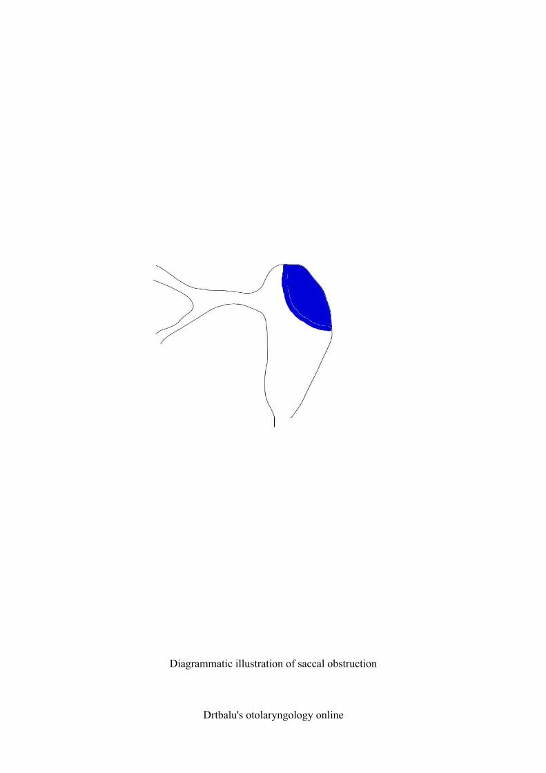
Diagrammatic illustration of saccal obstruction
Drtbalu's otolaryngology online

Subsaccal obstruction:
In this category the obstruction lies below the level of lacrimal sac. This condition commonly requires endoscopic dacryocystorhinostomy. This obstruction is more common than the rest. Causes include:
1. Congenital nasolacrimal duct obstruction2. Primary acquired nasolacrimal duct obstruction3. Nasolacrimal duct obstruction following FESS
Drtbalu's otolaryngology online

Diagrammatic representation of incomplete subsaccular obstruction
Drtbalu's otolaryngology online

Illustration showing total subsaccular obstruction
Functional obstruction:
This controversial term is used to explain those patients whose lacrimal system is patent to syringing but they still complain of epiphora. It should be borne in mind that the term obstruction should be used only to indicate anatomical obstrcution only.
Drtbalu's otolaryngology online

Diagnosis of epiphora the Phylosophy behind it:
Theoretically speaking excessive tearing may be caused by -
HypersecretionEpiphoraCombination of both
Diagnostic evaluation should include:
1. Quantification of tear production2. Assessment of nasolacrimal system patency3. Differentiating epiphora from lacrimation4. Defining the pathological process of epiphora5. Differentiating anatomical from functional obstruction6. Attempting to locate the obstruction in order to define the optimal surgical approach
Classification of Tests for lacrimal drainage pathway:
Anatomical testsFunctional testsSecretory tests
Anatomical tests:
These tests are performed to locate the probable area of lacrimal tract obstruction. These tests include:
Palpation of lacrimal sacSyringing / irrigationDiagnostic probingDacryocystographyNasal examinationCT / MRI
Functional tests:
These are performed to access the function of lacrimal apparatus under physiologic conditions. This test is performed if there is no obstruction as evdenced by negative anatomical tests.These tests include:
Flourescein dye disappearance testScintigraphyJones dye test ISacharin test
Drtbalu's otolaryngology online
Tests of secretion:
These tests are performed to access secretory function of the lacrimal apparatus. These tests are performed in examining dry eyes. These tests include:
Schrimers testBengal Rose testTear-film break upTear lysozyme
Knowledge of various causes of lacrimation and epiphora really helps in clinical examination of these patients.
Excess lacrimation:
Supranuclear causes – Psychogenic / emotionsStimulation of V cranial nerve – (Reflex tearing) Lid causes (Blepharitis / Trichiaris) Conjunctival diseases Corneal diseases Neuralgia Ocular inflammationInfranuclear causes – facial palsy, abberant innervation, crocodile tearsLacrimal gland stimulationOthers – Bright lights, sneezing
Epiphora:
Functional insufficiency Incorrect lid closure Lid malposition Punctal eversion Punctal medializationAnatomical obstruction
Combined lacrimation / epiphora – A combination of the above two categories
Facial nerve palsy – Corneal irritation and pump defects
Lower lid ectropion – Conjunctival irritation , ineffective pump mechanism
Thyroid diseases – Corneal irritation, defective canalicular function
Drtbalu's otolaryngology online

Clinical history:
This is a very important aspect of lacrimal apparatus examination. This will provide vital clues to the presence of canalicular disorders 12. History should include patient's present and past ophthalmological problems, nasal symptoms, medical and interventional relevant procedures also. Unilateral tearing usually indicate obstructive pathology whereas bilateral tearing could be physiological. A child with a history of tearing since birth should arouse suspicion of membranous obstruction to nasolacrimal duct. Nasal disorders like nasal polyposis / sinusitis can also cause unilateral epiphora.
Inspection and palpation should involve the following areas:
1. Eyelids 2. Medial canthus3. Palpation of lacrimal sac
Eye lid examination:
Look out for lower eyelid laxityEctropionPunctal eversionTrichiasisBlepharitisSnap-back test – This test is performed by pulling the lower eye lid down and away from the globe and held for several seconds. On release the lower lid resumes its normal position. The time taken for resumption of normal position is noted. The patient should not blink during the test. This test provides an assessment of laxity of lower lid. The longer it takes for the lower lid to spring back to position the more lax it is. This test is graded on a scale of 4 starting from 0. 0= normal and 4= lax lower lid.Medial canthal laxityLateral canthal laxityOrbicularis oculi muscle tone checkPinch test – This test helps to assess orbicularis oculi muscle tension.
Examination of medial canthus:
Lacrimal sac enlargement will be seen as mass below medial canthal tendon.
Enlargement above medial canthal tendon indicates neoplasm.
Drtbalu's otolaryngology online
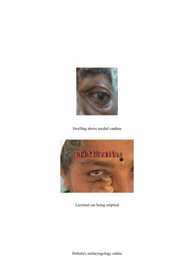
Swelling above medial canthus
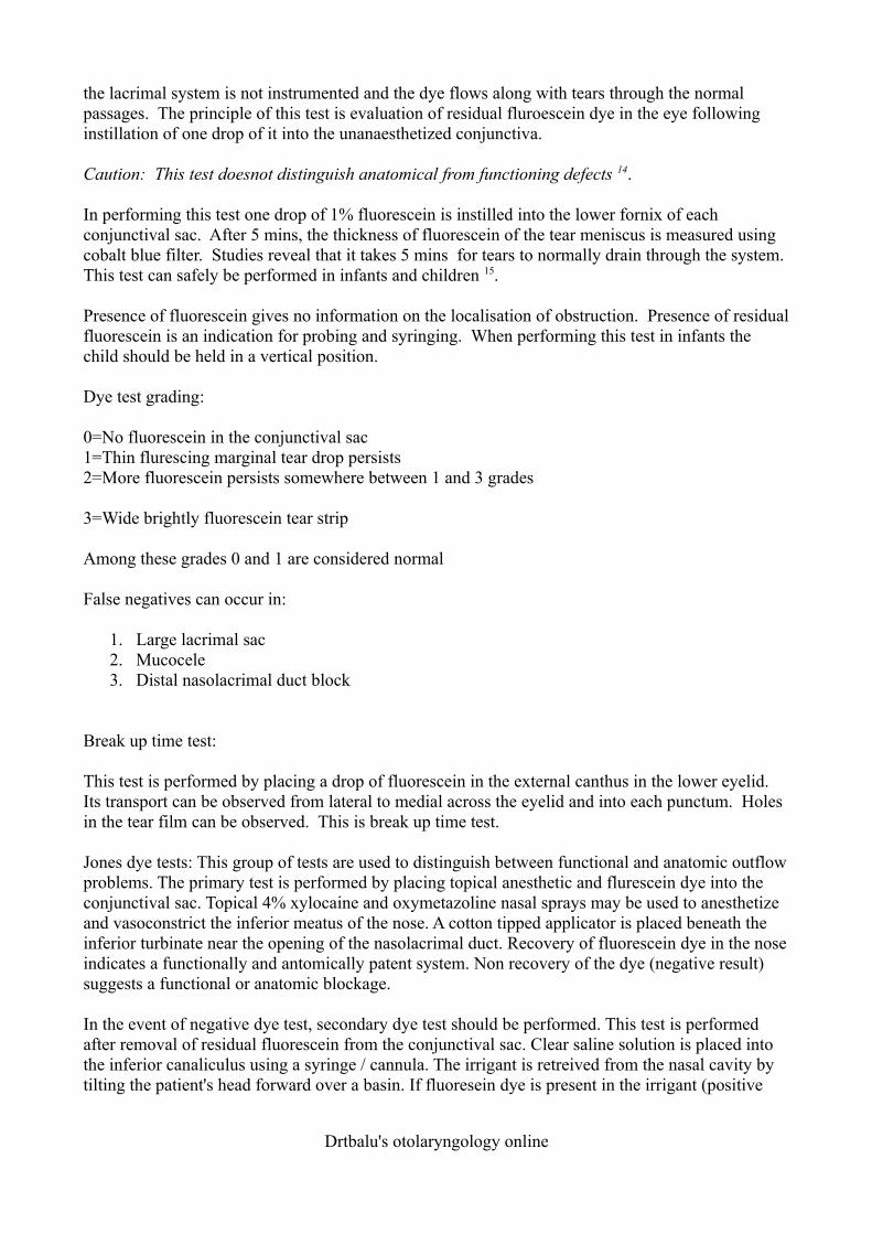
Lacrimal sac being emptied
Drtbalu's otolaryngology online

Swellling over medial canthus
Swelling below medial canthus
Palpation of lacrimal sac:Normal sac is not palpable. Sac swelling is usually confined to below the medial canthal tendon. If there is neoplasm then it is likely to extend above the medial canthus.Reflux of tears / pent up mucopurulent secretions can be seen on palpating the lacrimal sac area. Pain and tenderness over this area indicates acute dacryocystitis.
Dye excretion tests:
These tests help in ascertaining drainage functions and patency of the entire nasolacrimal system. Fluorescein dye is used for this purpose. This test is considered to be more physiological 13 since
Drtbalu's otolaryngology online
the lacrimal system is not instrumented and the dye flows along with tears through the normal passages. The principle of this test is evaluation of residual fluroescein dye in the eye following instillation of one drop of it into the unanaesthetized conjunctiva.
Caution: This test doesnot distinguish anatomical from functioning defects 14.
In performing this test one drop of 1% fluorescein is instilled into the lower fornix of each conjunctival sac. After 5 mins, the thickness of fluorescein of the tear meniscus is measured using cobalt blue filter. Studies reveal that it takes 5 mins for tears to normally drain through the system.This test can safely be performed in infants and children 15.
Presence of fluorescein gives no information on the localisation of obstruction. Presence of residual fluorescein is an indication for probing and syringing. When performing this test in infants the child should be held in a vertical position.
Dye test grading:
0=No fluorescein in the conjunctival sac1=Thin flurescing marginal tear drop persists2=More fluorescein persists somewhere between 1 and 3 grades
3=Wide brightly fluorescein tear strip
Among these grades 0 and 1 are considered normal
False negatives can occur in:
1. Large lacrimal sac2. Mucocele3. Distal nasolacrimal duct block
Break up time test:
This test is performed by placing a drop of fluorescein in the external canthus in the lower eyelid. Its transport can be observed from lateral to medial across the eyelid and into each punctum. Holes in the tear film can be observed. This is break up time test.
Jones dye tests: This group of tests are used to distinguish between functional and anatomic outflow problems. The primary test is performed by placing topical anesthetic and flurescein dye into the conjunctival sac. Topical 4% xylocaine and oxymetazoline nasal sprays may be used to anesthetize and vasoconstrict the inferior meatus of the nose. A cotton tipped applicator is placed beneath the inferior turbinate near the opening of the nasolacrimal duct. Recovery of fluorescein dye in the nose indicates a functionally and antomically patent system. Non recovery of the dye (negative result) suggests a functional or anatomic blockage.
In the event of negative dye test, secondary dye test should be performed. This test is performed after removal of residual fluorescein from the conjunctival sac. Clear saline solution is placed into the inferior canaliculus using a syringe / cannula. The irrigant is retreived from the nasal cavity by tilting the patient's head forward over a basin. If fluoresein dye is present in the irrigant (positive
Drtbalu's otolaryngology online
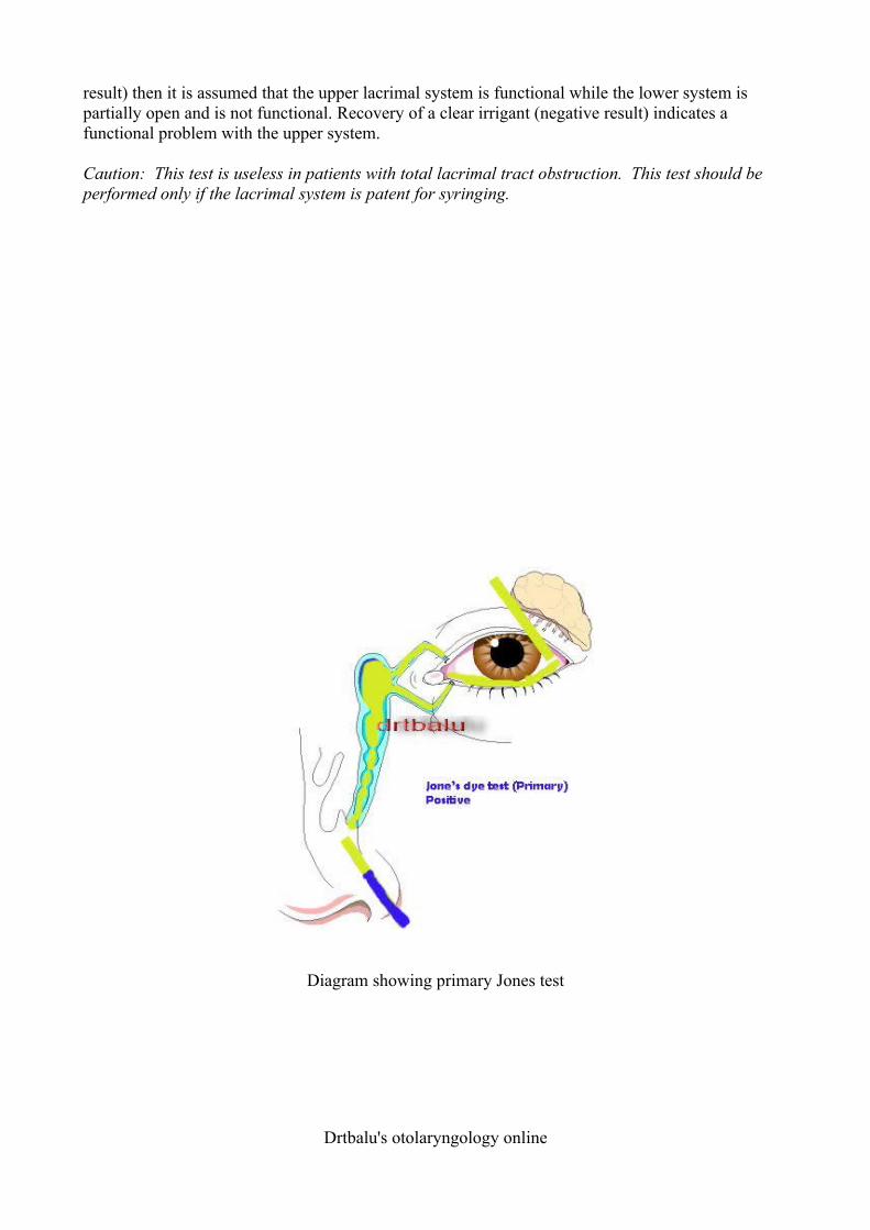
result) then it is assumed that the upper lacrimal system is functional while the lower system is partially open and is not functional. Recovery of a clear irrigant (negative result) indicates a functional problem with the upper system.
Caution: This test is useless in patients with total lacrimal tract obstruction. This test should be performed only if the lacrimal system is patent for syringing.
Diagram showing primary Jones test
Drtbalu's otolaryngology online
Saccharin test:
This test is more or less similar to fluorescein dye test. This test is also hence physiological. A drop of saccharin is placed into anesthetized and the time taken for the patient to taste saccharin is measured. Approximate time is about 3.5 mins. The flip side in this test is that the patient should have normal taste sensation.
Diagnostic probing and lacrimal syringing:
These are invasive tests. They provide valuable information on location of obstruction. They establish diagnosis of anatomical obstruction in the lacrimal system. This test is virtually useless in functional obstruction 16. Syringing / irrigation of lacrimal system is not a physiological test since the pressures used is more than the normal pressure of lacrimal syste. Hence this test should be interpreted with fluorescein dye test and clinical examination.
Procedure:
1. Topical 4% xylocaine drops applied to the conjunctiva2. Punctum dilator is used to dilate the punctum and ampulla3. A blunt cannula is placed in the inferior canaliculus. The lower eyelid is pulled down to
straighten the inferior canaliculus. Superior canaliculus is gently stretched laterally prior to irrigation
4. Tip of the irrigator is placed in the inferior canaliculus, first vertically and then horizontally with the eyelid on stretch. The tip is advanced 3-7 mm into the canaliculus and sterile saline is injected.
5. It is important to avoid forced irrigation to avoid damage to the canaliculiInterpretation: Regurgitation of irrigated saline through the opposite punctum indicates an obstruction in the common canaliculus or more distal structures. Regurgitation of fluid via the same canaliculus indicates punctal obstruction and syringing should be repeated via the opposite canaliculus. Irrigation into the nose indicates normal drainage function. It does not rule out functional obstruction.
Probing (Diagnostic):
This test should be performed if syringing test indicate obstruction and the location of the obstruction is to be ascertained. Obstruction can be located in the canaliculi and their assessment is vital in deciding the mangement modality in these patients. If irrigated fluid regurgitates through opposite punctum obstruction of common canaliculus or more distally is possible. The exact site in this scenario could be ascertained by careful probing of the entire system. Probing can be performed using blunt Bowman's probe which come in various sizes.
Procedure:
After instilling topical anesthetic drops into the conjunctiva the punctum is dilated using lacrimal probe. The probe is then passed vertically and then horizontally with the eyelid in stretch till the
Drtbalu's otolaryngology online
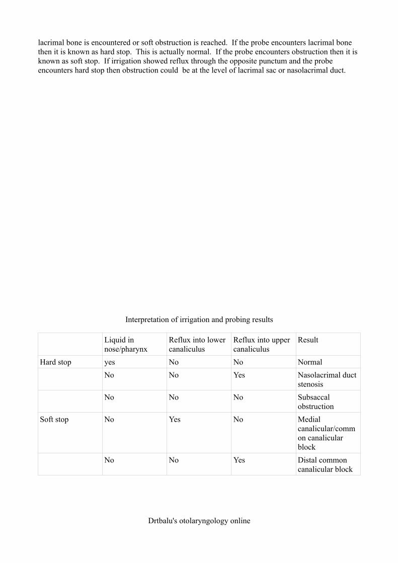
lacrimal bone is encountered or soft obstruction is reached. If the probe encounters lacrimal bone then it is known as hard stop. This is actually normal. If the probe encounters obstruction then it is known as soft stop. If irrigation showed reflux through the opposite punctum and the probe encounters hard stop then obstruction could be at the level of lacrimal sac or nasolacrimal duct.
Interpretation of irrigation and probing results
Liquid in nose/pharynx
Reflux into lower canaliculus
Reflux into upper canaliculus
Result
Hard stop yes No No Normal
No No Yes Nasolacrimal duct stenosis
No No No Subsaccal obstruction
Soft stop No Yes No Medial canalicular/common canalicular block
No No Yes Distal common canalicular block
Drtbalu's otolaryngology online

Figure showing hard stop
Figure showing soft stop
Drtbalu's otolaryngology online
Radiological evaluation:
Include:
DacryocystographyNuclear lacrimal scintigraphyCTMRI
Dacryocystography:
This is an anatomical investigation. This is indicated when there is block in the lacrimal system as indicated by syringing test. It helps in creating an internal image of the entire lacrimal system. In this test radio opaque water soluble dye is injected either into upper / lower canaliculus and magnified images are taken. Using digital subtraction techniques excellent images of the entire lacrimal system can be ensured.
Radiologic criteria of lacrimal pathology 17:
1. Regurgitation of radio-opaque fluid into the conjunctival sac2. Absence of fluid in the nose3. Fluctuation of lumen of lacrimal system4. Irregularity in contrast5. Deformation involving lacrimal sac
Nuclear lacrimal scintigraphy:
This is a non invasive physiological test. This test utilizes radiotracer technitium-99M pertechnitate. This can be analysed using a gama camera. This is useful only in patients whose lacrimal system is patent on syringing despite epiphora. This is found to be useful in difficult cases and incomplete obstruction.This test is performed without instilling topical anesthesia. A drop of technetium-99m is instilled into each conjunctival sac of a patient sitting in front of a gamma camera. Normal blinking of eyes are allowed. Patient stares at a distant target during a 20 mins test period while images are being recorded with a gamma camera.
Drtbalu's otolaryngology online
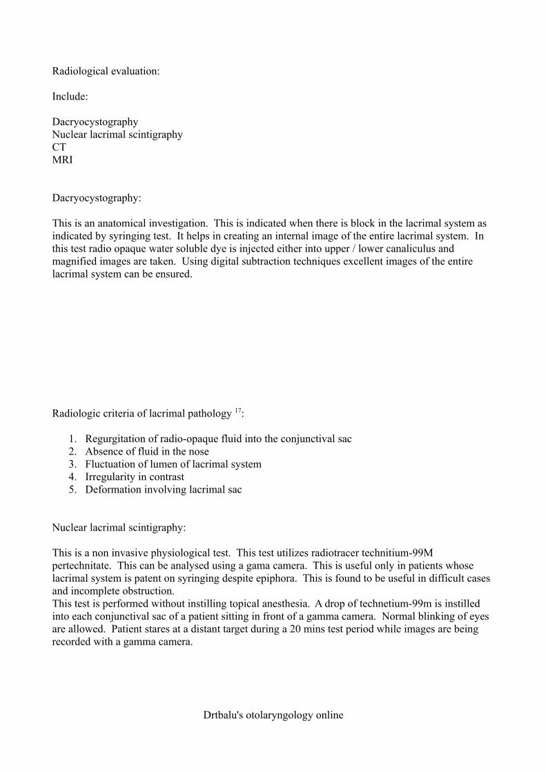
CT scan/ MRI scan:
Helpful in identifying tumors involving sac, or adjacent areas.
CT scan of a patient with rhinosporidiosis involving the lacrimal sac
Secretory tests:
These patients are useful in evaluating those with complaints of dry eye.
Schirmer's test: This test is basically prepared to quantitate tear production. This test is performed by placing strips of white filter paper 35x5 mm at the junction of the middle and lateral thirds of the lower eyelids after administration of a topical anesthetic agent. The tear production is measured with the eyes closed. Produced tears will wet the filter paper. The length of the filter paper which becomes wet is assessed at the end of 5 minutes. Normal test result is between 10mm and 30 mm of wet filter paper. Normally it should not exceed 30 mm. A value of more than 30 mm is considered to be epiphora. A value of less than 10 mm is considered to be dry eye (hyposecretion).
Drtbalu's otolaryngology online

Figure showing schirmer's test
Breakup time test:
This test indicates function of mucin layer / reflex hypersecretion of aquous component of the tears. One drop of fluorescein is instilled into the external canthus of a lower lid and the patient is instructed to blink once and then to keep his eyes open. The holes developed in the tear film are observed at the cornea through a slit-lamp with illumination through the cobalt filter. The normal breakup time should be approximately 15–30 s. A break-up time of less than 10 s indicates a deficiency and the epiphora should be treated with libricating eye drops .
Bengal Rose test:
This test is also similar to that of Break up time test. One drop of Bengal Rose dye is placed in the conjunctiva and the patient is instructed to blink several times within a minute. Interpalpebral staining is seen in patients with dry eye.
Lysozyme lysis test:
The amount of a lysozyme activity and concentration is decreased in hypersecretion and in hyposecretion, and it usually precedes clinical symptoms. A lysozyme activity (and concentration)
Drtbalu's otolaryngology online

is estimated on the basis of the inhibition of the growth of the bacterium Micrococcus lysodicticus.
References:
1. Onerci M. Dacryocystorhinostomy. Diagnosis and treatment of nasolacrimal canal obstructions. Rhinology 2002; 40:49-65.
2. Cokkeser Y, Er H. Comparative external vesus endoscópico dacryocystorhinostomy. Otolaryngol Head and Neck Surg 2000; 123:488-91
3. Hurwitz JJ (1996) The lacrimal system. Lippincott-Raven Publishers, Philadelphia 4. Guerry D, Kendig EL. Congenital impotency of the nasolacrimal duct. Arch Ophthalmol
1948, 39: 193-204.5. Price HW. Dacryostenosis. J Pediatr 1947, 30: 302-3056. Ballard EA.Excessive tearing in infancy and early childhood: the role and treatment of
congenital nasolacrimal duct obstruction. Postgrad Med 2000; 107:149-547. Kashkouli MB, Kassaee A, Tabatabaee Z. Initial Nasolacrimal Duct Probing in Children
under Age 5: Cure Rate and Factors Affecting Success. J AAPOS 2002; 6:360-38. Bouzas, A. (1965). Canalicular inflammation in ophthalmic cases of herpes zoster and
herpes simplex. American Journal of Ophthalmology, 60, 713-716.9. Hussain I, Bonshek RE, Loudon K, Armstrong M, Tullo AB (1993) Canalicular infection
caused by Actinomyces. Eye 7 (Pt 4): 542-544.10. .Sahlin S, Rose GE. (2001) Lacrimal drainage capacity and symptomatic improvement after
dacryocystorhinostomy in adults presenting with patent lacrimal drainage systems. Orbit, 20:173-179.
11. http://www.drtbalu.com/Endo_dcr.html 12. Watkins LM, Janfaza P, Tubin PA (2003) The evolution of endonasal
dacryocystorhinostomy. Surv Ophthalmol 48:73–84 13. Meyer DR, Antonello A, Linberg JV (1990) Assessment of tear drainage after canalicular
obstruction using fluorescein dye disappearance. Ophthalmology 97:370–374 14. Hurwitz JJ (1996) The lacrimal system. Lippincott-Raven Publishers, Philadelphia 15. Lloyd GAS, Welham RAN (1974) Subtraction macrodacryocystography. Br J Radiol
47:379–382 16. Sahlin S, Chen E (1996) Evaluation of the lacrimal drainage function by the drop test. Am J
Ophthalmol 122:701–708 17. Walther EK, Köster O, Straehler-Pohl HJ (1989) Dakryozystographie in digilater
Subtraktionstechnik. Laryngol Rhinol Otol 68:396–400
Drtbalu's otolaryngology online