
Evaluation of Castration-Resistant Prostate Cancer...
7
Evaluation of Castration-Resistant Prostate Cancer with Androgen Receptor–Axis Imaging Neeta Pandit-Taskar 1 , Darren R. Veach 1 , Josef J. Fox 1 , Howard I. Scher 2 , Michael J. Morris 2 , and Steven M. Larson 1 1 Department of Radiology, Memorial Sloan Kettering Cancer Center, New York, New York; and 2 Department of Medicine, Memorial Sloan Kettering Cancer Center, New York, New York Castration-resistant prostate cancer (CRPC) is the lethal form of prostate cancer, and more than 26,000 men will die from this disease in 2016. The pathophysiology of CRPC is clearly multi- factorial, but most often, androgen receptor (AR) upregulation is associated with its earliest beginnings and the AR increase is part of the multimolecular complex including downstream effector proteins linked to AR (AR-axis) responsible for rapid proliferation and malignant features of the malignant cell. In both animal models and patients, glycolysis (Warburg effect) is also an early manifestation of CRPC transformation. At Memorial Sloan Kettering Cancer Center, we have focused our energies on imaging studies of the AR-axis in CRPC, using 18 F-FDG, 18 F-16β-fluoro-5α-dihydrotestosterone ( 18 F-FDHT), and a variety of radiolabeled antibodies targeting downstream effectors, such as prostate-specific membrane antigen (PSMA). Small-molecular- weight PSMA-targeting agents are not part of this review. In this review, we will focus on molecular imaging of the AR-axis in metastatic CRPC (mCRPC) and discuss our personal experi- ence with these tracers. Our goal is to put these radiopharma- ceuticals in the context of mCRPC biology and diagnosis (e.g., 18 F-FDHT). Key Words: molecular imaging; oncology: GU; radioimmunoimaging; androgen receptor–axis imaging; CRPC; castration-resistant prostate cancer J Nucl Med 2016; 57:73S–78S DOI: 10.2967/jnumed.115.170134 Prostate cancer, when detected before spread from the pros- tate gland, may be completely eradicated by surgery or local radiotherapy. Approximately one third of patients will fail pri- mary treatment, and a rising prostate-specific antigen (PSA) level will herald the onset of recurrent or metastatic tumor. At this stage, PSA levels will usually decline with hormonal castration to levels of androgen hormone below 50 ng/dL. Typ- ically, in castrate patients, PSA begins to rise again within approximately 16–18 mo (median) despite castration, heralding the onset of CRPC. MOLECULAR IMAGING OF AR WITH 18 F-FDHT Discovery and Clinical Translation of 18 F-FDHT John Katzenellenbogen at the University of Illinois (Urbana- Champaign) and Michael Welch at Washington University (St. Louis) developed steroid-based radioligands with sufficient affinity to hormone receptors (i.e., estrogen receptor/AR) to quantify receptor status in breast and prostate cancer patients using whole-body PET. The highly versatile and easily obtained isotope 18 F with its 110-min half-life was chosen for labeling (1–3). In vitro and in vivo biologic data demonstrated that 18 F-FDHT was the best choice for AR imaging in prostate cancer in light of its good balance of in vivo stability, ease of production, and similar bind- ing affinity for AR, while retaining selectivity from other nuclear receptors (e.g., estrogen receptor, glucocorticoid receptor, and pro- gesterone receptor) (Supplemental Fig. 1 [supplemental materials are available at http://jnm.snmjournals.org]). The study that set the stage for clinical translation was imaging in baboons showing good, saturable focal uptake in AR-positive tissue, with an excellent tumor-to-background ratio (4). About 8 y later, the first in-human PET imaging studies in castrate patients were reported by Memorial Sloan Kettering Cancer Center and Washington University (5–7). The production methodology devel- oped by Washington University continues to be used to this day, though recent improvements have been made in the radiosynthetic chemistry (8) and to the production process by automation (9,10). Pharmacokinetics of 18 F-FDHT Administered as an intravenous infusion in the brachial vein, a typical dose of 185–370 MBq (5–10 mCi) of 18 F-FDHT (1–4 nmol; 0.3–1.2 mg mass) rapidly binds to serum proteins and albumin- and sex hormone–binding globulin-bound state in blood, as one would expect for a hydrophobic steroid (Fig. 1) (6). In men, most intact 18 F-FDHT has a plasma half-life of only 5–7 min and clears from the bloodstream in 15 min (Fig. 2A) (6). Via primarily hepatic metabolism, radiometabolites appear over the same timeframe, and the system plateaus at 20 min after injection. The metabolites are excreted in the bile, and a portion is refluxed into the blood. Fortunately, the metabolites do not compete with 18 F-FDHT binding to AR. Beattie et al. applied linear compartmental pharmacokinetic modeling to 18 F-FDHT uptake into tumor to a simple 2-compartment model in which the destination compartment was considered a per- manent, unidirectional parameter, k trap , attributed to intramolecular transport and binding to AR; a population-based input function was calculated by averaging aorta region-of-interest data from dynamic PET scans of a 25-patient cohort (11) (Fig. 2B). This kinetic modeling approach shows that uptake by PET imaging correlates with AR expression levels. In a recent study supported Received Jul. 14, 2016; revision accepted Aug. 15, 2016. For correspondence or reprints contact: Steven M. Larson, Department of Radiology, Memorial Sloan Kettering Cancer Center, 1275 York Ave., New York, NY 10065. E-mail: [email protected]. COPYRIGHT © 2016 by the Society of Nuclear Medicine and Molecular Imaging, Inc. AR-AXIS MOLECULAR IMAGING • Pandit-Taskar et al. 73S by on September 5, 2018. For personal use only. jnm.snmjournals.org Downloaded from
Transcript of Evaluation of Castration-Resistant Prostate Cancer...

Evaluation of Castration-Resistant Prostate Cancer withAndrogen Receptor–Axis Imaging
Neeta Pandit-Taskar1, Darren R. Veach1, Josef J. Fox1, Howard I. Scher2, Michael J. Morris2, and Steven M. Larson1
1Department of Radiology, Memorial Sloan Kettering Cancer Center, New York, New York; and 2Department of Medicine, MemorialSloan Kettering Cancer Center, New York, New York
Castration-resistant prostate cancer (CRPC) is the lethal form of
prostate cancer, and more than 26,000 men will die from this
disease in 2016. The pathophysiology of CRPC is clearly multi-
factorial, but most often, androgen receptor (AR) upregulation isassociated with its earliest beginnings and the AR increase
is part of the multimolecular complex including downstream
effector proteins linked to AR (AR-axis) responsible for rapid
proliferation and malignant features of the malignant cell. Inboth animal models and patients, glycolysis (Warburg effect)
is also an early manifestation of CRPC transformation. At Memorial
Sloan Kettering Cancer Center, we have focused our energieson imaging studies of the AR-axis in CRPC, using 18F-FDG,18F-16β-fluoro-5α-dihydrotestosterone (18F-FDHT), and a variety
of radiolabeled antibodies targeting downstream effectors, such
as prostate-specific membrane antigen (PSMA). Small-molecular-weight PSMA-targeting agents are not part of this review. In this
review, we will focus on molecular imaging of the AR-axis in
metastatic CRPC (mCRPC) and discuss our personal experi-
ence with these tracers. Our goal is to put these radiopharma-ceuticals in the context of mCRPC biology and diagnosis (e.g.,18F-FDHT).
Key Words:molecular imaging; oncology: GU; radioimmunoimaging;
androgen receptor–axis imaging; CRPC; castration-resistant prostate
cancer
J Nucl Med 2016; 57:73S–78SDOI: 10.2967/jnumed.115.170134
Prostate cancer, when detected before spread from the pros-tate gland, may be completely eradicated by surgery or localradiotherapy. Approximately one third of patients will fail pri-mary treatment, and a rising prostate-specific antigen (PSA)level will herald the onset of recurrent or metastatic tumor.At this stage, PSA levels will usually decline with hormonalcastration to levels of androgen hormone below 50 ng/dL. Typ-ically, in castrate patients, PSA begins to rise again withinapproximately 16–18 mo (median) despite castration, heraldingthe onset of CRPC.
MOLECULAR IMAGING OF AR WITH 18F-FDHT
Discovery and Clinical Translation of 18F-FDHT
John Katzenellenbogen at the University of Illinois (Urbana-Champaign) and Michael Welch at Washington University (St. Louis)developed steroid-based radioligands with sufficient affinity tohormone receptors (i.e., estrogen receptor/AR) to quantify receptorstatus in breast and prostate cancer patients using whole-bodyPET. The highly versatile and easily obtained isotope 18F with its110-min half-life was chosen for labeling (1–3). In vitro and invivo biologic data demonstrated that 18F-FDHT was the bestchoice for AR imaging in prostate cancer in light of its goodbalance of in vivo stability, ease of production, and similar bind-ing affinity for AR, while retaining selectivity from other nuclearreceptors (e.g., estrogen receptor, glucocorticoid receptor, and pro-gesterone receptor) (Supplemental Fig. 1 [supplemental materialsare available at http://jnm.snmjournals.org]).The study that set the stage for clinical translation was imaging
in baboons showing good, saturable focal uptake in AR-positivetissue, with an excellent tumor-to-background ratio (4). About 8 ylater, the first in-human PET imaging studies in castrate patientswere reported by Memorial Sloan Kettering Cancer Center andWashington University (5–7). The production methodology devel-oped by Washington University continues to be used to this day,though recent improvements have been made in the radiosyntheticchemistry (8) and to the production process by automation (9,10).
Pharmacokinetics of 18F-FDHT
Administered as an intravenous infusion in the brachial vein, atypical dose of 185–370 MBq (5–10 mCi) of 18F-FDHT (1–4 nmol;0.3–1.2 mg mass) rapidly binds to serum proteins and albumin- andsex hormone–binding globulin-bound state in blood, as one wouldexpect for a hydrophobic steroid (Fig. 1) (6). In men, most intact18F-FDHT has a plasma half-life of only 5–7 min and clears fromthe bloodstream in 15 min (Fig. 2A) (6). Via primarily hepaticmetabolism, radiometabolites appear over the same timeframe,and the system plateaus at 20 min after injection. The metabolitesare excreted in the bile, and a portion is refluxed into the blood.Fortunately, the metabolites do not compete with 18F-FDHT bindingto AR.Beattie et al. applied linear compartmental pharmacokinetic
modeling to 18F-FDHT uptake into tumor to a simple 2-compartmentmodel in which the destination compartment was considered a per-manent, unidirectional parameter, ktrap, attributed to intramoleculartransport and binding to AR; a population-based input functionwas calculated by averaging aorta region-of-interest data fromdynamic PET scans of a 25-patient cohort (11) (Fig. 2B). Thiskinetic modeling approach shows that uptake by PET imagingcorrelates with AR expression levels. In a recent study supported
Received Jul. 14, 2016; revision accepted Aug. 15, 2016.For correspondence or reprints contact: Steven M. Larson, Department of
Radiology, Memorial Sloan Kettering Cancer Center, 1275 York Ave., NewYork, NY 10065.E-mail: [email protected] © 2016 by the Society of Nuclear Medicine and Molecular
Imaging, Inc.
AR-AXIS MOLECULAR IMAGING • Pandit-Taskar et al. 73S
by on September 5, 2018. For personal use only. jnm.snmjournals.org Downloaded from
by the Movember GAP2 project, these findings were confirmedand refined by Kramer et al. at VU Amsterdam, who reported animprovement over using body weight–normalized SUV in dy-namic 18F-FDHT PET images of 31 lesions in a 4-patient cohort.They observed a high correlation (R2 5 0.97) between SUVAUC,PP
(SUV normalized to 18F-FDHT plasma area under the curve) and anirreversible trapping constant in a 2-tissue-compartment model, us-ing an input function derived from both PET and venous bloodsample data to correct for metabolites (12). Such studies may makeit more practical to obtain biologically relevant quantitation of re-ceptor number.
Clinical Molecular Imaging of AR in Men18F-FDHT binds with high affinity to AR-expressing tumor
tissues in men. Patient selection should include serum androgenmeasurements, because AR imaging is practical only in castrate
patients (currently) with testosterone levelsbelow 50 ng/dL. The active radiotracer18F-FDHT is an agonist of AR and ananalog of DHT, the most common andro-gen in prostate and prostate cancer at thecellular level in men. The 18F-FDHT up-take, as quantified by SUVmax or relatedSUV parameter, plateaus within about30 min, with no further uptake thereafter.A single patient dose at the conservative5-cGy threshold for diagnostic studies cor-responded to 331 MBq (8.9 mCi) (7).An example of a PET imaging study using
18F-FDHT is shown in Figure 3. Normally,subjects are studied within the same timewindow (a few days), with 18F-FDG as well,because we have found that there is consid-erable heterogeneity of biochemical featuresof individual CRPC lesions, when glycoly-sis and AR expression are compared. Inaddition, the companion CT provides an-atomic orientation to lesion site.
Hitting the Target: In Vivo
Displacement of 18F-FDHT by AR
Inhibitor Drugs
In the last few years, high-affinity AR inhibitors have beendeveloped and approved by the U.S. Food and Drug Administra-tion for use in CRPC on the basis of prolonged survival. One ofthese, enzalutamide, has been studied in men, using 18F-FDHTimaging to show whether or not the AR blockade of 18F-FDHT uptakeis occurring, as would be expected for effective therapy. Figure 4shows in vitro and in vivo studies in support of the concept that18F-FDHT may be used as a probe to demonstrate the high-affinitybinding requisite for pharmacologic AR blockade in vivo (13).
Determining Biologically Effective Dose for AR Blockade
In a first-in-human study of optimizing dosing schedules for anovel second-generation AR blockade drug, apalutamide, 18F-FDHTwas also used to explore progressive AR blockade in a phase I trialof dose escalation (Fig. 5) (14). Sixteen patients received a base-
line and follow-up 18F-FDHT scan duringthe course of dose escalation, with dosesranging from 30 to 390 mg. A background-corrected SUVmax was determined as pre-viously described (15), and all lesions inindividual patients were averaged. A meanof the SUVmax parameter at each drug levelwas then correlated with dose level, withsaturation levels beginning at 120 mg. Be-cause seizures had been observed at highestdoses of other AR inhibitors, a final phase IIdose of 240 mg was chosen to ensure satura-tion and minimize neurotoxicity.
PSMA IMAGING WITH
RADIOLABELED ANTIBODIES
AR-Directed Agents and PSMA
Targeting with Antibodies
PSMA is a well-established marker forprostate cancer that is highly expressedin prostate cancer cells. Although highly
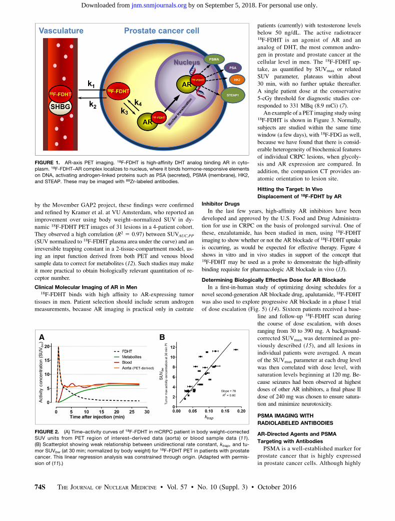
FIGURE 1. AR-axis PET imaging. 18F-FDHT is high-affinity DHT analog binding AR in cyto-
plasm. 18F-FDHT–AR complex localizes to nucleus, where it binds hormone-responsive elements
on DNA, activating androgen-linked proteins such as PSA (secreted), PSMA (membrane), HK2,
and STEAP. These may be imaged with 89Zr-labeled antibodies.
FIGURE 2. (A) Time–activity curves of 18F-FDHT in mCRPC patient in body weight–corrected
SUV units from PET region of interest–derived data (aorta) or blood sample data (11).
(B) Scatterplot showing weak relationship between unidirectional rate constant, ktrap, and tu-
mor SUVbw (at 30 min; normalized by body weight) for 18F-FDHT PET in patients with prostate
cancer. This linear regression analysis was constrained through origin. (Adapted with permis-
sion of (11).)
74S THE JOURNAL OF NUCLEAR MEDICINE • Vol. 57 • No. 10 (Suppl. 3) • October 2016
by on September 5, 2018. For personal use only. jnm.snmjournals.org Downloaded from
specific for prostate, it is also expressed in the cells of the smallintestine, proximal renal tubules, and salivary glands (16,17).However, the level of expression in the prostate cells is 100- to1,000-fold higher than in nonprostate tissue (18,19). PSMA is atype II integral cell surface membrane protein that is not se-creted, thereby making it an ideal target for monoclonal anti-body (mAb) imaging or therapy. PSMA has been found to havefolate hydrolase and glutamate carboxypeptidase activity (20–22). PSMA upregulation correlates with increased aggressive-ness and recurrence (23,24) and higher mortality (25), suggest-ing a functional role of PSMA in prostate cancer progression.PSMA-targeted imaging with 111In-labeled mAb was ap-
proved by the Food and Drug Administration as 7E11/CYT-356Capromab (ProstaScint) to detect disease in patients (26–29).Initial clinical enthusiasm was tempered by the fact that 7E11
targets the internal portion of the PSMAmolecule, which is less accessible, and tar-geting was suboptimal in intact cells versusnecrotic tissue, and viable tumor was missed(30,31).Given the limitation of Capromab, tar-
geting of the extracellular domain of PSMAwas explored (31–33) and found to be moreeffective. J591 binds to the external do-main of PSMA with high-affinity bindingto prostate cancer cells in tissue culture andanimal models (33,34).
Imaging with Radiolabeled J591
For human studies, J591 has been conju-gated with a metal chelating agent (e.g.,diethylene triamine pentaacetic acid orDOTA) to enable labeling with radiometalssuch as 111In or 177Lu (35). More recently,J591 has been radiolabeled with a PET ra-dioisotope, 89Zr, using the siderophore des-ferrioxamine as a chelator (36,37).Initial phase I studies of huJ591 trace-
labeled with 111In using a DOTA chelate showed that repetitivedosing was well tolerated, with total doses of up to 500 mg/m2
without the development of a human antihumanized (deimmu-nized) antibody response (38,39). No dose-limiting toxicity oc-curred, and the maximum tolerated dose was not reached. Excellenttumor targeting occurred at all dose levels of mAb. No mAbtargeting to sites other than those involved by prostate cancerwas observed, although, as seen in other trials using radiometals,the liver is the primary site of metabolism. Percentage injecteddose in the liver diminished with increasing dose of antibody,and higher doses were associated with longer plasma clearancetimes (40).In a dose escalation study, 111In-J591 antibody was given in
combination with cold J591 administered in escalating doses from25 to 100 mg. Dose-dependent plasma clearance of the antibody
occurred more slowly as antibody massincreased up to 100 mg (41). The meanbiologic half-life was 0.96, 1.9, 2.75, and3.47 d for the 10-, 25-, 50-, and 100-mgdoses, respectively. Hepatic saturation wasachieved at a 10- to 25-mg dose of anti-body, and the optimal tradeoff between in-creased J591 circulation and liver uptakewas obtained with 25 mg of total antibodydose (41).In a detailed analysis of lesion targeting
with 111In-J591, both bone and soft-tissuelesions were targeted well, with localizationin about 94% of skeletal lesions detected byconventional imaging (42). Visualization oflesions was better in delayed images and atlater infusions of higher antibody masses.Imaging was similarly high with 177Lu-J591 (43).PET allows for superior image resolu-
tion and ability to quantify uptake as com-pared with single-photon imaging, enabling
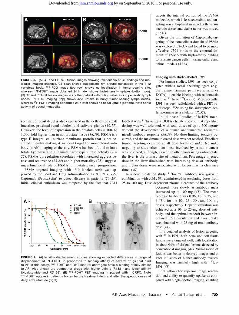
FIGURE 3. (A) CT and PET/CT fusion images showing relationship of CT findings and mo-
lecular imaging changes. CT scan shows osteoblastic rim around metastasis in the T-12
vertebrae body. 18F-FDG image (top row) shows no localization in tumor-bearing site,
whereas 18F-FDHT image obtained 24 h later shows high-intensity uptake (bottom row).
(B) CT and PET/CT fusion images in another patient with bulky metastasis in periaortic lymph
nodes. 18F-FDG imaging (top) shows avid uptake in bulky tumor-bearing lymph nodes,
whereas 18F-FDHT imaging performed 24 h later shows no nodal uptake (bottom). Note aortic
activity of bound metabolite.
FIGURE 4. (A) In vitro displacement studies showing expected differences in range of
displacement of 18F-FDHT, in proportion to binding affinity of several drugs that bind
to AR in this assay. 18F-FDHT and DHT (natural androgen) have a binding affinity similar
to AR. Also shown are competitor drugs with higher affinity (R1881) and lower affinity
(bicalutamide and RD162). (B) 18F-FDHT PET imaging in patient with mCRPC. Note18F-FDHT uptake in patient’s bones before treatment (left) and after therapeutic doses of
daily enzalutamide (right).
AR-AXIS MOLECULAR IMAGING • Pandit-Taskar et al. 75S
by on September 5, 2018. For personal use only. jnm.snmjournals.org Downloaded from
better dosimetry estimations for normal organs and tumors invivo. 89Zr has been increasingly used for immuno-PET imagingbecause of its favorable decay characteristics, including a radio-active half-life of 78.4 h, which is more suitable for imagingantibody uptake. Chelation methodology using desferrioxaminehas been described for labeling antibodies (37,44), and preclinicalstudies with 89Zr-J591 have demonstrated excellent targeting of pros-tate cancer in vivo (44).
In a first-in-human phase I study with 89Zr-J591 in prostate can-cer patients, the clearance of 89Zr-J591 from serum was biexponen-tial, with biologic half-lives of 7.0 6 4.5 h (range, 1.1–14 h) and62 6 13 h (range, 51–89 h). Whole-body clearance was monoex-ponential, with a mean half-life of 219 6 48 h (range, 153–317 h).Dosimetric estimates to critical organs showed highest dose to theliver (2086 40.5 cGy/GBq [7.76 1.5 cGy/mCi]), with lesser dosesto renal cortex (956 10.9 cGy/GBq [3.56 0.4 cGy/mCi]) and bonemarrow (32 6 5.3 cGy/GBq [1.2 6 0.2 cGy/mCi]). The optimaltime for PET imaging after injection was 7 6 1 d (45).A follow-up analysis of lesion targeting in 50 patients with CRPC
imaged using 89Zr-J591 PET/CT showed good localization of bothbone and soft-tissue lesions (46). Higher targeting, including bothuptake and lesion detection, was seen for bone lesions than for soft-tissue lesions. The median SUV was significantly higher for bonelesions than for soft-tissue lesions (8.9 vs. 4.8, respectively; P ,0.00003). In a comprehensive comparative analysis with conven-tional imaging, 89Zr-J591 detected more osseous sites relative toconventional methylene diphosphonate bone scanning and CT scan-ning (Fig. 6). However, soft-tissue disease detection was inferior toCT scanning. Pathology correlation showed a high overall accuracyof 89Zr-J591 (95.2%) for osseous lesions and slightly lower (60%)for soft-tissue lesions.Bayesian analysis was used to predict the number of positive
lesions among the unbiopsied sites for each modality separatelyfor bone and soft tissue (46). The results showed the highestpredicted number of positive findings for J591 in osseous lesions,as compared with other conventional modalities (SupplementalTable 1).
ANTI-PSMA MINIBODY IMAGING
It has been shown that antibody fragments such as minibodiesand diabodies clear faster and may allow for early imaging of
tumor sites (47,48). Fragments below 60kDa are filtered through the glomerularsystem, leading to significant kidney ex-cretion (49–52), which is not ideal forprostate cancer imaging agents; on theother hand, minibodies are slightly largerand do not have high renal clearance.IAB2M is an 80-kDa minibody geneti-cally engineered from the parent human-ized anti-PSMA mAb J591, consisting ofa bivalent homodimer, with each mono-mer comprising a single-chain variablefragment (scFv) linked to a human IgG1CH3 domain, that targets the extracellulardomain of PSMA (Supplemental Fig. 2).IAB2M lacks Fc receptor interactiondomains on the minibody that make itpharmacologically inert to Fc-mediatedeffector functions (50–56).In a phase I first-in-human study eval-
uating PET imaging with 89Zr- IAB2M(57), there was rapid clearance of theminibody from the blood. Lesions wereseen as early as 24 h after injection, withmost lesions seen by 48 h. Targeting toboth bone and soft-tissue lesions (Fig. 7)and high correlation with pathology wasobserved.
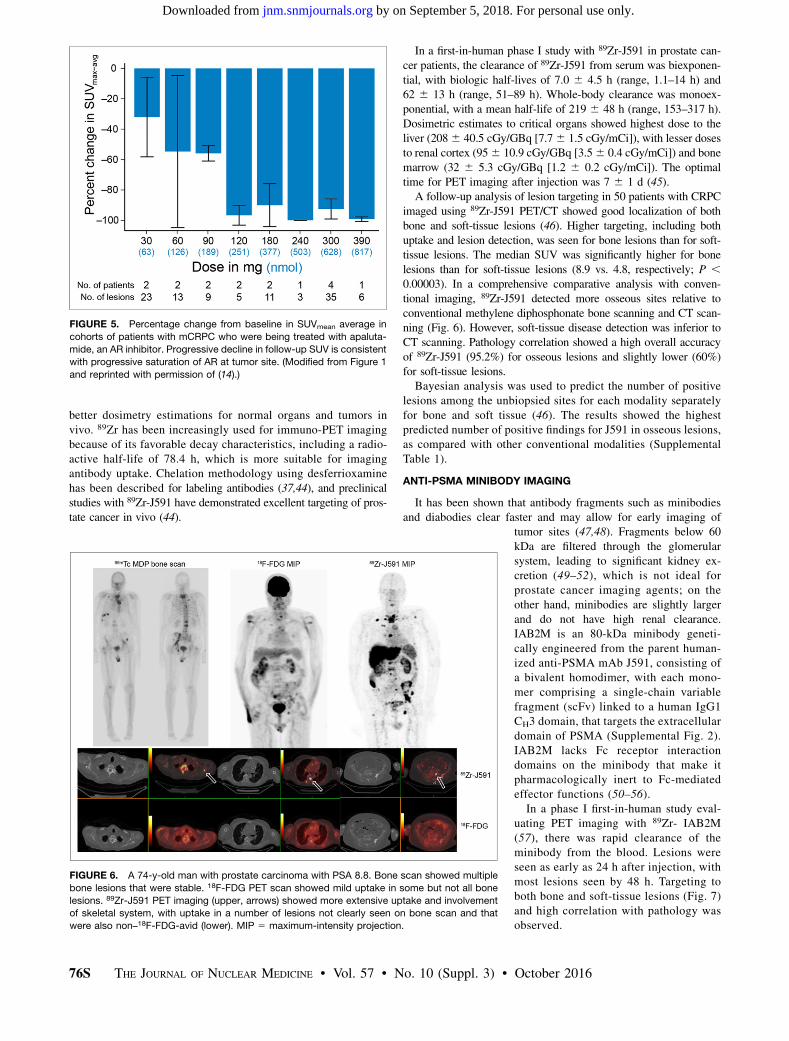
FIGURE 5. Percentage change from baseline in SUVmean average in
cohorts of patients with mCRPC who were being treated with apaluta-
mide, an AR inhibitor. Progressive decline in follow-up SUV is consistent
with progressive saturation of AR at tumor site. (Modified from Figure 1
and reprinted with permission of (14).)
FIGURE 6. A 74-y-old man with prostate carcinoma with PSA 8.8. Bone scan showed multiple
bone lesions that were stable. 18F-FDG PET scan showed mild uptake in some but not all bone
lesions. 89Zr-J591 PET imaging (upper, arrows) showed more extensive uptake and involvement
of skeletal system, with uptake in a number of lesions not clearly seen on bone scan and that
were also non–18F-FDG-avid (lower). MIP 5 maximum-intensity projection.
76S THE JOURNAL OF NUCLEAR MEDICINE • Vol. 57 • No. 10 (Suppl. 3) • October 2016
by on September 5, 2018. For personal use only. jnm.snmjournals.org Downloaded from
CONCLUSION
Prostate cancer is biologically heterogeneous, with clinicalbehavior ranging from more benign to lethal. Prostate cancerimaging with radionuclides has come a long way in the pastdecade, with the introduction of a variety of new agents that providedetection of early and late metastases for many of these clinicalstates, particularly early recurrence with PSA. Several reviewsprovide summaries of the role of molecular imaging agents forprostate cancer (e.g., Kircher et al. (58)). Our group has focusedprincipally on mCRPC, the lethal form of the disease, with tracersmost relevant to AR-axis stimulation and growth, such as 18F-FDHT,18F-FDG, and radioantibodies that target a variety of downstreameffector proteins that can be used to monitor the response of tumorto treatment as well as the status of AR-axis activity in late stagesof the disease, through PET imaging of antibodies. In this review,we have emphasized our work with AR imaging and PSMA-targeted antibodies.From a clinical point of view, 18F-FDHT has proven to be
highly accurate for detecting the presence of AR (data not shown),which in turn is the most common driver of the CRPC state. Theutility of 18F-FDG comes from the ability to monitor treatmentresponse in CRPC. 18F-FDHT has proven uniquely useful in doc-umenting the interaction of second-generation AR-axis inhibitorswith AR binding in human tumors during clinical trials, and in thisway has facilitated acceptance by regulatory agencies of thesedrugs for therapy in CRPC.The explosion of interest in small molecules targeting PSMA,
particularly in Europe (59), is one of the reasons we emphasized ourstudies with J591, a high-affinity anti-PSMA antibody. PSMA an-tibody imaging and therapy with radioantibodies is feasible andprovides a specific technique to evaluate viable disease. Given cer-tain practical limitations of longer circulation times of the antibody,methods to reduce circulation times and use of smaller moleculeswould be more suitable and should be further explored. PSMA-targeted therapeutics may offer another option for therapy espe-cially in those with refractory disease (supplemental materials).One final point: new targeting agents have recently been approved
by the Food and Drug Administration, such as 18F-fluciclovine (60),
11C-choline (61), and 68Ga-PSMA (59). Allof these agents provide high-quality imagesand are documented to be useful for image-guided biopsy to detect occult tumors, es-pecially in PSA recurrence after primarytreatment of prostate cancer. Often, weare called on to choose the best molecularimaging agent, which is a difficult ques-tion; enthusiasm based on superb imagesis not proof. Instead, we propose consid-ering biopsy-based approaches with carefulstatistics to estimate error rates, such as theBayesian method illustrated in Supplemen-tal Table 1. In a clinical research setting, inwhich we can perform a battery of tests onthe same patients, comparison of 95%confidence for accuracy of individual testsallows us to rank-order the effectivenessof a new test—in this case, 89Zr-J591—compared with standard tests, such as CT,18F-FDG, and 99mTc bone scanning. Eachof these exciting new small moleculeswill have to be studied in a similar fash-
ion, perhaps head-to-head, to choose which of these newly in-troduced imaging tracers will prove to be most effective.
DISCLOSURE
No potential conflict of interest relevant to this article wasreported.
REFERENCES
1. Chen Y, Sawyers CL, Scher HI. Targeting the androgen receptor pathway in
prostate cancer. Curr Opin Pharmacol. 2008;8:440–448.
2. Agus DB, Golde DW, Sgouros G, Ballangrud A, Cordon-Cardo C, Scher HI.
Positron emission tomography of a human prostate cancer xenograft: association
of changes in deoxyglucose accumulation with other measures of outcome fol-
lowing androgen withdrawal. Cancer Res. 1998;58:3009–3014.
3. Ido T, Wan CN, Casella V, et al. Labeled 2-deoxy-D-glucose analogs: F-18-
labeled 2-deoxy-2-fluoro-D-glucose, 2-deoxy-2-fluoro-D-mannose and C-14-2-
deoxy-2-fluoro-D-glucose. J Labelled Comp Radiopharm. 1978;14:175–183.
4. Bonasera TA, O’Neil JP, Xu M, et al. Preclinical evaluation of fluorine-18-labeled
androgen receptor ligands in baboons. J Nucl Med. 1996;37:1009–1015.
5. Dehdashti F, Picus J, Michalski JM, et al. Positron tomographic assessment
of androgen receptors in prostatic carcinoma. Eur J Nucl Med Mol Imaging.
2005;32:344–350.
6. Larson SM, Morris M, Gunther I, et al. Tumor localization of 16b-18F-fluoro-5a-
dihydrotestosterone versus 18F-FDG in patients with progressive, metastatic
prostate cancer. J Nucl Med. 2004;45:366–373.
7. Zanzonico PB, Finn R, Pentlow KS, et al. PET-based radiation dosimetry in man
of 18F-fluorodihydrotestosterone, a new radiotracer for imaging prostate cancer.
J Nucl Med. 2004;45:1966–1971.
8. Zhou D, Lin M, Yasui N, et al. Optimization of the preparation of fluorine-18-
labeled steroid receptor ligands 16alpha-[18F]fluoroestradiol (FES), [18F]fluoro
furanyl norprogesterone (FFNP), and 16beta-[18F]fluoro-5alpha-dihydrotestosterone
(FDHT) as radiopharmaceuticals. J Labelled Comp Radiopharm. 2014;57:
371–377.
9. Lazari M, Lyashchenko SK, Burnazi EM, Lewis JS, van Dam RM, Murphy JM.
Fully-automated synthesis of 16b-18F-fluoro-5a-dihydrotestosterone (FDHT) on
the ELIXYS radiosynthesizer. Appl Radiat Isot. 2015;103:9–14.
10. Ackermann U, Lewis JS, Young K, et al. Fully automated synthesis of
[18F]fluoro-dihydrotestosterone ([18F]FDHT) using the FlexLab module. J La-
belled Comp Radiopharm. 2016;59:424–428.
11. Beattie BJ, Smith-Jones PM, Jhanwar YS, et al. Pharmacokinetic assessment of
the uptake of 16b-18F-fluoro-5a-dihydrotestosterone (FDHT) in prostate tumors
as measured by PET. J Nucl Med. 2010;51:183–192.
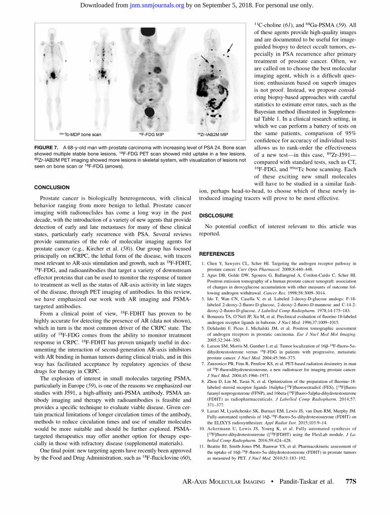
FIGURE 7. A 68-y-old man with prostate carcinoma with increasing level of PSA 24. Bone scan
showed multiple stable bone lesions. 18F-FDG PET scan showed mild uptake in a few lesions.89Zr-IAB2M PET imaging showed more lesions in skeletal system, with visualization of lesions not
seen on bone scan or 18F-FDG (arrows).
AR-AXIS MOLECULAR IMAGING • Pandit-Taskar et al. 77S
by on September 5, 2018. For personal use only. jnm.snmjournals.org Downloaded from
12. Kramer G, Yaqub M, Schuit R, et al. Assessment of simplified methods for
quantification of [18F]FDHT uptake in patients with metastasized castrate-resistant
prostate cancer [abstract]. J Nucl Med. 2016;57(suppl 2):464.
13. Scher HI, Beer TM, Higano CS, et al. Antitumour activity of MDV3100 in
castration-resistant prostate cancer: a phase 1-2 study. Lancet. 2010;375:1437–1446.
14. Rathkopf DE, Morris MJ, Fox JJ, et al. Phase I study of ARN-509, a novel
antiandrogen, in the treatment of castration-resistant prostate cancer. J Clin
Oncol. 2013;31:3525–3530.
15. Fox JJ, Autran-Blanc E, Morris MJ, et al. Practical approach for comparative
analysis of multilesion molecular imaging using a semiautomated program for
PET/CT. J Nucl Med. 2011;52:1727–1732.
16. Israeli RS, Grob M, Fair WR. Prostate-specific membrane antigen and other
prostatic tumor markers on the horizon. Urol Clin North Am. 1997;24:439–450.
17. Israeli RS, Powell CT, Corr JG, Fair WR, Heston WD. Expression of the prostate-
specific membrane antigen. Cancer Res. 1994;54:1807–1811.
18. Silver DA, Pellicer I, Fair WR, Heston WD, Cordon-Cardo C. Prostate-specific
membrane antigen expression in normal and malignant human tissues. Clin Cancer
Res. 1997;3:81–85.
19. Sokoloff RL, Norton KC, Gasior CL, Marker KM, Grauer LS. A dual-monoclonal
sandwich assay for prostate-specific membrane antigen: levels in tissues, seminal
fluid and urine. Prostate. 2000;43:150–157.
20. Carter RE, Feldman AR, Coyle JT. Prostate-specific membrane antigen is a
hydrolase with substrate and pharmacologic characteristics of a neuropeptidase.
Proc Natl Acad Sci USA. 1996;93:749–753.
21. Heston WD. Characterization and glutamyl preferring carboxypeptidase func-
tion of prostate-specific membrane antigen: a novel folate hydrolase. Urology.
1997;49:104–112.
22. Pinto JT, Suffoletto BP, Berzin TM, et al. Prostate-specific membrane antigen: a
novel folate hydrolase in human prostatic carcinoma cells. Clin Cancer Res.
1996;2:1445–1451.
23. Perner S, Hofer MD, Kim R, et al. Prostate-specific membrane antigen expression
as a predictor of prostate cancer progression. Hum Pathol. 2007;38:696–701.
24. Ross JS, Sheehan CE, Fisher HA, et al. Correlation of primary tumor prostate-
specific membrane antigen expression with disease recurrence in prostate cancer.
Clin Cancer Res. 2003;9:6357–6362.
25. Kasperzyk JL, Finn S, Hendrickson W, et al. PSMA expression and prostate
cancer survival. Paper presented at: Multi-Institutional Prostate Cancer SPORE
Meeting; April 12-14; 2010; Fort Lauderdale, FL.
26. Babaian RJ, Sayer J, Podoloff DA, Steelhammer LC, Bhadkamkar VA, Gulfo JV.
Radioimmunoscintigraphy of pelvic lymph nodes with 111indium-labeled mono-
clonal antibody CYT-356. J Urol. 1994;152:1952–1955.
27. Kahn D, Williams RD, Seldin DW, et al. Radioimmunoscintigraphy with 111in-
dium labeled CYT-356 for the detection of occult prostate cancer recurrence.
J Urol. 1994;152:1490–1495.
28. Kahn D, Williams RD, Manyak MJ, et al. 111Indium-capromab pendetide in the
evaluation of patients with residual or recurrent prostate cancer after radical
prostatectomy. The ProstaScint Study Group. J Urol. 1998;159:2041–2046.
29. Kahn D, Williams RD, Haseman MK, Reed NL, Miller SJ, Gerstbrein J. Radio-
immunoscintigraphy with In-111-labeled capromab pendetide predicts prostate
cancer response to salvage radiotherapy after failed radical prostatectomy. J Clin
Oncol. 1998;16:284–289.
30. Troyer JK, Feng Q, Beckett ML, Wright GL Jr. Biochemical characterization and
mapping of the 7E11-C5.3 epitope of the prostate-specific membrane antigen.
Urol Oncol. 1995;1:29–37.
31. Troyer JK, Beckett ML, Wright GL Jr. Location of prostate-specific membrane
antigen in the LNCaP prostate carcinoma cell line. Prostate. 1997;30:232–242.
32. Fair WR, Israeli RS, Heston WD. Prostate-specific membrane antigen. Prostate.
1997;32:140–148.
33. Liu H, Moy P, Kim S, et al. Monoclonal antibodies to the extracellular domain of
prostate-specific membrane antigen also react with tumor vascular endothelium.
Cancer Res. 1997;57:3629–3634.
34. Liu H, Rajasekaran AK, Moy P, et al. Constitutive and antibody-induced internali-
zation of prostate-specific membrane antigen. Cancer Res. 1998;58:4055–4060.
35. Smith-Jones PM, Vallabhajosula S, Navarro V, Bastidas D, Goldsmith SJ,
Bander NH. Radiolabeled monoclonal antibodies specific to the extracellular
domain of prostate-specific membrane antigen: preclinical studies in nude mice
bearing LNCaP human prostate tumor. J Nucl Med. 2003;44:610–617.
36. Holland JP, Divilov V, Bander NH, Smith-Jones PM, Larson SM, Lewis JS.
Zr-89-DFO-J591 for immunoPET of prostate-specific membrane antigen expres-
sion in vivo. J Nucl Med. 2010;51:1293–1300.
37. Holland JP, Sheh Y, Lewis JS. Standardized methods for the production of high
specific-activity zirconium-89. Nucl Med Biol. 2009;36:729–739.
38. Bander NH, Nanus D, Bremer S, et al. Phase I clinical trial targeting a mono-
clonal antibody (mAb) to the extracellular domain of prostate-specific membrane
antigen (PSMAext) in patients with hormone-independent prostate cancer [ab-
stract]. Proc Am Soc Clin Oncol. 2000;19:477a.
39. Bander NH, Nanus D, Goldsmith S, et al. Phase I trial of humanized monoclonal
antibody (mAb) to prostate-specific membrane antigen/extracellular domain
(PSMAext) [abstract]. Proc Am Soc Clin Oncol. 2001;20:722.
40. Vallabhajosula S, Kuji I, Hamacher KA, et al. Pharmacokinetics and biodistri-
bution of 111In- and 177Lu-labeled J591 antibody specific for prostate-specific
membrane antigen: prediction of 90Y-J591 radiation dosimetry based on 111In or177Lu? J Nucl Med. 2005;46:634–641.
41. Morris MJ, Divgi CR, Pandit-Taskar N, et al. Pilot trial of unlabeled and indium-
111-labeled anti-prostate-specific membrane antigen antibody J591 for castrate
metastatic prostate cancer. Clin Cancer Res. 2005;11:7454–7461.
42. Pandit-Taskar N, O’Donoghue JA, Morris MJ, et al. Antibody mass escalation
study in patients with castration-resistant prostate cancer using 111In-J591: lesion
detectability and dosimetric projections for 90Y radioimmunotherapy. J Nucl
Med. 2008;49:1066–1074.
43. Bander NH, Milowsky MI, Nanus DM, Kostakoglu L, Vallabhajosula S,
Goldsmith SJ. Phase I trial of 177lutetium-labeled J591, a monoclonal antibody
to prostate-specific membrane antigen, in patients with androgen-independent
prostate cancer. J Clin Oncol. 2005;23:4591–4601.
44. Holland JP, Caldas-Lopes E, Divilov V, et al. Measuring the pharmacodynamic
effects of a novel Hsp90 inhibitor on HER2/neu expression in mice using Zr-89-
DFO-trastuzumab. PLoS One. 2010;5:e8859.
45. Pandit-Taskar N, O’Donoghue JA, Beylergil V, et al. 89Zr-huJ591 immuno-PET
imaging in patients with advanced metastatic prostate cancer. Eur J Nucl Med
Mol Imaging. 2014;41:2093–2105.
46. Pandit-Taskar N, O’Donoghue JA, Durack JC, et al. A phase I/II study for
analytic validation of 89Zr-J591 immunoPET as a molecular imaging agent for
metastatic prostate cancer. Clin Cancer Res. 2015;21:5277–5285.
47. Knowles SM, Wu AM. Advances in immuno-positron emission tomography: anti-
bodies for molecular imaging in oncology. J Clin Oncol. 2012;30:3884–3892.
48. Olafsen T, Sirk SJ, Olma S, Shen CK, Wu AM. ImmunoPET using engineered
antibody fragments: fluorine-18 labeled diabodies for same-day imaging. Tu-
mour Biol. 2012;33:669–677.
49. Wong JY, Chu DZ, Williams LE, et al. Pilot trial evaluating an 123I-labeled
80-kilodalton engineered anticarcinoembryonic antigen antibody fragment
(cT84.66 minibody) in patients with colorectal cancer. Clin Cancer Res.
2004;10:5014–5021.
50. Wu AM, Olafsen T. Antibodies for molecular imaging of cancer. Cancer J.
2008;14:191–197.
51. Wu AM. Engineered antibodies for molecular imaging of cancer. Methods.
2014;65:139–147.
52. Wu AM. Antibodies and antimatter: the resurgence of immuno-PET. J Nucl Med.
2009;50:2–5.
53. Wu AM, Senter PD. Arming antibodies: prospects and challenges for immuno-
conjugates. Nat Biotechnol. 2005;23:1137–1146.
54. Leyton JV, Olafsen T, Lepin EJ, et al. Humanized radioiodinated minibody for
imaging of prostate stem cell antigen-expressing tumors. Clin Cancer Res.
2008;14:7488–7496.
55. Olafsen T, Betting D, Kenanova VE, et al. Recombinant anti-CD20 antibody
fragments for small-animal PET imaging of B-cell lymphomas. J Nucl Med.
2009;50:1500–1508.
56. Viola-Villegas NT, Sevak KK, Carlin SD, et al. Noninvasive imaging of PSMA
in prostate tumors with 89Zr-labeled huJ591 engineered antibody fragments: the
faster alternatives. Mol Pharm. 2014;11:3965–3973.
57. Pandit-Taskar N, Joseph O, Lyashchenko S, et al. 89Zr-IAB2M minibody imag-
ing in patients with prostate cancer: biodistribution, kinetics, lesion uptake and
organ dosimetry [abstract]. J Nucl Med. 2014;55(suppl 1):1670.
58. Kircher MF, Hricak H, Larson SM. Molecular imaging for personalized cancer
care. Mol Oncol. 2012;6:182–195.
59. Maurer T, Gschwend JE, Rauscher I, et al. Diagnostic efficacy of 68gallium-
PSMA positron emission tomography compared to conventional imaging for
lymph node staging of 130 consecutive patients with intermediate to high-risk
prostate cancer. J Urol. 2016;195:1436–1443.
60. Welle CL, Cullen EL, Peller PJ, et al. 11C-choline PET/CT in recurrent pros-
tate cancer and nonprostatic neoplastic processes. Radiographics. 2016;36:
279–292.
61. Odewole OA, Tade FI, Nieh PT, et al. Recurrent prostate cancer detection with
anti-3-[18F]FACBC PET/CT: comparison with CT. Eur J Nucl Med Mol Imaging.
2016;43:1773–1783.
78S THE JOURNAL OF NUCLEAR MEDICINE • Vol. 57 • No. 10 (Suppl. 3) • October 2016
by on September 5, 2018. For personal use only. jnm.snmjournals.org Downloaded from
Doi: 10.2967/jnumed.115.1701342016;57:73S-78S.J Nucl Med.
Neeta Pandit-Taskar, Darren R. Veach, Josef J. Fox, Howard I. Scher, Michael J. Morris and Steven M. Larson Imaging
Axis−Evaluation of Castration-Resistant Prostate Cancer with Androgen Receptor
http://jnm.snmjournals.org/content/57/Supplement_3/73SThis article and updated information are available at:
http://jnm.snmjournals.org/site/subscriptions/online.xhtml
Information about subscriptions to JNM can be found at:
http://jnm.snmjournals.org/site/misc/permission.xhtmlInformation about reproducing figures, tables, or other portions of this article can be found online at:
(Print ISSN: 0161-5505, Online ISSN: 2159-662X)1850 Samuel Morse Drive, Reston, VA 20190.SNMMI | Society of Nuclear Medicine and Molecular Imaging
is published monthly.The Journal of Nuclear Medicine
© Copyright 2016 SNMMI; all rights reserved.
by on September 5, 2018. For personal use only. jnm.snmjournals.org Downloaded from


