Evaluation of Cardiac Involvement in Children with Dengue ...
19
University of Rhode Island DigitalCommons@URI Institute for Immunology and Informatics Faculty Publications Institute for Immunology and Informatics (iCubed) 2015 Evaluation of Cardiac Involvement in Children with Dengue by Serial Echocardiographic Studies Tawatchai Kirawiaya In-Kyu Yoon See next page for additional authors Follow this and additional works at: hps://digitalcommons.uri.edu/immunology_facpubs is Article is brought to you for free and open access by the Institute for Immunology and Informatics (iCubed) at DigitalCommons@URI. It has been accepted for inclusion in Institute for Immunology and Informatics Faculty Publications by an authorized administrator of DigitalCommons@URI. For more information, please contact [email protected]. Citation/Publisher Aribution Kirawiaya T, Yoon IK, Wichit S, Green S, Ennis FA, Gibbons RV, omas SJ, Rothman AL, Kalayanarooj S, Srikiatkhachorn A. (2015). "Evaluation of Cardiac Involvement in Children with Dengue by Serial Echocardiographic Studies." PLoS Neglected Tropical Diseases, 9(7), e0003943. Available at: hp://journals.plos.org/plosntds/article?id=10.1371/journal.pntd.0003943
Transcript of Evaluation of Cardiac Involvement in Children with Dengue ...

University of Rhode IslandDigitalCommons@URIInstitute for Immunology and Informatics FacultyPublications Institute for Immunology and Informatics (iCubed)
2015
Evaluation of Cardiac Involvement in Childrenwith Dengue by Serial Echocardiographic StudiesTawatchai Kirawittaya
In-Kyu Yoon
See next page for additional authors
Follow this and additional works at: https://digitalcommons.uri.edu/immunology_facpubs
This Article is brought to you for free and open access by the Institute for Immunology and Informatics (iCubed) at DigitalCommons@URI. It hasbeen accepted for inclusion in Institute for Immunology and Informatics Faculty Publications by an authorized administrator ofDigitalCommons@URI. For more information, please contact [email protected].
Citation/Publisher AttributionKirawittaya T, Yoon IK, Wichit S, Green S, Ennis FA, Gibbons RV, Thomas SJ, Rothman AL, Kalayanarooj S, Srikiatkhachorn A.(2015). "Evaluation of Cardiac Involvement in Children with Dengue by Serial Echocardiographic Studies." PLoS Neglected TropicalDiseases, 9(7), e0003943. Available at: http://journals.plos.org/plosntds/article?id=10.1371/journal.pntd.0003943

AuthorsTawatchai Kirawittaya, In-Kyu Yoon, Sineewanlaya Wichit, Sharone Green, Francis A. Ennis, Robert V.Gibbons, Stephen J. Thomas, Alan L. Rothman, Siripen Kalayanarooj, and Anon Srikiatkhachorn
This article is available at DigitalCommons@URI: https://digitalcommons.uri.edu/immunology_facpubs/43

RESEARCH ARTICLE
Evaluation of Cardiac Involvement inChildren with Dengue by SerialEchocardiographic StudiesTawatchai Kirawittaya1, In-Kyu Yoon2, SineewanlayaWichit2¤, Sharone Green3, FrancisA. Ennis3, Robert V. Gibbons4, Stephen J. Thomas5, Alan L. Rothman6,Siripen Kalayanarooj1, Anon Srikiatkhachorn3*
1 Queen Sirikit National Institute of Child Health, Bangkok, Thailand, 2 Department of Virology, ArmedForces Research Institute of Medical Sciences, Bangkok, Thailand, 3 Division of Infectious Diseases andImmunology, Department of Medicine, University of Massachusetts Medical School, Worcester,Massachusetts, United States of America, 4 United States Army Institute of Surgical Research, Institute ofSurgical Research, Houston, Texas, United States of America, 5 Viral Diseases Branch, Walter Reed ArmyInstitute of Research, Silver Spring, Maryland, United States of America, 6 Institute for Immunology andInformatics, University of Rhode Island, Providence, Rhode Island, United States of America
¤ Current address: Institut de recherche pour le développement, Montpellier, France* [email protected]
Abstract
Background
Infection with dengue virus results in a wide range of clinical manifestations from dengue
fever (DF), a self-limited febrile illness, to dengue hemorrhagic fever (DHF) which is charac-
terized by plasma leakage and bleeding tendency. Although cardiac involvement has been
reported in dengue, the incidence and the extent of cardiac involvement are not well
defined.
Methods and Principal findings
We characterized the incidence and changes in cardiac function in a prospective in-patient
cohort of suspected dengue cases by serial echocardiography. Plasma leakage was
detected by serial chest and abdominal ultrasonography. Daily cardiac troponin-T levels
were measured. One hundred and eighty one dengue cases were enrolled. On the day of
enrollment, dengue cases that already developed plasma leakage had lower cardiac index
(2695 (127) vs 3188 (75) (L/min/m2), p = .003) and higher left ventricular myocardial perfor-
mance index (.413 (.021) vs .328 (.026), p = .021) and systemic vascular resistance (2478
(184) vs 1820 (133) (dynes�s/cm5), p = .005) compared to those without plasma leakage.
Early diastolic wall motion of the left ventricle was decreased in dengue cases with plasma
leakage compared to those without. Decreased left ventricular wall motility was more com-
mon in dengue patients compared to non-dengue cases particularly in cases with plasma
leakage. Differences in cardiac function between DF and DHF were most pronounced
around the time of plasma leakage. Cardiac dysfunction was transient and did not require
PLOS Neglected Tropical Diseases | DOI:10.1371/journal.pntd.0003943 July 30, 2015 1 / 17
OPEN ACCESS
Citation: Kirawittaya T, Yoon I-K, Wichit S, Green S,Ennis FA, Gibbons RV, et al. (2015) Evaluation ofCardiac Involvement in Children with Dengue bySerial Echocardiographic Studies. PLoS Negl TropDis 9(7): e0003943. doi:10.1371/journal.pntd.0003943
Editor:William B Messer, Oregon Health andScience University, UNITED STATES
Received: March 11, 2015
Accepted: July 1, 2015
Published: July 30, 2015
Copyright: This is an open access article, free of allcopyright, and may be freely reproduced, distributed,transmitted, modified, built upon, or otherwise usedby anyone for any lawful purpose. The work is madeavailable under the Creative Commons CC0 publicdomain dedication.
Data Availability Statement: Relevant dataunderlying the findings are included in the paper.
Funding: This research was supported by theNational Institutes of Health (NIH-P01AI34533 to SG,FAE, ALR, SK, AS) and Military Infectious DiseaseResearch Program (S0328_11_AF_PP to SJT, RVG,IKY). The funders had no role in study design, datacollection and analysis, decision to publish, orpreparation of the manuscript.
Competing Interests: The authors have declaredthat no competing interests exist.

treatment. Transient elevated troponin-T levels were more common in DHF cases com-
pared to DF (14.5% vs 5%, p = 0.028).
Conclusions
Transient left ventricular systolic and diastolic dysfunction was common in children hospital-
ized with dengue and related to severity of plasma leakage. The functional abnormality
spontaneously resolved without specific treatment. Cardiac structural changes including
myocarditis were uncommon.
Author Summary
Dengue is a viral infection with a wide range of symptoms from a self-limiting fever calleddengue fever (DF) to dengue hemorrhagic fever (DHF) which is characterized by leakyblood vessels and bleeding that can lead to shock in severe cases. Abnormal heart functionhas been reported but the frequencies and the progression of heart involvement are notwell defined. In this study children with dengue had serial evaluation of their heart func-tion during the course of the illness. Patients with DHF had comparatively low blood vol-ume at the time of fever resolution and had decreased blood flow into the left lower heartchamber compared to DF cases. Relaxation and contraction of the left side of the heartwere also relatively decreased in DHF. These abnormalities may contribute to the clinicalresponse and complications of fluid replacement in dengue.
IntroductionInfection by dengue viruses (DENV) results in clinical presentation ranging from asymptom-atic infection to a fatal viral hemorrhagic fever characterized by plasma leakage and bleeding[1–3]. Plasma leakage in dengue occurs around the time of defervescence, is localized primarilyin the serosal cavities, and usually lasts approximately 48–72 hours. Severe plasma leakage canlead to shock (dengue shock syndrome). Although the principal mechanism of shock is due todecreased intravascular volume, abnormal cardiac function may contribute to cardiovascularcompromise. Several studies have reported cardiac involvement in dengue including myocardi-tis and heart failure [4–10]. Functional impairment and electrocardiographic abnormalitieshave also been described [11–14]. The incidence and severity of cardiac involvement in denguehas varied between studies, likely as a result of differences in study design, methodology, andpopulations.
Early detection of plasma leakage and fluid replacement is critical in the treatment of den-gue. It has been observed that excessive fluid treatment can lead to pulmonary edema in somecases [15–17]. Increased intravascular volume due to fluid intake and reabsorption of fluidfrom serosal cavities has been thought to be the underlying mechanism. However, it remainspossible that abnormalities in cardiac function may also contribute to pulmonary edema.
To further our understanding of cardiac involvement in dengue, we undertook serial echocar-diographic studies of cardiac function in dengue cases. We characterized the dynamics of cardiacfunctional indices and analyzed them in the context of the clinical course and the extent ofplasma leakage. Our findings indicate that myocarditis is uncommon, but transient functionalchanges are common and correlate with the extent of plasma leakage. Diastolic dysfunction
Cardiac Involvement in Dengue
PLOS Neglected Tropical Diseases | DOI:10.1371/journal.pntd.0003943 July 30, 2015 2 / 17

characterized by impaired left ventricle (LV) relaxation was the most prominent abnormality.These findings have practical implications for fluid management in dengue.
Methods
Ethics statementThe study was approved by the Institutional Review Boards of the Thai Ministry of PublicHealth, the Walter Reed Army Institute of Research Institutional Review Board. Writteninformed consent was obtained from the parent or the legal guardian of each study subject.
PatientsChildren less than 15 years of age who were hospitalized for suspected dengue at Queen SirikitNational Institute of Child Health (QSNICH) in 2010 to 2012 were enrolled. Criteria for sus-pected dengue included cases with febrile illness without an obvious focal source of infectionand with compatible laboratory findings including leucopenia or thrombocytopenia. Patientswith chronic hematologic or immunologic conditions were excluded. Patients were treatedaccording to World Health Organization (WHO) guidelines [2]. The patients were encouragedto drink. Intravenous fluid was administered in cases with dehydration and inadequate oralfluid intake. The rate and amount of fluid were adjusted according to the clinical status and theseverity of dehydration. In shock cases, 10 ml/kg fluid was given as bolus or within one hourfor resuscitation and the rate of fluid was adjusted subsequently according to clinical status fol-lowing an established treatment guideline at QSNICH. DENV infections were confirmed byRT-PCR to detect viral RNA in plasma obtained on the day of study enrollment and by serol-ogy of paired acute and convalescent plasma [18, 19] Daily complete blood count, plasma albu-min and serum aspartate and alanine aminotransferase (AST, ALT) levels were obtained.Echocardiographic and chest and abdominal ultrasonographic studies were performed daily.
Cases with negative dengue serology were classified as non-dengue febrile illness. Con-firmed dengue cases were assigned as dengue fever (DF) or dengue hemorrhagic fever (DHF)according to the 1997 WHO case definitions [2]. DHF cases were graded according to severityas grades I-IV. Cases were also classified as dengue and severe dengue according to the 2009WHO guidelines[3]. Severe dengue was defined as cases with 1) plasma leakage requiring fluidresuscitation from shock, 2) significant bleeding (defined as cases that required blood transfu-sion in this study), and 3) evidence of organ failure including AST or ALT>1000 IU/ml. Caseclassification was performed after the completion of the study by investigators not involved inpatient care.
The day of defervescence (temperature<38°C) was defined as fever day 0. Days before andafter defervescence were defined as fever day –1, -2, and +1, +2, etc. The study was approvedby the Institutional Review Boards of the QSNICH, Thai Ministry of Public Health, and theWalter Reed Army Institute of Research. Written informed consent was obtained from thelegal guardian of each participant. Assent was obtained from children who were at least 7 yearsold.
Cardiac enzymesDaily plasma samples obtained during hospitalization and at the early convalescence follow upvisit (approximately 5 days after hospital discharge) were assayed for troponin-T levels usingthe Elecsys Troponin T hs assay (Roche Diagnostics, Elcsys, Germany). Levels>30 pg/ml wereconsidered elevated. Plasma samples collected on the first day of hospitalization and at theearly convalescence visit were also analyzed for creatine kinase MB (CPK-MB) isoenzyme
Cardiac Involvement in Dengue
PLOS Neglected Tropical Diseases | DOI:10.1371/journal.pntd.0003943 July 30, 2015 3 / 17

levels by ELISA (MyBioSource, U.S.A.). The assay range was 0.2–60 ng/ml. All tests were per-formed in batch after study completion and without the knowledge of the clinical diagnosis.
UltrasonographyDaily ultrasonography was performed as previously described [20]. The vertical dimensions ofpleural effusions were measured as the distance between the top of the dome of the diaphragmand the base of the lung visualized by upright midaxillary longitudinal scans of the right hemi-thorax. The dimensions of ascitic fluids in the perivesicular area were measured in the trans-verse scan of the lower abdomen.
EchocardiographyDaily echocardiography was performed by a single cardiologist (TK) using a CX50 CompactX-treme ultrasound system (Philips Healthcare). All measurements were obtained on a dailybasis without knowledge of the diagnostic laboratory results. Systolic and diastolic blood pres-sures and electrocardiograms were recorded during the examinations. Routine 2-D echocardio-gram and color flow Doppler were obtained in subcostal and apical 4-chamber views. An M-mode scan of the LV obtained from a standard parasternal long-axis view, at the level of themitral valve (MV) tip, was recorded simultaneously with the electrocardiogram. Measurementsof LV walls and dimensions were performed in accordance with published guidelines [21, 22].Transmitral pulsed-wave Doppler velocities (peak E- and A-wave velocities) were measured inthe apical four chamber view with the sample volume positioned at the MV. Tissue DopplerImaging (TDI) of the LV was performed using pulsed wave Doppler assessment of the medialand lateral MV annulus. Peak tissue medial and lateral S-wave (S), E-wave (Ea) and A-wave(Aa) velocities were measured. Myocardial performance index (MPI) was calculated from TDIof the LV using the following formula: (isovolumic contraction time + isovolumic relaxationtime)/ejection time). Blood pressure was measured by oscillometric method at the time ofechocardiography. An average value from at least three consecutive measurements was calcu-lated. TDI parameters were assessed using published age-specific normal values [23]. Valuesbelow the 5th or above the 95th percentile of normal values were deemed abnormal.
The inferior vena cava (IVC) diameter was measured using the subcostal view, below thelevel of the hepatic veins. Pericardial effusions, abnormal electrocardiograms, and other ana-tomical and functional findings were recorded when present.
Statistical methodsVariables are reported as mean (SE) or number (%) as appropriate. Analysis of categorical vari-ables was performed using Chi square. Continuous variables were tested with Shapiro-Wilk’stests for normality of distribution. Normally distributed continuous variables were comparedusing ANOVA with post hoc analysis or by Student’s t-test. Mann-Whitney U test was usedfor covariates with non-normal distribution. Correlations were analyzed using Spearman’sanalysis for non-parametric data and Pearson’s analysis for parametric data. A p value� 0.05was considered significant. All analyses were performed using SPSS (version 14).
Results
Patient characteristicsBetween 2010 and 2012, 861 cases were screened, 320 cases met enrollment criteria; 181 denguecases and 35 non-dengue cases were enrolled. The reasons for not being enrolled were: 1)patients already developed clinical signs of shock at the time of screening, 2) failure to obtain
Cardiac Involvement in Dengue
PLOS Neglected Tropical Diseases | DOI:10.1371/journal.pntd.0003943 July 30, 2015 4 / 17

informed consent and/or assent, 3) the number of cases exceeded the weekly enrollment limit(6 cases per week). Table 1 shows clinical and laboratory findings on the first day of the study.Dengue cases were classified as 119 DF cases and 12, 28, 21, and 1 cases of DHF Grades I, II,III, and IV, respectively (Table 1). Twenty-three cases were classified as severe dengue (SD)according to 2009 WHO guidelines; all met the DHF case definition. The diagnosis of non-dengue cases were: unspecified viral illness (28 cases), influenza (3 cases), respiratory syncytialvirus infection (1 case), pneumonia (1 case), and sinusitis (2 cases). Dengue cases were olderthan non-dengue febrile illness cases (p = .002, ANOVA) and had higher hematocrit levels onthe day of enrollment (p = .005, ANOVA). DHF cases had higher levels of alanine aminotrans-ferase (ALT) and hematocrit and lower albumin and platelet counts compared to DF cases (allat p<0.05, ANOVA). There were no deaths or intensive care unit admissions, and nonereceived vasopressive or inotropic support.
Hemodynamic and functional cardiac indices on the first day of studyTable 2 shows the hemodynamic and cardiac functions of dengue cases on the first study day.Cases were classified into DF and DHF. To examine cardiovascular functions in the context ofplasma leakage, we further divided DHF cases based on the presence of ultrasonographic
Table 1. Clinical characteristics of study participants.
Diagnosis
Non-dengue DF DHF
N 35 119 Gr I 12
Gr II 28
Gr III 21
Gr IV 1
M/F 18/17 71/48 34/28
Age, y (SE) 7.74 (.40) 9.78 (.27) a 9.48 (.43) a
Duration of fever prior to enrollment (days) 4.7 (.27) 4.5 (.10) 4.4 (.15)
2009 classification (SD/D) N/A 0/119 23/39
Shock (N) 22
Significant bleeding 7
Liver enzyme > 1000 3
Other isolated organ failure 0
AST, IU/ml 48.5 (6) 126 (11) 429 (238)
ALT, IU/ml 23 (4) 61 (6) 145 (59) a
Platelet counts, cells/μl 172600 (8314) 115932 (3807) a 61177 (4348) a, b
Hematocrit, % 36 (.5) 38 (.4) a 41 (.6) a, b
White blood cell counts, cells/μl 4262 (259) 3026 (132) a 4230 (384)
Albumin, g/dl 3.9 (.04) 3.9 (.03) 3.5 (.06) a, b
Dengue immune status
Primary N/A 14 0
Secondary N/A 105 62
Dengue serotypes
D1/D2/D3/D4/unidentified N/A 34/27/38/2/18 12/24/14/1/11
Fever day and laboratory findings were from the day of study entry. Values represent mean (SE), or number of cases.a Different from non-dengue cases (P < .05)b different from DF (P < .05).
doi:10.1371/journal.pntd.0003943.t001
Cardiac Involvement in Dengue
PLOS Neglected Tropical Diseases | DOI:10.1371/journal.pntd.0003943 July 30, 2015 5 / 17

evidence of plasma leakage at this time point. Differences were observed between DHF casesthat already showed plasma leakage at this time point (n = 36) compared to DHF cases thathad not yet developed plasma leakage (n = 24) and DF cases (n = 119). DHF cases with plasmaleakage had decreased stroke volume and cardiac index (CI), and elevated systemic vascularresistance (SVR) (p = .001, .003, .003, respectively, Student’s t-test). These indices were not dif-ferent between DF cases and DHF cases that had not yet developed plasma leakage. Diastolicand systolic cardiac indices also differed between groups. MV-E wave and ejection fraction(EF) were lower in DHF cases with plasma leakage compared to those without (p = .01, and.068) and to DF (p = .001, and .006). There were 6 cases with EF< 56%; all had evidence ofplasma leakage. There were no differences in age, sex, and viral serotypes of DHF cases with orwithout plasma leakage at this time point.
The above findings suggest that differences in hemodynamic status and LV functions wereassociated with plasma leakage and decreased intravascular volume. To examine cardiac func-tions that are less volume dependent, we performed tissue Doppler imaging (TDI) of the LV.TDI measurements of myocardial excursion at the MV during diastole and systole revealed
Table 2. Laboratory findings and cardiac function measurements on the first study day in subjects with dengue.
Diagnosis
DF DHF (no leakage)a DHF(leakage)
Number 119 24 36
Fever day .34 .83 (.007) 0 (.013) b
Liver enzymes
AST, IU/ml 126 (11) 142(22) 636(408)
ALT, IU/ml 61 (6) 65(14) 202(102)
Platelet counts, cells/μl 115932 (3807) 86125. (7362) 41638(3014) b
Hematocrit, % 38 (.4) 40.0(.78) 41.7 (.84)
Albumin, g/dl 3.99 (.42) 3.8 (.05) 3.3 (.09) b
Cardiac enzymes
Troponin-T (pg/ml) 4.72 (2.9) 5.98 (2.1) 4.9 (.6)
CPK-MB (ng/ml) 11.4 (.6) 10.3 (.5) 11.4 (1.3)
HR, beats/min 91 (1.4) 94 (3.7) 99 (2.6)
Pulse pressure, mm Hg 32.7 (0.6) 32.2(1.8) 22.5(1.3) b
Stroke volume (ml) 41.4 (1.3) 43.6 (3.1) 30.0 (2.6) b
Cardiac index (L/min/m2). 3189 (75) 3173 (857) 2695 (767) c
SVR (dynes-s/cm5) 1795 (63) 1820 (133) 2478 (184) b
Left ventricular systolic functions
EF, % 68.1 (1.3) 67.6 (1.3) 64.3 (1.2)(p = .068 compared to DHF (no leakage)
Left ventricular diastolic functions
MV-E (cm/s) 99.9 (6.63) 94.6 (3.8) 75.4 (2.5) b
MV-A (cm/s) 56.50(1.34) 54.63 (2.78) 49.73 (1.79)
MV-E/A 2.02(.125) 1.79 (0.8) 1.56 (0/6) c
Intravascular volume indicator
Inferior vena cava diameter, mm 10.22 (.34) 11.25 (.76) 8.42 (.62) c
Values represent mean (SE), or number of cases.a two DHF cases did not have ultrasonography performed on the day of enrollment and were not included in this analysis. Differences between groups
were analyzed by ANOVA with post hoc test or by Man Whitney’s test.b, c different from DHF without leakage at P < .005, and .05 respectively).
doi:10.1371/journal.pntd.0003943.t002
Cardiac Involvement in Dengue
PLOS Neglected Tropical Diseases | DOI:10.1371/journal.pntd.0003943 July 30, 2015 6 / 17
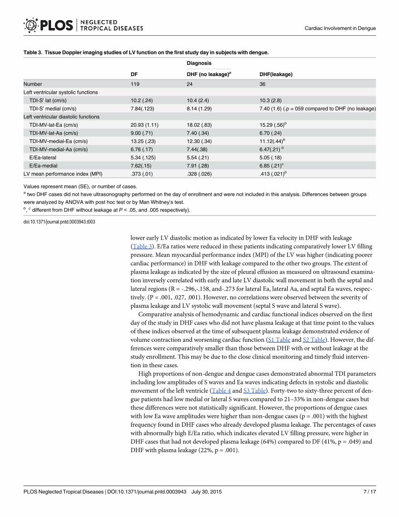
lower early LV diastolic motion as indicated by lower Ea velocity in DHF with leakage(Table 3). E/Ea ratios were reduced in these patients indicating comparatively lower LV fillingpressure. Mean myocardial performance index (MPI) of the LV was higher (indicating poorercardiac performance) in DHF with leakage compared to the other two groups. The extent ofplasma leakage as indicated by the size of pleural effusion as measured on ultrasound examina-tion inversely correlated with early and late LV diastolic wall movement in both the septal andlateral regions (R = -.296,-.158, and-.273 for lateral Ea, lateral Aa, and septal Ea waves, respec-tively. (P = .001, .027, .001). However, no correlations were observed between the severity ofplasma leakage and LV systolic wall movement (septal S wave and lateral S wave).
Comparative analysis of hemodynamic and cardiac functional indices observed on the firstday of the study in DHF cases who did not have plasma leakage at that time point to the valuesof these indices observed at the time of subsequent plasma leakage demonstrated evidence ofvolume contraction and worsening cardiac function (S1 Table and S2 Table). However, the dif-ferences were comparatively smaller than those between DHF with or without leakage at thestudy enrollment. This may be due to the close clinical monitoring and timely fluid interven-tion in these cases.
High proportions of non-dengue and dengue cases demonstrated abnormal TDI parametersincluding low amplitudes of S waves and Ea waves indicating defects in systolic and diastolicmovement of the left ventricle (Table 4 and S3 Table). Forty-two to sixty-three percent of den-gue patients had low medial or lateral S waves compared to 21–33% in non-dengue cases butthese differences were not statistically significant. However, the proportions of dengue caseswith low Ea wave amplitudes were higher than non-dengue cases (p = .001) with the highestfrequency found in DHF cases who already developed plasma leakage. The percentages of caseswith abnormally high E/Ea ratio, which indicates elevated LV filling pressure, were higher inDHF cases that had not developed plasma leakage (64%) compared to DF (41%, p = .049) andDHF with plasma leakage (22%, p = .001).
Table 3. Tissue Doppler imaging studies of LV function on the first study day in subjects with dengue.
Diagnosis
DF DHF (no leakage)a DHF(leakage)
Number 119 24 36
Left ventricular systolic functions
TDI-S’ lat (cm/s) 10.2 (.24) 10.4 (2.4) 10.3 (2.8)
TDI-S’ medial (cm/s) 7.84(.123) 8.14 (1.29) 7.40 (1.6) (.p = 059 compared to DHF (no leakage)
Left ventricular diastolic functions
TDI-MV-lat-Ea (cm/s) 20.93 (1.11) 18.02 (.83) 15.29 (.56)b
TDI-MV-lat-Aa (cm/s) 9.00 (.71) 7.40 (.34) 6.70 (.24)
TDI-MV-medial-Ea (cm/s) 13.25 (.23) 12.30 (.34) 11.12(.44)b
TDI-MV-medial-Aa (cm/s) 6.76 (.17) 7.44(.38) 6.47(.21) b
E/Ea-lateral 5.34 (.125) 5.54 (.21) 5.05 (.18)
E/Ea-medial 7.62(.15) 7.91 (.28) 6.85 (.21)c
LV mean performance index (MPI) .373 (.01) .328 (.026) .413 (.021)b
Values represent mean (SE), or number of cases.a two DHF cases did not have ultrasonography performed on the day of enrollment and were not included in this analysis. Differences between groups
were analyzed by ANOVA with post hoc test or by Man Whitney’s test.b, c different from DHF without leakage at P < .05, and .005 respectively).
doi:10.1371/journal.pntd.0003943.t003
Cardiac Involvement in Dengue
PLOS Neglected Tropical Diseases | DOI:10.1371/journal.pntd.0003943 July 30, 2015 7 / 17

Changes in hemodynamic status, cardiac function, and plasma leakageover the course of illnessFig 1 shows the patterns of hemodynamic and volume indices and fluid intake over the courseof the illness of DF and DHF cases. DHF cases had higher heart rate compared to DF earlyin the course of illness and this persisted throughout the acute illness (A). There was no differ-ence in systolic blood pressure (B), but mean diastolic pressures were higher in DHF casesaround the day of fever resolution (fever day 0) and shortly thereafter (C). This coincided withdecreased intravascular volume as indicated by a decrease in the average IVC diameter in DHFcases (D). DHF cases had lower CI (E) at defervescence (fever day 0) and increased SVR (F) onfever day 0 and +1 compared to DF cases. Ultrasonography revealed a gradual increase in thepercentage of cases with plasma leakage, with peak incidence on fever day +1 (G). Patientswith DHF also received more fluid compared to those with DF starting on fever day 0 andafterward.
Evaluation of LV systolic and diastolic functions revealed lower EFs (Fig 2A) on fever day -1and 0, and lower early component of LV inflow (MV-E wave) on fever days 0 and +1 (Fig 2B)in DHF compared to DF. The late component LV inflow was also decreased in DHF on feverday +1 (Fig 2C). Seventeen dengue cases had at least one abnormal EF (<56%) detected duringthe illness. They represented 7%, 9%, 15%, and 32% of DF, DHF grade I, II, and DHF Gr III/IVcases, respectively. The relative frequencies of abnormal EFs were higher in DHF grade III/IVcompared to DHF grade I/II and DF (p< .001, Chi-square). The majority of cases (67%) hadlow EF detected on only a single day, usually fever day 0 or +1. The EFs on the day of dischargewere improved compared to the lowest EF. Taken together, this indicates that changes in
Table 4. Frequencies of cases with abnormal TDI findings on the first day of the study.
Clinical classification (%)a
TDI parameters Non-dengue DF DHF without leakage DHF with leakage
Low S medial 33 47 45 66
Low S lateral 21 46 42 44
Low Ea medial 30 52 b 59b 80b,c
Low Ea lateral 14 28 42 d 64 d,e
Low Aa medial 18 25 27 36
Low Aa lateral 3 f,g 21 29 39h
High E/Ea 44 41 64 i, j, k 22
a, Percentages of cases with abnormal TDI findings were calculated from the numbers of records with abnormal tracings divided by the numbers of all
records with analyzable tracings and multiplied by one hundred. The numbers of records with abnormal tracings and the number of analyzable records for
each parameters and diagnostic category are shown in S3 Table. Two DHF cases did not have ultrasonography performed on the day of enrollment and
were not included in this analysis.
b, different from non-dengue cases (p = .033)
c, different from DF (p = .001)
d, different from non-dengue (p = .001)
e, different from DF (p = .001)
f, different from DF (p = .013)
g, different from DHF with or without leakage, (p = .004)
h, different from DF (p = .045)
i, different from non-dengue (p = .045)
j, different from DF (p = .049)
k, different from DHF with leakage (p = .001)
doi:10.1371/journal.pntd.0003943.t004
Cardiac Involvement in Dengue
PLOS Neglected Tropical Diseases | DOI:10.1371/journal.pntd.0003943 July 30, 2015 8 / 17

hemodynamic status in DHF were temporally associated with plasma leakage and were charac-terized by a compensatory autonomic response to contracted intravascular volume which wascorrected by fluid replacement.
Tissue Doppler assessment of cardiac wall motionTDI of the MV showed slower septal movement during systole in DHF compared to DF onfever day +1 (p = .031) (Fig 3B). Early and late diastolic LV annulus motions (Ea and Aa wave)were lower in DHF cases compared to DF cases at the end of the febrile period and after
Fig 1. Hemodynamic status and plasma leakage in dengue by fever day.Heart rate (A), systolic blood pressure (B), diastolic blood pressure (C), inferiorvena cava diameter (D), mean cardiac index (E), systemic vascular resistance (F), frequencies of cases with a pleural effusion or ascites (G), and daily fluidintake (H) of dengue fever (dotted line) and dengue hemorrhagic fever cases (solid line) over the course of the illness. Fever day 0 denotes day ofdefervescence. * different from dengue fever cases (P < .05). The numbers of cases were: fever-day -3 (DF 4, DHF 5), fever-day-2 (DF 13, DHF 9), fever-day-1(DF 43, DHF 26), fever-day-0(DF 104, DHF 56), fever-day+1(DF 119, DHF 62), fever-day+2(DF 70, DHF 60), fever-day+3(DF 13, DHF 38).
doi:10.1371/journal.pntd.0003943.g001
Cardiac Involvement in Dengue
PLOS Neglected Tropical Diseases | DOI:10.1371/journal.pntd.0003943 July 30, 2015 9 / 17

(Fig 3D, 3E and 3F). The decreased early diastolic annulus motion was more pronounced inthe septal area. The E/Ea ratio, which indicates LV filling pressure, was not different betweenDHF and DF cases on most days except on fever day +1 when the average E/Ea ratio was signif-icantly lower in DHF cases (I).
Twenty to seventy percent of dengue cases had abnormally low diastolic and systolic wallmotion during the course of the illness (Fig 4A–4D). The frequencies of abnormally low dia-stolic wall motion were higher in DHF compared to DF on fever day 0 and after (Fig 4C and4D). Elevated E/Ea ratios were found in both DF and DHF cases with similar frequencies.None had E/Ea ratio above 15.
Troponin and CPK-MB levels were similar between dengue and non-dengue cases on theday of study enrollment. Plasma levels of cardiac troponin-T were low to non-detectable inmost cases with comparable levels found in DF and DHF cases throughout the acute illnessperiod. Consistent with this finding, the mean levels of CPK-MB were similar between DF andDHF. However, 14.5% of DHF cases had troponin-T levels>30 ng/L at any time point com-pared to 5% in DF (p = 0.028, Chi-square test). All of these cases had only one sample with ele-vated troponin-T levels during the course of illness. These elevated troponin levels weredetected later in the course of the illness (Fig 5). There were no cases with clinical heart failureor conductive defects consistent with clinical myocarditis.
Pericardial effusion was detected in 7 DHF cases. The size of the effusion was minimal in 6cases and substantial in 1 case. Sinus bradycardia was documented in 1 DF case. Four cases ofmild mitral regurgitation (2 DF and 2 DHF) and 2 DF cases with tricuspid regurgitation werefound. These findings were transient and not clinically significant.
Twelve patients (4 DF, 8 DHF) had a follow up examination performed at early convales-cence (< 1 week after discharge). Trends toward improvement were found for EF and LVinflow at the MV (MV-E) and diastolic medial LV wall movement (medial Ea wave) in DHFcases at convalescence compared to fever day +1 (Table 5).
DiscussionMuch of the literature on cardiac manifestations in dengue consists of case reports and smallcase series. More recent prospective studies have reported varied incidence of abnormal cardiac
Fig 2. Left ventricular functions in dengue by fever day. Ejection fraction (A), early diastolic LV inflow flow (B), and late diastolic LV inflow (C) of denguefever (dotted line) and dengue hemorrhagic fever cases (solid line) over the course of the illness. Fever day 0 denotes day of defervescence. * different fromdengue fever (P < .05). The numbers of cases were: fever-day -3 (DF 4, DHF 5), fever-day-2 (DF 13, DHF 9), fever-day-1(DF 43, DHF 26), fever-day-0(DF104, DHF 56), fever-day+1(DF 119, DHF 62), fever-day+2(DF 70, DHF 60), fever-day+3(DF 13, DHF 38).
doi:10.1371/journal.pntd.0003943.g002
Cardiac Involvement in Dengue
PLOS Neglected Tropical Diseases | DOI:10.1371/journal.pntd.0003943 July 30, 2015 10 / 17

findings from approximately 15–27% for myocarditis [8, 24, 25] and up to 40% for functionalabnormalities[26]. Cardiac performance and hemodynamic status are affected by intravascularvolume, cardiac functions and autonomic response. The present study extends our knowledgeof cardiac involvement in dengue in several ways. We obtained daily echocardiographic studiesover the critical phase of illness in a cohort that included both milder and more severe illness,and analyzed cardiac function data in conjunction with serial assessments of intravascular vol-ume status and plasma leakage. The data show that cardiac functional abnormalities are com-mon in dengue and correlate with disease severity. However, these abnormalities weretransient, did not require specific treatment, and were not accompanied by evidence of struc-tural damage to the myocardium.
Our study found that cardiac functional abnormalities were related to the severity of plasmaleakage. Decreased EF, CI, LV diastolic inflow, and the elevated SVR in DHF are therefore
Fig 3. Tissue Doppler image of LV functions by fever day. Left ventricular lateral (A) and septal S wave (B), LVmean performance index (MPI) (C), lateral(D) and septal (E) annulus movement during early diastole, lateral (F) and septal (G) annulus movement during late diastole, and lateral (H) and medial (I) E/Earatios of dengue fever (dotted line) and dengue hemorrhagic fever (solid line) cases over the course of the illness. Fever day 0 denotes day of defervescence.* different from DF (P < .05). The numbers of cases were: fever-day -3 (DF 4, DHF 5), fever-day-2 (DF 13, DHF 9), fever-day-1(DF 43, DHF 26), fever-day-0(DF104, DHF 56), fever-day+1(DF 119, DHF 62), fever-day+2(DF 70, DHF 60), fever-day+3(DF 13, DHF 38).
doi:10.1371/journal.pntd.0003943.g003
Cardiac Involvement in Dengue
PLOS Neglected Tropical Diseases | DOI:10.1371/journal.pntd.0003943 July 30, 2015 11 / 17

likely to be affected by contracted intravascular volume. TDI indices, which are less preloaddependent [27, 28], also differed between dengue and non-dengue cases, and between DF and
Fig 4. Frequencies of abnormal TDI during the course of the illness. Percentages of DF (dotted lines) and DHF cases (solid lines) with low septal andlateral S wave (A, B) and septal and lateral Ea wave (C, D) and high E/Ea ratios (E) determined based on published age-specific normal values (21).* different from DF (P < .05).
doi:10.1371/journal.pntd.0003943.g004
Fig 5. Plasma troponin-T levels in individual DF (white circles) or DHF cases (black circles) during thecourse of the illness. The horizontal line indicates a cut-off for an abnormal value at 30 pg/ml.
doi:10.1371/journal.pntd.0003943.g005
Cardiac Involvement in Dengue
PLOS Neglected Tropical Diseases | DOI:10.1371/journal.pntd.0003943 July 30, 2015 12 / 17

DHF. This was most evident in decreased LV wall movement during early diastole indicating arelaxation defect. The higher frequencies of cases with decreased LV early diastolic wall move-ment in DF compared to non-dengue cases suggests a dengue specific process independent ofplasma leakage. However, the correlation between abnormal LV diastolic movement and theextent of plasma leakage among dengue cases suggests that plasma leakage may also directlycontribute to this functional abnormality by reducing intravascular volume. Alternatively, plasmaleakage may be a correlate of the disease mechanism that affects cardiac function. The relativelysimilar E/Ea ratios in DF and DHF despite lower E in DHF reflect this diastolic defect. Thepathology underlying this finding is unknown. Nevertheless, the abnormal LV relaxation mayfurther compromise LV filling. Decreased LV relaxation may also contribute to high LV fillingpressure when intravascular volume has been restored by intravenous fluid treatment and byreabsorption of effusion fluid, and may lead to the pulmonary edema observed in some DHFcases. The prevalence of subclinical diastolic dysfunction has been reported to increase with age,in individuals with hypertension, LV hypertrophy, and diabetes [29–31]. This has importantimplications for clinical care considering the shift in the demographics of dengue cases to adultswho may have these common co-morbidities. Further studies in adults with dengue will beneeded to address this question.
Fatal dengue associated with myocardial injury and inflammation has been reported. In astudy of adult and pediatric cases from Brazil, the incidence of myocarditis on the basis of clini-cal diagnosis or elevated biomarkers was approximately 15% [24]. A subset of these cases hadabnormal echocardiography or MRI [8, 24]. Viral antigen has been detected in cardiac myo-cytes, monocytes, and endothelial cells by immunostaining [7, 8, 32]. Contrary to these reports,we did not observe cases with myocarditis based on clinical information and cardiac enzymes.Our findings are consistent with other reports in pediatric dengue cases in Southeast Asia [26,33]. The difference in the incidence of myocarditis in various reports may be related to hostgenetic factors and locally circulating viruses, which may influence tissue tropism and hostinflammatory responses. In addition, different temporal patterns of circulating serotypes maylead to distinct dengue immune status that predisposes individuals for more severe cardiacmanifestation during a secondary infection. However, differences in age groups, co-morbidi-ties, and study design may also contribute to differences in manifestations and incidence
Table 5. Comparison between cardiac function at fever day +1 and early convalescence.
DF DHF
feverday+1 earlyconvalescence
feverday+1 earlyconvalescence
average (SEM) average (SEM) aP average (SEM) average (SEM) aP
EF (%) 70.88 4.29 71.03 1.01 0.967 66.46 2.58 72.48 1.23 0.075
MV-E (cm/s)
99.90 11.50 98.35 11.02 0.414 83.63 8.86 98.93 6.32 0.049
MV Lat Ea(cm/s)
21.68 1.53 20.03 1.77 0.472 18.01 1.94 22.90 2.18 0.184
MV MedEa (cm/s)
13.98 0.55 14.08 1.41 0.937 11.73 0.61 14.18 0.36 0.014
Lat E/Earatio
4.57 0.27 4.99 0.68 0.442 4.94 0.25 4.50 0.20 0.538
Med E/Earatio
7.24 1.04 7.41 1.49 0.858 7.11 0.16 7.0 0.09 0.794
a P value comparing values at fever day +1 and early convalescence (Student’s paired t-test).
doi:10.1371/journal.pntd.0003943.t005
Cardiac Involvement in Dengue
PLOS Neglected Tropical Diseases | DOI:10.1371/journal.pntd.0003943 July 30, 2015 13 / 17

Although sustained elevated enzyme levels were not found, transient increased levels werefound in some cases, particularly in DHF, which may indicate subtle myocardial injury in thesecases.
Cardiac dysfunction has been reported in other viral hemorrhagic fevers such as Crimean-Congo hemorrhagic fever and Puumala virus infection [34–36]. The patterns of cardiacinvolvement are similar to dengue and are characterized by transient functional impairmentand normal cardiac enzyme levels. Another similarity is more severe involvement in adults.These findings suggest common pathophysiologic pathways including endothelial cell activa-tion, perturbation of vascular permeability, and cardiac muscle cell dysfunction. Cardiac dys-function is also common in sepsis in which TNF-α and nitric oxide are considered to beinvolved [37, 38]. These mediators have been reported to be altered in dengue as well [39–41].These mediators may alter cardiac function and hemodynamic status by their permeabilityenhancing effects, which lead to decreased preload, as well as direct effects on cardiac myocytesand on vascular resistance. The relative contribution of these mediators will need to be furtheraddressed in relevant animal models.
Declining heart rate during defervescence has been observed in dengue and attributed toincreased parasympathetic activity[42]. Benign bradyarrhythmias and ectopic beats have beenreported in patients with DENV infection following defervescence and were not related to dis-ease severity[43]. The prevalence of cardiac arrhythmia in the present study was lower thanpreviously reported. We did not perform continuous electrocardiographic monitoring andtherefore some cases might have been missed. Another limitation was the lack of adult casesthat may exhibit distinct cardiac manifestations. Due to the conservative use of intravenousfluid there were no cases with pulmonary edema and fluid overload in this series. However, therelatively normal or elevated E/Ea ratio in DHF cases in the presence of contracted intravascu-lar volume suggests that diastolic dysfunction may predispose some DHF cases to pulmonaryedema with aggressive administration of intravenous fluid.
In summary, functional cardiac abnormalities in dengue involved both systolic and diastolicfunctions and correlated with the severity of plasma leakage. Cardiac structural changes suchas infarction and myocarditis were not likely the underlying mechanism. Cardiac dysfunctionwas transient and did not require specific treatment. Decreased LV wall diastolic movementmay contribute to abnormal LV filling and the predisposition for pulmonary edema in DHF.These findings underscore the importance of vigilance in fluid management and hemodynamicstatus monitoring in the treatment of dengue.
Supporting InformationS1 Checklist. STROBE checklist.(DOCX)
S1 Table. Laboratory findings and cardiac function measurements at study enrollment ofDHF cases without plasma leakage and the subsequent findings on day of plasma leakage.(DOCX)
S2 Table. Tissue Doppler imaging values at study enrollment of DHF cases without plasmaleakage and the subsequent findings on day of plasma leakage.(DOCX)
S3 Table. Numbers of cases with abnormal TDI findings on the first day of the study.(DOCX)
Cardiac Involvement in Dengue
PLOS Neglected Tropical Diseases | DOI:10.1371/journal.pntd.0003943 July 30, 2015 14 / 17

AcknowledgmentsWe thank Dr. Jennifer Friedman for reviewing the manuscript. We thank the arbovirology andmolecular sections of the Armed Forces Research Institute of Medical Sciences for diagnostictesting; doctors and nurses of Queen Sirikit National Institute of Child Health and the staff ofthe Armed Forces Research Institute of Medical Sciences for patient care and samplecollection.
Author ContributionsConceived and designed the experiments: AS SG ALR FAE. Performed the experiments: ASTK SW SK. Analyzed the data: AS ALR RVG IKY SW. Wrote the paper: AS TK SG ALR SJTIKY RVG SK FAE SW.
References1. Gubler D, Kuno G, Markoff L. Flavivirus, Field's Virology. 5th ed. Knipe David M, Howly P, editors: Lip-
pincott Williams &Wilkins; 2007. 1154–61 p.
2. Dengue Hemorrhagic Fever: diagnosis, treatment, prevention and control. 2nd ed. World Health Orga-nization; 1997.
3. Dengue, guidelines for diagnosis, treatment, prevention and control: World Health Organization; 2009.
4. Sam SS, Omar SF, Teoh BT, Abd-Jamil J, AbuBakar S. Review of Dengue hemorrhagic fever fatalcases seen among adults: a retrospective study. PLoS Negl Trop Dis. 2013; 7(5):e2194. Epub 2013/05/10. doi: 10.1371/journal.pntd.0002194 PNTD-D-12-00967 [pii]. PMID: 23658849; PubMed CentralPMCID: PMC3642057.
5. Miranda CH, Borges Mde C, Schmidt A, Pazin-Filho A, Rossi MA, Ramos SG, et al. A case presenta-tion of a fatal dengue myocarditis showing evidence for dengue virus-induced lesion. Eur Heart J AcuteCardiovasc Care. 2013; 2(2):127–30. Epub 2013/11/14. doi: 10.1177/2048872613475889 [pii]. PMID:24222821; PubMed Central PMCID: PMC3821802.
6. Marques N, Gan VC, Leo YS. Dengue myocarditis in Singapore: two case reports. Infection. 2013; 41(3):709–14. Epub 2013/01/02. doi: 10.1007/s15010-012-0392-9 PMID: 23277366.
7. Weerakoon KG, Kularatne SA, Edussuriya DH, Kodikara SK, Gunatilake LP, Pinto VG, et al. Histopath-ological diagnosis of myocarditis in a dengue outbreak in Sri Lanka, 2009. BMC Res Notes. 2011;4:268. Epub 2011/07/30. doi: 1756-0500-4-268 [pii] doi: 10.1186/1756-0500-4-268 PMID: 21798066;PubMed Central PMCID: PMC3160397.
8. Salgado DM, Eltit JM, Mansfield K, Panqueba C, Castro D, Vega MR, et al. Heart and skeletal muscleare targets of dengue virus infection. Pediatr Infect Dis J. 2010; 29(3):238–42. Epub 2009/12/25. doi:10.1097/INF.0b013e3181bc3c5b PMID: 20032806; PubMed Central PMCID: PMC2833338.
9. Lee CH, Teo C, Low AF. Fulminant dengue myocarditis masquerading as acute myocardial infarction.Int J Cardiol. 2009; 136(3):e69–71. Epub 2008/08/15. doi: S0167-5273(08)00733-X [pii] doi: 10.1016/j.ijcard.2008.05.023 PMID: 18701172.
10. PromphanW, Sopontammarak S, Pruekprasert P, Kajornwattanakul W, Kongpattanayothin A. Denguemyocarditis. Southeast Asian J Trop Med Public Health. 2004; 35(3):611–3. Epub 2005/02/04. PMID:15689075.
11. Sengupta SP, Nugurwar A, Jaju R, Khandheria BK. Left ventricular myocardial performance in patientswith dengue hemorrhagic fever and thrombocytopenia as assessed by two-dimensional speckle track-ing echocardiography. Indian Heart J. 2013; 65(3):276–82. Epub 2013/07/03. doi: S0019-4832(13)00106-5 [pii]doi: 10.1016/j.ihj.2013.04.017 PMID: 23809381; PubMed Central PMCID: PMC3860609.
12. Yacoub S, Griffiths A, Chau TT, Simmons CP, Wills B, Hien TT, et al. Cardiac function in Vietnamesepatients with different dengue severity grades. Crit Care Med. 2012; 40(2):477–83. Epub 2011/09/29.doi: 10.1097/CCM.0b013e318232d966 PMID: 21946658.
13. Khongphatthanayothin A, Lertsapcharoen P, Supachokchaiwattana P, La-Orkhun V, Khumtonvong A,Boonlarptaveechoke C, et al. Myocardial depression in dengue hemorrhagic fever: prevalence and clin-ical description. Pediatr Crit Care Med. 2007; 8(6):524–9. Epub 2007/10/02. doi: 10.1097/01.PCC.0000288672.77782.D4 PMID: 17906598.
14. Khongphatthanayothin A, Suesaowalak M, Muangmingsook S, Bhattarakosol P, Pancharoen C. Hemo-dynamic profiles of patients with dengue hemorrhagic fever during toxic stage: an echocardiographicstudy. Intensive Care Med. 2003; 29(4):570–4. Epub 2003/02/22. doi: 10.1007/s00134-003-1671-9PMID: 12595978.
Cardiac Involvement in Dengue
PLOS Neglected Tropical Diseases | DOI:10.1371/journal.pntd.0003943 July 30, 2015 15 / 17

15. Lee IK, LeeWH, Yang KD, Liu JW. Comparison of the effects of oral hydration and intravenous fluidreplacement in adult patients with non-shock dengue hemorrhagic fever in Taiwan. Trans R Soc TropMed Hyg. 2010; 104(8):541–5. Epub 2010/07/02. doi: S0035-9203(10)00112-4 [pii] doi: 10.1016/j.trstmh.2010.05.003 PMID: 20591457.
16. Nguyen TH, Nguyen TL, Lei HY, Lin YS, Le BL, Huang KJ, et al. Volume replacement in infants withdengue hemorrhagic fever/dengue shock syndrome. Am J Trop Med Hyg. 2006; 74(4):684–91. Epub2006/04/12. doi: 74/4/684 [pii]. PMID: 16607006.
17. Premaratna R, Liyanaarachchi E, Weerasinghe M, de Silva HJ. Should colloid boluses be prioritizedover crystalloid boluses for the management of dengue shock syndrome in the presence of ascites andpleural effusions? BMC Infect Dis. 2011; 11:52. Epub 2011/03/02. doi: 1471-2334-11-52 [pii] doi: 10.1186/1471-2334-11-52 PMID: 21356095; PubMed Central PMCID: PMC3056793.
18. Innis BL, Nisalak A, Nimmannitya S, Kusalerdchariya S, Chongswasdi V, Suntayakorn S, et al. Anenzyme-linked immunosorbent assay to characterize dengue infections where dengue and Japaneseencephalitis co-circulate. Am J Trop Med Hyg. 1989; 40(4):418–27. PMID: 2540664.
19. Lanciotti RS, Calisher CH, Gubler DJ, Chang GJ, Vorndam AV. Rapid detection and typing of dengueviruses from clinical samples by using reverse transcriptase-polymerase chain reaction. J Clin Micro-biol. 1992; 30(3):545–51. Epub 1992/03/01. PMID: 1372617; PubMed Central PMCID: PMC265106.
20. Srikiatkhachorn A, Krautrachue A, Ratanaprakarn W, Wongtapradit L, Nithipanya N, Kalayanarooj S,et al. Natural history of plasma leakage in dengue hemorrhagic fever: a serial ultrasonographic study.Pediatr Infect Dis J. 2007; 26(4):283–90; discussion 91–2. Epub 2007/04/07. doi: 10.1097/01.inf.0000258612.2674310 00006454-200704000-00002 [pii]. PMID: 17414388.
21. Lopez L, Colan SD, Frommelt PC, Ensing GJ, Kendall K, Younoszai AK, et al. Recommendations forquantification methods during the performance of a pediatric echocardiogram: a report from the Pediat-ric Measurements Writing Group of the American Society of Echocardiography Pediatric and Congeni-tal Heart Disease Council. J Am Soc Echocardiogr. 2010; 23(5):465–95; quiz 576–7. Epub 2010/05/11.doi: S0894-7317(10)00266-X [pii] doi: 10.1016/j.echo.2010.03.019 PMID: 20451803.
22. Mor-Avi V, Lang RM, Badano LP, Belohlavek M, Cardim NM, Derumeaux G, et al. Current and evolvingechocardiographic techniques for the quantitative evaluation of cardiac mechanics: ASE/EAE consen-sus statement on methodology and indications endorsed by the Japanese Society of Echocardiogra-phy. J Am Soc Echocardiogr. 2011; 24(3):277–313. Epub 2011/02/23. doi: S0894-7317(11)00048-4[pii] doi: 10.1016/j.echo.2011.01.015 PMID: 21338865.
23. Eidem BW, McMahon CJ, Cohen RR, Wu J, Finkelshteyn I, Kovalchin JP, et al. Impact of cardiacgrowth on Doppler tissue imaging velocities: a study in healthy children. J Am Soc Echocardiogr. 2004;17(3):212–21. Epub 2004/02/26. doi: 10.1016/j.echo.2003.12.005 S089473170301085X [pii]. PMID:14981417.
24. Miranda CH, Borges Mde C, Matsuno AK, Vilar FC, Gali LG, Volpe GJ, et al. Evaluation of cardiacinvolvement during dengue viral infection. Clin Infect Dis. 2013; 57(6):812–9. Epub 2013/06/21. doi:cit403 [pii] doi: 10.1093/cid/cit403 PMID: 23784923.
25. Wichmann D, Kularatne S, Ehrhardt S, Wijesinghe S, Brattig NW, Abel W, et al. Cardiac involvement indengue virus infections during the 2004/2005 dengue fever season in Sri Lanka. Southeast Asian JTrop Med Public Health. 2009; 40(4):727–30. Epub 2009/10/22. PMID: 19842405.
26. Yacoub S, Wertheim H, Simmons CP, Screaton G, Wills B. Cardiovascular manifestations of theemerging dengue pandemic. Nat Rev Cardiol. 2014; 11(6):335–45. Epub 2014/04/09. doi: nrcar-dio.2014.40 [pii] doi: 10.1038/nrcardio.2014.40 PMID: 24710495.
27. Oki T, Tabata T, Yamada H, Wakatsuki T, Shinohara H, Nishikado A, et al. Clinical application of pulsedDoppler tissue imaging for assessing abnormal left ventricular relaxation. Am J Cardiol. 1997; 79(7):921–8. Epub 1997/04/01. doi: S0002914997000155 [pii]. PMID: 9104907.
28. Sohn DW, Chai IH, Lee DJ, Kim HC, Kim HS, Oh BH, et al. Assessment of mitral annulus velocity byDoppler tissue imaging in the evaluation of left ventricular diastolic function. J Am Coll Cardiol. 1997; 30(2):474–80. Epub 1997/08/01. doi: S0735-1097(97)88335-0 [pii]. PMID: 9247521.
29. AbhayaratnaWP, Marwick TH, Smith WT, Becker NG. Characteristics of left ventricular diastolic dys-function in the community: an echocardiographic survey. Heart. 2006; 92(9):1259–64. Epub 2006/02/21. doi: hrt.2005.080150 [pii] doi: 10.1136/hrt.2005.080150 PMID: 16488928; PubMed Central PMCID:PMC1861192.
30. Redfield MM, Jacobsen SJ, Burnett JC Jr., Mahoney DW, Bailey KR, Rodeheffer RJ. Burden of systolicand diastolic ventricular dysfunction in the community: appreciating the scope of the heart failure epi-demic. JAMA. 2003; 289(2):194–202. Epub 2003/01/09. doi: joc21616 [pii]. PMID: 12517230.
31. Wan SH, Vogel MW, Chen HH. Pre-clinical diastolic dysfunction. J Am Coll Cardiol. 2014; 63(5):407–16. Epub 2013/12/03. doi: S0735-1097(13)06160-3 [pii] doi: 10.1016/j.jacc.2013.10.063 PMID:24291270; PubMed Central PMCID: PMC3934927.
Cardiac Involvement in Dengue
PLOS Neglected Tropical Diseases | DOI:10.1371/journal.pntd.0003943 July 30, 2015 16 / 17

32. Povoa TF, Alves AM, Oliveira CA, Nuovo GJ, Chagas VL, Paes MV. The pathology of severe denguein multiple organs of human fatal cases: histopathology, ultrastructure and virus replication. PLoS One.2014; 9(4):e83386. Epub 2014/04/17. doi: 10.1371/journal.pone.0083386 PONE-D-13-18463 [pii].PMID: 24736395; PubMed Central PMCID: PMC3987999.
33. Khongphatthanayothin A, Lertsapcharoen P, Supachokchaiwattana P, Satupan P, Thongchaiprasit K,Poovorawan Y, et al. Hepatosplanchnic circulatory dysfunction in acute hepatic infection: the case ofdengue hemorrhagic fever. Shock. 2005; 24(5):407–11. Epub 2005/10/26. doi: 00024382-200511000-00002 [pii]. PMID: 16247324.
34. Engin A, Yilmaz MB, Elaldi N, Erdem A, Yalta K, Tandogan I, et al. Crimean-Congo hemorrhagic fever:does it involve the heart? Int J Infect Dis. 2009; 13(3):369–73. Epub 2008/11/05. doi: S1201-9712(08)01492-6 [pii] doi: 10.1016/j.ijid.2008.07.019 PMID: 18980852.
35. Makela S, Kokkonen L, Ala-Houhala I, Groundstroem K, Harmoinen A, Huhtala H, et al. More than halfof the patients with acute Puumala hantavirus infection have abnormal cardiac findings. Scand J InfectDis. 2009; 41(1):57–62. Epub 2008/10/22. doi: 904383584 [pii] doi: 10.1080/00365540802502629PMID: 18932105.
36. Peters CJ, Liu CT, Anderson GW Jr., Morrill JC, Jahrling PB. Pathogenesis of viral hemorrhagic fevers:Rift Valley fever and Lassa fever contrasted. Rev Infect Dis. 1989; 11 Suppl 4:S743–9. Epub 1989/05/01. PMID: 2665011.
37. Flynn A, ChokkalingamMani B, Mather PJ. Sepsis-induced cardiomyopathy: a review of pathophysio-logic mechanisms. Heart Fail Rev. 2010; 15(6):605–11. Epub 2010/06/24. doi: 10.1007/s10741-010-9176-4 PMID: 20571889.
38. Zanotti-Cavazzoni SL, Hollenberg SM. Cardiac dysfunction in severe sepsis and septic shock. CurrOpin Crit Care. 2009; 15(5):392–7. Epub 2009/07/28. doi: 10.1097/MCC.0b013e3283307a4e PMID:19633546.
39. Hober D, Poli L, Roblin B, Gestas P, Chungue E, Granic G, et al. Serum levels of tumor necrosis factor-alpha (TNF-alpha), interleukin-6 (IL-6), and interleukin-1 beta (IL-1 beta) in dengue-infected patients.Am J Trop Med Hyg. 1993; 48(3):324–31. Epub 1993/03/01. PMID: 8470771.
40. Trairatvorakul P, Chongsrisawat V, Ngamvasinont D, Asawarachun D, Nantasook J, Poovorawan Y.Serum nitric oxide in children with dengue infection. Asian Pac J Allergy Immunol. 2005; 23(2–3):115–9. Epub 2005/10/29. PMID: 16252841.
41. Valero N, Espina LM, Anez G, Torres E, Mosquera JA. Short report: increased level of serum nitricoxide in patients with dengue. Am J Trop Med Hyg. 2002; 66(6):762–4. Epub 2002/09/13. PMID:12224588.
42. Carter R 3rd, Hinojosa-Laborde C, Convertino VA. Heart rate variability in patients being treated fordengue viral infection: new insights frommathematical correction of heart rate. Front Physiol. 2014;5:46. Epub 2014/03/13. doi: 10.3389/fphys.2014.00046 PMID: 24611050; PubMed Central PMCID:PMC3933783.
43. La-Orkhun V, Supachokchaiwattana P, Lertsapcharoen P, Khongphatthanayothin A. Spectrum of car-diac rhythm abnormalities and heart rate variability during the convalescent stage of dengue virus infec-tion: a Holter study. Ann Trop Paediatr. 2011; 31(2):123–8. Epub 2011/05/18. doi: 10.1179/1465328111Y.0000000008 PMID: 21575316.
Cardiac Involvement in Dengue
PLOS Neglected Tropical Diseases | DOI:10.1371/journal.pntd.0003943 July 30, 2015 17 / 17