Evaluation of Breast Specimens after Neoadjuvant...
83
Evaluation of Breast Specimens after Neoadjuvant Chemotherapy A.A. Sahin, M.D. Professor of Pathology and Translation Molecular Pathology Section Chief of Breast Pathology
Transcript of Evaluation of Breast Specimens after Neoadjuvant...

Evaluation of Breast Specimens after Neoadjuvant Chemotherapy
A.A. Sahin, M.D.
Professor of Pathology and Translation Molecular Pathology
Section Chief of Breast Pathology

• Management of locally advanced invasive breast ca including inflammatory breast ca
• ‘Down-staging’ of large inoperable cancers to permit surgical resection
• Routine management of women with high risk disease who would require adjuvant chemotherapy based on biological tumorcharacteristics and clinical-radiological findings
Indications of Neoadjuvant Chemotherapy

Advantages of Neoadjuvant Therapy
• Potential for tumor down sizing with an increase in the rate of breast conserving surgery
• Ability to monitor treatment response and tailor subsequent locoregional and systemic therapy - more individualized patient care• Patients with pCR may not benefit from further regional
therapy such as adjuvant radiotherapy
• Patients with poor response can be identified and entered into trials of novel targeted agents

Advantages of Neoadjuvant Therapy
• Evaluation of treatment response to new
agents using pathological complete
response (pCR) as a surrogate marker of
outcome
• Neoadjuvant studies smaller, cheaper,
faster results

Federal Register 2014; 79 (194): 60476–77
Pre Treatment Evaluation - Breast
• Pre treatment breast CNB must be adequate for unequivocal diagnosis of invasive carcinoma and assessment of key prognostic/predictive factors
– Histological type and grade
– ER/ PR /HER2 status
– Other biomarkers- Ki67, multigene assays
• If multiple lesions biopsy of at least 2 foci is advised to confirm multifocality and look for heterogeneity
• Additional biopsies for translational research studies

Pre Treatment Evaluation - Axilla
• Routine axillary U/S with histological assessment of abnormal nodes by CNB or FNA
• Pre-treatment SLNB not advised unless positive result will influence decision to give chemotherapy
• Nodal response is an important prognostic factor independent of response in the breast
Specimen Handling
• One of the most critical steps in accurately evaluate response to NAC is the macroscopic (gross) assessment of the specimen
• A multidisciplinary approach with close clinical/radiological correlation to map the precise location of the tumor bed is preferable to exhaustive blind sampling
Specimen Handling
• To achieve this it is essential that the surgical request form contains adequately detailed clinical information
• Access to radiological images, particularly MRI scans, at the time of specimen dissection is also useful

• 38 yrs female presented with a 2.5 cm palpable breast mass. Physical exam revealed prominent ALN
• She had a core bx of her breast mass and FNA of ALN
• Her breast bx showed a high-grade IDC and ALN FNA was positive for metastatic carcinoma
Neoadjuvant Chemotherapy
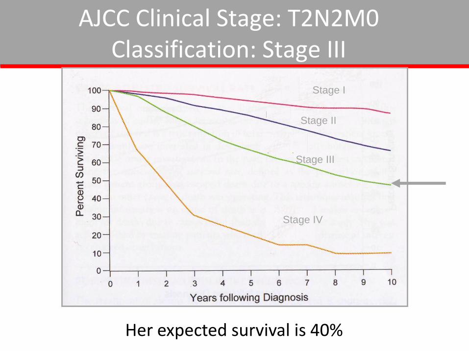
AJCC Clinical Stage: T2N2M0 Classification: Stage III
Stage I
Stage II
Stage III
Stage IV
Her expected survival is 40%

• The patient was treated with neoadjuvant chemotherapy including four cycles of cytoxanadriamycin and taxol
• Clinically the mass became softer and smaller
• Lymph nodes were no longer palpable
Neoadjuvant Chemotherapy

Neoadjuvant Chemotherapy
• MRI showed complete resolution of the mass
• Patient underwent an excision of the mass and complete axillary node dissection

10-1210-12-2010

4-22
4-22-2011


17

Neoadjuvant Chemotherapy
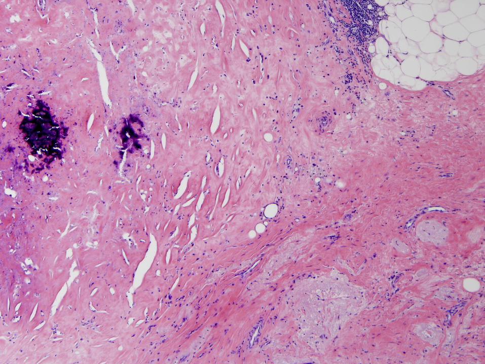
• The tumor bed consisted of an area of histiocytes and lymphocytes. No residual carcinoma was identified
• Sixteen lymph nodes were excised
• All were negative for metastatic carcinoma

Neoadjuvant Chemotherpay
Stage I
Stage II
Stage III
Stage IV
Her expected survival is over 90%
Neoadjuvant Chemotherapy
• Standard therapy for locally
advanced breast carcinoma
• Increasingly used for early stage
operable disease
• A wide range of pathologic changes
can occur after neoadjuvant
chemotherapy
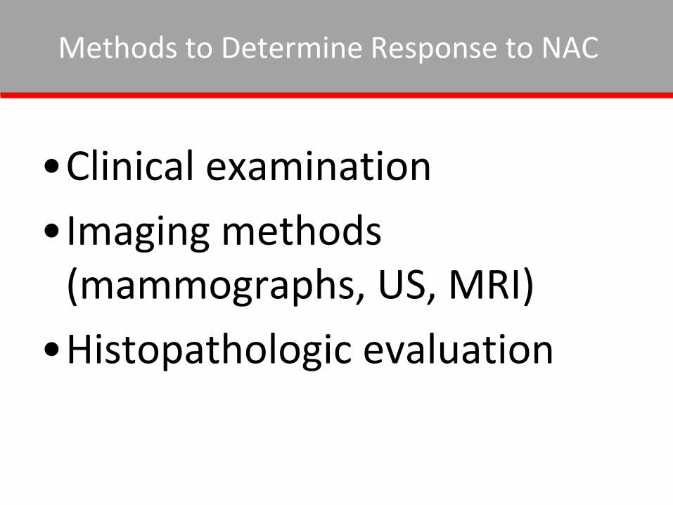
Methods to Determine Response to NAC
•Clinical examination
•Imaging methods (mammographs, US, MRI)
•Histopathologic evaluation

Neoadjuvant Chemotherapy
• 60-80% patients with locally
advanced breast carcinoma show
measurable clinical response
• Imprecise
Clinical Response

Methods to Determine Response to NAC
• Clinical/imaging methods
–False negative 40-60%
–False positive 20-30%
Histopathologic evaluation is gold standard
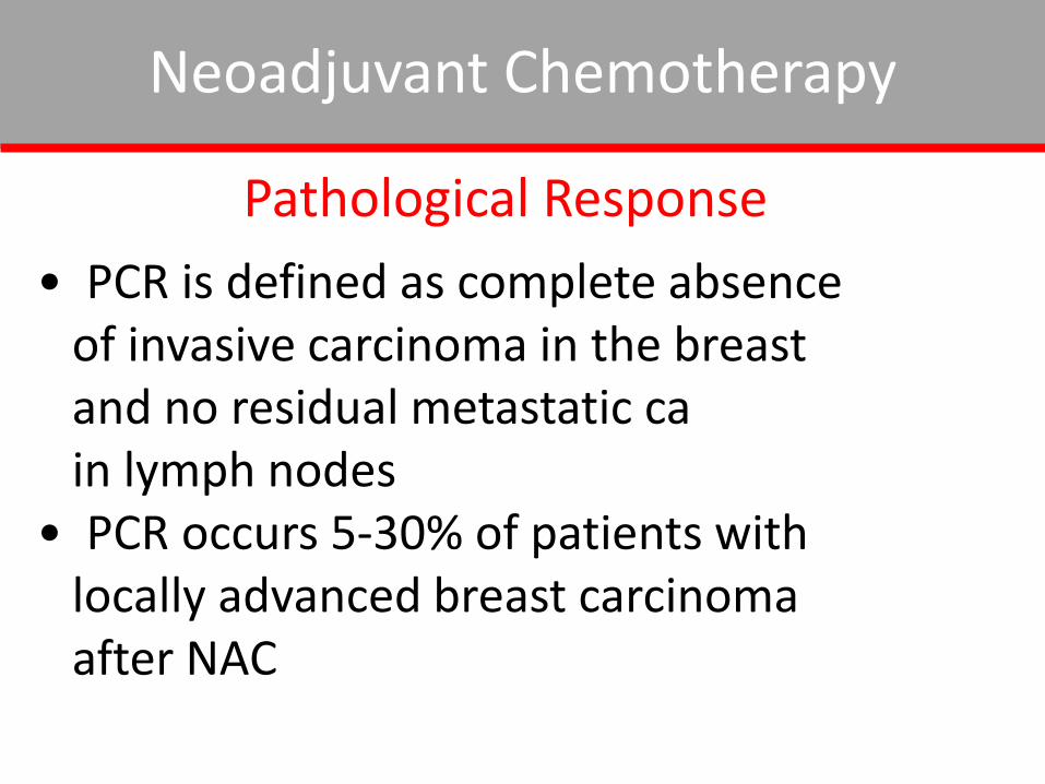
Neoadjuvant Chemotherapy
• PCR is defined as complete absence of invasive carcinoma in the breastand no residual metastatic ca in lymph nodes
• PCR occurs 5-30% of patients with locally advanced breast carcinomaafter NAC
Pathological Response

Provenzano E et al. Mod Pathol 2015;1185-201
Standardization of pathologic evaluation and reporting of postneoadjuvant specimens in clinical trials of breast cancer: recommendations from an international working group
In breast and axilla,
DCIS not allowedIn breast and axilla,
DCIS allowed
In breast,
DCIS allowedIn breast, DCIS
and few scattered
tumor cells allowed
16
14
12
10
8
6
4
2
0
# o
f t
rials

pCR = no invasive cancer in breast or lymph nodes
Cortazar, et al. Lancet 2014; 384: 164–72
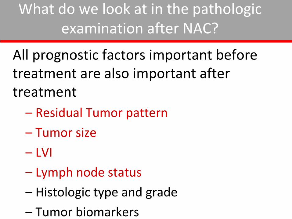
What do we look at in the pathologic examination after NAC?
All prognostic factors important before treatment are also important after treatment
– Residual Tumor pattern
– Tumor size
– LVI
– Lymph node status
– Histologic type and grade
– Tumor biomarkers

Neoadjuvant Chemotherapy
• Less than complete response (partial response) is difficult to classify
• There are different classification systems
Pathological Response

Concentric shrinking
Patterns of Tumor Response
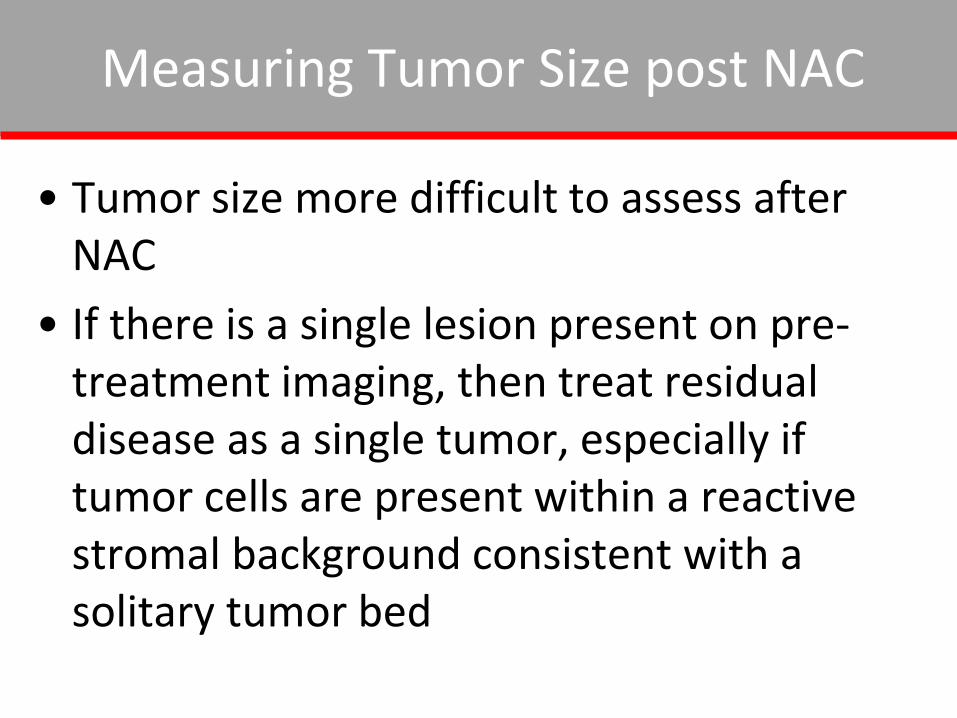
Measuring Tumor Size post NAC
• Tumor size more difficult to assess after NAC
• If there is a single lesion present on pre-treatment imaging, then treat residual disease as a single tumor, especially if tumor cells are present within a reactive stromal background consistent with a solitary tumor bed

Scatter pattern
Patterns of Tumor Response

• 7th edition AJCC –largest contiguous area of tumour cells (B)
• The combination of size and residual tumor cellularity is the best indicator of response
Measuring Tumor Size post NAC

Measuring Tumor Size post NAC

Provenzano E et al. Mod Pathol 2015;1185-201
Standardization of pathologic evaluation and reporting of postneoadjuvant specimens in clinical trials of breast cancer:
recommendations from an international working group

36


Placement of clip prior to treatment is very helpful


Neoadjuvant Chemotherapy
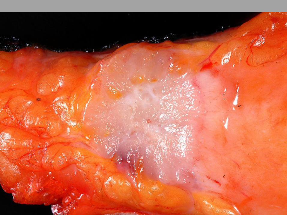
• Identification of “Tumor Bed” essential
• Can be very difficult if there is a marked clinical/imaging response
• Requires thorough evaluation

Tumor Bed
How extensively these specimens need to be sampled?
• If gross tumor is present limited sampling is adequate to establish the presence, size and cellularity of residual tumor
• 1-2 sections/cm of tumor is reasonable
• If tumor bed is ill defined more extensive sampling is necessary


Neoadjuvant Chemotherapy
Buchholz et al., Cancer, 2003
Responses Are Not Uniform

pCR: No recognizable invasive tumor cells present
pPR: The presence of scattered individual or small clusters of
tumor cells in a demosplastic or hyaline stroma
pNR: Tumors not exhibiting therapy related changes
Systems of Categorizing Response to Neoadjuvant Treatment
NSABP B-18

Miller-Payne System
• Grade 1: no reduction in overall cellularity
• Grade 2: a minor loss in overall cellularity (up to 30% loss)
• Grade 3: 30-90% reduction in cellularity
• Grade 4: >90% reduction in cellularity
• Grade 5: no residual invasive carcinoma

Classification of Breast Ca After NAC
Miller-Payne Grading System
Overall survival compared with histological response to chemotherapy
Disease free survival compared with histological response to chemotherapy
Breast. 12(5):320-7, 2003.

Miller-Payne System
• Pts with grade 4 response have a significantly worse prognosis
• Identification of small foci of residual invasive carcinoma is important
• Main limitation is that it does not include response in lymph nodes

Residual Cancer Burden System MDACC
• Cellularity of residual carcinoma over the tumor bed
• Presence of lymph node metastasis
• Size of the largest lymph node metastasis

Pathologic Assessment of Tumor Bed
See downloadable protocol and illustrations at www.mdanderson.org/breastcancer_RCB

Pathologic Assessment of Tumor Bed

Residual Cancer Burden

RCB-0 No carcinoma in breast or
lymph nodes (pCR)
RCB-1 Minimal residual disease
(marked response)
RCB-2 Moderate response
RCB-3 Minimal or no response
(chemoresistant)
Systems of Categorizing Response To Neoadjuvant Treatment
Residual Cancer Burden System (MDACC)

J Clin Oncol. 25(28):4414-22, 2007
Likelihood of distant relapse in patients with residual cancer burdenA: entire paclitaxel plus fluorouracil, doxorubicin, and cyclophosphamide cohortB: subset without adjuvant hormone treatmentC: subset who received adjuvant hormone treatment
Residual Cancer Burden

TNBC
This presentation is the intellectual
property of the presenter. Contact
permission to reprint and/or
Class N %
pCR 43 34
RCB-I 18 14
RCB-II 42 34
RCB-III 22 18
Class N %
pCR 26 10
RCB-I 34 13
RCB-II 156 60
RCB-III 45 17
Class N %
pCR 38 37
RCB-I 17 17
RCB-II 30 29
RCB-III 17 17
HR+/HER2- HER2+

Residual Disease in Breast and Nodes (RDBN)
• Modification of the Nottingham Prognostic Index (NPI)
RDBN= 0.2 x Tumor size(cm) + LN stage (0-3) + Histologic grade (1-3)
Chollet P aet al. A new Prognostic classification after primary chemotherapy for breast cancer. Cancer J 14:128-132, 2008

Residual Disease in Breast and Nodes (RDBN)
• Lymph node stage:
– 0 = neg
– 1 = 1-4 LNs
– 2 = 5-7 LNs
– 3 = > 8 LNs
• Grade is determined after therapy
Chollet P aet al. A new Prognostic classification after primary chemotherapy for breast cancer. Cancer J 14:128-132, 2008

Residual Disease in Breast and Nodes (RDBN)
4 groups are identified with significant prognostic differences
Chollet P aet al. A new Prognostic classification after primary chemotherapy for breast cancer. Cancer J 14:128-132, 2008

Provide the following information:
1. pCR (ypT0 ypN0 and ypT0/is ypN0) versus residual disease
2. ypT and ypN Stage using the current AJCC/UICC staging system
A single standardized approach to macroscopic and
microscopic pathologic examination makes it easy to reliably
provide information for clinical practice
Recommendations
Modern Pathol 00: 1–17, 2015 doi:10.1038/modpathol.2015.74

Neoadjuvant Chemotherapy
• Lobular atrophy and calcification• Epithelial atypia• Stromal fibrosis• Inflammation• Cytoplasmic vacuolization• Pigmented and foamy macrophages• Interlobular fibrosis• Fat necrosis• Duct ectasia
Histopathological Changes


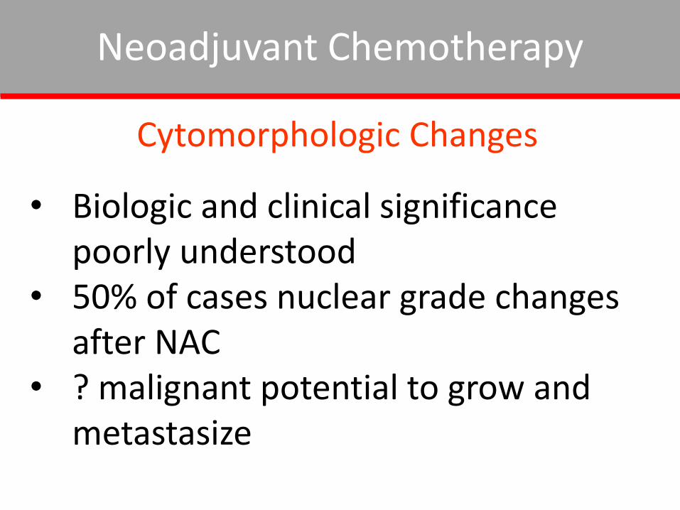
Neoadjuvant Chemotherapy
•
• Biologic and clinical significance poorly understood
• 50% of cases nuclear grade changes after NAC
• ? malignant potential to grow and metastasize
Cytomorphologic Changes

Neoadjuvant Chemotherapy
• Similar changes can occur in lymph node metastases
Cytomorphologic Changes


Lymph node changes
pCR breastPartial response LN
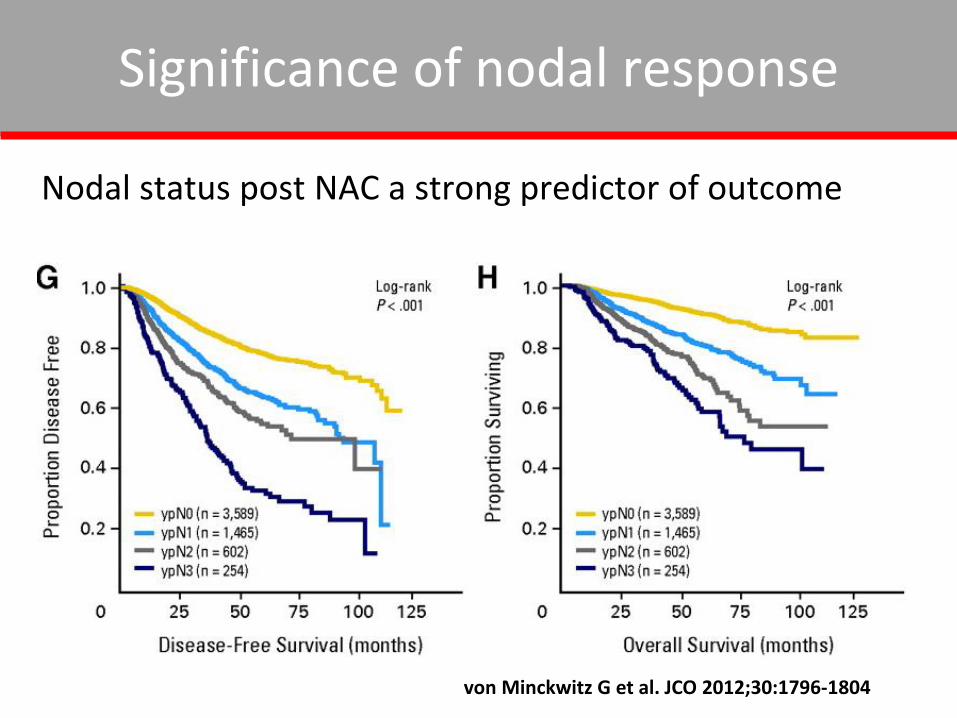
Significance of nodal response
Nodal status post NAC a strong predictor of outcome
von Minckwitz G et al. JCO 2012;30:1796-1804
Neoadjuvant Chemotherapy
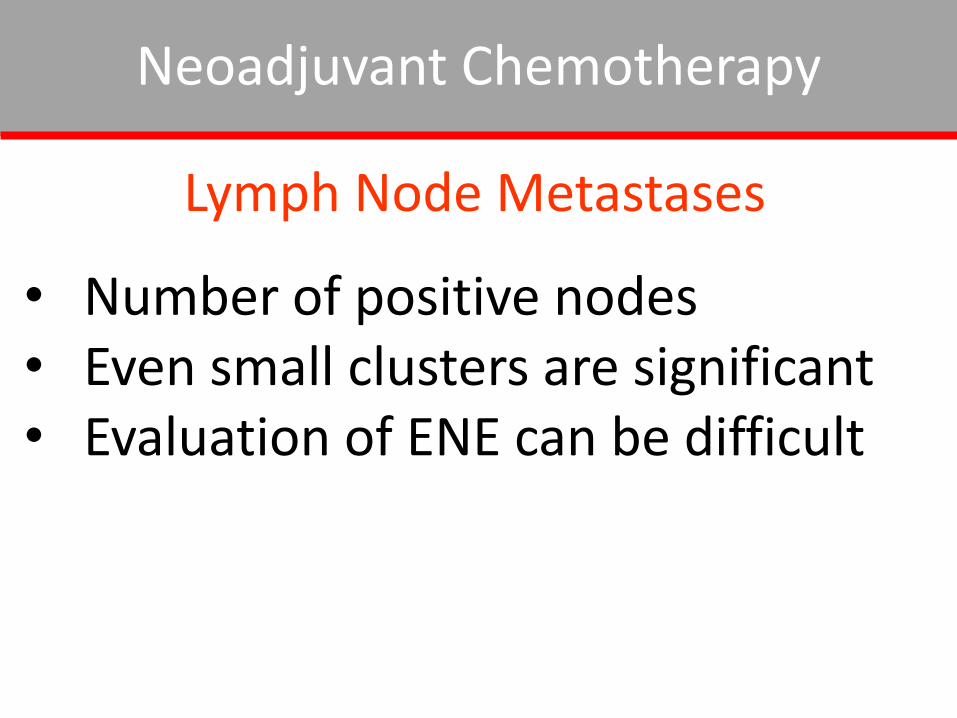
•
• Number of positive nodes• Even small clusters are significant• Evaluation of ENE can be difficult
Lymph Node Metastases

Receptor Testing post NAC
• Limited data about correlation of hormone receptor and HER2 status pre and post treatment
• ER - 2-30% change in status
• Similar outcome in patients who converted from ER+ vs to ER-ve after treatment
• PR - 5-50% change in status

• HER2 - results vary depending upon whether IHC or ISH is used
• 0-21% conversion on HER2 IHC
• 0-43% loss of HER2 gene amplification
• Association between loss of HER2 positivity and worse outcome – resistance vs negative subclone
Mittendorf et al, Clin Cancer Res 2009
Receptor Testing post NAC

Consider retesting if:
• Identification of morphologically distinct areas on final excision
• Inadequate tumor on CNB
• If there is a change in status which is predictive of response to therapy?
Treat positive result
Receptor Testing post NAC

Take Home Messages
Neoadjuvant Chemotherapy
• NAC is being used more frequently
• Pathologic response is an important
predictor of survival
• pCR provides the best prognosis
• Better classification of pPR category is
needed

Neoadjuvant Chemotherapy
•
• Pathologists should be familiar to ensure that chemotherapy induced changes in non-neoplastic tissue is not mistaken for residual tumor or that residual tumor is not confused with non-neoplastic cells
Take Home Messages

Summary
• Record pretreatment phenotype and grade
• Accurate assessment of residual tumor requires multidisciplinary teamwork
• Require standardized procedures to evaluate the gross specimen, record a map of the tissue sections related to the gross & imaging findings, and relate the histopathologic findings to that map
Summary
• Uniform terminology is helpful
- pCR in breast and nodes
- Report presence and extent of in situ residual disease

Summary
•Pathology reports should include: Histologic type Size (as a single focus or tumor bed)
Cellularity Margins LVI Lymph nodes