Evaluation of Antibacterial, Antioxidant Activity and ...rdo.psu.ac.th › sjstweb › Ar-Press ›...
25
Evaluation of Antibacterial, Antioxidant Activity and Calmodulin Gene Expression of Scoparia dulcis Linn. Journal: Songklanakarin Journal of Science and Technology Manuscript ID SJST-2017-0227.R1 Manuscript Type: Original Article Date Submitted by the Author: 18-Oct-2017 Complete List of Authors: Chana, Netnapa; Thaksin University Phatthalung Campus, Department of Chemistry Supaphon , Preuttiporn; Thaksin University Phatthalung Campus, Department of Biology Phongdara, Amornrat; Center for Genomics and Bioinformatics Research, Department of Molecular Biotechnology and Bioinformatics,Prince of Songkla University, Keyword: Agricultural and Biological Sciences, Biotechnology and Agro-Industry Songklanakarin Journal of Science and Technology SJST-2017-0227.R1 Chana
Transcript of Evaluation of Antibacterial, Antioxidant Activity and ...rdo.psu.ac.th › sjstweb › Ar-Press ›...

Evaluation of Antibacterial, Antioxidant Activity and
Calmodulin Gene Expression of Scoparia dulcis Linn.
Journal: Songklanakarin Journal of Science and Technology
Manuscript ID SJST-2017-0227.R1
Manuscript Type: Original Article
Date Submitted by the Author: 18-Oct-2017
Complete List of Authors: Chana, Netnapa; Thaksin University Phatthalung Campus, Department of Chemistry Supaphon , Preuttiporn; Thaksin University Phatthalung Campus, Department of Biology Phongdara, Amornrat; Center for Genomics and Bioinformatics Research, Department of Molecular Biotechnology and Bioinformatics,Prince of
Songkla University,
Keyword: Agricultural and Biological Sciences, Biotechnology and Agro-Industry
Songklanakarin Journal of Science and Technology SJST-2017-0227.R1 Chana

1
Evaluation of antibacterial, antioxidant activity and Calmodulin gene expression of
Scoparia dulcis Linn.
Netnapa Chana1,*
, Preuttiporn Supaphon2, and Amornrat Phongdara
3
1 Department of Chemistry, Faculty of Science, Thaksin University, Phatthalung campus, Phatthalung, 93210, Thailand
2 Department of Biology, Faculty of Science, Thaksin University, Phatthalung campus, Phatthalung, 93210, Thailand
3 Center for genomics and bioinformatics research, Faculty of Science, Prince of Songkla University, Songkhla, 90112,
Thailand
*Corresponding Author: [email protected] ; Tel : +66-9-0665-7531
Abstract
In Thailand, Scoparia dulcis is being used as a traditional medicine. The extract
displayed the best inhibitory effect against Pseudomonas aeruginosa and its minimum
inhibitory concentration (MIC) was 400 µg/ml, followed by Escherichia coli and
Staphylococcus aureus with MIC 500 µg/ml. The presence of the extract significantly
protected Sf9 cells against H2O2-induced cell death. In addition, the extract also showed DNA
damage inhibitory effect in a concentration-dependent manner (0.5-4 mg/ml). However, at
high concentration (7.5-30 mg/ml) it might induce damage to the DNA. The prevention of
DNA damage differs in different parts of the plant. To support the secondary metabolites
synthesis in different parts of S. dulcis, we investigated the expression of the Calmodulin
gene that is involved in secondary metabolite production. The Calmodulin gene showed the
highest expression in the fruit. This finding justifies the use of S. dulcis in the treatment of
diseases caused by bacteria and free radicals.
Keywords: Scoparia dulcis Linn., antibacterial, H2O2, DNA nicking, Calmodulin gene
expression

2 1. Introduction
The exposure of organisms to various infections has been reported to generate a wide
range of reactive oxygen species (ROS), resulting in hundreds of disorders especially in
diabetic wound infection (Ivanov, Bartosch, & Isaguliants, 2017). Traditional medicinal
plants are being used as a strategy for the production of effective novel compounds to
develop new antimicrobial and antioxidant drugs. Medicinal plants contain a wide variety of
therapeutic agents with biochemical and therapeutic properties.
Scoparia dulcis (SD) Linn. (Plantaginaceae), commonly known as sweet broom, is a
herbal medical plant widely used in the indigenous system of medicine. S. dulcis is found in
tropical and subtropical regions. Fresh or dried S. dulcis have been used for treating liver
ailments, stomach troubles, ulcer, cancer, wounds, cough and tuberculosis. The previous
report revealed that an aqueous crude extract from S. dulcis effectively protected rodents
from gastric ulcers (Mesia-Vela et al., 2007). The SD extract and its active compounds may
exert effects in cases of diabetes and anticancer (Beh, Latip, Abdullah, Ismail, & Hamid,
2010; Wu, Chen, Lu, Chen, & Chang, 2012). In addition, SD extract was reported to promote
wound healing and showed good blood-clotting ability in the rat model (Ediriweera,
Jayakody, & Ratnasooriya, 2011; Krishna, Vijay, Mayank, & Megha, 2011). These biological
activities are conceivably attributed to its active phytochemical constituents. S. dulcis
accumulates a variety of secondary metabolites such as scoparic acid A, scoparic acid B,
scopadulcic acid A, scopadulcic acid B, scopadulciol, scopadulin, scopanolal and scopadiol
with a group of saponins (Pamunuwa, Karunaratne, & Waisundara, 2016). Nkembo et al
showed that the synthesis of secondary metabolites was enhanced when S. dulcis was
stimulated by methyl jasmonate and calcium. The Calmodulin gene functions as the key
component in the activation of the calcium cascade in these processes. The scopadulcic acid
B (SDB) production was almost completely blocked by pretreatments with trifluoperazine

3 (Calmodulin antagonists) (Nkembo, Kurosaki, Lee, & Hayashi, 2005; Saitoh et al., 2007).
Plant Calmodulin is involved in plant responses to environmental stimuli and stresses, such as
salinity, cold, heat, oxidative stress, and pathogens. In tobacco, the Calmodulin gene
expression was enhanced in response to pathogenic Ralstonia solanacearum infection
(Takabatake et al., 2007). Transgenic soybean expressing a foreign Calmodulin gene showed
pathogens stress tolerance (Rao et al., 2014).
Hence, the present study was aimed to provide scientific data on the beneficial
biological properties including antibacterial and antioxidant properties of S. dulcis. In
addition, gene expression analysis of Calmodulin was performed in different plant tissues.
This investigation suggests that Calmodulin may function related to the secondary
metabolites production of S. dulcis, and support the extract exert potent antimicrobial effect
and protection of cells, and DNA damage from free radicals. The consequence of this
investigation supports that S. dulcis can be used for the treatment of microbial infections in
wounds especially in diabetic wounds.
2. Materials and Methods
2.1 Collection and identification of plant samples
Plant samples were collected from the local area of Songkhla Province, Thailand. The
subject plant material specimen (N. Chana 001) was authenticated by a taxonomist, Assistant
Professor Dr. Jarun Leerativong, Department of Biology, Faculty of Science, Prince of
Songkla University, Songkhla, Thailand. The specimen was deposited at the Herbarium of
Prince of Songkla University, Thailand. The fresh young leaves were grounded into powder
in liquid nitrogen, and used for the genomic DNA extraction. Genomic DNA isolation was
performed by using a GeneJET Plant Genomic DNA Purification Kit (Thermo Scientific)
according to the manufacturer's instructions. The ITS2 region was amplified using the
following pair of universal primers: ITS-2F, 5′-GTT TCC GTA GGT GAA CCT-3′; and ITS-

4 2R, 5′-GCT TAT TAA TAT GCT TAA ATC ACG-3′. The PCR products were cloned into
pGEM-T easy vector and sent for DNA sequencing.
2.2 Preparation of S. dulcis extract
The aerial parts of the plant were collected, 200 grams of fresh plant were soaked in
300 ml of methanol for 3 days with occasional shaking. The resultant mixture was then
successively filtered through a Whatman filter paper No1. The rotary evaporator was used to
concentrate the filtrate at 40 °C. These extracts were then used for a biological properties
study. The concentrate sample was dissolved in DMSO/MHB to carry out the antibacterial
activity. In a separate experiment, the root, leaves, stem and fruit of S. dulcis were extracted
according to the method described above, and dissolved in distilled water for DNA nicking
assay.
2.3 Preparation of inoculums
Four strains of bacteria were used in this study, including Gram-positive bacteria
(Staphylococcus aureus ATCC25923 and methicillin - resistant Staphylococcus aureus
SK1) and Gram-negative bacteria (Pseudomonas aeruginosa ATCC27853 and Escherichia
coli ATCC25922). The bacteria were subcultured on nutrient agar (NA). Five single colonies
were transferred into nutrient broth (NB) and incubated in a shaking incubator at 35 ºC, 150
rpm for 3 hours. McFarland standard (no. 0.5) was used to adjust the turbidity of bacterial
suspension. After that, the suspension was diluted to 1:200 ratio with Mueller Hinton Broth
(MHB).
2.4 Antibacterial assay
The antibacterial activity of crude extract was evaluated with the colorimetric broth
microdilution method. The extract which exhibited the antibacterial activity at 1 mg/ml were
further subjected to the same method to find the minimum inhibitory concentration (MIC)
and the minimum bactericidal concentration (MBC), respectively.

5 2.4.1 Preliminary assay
Antibacterial activity tests were conducted using the 96- well microtiter plate by
colorimetric broth microdilution method (CLSI, 2012) with some modifications. Fifty-micro
liters of crude extract (2 mg/ml) and 50 µL of bacterial suspension (106 CFU/ml) were added
in each well of the microtiter plate according to the method of Supaphon et al. Plates were
incubated at 35 o
C for 18 hours. Then, 10 µl of resazurin indicator was added to the plates.
The results were recorded as growth or no growth based on the color of the resazurin (blue
color) as a positive result and resorufin (pink color) as a negative result. (Supaphon,
Phongpaichit, Rukachaisirikul, & Sakayaroj, 2013).
2.4.2 Determination of minimum inhibitory concentration and minimum bactericidal
concentration
The minimum inhibitory concentration was determined by the previous assay using a
2-flow dilution of crude extract with final concentration (1000-100 µg/ml). The lowest
concentration of extract that inhibited growth of microorganisms was recorded as the MIC
value. All extract in microtiter plate that showed a blue color were further tested to find the
minimum bactericidal concentration. The blue color in each well was streaked onto nutrient
agar (NA). Plates were incubated at 35 o
C for 18-24 hours. The lowest concentration of
extract that showed no growth of bacteria on NA was recorded as MBC value. Antibiotic
drugs (vancomycin and gentamicin) were used as positive control and DMSO was used as
negative control.
2.5 Spodoptera frugiperda (Sf9) insect cell viability assay
Sf9 insect cells (Invitrogen) were cultured in insect express Sf9-S2 medium (PAA).
The cells were seeded onto a 96-well plate (5 × 10⁴ cells/well) and grown to approximately
70-90% confluence for 24 hours. The cells were incubated with SD extract (2 mg/ml or 4
mg/ml), and the mixture also combined with 750 µM or 1 mM H2O2 diluted in the Sf9 culture

6 medium for 16 hours. The viability cells were assessed using the MTT survival assay. The
percentage of cell survival was calculated, and compared with untreated cells.
2.6 DNA nicking assay
To study the ability of SD extract to protect supercoiled DNA from metal-catalyzed
oxidation (MCO), in vitro tests were performed essentially as described by Saelee et al with
slight modifications (Saelee, Tonganunt-Srithaworn, Wanna, & Phongdara, 2012).
Approximately 200 ng of pUC19 plasmid incubated with different concentrations of the
extract followed by addition of 10 mM DTT and 3 µM of FeCl3. The negative control was
without addition of the extract. The reaction was then incubated 2 hours at 37 °C. The
samples were then analyzed by 0.8% agarose gel electrophoresis.
2.7 Tissue distribution analysis of Calmodulin
To determine the gene related to secondary metabolite production, the expression
level of Sd-Calmodulin was analyzed using the RT-PCR technique. The leaves, stem, fruit,
and root were detached from S. dulcis. Total RNA of S. dulcis was extracted using the
GeneJET RNA Purification Kit according to the manufacturer's instructions (Thermo Fisher
Scientific). The single-stranded cDNA was synthesized using SuperScript® III Reverse
Transcriptase (RT) and oligo (dT) primer, following the manufacturer’s protocol (Invitrogen).
The transcript level of Sd-Calmodulin gene was amplified using the following primer pair:
CAM-SdF ; 5’-GAG GCT GAG CTT CAG GAT ATG ATC AA as the forward primer and ;
CAM-SdR; 5’-ACG TCG GCC TCG CGG ATC ATC TCG T-3’as the reverse primer. Actin
was used as internal control with specific primers (Actin-SdF; 5’-TCT ACA ATG AGC TCC
GTG TTG C-3’ and Actin-SdR; 5’- CTT GTT CAT AAT CAA GAG CAA TGT A-3’). The
amplified DNA fragments were subcloned into pGEM-T Easy vector (Promega Corporation),
and sent for DNA sequencing.
2.8 Guava ViaCount assay

7
A ViaCount assay was used with the Guava easyCyte 5HT Flow cytometer to
evaluate viable and dead cell populations on Chinese hamster ovary cells (CHO cells)
exposed to SD extract. The cells were exposed to the SD extract for 24 hours. Guava
ViaCount Reagent was added to the cells. The percentage of viable cells was assessed by
flow cytometry and analyzed with Guava ViaCount software. The obtained results were
expressed as the percentage of live cells in treated cultures compared to that present in the
control cultures.
2.9 Guava Nexin assay
A Nexin assay was used to evaluate viable, early and late apoptotic cell populations
on CHO cells exposed to SD extract. CHO cells were exposed to the extract for 24 hours. The
cells were stained with Guava Nexin Reagent and data analysis was performed using the
Nexin software module.
3. Results and discussion
3.1 Identification of S. dulcis
In order to facilitate accurate species resolution when testing the herbal products, we
tested the nuclear ribosomal internal transcribed spacer 2 (ITS2) gene in identification of
herbal medicine species. ITS is one of the most sequenced regions in molecular systematics.
The ITS2 sequences were analyzed using the BLAST method in the GenBank database. DNA
analysis showed that S. dulcis ITS2 sequence from Songkhla province in Thailand has 99%
identity with Scoparia dulcis species (Accession number AY963776.1). The S. dulcis is a
member of the Gratioleae tribe in the Plantaginaceae family. Our results showed that the
ITS2 gene could successfully identify this species of herbal medicine.
3.2 Antibacterial property
Bacterial infections caused by drug-resistant bacteria are at an increased risk in
Thailand. Natural products are alternative sources of new pharmaceutical agents, especially

8 Thai medicinal plants. S. dulcis is a flowering plant distributed in Southern Thailand. It has
been widely reported that there are various phytochemical compounds in whole plants such
as flavonoids, saponins, tannins, alkaloids, steroids and terpenes (Wankhar, Srinivasana, &
Rathinasamy, 2015). The extract of this plant species were used to treat diarrhea, ulcer and
wounds (Ediriweera et al., 2011). Wounds with bacterial infection are the most common
public health problems. Most studies of S. dulcis used dried plant material to extract the
metabolites and tested their ability against pathogenic microorganisms (Jose, & Sinha, 2017;
Latha et al., 2006; Zulfiker et al., 2011; Mohandas, Valsalakumari, William, & Narayanan,
2014). There is no report on the antibacterial activity of extract from fresh plant material. We
first investigated the effect of methanolic extract from the fresh aerial part of S. dulcis (SD
extract) on the antibacterial activity. SD extract was found to inhibit both bacterial strains
(Gram-positive and Gram-negative bacteria). The extract was most active against P.
aeruginosa with MIC 400 µg/ml, followed by E. coli and S. aureus with MIC 500 µg/ml
(Table 1).
Coulibaly et al revealed that dried S. dulcis extract exhibited significant antimicrobial
activities, methanolic extract and aqueous-acetone extract on E. coli (MIC 12.5 mg/ml),
aqueous-acetone extract on S. typhimurium (MIC 1.56 mg/ml), chloroform extract on B.
cereus (MIC 1.56 mg/ml). Furthermore, hexane extract from this plant displayed the best
antifungal activity against A. niger and P. roquefortii with MIC 6.25 mg/ml (Coulibaly et al.,
2012). The result in this study indicated that fresh SD extract yielded more antibacterial
activity than dried SD extract as showed in the previous report. It could also be suggested that
the extraction components in fresh and dried SD extract might have a different composition.
SD extract was also reported to have activities against the viruses such as herpes simplex
virus type 1, pathogenic fungi and Aedes aegypti larvae (Wankhar et al., 2015; Hayashi,

9 Masaru, Yoshihisa, Tooru, & Naokata, 1990). These findings confirm that S. dulcis is a good
source of active metabolites.
3.3 Protective effects of SD extract against H2O2 damage in Sf9 cells
Wound infections are a common problem for the diabetic population. Reactive
oxygen species (ROS) such as superoxide anion (⋅O2-), hydroxyl radicals (⋅OH), and
hydrogen peroxide (H2O2) play an integral role in the wound healing process particularly
hydrogen peroxide radical. However, excessive ROS can damage the structure of nearby
molecules via an oxidation reaction. The antioxidants were designed to remove the
deleterious effects of ROS in wound infection (Dunnill et al., 2015). Plant natural products
have been a useful source of antioxidant properties that may act as free radical scavengers to
prevent oxidative stress imbalance. SD extract has been reported to exhibit various
pharmacological activities, including wound healing, anti-diabetic, anti-inflammatory and
anti-oxidative effects (Ediriweera et al., 2011; Latha, Pari, Sitasawad, & Bhonde, 2004;
Murti, Panchal, Taya, Singh, 2012). The present study focused on the ability of SD extract to
protect Sf9 cells against H2O2 exposure. Cell viability was measured by a MTT assay. The
MTT assay revealed that exposure of Sf9 cells to H2O2 at 750 M and 1 mM, significantly
decreased cell viability (Figure 1). However, treatment of Sf9 cells with fresh SD extract
increased cell viability. These results suggest that SD extract exerts a cytoprotective effect
against H2O2-induced oxidative stress in Sf9 cell. Latha et al showed that the S. dulcis extract
also prevented streptozotocin-induced rat insulinoma cell death (Latha et al., 2004). The
aqueous extract of S. dulcis play a role in hepatoprotective and antioxidant activity against N-
Nitrosodiethylamine (DEN) induced hepatotoxicity in experimental rats (Langeswaran,
Jagadeesan, & Vijayaprakash, 2012). Thus, SD extract has a cytoprotective role against
cytotoxic agents.
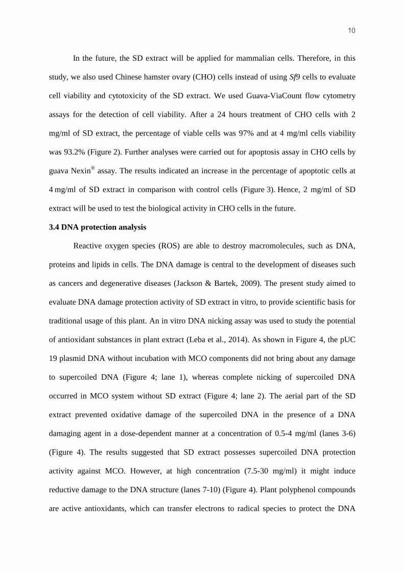
10 In the future, the SD extract will be applied for mammalian cells. Therefore, in this
study, we also used Chinese hamster ovary (CHO) cells instead of using Sf9 cells to evaluate
cell viability and cytotoxicity of the SD extract. We used Guava-ViaCount flow cytometry
assays for the detection of cell viability. After a 24 hours treatment of CHO cells with 2
mg/ml of SD extract, the percentage of viable cells was 97% and at 4 mg/ml cells viability
was 93.2% (Figure 2). Further analyses were carried out for apoptosis assay in CHO cells by
guava Nexin® assay. The results indicated an increase in the percentage of apoptotic cells at
4 mg/ml of SD extract in comparison with control cells (Figure 3). Hence, 2 mg/ml of SD
extract will be used to test the biological activity in CHO cells in the future.
3.4 DNA protection analysis
Reactive oxygen species (ROS) are able to destroy macromolecules, such as DNA,
proteins and lipids in cells. The DNA damage is central to the development of diseases such
as cancers and degenerative diseases (Jackson & Bartek, 2009). The present study aimed to
evaluate DNA damage protection activity of SD extract in vitro, to provide scientific basis for
traditional usage of this plant. An in vitro DNA nicking assay was used to study the potential
of antioxidant substances in plant extract (Leba et al., 2014). As shown in Figure 4, the pUC
19 plasmid DNA without incubation with MCO components did not bring about any damage
to supercoiled DNA (Figure 4; lane 1), whereas complete nicking of supercoiled DNA
occurred in MCO system without SD extract (Figure 4; lane 2). The aerial part of the SD
extract prevented oxidative damage of the supercoiled DNA in the presence of a DNA
damaging agent in a dose-dependent manner at a concentration of 0.5-4 mg/ml (lanes 3-6)
(Figure 4). The results suggested that SD extract possesses supercoiled DNA protection
activity against MCO. However, at high concentration (7.5-30 mg/ml) it might induce
reductive damage to the DNA structure (lanes 7-10) (Figure 4). Plant polyphenol compounds
are active antioxidants, which can transfer electrons to radical species to protect the DNA

11 from radicals (Nimse & Pal, 2015; Jackson et al., 2009). However, excessive antioxidants
might cause these compounds to transfer their electrons to nucleotide bases and induce
radical species which induce chemical bond cleavage in DNA (Lu, Ou, & Lu, 2013). This
study showed similar results with fermented red brown rice extract for DNA nicking analysis
(Kong, Lee, Michelle, Ginjom, & Nissom, 2015). These activities of SD extract may be
attributed to the presence of various secondary metabolites such as flavonoids and phenolic
acids which were reported earlier to neutralize the radicals. Hence, the secondary metabolites
accumulation in different tissues of S. dulcis may result in different DNA damage protection
levels.
The leaves, stem, fruit, and root were detached from S. dulcis to study the DNA
damage protection. The results showed that the radicals produced in the MCO system caused
complete degradation of the control DNA within 2 hours of incubation (Figure 5; lane 2). The
root part of SD inhibited DNA damage at 4 mg/ml (Figure 5; lane 4). However, at the
concentration of 0.5 mg/ml it did not show protective action against DNA damage (Figure 5;
lane 3). The stem extract prevented DNA damage at concentrations of 0.5 and 4 mg/ml
(Figure 5; lanes 5-6). Leaves extract showed DNA damage protection at 0.5 mg/ml (Figure 5;
lane 7), but at a concentration of 4 mg/ml it might induce DNA damage about 50% (Figure 5;
lane 8). The protective effects of fruit extract against DNA damage were shown at a
concentration of 0.5 mg/ml (Figure 5; lane 9). However, it completely damaged the DNA at a
concentration of 4 mg/ml (Figure 5; lane 10). Thus, the prevention of DNA damage depends
not only on concentration but also on the origin of the extract. SD extract from the stem part
was most effective in preventing DNA damage. However, only the fruit extract completely
damaged the DNA at a concentration of 4 mg/ml. These results may imply that the fruit part
may have the highest secondary metabolites content because it can induce reductive damage

12 to the DNA structure. However, further chemical composition level analysis in SD extract is
necessary to confirm this suggestion.
3.5 Tissue distribution analysis of Calmodulin gene
Various parts of S. dulcis are used extensively in traditional medicine to cure various
human ailments and its extract has antibacterial and antioxidant properties. However, the
mechanism is still unknown. It has been documented that Calmodulin gene expression
increased in ROS treatment in the protoplasts of mesophyll cells. This mechanism enhanced
the expression of antioxidant genes such as superoxide dismutase 4 (SOD4), cytosolic
corbate peroxidase (cAPX), and glutathione reductase 1 (GR1). The up-regulation of the
antioxidant enzymes was almost completely blocked by pretreatments with Calmodulin
antagonists (Hu et al., 2007). Moreover, Nkembo et al revealed that the scopadulcic acid B
(SDB) production was almost completely blocked by pretreatments with trifluoperazine
(Calmodulin antagonists) (Nkembo et al., 2005; Saitoh et al., 2007). SDB was found to
possess various biological activities, such as an inhibitor against replication of herpes simplex
virus type 1, gastric H+ ,K+ ATPase (Riel, Kyle, & Milhous, 2002) and antitumor promoting
activity (Nishino, Hayashi, Arisawa, Satomi, & Iwashima, 1993). The DNA nicking assay in
the present study revealed that the fruit extract may have higher secondary metabolites
content in comparison to other plant parts. To support the secondary metabolites synthesis in
different parts of S. dulcis, the Calmodulin gene participating in secondary metabolites
production was isolated and its gene expression was investigated in different organs by using
reverse transcription polymerase chain reaction. As shown in Figure 6, Sd-Calmodulin
showed the highest expression in fruit and this may result in higher concentrations of
secondary compounds in the fruit compared to other tissues.
In conclusion, the SD extract was found to inhibit both Gram-positive and Gram-
negative bacteria, and protect Sf9 cells against H2O2-induced oxidative damage. The extract

13 also prevents DNA damage from metal-catalyzed oxidation. These activities of SD extract
may be attributed to the presence of various secondary metabolites. The expression of the
Calmodulin gene that is involved in secondary metabolites production is highest in fruit. Our
data suggest that S. dulcis should be considered as a useful source of material for the
treatment of wound infection, as an antioxidant and antimicrobial agent.
Acknowledgments
This research was supported by research grants from Thaksin University. Our thanks
go to the Center for Genomics and Bioinformatics Research, Faculty of Science, Prince of
Songkla University, and the Faculty of Science, Thaksin University for allowing us to use
their laboratory facilities. We would like to thank Mr. Christopher Joseph Forti and Dr.
Stefano G.A. Draisma for language editing this manuscript for publication.
References
Beh, J. E., Latip, J., Abdullah, M. P., Ismail, A., & Hamid, M. (2010). Scoparia dulcis
(SDF7) endowed with glucose uptake properties on L6 myotubes compared insulin.
Journal of Ethnopharmacology, 129(1), 23-33. doi:10.1016/j.jep.2010.02.009.
Coulibaly, A. Y., Konate, K., Youl, E. N. H., Sombie, P. A. E. D., Kiendrebeogo, M., Meda,
N-T. R., & Nacoulma, O. G. (2012). Anti-proliferative effect of Scoparia dulcis L.
against bacterial and fungal strains. International Journal of Biological and Chemical
Sciences, 6(6), 3055-3063.
CLSI. (2012). Methods for Dilution Antimicrobial Susceptibility Tests for Bacteria that Grow
Aerobically. 9th
Edn., Clinical and Laboratory Standards Institute, Wayne, PA., USA.
Dunnill, C., Patton, T., Brennan, J., Barrett, J., Dryden, M., Cooke, J., … Georgopoulos, N.
T. (2015). Reactive oxygen species (ROS) and wound healing: the functional role of

14
ROS and emerging ROS-modulating technologies for augmentation of the healing
process. International Wound Journal, 14(1), 89-96.
Ediriweera, E., Jayakody, J., & Ratnasooriya, W. (2011). Pro blood clotting activity of
Scoparia dulcis in rats. An International Quarterly Journal of Research in Ayurveda,
32, 271-274.
Hayashi, T., Masaru, K., Yoshihisa, M., Tooru, T., & Naokata, M. (1990). Antiviral agents of
plant origin. III. Scopadulin, a novel tetracyclic diterpene from S. dulcis. Chemical
and Pharmaceutical Bulletin, 38, 945-947.
Hu, X., Jiang, M., Zhang, J., Zhang, A., Lin, F., & Tan, M. (2007). Calcium-calmodulin is
required for abscisic acid-induced antioxidant defense and functions both upstream
and downstream of H2O2 production in leaves of maize (Zea mays) plants. New
Phytologist, 173(1), 27-38.
Ivanov, A. V., Bartosch, B., & Isaguliants, M. G. (2017). Oxidative Stress in Infection and
Consequent Disease. Oxidative Medicine and Cellular Longevity,
doi:10.1155/2017/3496043
Jackson, S. P., & Bartek, J. (2009). The DNA-damage response in human biology and
disease. Nature, 461(7267), 1071-1078.
Jose, S., & Sinha, M. P. (2017). Study of Antibacterial Efficacy of Methanolic and Aqueous
Leaf Extracts of Scoparia dulcis on Some Human Pathogenic Bacteria. International
Journal of Current Microbiology and Applied Sciences, 6, 423-432.
Kong, E. L., Lee, B. K., Michelle, Ginjom, I., & Nissom, P. M. (2015). DNA damage
inhibitory effect and phytochemicals of fermented red brown rice extract. Asian
Pacific Journal of Tropical Disease, 5(9), 732-736.
Krishna, M., Vijay, L., Mayank, P., & Megha, S. (2011). Healing promoting potentials of
roots of scoparia dulcis in albino rats. Phamacologia, 2, 369-373.

15 Langeswaran, K., Jagadeesan, A., & Vijayaprakash, J. (2012). Hepatoprotective and
Antioxidant activity of Scoparia dulcis Linn, against N-Nitrosodiethylamine (DEN)
induced hepatotoxicity in experimental rats. International Journal of Drug
Development and Research, 4, 295-303.
Latha, M., Pari, L., Sitasawad, S., & Bhonde, R. (2004). Scoparia dulcis, a traditional
antidiabetic plant, protects against streptozotocin induced oxidative stress and
apoptosis in vitro and in vivo. Journal of Biochemical and Molecular Toxicology,
18(5), 261-272.
Latha, M., Ramkumar, K. M., Pari, L. Damodaran, P. N., Rajeshkannan, V., & Suresh, T.
(2006). Phytochemical and Antimicrobial Study of an Antidiabetic Plant: Scoparia
dulcis L. Journal of medicinal food, 9(3), 391-394.
Leba, L. J., Brunschwig, C., Saout, M., Martial, K., Vulcain, E., Bereau, D., & Robinson, J.
C. (2014). Optimization of a DNA Nicking Assay to Evaluate Oenocarpus bataua and
Camellia sinensis Antioxidant Capacity. International Journal of Molecular Sciences,
15, 18023-18039.
Lu, L. Y., Ou, N., & Lu, Q. B. (2013). Antioxidant induces DNA damage, cell death and
mutagenicity in human lung and skin normal cells. Scientific Reports, 3, 3169.
Mesia-Vela, S., Bielavsky, M., Torres, L. M. B., Freire, S. M., Lima-Landman, T. R.,
Souccar, C., & Lapa, A. J. (2007). In vivo inhibition of gastric acid secretion by the
aqueous extract of Scoparia dulcis L. in rodents. Journal of Ethnopharmacology,
111(2), 403-408.
Mohandas, C. K., Valsalakumari, P. K., William, H., & Narayanan, N. (2014). Antibacterial
Activity of Clerodendron Infortunatum and Scoparia Dulcis - A Comparative Study.
IOSR Journal of Pharmacy and Biological Sciences (IOSR-JPBS), 9(6), 25-29.

16 Murti, K., Panchal, M., Taya, P., & Singh, R. (2012). Pharmacological properties of Scoparia
dulcis: a review. Pharmacologia, 3, 344-347.
Nimse, S. B., & Pal, D. (2015). Free radicals, natural antioxidants, and their reaction
Mechanisms. The Royal Society of Chemistry Advances, 5, 27986-28006 .
Nishino , H., Hayashi, T., Arisawa, M., Satomi, Y., & Iwashima, A. (1993). Antitumor-
promoting activity of scopadulcic acid B, isolated from the medicinal plant Scoparia
dulcis L. Oncology, 50(2), 100-103.
Nkembo, M. K., Kurosaki, F., Lee, J. B., & Hayashi, T. (2005). Stimulation of calcium signal
transduction results in enhancement of production of scopadulcic acid B by methyl
jasmonate in the cultured tissues of Scoparia dulcis. Plant Biotechnology, 22, 333-
337.
Pamunuwa, G., Karunaratne, D. N., & Waisundara, V. Y. (2016). Antidiabetic Properties,
Bioactive Constituents, and Other Therapeutic Effects of Scoparia dulcis. Evidence-
Based Complementary and Alternative Medicine, 2016, 8243215.
Rao, S. S., El-Habbak, M. H., Havens, W. M., Singh, A., Zheng, D., Vaughn, L., … Ghabrial,
S.A. (2014). Overexpression of GmCaM4 in soybean enhances resistance to
pathogens and tolerance to salt stress. Molecular Plant Pathology, 15(2), 145-160.
Riel, M. A., Kyle, D. E., & Milhous, W. K. (2002). Efficacy of scopadulcic acid A against
Plasmodium falciparum in vitro. Journal of Natural Products, 65(4), 614-615.
Saelee, N., Tonganunt-Srithaworn, M., Wanna, W., & Phongdara, A. (2011). Receptor for
Activated C Kinase-1 protein from Penaeus monodon (Pm-RACK1) participates in
the shrimp antioxidant response. International Journal of Biological Macromolecules,
49(1), 32-36.
Saitoh, D., Asakura, Y., Nkembo, M. K., Shite, M., Sugiyama, R., Lee, J. B., … Kurosaki, F.
(2007). Cloning and expression of calmodulin gene in Scoparia dulcis.

17 Biological & pharmaceutical bulletin, 30(6), 1161-1163.
Supaphon, P., Phongpaichit, S., Rukachaisirikul, V., & Sakayaroj, J. (2013). Antimicrobial
potential of endophytic fungi derived from three seagrass species: Cymodocae
serrulata, Halophila ovalis and Thalassia hermprichii. PLOS ONE, 8, e72520.
Takabatake, R., Karita, E., Seo, S., Mitsuhara, I., Kuchitsu, K., & Ohashi, Y. (2007).
Pathogen-Induced Calmodulin Isoforms in Basal Resistance Against Bacterial and
Fungal Pathogens in Tobacco. Plant Cell Physiol, 48(3), 414-423.
Wankhar, W., Srinivasana, S., & Rathinasamy, S. (2015). HPTLC analysis of Scoparia dulcis
Linn (Scrophulariaceae) and its larvicidal potential against dengue vector Aedes
aegypti. Natural product research, doi:10.1080/14786419.2014.999060
Wu, W., Chen, T., Lu, R., Chen, S., & Chang, C. (2012). Benzoxazinoids from Scoparia
dulcis (sweet broomweed) with antiproliferative activity against the DU-145 human
prostate cancer cell line. Phytochemistry, 83, 110-115.
Zulfiker, A. H. M., Siddiqua, M., Nahar, L., Habib, M.R., Uddin, N., Hasan, N., & Rana, M.
S. (2011). In vitro antibacterial, antifungal &cytotoxic activity of Scoparia dulcis L.
International Journal of Pharmacy and Pharmaceutical Sciences, 3(2), 198-203.

18
Figure 1. The percentage of Sf9 viable cellsafter incubation with or without different
concentrations of SD extract and treatment with or without H2O2. The viable cells were
monitored using MTT assay. *p< 0.05 compared with H2O2 treated cells. Data are expressed
as mean ± SD (n=3).

19
Figure 2.Evaluation of the effect of SD extracton cell viability by ViaCount assay in CHO
cells. Cell populations obtained by Guava ViaCount flow cytometry after 24 hours incubation
of CHO cells with SD extract at 2 mg/ml or 4 mg/ml. Results are expressed as the percentage
(%) of live or dead cells. Each bar represents the mean ± SD of three independent
experiments.

20
Figure 3.Cell apoptosis was evaluated by guava Nexin® assayfollowing 24 hours incubation
of CHO cells with or without 2 mg/ml or 4 mg/ml of SD extract. (A)Data representative dot
plot of apoptosis analysis. (B)Results expressed as the mean percentage (%) of viable and
apoptotic cells. Results are expressed as mean ± SD of three independent experiments.

21
Figure 4.The DNA protection assay of SD extract from the aerial part of the plant. Treatment
of pUC19 plasmid with SD extract in absence and presence of metal-catalyzed oxidation
followed by 0.8% agarose gel electrophoresis analysis.

22
Figure 5. Protection of supercoiled DNA cleavage by SD extract from different tissues.
Treatment of pUC19 plasmid with SD extract in absence and presence of metal-catalyzed
oxidation followed by 0.8% agarose gel electrophoresis analysis.

23
Figure 6.The transcriptional
activity of Sd-calmodulin was determined by RT-PCR.(A)The PCR products were analyzed
by 1.5% agarose gel electrophoresis. (B)Image analysis with Quantity One software from
Bio-Rad.
(A)
(B)
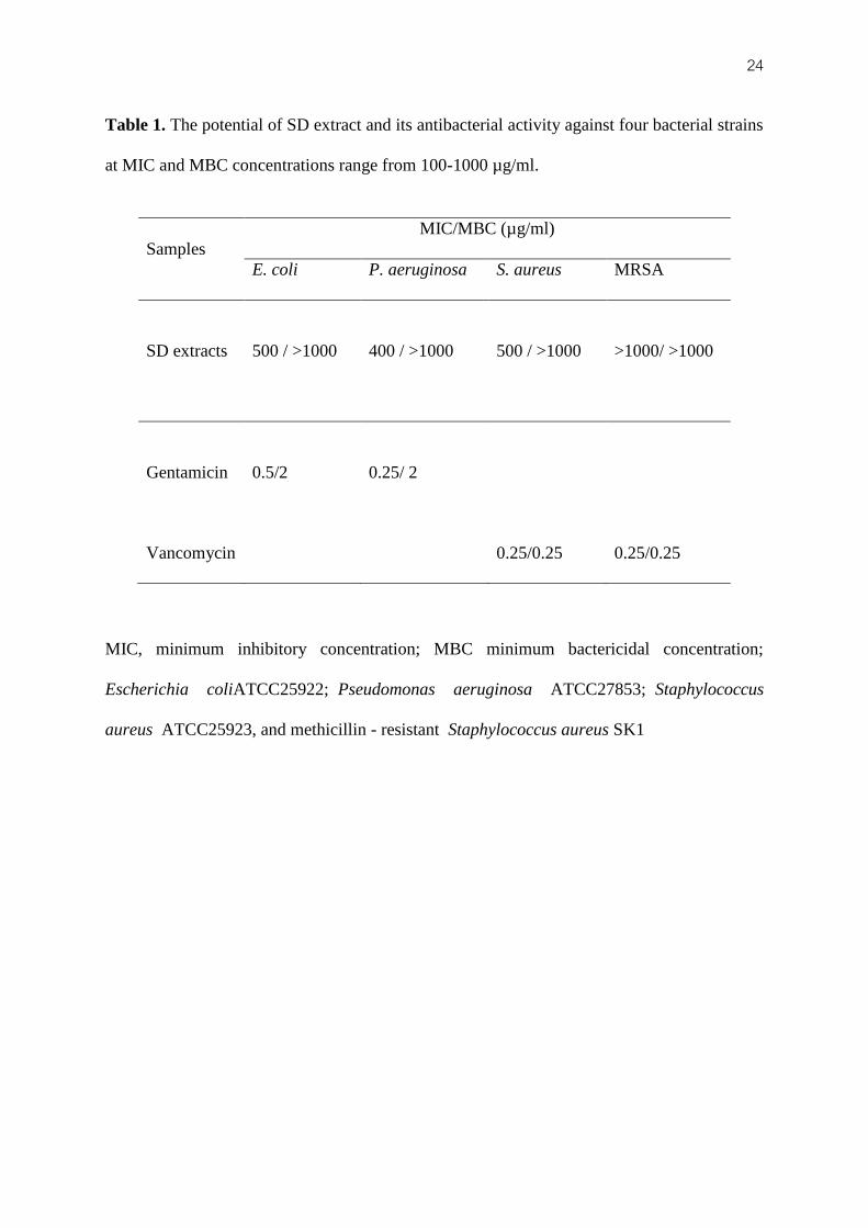
24 Table 1. The potential of SD extract and its antibacterial activity against four bacterial strains
at MIC and MBC concentrations range from 100-1000 µg/ml.
MIC, minimum inhibitory concentration; MBC minimum bactericidal concentration;
Escherichia coliATCC25922; Pseudomonas aeruginosa ATCC27853; Staphylococcus
aureus ATCC25923, and methicillin - resistant Staphylococcus aureus SK1
Samples
MIC/MBC (µg/ml)
E. coli P. aeruginosa S. aureus MRSA
SD extracts
500 / >1000 400 / >1000 500 / >1000 >1000/ >1000
Gentamicin
0.5/2 0.25/ 2
Vancomycin
0.25/0.25 0.25/0.25