Evaluation of an autologous cancer vaccine for the ...
12
RESEARCH ARTICLE Open Access Evaluation of an autologous cancer vaccine for the treatment of metastatic canine hemangiosarcoma: a preliminary study Michael D. Lucroy 1* , Ryan M. Clauson 1 , Mark A. Suckow 2 , Ferris El-Tayyeb 1 and Ashley Kalinauskas 1 Abstract Background: Canine hemangiosarcoma (HSA) is an aggressive cancer arising from multipotential bone marrow- derived stem cells. Anthracycline chemotherapy drugs have been the mainstay adjuvant chemotherapy following surgery with only modest improvement in survival and an attendant risk for adverse events. Immunotherapy, using a whole cell autologous cancer vaccine adjuvanted with MIM-SIS, may improve outcomes for dogs with HSA with a lower risk for adverse events compared with chemotherapy. Results: In cultured DH82 canine monocyte-like cells, autologous cancer vaccines prepared from 13 dogs with HSA increased MHC-II surface expression ranging from 20.0-60.4% on single-stained cells, CD80 surface expression ranging from 23.7–45.9% on single-stained cells, and MHC-II/CD80 surface expression ranging from 7.2–20.1% on double-stained cells. Autologous cancer vaccines were able to, on average, stimulate an up-regulation of MHC-II and CD80 by 48-fold as compared to media only (MHC-II + CD80 + cells: 12.19 ± 3.70% vs. 0.25 ± 0.06%; p < 0.001). The overall median survival time for dogs treated with the autologous cancer vaccine was 142 days (range, 61 to 373 days). Dogs treated with the autologous cancer vaccine or maximum tolerated dose (MTD) chemotherapy had significantly (P < 0.001) longer survival than dogs treated with surgery alone. The 1-year survival rate was 12.5% for dogs treated with the autologous cancer vaccine, and 0% for dogs treated with surgery alone or MTD chemotherapy. No adverse events were observed in the dogs treated with the autologous cancer vaccine. Conclusions: The adjuvanted autologous cancer vaccine is capable of up-regulating MHC-II and CD80 in cultured canine monocyte-derived cells, which are important stimulatory molecules in generating an immune response and improves survival time in dogs with metastatic (stage III) HSA when compared to surgical treatment alone. Autologous cancer vaccine-treated dogs had survival similar to those dogs treated with MTD chemotherapy without any observed adverse events. This autologous cancer vaccine represents an effective form of individualized immunotherapy that is an appealing option for dog owners not wanting to pursue adjuvant chemotherapy for HSA. Keywords: Immunotherapy, Canine hemangiosarcoma, CD80, MCHII, Autologous cancer vaccine © The Author(s). 2020 Open Access This article is licensed under a Creative Commons Attribution 4.0 International License, which permits use, sharing, adaptation, distribution and reproduction in any medium or format, as long as you give appropriate credit to the original author(s) and the source, provide a link to the Creative Commons licence, and indicate if changes were made. The images or other third party material in this article are included in the article's Creative Commons licence, unless indicated otherwise in a credit line to the material. If material is not included in the article's Creative Commons licence and your intended use is not permitted by statutory regulation or exceeds the permitted use, you will need to obtain permission directly from the copyright holder. To view a copy of this licence, visit http://creativecommons.org/licenses/by/4.0/. The Creative Commons Public Domain Dedication waiver (http://creativecommons.org/publicdomain/zero/1.0/) applies to the data made available in this article, unless otherwise stated in a credit line to the data. * Correspondence: [email protected] 1 Torigen Pharmaceuticals, Inc, 400 Farmington Avenue R1855 CB129, 06032 Farmington, CT, USA Full list of author information is available at the end of the article Lucroy et al. BMC Veterinary Research (2020) 16:447 https://doi.org/10.1186/s12917-020-02675-y
Transcript of Evaluation of an autologous cancer vaccine for the ...

RESEARCH ARTICLE Open Access
Evaluation of an autologous cancer vaccinefor the treatment of metastatic caninehemangiosarcoma: a preliminary studyMichael D. Lucroy1* , Ryan M. Clauson1, Mark A. Suckow2, Ferris El-Tayyeb1 and Ashley Kalinauskas1
Abstract
Background: Canine hemangiosarcoma (HSA) is an aggressive cancer arising from multipotential bone marrow-derived stem cells. Anthracycline chemotherapy drugs have been the mainstay adjuvant chemotherapy followingsurgery with only modest improvement in survival and an attendant risk for adverse events. Immunotherapy, usinga whole cell autologous cancer vaccine adjuvanted with MIM-SIS, may improve outcomes for dogs with HSA with alower risk for adverse events compared with chemotherapy.
Results: In cultured DH82 canine monocyte-like cells, autologous cancer vaccines prepared from 13 dogs with HSAincreased MHC-II surface expression ranging from 20.0-60.4% on single-stained cells, CD80 surface expressionranging from 23.7–45.9% on single-stained cells, and MHC-II/CD80 surface expression ranging from 7.2–20.1% ondouble-stained cells. Autologous cancer vaccines were able to, on average, stimulate an up-regulation of MHC-IIand CD80 by 48-fold as compared to media only (MHC-II + CD80 + cells: 12.19 ± 3.70% vs. 0.25 ± 0.06%; p < 0.001).The overall median survival time for dogs treated with the autologous cancer vaccine was 142 days (range, 61 to373 days). Dogs treated with the autologous cancer vaccine or maximum tolerated dose (MTD) chemotherapy hadsignificantly (P < 0.001) longer survival than dogs treated with surgery alone. The 1-year survival rate was 12.5% fordogs treated with the autologous cancer vaccine, and 0% for dogs treated with surgery alone or MTDchemotherapy. No adverse events were observed in the dogs treated with the autologous cancer vaccine.
Conclusions: The adjuvanted autologous cancer vaccine is capable of up-regulating MHC-II and CD80 in culturedcanine monocyte-derived cells, which are important stimulatory molecules in generating an immune response andimproves survival time in dogs with metastatic (stage III) HSA when compared to surgical treatment alone.Autologous cancer vaccine-treated dogs had survival similar to those dogs treated with MTD chemotherapywithout any observed adverse events. This autologous cancer vaccine represents an effective form of individualizedimmunotherapy that is an appealing option for dog owners not wanting to pursue adjuvant chemotherapy forHSA.
Keywords: Immunotherapy, Canine hemangiosarcoma, CD80, MCHII, Autologous cancer vaccine
© The Author(s). 2020 Open Access This article is licensed under a Creative Commons Attribution 4.0 International License,which permits use, sharing, adaptation, distribution and reproduction in any medium or format, as long as you giveappropriate credit to the original author(s) and the source, provide a link to the Creative Commons licence, and indicate ifchanges were made. The images or other third party material in this article are included in the article's Creative Commonslicence, unless indicated otherwise in a credit line to the material. If material is not included in the article's Creative Commonslicence and your intended use is not permitted by statutory regulation or exceeds the permitted use, you will need to obtainpermission directly from the copyright holder. To view a copy of this licence, visit http://creativecommons.org/licenses/by/4.0/.The Creative Commons Public Domain Dedication waiver (http://creativecommons.org/publicdomain/zero/1.0/) applies to thedata made available in this article, unless otherwise stated in a credit line to the data.
* Correspondence: [email protected] Pharmaceuticals, Inc, 400 Farmington Avenue R1855 CB129, 06032Farmington, CT, USAFull list of author information is available at the end of the article
Lucroy et al. BMC Veterinary Research (2020) 16:447 https://doi.org/10.1186/s12917-020-02675-y
BackgroundCanine hemangiosarcoma (HSA) is an aggressive, ultim-ately fatal, cancer arising from multipotential bonemarrow-derived stem cells that arrest their differenti-ation at the hemangioblast or angioblast stage [1]. Basedon gene expression profiling revealing upregulation ofVEGF, MMPs, TIMPs, etc. and enrichment of cytokines,it appears inflammation and angiogenesis are two im-portant processes in the pathogenesis of canine HSA [2].The HSA microenvironment has also been shown to en-hance tumor growth and promote migration of tumorcells through chemokines such as IL-8 and CXCL12 andmodified sphingosines [3]. The spleen is the most com-mon site of HSA in the dog, although liver, heart, andskeletal muscle may also be sites of origin [3, 4]. Aftersplenectomy, or wide surgical excision at other sites, re-ported median survival times range from 19 to 110 days,with death typically due to widespread metastasis whichoften results in significant hemorrhage [4–10]. Dogswith metastatic disease at the time of diagnosis (stageIII) have a poor prognosis, with reported median survivaltimes from 23 to 40 days following surgical removal ofthe primary tumor [5, 9, 10].For nearly 30 years, anthracycline chemotherapy drugs
(most notably doxorubicin) have been the mainstay adju-vant chemotherapy following splenectomy, and modestlyprolongs survival of affected dogs. When combined withsurgery, doxorubicin-based chemotherapy protocols havereported median survival times ranging from 140 to202 days [11]. Epirubicin, another anthracycline drug, hasa reported median survival time of 144 days when used totreat canine splenic HSA after splenectomy [12]. Combin-ing doxorubicin with other chemotherapy drugs does nottypically prolong patient survival, and additions of tocera-nib, vincristine with cyclophosphamide, or metronomiccyclophosphamide, have reported median survival timesof 172, 145 and 202 days, respectively, in populations ofdogs that included those without metastatic disease (stageI and II) [13–15]. However, the addition of dacarbazinehas been reported to increase the median survival time to> 550 days in a group of 9 dogs with stage II HSA [16].Doxorubicin chemotherapy is modestly effective for dogswith stage III HSA, extending the median survival time to107 to 140 days [10, 17]. Altered expression of the ATP-binding cassette transporters ABCB1 and ABCG2 may bepartly responsible for the inherent drug resistance ob-served in canine HSA [18].In addition to the potential for prolonging survival in
dogs with HSA, chemotherapy also has the potential tocause adverse events. In dogs with stage III HSA treatedwith anthracycline chemotherapy, 43.5% had adverseevents, with 21.7% of treated dogs requiringhospitalization for supportive care [10]. The combin-ation of doxorubicin, vincristine and cyclophosphamide
(VAC protocol) to treat canine HSA caused neutropeniain 73% of dogs, 27% had severe gastrointestinal (GI) ad-verse events, and 13% developed sepsis [14]. Epirubicinis also reported to have a high rate of adverse eventswhen used to treat cancer-bearing dogs [19]. Despite thepositive effect on survival for cancer-bearing dogs, recentfindings show that the majority of surveyed pet ownerswould not pursue chemotherapy due to concerns aboutthe risk for adverse events [20].Immunotherapy is another treatment option that may
improve patient outcomes for dogs with HSA with a lowerrisk for adverse events compared with chemotherapy. Anallogenic vaccine, consisting of lysates from two canineHSA cell lines and a liposome-DNA adjuvant, was admin-istered to 28 dogs with various forms of HSA via intraperi-toneal (IP) injection [21]. In subset analysis of 13 dogswith stage II HSA treated with only doxorubicin and thevaccine, the reported median survival time was 182 days,which is similar to chemotherapy alone. A dendritic cellvaccine was evaluated in 5 dogs with splenic HSA [22].Monocytes isolated from peripheral blood were incubatedwith tumor-derived mRNA to create the vaccine, whichwas administered via IP injection. The dogs were alsotreated with doxorubicin (20 mg/m2). The reported me-dian survival time was 109 days. Both of these vaccine ap-proaches were reportedly well-tolerated.Autologous tissue vaccine is another immunotherapy
option for dogs with HSA. Autologous tissue vaccinesconsist of cells harvested from the patient’s own tumor,which presents the full repertoire of the patient’s uniquetumor-associated antigens (TAA) to the immune system.A method of creating autologous tumor vaccines fordogs has been recently described, and relies on enzym-atic cell dissociation, which could destroy TAA [23]. Al-though some humoral response was described in thetreated dogs, survival outcome was not reported for thesingle dog with HSA in that study population.Another method of autologous cancer immunotherapy
is a whole cell autologous cancer vaccine, describedherein. Cancer cells reproduce within a mesh of support-ing tumor stroma, and reciprocal interactions betweenneoplastic cells and their surrounding microenvironmentare critical factors in tumor progression. Many antican-cer immunotherapies have focused on persuading thehost immune system to recognize specific TAA; how-ever, the ability for tumor destruction by such vaccineshas been disappointing, perhaps because TAA are lostover time in the growing tumor, do not represent thefull repertoire of antigens, and/or the host grows toler-ant to them. For this reason, we chose an approach thatpresents the immune system with a broad menu of anti-gen targets, including those that are associated with neo-plastic cells and those that are associated with the tumorstroma that is essential to tumor growth and spread.
Lucroy et al. BMC Veterinary Research (2020) 16:447 Page 2 of 12

This whole cell autologous cancer vaccine is generatedby a novel method of mechanically dissociating tumorcells followed by chemical inactivation, to preserve TAA,and then mixing the cellular material with MIM-SIS(matrix immunomodulator, small intestine submucosa-derived), a protein immune adjuvant [24]. Extracellularmatrix bioscaffolds used in tissue and wound repair appli-cations are pro-inflammatory, and modulation of macro-phage phenotype is believed to play an important role inthe mechanism of action [25]. Because of this ability todrive the immune response, medical grade SIS has beenevaluated for use as an adjuvant for therapeutic cancervaccines. Immunotherapy with this whole cell MIM-SISadjuvanted autologous cancer vaccine has shown efficacyin rodent models of prostate cancer [26–28]. These datasupport the idea that MIM-SIS may be effective whenused as an adjuvant for tissue vaccines in a clinical situ-ation. With this approach, tumor cells are not cultured,avoiding the changes in antigenic profiles that occurin vitro as demonstrated by microarray analysis [29]. Thisautologous cancer vaccine also appears to be well-tolerated by cancer-bearing dogs [24].With therapeutic cancer vaccines, the goal is to in-
crease presentation of TAA to the immune system,thereby increasing activation of tumor-specific T cells,favoring the elimination and equilibrium stages of cancerimmunoediting in the host [30]. The result is an en-hanced ability of the immune system to destroy cancercells, stop or slow the growth of cancer cells, or delaymetastasis which can improve patient survival. The pur-pose of this study was to determine the in vitro effects ofthis whole cell adjuvanted autologous cancer vaccine ona canine monocyte cell line, and determine the effect on
postoperative survival of dogs with metastatic HSA whencompared to a historical control group of dogs withmetastatic HSA treated with either surgery alone or sur-gery followed by standard anthracycline-based chemo-therapy protocols.
ResultsMechanism of action assaySurrogate autologous cancer vaccine preparations wereformulated from 13 dogs with HSA of variable originand stage (Table 1). These surrogate vaccines were uti-lized to assess their immunostimulatory potentialin vitro in terms of their ability to stimulate antigenpresentation (MHC-II) and co-stimulation (CD80) in ca-nine DH82 cells evaluated via flow cytometry (Fig. 1).From this analysis, it was demonstrated that autologouscancer vaccines increased MHC-II surface expressionranging from 20.0-60.4% on single-stained cells (Fig. 2a-b), CD80 surface expression ranging from 23.7–45.9%on single-stained cells (Fig. 2c-d), and MHC-II/CD80surface expression ranging from 7.2–20.1% on double-stained cells (Fig. 2e-f). Notably, autologous cancervaccines (n = 13) were able to, on average, stimulate anup-regulation of MHC-II and CD80 by 48-fold as com-pared to media only (MHC-II + CD80 + cells: 12.19 ±3.70% vs. 0.25 ± 0.06%; p < 0.001). As compared toautologous cells alone without MIM-SIS adjuvant, au-tologous cancer vaccines increased the expression ofMHC-II and CD80 by 53.7% (MHC-II + CD80 + cells:12.19 ± 3.70% vs. 7.93 ± 0.46%; p = 0.18). Lastly, as com-pared to MIM-SIS adjuvant only, autologous cancer vac-cines increased the expression of MHC-II and CD80 by
Table 1 Characteristics of dogs with hemangiosarcoma utilized for mechanism of action evaluation
Patient Breed Age (y) Sex/Statusb Weight (kg) Primary Metastasis
16–034 Mixed 6 F/S 12.2 Subcutaneous No
18–082 Golden retriever NRa M/C 40.9 Liver Yes
19–039 Mixed 12 M/C 36.2 Subcutaneous No
19–060 Boxer 7 M/I 29.5 Subcutaneous No
19–195 Rottweiler 3 F/S 34.9 Bonec No
19–199 Labrador retriever 12 M/C NR Spleen No
19–261 Golden retriever 10 F/S 25.9 Spleen No
20−008 Mixed NR M/C 12.2 Spleen No
20−010 Coonhound 7 F/S 31.8 Spleen No
20–020 Mixed 13 M/C 22.7 Subcutaneous No
20–025 English Setter 14 M/C 31.6 Spleen No
20–027 Mixed 10 M/C 24.0 Spleen No
20–040 Mixed 10 M/C 41.5 Spleen YesaNR Not reportedbF female, M male, S spayed, C castrated, I intactcThe submitting veterinarian did not pursue immunohistochemisty, so telangiectatic osteosarcoma is not excluded. However the pathologist did not find anythingto support osteosarcoma in the tissue sectons examined
Lucroy et al. BMC Veterinary Research (2020) 16:447 Page 3 of 12
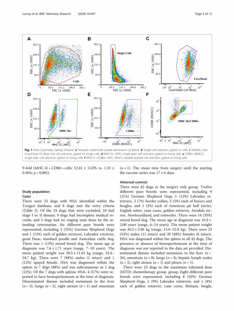
9-fold (MHC-II + CD80 + cells: 12.45 ± 3.53% vs. 1.33 ±0.36%; p < 0.001).
Study populationCasesThere were 32 dogs with HSA identified within theTorigen database, and 8 dogs met the entry criteria(Table 2). Of the 24 dogs that were excluded, 10 hadstage I or II disease, 9 dogs had incomplete medical re-cords, and 5 dogs had no staging tests done by the at-tending veterinarian. Six different pure breeds wererepresented, including 2 (25%) German Shepherd Dogsand 1 (13%) each of golden retriever, Labrador retriever,great Dane, standard poodle and Australian cattle dog.There was 1 (13%) mixed breed dog. The mean age atdiagnosis was 7.6 ± 1.71 years (range, 7–10 years). Themean patient weight was 39.5 ± 11.83 kg (range, 22.4–54.7 kg). There were 7 (88%) males (1 intact) and 1(12%) spayed female. HSA was diagnosed within thespleen in 7 dogs (88%) and was subcutaneous in 1 dog(12%). Of the 7 dogs with splenic HSA, 4 (57%) were re-ported to have hemoperitoneum at the time of diagnosis.Disseminated disease included metastasis to the liver(n = 3), lungs (n = 3), right atrium (n = 1) and omentum
(n = 1). The mean time from surgery until the startingthe vaccine series was 17 ± 6 days.
Historical controlsThere were 42 dogs in the surgery only group. Twelvedifferent pure breeds were represented, including 9(21%) German Shepherd Dogs 5 (12%) Labrador re-trievers, 3 (7%) border collies, 2 (5%) each of boxers andbeagles, and 1 (2%) each of American pit bull terrier,English setter, cane corso, golden retriever, Airedale ter-rier, Newfoundland, and rottweiler. There were 14 (33%)mixed breed dog. The mean age at diagnosis was 10.3 ±2.00 years (range, 6–14 years). The mean patient weightwas 30.2 ± 5.96 kg (range, 15.0–52.8 kg). There were 22(52%) males (11 intact) and 20 (48%) females (6 intact).HSA was diagnosed within the spleen in all 42 dogs. Thepresence or absence of hemoperitoneum at the time ofdiagnosis was not reported in the data set provided. Dis-seminated disease included metastasis to the liver (n =26), omentum (n = 8), lungs (n = 4), hepatic lymph nodes(n = 2), right atrium (n = 1) and pleura (n = 1).There were 23 dogs in the maximum tolerated dose
(MTD) chemotherapy group. group. Eight different purebreeds were represented, including 8 (35%) GermanShepherd Dogs, 2 (9%) Labrador retrievers, and 1 (4%)each of golden retriever, cane corso, Brittany, beagle,
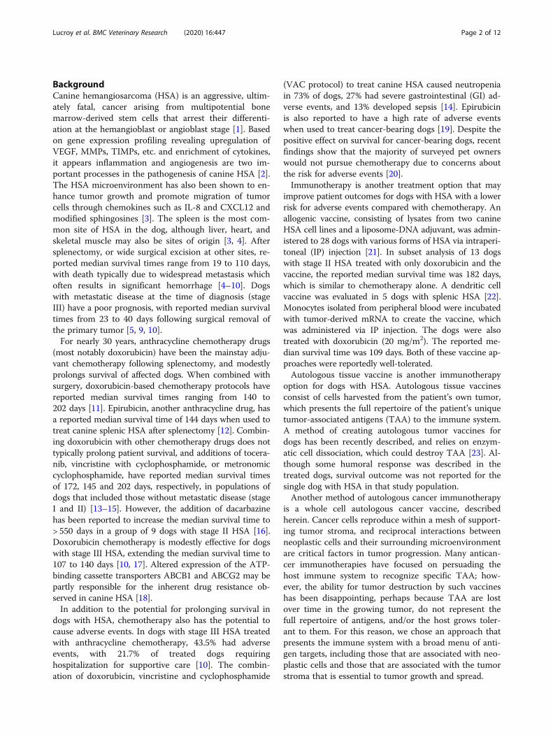
Fig. 1 Flow Cytometry Gating Scheme. a Forward scatter/side scatter elimination of debris. b Single cell selection, gated on cells. c Viability stain(Live/Dead UV Blue) live cell selection, gated on single cells. d MHC-II+ (APC) single-stain cell selection, gated on living cells. e CD80+ (BV421)single-stain cell selection, gated on living cells. f MHC-II + CD80+ (APC, BV421) double-stained cell selection, gated on living cells
Lucroy et al. BMC Veterinary Research (2020) 16:447 Page 4 of 12
dachshund, and Czechoslovakian wolf dog. There were 7(30%) mixed breed dogs. The mean age at diagnosis was10.0 ± 2.21 years (range, 7–14 years). The mean patientweight was 31.6 ± 9.84 kg (range, 5.1–47.0 kg). There were14 (61%) males (12 intact) and 9 (39%) females (4 intact).HSA was diagnosed within the spleen in all 23 dogs. Infor-mation regarding hemoperitoneum at diagnosis was notdescribed in the shared data set. Disseminated disease in-cluded metastasis to the liver (n = 16), omentum (n = 5)and lungs (n = 1). In the MTD chemotherapy group,standard anthracycline-based protocols were used, and 17(74%) dogs were treated with doxorubicin, 3 (13%) dogswere treated with epirubicin, 2 (9%) dogs were treatedwith doxorubicin and cyclophosphamide, and 1 (4%) dogwas treated with doxorubicin and dacarbazine.The proportion of male dogs was significantly higher
in the autologous cancer vaccine group when comparedto the surgery only group (p < 0.05). The proportion ofmale dogs was not significantly different between the au-tologous cancer vaccine and MTD chemotherapy
groups. There were no significant differences in varianceof weights or ages among of the treatment groups.
Clinical outcomesAdverse eventsIn the 8 dogs treated with the adjuvanted autologous can-cer vaccine, no episodes of anaphylaxis or other adverseevents were reported following any of the 24 doses admin-istered. In the dogs treated with MTD chemotherapy, 10(43%) dogs were reported to have at least 1 adverse event,and 4 of the 10 dogs had multiple adverse events. Overall,4 (17%) dogs required hospitalization for grade 3–4 bonemarrow toxicity (n = 2) and grade 3 GI toxicity (n = 2).
SurvivalProgressive HSA was the cause of death in all dogs thatdied during the follow-up period in both the historicalcontrol group and autologous cancer vaccine group.Four dogs were right-censored (1 dog treated with the
Fig. 2 Antigen Presentation Assay, CD80 and MHC-II Expression. Thirteen autologous cancer vaccines (ACV), media only control, glutaraldehyde-fixed (GFT) cell only control and MIM-SIS only control. a MHC-II+ single stained cell percentage of total live cells, individual ACV preparationsplotted; data represent mean ± SD, n = 3. b MHC-II+ single stained cell percentage of total live cells, pooled ACV preparations plotted; datarepresent mean ± SD, n = 3 (controls) and n = 39 (ACV). c CD80 + single stained cell percentage of total live cells, individual ACV preparationsplotted; data represent mean ± SD, n = 3. d CD80+ single stained cell percentage of total live cells, pooled ACV preparations plotted; datarepresent mean ± SD, n = 3 (controls) and n = 39 (ACV). e MHC-II + CD80 + double stained cell percentage of total live cells, individual ACVpreparations plotted; data represent mean ± SD, n = 3. (F) MHC-II + CD80 + double stained cell percentage of total live cells, pooled ACVpreparations plotted; data represent mean ± SD, n = 3 (controls) and n = 39 (ACV). Statistical comparisons are based on one-way ANOVA, followedby post hoc Tukey’s pairwise comparisons. The asterisks denote statistical significance at the level of * p < 0.05, ** p < 0.01, *** p < 0.001. ANOVA,analysis of variance; SD, standard deviation
Lucroy et al. BMC Veterinary Research (2020) 16:447 Page 5 of 12
autologous cancer vaccine and 3 dogs treated with sur-gery alone).The median survival time for dogs treated with surgery
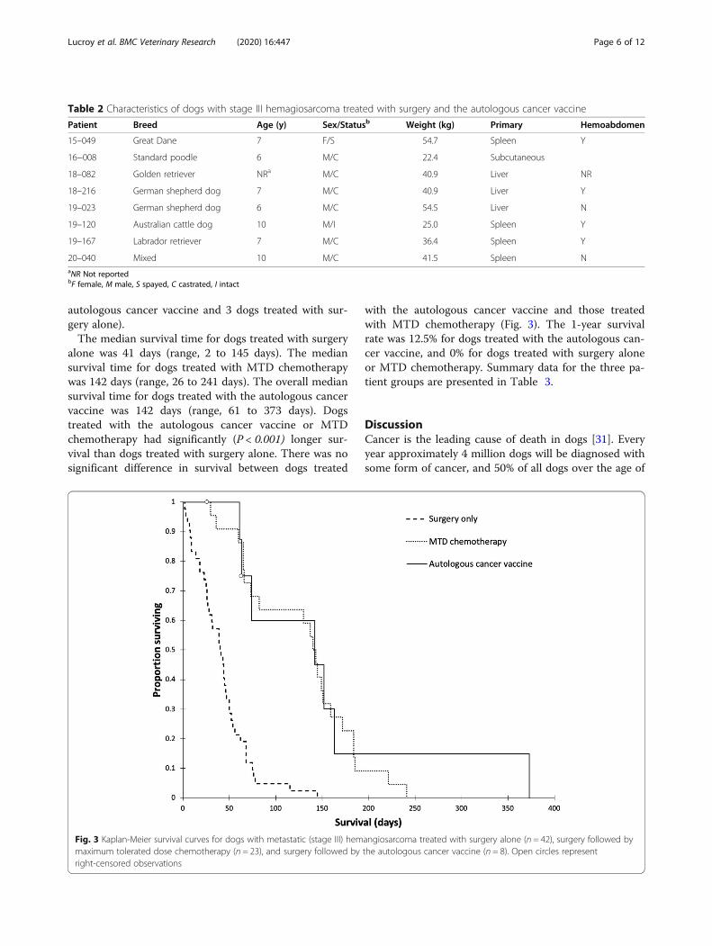
alone was 41 days (range, 2 to 145 days). The mediansurvival time for dogs treated with MTD chemotherapywas 142 days (range, 26 to 241 days). The overall mediansurvival time for dogs treated with the autologous cancervaccine was 142 days (range, 61 to 373 days). Dogstreated with the autologous cancer vaccine or MTDchemotherapy had significantly (P < 0.001) longer sur-vival than dogs treated with surgery alone. There was nosignificant difference in survival between dogs treated
with the autologous cancer vaccine and those treatedwith MTD chemotherapy (Fig. 3). The 1-year survivalrate was 12.5% for dogs treated with the autologous can-cer vaccine, and 0% for dogs treated with surgery aloneor MTD chemotherapy. Summary data for the three pa-tient groups are presented in Table 3.
DiscussionCancer is the leading cause of death in dogs [31]. Everyyear approximately 4 million dogs will be diagnosed withsome form of cancer, and 50% of all dogs over the age of
Fig. 3 Kaplan-Meier survival curves for dogs with metastatic (stage III) hemangiosarcoma treated with surgery alone (n = 42), surgery followed bymaximum tolerated dose chemotherapy (n = 23), and surgery followed by the autologous cancer vaccine (n = 8). Open circles representright-censored observations
Table 2 Characteristics of dogs with stage III hemagiosarcoma treated with surgery and the autologous cancer vaccine
Patient Breed Age (y) Sex/Statusb Weight (kg) Primary Hemoabdomen
15–049 Great Dane 7 F/S 54.7 Spleen Y
16−008 Standard poodle 6 M/C 22.4 Subcutaneous
18–082 Golden retriever NRa M/C 40.9 Liver NR
18–216 German shepherd dog 7 M/C 40.9 Liver Y
19–023 German shepherd dog 6 M/C 54.5 Liver N
19–120 Australian cattle dog 10 M/I 25.0 Spleen Y
19–167 Labrador retriever 7 M/C 36.4 Spleen Y
20–040 Mixed 10 M/C 41.5 Spleen NaNR Not reportedbF female, M male, S spayed, C castrated, I intact
Lucroy et al. BMC Veterinary Research (2020) 16:447 Page 6 of 12

10 will die as a result of developing the disease [31, 32].A major driving force behind these statistics is that,unfortunately, the therapeutic approaches used fortreating dogs with cancer has lagged behind that ofhumans, resulting in sub-optimal clinical outcomes[33]. Immunotherapy offers a new approach to cancertreatment by re-educating the patient’s immune sys-tem to combat the disease [34, 35]. This re-educationprocess is accomplished by providing back stimulatoryfactors and tumor-associated antigens that allow for thehost immune system to recognize the tumor as foreign.One such immunotherapeutic modality that is capable ofperforming such function is the autologous whole tissuecancer vaccine being developed by Torigen Pharmaceuti-cals [24].Utilizing an in vitro assay of antigen presentation and
DH82 canine monocyte-like cells, it was possible tohighlight the mechanism of action of autologous cancervaccines combined with MIM-SIS adjuvant. Major histo-compatibility complex II (MHC-II) are molecules foundon professional antigen-presenting cells (amongstothers) that serve to present epitopes to T lymphocytes.Specifically, MHC-II molecules present longer peptide-based epitopes utilized for priming of CD4 + T helpercells. Notably, MHC-I molecules facilitate priming ofCD8 + cytotoxic T-cells that would be more directly rele-vant in the context of cancer immunotherapy, but nocommercially available or literature published canine spe-cific antibody is available at present for diagnostics. Be-yond MHC, CD80 (or CD86) is a co- stimulatorymolecule that increases in expression in response to anti-gen and facilitates T-cell priming by co-ligation withCD28 T-cells. Together, MHC and CD80 represent two ofthe three required signals for T lymphocyte activation anddifferentiation; the third being a local cytokine environ-ment. Accordingly, it is reasonable to rationalize that theability to upregulate the expression of these surface mole-cules on canine monocytes could be indicative of greaterpotential T lymphocyte response in vivo. As such, thistechnique has been applied in numerous studies evaluat-ing new vaccine technologies including recombinant anti-gens, novel adjuvants and variable formulation schemes
[36–42]. Specifically, the extent of major histocompatibil-ity complex II (MHC-II) and CD80 co-stimulatory mole-cules were up-regulated in response to incubation withvaccine or relevant controls. These signals are traditionalmarkers of antigen-presentation and are correlated to theability to stimulate T-cell activation in vivo [43–45]. Not-ably, the MHC-II signal is indicative of helper T-cell acti-vation, not cytotoxic T-cell activation [46]; at this time, novalidated canine-specific antibody is commercially avail-able for MHC-I quantification, a marker of cytotoxic T-cell activation, via flow cytometry.The results of the antigen presentation assay sug-
gest that there is an immunostimulatory synergy be-tween autologous cancer cells and MIM-SIS adjuvant.Specifically, while autologous cancer cells alone (GFTcells) are capable of stimulating a robust extent ofantigen-presentation, they fail to provide the co-stimulatory signal that is absolutely required for T-cell activation. Moreover, MIM-SIS alone promotesonly weak co-stimulatory responses via CD80 surfaceexpression. It is only when autologous cancer cellsand MIM-SIS adjuvant are combined that there is a53.7% increase in dual signaling of MHC-II and CD80on canine cells, and thus a more robust potential forimmune activation. The results of this assay indicatethat there is no clear correlation between antigen-presentation/co-stimulation and tumor stage evaluatedin an in vitro setting. In vivo, there could absolutelybe a difference based upon the immunological statusof that individual patient, which is highly dependenton manner factors even beyond the tumor type andgrade. The most dominant discrepancy betweenin vitro and in vivo evaluations of the immune re-sponse would be the immunosuppressive tumor envir-onment (negative impact), as well as tumormutational burden (positive impact). Ultimately, theability to upregulate CD80 and MHC-II would be de-rived from a balance of these two factors. Accord-ingly, this evidence, combined with our previous pre-clinical evaluations [26–29], provides justification forfurther research and warrants initial clinical validationin canine models.
Table 3 Summary data from 73 dogs with metastatic hemangiosarcoma
Surgery alone(n = 42)
Surgery plus chemotherapy(n = 23)
Surgery plus autologous cancer vaccine(n = 8)
Age, years (mean ± SD) 10.3 ± 2.00 10.0 ± 2.21 7.6 ± 1.71
Weight, kg (mean ± SD) 30.2 ± 5.96 31.6 ± 9.84 39.5 ± 11.83
Adverse events (%) 0 43 0
Hopitalizations (%) 0 17 0
Median survival time, days 41 142 142
Survival range, days 2 to 145 26 to 241 61 to 373
Alive at 1 year (%) 0 0 12.5
Lucroy et al. BMC Veterinary Research (2020) 16:447 Page 7 of 12

The population of dogs described herein was similarin age, weight, and male predominance described to pre-vious reports of HSA [5, 11, 12, 14, 47]. The preponder-ance of German shepherd dogs observed herein has alsobeen previously described [4, 11, 47, 48]. The dogstreated with immunotherapy in this study all toleratedthe autologous cancer vaccine well, with no reported ad-verse events described. This group of dogs had an evenlower proportion of adverse event rate than the 12% re-ported in a population of 93 dogs with various cancerstreated with the autologous cancer vaccine immunother-apy after surgery [24]. This is in stark comparison to theobserved proportion of adverse events (43%) and hospi-talizations (17%) within the MTD chemotherapy group.Although all of the deaths during the follow-up period
were due to progressive HSA, the observed median sur-vival time of 142 days for dogs with metastatic HSA rep-resented a more than 3-fold increase over the 41 daymedian survival time in the historical control grouptreated with surgery alone, and is more than 2.5-foldlonger than the recently reported 54 day median survivaltime following surgery alone [9]. The majority of dogstreated with the adjuvanted autologous cancer vaccinehad hemoperitoneum at presentation. Although therewere no available comparison data from the historicalcontrol group, recent findings that hemoperitoneumdoes not affect survival time of dogs with HSA [49], sug-gests hemoperitoneum would be unlikely to influenceoutcome in the dogs described herein.There were several limitations of this study. First, due
to lack of available specimens, the in vitro work was con-ducted with surrogate HSA tumor vaccines created frompatients not included in the clinical case evaluations andutilized a canine monocyte-like cell line rather thanmonocytes isolated from each specific dog. Although theupregulation of MCH-II and CD80 was demonstrated inthe canine model, the findings were not specific to thedogs studied for survival outcome. However, the advan-tage of using the DH82 cell line is standardization andreplication to facilitate vaccine-to-vaccine comparisons.If patient-specific monocytes were used, there would betwo variables within the assay.Another limitation was the retrospective nature of case
evaluations. Because tumor specimens for the autolo-gous cancer vaccine creation were submitted from a var-iety of clinicians (oncologists, surgeons, and generalpractitioners), pre-operative cancer staging, and post-treatment follow-up varied between patients. This lim-ited the evaluation of vaccine efficacy in this populationof dogs to simple survival analysis. Evaluating time toprogression is an objective measurement of clinical effi-cacy that is free from bias introduced by dog owners’ de-cisions on euthanasia. However, given the aggressivenature of HSA in dogs, the lengths of progression-free
interval and overall survival are typically similar amongdifferent study populations [10, 15, 50, 51].The small number of available dogs with stage III HSA
treated with only surgery and the autologous cancer vac-cine, and heterogeny among the cases, represent otherlimitations of the current study. Regardless, these pre-liminary findings are encouraging; the median survivaltime of vaccinated dogs was significantly longer thandogs treated with surgery alone. Results from a larger,prospective study will be needed to confirm these pre-liminary findings. In addition to treating dogs with meta-static HSA, studies of dogs with stage I and stage II arealso warranted, as the adjuvanted autologous cancer vac-cine may be more effective in dogs with a lower cancerburden, and vaccine effects on preventing metastatic dis-ease could be demonstrated.Compared with other techniques for creating autolo-
gous tumor vaccines, the method used herein to createthe adjuvanted autologous cancer vaccine does not re-quire culturing tumor cells, which allows for rapidmanufacturing of the patient-specific vaccine. Culturingtumor cells increases the chances for microbial contam-ination of the vaccine product. Using mechanical dis-sociation of tumor tissue also preserves TAA which mayinduce a more robust immunologic response.The autologous cancer vaccine protocol used here, ad-
ministering 3 weekly subcutaneous autologous cancervaccine doses, allows a patient with stage III HSA tocomplete the course of therapy quickly, compared todogs undergoing a standard anthracycline (doxorubicinor epirubicin) chemotherapy protocol receiving an intra-venous injection every 3 weeks for 5 to 6 doses [11]. Theautologous cancer vaccine also provides a significantsurvival advantage over surgery alone for dogs with stageIII HSA, with minimal risk for adverse events. Many dogowners are concerned about adverse events associatedwith chemotherapy [20], and the anthracycline drugshave been associated with acute allergic reactions, GIupset, neutropenia, thrombocytopenia, and rarely cardio-toxicity [10, 12, 19, 52–60]. In the present study, adverseevents were common in the historical control grouptreated with standard anthracycline chemotherapy. Add-itionally, for many dog owners, the time and financialcommitments associated with chemotherapy for stage IIIHSA are not feasible, and the outcome expectations alsofactor into the decision-making process [61]. Treatmentwith the autologous cancer vaccine as reported here, andpreviously described, has a low risk of adverse events[24], and may be more affordable than chemotherapy.This method of generating an adjuvanted autologous
cancer vaccine represents an individualized form of im-munotherapy, presenting a range of tumor-specific andhost-specific antigens to the patient’s immune system,that may be appropriate for any solid tumor where
Lucroy et al. BMC Veterinary Research (2020) 16:447 Page 8 of 12

sufficient cells can be collected for vaccine preparation.This is in contrast to other immunotherapy options,such as the canine melanoma vaccine and canine osteo-sarcoma vaccine, which present a single antigen to thepatient’s immune system (human tyrosinase and HER2/neu, respectively) [62, 63].
ConclusionsThe data presented herein demonstrate that immunother-apy with the adjuvanted autologous cancer vaccine is cap-able of up-regulating MHC-II and CD80 in culturedcanine monocyte-derived cells, which are importantstimulatory molecules in generating an immune response,and the vaccine improves survival time in dogs with meta-static (stage III) HSA when compared to surgical treat-ment alone. The autologous cancer vaccine-treated dogshad survival similar to those dogs treated with maximumtolerated dose chemotherapy without any observed ad-verse events. This autologous cancer vaccine representsan effective form of individualized immunotherapy that isan appealing option for dog owners not wanting to pursueadjuvant chemotherapy for HSA.
MethodsMaterialsAll reagents were used as obtained from commercialsources without further purification. DH82 canine ma-lignant histiocytoma cells (macrophage-like, CRL-10,389) were purchased from ATCC (Manassas, VA, USA).Anti-CD80 (Brilliant Violet 421, clone 16-10A1) was ob-tained from BioLegend (San Diego, CA, USA). Anti-MHC-II (APC, YKIX334.2) and Live/Dead Fixable BlueStain Kit (L34962) were obtained from Thermo FisherScientific (Waltham, MA, USA).
DH82 antigen presentation assayThe ability of autologous cancer vaccines to facilitate anti-gen presentation (MHC-II) and co-stimulation (CD80)was evaluated in-vitro using DH82 canine macrophagesand flow cytometry. DH82 cells were maintained at 37 °C,5% CO2/95% air atmosphere and approximately 85% rela-tive humidity. DH82 cell culture media consisted of Dul-becco’s Modified Eagles Media, 15% heat-inactivated fetalbovine serum (VWR, Radnor, PA, USA), 1% SG-200 (GEHealthcare, Chicago, IL, USA), 1% sodium pyruvate (GEHealthcare), 1% antibiotic-antimycotic (Thermo FisherScientific). Prior to performing the assay, DH82 cells wereseeded overnight at 2.5e5 cells/well in 24-well non-treatedcell culture dishes. The antigen presentation assay wasperformed by incubating autologous cancer vaccine prep-arations (0.25e6 cells/well, 0.5 mg/mL MIM-SIS) or rele-vant controls with cells for 48 hours (performed intriplicate). Autologous cancer vaccines were prepared forthirteen unique HSA samples (Table 1) as previously
described [24], from submitted patient samples with a suf-ficient amount of tissue available after creation of theirvaccine for clinical use. After 48 hours, cells are harvestedand prepared for analysis by flow cytometry using the fol-lowing stains: Live/Dead Fixable, anti-CD80 Brilliant Vio-let 421 and anti-MHC-II APC. Cells were analyzed usinga Becton-Dickinson LSR II flow cytometer (FranklinLakes, NJ, USA) at the University of Connecticut HealthCenter Flow Cytometry Core (Farmington, CT, USA) withthe FCS Express 7 software package (DeNovo Software,Pasadena, CA, USA). Fluorescence minus one (FMO) con-trols were utilized to established gating scheme.
Case selectionThe case accession database at Torigen Pharmaceuticals,Inc. was queried to identify dogs diagnosed with stageIII HSA between January 2015 and January 2020 thatwere treated with surgery followed by the adjuvanted au-tologous cancer vaccine only.Dogs were eligible for inclusion in this study if they had a
histopathological diagnosis of non-cutaneous HSA and evi-dence of distant metastasis (i.e., stage III disease) identifiedvia imaging, cytopathology or histopathology, and weretreated by surgery and the adjuvanted autologous cancervaccine only. Dogs were excluded from study if they werediagnosed with cutaneous HSA, had visceral, subcutaneousor intramuscular HSA with no evidence of metastasis (i.e.,stage I or II), did not receive all three doses of the autolo-gous cancer vaccine, received adjuvant chemotherapy, orhad incomplete outcome information available. Histopatho-logic diagnosis was reported by board-certified veterinarypathologists via commercial laboratory services. Patientdata collected included signalment, body weight, histologyresults, primary tumor location, adverse events reportedafter autologous tumor vaccine administration, and survivalfrom the time of tumor removal. Follow up information oneach dog was obtained through direct communication withthe submitting veterinarian. Adverse events were classifiedbased on the Veterinary Cooperative Oncology Groupcommon terminology criteria for adverse events (VCOG-CTCAE) [64].
Historical controlsThe historical controls cases were dogs identified be-tween 2011 and 2018 with stage III HSA that were eithertreated with surgery alone, or surgery followed withstandard maximum tolerated dose (MTD) anthracycline-based chemotherapy. The outcomes of these dogs havebeen previously published [10], and the authors provideda raw data set which included signalment, adverse eventsand hospitalization reported after chemotherapy admin-istration, and survival from the time of tumor removal.Adverse events were classified based on the VCOG-CTCAE [64].
Lucroy et al. BMC Veterinary Research (2020) 16:447 Page 9 of 12
Vaccine protocolPreparation of the adjuvanted autologous cancer vaccinehas been described elsewhere [24]. Briefly, unfixed tumortissue was mechanically dissociated, fixed with glutaralde-hyde, and following multiple washing steps, the fixed cellswere combined with MIM-SIS to create the final vaccineproduct. Veterinarians were instructed to give the vaccineas a subcutaneous injection, as three 1 mL doses, atweekly intervals. The attending veterinarian was advisedto monitor the dog for acute AE for 30 minutes after eachof the three injections. At hospital discharge, dog ownerswere informed of possible vaccine reactions, andinstructed to report any observed abnormalities immedi-ately upon their occurrence. Written owner informed con-sent was obtained before the vaccine was administered.
Statistical analysisIn-vitro data are expressed as mean ± standard deviation(SD). Means of multiple groups were compared with theone-way analysis of variance (ANOVA), followed by posthoc Tukey’s pairwise comparisons. All probability valuesare two-sided, and values of p < 0.05 were consideredstatistically significant. Statistical analyses were carriedout using the GraphPad Prism 7 software package.Among treatment groups, the variance of continuous
variables was compared using Levene’s test, and propor-tions were compared using the Marascuilo procedure.Dogs were right-censored from survival analysis if theywere lost to follow-up, died from an unrelated cause, orwere still alive at the time of data analysis. Progression-free survival was not calculated due to insufficient infor-mation in the database or attending veterinarian medicalrecords. Survival estimates were generated using theKaplan-Meier method, and survival curves were com-pared using the Log-Rank test. Statistical testing wasdone using XLSTAT Life Science (Addinsoft, 2020, NewYork, USA). P values < 0.05 were used to indicate statis-tical significance. Results are reported as mean ± stand-ard deviation unless otherwise noted.
AbbreviationsABC: ATP-binding cassette; GI: Gastrointestinal; HSA: Hemangiosarcoma;IP: Intraperitoneal; MIM-SIS: Matrix immunomodulator, small intestinesubmucosa-derived; MTD: Maximum tolerated dose; TAA: Tumor-associatedantigens
AcknowledgementsSpecial thanks to Laura Marconato, DVM, Dipl. ECVIM-CA (oncology),Professore Associato, Dipartimento di Scienze Mediche Veterinarie, Universitàdi Bologna for providing the historical data set.
Authors' contributionsRC and FE performed the in vitro assays and analyzed and interpreted theflow cytometry data. ML analyzed and interpreted the patient outcome data.MS and AK were instrumental in the conception of and subsequent revisionsof this work. All authors read and approved the final manuscript.
FundingThere was no external funding for this study.
Availability of data and materialsThe datasets generated and/or analyzed during the current study areavailable from the corresponding author on reasonable request.
Ethics approval and consent to participateThe retrospective study described here involved review of medical recordsfrom privately-owned dogs, all receiving care as prescribed by licensed veter-inarians. Under such circumstances, U. S. Federal regulatory agencies do notrequire formal review by an Institutional Animal Care and Use Committee(IACUC) [53]. Ethical assurance regarding the clinical use of the investiga-tional, commercially available, autologous cancer vaccine was providedthrough a mechanism of written informed consent from dog owners, as re-quired by the USDA for unlicensed veterinary biologics.
Consent for publicationNot applicable.
Competing interestsAll authors are affiliated with Torigen Pharmaceuticals, Inc.
Author details1Torigen Pharmaceuticals, Inc, 400 Farmington Avenue R1855 CB129, 06032Farmington, CT, USA. 2Office of the Vice President for Research, University ofKentucky, 445 Bowman Hall, KY 40506-0032 Lexington, USA.
Received: 29 April 2020 Accepted: 9 November 2020
References1. Lamerato-Kozicki AR, Helm KM, Jubala CM, Cutter GC, Modiano JF. Canine
hemangiosarcoma originates from hematopoietic precursors with potentialfor endothelial differentiation. Exp Hematol. 2006;34(7):870–8.
2. Tamburini BA, Phang TL, Fosmire SP, Scott MC, Trapp SC, Duckett MM,Robinson SR, Slansky JE, Sharkey LC, Cutter GR, et al. Gene expressionprofiling identifies inflammation and angiogenesis as distinguishing featuresof canine hemangiosarcoma. BMC Cancer. 2010;10:619.
3. Kim JH, Graef AJ, Dickerson EB, Modiano JF. Pathobiology ofHemangiosarcoma in Dogs: Research Advances and Future Perspectives. VetSci. 2015;2(4):388–405.
4. Brown NO, Patnaik AK, MacEwen EG. Canine hemangiosarcoma:retrospective analysis of 104 cases. J Am Vet Med Assoc. 1985;186(1):56–8.
5. Batschinski K, Nobre A, Vargas-Mendez E, Tedardi MV, Cirillo J, Cestari G,Ubukata R, Dagli MLZ. Canine visceral hemangiosarcoma treated withsurgery alone or surgery and doxorubicin: 37 cases (2005–2014). Can Vet J.2018;59(9):967–72.
6. Wood CA, Moore AS, Gliatto JM, Ablin LA, Berg RJ, Rand WM. Prognosis fordogs with stage I or II splenic hemangiosarcoma treated by splenectomyalone: 32 cases (1991–1993). J Am Anim Hosp Assoc. 1998;34(5):417–21.
7. Prymak C, McKee LJ, Goldschmidt MH, Glickman LT. Epidemiologic, clinical,pathologic, and prognostic characteristics of splenic hemangiosarcoma andsplenic hematoma in dogs: 217 cases (1985). J Am Vet Med Assoc. 1988;193(6):706–12.
8. Cleveland MJ, Casale S. Incidence of malignancy and outcomes for dogsundergoing splenectomy for incidentally detected nonruptured splenicnodules or masses: 105 cases (2009–2013). J Am Vet Med Assoc. 2016;248(11):1267–73.
9. Wendelburg KM, Price LL, Burgess KE, Lyons JA, Lew FH, Berg J. Survivaltime of dogs with splenic hemangiosarcoma treated by splenectomy withor without adjuvant chemotherapy: 208 cases (2001–2012). J Am Vet MedAssoc. 2015;247(4):393–403.
10. Marconato L, Chalfon C, Finotello R, Polton G, Vasconi ME, Annoni M,Stefanello D, Mesto P, Capitani O, Agnoli C, et al. Adjuvant anthracycline-based vs metronomic chemotherapy vs no medical treatment for dogswith metastatic splenic hemangiosarcoma: A multi‐institutionalretrospective study of the Italian Society of Veterinary Oncology. VeterinaryComparative Oncology. 2019;17(4):537–44.
11. Clifford CA, Mackin AJ, Henry CJ. Treatment of canine hemangiosarcoma:2000 and beyond. J Vet Intern Med. 2000;14(5):479–85.
12. Kim SE, Liptak JM, Gall TT, Monteith GJ, Woods JP. Epirubicin in the adjuvanttreatment of splenic hemangiosarcoma in dogs: 59 cases (1997–2004). J AmVet Med Assoc. 2007;231(10):1550–7.
Lucroy et al. BMC Veterinary Research (2020) 16:447 Page 10 of 12

13. Gardner HL, London CA, Portela RA, Nguyen S, Rosenberg MP, Klein MK,Clifford C, Thamm DH, Vail DM, Bergman P, et al. Maintenance therapy withtoceranib following doxorubicin-based chemotherapy for canine splenichemangiosarcoma. BMC Vet Res. 2015;11:131.
14. Hammer AS, Couto CG, Filppi J, Getzy D, Shank K. Efficacy and toxicity ofVAC chemotherapy (vincristine, doxorubicin, and cyclophosphamide) indogs with hemangiosarcoma. J Vet Intern Med. 1991;5(3):160–6.
15. Alexander CK, Cronin KL, Silver M, Gardner HL, London C. The addition ofmetronomic chemotherapy does not improve outcome for canine splenichaemangiosarcoma. J Small Anim Pract. 2019;60(1):32–7.
16. Finotello R, Stefanello D, Zini E, Marconato L. Comparison of doxorubicin-cyclophosphamide with doxorubicin-dacarbazine for the adjuvanttreatment of canine hemangiosarcoma. Vet Comp Oncol. 2017;15(1):25–35.
17. Sorenmo KU, Baez JL, Clifford CA, Mauldin E, Overley B, Skorupski K,Bachman R, Samluk M, Shofer F. Efficacy and toxicity of a dose-intensifieddoxorubicin protocol in canine hemangiosarcoma. J Vet Intern Med. 2004;18(2):209–13.
18. Khammanivong A, Gorden BH, Frantz AM, Graef AJ, Dickerson EB.Identification of drug-resistant subpopulations in canine hemangiosarcoma.Vet Comp Oncol. 2016;14(3):e113–25.
19. Marrington AM, Killick DR, Grant IA, Blackwood L. Toxicity associated withepirubicin treatments in a large case series of dogs. Vet Comp Oncol. 2012;10(2):113–23.
20. Williams J, Phillips C, Byrd HM. Factors Which Influence Owners WhenDeciding to Use Chemotherapy in Terminally Ill Pets. Animals (Basel).2017;7(3):18. Published 2017 Mar 7. https://doi.org/10.3390/ani7030018.
21. U’Ren LW, Biller BJ, Elmslie RE, Thamm DH, Dow SW. Evaluation of a novel tumorvaccine in dogs with hemangiosarcoma. J Vet Intern Med. 2007;21(1):113–20.
22. Konduri V, Halpert MM, Baig YC, Coronado R, Rodgers JR, Levitt JM, CerroniB, Piscoya S, Wilson N, DiBernardi L, et al. Dendritic cell vaccination pluslow-dose doxorubicin for the treatment of spontaneous caninehemangiosarcoma. Cancer Gene Ther. 2019;26(9–10):282–91.
23. Yannelli JR, Wouda R, Masterson TJ, Avdiushko MG, Cohen DA.Development of an autologous canine cancer vaccine system for resectablemalignant tumors in dogs. Vet Immunol Immunopathol. 2016;182:95–100.
24. Crossley RA, Matz A, Dew T, Kalinauskas A, Faucette N, Poff B, Silbart LK,Suckow MA. Safety Evaluation of Autologous Tissue Vaccine CancerImmunotherapy in a Canine Model. Anticancer Res. 2019;39(4):1699–703.
25. Brown BN, Ratner BD, Goodman SB, Amar S, Badylak SF. Macrophagepolarization: an opportunity for improved outcomes in biomaterials andregenerative medicine. Biomaterials. 2012;33(15):3792–802.
26. Suckow MA, Hall P, Wolter W, Sailes V, Hiles MC. Use of an extracellular matrixmaterial as a vaccine carrier and adjuvant. Anticancer Res. 2008;28(5A):2529–34.
27. Suckow MA, Wolter WR, Sailes VT. Inhibition of prostate cancer metastasisby administration of a tissue vaccine. Clin Exp Metastasis. 2008;25(8):913–8.
28. Suckow MA, Wolter WR, Pollard M. Prevention of de novo prostate cancerby immunization with tumor-derived vaccines. Cancer ImmunolImmunother. 2005;54(6):571–6.
29. Suckow MA, Heinrich J, Rosen ED. Tissue vaccines for cancer. Expert RevVaccines. 2007;6(6):925–37.
30. Disis ML. Mechanism of action of immunotherapy. Semin Oncol. 2014;41(Suppl 5):3–13.
31. Davis BW, Ostrander EA. Domestic dogs and cancer research: a breed-basedgenomics approach. ILAR J. 2014;55(1):59–68.
32. Schiffman JD, Breen M. Comparative oncology: what dogs and otherspecies can teach us about humans with cancer. Philos Trans R Soc Lond BBiol Sci.2015;370(1673):20140231. https://doi.org/10.1098/rstb.2014.0231.
33. Addissie S, Klingemann H. Cellular Immunotherapy of Canine Cancer. VetSci. 2018;5(4):100. Published 2018 Dec 6. https://doi.org/10.3390/vetsci5040100.
34. Kaufman HL, Atkins MB, Subedi P, Wu J, Chambers J, Joseph Mattingly T2nd, Campbell JD, Allen J, Ferris AE, Schilsky RL, et al. The promise ofImmuno-oncology: implications for defining the value of cancer treatment.J Immunother Cancer. 2019;7(1):129.
35. Sambi M, Bagheri L, Szewczuk MR. Current Challenges in CancerImmunotherapy: Multimodal Approaches to Improve Efficacy and PatientResponse Rates. J Oncol. 2019;2019:4508794.
36. Ilkovitch D, Ostrand-Rosenberg S. MHC class II and CD80 tumor cell-basedvaccines are potent activators of type 1 CD4 + T lymphocytes providedthey do not coexpress invariant chain. Cancer Immunol Immunother. 2004;53(6):525–32.
37. Wu C, Liu Y, Zhao Q, Chen G, Chen J, Yan X, Zhou YH, Huang Z. SolubleCD40 ligand-activated human peripheral B cells as surrogated antigenpresenting cells: A preliminary approach for anti-HBV immunotherapy. VirolJ. 2010;7:370.
38. Vono M, Lin A, Norrby-Teglund A, Koup RA, Liang F, Lore K. Neutrophilsacquire the capacity for antigen presentation to memory CD4(+) T cellsin vitro and ex vivo. Blood. 2017;129(14):1991–2001.
39. Lee OE, Ko YT, Kim EJ, Lee KH, Song YN, Kwon JM, Kim YM, Perez MC, Kang DR.SM: Roles of major histocompatibility complex class II in inducing protectiveimmune responses to influenza vaccination. J Virol. 2014;88(14):7764–75.
40. Aguilera R, Saffie C, Tittarelli A, Gonzalez FE, Ramirez M, Reyes D, Pereda C,Hevia D, Garcia T, Salazar L, et al. Heat-shock induction of tumor-deriveddanger signals mediates rapid monocyte differentiation into clinicallyeffective dendritic cells. Clin Cancer Res. 2011;17(8):2474–83.
41. Zhai J, Gao W, Zhao L, Gao Z, Jiang X, Lu C. Dendritic cell vaccine with Ag85Aenhances anti-colorectal carcinoma immunity. Exp Ther Med. 2018;16(6):5123–9.
42. Zhang C, Wang GX, Zhu B. Application of antigen presenting cell-targetednanovaccine delivery system in rhabdovirus disease prophylactics using fishas a model organism. J Nanobiotechnology. 2020;18(1):24.
43. Mendonca PHB, da Rocha R, Moraes JBB, LaRocque-de-Freitas IF, Logullo J,Morrot A, Nunes MP, Freire-de-Lima CG, Decote-Ricardo D. CanineMacrophage DH82 Cell Line As a Model to Study Susceptibility toTrypanosoma cruzi Infection. Front Immunol. 2017;8:604.
44. de Charette M, Marabelle A, Houot R. Turning tumour cells into antigenpresenting cells: The next step to improve cancer immunotherapy? Eur JCancer. 2016;68:134–47.
45. Bodewes R, Geelhoed-Mieras MM, Heldens JG, Glover J, Lambrecht BN,Fouchier RA, Osterhaus AD, Rimmelzwaan GF. The novel adjuvantCoVaccineHT increases the immunogenicity of cell-culture derived influenzaA/H5N1 vaccine and induces the maturation of murine and humandendritic cells in vitro. Vaccine. 2009;27(49):6833–9.
46. Zhu J, Paul WE. CD4 T cells: fates, functions, and faults. Blood. 2008;112(5):1557–69.47. Moore AS, Rassnick KM, Frimberger AE. Evaluation of clinical and histologic
factors associated with survival time in dogs with stage II splenichemangiosarcoma treated by splenectomy and adjuvant chemotherapy: 30cases (2011–2014). J Am Vet Med Assoc. 2017;251(5):559–65.
48. Ng CY, Mills JN. Clinical and haematological features of haemangiosarcomain dogs. Aust Vet J. 1985;62(1):1–4.
49. Patten SG, Boston SE, Monteith GJ. Outcome and prognostic factors fordogs with a histological diagnosis of splenic hematoma followingsplenectomy: 35 cases (2001–2013). Can Vet J. 2016;57(8):842–6.
50. Kahn SA, Mullin CM, de Lorimier LP, Burgess KE, Risbon RE, Fred RM 3rd,Drobatz K, Clifford CA. Doxorubicin and deracoxib adjuvant therapy for caninesplenic hemangiosarcoma: a pilot study. Can Vet J. 2013;54(3):237–42.
51. Vail DM, MacEwen EG, Kurzman ID, Dubielzig RR, Helfand SC, KisseberthWC, London CA, Obradovich JE, Madewell BR, Rodriguez CO Jr, et al.Liposome-encapsulated muramyl tripeptide phosphatidylethanolamineadjuvant immunotherapy for splenic hemangiosarcoma in the dog: arandomized multi-institutional clinical trial. Clin Cancer Res. 1995;1(10):1165–70.
52. Phillips BS, Kraegel SA, Simonson E, Madewell BR. Acute reactions in dogstreated with doxorubicin: increased frequency with the use of a genericformulation. J Vet Intern Med. 1998;12(3):171–2.
53. Ogilvie GK, Reynolds HA, Richardson RC, Withrow SJ, Norris AM, Henderson RA,Klausner JS, Fowler JD, McCaw D. Phase II evaluation of doxorubicin for treatmentof various canine neoplasms. J Am Vet Med Assoc. 1989;195(11):1580–3.
54. Sorenmo K, Duda L, Barber L, Cronin K, Sammarco C, Usborne A,Goldschmidt M, Shofer F. Canine hemangiosarcoma treated with standardchemotherapy and minocycline. J Vet Intern Med. 2000;14(4):395–8.
55. Ogilvie GK, Richardson RC, Curtis CR, Withrow SJ, Reynolds HA, Norris AM,Henderson RA, Klausner JS, Fowler JD, McCaw D. Acute and short-termtoxicoses associated with the administration of doxorubicin to dogs withmalignant tumors. J Am Vet Med Assoc. 1989;195(11):1584–7.
56. Banco B, Grieco V, Servida F, Giudice C. Sudden death in a dog afterdoxorubicin chemotherapy. Vet Pathol. 2011;48(5):1035–7.
57. Gallay-Lepoutre J, Belanger MC, Nadeau ME. Prospective evaluation ofDoppler echocardiography, tissue Doppler imaging and biomarkersmeasurement for the detection of doxorubicin-induced cardiotoxicity indogs: A pilot study. Res Vet Sci. 2016;105:153–9.
58. Loar AS, Susaneck SJ. Doxorubicin-induced cardiotoxicity in five dogs.Semin Vet Med Surg (Small Anim). 1986;1(1):68–71.
Lucroy et al. BMC Veterinary Research (2020) 16:447 Page 11 of 12
59. Surachetpong SD, Teewasutrakul P, Rungsipipat A. Serial measurements ofcardiac troponin I (cTnI) in dogs treated with doxorubicin. Jpn J Vet Res.2016;64(4):221–33.
60. Tomlinson CW, Godin DV, Rabkin SW. Adriamycin cardiomyopathy:implications of cellular changes in a canine model with mild impairment ofleft ventricular function. Biochem Pharmacol. 1985;34(22):4033–41.
61. Brockman BK, Taylor VA, Brockman CM. The price of unconditional love:Consumer decision making for high-dollar veterinary care. J Business Res.2008;61:397–405.
62. Grosenbaugh DA, Leard AT, Bergman PJ, Klein MK, Meleo K, Susaneck S,Hess PR, Jankowski MK, Jones PD, Leibman NF, et al. Safety and efficacy of axenogeneic DNA vaccine encoding for human tyrosinase as adjunctivetreatment for oral malignant melanoma in dogs following surgical excisionof the primary tumor. Am J Vet Res. 2011;72(12):1631–8.
63. Mason NJ, Gnanandarajah JS, Engiles JB, Gray F, Laughlin D, Gaurnier-Hausser A, Wallecha A, Huebner M, Paterson Y. Immunotherapy with aHER2-Targeting Listeria Induces HER2-Specific Immunity and DemonstratesPotential Therapeutic Effects in a Phase I Trial in Canine Osteosarcoma. ClinCancer Res. 2016;22(17):4380–90.
64. Caballero-Banos M, Benitez-Ribas D, Tabera J, Varea S, Vilana R, Bianchi L,Ayuso JR, Pages M, Carrera G, Cuatrecasas M, et al. Phase II randomised trialof autologous tumour lysate dendritic cell plus best supportive carecompared with best supportive care in pre-treated advanced colorectalcancer patients. Eur J Cancer. 2016;64:167–74.
Publisher’s NoteSpringer Nature remains neutral with regard to jurisdictional claims inpublished maps and institutional affiliations.
Lucroy et al. BMC Veterinary Research (2020) 16:447 Page 12 of 12