
EUROSPINE 2019 scientific programme quick fires
24
EUROSPINE 2019 scientific programme quick fires Wednesday, 16 September, 2019 14:00 – 15:20 Basic Science QF1 ENDPLATE AND INTERVERTEBRAL DISC CHANGES IN RAT LUMBAR SPINE INDUCED BY PROLONGED HINDLIMB TAIL SUSPENSION Yueli Sun, Xiaofei Li, Jiangmeng Han, Dongye Zhang, Minyi Hu Department of Biomedical Engineering, Stony Brook Universit, New York, USA Prolonged exposure to microgravity has deleterious effects on the human spinal disc. However, the pathogenesis of disc degeneration under unloading conditions is poorly understood. The objective of this study was to evaluate disc degeneration and notochordal cell migration in the fiber layers under unloading conditions. We employed the hindlimb suspension rat model to investigate superior and inferior endplate changes associated with intervertebral disc volume, considering them as a functional unit. A total of 54 rats were studied for 2 or 6 weeks by micro-CT and histology. Aberrant mechanical unloading led to endplate and disc changes (43% bone density loss in the fifth lumbar vertebra and 23% decrease in disc height after 6 weeks of hindlimb suspension). Compared to controls, HLS induced a significant increase in the NP area after 2 weeks. However, a significant decrease was observed with HLS after 6 weeks compared to controls. Alcian Blue staining intensity was lower in the 2- and 6-week HLS groups than in the control groups, indicating that the proteoglycan content in the unloading condition is decreased once mechanically unloaded. We also observed rare notochordal cells in the disarrayed inner disc fiber layers after 6 weeks of hindlimb suspension, while notochordal cells were equally distributed in in the fiber layers of control animals. Intervertebral discs are avascular; cyclic compression from daily loading and induced dynamic fluid flow through collagen channels may be important for nutrition supply and waste removal. Aberrant spinal unloading changes endplate microstructure by interrupting the daily fluid inflow-outflow cycle in the discs. This interruption might misdirect notochordal cells from the inner to the outer fiber layers; inner layers may remain unrepaired, increasing the risk of disc degeneration. Although most intervertebral degeneration can result from repetitive or persistent spinal compressive forces, our study revealed the opposite: persistent spinal unloading, as with long-term bedrest or space flight might also accelerate disc degeneration. Disclosures: author 1: none; author 2: none; author 3: none; author 4: none; author 5: none
Transcript of EUROSPINE 2019 scientific programme quick fires

EUROSPINE 2019 scientific programme quick fires Wednesday, 16 September, 2019 14:00 – 15:20 Basic Science
QF1
ENDPLATE AND INTERVERTEBRAL DISC CHANGES IN RAT LUMBAR SPINE INDUCED BY PROLONGED HINDLIMB TAIL SUSPENSION
Yueli Sun, Xiaofei Li, Jiangmeng Han, Dongye Zhang, Minyi Hu Department of Biomedical Engineering, Stony Brook Universit, New York, USA
Prolonged exposure to microgravity has deleterious effects on the human spinal disc. However, the pathogenesis of disc degeneration under unloading conditions is poorly understood. The objective of this study was to evaluate disc degeneration and notochordal cell migration in the fiber layers under unloading conditions. We employed the hindlimb suspension rat model to investigate superior and inferior endplate changes associated with intervertebral disc volume, considering them as a functional unit. A total of 54 rats were studied for 2 or 6 weeks by micro-CT and histology. Aberrant mechanical unloading led to endplate and disc changes (43% bone density loss in the fifth lumbar vertebra and 23% decrease in disc height after 6 weeks of hindlimb suspension). Compared to controls, HLS induced a significant increase in the NP area after 2 weeks. However, a significant decrease was observed with HLS after 6 weeks compared to controls. Alcian Blue staining intensity was lower in the 2- and 6-week HLS groups than in the control groups, indicating that the proteoglycan content in the unloading condition is decreased once mechanically unloaded. We also observed rare notochordal cells in the disarrayed inner disc fiber layers after 6 weeks of hindlimb suspension, while notochordal cells were equally distributed in in the fiber layers of control animals. Intervertebral discs are avascular; cyclic compression from daily loading and induced dynamic fluid flow through collagen channels may be important for nutrition supply and waste removal. Aberrant spinal unloading changes endplate microstructure by interrupting the daily fluid inflow-outflow cycle in the discs. This interruption might misdirect notochordal cells from the inner to the outer fiber layers; inner layers may remain unrepaired, increasing the risk of disc degeneration. Although most intervertebral degeneration can result from repetitive or persistent spinal compressive forces, our study revealed the opposite: persistent spinal unloading, as with long-term bedrest or space flight might also accelerate disc degeneration.
Disclosures: author 1: none; author 2: none; author 3: none; author 4: none; author 5: none
QF2
FACET JOINT CAPSULAR LAXITY IN DEGENERATIVE LUMBAR SPONDYLOLISTHESIS ASSOCIATED WITH THE INCREASED EXPRESSION OF FRACTALKINE CX3CL1/CX3CR1 CHEMOKINE
Hyun-Woo Lee, Kee-Yong Ha, In-Soo Oh Dept of Orthopedic surgery, Incheon, Korea
Introduction Involvement of CX3CL1 and its receptor CX3CR1 in leukocyte recruitment and adhesion in chronic inflammatory disease and their activity have been established in ligament flavum, synovial membrane, and intervertebral discs. The purpose of this study was to investigate the role of fractalkine CX3CL1/CX3CR1 chemokine on facet joint capsular laxity in degenerative lumbar spondylolisthesis (DLS) and the correlation between CX3CL1/CX3CR1 chemokine and degree of slippage in DLS Methods The mRNA concentrations of CX3CL1/CX3CR1 chemokine were analyzed in facet joint capsule surgically obtained from grade 1 (n =12), grade 2 (n =12) and more than grade 3 (n =11) DLS by real-time PCR. Grade 1 to 3 was decided upon degree of slippage which is, < 5mm, 5 - 10mm, and > 10mm. The localization of CX3CL1/CX3CR1 chemokine within the facet capsule was determined using immunohistochemical study. Plasma level of soluble fractalkine (sFKN) was measured by enzyme-linked immunosorbent assay (ELISA). Results The ratio of CX3CL1/CX3CR1 positive cell in the facet joint capsule obtained in high grade DLS patients was significantly higher than in low grade DLS patients. Both of CX3CL1 and CX3CR1 were expressed on infiltrated mononuclear cells in the facet joint capsule. In the quantitative RT-PCR, CX3CL1/CX3CR1 expression in the high grade DLS patients was higher compared to that in the low grade DLS patients. An amount of CX3CL1/CX3CR1 mRNA expression in the high grade DLS patients was relatively greater than in the low grade DLS patients (P = 0.000, 0.003). Serum CX3CL1 in high grade DLS patients was prominently elevated compared to that in low grade DLS patients. (P =0.002). Degree of slippage in DLS patients was significantly correlated with both serum CX3CL1 level (R2 = 0.451, P =0.000) and mRNA expression of CX3CL1/CX3CR1 (R2 = 0.360, P =0.000) (R2 = 0.205, P =0.006) Conclusion It was assumed that CX3CL1/CX3CR1 would be very important in migration of inflammatory cells into diseased facet joint capsule. First, overexpressed sFKN promotes the recruitment of CX3CR1-expressed mononuclear cells into the diseased capsule. And then inflammation, vascular injury and angiogenesis occur. CX3CL1, expressed on the cell membrane of endothelium by stimulation of pro-inflammatory cytokines, mediates activation and adhesion of leucocytes to express CX3CR1. The recruitment of mononuclear cells induces the proliferation of fibroblast and inflammatory cell in the facet joint capsule of DLS. Second, the enhanced CX3CL1/CX3CR1 activity induces the degradation of the capsular matrix leading to facet joint capsular laxity. This study identified for the first time that increases in CX3CL1 and CX3CR1-expressing cells are significantly related to facet joint capsular laxity, which may provide new conceptual and therapeutic approaches for treating DLS. Disclosures: author 1: none; author 2: none; author 3: none
QF3

COMPARISON OF ANNULUS FIBROSUS CELL COLLAGEN REMODELING RATES IN A MICRO-TISSUE SYSTEM
Isabel Tromp, Jasper Foolen, Ying Zhang, Danny Chan, Laura Creemers, Moyo Kruyt, Rene Castelein, Keita Ito Department of Orthopaedic Surgery, University Medical Center Utrecht, The Netherlands; Eindhoven University of Technology, The Netherlands; Hong Kong University, Hong Kong
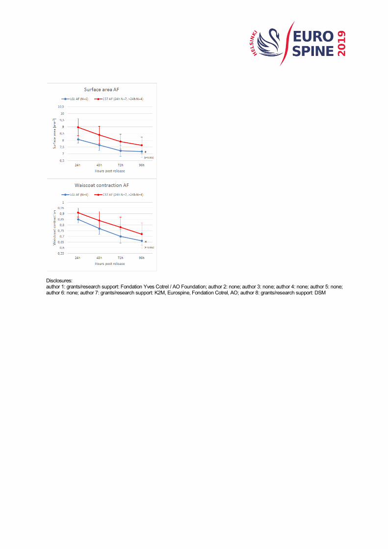
Introduction: In scoliosis, most of the early deformity is in the disc. We hypothesize that all spines experience asymmetric loading during rapid adolescent growth resulting in temporary bending of the disc. The difference between individuals who develop persistent and progressive disc deformation, and those who spontaneously correct the same initial deformation of the disc, could be dependent on the individual’s ability or lack thereof to alter their collagen network, in a faster and more permanent manner. Purpose of this study: We hypothesize that the collagen network remodeling response to asymmetrical loading of the disc determines whether a curvature becomes structural or not, and also whether an established curve progresses more or less rapidly. Recently, strains of mice (C57BL/6J and LG/J mice) have been identified with differences in their connective tissue healing capacity and disc wedging as part of an experimental model of scoliosis. In this experiment, we determined whether an in-vitro micro-tissue assay can distinguish the differences in collagen remodeling rates of annulus fibrosus (AF) cells between these strains. Materials and Methods: AF cells, harvested from 8-10 week-old C57BL/6J and LG/J mice were expanded in a hydrogel in a tissue remodeling platform, consisting of ten constraining posts in a hexagon shape and cultured for 48 hours. Hereafter the micro-tissues were released from one opposing set of posts and cultured for an extra 96 hours. With the alteration in support, the cells are stimulated to remodel the collagen in the micro-tissue from an isotropic to anisotropic organization. Micro-tissue surface area and waistcoat contraction were analysed by making digital top-down images of the micro-tissue assay system with a stereo microscope at different time points. Collagen orientation was analysed by staining tissues at 24 hours and 96 hours post-release with CNAmCherry for collagen and visualized using a confocal microscope. Results: For LG/J N=6 and for C57BL/6J N=7 (at 24 hours) and N=4 (after 24 hours) micro-tissues were analyzed. 96 hours post-release, LG/J cells contracted the tissue more than the C57cells as evidenced by a smaller micro-tissues surface area, 7.14±0.44 vs. 7.62±0.62 mm (p<0.001) and more waist coat contraction, 0.66±0.07 vs 0.72±0.1 mm (p=0.002) (Figure 1). Although no significant difference in collagen orientation between the groups was seen at 24 hours, at 96 hours, LG/J AF cells oriented the collagen more than C57 cells (p<0.001). Conclusion: The micro-tissue remodeling assay system shows promising results in terms of distinguishing between the collagen network remodeling capability of AF cells of these different mice. The next step is to determine whether these differences are reproducible with skin fibroblasts which are more clinically accessible. This could pave the way towards a bioassay for the prediction of curve progression in human AIS patients.
Disclosures: author 1: grants/research support: Fondation Yves Cotrel / AO Foundation; author 2: none; author 3: none; author 4: none; author 5: none; author 6: none; author 7: grants/research support: K2M, Eurospine, Fondation Cotrel, AO; author 8: grants/research support: DSM
QF4
AN INVESTIGATIONAL STUDY OF A DUAL-LAYER, CHORION-FREE AMNION PATCH AS A PROTECTIVE BARRIER FOLLOWING LUMBAR LAMINECTOMY IN A SHEEP MODEL
Bryan Cunningham, Breanna Seiber, Jessica Riggleman, Margaret Van Horn, Archana Bhat Musculoskeletal Education and Research Center, Globus Medical Inc., Audubon, USA
Background Context: A decompressive laminectomy is the most common surgery performed to relieve symptoms of lumbar spinal stenosis. While this surgery has been shown to be successful in relieving patient symptoms, partially removing the lamina exposes the dura to elements, and potential damage, from the surrounding environment. While a number of different biomaterials have been investigated for use as a protective barrier in spine surgery, the inherent properties of the human amniotic membrane (HAM) may make it favorable to protect neural elements and anterior vessels from the surrounding environment. The effect of a dual-layer HAM patch used as a physical barrier following a laminectomy in an animal model has yet to be investigated. Purpose: The objective of this study was to evaluate the effect of a dual-layer, chorion-free amnion patch (DLAM) processed from HAM as a protective barrier following lumbar laminectomy in a sheep model. Methods: Twelve adult sheep each underwent a laminectomy at L3 and L5, and one surgical site randomly received DLAM treatment. A multiplex immunoassay was performed to quantify the inherent cytokines present in the amnion after processing. Epidural fibrosis and neurohistopathological responses were assessed based on epidural fibrosis-dura tenacity scores and decalcified histology, respectively, at 4 and 10 weeks post-operatively. Results: Immunoassay results showed that inflammatory mediators and immunomodulatory cytokines were present in the amnion after processing, but no pro-angiogenic cytokines were detected. At 10 weeks, tissue tenacity was significantly less in the DLAM treatment group compared to the operative control (1.2±0.4 vs 2.8±0.4, p<0.05), demonstrating the ability of DLAM to act as a barrier and cover the dura. At both 4 and 10 weeks, there were significantly more infiltrated fibroblasts at the operative control sites than in the DLAM treated sites (Fig 1), expressed as a percentage of the total number of fibroblasts present (4 weeks: 72.3±10.2% vs 10.8±10.1%, p<0.05; 10 weeks: 84.9±15.8% vs 43.1±11.6%, p<0.05). Additionally, fibroblasts traveled further into the dura in the operative control group compared to the DLAM treated group at both time points. Conclusion: This study found that DLAM reduced fibroblast infiltration and tissue tenacity 10-weeks post-lumbar laminectomy in an animal model. DLAM reduced fibroblast infiltration and tissue tenacity post-surgically in an animal model, supporting its potential use as a protective barrier for neural elements and anterior vessels.

Disclosures: author 1: none; author 2: employee: Globus Medical; author 3: stock/shareholder: Globus Medical,employee: Globus Medical; author 4: grants/research support: Globus Medical,employee: Globus Medical; author 5: employee: Globus Medical

QF5
THE MECHANISM OF DURAL ADHESION IN THE EXCESSIVE FIBROSIS CAUSING PAIN AFTER SPINE SURGERY; THE AFFINITY TO COLLAGEN THROUGH INTEGRIN AND MATRIX METALLOPROTEINASE
Joo Han Kim, Kyuha Chong, Woo Keun Kwon, Hong Joo Moon Department of Neurosurgery, Guro Hospital, College of Medicine, Korea University, Korea, Seoul 1. Introduction Dural traction by peridural adhesion and direct neural compression by excessive fibrosis might be one of major factors causing axial and radicular pain after spine surgery. 2. Purpose : This study was designed to explore the molecular mechanism of dural adhesion by using primary culture of human dura mater cells (hDMCs). 3. Materials and Methods hDMCs were cultured on the collagen I coated plate from human dura mater tissue(8 men and 2 women, mean age 55.4 ± 17.86) obtained during duroplasty following decompressive craniectomy. The comparison between naive hDMCs and co-culturing hDMC with macrophage like THP-1 cells (MΦ) is performed in various assays to reflect inflammatory circumstances of post-operation. Adhesion assay was performed for investigating major adhesive substrates to human dura matter cells (hDMCs). Flow cytometry about integrins and western blot about intracellular proteins related with adhesion (focal adhesion kinase (FAK), talin 1, and Factin) was performed for investigating the alteration on adhesion-related molecules in hDMCs. ELISA about secreted matrix metalloproteinases (MMPs) was performed for investigating ECM remodeling factors for adhesion). 4. Results : The human dura mater tissue was sufficiently well adhered to the collagen I coated-dishes and plates within 3 days of gentle compression. After 3 days, the hDMCs which had similar compatible morphology of fibroblasts were appeared from the edge of the explanted tissues. The cells had elongated or multiple protruding pseudopods in shape initially, and gradually migrated away from the tissue. The adhesion profiles of hDMCs with collagen I, IV, fibrinogen and fibronectin after co-culture was significantly increased 6.4, 5.0, 3.0 and 1.6 times comparing to those of control group respectively (P<.001). There were significant increase in expression of the integrin subtype α2β1 about 6.3 times (P < .001) and αIIbβ3 about 2.0 times (P < .001). α1, which had highly expressed in naïve hDMCs, showed significant decrease after co-culture (P < .001). FAK showed significant increase 1.99-fold in co-culture (P<.001). There was no significant alteration between control and co-culture group in talin and F-actin. Co-culturing of the hDMCs with the MΦ induced significant increment of MMP-1, MMP-3, and VEGF concentrations compared to the sum of naive hDMCs and MΦ (MMP-1: p < 0.01, 13.9 fold change; MMP-3: p < 0.01, 7.6 fold change; VEGF: p < 0.01, 3.8 fold change). MMP-9 secretion at co-culture was significantly suppressed into 9.7% comparing to that of activated THP-1 cells only. 5. Conclusion: hDMCs could be successfully utilized for the study of dural adhesion. Collagen might be a critical substrate of hDMCs in terms of adhesion, mediated by integrin α2β1 rather than α1. The increase of MMP 1, 3 and suppression of MMP 9 from hDMCs after exposure to inflammation might have critical roles in adhesion of hDMCs through ECM remodeling after peridural tissue injury. Disclosures: author 1: not indicated; author 2: not indicated; author 3: none; author 4: none;

QF6
SEQUENTIAL CHANGES IN LUMBAR LORDOSIS AND SEGMENTAL STABILITY FOLLOWING LATERAL INTERBODY CAGE PLACEMENT, SMITH-PETERSON OSTEOTOMY, AND ANTERIOR LONGITUDINAL LIGAMENT RELEASE
Zachary Child, Richard Hurley, Jr, Amy Claeson, Vijay Permeswaran, Jason Inzana, Anup Gandhi Spine Surgery and Musculoskeletal Oncology; Tahoe Center for Orthopedics; South Lake Tahoe, CA, USA; Zimmer Biomet, Westminster, CO, USA
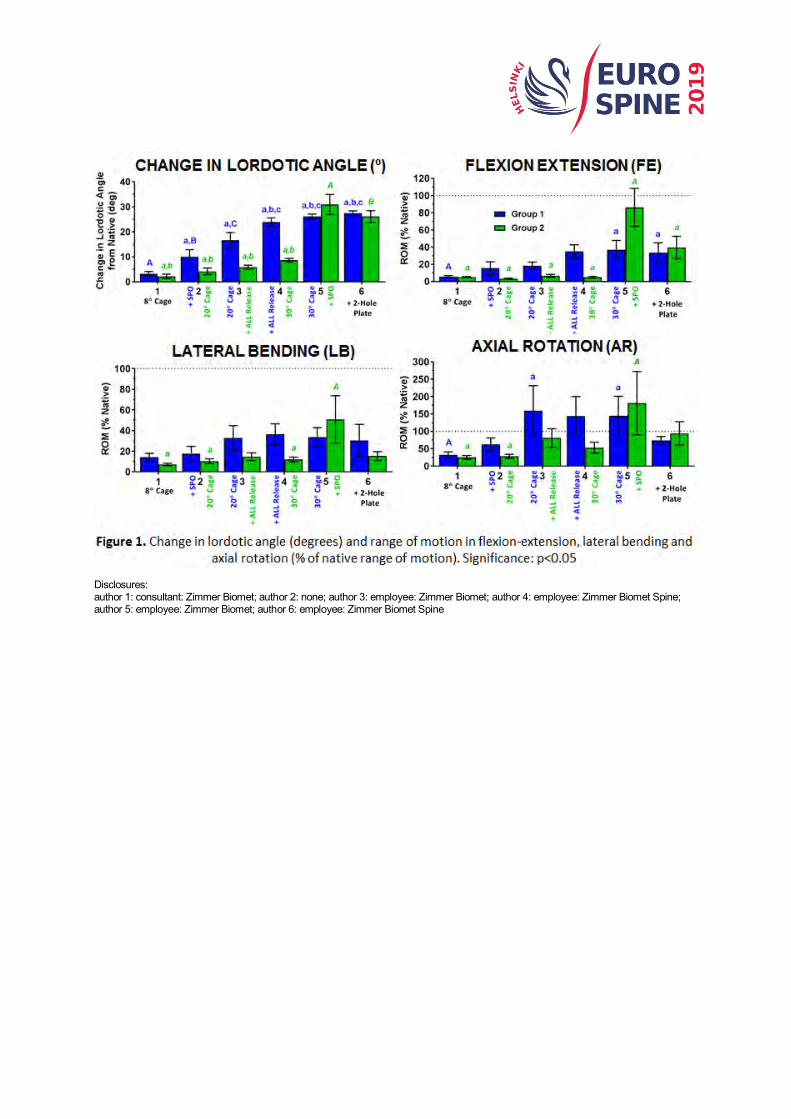
Introduction: Interbody cage systems are used in isolation to achieve lumbar lordosis correction, but can also be deployed using techniques to shorten the posterior or lengthen the anterior spinal column to achieve even larger corrections. The Thomasen pedicle subtraction osteotomy (PSO) yields ~30° of correction, but with high degree of difficulty and surgical complication rates. A lateral lordotic cage with Smith-Petersen Osteotomy (SPO), a hyper-lordotic lateral cage with anterior longitudinal ligament (ALL) release, or a lateral cage with combined techniques show potential for substantial lordosis correction. The removal or release of local spinal anatomy, however, can increase segmental motion and may inhibit fusion. The objective of this biomechanical cadaver study was to identify the lordosis correction and resulting segmental stability achieved with implantation of lateral lordotic cages of increasing angle coupled with either or both minimally invasive surgical techniques of ALL-release and SPO. We hypothesize that a combination of lordotic cage placement, ALL-release and SPO yields more segmental lordosis than with these interventions in isolation and that the biomechanical stability of the construct will be sufficiently rigid to promote fusion. Materials and Methods: Cadaveric lumbar spines (n=6) were divided into L1-L2 and L3-L4 segments, potted in plaster with pedicles exposed and randomly distributed (Figure 1A, n=6 segments/group); Group 1: SPO followed by ALL-release, Group 2: ALL-release followed by SPO. Sagittal fluoroscopic images for lordosis angle measurement and range of motion (ROM) data were collected after each intervention. A spine simulator applied 7.5Nm pure moments to segments in flexion-extension (FE), lateral bending (LB), and axial rotation (AR), while optical motion capture recorded ROM. Linear mixed models with repeated measures provided statistical comparisons at α=0.05 within each group for each metric (angle, FE-, LB-, AR-ROM); ROM data reported as percent of native. Results: The combination of SPO with ALL-release yielded the largest lordotic angle regardless of procedure order (28.5º ± 7.3º), but also greatest ROM in all planes. A 30° cage provided no additional gain in lordotic angle and increased ROM in FE and AR with ALL-release and SPO. An 8º lordotic cage with SPO achieved the largest angle correction (10.1º ± 6.8º) while reducing ROM in all planes. The addition of a 2-hole plate does not decrease correction and tends to reduce ROM in all planes compared to the 1-hole plate. Conclusion: We show that lordosis >15° is possible through these minimally invasive techniques of SPO and ALL-release and that performing a SPO without ALL-release yielded substantial correction without compromising stability. Performing a SPO after ALL-release increases segment ROM in FE, LB, AR, thus a 2-hole plate is recommended to aid in stability with ALL-release.
Disclosures: author 1: consultant: Zimmer Biomet; author 2: none; author 3: employee: Zimmer Biomet; author 4: employee: Zimmer Biomet Spine; author 5: employee: Zimmer Biomet; author 6: employee: Zimmer Biomet Spine

QF7
INFLUENCES OF CERVICAL STRUCTURES ON THE KINEMATIC BEHAVIOR OF THE CERVICAL SPINE
René Jonas, Robert Demmelmaier, Hans-Joachim Wilke Institute of Orthopaedic Research and Biomechanics, Ulm, Germany
Surgical treatment of the degenerated cervical spine (CS) often requires resections to a series of structures. Thereby, a broad range of treatment options aim to maintain physiological motion. However, it is not yet fully understood which influences each structure of the CS and its resection might have on its kinematic behavior. Knowledge of these influences could help to reduce or even prevent iatrogenic degeneration after surgical intervention. Therefore, we conducted an in vitro study in order to investigate the influences of different structures on the kinematic behavior of the CS using 3D helical axes (HA). We extracted motion segment C4-C5 from six human cadaveric specimens with an average age of 48 years. For the in vitro experiments, 7 states were defined. The first state represented the intact state. The remaining 6 states correspond with the subsequent resection of the following structures in the given order: interspinous ligament (IS), ligamentum flavum (FL), facet capsule (FC), vertebral arch (AV), posterior longitudinal ligament (PL) and anterior longitudinal ligament (AL). Each state was tested using a well-established spine tester. Each test sequence included 3.5 quasi-static motion cycles in all three bending directions using 1 Nm. We calculated the 3D-HA using Vicon motion recordings and matched them with x-ray images. Due to the small number of specimens quantitative data was analyzed using descriptive statistics. The least change in the kinematic behavior of the CS was observed during flexion/extension. For lateral bending (LB) and axial rotation (AR) the greatest change in the pattern of the HA was observed during the resection of the vertebral arch. For LB, it could be observed that the deviation in the axes’ orientation increased (Figure 1) whereas for AR it decreased. Furthermore, a great variety among the specimens was observed regarding the influences of each resection step on the mean HA’ orientation and deviation. To the knowledge of the authors, this is the first investigation of the influences of cervical structures on the kinematic behavior of the CS using 3D-HA. It is assumed that the angular orientation of the cervical facet cartilage has the greatest effect on cervical kinematics. However, the herein calculated HA indicate that other structure may also play a relevant role in the kinematic behavior of the CS depending on the pathology of the respective specimen. The data also indicate that the Uncinate processes and the overall shape of the vertebral body greatly influence cervical kinematics due to the kinematic behavior after the last step of resection. Regarding surgical treatment, the results of this study suggest that the resection of IS, FL and PL may cause less iatrogenic degeneration compared to the resection of FC, AV and AL
Disclosures: author 1: none; author 2: none; author 3: grants/research support: Signus, Neos, ApiFix, Spinol, 3D Matrix
QF8
GENDER SPECIFIC BIOMECHANICAL EVALUATION OF FORTY-FOUR HUMAN CADAVERIC LUMBOPELVIC SPINES - EMPHASIS ON THE KINEMATIC DIFFERENCES IN SACROILIAC JOINT RANGE OF MOTION
Bryan W. Cunningham, Daina M. Brooks, David A. Weiner, Jessica B. Hawken, P. Justin Tortolani Medstar Union Memorial Hospital, Department of Orthopaedic Surgery, Baltimore, Maryland, USA
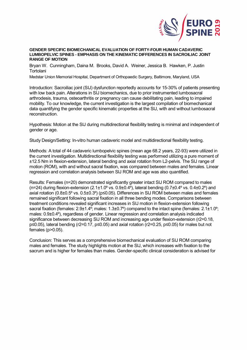
Introduction: Sacroiliac joint (SIJ) dysfunction reportedly accounts for 15-30% of patients presenting with low back pain. Alterations in SIJ biomechanics, due to prior instrumented lumbosacral arthrodesis, trauma, osteoarthritis or pregnancy can cause debilitating pain, leading to impaired mobility. To our knowledge, the current investigation is the largest compilation of biomechanical data quantifying the gender specific kinematic properties at the SIJ, with and without lumbosacral reconstruction. Hypothesis: Motion at the SIJ during multidirectional flexibility testing is minimal and independent of gender or age. Study Design/Setting: In-vitro human cadaveric model and multidirectional flexibility testing. Methods: A total of 44 cadaveric lumbopelvic spines (mean age 68.2 years, 22-93) were utilized in the current investigation. Multidirectional flexibility testing was performed utilizing a pure moment of ±12.5 Nm in flexion-extension, lateral bending and axial rotation from L2-pelvis. The SIJ range of motion (ROM), with and without sacral fixation, was compared between males and females. Linear regression and correlation analysis between SIJ ROM and age was also quantified. Results: Females (n=20) demonstrated significantly greater intact SIJ ROM compared to males (n=24) during flexion-extension (2.1±1.0⁰ vs. 0.9±0.4⁰), lateral bending (0.7±0.4⁰ vs. 0.4±0.2⁰) and axial rotation (0.8±0.5⁰ vs. 0.5±0.3⁰) (p≤0.05). Differences in SIJ ROM between males and females remained significant following sacral fixation in all three bending modes. Comparisons between treatment conditions revealed significant increases in SIJ motion in flexion-extension following sacral fixation (females: 2.9±1.4⁰; males: 1.3±0.7⁰) compared to the intact spine (females: 2.1±1.0⁰; males: 0.9±0.4⁰), regardless of gender. Linear regression and correlation analysis indicated significance between decreasing SIJ ROM and increasing age under flexion-extension (r2=0.18, p≤0.05), lateral bending (r2=0.17, p≤0.05) and axial rotation (r2=0.25, p≤0.05) for males but not females (p>0.05). Conclusion: This serves as a comprehensive biomechanical evaluation of SIJ ROM comparing males and females. The study highlights motion at the SIJ, which increases with fixation to the sacrum and is higher for females than males. Gender-specific clinical consideration is advised for
patients presenting for index and revision lumbosacral surgical procedures.
Disclosures: author 1: none; author 2: none; author 3: none; author 4: none; author 5: grants/research support: Spineology, K2M,consultant: Globus Medical, Innovasis,royalties: Globus Medical,other financial report: Innovasis
QF9
RADIOGRAPHIC SPINOPELVIC PARAMETERS IN ASD: A DYNAMIC EVALUATION
Thomas Overbergh, Pieter Severijns, Ilse Jonkers, Lieven Moke, Lennart Scheys Institute for Orthopaedic Research and Training (IORT), KU Leuven, Belgium
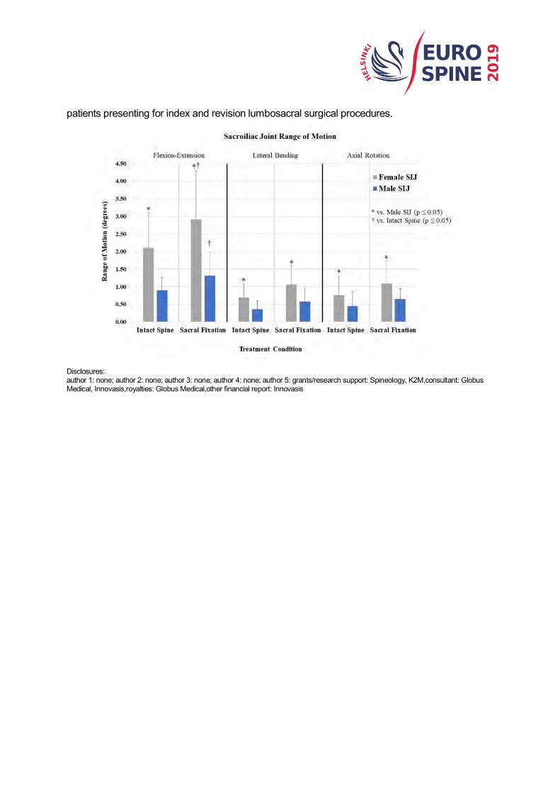
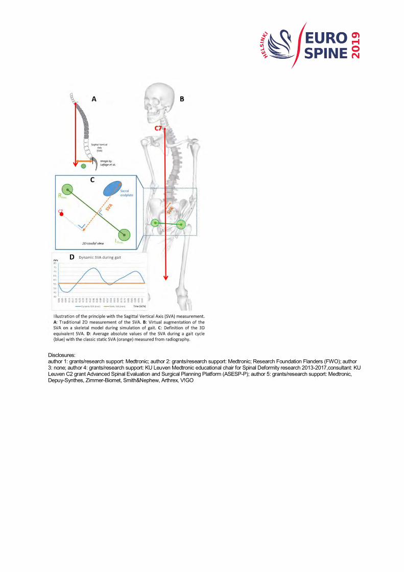
Summary Conventional radiographic analysis in spinal deformity only quantifies the static skeletal body structure from which no conclusions can be drawn with respect to functional abilities. The aim of this work was to extend some of the currently used 2D, static, radiographic concepts to 3D, dynamic, equivalent parameters. Hypothesis Motion analysis enables the use of 3D, dynamic, equivalents of 2D, static, radiographic measurements. Introduction At present, assessment of spinal alignment in Adult Spinal Deformity (ASD) is mainly based on static standing 2D radiographic spinopelvic parameters. Radiographic images, however, are but instantaneous snapshots of a patient's spinal alignment, which don’t provide objective information about the patient’s spinopelvic anatomy in more dynamic activities of daily living. Methods Eight ASD patients were included in this study. All of them underwent a spinopelvic analysis using a biplanar imaging system followed by performance of a series of motion tasks in the 3D motion lab, including gait. Subsequently, multi-body analysis (MBA) of a subject-specific skeletal model, based on the radiographic images, was used to simulate the motion of each subject performed in the motion lab. 3D equivalent parameters for sagittal vertical axis (SVA, Figure A-C), pelvic tilt, sacral slope, lumbar lordosis, thoracic kyphosis and spino-pelvic inclinations were automatically calculated for each time frame based on the 3D trajectories of predefined anatomical landmarks (such as the C7 body center) on the skeletal model. This 3D, dynamic, equivalent parameter was defined ensuring that when the patient is perfectly aligned with the radiograph plane, the 2D projection of the 3D measurement in the image produces identical values. Results As shown in figure D it is now possible to use multi-body simulations to estimate dynamic equivalents of radiographic measurements. Furthermore, classic 2D radiographic analysis of the eight subjects provided an average (SD) SVA of 40.4mm (38.4mm) while the dynamic analysis calculated a time-averaged SVA of 69.2mm (30.2mm) with an average dynamic range of 20.4mm (7.7mm) during gait. Conclusion Load-bearing skeletal models of the spine, created based on biplanar images and driven by recorded motion capture data, can be used to calculate dynamic and 3D equivalent radiographic measures. Future research should further analyze the clinical added value over the current static 2D evaluation. Take Home Message 2D, static, radiographic measurements can be extended to their 3D equivalent and thereafter evaluated in dynamic conditions using MBA.
Disclosures: author 1: grants/research support: Medtronic; author 2: grants/research support: Medtronic; Research Foundation Flanders (FWO); author 3: none; author 4: grants/research support: KU Leuven Medtronic educational chair for Spinal Deformity research 2013-2017,consultant: KU Leuven C2 grant Advanced Spinal Evaluation and Surgical Planning Platform (ASESP-P); author 5: grants/research support: Medtronic, Depuy-Synthes, Zimmer-Biomet, Smith&Nephew, Arthrex, V!GO

QF10
DIFFERENCES IN VERTEBRAL MOTION BETWEEN AN ADULT SPINAL DEFORMITY PATIENT AND A HEALTHY SUBJECT: A CASE STUDY
Thomas Overbergh, Pieter Severijns, Ilse Jonkers, Lieven Moke, Lennart Scheys Institute for Orthopaedic Research and Training (IORT); KU Leuven, Leuven, Belgium
Summary Conventional radiographic analysis in spinal deformity only quantifies the static skeletal body structure which inherently impedes conclusions with respect to dynamic functional abilities. The aim of this case study was to investigate the kinematic strategies of an adult spinal deformity (ASD) patient compared to a healthy subject as an illustration of the possible added value of integrating motion analysis in clinical decision making in spinal deformity. Hypothesis The kinematic strategy to perform a well-defined spinal motion is related to the structural spinal deformity in an ASD patient. Introduction Radiographic measurements are the main parameters in the clinical decision making in spinal deformity. They, however, don’t provide objective information about the patient’s spinopelvic anatomy in more dynamic activities of daily living. Methods The spinopelvic anatomy of one healthy subject (61y) and one ASD patient (59y, lumbar scoliosis: Fig.1) were reconstructed in 3D (Fig.1) based on load-bearing biplanar radiographic images and CT data. Both subjects were asked to perform a maximal forward bending motion while seated, with an optical motion tracking system tracking retroreflective markers placed on the back of the subjects. An inverse kinematics simulation provided intervertebral joint (IVJ) motions from the lumbosacral joint up to the T3-T4 joint. The range of motion (ROM) at each IVJ was determined for every axis (i.e. lateroflexion, axial rotation and flexion). Results The ROM graphs (Fig.1) show that the ASD subject used a clearly different kinematic strategy to reach the bent position, compared to the healthy subject. First, the ASD patient used a combined kinematic strategy involving all three degrees of freedom, which contrasts with the healthy subject almost solely relying on flexion to reach the forward bent posture. Secondly, the thoracic region is clearly more involved in the ASD-specific kinematic strategy compared to a primarily lumbar involvement in the healthy subject. Conclusion This case study illustrates the additional information that motion analysis may provide about vertebral kinematic strategies in the ASD population, possibly leading to more informed clinical decision making. The difference at lumbar level between ASD patient and control can be attributed to the spinal deformity realigning the directions of motion and thus forcing a portion of the global spine flexion to be realized through axial rotation and lateroflexion. The difference at the thoracic region may be a compensation mechanism for the lowered mobility in the lumbar region. Take Home Message Motion analysis may provide additional insights to the radiographic evaluation of spinal deformity
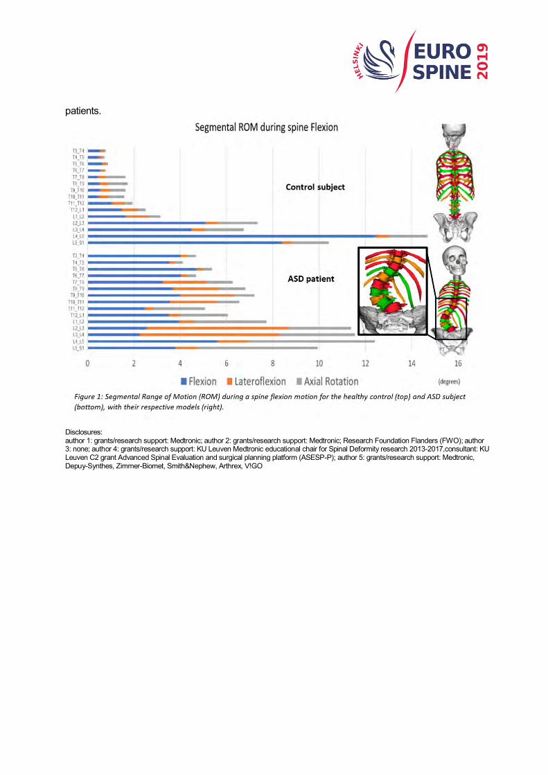
patients.
Disclosures: author 1: grants/research support: Medtronic; author 2: grants/research support: Medtronic; Research Foundation Flanders (FWO); author 3: none; author 4: grants/research support: KU Leuven Medtronic educational chair for Spinal Deformity research 2013-2017,consultant: KU Leuven C2 grant Advanced Spinal Evaluation and surgical planning platform (ASESP-P); author 5: grants/research support: Medtronic, Depuy-Synthes, Zimmer-Biomet, Smith&Nephew, Arthrex, V!GO
QF11
POC5 VARIANT LEADS TO CELLULAR ALTERATIONS AND RETINAL ANOMALIES IN ADOLESCENT IDIOPATHIC SCOLIOSIS
Florina Moldovan, Amani Hassan, StefanParent, Shunmoogum A. Patten CHU Sainte Justine and Université de Montréal, Faculty of dentistry, Montreal, Canada
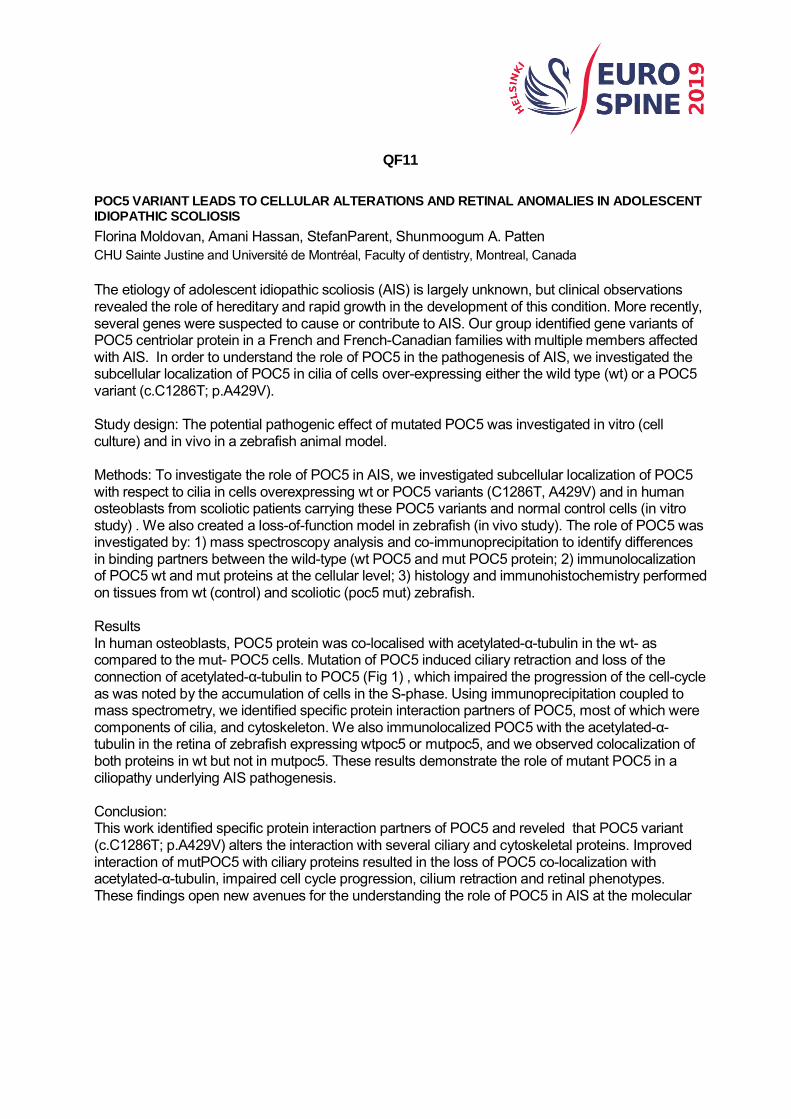
The etiology of adolescent idiopathic scoliosis (AIS) is largely unknown, but clinical observations revealed the role of hereditary and rapid growth in the development of this condition. More recently, several genes were suspected to cause or contribute to AIS. Our group identified gene variants of POC5 centriolar protein in a French and French-Canadian families with multiple members affected with AIS. In order to understand the role of POC5 in the pathogenesis of AIS, we investigated the subcellular localization of POC5 in cilia of cells over-expressing either the wild type (wt) or a POC5 variant (c.C1286T; p.A429V). Study design: The potential pathogenic effect of mutated POC5 was investigated in vitro (cell culture) and in vivo in a zebrafish animal model. Methods: To investigate the role of POC5 in AIS, we investigated subcellular localization of POC5 with respect to cilia in cells overexpressing wt or POC5 variants (C1286T, A429V) and in human osteoblasts from scoliotic patients carrying these POC5 variants and normal control cells (in vitro study) . We also created a loss-of-function model in zebrafish (in vivo study). The role of POC5 was investigated by: 1) mass spectroscopy analysis and co-immunoprecipitation to identify differences in binding partners between the wild-type (wt POC5 and mut POC5 protein; 2) immunolocalization of POC5 wt and mut proteins at the cellular level; 3) histology and immunohistochemistry performed on tissues from wt (control) and scoliotic (poc5 mut) zebrafish. Results In human osteoblasts, POC5 protein was co-localised with acetylated-α-tubulin in the wt- as compared to the mut- POC5 cells. Mutation of POC5 induced ciliary retraction and loss of the connection of acetylated-α-tubulin to POC5 (Fig 1) , which impaired the progression of the cell-cycle as was noted by the accumulation of cells in the S-phase. Using immunoprecipitation coupled to mass spectrometry, we identified specific protein interaction partners of POC5, most of which were components of cilia, and cytoskeleton. We also immunolocalized POC5 with the acetylated-α-tubulin in the retina of zebrafish expressing wtpoc5 or mutpoc5, and we observed colocalization of both proteins in wt but not in mutpoc5. These results demonstrate the role of mutant POC5 in a ciliopathy underlying AIS pathogenesis. Conclusion: This work identified specific protein interaction partners of POC5 and reveled that POC5 variant (c.C1286T; p.A429V) alters the interaction with several ciliary and cytoskeletal proteins. Improved interaction of mutPOC5 with ciliary proteins resulted in the loss of POC5 co-localization with acetylated-α-tubulin, impaired cell cycle progression, cilium retraction and retinal phenotypes. These findings open new avenues for the understanding the role of POC5 in AIS at the molecular
and cellular levels.
Disclosures: author 1: grants/research support: Yves Cotrel Foundation, and SRS; author 2: none; author 3: grants/research support: DePuy Synthes Spine, Medtronic, EOS-Imaging, Spinologics, K2M,consultant: DePuy Synthes Spine, EOS-Imaging,stock/shareholder: Spinologics,royalties: EOS-Imaging
QF12
MACROPHAGES CONTRIBUTE TO AGGRAVATE INTERVERTEBRAL DISC DEGENERATION UNDER A PRO-INFLAMMATORY/DEGENERATIVE MICROENVIRONMENT
Ana João Silva, João Vasco Corte-Real, Joana Rita Ferreira, Carla Cunha, Mafalda Bessa-Gonçalves, Mário A. Barbosa, Susana G. Santos, Raquel M. Gonçalves 1i3S – Instituto de Investigação e Inovação em Saúde, Porto, Portugal; 2INEB – Instituto de Engenharia Biomédica, Porto, Portugal;3FCUP – Faculdade de Ciências da Universidade do Porto, Porto, Portugal; 4ICBAS – Instituto de Ciências Biomédicas Abel Salazar, Universidade do Porto, Porto, Portugal Introduction: Low back pain is a highly prevalent clinical problem and intervertebral disc (IVD) degeneration is now accepted as the major pathophysiological mechanism responsible for this condition. Inflammation plays a crucial role in the progression of human IVD degeneration [1], with macrophages being pointed as the key immune cell players in this process. Macrophages are highly plastic and may play different roles depending on the microenvironmental cues. The study of inflammation associated with IVD degeneration has been somehow neglected and one of the reasons is related with a lack of adequate models. To overcome this, we propose the establishment of a new model of degenerated IVD in 3D organ culture to further dissect the role of macrophages in IVD associated immune response. Methods: For that, human monocyte-derived macrophages were co-cultured either with bovine caudal IVD punches in the presence of the pro-inflammatory cytokine IL-1β, as previously described [2], or IVD-conditioned medium (CM), to investigate how IVD-produced factors influence macrophage phenotype. After 72h, metabolic activity, gene expression and cytokine profile of macrophages and IVD cells were measured. Statistical analysis was performed using non-parametric tests: paired Friedman test for macrophages and unpaired Kruskal-Wallis for IVDs. Results: Macrophages remain metabolically active in the presence of IL-1β-stimulated IVDs, with significantly higher upregulation of CCR7 gene expression (p<0.001) and increased production of IL-6 (p<0.05). When treating macrophages with IL-1β-IVD-CM, CCR7 upregulation follows the same trend (p=0.06), while for IL-6 an opposite effect was observed. On the other hand, macrophages interfere with IVD ECM remodeling, decreasing MMP3 expression and significantly downregulating aggrecan (p=0.09) and collagen II (p<0.05) gene expression in the presence of IL-1β. Conclusions: Overall, the co-culture model established in this study can be considered a suitable approach to address the cellular and molecular pathways that regulate macrophage-IVD crosstalk, suggesting that degenerated IVD tissue tends to polarize human macrophages towards a more pro-inflammatory profile, which seems to aggravate IVD degeneration. This model could be used to improve the knowledge of the mechanisms that link IVD degeneration and the immune response. Reference: [1]Molinos, J Royal Soc Interf, 2015; [2] Teixeira, Tissue Eng, Part C, 2016
Disclosures: author 1: none; author 2: none; author 3: none; author 4: none; author 5: none; author 6: none; author 7: none; author 8: none;

QF13
A ROLE FOR MSCS SECRETOME TO MODULATE THE INFLAMMATORY RESPONSE IN DISC DEGENERATION
Joana Rita Ferreira, Graciosa Quelhas Teixeira, Cláudia Ribeiro-Machado, Catarina Leite Pereira, Mário Adolfo Barbosa, Raquel Madeira Gonçalves ICBAS - Instituto de Ciências Biomédicas Abel Salazar, Universidade do Porto, Porto, Portugal; INEB - Instituto Nacional de Engenharia Biomédica, Porto, Portugal; i3S - Instituto de Investigação e Inovação em Saúde, Porto, Portugal Introduction: Intervertebral Disc (IVD) degeneration, a major cause of spine disorders and disability worldwide, occurs naturally with age. It’s associated with matrix degradation, inflammation and IVD vascularization and innervation (in a tissue physiologically avascular and non-innervated) which lead to the onset of low back pain (LBP). Current treatments can alleviate associated symptoms but don’t solve the underlying cause. Mesenchymal stem/stromal cells (MSCs) clinical potential has been widely demonstrated in a variety of disorders including LBP. Still, the MSCs paracrine competence is frequently pointed out as its main therapeutic factor, particularly considering the questionable MSCs survival upon transplantation to the harsh environment of the degenerated IVD. Previous work has shown that in the pro-inflammatory/degenerative IVD environment MSCs assume an immunoregulatory role [1]. Here, we aimed to evaluate if we can replicate this effect using only the MSCs secretome. Methods: Human bone marrow-derived MSCs were pre-conditioned for 48h with IL-1β (10 ng/mL), in hypoxic (6%O2) conditions, the microenvironment used to culture bovine IVDs (bIVDs) [1]. MSCs secretome (secMSC) was collected and used to culture bIVD punches in pro-inflammatory/degenerative conditions (puncture+IL-1β) plus hypoxia (6% O2), using an ex vivo model previously established in the group [2]. Control groups of non-stimulated IVDs and IVDs co-cultured with MSCs were analysed in parallel. Results: MSCs preconditioning with hypoxia and IL-1β shifted their secretome towards a pro-inflammatory profile with increased production of pro-inflammatory cytokines IL-6, IL-8, MCP-1, RANTES and PGE2. After 48h, the levels of pro-inflammatory markers of degenerated IVDs were down-regulated (IL-6, IL-8) comparatively to non-degenerated discs, which was also observed with MSCs-treated IVDs. Regarding the matrix degenerative process, 48h after the proinflammatory/degenerative stimulus, neither secMSCs nor MSCs seemed to be able to counteract the down-regulation of collagen type II and aggrecan expression induced by the model. Yet, treatment with secMSCs significantly altered the expression of matrix degrading enzymes MMP1 and MMP3, decreasing the first and increasing the last. Co-culture with MSCs didn’t have a significant effect. Interestingly, only secMSC treatment was able to increase Aggrecan deposition relatively to the degenerated IVDs at a later timepoint (14 days) (Figure 1). Conclusions: Overall, the results show it is possible to replicate the immunomodulatory effect observed with MSCs in a pro-inflammatory/degenerative IVD model using only their secretome. Additionally, secMSCs seems to have the ability to inhibit the loss of aggrecan from the IVD’s ECM in response to the pro-inflammatory/degenerative stimulus applied. References: [1] G.Q. Teixeira et al., Spine 43(12) E673-E682, (2018). [2] G.Q. Teixeira et al.,
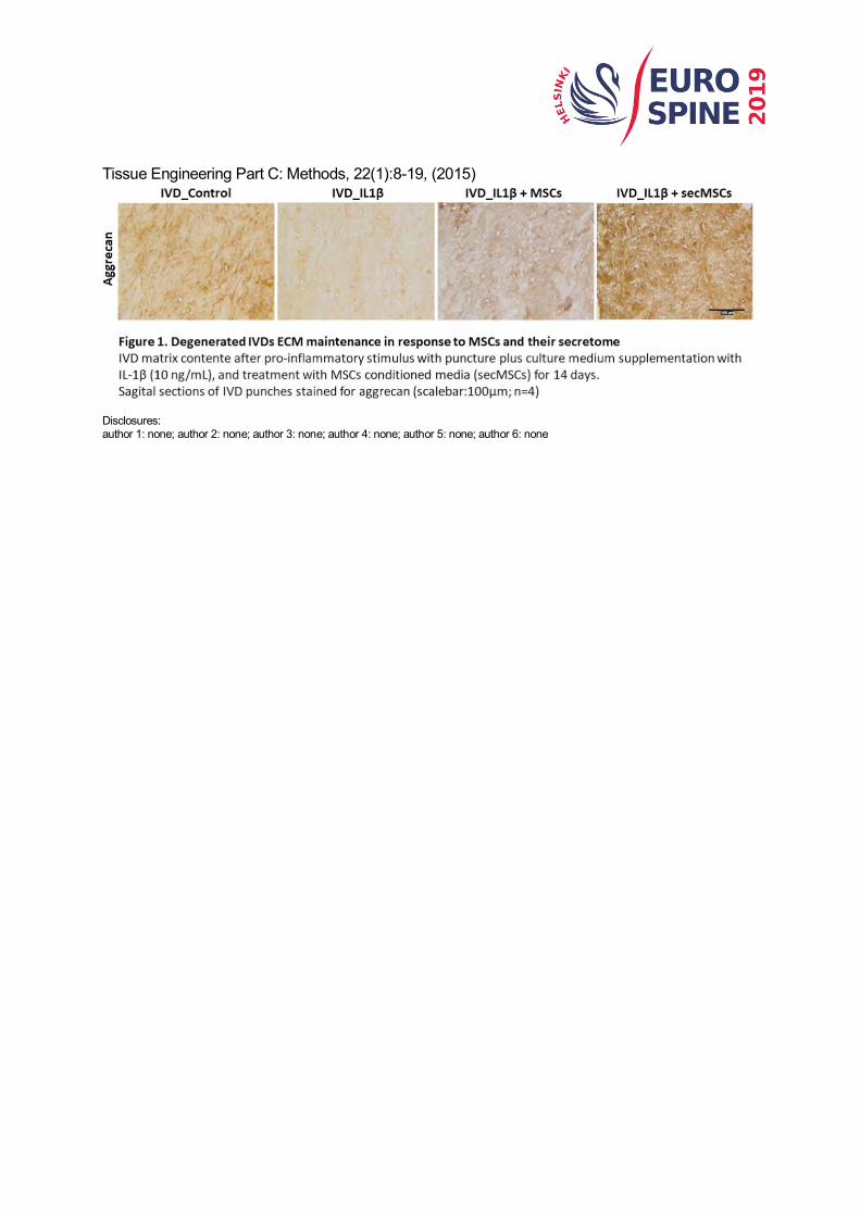
Tissue Engineering Part C: Methods, 22(1):8-19, (2015)
Disclosures: author 1: none; author 2: none; author 3: none; author 4: none; author 5: none; author 6: none

QF14
FROM STATIC TO DYNAMIC: SAGITTAL ALIGNMENT AND COMPENSATION STRATEGIES DURING WALKING IN ADULT SPINAL DEFORMITY
Pieter Severijns, Thomas Overbergh, Kaat Desloovere, Lennart Scheys, Lieven Moke Institute for Orthopaedic Research and Training; KU Leuven, Belgium
Hypothesis Adult spinal deformity (ASD) patients are not able to preserve their static compensations during walking. Introduction Although static spinal alignment and compensation mechanisms in ASD are well documented, what happens to them during motion is less clear. The primary goal of this study was to compare the static and dynamic sagittal profile of these patients during standing and walking in the motion lab. Secondly, also spatiotemporal parameters and balance performance are compared between different deformity types to document their functionality. Methods 51 ASD patients and 20 controls were recruited. The ASD group was divided into three groups: a decompensated sagittal deformity group (ASD1: SVA>4cm), a compensated sagittal deformity group (ASD2: SVA<4cm), a primary coronal deformity group (ASD3: Cobb>30°, SVA<4cm, PT<20°). All subjects performed 3D motion analysis and balance assessment using the Balance Evaluation Systems Test (BESTest). A static and walking trial were processed (full body Plug-In-Gait model, Vicon Nexus) for each subject resulting in sagittal kinematics of the spine and lower limbs. Between group differences were analyzed using the Kruskal-Wallis test. Results Decompensated ASD patients showed increased trunk flexion during stance compared to all groups (p=0.002-0.014), for which they tried to compensate with increased knee (p=0.007) and ankle (p=0.008) (dorsi)flexion and non-significant pelvic retroversion. These findings are in accordance with the static alignment and compensations reported in the literature, measured on radiographic images. No static differences with controls could be found for compensated sagittal or primary coronal patients. During walking the static differences at the lower limb level disappeared, suggesting decompensated ASD patients adopt a normal walking pattern. Within group analysis of sagittal kinematics revealed that from stance to walk all groups performed similar spinal and pelvic strategies, namely trunk flexion and pelvic anteversion. Overall, ASD patients showed altered spatiotemporal parameters during walking and decreased balance control. However, between group analysis revealed that these functional differences were mainly due to sagittal deformity (BESTest: ASD1: p<0.001; ASD2: p=0.002) since no differences were found for balance control between the primary coronal deformity group and controls. All results and p-values are displayed in Table 1. Conclusion ASD patients with sagittal malalignment seem to adopt the same strategies from stance to walking, measured with motion analysis, compared to controls, although their functionality, measured by spatiotemporal parameters and balance assessment, was clearly decreased. Therefor it can be assumed that these functional strategies are rather a loss of their static compensations, possibly compromising their functionality. Also, sagittal malalignment seems to be a better predictor of functionality than coronal malalignment.

Disclosures: author 1: grants/research support: MEdtronic; Research Foundation - Flanders (FWO); author 2: grants/research support: Medtronic; author 3: none; author 4: grants/research support: Medtronic, Depuy-Synthes, Zimmer-Biomet, Smith&Nephew, Arthrex, V!GO; author 5: grants/research support: KU Leuven Medtronic Educational Chair for Spinal Deformity Research 2013-2017,consultant: KU Leuven C2 grant Advanced Spinal Evaluation and Surgical Planning Platform (ASESP-P)

QF15
BIOMECHANICAL EFFECTS OF LATERAL BENDING POSITION ON PERFORMING CERVICAL SPINAL MANIPULATION FOR CERVICAL DISC HERNIATION: A THREE-DIMENSIONAL FINITE ELEMENT ANALYSIS
Xuecheng Huang, Linqiang Ye, Wenhua Huang, Xiaobing Jiang Department of Human Anatomy, Guangzhou, China
Background. Most studies report that the common position of cervical spinal manipulation (CSM) for treating symptomatic cervical disc herniation (CDH) is lateral bending to the herniated side. However, the rationality of lateral bending position on performing CSM for CDH is still unclear. Objective. The purpose of this study is to investigate the biomechanical effects of lateral bending position on performing CSM for CDH. Study design. A finite element analysis study. Methods. A finite element (FE) model of CDH (herniated on the left side) was generated in C5-C6 segment based on the normal FE model. The FE model was performed CSM in left lateral bending position, neutral position and right lateral bending position, respectively. Cervical disc displacement, annulus fiber stress and facet joint stress were observed during the simulation of CSM. Results. The cervical disc displacement on herniated side moved forward during CSM, and the maximum forward displacements were 0.23, 0.36 and 0.45mm in left lateral bending position, neutral position and right lateral bending position, respectively. As the same trend of cervical disc displacement, the annulus fiber stress on herniated side from small to large were 7.40, 16.39 and 22.75 MPa in left lateral bending position, neutral position and right lateral bending position, respectively. However, the maximum facet stresses at left superior cartilage of C6 in left lateral bending position, neutral position and right lateral bending position were 6.88, 3.60 and 0.12MPa, respectively. Conclusion. Compared with neutral position and right lateral bending position, though the forward displacement of cervical disc on herniated side was smaller in left lateral bending position, the annulus fiber stress on herniated side was declined by sharing load on the left facet joint. The results suggested that lateral bending to the herniated side on performing CSM tend to protect the cervical disc on herniated side. Future clinical studies are in need to verify that. Key words: cervical spinal manipulation; cervical disc herniation; lateral bending position; finite element analysis Disclosures: author 1: none; author 2: not indicated; author 3: not indicated; author 4: none