
nuova questione abitativa: programmazione della spesa e livelli di governo Maria Bianca Berruti
European Society of Endocrinology Clinical Practice Guidelines on the Management of Adrenocortical
Carcinoma in Adults, in collaboration with the European Network for the Study of Adrenal Tumors
Martin Fassnacht1,2*, Olaf M. Dekkers3,4,5, Tobias Else5, Eric Baudin7,8, Alfredo Berruti9, Ronald R. de Krijger10, 11, 12, 13, Harm R. Haak14,15, 16, Radu Mihai17, Guillaume Assie19, 20, Massimo Terzolo20* 1 Dept. of Internal Medicine I, Div. of Endocrinology and Diabetes, University Hospital, University of Würzburg, Würzburg, Germany 2 Comprehensive Cancer Center Mainfranken, University of Würzburg, Würzburg, Germany 3 Department of Clinical Epidemiology, Leiden University Medical Centre, Leiden, the Netherlands 4 Department of Clinical Endocrinology and Metabolism, Leiden University Medical Centre, Leiden, the Netherlands 5 Department of Clinical Epidemiology, Aarhus University Hospital, Aarhus, Denmark 6 Department of Internal Medicine, Division of Metabolism, Endocrinology and Diabetes, University of Michigan, Ann Arbor, MI, USA 7 Endocrine Oncology and Nuclear Medicine, Institut Gustave Roussy, Villejuif, France 8 INSERM UMR 1185, Faculté de Médecine, Le Kremlin-Bicêtre, Université Paris Sud, Paris, France 9 Department of Medical and Surgical Specialties, Radiological Sciences, and Public Health, Medical Oncology, University of Brescia at ASST Spedali Civili, Brescia, Italy. 10 Dept. of Pathology, Erasmus MC University Medical Center, Rotterdam, The Netherlands 11 Dept. of Pathology, University Medical Center Utrecht, Utrecht, The Netherlands 12 Dept. of Pathology, Reinier de Graaf Hospital, Delft, The Netherlands 13 Princess Maxima Center for Pediatric Oncology, Utrecht, The Netherlands 14 Department of Internal Medicine, Máxima Medical Centre, Eindhoven/Veldhoven, the Netherlands 15 Maastricht University, CAPHRI School for Public Health and Primary Care, Ageing and Long-Term Care, Maastricht, the Netherlands 16 Department of Internal Medicine, Division of General Internal Medicine, Maastricht University Medical Centre+, Maastricht, the Netherlands. 17 Department of Endocrine Surgery, Churchill Cancer Centre, Oxford University Hospitals NHS Foundation Trust, Oxford, UK 18 Department of Endocrinology, Reference Center for Rare Adrenal Diseases, Reference Center dor Rare Adrenal Cancers, Hôpital Cochin, Assistance Publique Hôpitaux de Paris, France 19 Institut Cochin, Institut National de la Santé et de la Recherche Médicale U1016, Centre national de la recherche scientifique UMR8104, Université Paris Descartes, Sorbonne Paris Cité, Paris, France 20 Internal Medicine, San Luigi Hospital, Dept. of Clinical and Biological Sciences, University of Turin, Orbassano, Italy *corresponding authors Correspondence should be addressed to Martin Fassnacht (Email [email protected]) and Massimo Terzolo (Email: [email protected])

Abstract 1 2 Adrenocortical carcinoma (ACC) is a rare and in most cases steroid hormone producing 3 tumor with heterogeneous prognosis. The purpose of these guidelines is to provide clinicians 4 with best possible evidence-based recommendations for clinical management of patients 5 with ACC based on the GRADE (Grading of Recommendations Assessment, Development 6 and Evaluation) system. We predefined four main clinical questions, which we judged as 7 particularly important for the management of ACC patients and performed systematic 8 literature searches: (A) What is needed to diagnose an ACC by histopathology? (B) Which 9 are the best prognostic markers in ACC? (C) Is adjuvant therapy able to prevent recurrent 10 disease or reduce mortality after radical resection? (D) What is the best treatment option for 11 macroscopically incompletely resected, recurrent or metastatic disease? Other relevant 12 questions were discussed within the group. SELECTED RECOMMENDATIONS: (i) We 13 recommend that all patients with suspected and proven ACC are discussed in a 14 multidisciplinary expert team meeting (ii) We recommend that every patient with (suspected) 15 ACC should undergo careful clinical assessment, detailed endocrine work-up to identify 16 autonomous hormone excess, and adrenal-focused imaging. (iii) We recommend that 17 adrenal surgery for (suspected) ACC should be performed only by surgeons experienced in 18 adrenal and oncological surgery aiming at a complete en bloc resection. (iv) We suggest that 19 all suspected ACC should be reviewed by an expert adrenal pathologist using the Weiss 20 score and providing Ki67 index. (v) We suggest adjuvant mitotane treatment in patients after 21 radical surgery that have a perceived high risk of recurrence (ENSAT stage III, or R1 22 resection, or Ki67 >10%). (vi) For advanced ACC not amenable to complete surgical 23 resection, local therapeutic measures (e.g. radiation therapy, radiofrequency ablation, 24 chemo-embolization) are of particular value. However, we suggest against the routine use of 25 adrenal surgery in case of widespread metastatic disease. In these patients we recommend 26 either mitotane monotherapy or mitotane, etoposide, doxorubicin, and cisplatin depending on 27 prognostic parameters. (vii) In patients with recurrent disease and a disease-free interval of 28 at least 12 months, in whom a complete resection/ablation seems feasible, we recommend 29 surgery or alternatively other local therapies. Furthermore, we offer detailed 30 recommendations about the management of mitotane treatment and other supportive 31 therapies. Finally, we suggest directions for future research. 32

1. Summary of recommendations 33 34 After the review process all Recommendations without Rational will be provided here as 35 summary. 36 37
38
2. Adrenocortical Carcinoma – epidemiology, pathogenesis, clinical 39
presentation, and general prognosis 40 41 Epidemiology and pathogenesis 42 The estimated incidence of adult adrenocortical carcinoma (ACC) is between 0.7 – 2.0 per 43 million per year (1, 2). ACC can occur at any age with a peak incidence between 40 and 60 44 years, and with women being more often affected (55-60%). In adults, the vast majority of 45 ACCs are sporadic. Occasionally, however, they occur as part of hereditary syndromes such 46 as Li-Fraumeni syndrome, Lynch syndrome, multiple endocrine neoplasia (MEN) 1 and 47 familial adenomatous polyposis (3, 4). In recent years several multi-center studies have shed 48 light on the pathogenesis of ACC (5-7)(8), but ‘multi-omic’ studies (9-11) reveal that only a 49 minority of ACC cases have pathogenic driver mutations. For details on this topic we refer to 50 recent reviews (12-14). 51 52 Clinical presentation (Table 1) 53 ACC may present with autonomous adrenal hormone excess or with symptoms caused by 54 an abdominal mass. An increasing number of cases are diagnosed within the group of 55 incidentally discovered adrenal masses (incidentalomas) (≈ 10-15%). However, the likelihood 56 of an adrenal incidentaloma being an ACC is low (15-17). About 50-60% of patients with 57 ACC have clinical hormone excess. Hypercortisolism (Cushing’s syndrome), or mixed 58 Cushing’s and virilizing syndromes are observed in the majority of these patients. Pure 59 androgen excess is less frequent while estrogen or mineralocorticoid excess are very rare 60 (13, 18-22). Non-specific symptoms from an abdominal mass include abdominal discomfort 61 (nausea, vomiting, abdominal fullness) or back pain. Classical malignancy-associated 62 symptoms such as weight loss, night sweats, fatigue or fever are rarely present. 63 64 Table 1: Clinical presentation of ACC# 65 66
Autonomous adrenal hormone excess 50-60 % Hypercortisolism (Cushing’s syndrome)* 50-70 % Androgen excess (virilization) in female patients* 20-30 % Estrogen excess (feminization) in male patients* 5 % Mineralocorticoid excess* 2-3 %
Non-specific symptoms from an abdominal mass 30-40 % Incidentally detected by imaging for other purpose 10-15 %
# number derived from: (20, 23, 24), and the ENSAT ACC registry 67 * frequently combined 68 69 General prognosis 70 The median overall survival of all ACC patients is about 3-4 years. The prognosis is, 71 however, heterogeneous. Complete surgical resection provides the only means of cure. In 72
3

addition to radical surgery, disease stage, proliferative activity, and cortisol excess are 73 independent prognostic parameters (see also section 4.2. and 5.5.). Five-year survival is 60-74 80% for tumors confined to the adrenal space, 35-50% for locally advanced disease, and 75 much lower in case of metastatic disease with reported percentages ranging from 0% to 28% 76 (19, 21, 25-30). 77 78 79 80 3. Methods 81 82 3.1. Guideline working group 83 This guideline was developed by The European Society of Endocrinology (ESE) in 84 collaboration with the European Network for the Study of Adrenal Tumours (ENSAT). The 85 chairs of the working group Martin Fassnacht and Massimo Terzolo as well as the 86 methodological expert Olaf Dekkers were appointed by the ESE Clinical Committee. Tobias 87 Else served as representative of The Endocrine Society, USA, and Radu Mihai as 88 representative of the European Society of Endocrine Surgeons. The other members were 89 suggested by the chairs and approved by the Clinical Committee of ESE. The 90 multidisciplinary team consisted of the following experts: endocrinologists (Guillaume Assie 91 (France), Olaf Dekkers (The Netherlands), Tobias Else (USA), Martin Fassnacht (Germany), 92 Harm Haak (The Netherlands), Massimo Terzolo (Italy), oncologists (Eric Baudin (France), 93 Alfredo Berruti (Italy), a pathologist Ronald de Krijger (The Netherlands), and an endocrine 94 surgeon Radu Mihai (UK). The working group had three in-person meetings (November 95 2016, September 2017, and March 2018) and communicated by phone and email. 96 Consensus was reached upon discussion; minority positions were taken into account in the 97 rationale behind recommendations. Prior to the process, all participants completed conflict of 98 interest forms. 99 100 3.2 Target group 101 This guideline was developed for healthcare providers involved in the care of patients with 102 adrenocortical carcinoma i.e., endocrinologists, oncologists, surgeons, radiologists, nuclear 103 medicine physicians, radio-oncologists, pathologists, and specialists in general internal 104 medicine. However, general practitioners might also find the guideline useful, as might our 105 patients. In addition, the guideline document can serve as a source document for the 106 preparation of patient information leaflets. 107 108 3.3 Aims 109 The overall purpose of this guideline is to provide clinicians with practical guidance for the 110 management of patients with adrenocortical carcinoma. In clinical practice, treatment 111 decisions should take into account the recommendations but also the clinical judgment of the 112 treating physician. Recommendations are thus never meant to replace clinical judgment. 113 114 3.4 Summary of methods used for guideline development 115 The methods used have been described in more detail previously (31). In short, the guideline 116 used GRADE (Grading of Recommendations Assessment, Development and Evaluation) as 117 a methodological base. The first step was to define clinical question(s) (see section 3.5), the 118 second being a systematic literature search (see Section 3.6). After including all relevant 119 articles, we 1), rated the quality of the evidence, and 2) estimated an average effect for 120
4

specific outcomes (if possible). The quality of evidence behind the recommendations is 121 classified as very low (+OOO), low (++OO), moderate (+++O) and strong (++++). 122 For the recommendations we took into account: 1) quality of the evidence, 2) balance of 123 desirable and undesirable outcomes, 3) values and preferences (patient preferences, goals 124 for health, costs, management inconvenience, feasibility of implementation, etc) (32, 33). The 125 recommendations are worded as recommend (strong recommendation) and suggest (weak 126 recommendation). The meaning of a strong recommendation can be stated as follows: 127 reasonably informed persons (clinicians, politicians and patients) would want the 128 management in accordance with the recommendation. For a weak recommendation, most 129 persons would still act in accordance with the guideline, but a substantial number would not 130 (33). Formal evidence syntheses were performed and graded only for recommendations 131 addressing our initial four questions. Recommendations based on good practice and 132 experience of the panelists were not graded (34). Recommendations were derived from 133 majority consensus of the guideline development committee, but if members had substantial 134 disagreements, this is acknowledged in the manuscript. For transparency, all 135 recommendations are accompanied by text explaining why specific recommendations were 136 made. 137 138 3.5. Clinical question, eligibility criteria and endpoint definition 139 At the beginning of the guideline development process, the panel agreed on 30 clinical 140 questions in the management of patients with ACC that should be addressed in the 141 guidelines. In a next step, we agreed on four most relevant clinical questions (Table 2), for 142 which a detailed literature search and review was subsequently performed. 143 144 3.6 Description of search and selection of literature 145 A literature search of electronic medical databases was performed for all four clinical 146 questions. As we expected that single publications could contribute to different questions (for 147 example 2 and 4) we decided to perform one overarching search using broad search terms. 148 The search revealed 5988 papers, of which 615 were duplicates. In summary, we included 149 18 publications for clinical question 1 (diagnostics for ACC), 35 studies for clinical question 2 150 (prognosis), 10 publications for clinical question 3 (adjuvant therapy) and 48 publications for 151 clinical question 4 (recurrent/advanced disease). The review of hormonal overproduction as 152 prognostic factor was published as stand-alone paper (35). For question 3, we included one 153 study after having been provided with baseline characteristics and adjusted estimates for 154 mitotane therapy not reported in the original publication (36). 155 156 157 3.7. Review process and endorsement of other societies 158 A draft of the guideline was reviewed by four experts in the field (see “Acknowledgment’ 159 section) and has been submitted for comments by ESE and ENSAT members. In addition, 160 the following societies and networks were asked for review and finally endorsed the 161 guidelines: XXX. Furthermore, patient groups were approached to review the guidelines. 162
5

Table 2: Overview of the key clinical questions and predefined outcome parameters 163
Clinical Question Predefined selection criteria and key outcome parameters
Metrics of the literature search
Question 1: Pathology - what is needed to diagnose an ACC? Sub-question 1A: How to make a distinction between adrenocortical/non-adrenocortical tumor? Sub-question 1B How to make a distinction between benign or malignant or indeterminate behavior in adrenocortical tumors
Population • Adrenal masses Restriction • Minimum 25 ACC patients • Each marker has to be reported in at least 2 independent cohorts Outcome • Diagnostic accuracy (Sensitivity/specificity/NPV/PPV) Diagnostic marker: • (Weiss Score), Ki67, reticulin, Helsinki, SF-1, melan A, inhibin,
calretinin, chromogranin, SRC1 Reference standard: • Weiss-Score1 • Recurrence
Number of papers included:
1a: n=4
1b: n=15
(2 papers contributed to both)
Question 2: Which are the best prognostic markers in ACC?
Population (minimum 100 ACC patients): 1) Patients after radically resected ACC 2) Patients with advanced ACC Restriction: • Prognostic marker has to be reported in at least 2 independent
cohorts Prognostic markers to be considered: • Tumor stage (different systems: Sullivan, Lee, UICC, ENSAT,
etc.), sex, age, Ki67, hormone section, Weiss score, mitotic index, R status, molecular/immunohistological markers
Outcome • Overall survival, disease-free and progression-free survival,
prognostic ability
Number of papers included: 35
Question 3: Is adjuvant therapy able to prevent recurrent disease or reduce mortality after radical resection?
Population: • Diagnosis of ACC with macroscopic radical resection (R0, R1,
Rx) Restriction: • Studies with > 10 patients in the intervention group • Only studies providing baseline data per treatment group, and
providing age and stage adjusted estimates • In case of >25% overlap only inclusion of the largest study
Number of papers included:
Mitotane n=6
Radiation therapy n=4
6

Intervention: • Adjuvant treatment with either mitotane, radiation therapy or
cytotoxic chemotherapy Control group: • Without therapy or other treatment Outcomes: • Disease-free survival, overall survival, quality of life, adverse
events Question 4: What is the best treatment option for macroscopically incompletely resected, recurrent or metastatic disease?
Population: • Macroscopically incompletely resected, recurrent or metastatic
ACC Restriction: • Studies > 10 patients in the intervention group. Only studies
providing baseline data per treatment group Interventions • Cytotoxic drugs including mitotane, surgery, radiation therapy,
radiofrequency ablation, chemoembolization Control • Not mandatory (single arm cohort studies eligible) Outcome • Overall survival, progression-free survival, tumor response,
quality of life, adverse events
Number of papers included:
cytotoxic drugs including mitotane: n=27
surgery: n= 16
radiation therapy: n=1
radiofrequency ablation: n=1
radionuclide therapy: n=1
164 NPV negative predictive value, PPV positive predictive value, SF-1 steroidogenic factor 1, SRC1 steroid receptor coactivator 1, R status Resection status, R0 165 microscopically complete resection, R1 microscopically incomplete resection, Rx uncertain resection status 166 1 we are aware that the Weiss score was never properly validated, but we decided that there is no other “gold standard”) 167
168
7

4. Summary and conclusions from systematic literature reviews 169 170 4.1. Clinical question 1: Pathology 171 We included 17 publications (37-53) that contributed data to either the diagnosis of ACC in 172 the context of adrenal vs. non-adrenal distinction (4 studies), or in the context of benign vs. 173 malignant adrenocortical tumor distinction (15 studies) (two of them contributing to both 174 subquestions (40, 45)). Details of studies are shown in Appendix 1. Melan-A and inhibin-175 alpha were studied in three publications; all other markers were studied in one or 2 176 publications only. In total data for twenty-seven diagnostic markers were reported. Since 177 many publications included patients who did not reflect the target population in question for 178 this guideline (i.e. patients with a suspicion for ACC), positive or negative predictive values 179 were not provided. A formal meta-analysis was not performed given the low number of 180 studies per marker. Importantly, no study reported on the combined diagnostic ability of a set 181 of markers, which actually may reflect the approach in clinical practice. 182 183 4.2. Clinical question 2: Prognostic factors 184 Thirty-five studies reporting on risk factors for recurrence and/or mortality, and that included 185 more than 100 patients with histologically proven ACC, were analyzed (1, 8, 20, 25, 26, 29, 186 54-82)(see Appendix 2 for details of studies included, and Appendix 3 for an overview of all 187 prognostic factors studied). The threshold of 100 cases was defined upfront as with n=100 188 and an expected number of deaths of 50, statistical power was considered sufficient. Almost 189 all studies reported age, sex and tumor stage as prognostic factors, although several 190 different staging systems were used. A formal comparison of the studies was difficult due to 191 heterogeneity regarding clinical characteristics, use of varying definitions of characteristics 192 (e.g. stage) and different cut-offs (e.g. tumor size, age). Furthermore, the multivariable 193 models presented include adjustment for different additional variables. We acknowledge a 194 concern over the number of variables included in models relative to the number of events, 195 and that this may have the potential to lead to false positive results. 196 The association between staging and prognosis was robust (+++O), despite different 197 systems being used (29, 55, 70, 83-89). In a formal comparison, the ENSAT staging (29) 198 was slightly superior to the UICC staging (88). Additionally, the association between 199 hypercortisolism and mortality was consistent, and remained with a positive hazard ratio after 200 adjustments for tumor stage HR 1.71, 95% CI 1.18-2.47 (35). Ki67 was studied in five 201 publications, showing worse prognosis with increasing Ki67 in all studies. Other molecular 202 markers have been studied in single cohorts only (Appendix 2+3). 203 It is important to mention that relative risks, even if statistically significant, cannot inform 204 clinical decision making unless translated into predictive values or incorporated in prediction 205 models. Only one study presented a formal prediction model (including the variables tumor 206 size, stage, mitotic index, venous invasion, and endocrine activity), showing a sensitivity of 207 0.91 and a specificity of 0.90 (63) Another study provided nomograms to facilitate prognosis 208 in individual patients (68). None of these models, however, has been validated externally. 209 210 4.3. Clinical question 3: Adjuvant therapy 211 No randomized clinical trial has been published yet exploring adjuvant therapies; no studies 212 comparing quality of life after different treatment modalities were found. We included six 213 studies that assessed the effect of mitotane on recurrence and mortality (36, 58, 90-93). See 214 Appendix 4 for details and Appendix 5 for risk of bias assessment. Due to an overlap of the 215 study population of >25% between studies (36, 58, 90) only the German study cohort from 216
8

Beuschlein et al. was considered, but not the validation cohort (58). In one study, forty-seven 217 patients were enrolled in 4 Italian centers where adjuvant mitotane was routinely 218 recommended, 55 patients in 4 Italian centers where no adjuvant strategy was undertaken 219 (control group 1), and 75 German patients left untreated after surgery (control group 2) (90, 220 94). However, only the most recent update of these series was included in the analysis (90). 221 In order to avoid counting data twice only control group 1 was included. 222 In a meta-analysis the pooled hazard ratio for recurrence was 0.7, 95%CI 0.5-1.1; for 223 mortality (5 studies) the pooled hazard ratio was 0.7, 95%CI 0.5-0.9 (Figure 1). All six studies 224 were non-randomized with the potential of a (residual) confounding effect, meaning that 225 treatment choices are based on prognosis (such as performance status of the patient, tumor 226 stage etc.), which introduces imbalance in prognostic factors. It is known that when studying 227 therapeutic effects this confounding effect is difficult to remedy statistically (95). One study 228 (90) circumvented the confounding effect by comparing two treatment strategies applied in 229 different settings; such comparison relies on other assumptions (96). A further bias in this 230 context is immortal time bias, which can occur if treatment is initiated after follow-up time 231 starts and this is not accounted for in the analysis. Such biases tend to overestimate 232 treatment effects (97), and were not explicitly accounted for in most studies. Only one study 233 applied a landmark analysis to address this bias (90). The overall quality rating was very low 234 (+OOO). 235 Four studies assessed the impact of adjuvant radiation therapy (91, 98-100). See Appendix 4 236 for details and Appendix 5 for risk of bias assessment. The study by Sabolch et al. (100) was 237 only considered for data on local recurrence, not for recurrence and mortality given the 238 overlap with another study of the same group (91). All but one study (59 patients treated with 239 adjuvant radiation therapy (91) were small. We found a pooled hazard ratio of 0.8 (95% CI 240 0.6-1.1) for recurrence and for mortality of 1.0 (95% CI 0.7-1.5)(Figure 1). The pooled hazard 241 ratio for local recurrence (three studies) after treatment with radiotherapy was 0.3 (93% CI 242 0.1-1.9). 243 All studies were observational with the potential of (residual) confounding effects, immortal 244 time bias was not explicitly accounted for in most studies, and the studies were small with 245 imprecise effect estimates; the overall quality rating was therefore very low (+OOO). 246
9

247 248
249 250 Figure 1 Meta-analysis of recurrence (A) and mortality (B) of included studies on 251 adjuvant therapy after radical resection in ACC 252 253 254 4.4. Question 4: Therapy for advanced or recurrent disease. 255 A total of twenty-seven publications reported outcomes of 29 different systemic therapies for 256 advanced or recurrent ACC (30, 66, 101-125); two were randomized controlled trials ((30, 257
10

102); see Appendix 6 for details of studies included). The first randomized trial compared 258 mitotane plus a combination of etoposide, doxorubicin, and cisplatin (EDP-M) to mitotane 259 plus streptozocin in 204 patients with advanced ACC (30). The trial showed a positive effect 260 of EPD-M on progression-free survival HR 0.55 (95% CI, 0.43 to 0.69; P<0.001), but failed to 261 show a significant effect on mortality (HR 0.79; 95% CI, 0.61 to 1.02; p=0.07); (+++O). The 262 second randomized trial compared linsitinib to placebo (total 139 patients, 2:1 randomization 263 to therapy) and did not show a clear effect on either progression free (HR 0.83, 95% CI 0.56–264 1.21; p=0.30) or overall survival (HR 0.94; 95%CI 0.61–1.44; p=0.77)(102); (+++O). 265 Many publications reported on single arm studies of different therapeutic regimens. These 266 single arm studies have an inherent risk of selection bias, and direct comparison is not 267 possible. Differences in patient characteristics, definition of response criteria and follow-up 268 duration are a concern (+OOO). Given the uncontrolled design a final conclusion about the 269 optimal treatment for advanced recurrent ACC cannot be given. Figure 2 shows response 270 rates from all studies with data for at least one regimen. For most regimens at least some 271 responses (partial or even complete) were reported; treatment merits in case of stable 272 disease is more difficult to judge as this depends highly on duration of follow-up and biology 273 of the disease. Adverse effects from chemotherapy, however, are common and diverse (see 274 Appendix 6). 275 276
11

Study Henning, 2017 (124)
Therapy Gemcitabine and capecitabine
Fassnacht, 2012 A (30) etoposide, doxorubicin, cisplatin, and mitotane Fassnacht, 2012 B (30) Streptozocin and mitotane Hermsen, 2011 (103) Mitotane and different cytotoxic drug Fassnacht, 2015 (102) Linsitinib Berruti, 2005 (101) Etoposide, doxorubicin, cisplatin, and mitotane Gonzalez, 2007 (66) Mitotane Williamson, 2000 (122) Cisplatin and etoposide Bukowski, 1993 (110) Cisplatin and mitotane Decker, 1991 B (111) Mitotane Abraham, 2002 (105) Doxorubicin, etoposide, vincristine, and mitotane Sperone, 2010 (104) Gemictabine + capecitabine/5-fluorouracil Kroiss, 2012 (116) Sunitinib Naing, 2013 (117) Cixutumumab and temsirolimus Haak, 1994 (112) Mitotane Kroiss, 2016 (115) Trofosfamide Bonacci, 1998 (109) Etoposide and cisplatin Urup, 2013 (121) Cisplatin and docetaxel Decker, 1991 A (111) Doxorubicin Lerario, 2014 (125) Cixutumumab and mitotane Haluska, 2010 (113) Figitumumab Schlumberger, 1991 (120) 5-fluorouracil, doxorubicin, and cisplatin O’Sullivan, 2014 (118) Axitinib Baudin, 2001 (107) Mitotane Baudin, 2002 (106) Irinotecan Kahn, 2004 (114) Vincristine, teniposide, cisplatin, and cyclophosphamide Wortmann, 2010 (123) Bevacizumab and capecitabine Quinkler, 2008 (119) Erlotinib Berruti, 2012 (108) Sorafenib and metronomic paclitaxel
Figure 2: Overview of the objective response rates in studies with systemic therapies in ACC 277 The studies are ordered by number of included patients per regimen. This figure has to be interpreted very cautiously, because study protocols, patient cohorts 278 and characteristics as well as outcome measurements are quite different precluding a direct comparison. CR: complete response; PR: partial response; SD: 279 stable disease; PD: Progression of the Disease. Some of the older studies did not report stable disease or progression, thus these columns don't sum up to 100% 280
0 20 40 60 80 100
145127125
919071674537363628282623211817161514131313121010
99
N
CR PR SD PD
12

Sixteen studies focused on surgery in recurrent and advanced ACC; six publications reported 281 on oligo-metastasectomy (lung, liver) (126-131), whereas 10 publications assessed the effect 282 of surgery in local recurrent and/or metastatic disease (61, 66, 78, 132-138). In patients with 283 metastasectomy 5-survival rates up to 40% were reported (126, 127), although control 284 groups were lacking (+OOO). There were large differences regarding extent of disease, 285 indication, and concurrent treatment in studies comparing a surgical approach to a non-286 surgical approach for recurrent or advanced disease. The reported benefit of surgery is 287 confounded by differing indications for surgery, and this precludes firm conclusions from 288 being drawn (+OOO). Therefore, the main conclusion is that in patients deemed radically 289 operable by the surgeon/team operation is a treatment option. However, a key influencing 290 factor in case of recurrence is the disease-free interval prior recurrence. 291 For radionuclide therapy (139), transcatheter arterial chemoembolization (140), 292 radiofrequency ablation (141) and radiation (142) only one small study per procedure was 293 found, and no conclusions can be drawn. 294 295 296 297
5. Recommendations 298 299 5.1. General remarks 300 The main part of this guideline addresses the management of adult patients with ACC. We 301 divided the 62 recommendations in 12 sections. In addition, we provide two flow-charts on 302 the management of patients with ACC amenable to radical resection (Figure 3) and on the 303 management of patients with advanced ACC not amenable to radical resection (Figure 4) to 304 give an efficient overview. However, we would like to emphasize once more that none of 305 these flow-charts nor the entire recommendations can replace clinical judgment of the 306 treating physician and joint decision-making with the patient. 307 308 309 5.1. Overarching recommendations 310
311 R.1.1. We recommend that all patients with suspected and proven adrenocortical 312
carcinoma (ACC) are discussed in a multidisciplinary expert team meeting 313 (including health care providers experienced in care of adrenal tumors, 314 including at least the following disciplines: endocrinology, oncology, 315 pathology, radiology, surgery) at least at the time of initial diagnosis. In 316 addition, this team should have access to adrenal-specific expertise in 317 interventional radiology, radiation therapy, nuclear medicine, and genetics as 318 well as to palliative care teams. 319
320 Reasoning: 321 Despite the lack of studies, the panel was convinced that patients with ACC benefit from 322 multidisciplinary management by a team of experts with experience in care for patients with 323 this rare disease. Ideally, all patients would be managed by such a team throughout the 324 course of their disease, but in many health care settings this is yet an unrealistic expectation. 325 Therefore, we envision that in the future at least one reference center, that fulfills the above-326 mentioned criteria, will be established in every country. We believe that it is crucial that every 327 case of suspected ACC is discussed in detail with a panel of experts for this disease at the 328
13
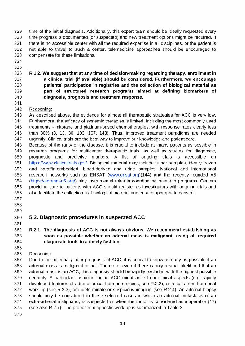
time of the initial diagnosis. Additionally, this expert team should be ideally requested every 329 time progress is documented (or suspected) and new treatment options might be required. If 330 there is no accessible center with all the required expertise in all disciplines, or the patient is 331 not able to travel to such a center, telemedicine approaches should be encouraged to 332 compensate for these limitations. 333
334 335
R.1.2. We suggest that at any time of decision-making regarding therapy, enrollment in 336 a clinical trial (if available) should be considered. Furthermore, we encourage 337 patients’ participation in registries and the collection of biological material as 338 part of structured research programs aimed at defining biomarkers of 339 diagnosis, prognosis and treatment response. 340
341 Reasoning: 342 As described above, the evidence for almost all therapeutic strategies for ACC is very low. 343 Furthermore, the efficacy of systemic therapies is limited, including the most commonly used 344 treatments - mitotane and platinum-based chemotherapies, with response rates clearly less 345 than 30% (3, 13, 30, 103, 107, 143). Thus, improved treatment paradigms are needed 346 urgently. Clinical trials are the best way to improve our knowledge and patient care. 347 Because of the rarity of the disease, it is crucial to include as many patients as possible in 348 research programs for multicenter therapeutic trials, as well as studies for diagnostic, 349 prognostic and predictive markers. A list of ongoing trials is accessible on 350 https://www.clinicaltrials.gov/. Biological material may include tumor samples, ideally frozen 351 and paraffin-embedded, blood-derived and urine samples. National and international 352 research networks such as ENSAT (www.ensat.org)(144) and the recently founded A5 353 (https://adrenal-a5.org/) play instrumental roles in coordinating research programs. Centers 354 providing care to patients with ACC should register as investigators with ongoing trials and 355 also facilitate the collection a of biological material and ensure appropriate consent. 356
357 358 359
5.2. Diagnostic procedures in suspected ACC 360 361
R.2.1. The diagnosis of ACC is not always obvious. We recommend establishing as 362 soon as possible whether an adrenal mass is malignant, using all required 363 diagnostic tools in a timely fashion. 364
365 Reasoning 366 Due to the potentially poor prognosis of ACC, it is critical to know as early as possible if an 367 adrenal mass is malignant or not. Therefore, even if there is only a small likelihood that an 368 adrenal mass is an ACC, this diagnosis should be rapidly excluded with the highest possible 369 certainty. A particular suspicion for an ACC might arise from clinical aspects (e.g. rapidly 370 developed features of adrenocortical hormone excess, see R.2.2), or results from hormonal 371 work-up (see R.2.3), or indeterminate or suspicious imaging (see R.2.4). An adrenal biopsy 372 should only be considered in those selected cases in which an adrenal metastasis of an 373 extra-adrenal malignancy is suspected or when the tumor is considered as inoperable (17) 374 (see also R.2.7). The proposed diagnostic work-up is summarized in Table 3. 375 376
14

Table 3: Diagnostic work-up in patients with suspected or proven ACC 377 378 Hormonal work up
• Glucocorticoid excess - 1mg dexamethasone suppression test or free cortisol in 24-h urine1
- basal ACTH (plasma)2 • Sex steroids and steroid precursors3 - DHEA-S
- 17-OH-progesterone - androstenedione - testosterone (only in women) - 17-beta-estradiol (only in men and
postmenopausal women) - 11-deoxycortisol
• Mineralocorticoid excess - potassium - aldosterone/renin ratio (only in patients with
arterial hypertension and/or hypokalemia) • Exclusion of a pheochromocytoma - Fractionated metanephrines in 24h urine or
free plasma-metanephrines Imaging - CT or MRI of abdomen and pelvis,
- Chest CT - FDG-PET/CT4 - Bone or brain imaging (when skeletal or
cerebral metastases are suspected) 379 1 The 1-mg dexamethasone test is the preferred method to exclude relevant hypercortisolism. 380 However, if overt Cushing syndrome is evident, then cortisol in 24-h urine might be at least as good to 381 quantify the cortisol excess. Alternatively, salivary or serum bedtime cortisol can be used. 382 2 ACTH can be skipped if hypercortisolism is excluded. 383 3 The most suitable set of precursors and sex hormones has not yet been established and local 384 availability might be taken into account. 385 4 The panel did not agree on the systematic use of FDG-PET/CT (see R.2.4). 386 387
388 R.2.2. We recommend that every patient with (suspected) ACC should undergo careful 389
assessment including case history, clinical examination for symptoms and 390 signs of adrenal hormone excess. 391
392 Reasoning 393 All patients should undergo a careful evaluation with detailed history and physical 394 examination. In particular, patients should be evaluated for rapidly developing Cushing’s 395 syndrome (which frequently presents not as 'full blown' Cushing, but rather predominantly 396 with muscle weakness, hypokalemia, wasting and constitutional symptoms), and symptoms 397 and signs of a large abdominal mass. Clinical evaluation should additionally focus on 398 symptoms and signs of androgen excess, hirsutism or virilization in women or recent onset of 399 gynecomastia in men, because these might be clinical indicators for a cortisol-, androgen- or 400 estrogen-producing ACC, respectively (13, 23, 145-148). Any evidence of co-secretion of 401 different steroids raises the suspicion of an ACC (especially if sex-hormones are involved). In 402 contrast, mild, long standing hirsutism is usually not caused by an ACC, but rather due to 403 (among other diagnoses) polycystic ovary syndrome and non-classical congenital adrenal 404 hyperplasia (149). Primary aldosteronism is rare in ACC and usually accompanied by severe 405 hypokalemia (150). However, hypokalemia in ACC is more frequently caused by massive 406 cortisol excess overwhelming the renal 11-β hydroxysteroid dehydrogenase type 2 system. 407
408 409
15
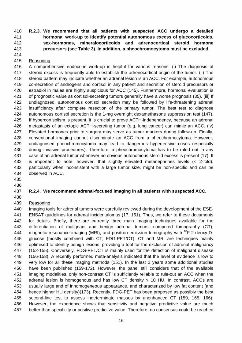
R.2.3. We recommend that all patients with suspected ACC undergo a detailed 410 hormonal work-up to identify potential autonomous excess of glucocorticoids, 411 sex-hormones, mineralocorticoids and adrenocortical steroid hormone 412 precursors (see Table 3). In addition, a pheochromocytoma must be excluded. 413
414 Reasoning 415 A comprehensive endocrine work-up is helpful for various reasons. (i) The diagnosis of 416 steroid excess is frequently able to establish the adrenocortical origin of the tumor. (ii) The 417 steroid pattern may indicate whether an adrenal lesion is an ACC. For example, autonomous 418 co-secretion of androgens and cortisol in any patient and secretion of steroid precursors or 419 estradiol in males are highly suspicious for ACC (145). Furthermore, hormonal evaluation is 420 of prognostic value as cortisol-secreting tumors generally have a worse prognosis (35). (iii) If 421 undiagnosed, autonomous cortisol secretion may be followed by life-threatening adrenal 422 insufficiency after complete resection of the primary tumor. The best test to diagnose 423 autonomous cortisol secretion is the 1-mg overnight dexamethasone suppression test (147). 424 If hypercortisolism is present, it is crucial to prove ACTH-independency, because an adrenal 425 metastasis of an ectopic ACTH-secreting tumor (e.g. lung cancer) can mimic an ACC. (iv) 426 Elevated hormones prior to surgery may serve as tumor markers during follow-up. Finally, 427 conventional imaging cannot discriminate an ACC from a pheochromocytoma. However, 428 undiagnosed pheochromocytoma may lead to dangerous hypertensive crises (especially 429 during invasive procedures). Therefore, a pheochromocytoma has to be ruled out in any 430 case of an adrenal tumor whenever no obvious autonomous steroid excess is present (17). It 431 is important to note, however, that slightly elevated metanephrines levels (< 2-fold), 432 particularly when inconsistent with a large tumor size, might be non-specific and can be 433 observed in ACC. 434
435 436
R.2.4. We recommend adrenal-focused imaging in all patients with suspected ACC. 437 438
Reasoning 439 Imaging tools for adrenal tumors were carefully reviewed during the development of the ESE-440 ENSAT guidelines for adrenal incidentalomas (17, 151). Thus, we refer to these documents 441 for details. Briefly, there are currently three main imaging techniques available for the 442 differentiation of malignant and benign adrenal tumors: computed tomography (CT), 443 magnetic resonance imaging (MRI), and positron emission tomography with 18F-2-deoxy-D-444 glucose (mostly combined with CT; FDG-PET/CT). CT and MRI are techniques mainly 445 optimised to identify benign lesions, providing a tool for the exclusion of adrenal malignancy 446 (152-155). Conversely, FDG-PET/CT is mainly used for the detection of malignant disease 447 (156-158). A recently performed meta-analysis indicated that the level of evidence is low to 448 very low for all these imaging methods (151). In the last 2 years some additional studies 449 have been published (159-172). However, the panel still considers that of the available 450 imaging modalities, only non-contrast CT is sufficiently reliable to rule-out an ACC when the 451 adrenal lesion is homogenous and has low CT density ≤ 10 HU. In contrast, ACCs are 452 usually large and of inhomogeneous appearance, and characterized by low fat content (and 453 hence higher HU density)(173). Recently, FDG-PET has been proposed as possibly the best 454 second-line test to assess indeterminate masses by unenhanced CT (159, 165, 166). 455 However, the experience shows that sensitivity and negative predictive value are much 456 better than specificity or positive predictive value. Therefore, no consensus could be reached 457
16

for a general recommendation on FDG-PET in all patients. Additional reasons in favor of 458 systematic FDG-PET are: whole body imaging (beyond thorax and abdomen, particularly for 459 distant bone metastasis) and in advanced disease, a reference uptake value for all 460 metastases can be established, which can help judging the future evolution of disease. 461 Evidence against FDG-PET includes cost, additional radiation exposure, false-positive 462 findings, and difficult access in some countries. 463 If adrenal imaging indicates an indeterminate mass, other parameters should be considered: 464 For instance, in such a situation a tumor size > 4 cm, combined adrenocortical hormone 465 excess (see also R.2.3), rapidly developing symptoms or young age (e.g. < 40 years) might 466 point to an ACC. However, it is important to note that no single imaging method can 467 definitively prove the diagnosis of ACC. 468
469 470
R.2.5. We recommend in any case where there is high suspicion for ACC performing a 471 chest CT, in addition to an abdominal-pelvic cross-sectional imaging (CT or 472 MRI), because the results might influence therapeutic decision-making. 473
474 Reasoning: 475 Since decisions for treatment strategy, particularly decisions for surgery, and prognostication 476 rely on tumor stage, it is mandatory to systematically and rapidly evaluate for metastases, 477 before initiation of any anti-tumor treatment. Thoraco-abdomino-pelvic imaging will cover the 478 vast majority of metastatic locations, which most often are lung and liver, and will assess 479 locoregional tumor extent. Imaging should include contrast-enhanced imaging. For 480 abdominal imaging there are advantages and disadvantages for both CT and MRI, but for 481 thoracic imaging CT is the method of choice, because it outperforms all other methods in 482 detecting small pulmonary lesions. 483 Additional imaging may be required to better characterize tumor vascularization, or specific 484 tumor extent such as a vena cava thrombus. 485 486 487 R.2.6. We suggest performing additional imaging (e.g. bone and brain imaging) only in 488
case of clinical suspicion of metastatic lesions. 489 490 Reasoning: 491 Bone and brain metastases are rare events (especially in patients without other metastatic 492 lesions). Therefore, additional imaging focusing on these sites is only warranted when there 493 is increased clinical suspicion or other imaging is suggestive for bone metastases. It should 494 be noted, however, that the basis for this advice has never been studied systematically. 495
496 497 R.2.7. We recommend against the use of an adrenal biopsy in the diagnostic work-up 498
of patients with suspected ACC unless there is evidence of metastatic disease 499 that precludes surgery and histopathologic proof is required to inform 500 oncological management. 501
502 Reasoning: 503 Differentiating benign from malignant adrenocortical tumors is very challenging on a biopsy 504 only and may lead to misdiagnosis (17, 174). Furthermore, the biopsy comes with significant 505
17

risks such as hemorrhage (175). The risk of tumor dissemination precluding a R0 resection is 506 very low (175). However, a biopsy might be indicated in an adrenal mass without any 507 hormone excess in patients with a history of extra-adrenal cancers to exclude or prove an 508 adrenal metastasis of an extra-adrenal malignancy. For details see the adrenal 509 incidentaloma guidelines (17). 510 511 512 513 5.3. Surgery for suspected localized ACC 514 515 R.3.1. We recommend that adrenal surgery for suspected/confirmed ACC should be 516
performed only by surgeons experienced in adrenal and oncological surgery. 517 518 Reasoning 519 ACC surgery requires expertise in both adrenal and oncological surgery due to the specific 520 anatomy, the malignant character of the disease and the potential need for multi-organ en-521 bloc resection to optimize the probability of a R0 resection and minimize the risk of 522 complications. 523 Data comparing outcome between 'high-volume' and 'low-volume' centers for ACC are 524 limited. Published reports from the UK, USA and Spain show an unacceptable low annual 525 workload for the majority of surgeons involved in any adrenal surgery, with a median 1 526 case/year (176-179). This situation is likely to have a negative impact on patient care and 527 contrasts significantly with the current status in other surgical specialties. 528 Based on the upper quartile distribution of workload of surgeons in USA, a volume of 4 529 adrenalectomies/year was used to define a ‘high-volume’ surgeon (177) but this threshold 530 might be too low to inspire confidence. Several studies showed that those doing more than 6-531 7 cases per year have shorter length of stay and less complications (176, 177, 180). Despite 532 the perceived benefit of being operated in a high-volume center, published data from Italy 533 and USA showed no significant association between overall survival / disease-free survival 534 and workload even though patients operated in high-volume centers had more radical 535 surgery, more lymph node assessment and more use of chemotherapy (181, 182). In 536 contrast, the creation of national centers for adrenal surgery in The Netherlands led to 537 significantly improved disease-free survival (1y: 93% vs. 78%, 5y: 63% vs. 42 %) (183, 184). 538 Therefore, the panel believes that a minimal annual workload of 6 adrenalectomies/year 539 seems to be required to ensure sufficient experience in adrenal surgery, but > 20 540 adrenalectomies/year are desirable for those involved in surgery for ACC. Furthermore, due 541 to the complexity of some operations, it is essential to involve surgeons with different 542 expertise (e.g. vascular, liver, and cardiac surgeons) for pre-surgical planning and during 543 these complex operations. 544 Protocols ensuring referral to regional or national centers should be established and patients 545 should feel empowered to ask about the previous experience of individual surgeons. 546
547 548 R.3.2. We recommend complete en bloc resection of all adrenal tumors suspected to 549
be ACC including the peritumoral/periadrenal retroperitoneal fat. We 550 recommend against enucleation and partial adrenal resection for suspected 551 ACC. If adjacent organs are suspected to be invaded, we recommend en bloc 552 resection. However, we suggest against the routine resection of the ipsilateral 553 kidney in the absence of direct renal invasion. 554
18

555 Reasoning 556 Complete resection is of utmost importance for all ACCs and successful surgery is a 557 prerequisite for cure. As the diagnosis of ACC might only become apparent after histological 558 analysis, it remains imperative for all adrenalectomies (laparoscopic or open) in patients with 559 a reasonable suspicion for ACC to respect the principles of oncological surgery in order to 560 ensure complete resection (R0 status) (185, 186). 561 To ensure that the pathologist can judge the completeness of surgery, any fragmentation of 562 the tumor has to be avoided. Intraoperative tumour rupture or spillage and R2 resection are 563 associated with very high recurrence rates and poor overall survival (26) (133). 564 Although there are no specific studies comparing outcome of surgery with and without 565 resection of invaded adjacent organs, it is deemed to be ‘good surgical practice’ to resect 566 adjacent tissues that are/could be invaded by tumor. This holds true for involvement of 567 spleen, distal pancreas, stomach, kidney, right liver, colon, diaphragm, the wall of the IVC or 568 left renal vein. A cohort study compared the oncological results of patients with stage II ACC 569 treated by radical adrenalectomy alone or by en-bloc resection with kidney. The results did 570 not support the hypothesis that nephrectomy improves the oncological outcome (187). 571 Combined nephrectomy, however, offers a lower risk of capsular rupture and can include 572 complete lymphadenectomy of the renal hilum, but impairs kidney function and this may limit 573 further access to chemotherapy. 574
575 576 R.3.3. Open surgery is the standard surgical approach for confirmed or highly 577
suspected ACC. Therefore, we recommend open surgery for all tumors with 578 radiological findings suspicious of malignancy and evidence for local invasion. 579 However, for tumors < 6 cm without any evidence of local invasion, 580 laparoscopic adrenalectomy (respecting the principles of oncological surgery) 581 is reasonable. 582
583 Reasoning 584 There is an ongoing debate if laparoscopic adrenalectomy is an acceptable alternative for 585 adrenal tumors with suspicion of ACC. Based on the systematic review on this topic until July 586 2014 (17) and an additional literature search until December 2017 (188-195), we conclude 587 that the quality of evidence from these observational studies is still very low. The main 588 concerns with all these studies are differences of baseline characteristics between groups, 589 and between important prognostic factors, such as tumor stage or size. The lack of any 590 randomized trial prevents any final conclusions. However, in order to provide guidance for 591 clinicians the panel concurs with two other recent European guidelines (17, 185) and agrees 592 that all tumors with some radiological evidence of local invasion (including enlarged lymph 593 nodes) should undergo surgery with an open approach. The likelihood of a benign adrenal 594 tumor is higher in the group of adrenal incidentalomas ≤ 6 cm, for whom a laparoscopic 595 approach is reasonable. However, this cut-off is arbitrary and the experience of the surgeon 596 is the single most important factor. For detailed discussion we refer to the recent 597 recommendations for the surgical management of ACC by ESES and ENSAT (185) and the 598 guidelines on adrenal incidentaloma (17). 599 Although retroperitoneoscopic adrenalectomy is gaining popularity, only a small number of 600 surgeons are likely to have completed the learning curve to reach sufficient expertise, which 601 is estimated to be at least 20 cases (196, 197). This is a very significant issue in the context 602
19

of the overall minimal experience of most surgeons offering adrenalectomy (see above). 603 Outside specialized centers with large volume practice, retroperitoneoscopic adrenalectomy 604 should only be considered for benign tumors <4 cm. 605 606 607 R.3.4. We suggest that routine locoregional lymphadenectomy should be performed 608
with adrenalectomy for highly suspected or proven ACC. It should include (as a 609 minimum) the periadrenal and renal hilum nodes. All suspicious or enlarged 610 lymph nodes identified on preoperative imaging or intraoperatively should be 611 removed. 612
613 Reasoning 614 Reports from several databases indicated that patients with stage III tumors and positive 615 lymph nodes can have a 10-year overall survival rate of up to 40 per cent after resection (29, 616 70, 88, 198, 199). However, the wide range of reported lymph node involvement in ACC 617 (from 4 to 73%) (25, 26, 200) demonstrates that regional lymphadenectomy is neither 618 formally performed by all surgeons nor accurately assessed or reported by all pathologists. 619 According to large American and French series, approximately 10-30% of patients with ACC 620 had formal lymphadenectomy as part of the tumor resection, reflecting the heterogeneity of 621 operative management (25, 198). A minimum of four lymph nodes should be retrieved in 622 order to declare lymph node negative cases (201) Furthermore, in an analysis of 120 cases 623 identified from a multi-institutional database, the benefit of lymphadenectomy on overall 624 survival persisted on multivariable analysis controlling for adverse preoperative and 625 intraoperative factors associated with lymphadenectomy, such as tumor size, palpable mass, 626 irregular tumor edges, suspicious nodes on imaging, and multivisceral resection (202). The 627 largest series so far included 283 patients and the resection of more than five lymph nodes 628 reduced also the risk of local recurrence and disease-related death in a multivariate analysis 629 (203). 630 However, the panel is not in favor of a re-do operation if complete adrenalectomy was 631 performed without lymphadenectomy (e.g. due to perceived benign tumor). The clinical 632 benefit is uncertain and probably lower than the harm (e.g. delayed adjuvant therapy). 633 634 635 R.3.5. We recommend that individualized treatment decisions are made in case of 636
tumors with extension into large vessels based on multidisciplinary surgical 637 team. Such tumors should not be regarded ‘unresectable’ until reviewed in an 638 expert center. 639
640 Reasoning 641 Extension of ACC into the adrenal vein, renal vein or inferior vena cava occurs in 642 approximately 15-25% (29, 204, 205). Venous involvement consists mostly of intravenous 643 tumor thrombus. Thrombectomy might require vena cava cross-clamping above or below the 644 hepatic vein confluence or cardiopulmonary bypass, depending on the upper level of extent 645 of the thrombus. The resection might include a complete thrombectomy, a flush manoeuvre 646 and, occasionally, vascular cuff or prosthetic IVC replacement. A 3-year overall survival rate 647 of about 25% in a large series (206) encourages the performance of a venous resection in 648 the presence of vena cava or renal vein invasion but without distant metastases. 649 650
20

651 R.3.6. If the first surgery was suboptimal and macroscopically incomplete (R2 652
resection), we suggest to discuss re-do surgery in a multidisciplinary expert 653 team. 654
655 Reasoning 656 There has been no prospective study assessing the benefits (or the lack thereof) of early 657 reoperation in patients whose initial adrenalectomy was incomplete (R2 status). It is the 658 panel’s view that such patients should have intensive postoperative monitoring and if local 659 recurrence is detected radiologically, in the absence of other metastases, they should 660 undergo surgery with a curative intent at an expert center, if it is deemed likely to lead to an 661 R0 resection. 662 663 664 R.3.7. We recommend perioperative hydrocortisone replacement in all patients with 665
hypercortisolism that undergo surgery for ACC. 666 667 Reasoning: 668 Overt ACTH-independent Cushing's syndrome or biochemical autonomous cortisol secretion 669 might lead to adrenal insufficiency after removal of the adrenal source of cortisol (even in 670 patients with incompletely suppressed ACTH) (207). Therefore, the group unanimously sees 671 a clear indication of intra- and postoperative glucocorticoid replacement, preferably with 672 hydrocortisone, in all patients with evidence for ‘(possible) autonomous cortisol secretion’ 673 (post-dexamethasone cortisol >50 nmol/L (>1.8 μg/dL)). This should follow the suggestions 674 for major stress dose replacement as per recent international guidelines (208). 675 Postoperatively, the dose of glucocorticoid should be tapered on an individualized basis by a 676 physician experienced with this clinical scenario. 677 678 679 680 5.4. Pathological work-up 681 682 R.4.1. We recommend that the diagnosis of ACC should be confirmed by 683
histopathology (+++0). 684 685 Reasoning: 686 Histopathology is the gold-standard of diagnosing ACC and should in principle be obtained in 687 all patients. For patients deemed operable this will be done on the basis of the resection 688 specimen and for those patients that are inoperable, a biopsy will be taken in accordance 689 with good oncological practice. However, the majority of panelists argued that in selected 690 cases biopsy might be omitted when there is advanced disease with unequivocal ACTH-691 independent cortisol excess, androgen excess (testosterone, DHEAS) or estradiol excess. 692 There is no role for biopsy in a patient who is considered suitable for surgery of the adrenal 693 mass. 694
695 696
R.4.2. We suggest that all adrenal tumors, which cannot be readily classified, and all 697 suspected ACC, should be reviewed by an expert adrenal pathologist (++OO). 698
21

699 Reasoning: 700 Diagnosing ACC can be challenging and misdiagnoses are relatively frequent events. In 21 701 of 161 of the patients (13%) registered with the German ACC Registry between 2006 and 702 2009, the diagnosis of ACC had to be revised by the reference pathologist (24). Similar 703 results were found in a large series from Italy with a rate of misdiagnosis in 26 out of 300 704 cases (9%) (209). 705 706 707 R.4.3. We suggest the use of immunohistochemistry for steroidogenic factor-1 (SF1) 708
for the distinction of primary adrenocortical tumors and non-adrenocortical 709 tumors (+OOO). 710
711 Reasoning: 712 Generally, the distinction between adrenocortical and non-adrenocortical tumors is clear and 713 can be made on the basis of hematoxylin and eosin-stained slides. In case of doubt, on the 714 basis of histology only, whether a tumor originates from the adrenal cortex or not, 715 immunohistochemistry with SF1 is the most sensitive and specific marker currently available 716 to establish if the tumor in question is of adrenocortical origin, with a sensitivity of 98% and a 717 specificity of 100% (47). If this marker is not available, we advise a combination of markers, 718 which should include inhibin-alpha, melan-A, and calretinin (210, 211). Depending on the 719 differential diagnosis, other immunohistochemistry markers used to make alternative 720 diagnoses may be considered following local standard procedures. 721 722 723 R.4.4. We recommend the use of the Weiss system, based on a combination of 9 724
histological criteria that can be applied on hematoxylin and eosin-stained 725 slides, for the distinction of benign and malignant adrenocortical tumors 726 (++OO). 727
728 Reasoning: 729 There are many classification systems based on histology and/or a limited number of 730 additional markers for the distinction of benign and malignant adrenocortical tumors. The 731 Weiss system is the most widely used, and although it is not fully standardized (212, 213) the 732 panel favors use of this score. It should be noted that all scoring systems have similar 733 inherent problems. Using the Weiss system, a score of 3 or higher (on a total of 9 criteria, 734 see Table 4) indicates ACC (214, 215). A score of 2 and 3 may be considered as borderline 735 between benign and malignant tumors (tumors of uncertain malignant potential). In such 736 instance, one of several other classification systems, including the van Slooten index (216), 737 the modified Weiss score (41), the Helsinki classification (60, 77), and the addition of reticulin 738 stain assessment (217) may be used. 739 740 Special attention should be paid to histological variants of adrenocortical tumors, mainly 741 oncocytic tumors, which, because of their specific characteristics, will always have a Weiss 742 score of least 3, whether they are benign or malignant. For these tumors, an adapted scoring 743 system should be used (218-220). 744
22

Table 4 Histopathologic criteria by Weiss (214, 215) 745
The presence of three or more of the following criteria highly correlated with subsequent malignant behavior:
• High nuclear grade (Fuhrman criteria (221)) • > 5 mitoses per 50 high-power field • Atypical mitotic figures • < 25% of tumor cells are clear cells • Diffuse architecture (> 33% of tumor) • Necrosis • Venous invasion (smooth muscle in wall) • Sinusoidal invasion (no smooth muscle in wall) • Capsular invasion
746 747
748 R.4.5. We recommend the use of Ki67 immunohistochemistry for every resection 749
specimen of an adrenocortical tumor (++OO). 750 Ki67 immunohistochemistry has been proposed for prognostic purposes. Higher Ki67 levels 751 are consistently associated with poor prognosis. Threshold levels of 10% and 20% have 752 been considered for discriminating low from high Ki67 labeling index (58, 70). However it is 753 not clear whether any single significant threshold can be determined (see R.5.2.). 754 Ki67 labeling has been shown to be unevenly distributed in tumors. Therefore, determination 755 of the labeling index should be done on whole tumors, with specific attention to the area of 756 highest Ki67 labeling, preferably by use of an image analysis system (222, 223). If only a 757 biopsy is available a low Ki67 labeling may not be representative and therefore can be 758 misleading. 759 If Ki67 immunohistochemistry is not available, mitotic count may help in prognostic 760 stratification of ACC. Mitotic count has been proposed for grading of ACC, using >20 mitoses 761 per 50 high-power field to define high-grade tumors (56, 87, 215). However, the precise 762 correlation between mitotic count and Ki67 labeling is undetermined. 763 764 765 R.4.6. We recommend that the pathology report of a suspected ACC should at least 766
contain the following information: Weiss score (including the exact mitotic 767 count), exact Ki67 index, resection status, and pathological tumor stage 768 (indicating invasion or not of the capsule and/or surrounding tissue and 769 organs) and nodal status (+OOO). 770
771 Reasoning 772 The importance of Weiss score and Ki67 index has been discussed in R4.4 and R4.5, 773 respectively. It is important that the exact values are given, because this is of prognostic 774 relevance. Resection status is a major prognostic factor (see R 5.2.). Tumor stage, including 775 nodal involvement, is discussed below (see R.5.1). 776 777 778 779 780
23
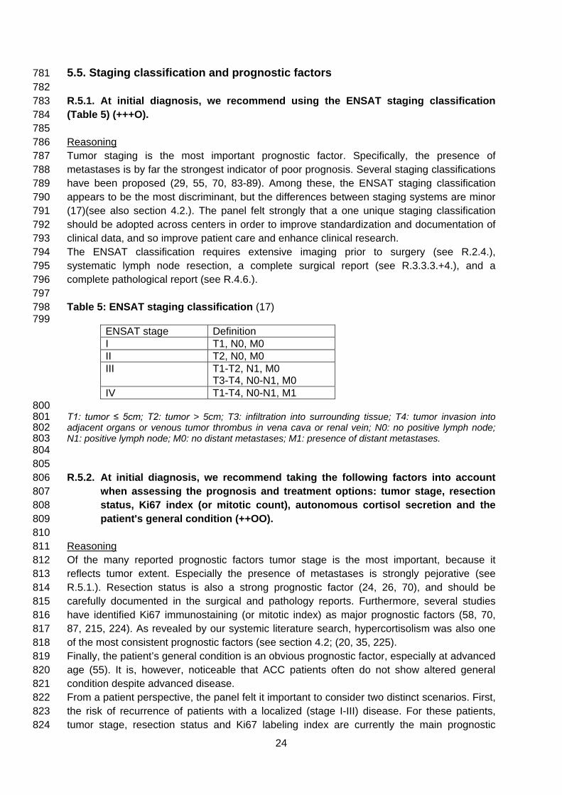
5.5. Staging classification and prognostic factors 781 782 R.5.1. At initial diagnosis, we recommend using the ENSAT staging classification 783 (Table 5) (+++O). 784 785 Reasoning 786 Tumor staging is the most important prognostic factor. Specifically, the presence of 787 metastases is by far the strongest indicator of poor prognosis. Several staging classifications 788 have been proposed (29, 55, 70, 83-89). Among these, the ENSAT staging classification 789 appears to be the most discriminant, but the differences between staging systems are minor 790 (17)(see also section 4.2.). The panel felt strongly that a one unique staging classification 791 should be adopted across centers in order to improve standardization and documentation of 792 clinical data, and so improve patient care and enhance clinical research. 793 The ENSAT classification requires extensive imaging prior to surgery (see R.2.4.), 794 systematic lymph node resection, a complete surgical report (see R.3.3.3.+4.), and a 795 complete pathological report (see R.4.6.). 796 797 Table 5: ENSAT staging classification (17) 798 799
ENSAT stage Definition I T1, N0, M0 II T2, N0, M0 III T1-T2, N1, M0
T3-T4, N0-N1, M0 IV T1-T4, N0-N1, M1
800 T1: tumor ≤ 5cm; T2: tumor > 5cm; T3: infiltration into surrounding tissue; T4: tumor invasion into 801 adjacent organs or venous tumor thrombus in vena cava or renal vein; N0: no positive lymph node; 802 N1: positive lymph node; M0: no distant metastases; M1: presence of distant metastases. 803 804 805 R.5.2. At initial diagnosis, we recommend taking the following factors into account 806
when assessing the prognosis and treatment options: tumor stage, resection 807 status, Ki67 index (or mitotic count), autonomous cortisol secretion and the 808 patient's general condition (++OO). 809
810 Reasoning 811 Of the many reported prognostic factors tumor stage is the most important, because it 812 reflects tumor extent. Especially the presence of metastases is strongly pejorative (see 813 R.5.1.). Resection status is also a strong prognostic factor (24, 26, 70), and should be 814 carefully documented in the surgical and pathology reports. Furthermore, several studies 815 have identified Ki67 immunostaining (or mitotic index) as major prognostic factors (58, 70, 816 87, 215, 224). As revealed by our systemic literature search, hypercortisolism was also one 817 of the most consistent prognostic factors (see section 4.2; (20, 35, 225). 818 Finally, the patient’s general condition is an obvious prognostic factor, especially at advanced 819 age (55). It is, however, noticeable that ACC patients often do not show altered general 820 condition despite advanced disease. 821 From a patient perspective, the panel felt it important to consider two distinct scenarios. First, 822 the risk of recurrence of patients with a localized (stage I-III) disease. For these patients, 823 tumor stage, resection status and Ki67 labeling index are currently the main prognostic 824
24

factors. This panel proposes to define two classes of localized ACC: low/moderate risk ACC 825 includes stage I-II and R0 and Ki67 ≤10%, whereas high risk ACC includes stage III, R1, or 826 Ki67 >10%. However, the panel is aware that the dichotomy is arbitrary. 827 Second, the prognosis of patients with advanced disease (stage IV or recurrent disease not 828 amenable to complete resection or R2 resection). High tumor burden, high tumor grade, high 829 Ki67 index, and uncontrolled symptoms are major factors associated with worse prognosis 830 (56, 70). However, there is consensus that the kinetics of tumor growth might be also 831 relevant, particularly when making the decision for initiation of cytotoxic chemotherapy. 832 However, this parameter has not been formally assessed. Although a correlation of tumor 833 growth and tumor grade exists, it is not true for all tumors. 834
835 836
R.5.3. During follow-up, we recommend re-assessing prognosis at each evaluation, to 837 guide treatment strategy (++OO). 838 839 Reasoning 840 After complete surgery, the major prognostic factor is whether there is any tumor recurrence. 841 At the time of recurrence the main prognostic factors are time between initial surgery and 842 recurrence, tumor burden and resectability (61, 62, 126, 136). 843 For patients with advanced disease, prognostic factors include Ki67 index, tumor burden, 844 general patient condition, and kinetics of tumor growth, as well as response to treatment. 845 Limited evidence is available, but these factors make clinical sense and are corroborated by 846 this panel’s experience. 847 848 849 5.6. Methods and time interval for imaging and hormonal assessment during 850 follow-up 851 852 R.6.1. We recommend following patients with regular cross-sectional imaging of the 853
abdomen, pelvis and chest for disease recurrence or progression. 854 855 Reasoning 856 A majority of disease recurrence and progression occurs either loco-regionally, or with 857 metastases to lung or liver and therefore should be identified by thoraco-abdomino-pelvic 858 imaging. Bone metastases are rare and brain involvement is exceptional (23, 70, 226). In 859 general, 18-FDG-PET/CT might provide additional information (see R.2.4.) particularly prior 860 to any surgical intervention (156, 227, 228). In addition, change in tracer uptake might inform 861 about disease evolution. 862 863 864 R.6.2. After complete resection, we suggest radiological imaging every 3 months for 2 865
years, then every 3-6 months for a further 3 years. The majority of the panel 866 suggests continuation of follow-up imaging beyond 5 years, but surveillance 867 should then be adapted. 868
869 Reasoning 870 There are no published studies that address specifically this issue. Therefore, the suggested 871 imaging interval is in accordance with the practice at many expert centers, and with 872
25

standards for other malignant tumors. In the experience of the panel few tumors with initial 873 curative surgery will recur after more than five years and therefore a 5-yr surveillance is likely 874 to include >90% of the ACC population that will experience disease recurrence. However, the 875 majority of the panel felt uncomfortable with the notion of complete cessation of imaging after 876 5 years and preferred for instance an annual imaging for another 5 years. After stopping 877 regular imaging, patients and primary care physicians should remain vigilant in terms of 878 potential symptoms or signs of late recurrences. 879 880 881 R.6.3. For advanced ACC, we recommend surveillance based on prognostic factors, 882
expected treatment efficacy and treatment-related toxicity, as well as the 883 available alternative treatment options. 884
885 Reasoning 886 The imaging interval in advanced ACC depends on the ongoing treatment and the overall 887 prognosis, but will usually be in 2-3 monthly intervals. For patients receiving mitotane alone, 888 imaging intervals might be even more individualized (e.g. 2-5 months) based on tolerability 889 and tumor kinetics. For patients undergoing loco-regional treatments, specific surveillance 890 following procedures must be determined by the team performing these procedures, both to 891 assess efficacy and adverse effects. For patients opting for entirely palliative management, 892 without any anti-neoplastic therapy, no systematic imaging is advised. 893 894 895 R.6.4. In all patients, we recommend regular screening for hormone secretion. 896 897 Reasoning 898 Biochemical evaluation together with clinical evaluation fulfills two purposes: (i) it allows in 899 few patients the early detection of recurrences and (ii) it also identifies patients that might 900 benefit from early anti-hormonal therapy. Biochemical evaluation should focus on steroid 901 hormones or metabolites that were present at the time of diagnosis of the initial tumor. 902 However, some panelists favored a more complete hormonal evaluation, because some 903 tumors might change their steroid secretion pattern over time. 904 905 906 907 5.7. Adjuvant therapy 908 909 R.7.1. For adrenal tumors with uncertain malignant potential, we recommend against 910
adjuvant therapy (+OOO). 911 Reasoning: 912 In certain tumors it is difficult to define if the tumor is truly malignant (see R.4.4.). Since all 913 adjuvant therapies are associated with potential toxicity, only patients with a definitive 914 diagnosis of ACC should be considered for adjuvant treatment. 915
916 R.7.2. We suggest adjuvant mitotane treatment in those patients without macroscopic 917
residual tumor after surgery that have a perceived high risk of recurrence 918 (+OOO). However, we cannot suggest for or against adjuvant therapy for 919
26

patients at low/moderate risk of recurrence (stage I-II, R0 resection and Ki67 ≤ 920 10%) and adjuvant therapy options should be discussed on an individual basis. 921
922 Reasoning: 923 The panel is in favor of offering mitotane to patients with high risk of recurrence (stage III, or 924 R1 resection, or Ki67 >10%; see R.5.2.) despite the absence of completely convincing 925 evidence (see section 4.3). The panel decided to use of mitotane in the adjuvant setting 926 based on three arguments: (i) the perceived effects (28, 36, 90-94, 229) (acknowledging this 927 is based on low quality evidence), see Figures 1A + B; (ii) published data showing a tumor 928 response in ~20% of patients with advanced disease treated with mitotane (13, 107, 143, 929 230); (iii) clinical experience of the panelists. For details on mitotane management see 930 section 5.9. 931 Ki67 has emerged as the most powerful predictor of recurrence, and tumors with Ki67 ≤10% 932 might represent a subset of patients with a good prognosis. For these patients mitotane 933 might be considered overtreatment. For this subset of patients (<30% of all localized ACCs) 934 the ongoing ADIUVO trial, a prospective study where patients are randomized to adjuvant 935 mitotane vs. observation, will provide guidance in a few years. 936 There is no clinical, histopathological, or molecular marker that reliably predicts response to 937 mitotane although few markers have been proposed (231, 232). A study showed that 938 mitotane levels may influence patient outcome in adjuvant setting (233) as it has been 939 reported in advanced ACC. The secretory status of the tumor has a negative prognostic 940 value but does not seem to influence response to treatment (20, 90, 230). 941 In patients who undergo surgery for recurrence of ACC but who have not previously had 942 medical therapy, the decision on adjuvant mitotane should follow the same lines of 943 reasoning. 944
945 946
R.7.3. Once the decision for mitotane treatment is established, we recommend 947 starting mitotane as soon as clinically possible after surgery (+OOO). 948
Reasoning: 949 The ideal timing to start adjuvant mitotane is unknown; however, by analogy with other 950 oncological adjuvant treatments we are convinced that starting mitotane within six weeks is 951 ideal, and would not initiate the treatment later than 3 months. This reasoning is sound with 952 the biological concept of adjuvant therapy in general, and with the latency of mitotane to 953 reach effective levels and anti-tumor activity. However, no published data are available to 954 demonstrate the superiority of an early start of treatment or the lack of efficacy when started 955 later than 3 months. 956 957 958 R.7.4. In patients without recurrence who tolerate mitotane in an acceptable manner, 959
we suggest to administer adjuvant mitotane for at least 2 years, but not longer 960 than 5 years (+OOO). 961
962 Reasoning: 963 The optimal duration of mitotane treatment is unknown and practice varies among different 964 centers. Some members of the panel continue treatment for 3 to 5 years if tolerated (234), 965 while others discontinue after 2 to 3 years (3, 13, 19). Prognostic factors at diagnosis, patient 966 compliance with treatment and plasma mitotane levels reached during treatment are factors 967
27

that influence duration of treatment. Mitotane possibly acts as an oncostatic measure in 968 those patients (235, 236). However, the rate of recurrence 5 years after surgery is potentially 969 too low to advise continuation of therapy treatment beyond this time point. Treatment-related 970 toxicity, lack of experience in long-term administration are additional factors portending 971 against indefinite treatment. 972
973 974
R.7.5. The panel did not come to a definitive consensus on adjuvant radiation therapy. 975 However, we suggest against the routine use of radiation therapy in patients 976 with stage I-II and R0 resection (+OOO). The panel suggests considering 977 radiation in addition to mitotane therapy on an individualized basis therapy in 978 patients with R1 or Rx resection or in stage III. 979
980 Reasoning: 981 The systematic literature search indicated that radiation therapy is able to prevent local 982 recurrence but does not significantly affect distant recurrences or overall survival (91, 98-983 100, 237, 238) (see section 4.3. and Figure 1). However, distant metastases account for 984 about 40-60% of tumor relapses (54, 61, 90) and have large impact on the patient prognosis, 985 and are more difficult to treat effectively. Conversely, prevention of the complications due to 986 local recurrence argues in favor of radiation therapy. Adjuvant radiation therapy might be 987 particularly reasonable in patients with R1 resection. This was already suggested by earlier 988 studies, but also by a very recent study that was published after our meta-analysis (239). 989 990 Radiation therapy is not advised for patients who experienced widespread tumor spillage 991 during surgery. The combination of radiation therapy and mitotane is biologically sound (240, 992 241) and possible but at the cost of greater toxicity (e.g. constitutional, gastrointestinal and 993 liver toxicity). In addition, there is concern that radiation therapy may delay systemic therapy 994 or prevent effective mitotane administration resulting in lower drug levels. 995
996 997
R.7.6. If adjuvant radiation therapy is administered, we recommend starting 998 treatment as soon as clinically possible after surgery and to deliver radiation 999 therapy at the dose of 50-60 Gy to the previous tumor bed in fractionated 1000 doses of approximately 2 Gy each (+OOO). 1001
Reasoning: 1002 Radiation therapy was delivered following this scheme in previous observational studies (91, 1003 98-100, 238) and lower dosage seems to be less effective (237). 1004 1005 1006 R.7.7. The panel did not come to a definitive consensus on adjuvant use of cytotoxic 1007
drugs. We suggest against the routine use of cytotoxic drugs in the adjuvant 1008 setting. However, the panel suggests considering adjuvant chemotherapy in 1009 selected patients with very high risk for recurrence. 1010
1011 Reasoning: 1012 Scant data are available on the use of cytotoxic drugs in an adjuvant setting and the studies 1013 did not control the results of treatment with a matched control group of untreated patients, or 1014 patients treated undergoing mitotane therapy (242). However, the majority of panelists favors 1015
28

discussion of this option with patients with high risk of recurrence (ideally in the setting of 1016 clinical trials). Despite the lack of published data, some members of the panel are currently 1017 using cisplatin, with or without etoposide, in patients at perceived very high risk of recurrence 1018 (e.g. Ki67 >30%. large tumor thrombus in the vena cava, stage IV, or R1 resection). 1019 In patients with R2 resection or tumor spillage, the same considerations for treatment of 1020 (locally) advanced disease should apply (see section 5.8.). 1021 1022 1023 Figure 3: Treatment for ACC amenable to complete resection 1024 1025
1026 1027 1028 DFI disease-free interval between complete resection and recurrence 1029 1 All patients with stage I+II and most patients with stage III should be amenable to radical resection. If complete 1030
resection is not feasible, consider neo-adjuvant treatment (e.g. cisplatin or EDP). In selected patients with single 1031 metastases complete resection might be possible as well. 1032
2 In patients with R2 resection, consider re-surgery by an expert surgeon (see R.3.6) or see Figure 1B 1033 3 If Ki67 staining is not available, a low (<20 mitoses / 50 high power fields) or a high mitotic rate (> 20 mitoses / 1034
50 high power fields) may be used for risk stratification. 1035 4 Individual decision (see R.7.2.). If possible enroll in clinical trial like ADIUVO (www.adiuvo-trial.org). 1036 5 In some patients (e.g. Ki67 >30%. large tumor thrombus in the vena cava, stage IV, or R1 resection) consider 1037
additional cytotoxic therapy (e.g. 3-4 cycle of cisplatin + etoposide). 1038 6 After two years the time intervals are gradually extended. 1039 7 If the disease-free interval is between 6 and 12 months or in patients with DFI > 12, in whom complete resection 1040
is not possible, an individual approach is required (see R.8.7.) 1041
29

5.8. Treatment of recurrent and/or advanced ACC 1042 1043 Clinical scenarios of patients with recurrent and/or advanced ACC are highly variable. 1044 Therefore, we try to provide recommendations for at least the most frequent presentations 1045 (see also Figure 4). Although a (small) proportion of patients experience a relatively long 1046 survival (13, 29, 70, 243), the prognosis of advanced/metastatic is generally limited. The goal 1047 of any therapy is to palliate symptoms and prolong survival. In this situation it is even more 1048 important than in other scenarios to tailor treatment on an individual basis taking into account 1049 the disease extent, the patient performance status and particularly the preference of the 1050 patient. 1051 1052 1053 Figure 4: Treatment of advanced ACC 1054 1055
1056 1057 EDP Etoposide, Doxorubicin, cisPlatin 1058 1 only in selected patients (e.g. with severe hormone excess) 1059 2 The following factors might guide the decision: site of disease involvement, tumor burden, symptoms, tumor 1060
grade/Ki67 index 1061 3 The following factors might guide the decision: site of disease involvement, tumor burden, symptoms, tumor 1062
grade/Ki67 index, and importantly kinetics of tumor growth 1063 4 radiotherapy, radiofrequency ablation, cryo ablation, microwave ablation, (chemo-)embolization 1064 5 Few panelist favored cisplatin + etoposide 1065 6 For the currently available cytotoxic regimens see Table 6 and contact specialized center. 1066 1067 1068 R.8.1. For patients presenting at time of initial diagnosis with limited intra-abdominal 1069
metastases we suggest surgical therapy if complete resection of all lesions 1070 seems feasible (+OOO). In case of limited extra-abdominal lesions, we suggest 1071 adrenal tumor resection in conjunction with therapy aiming at long-term tumor 1072
30

control of the other lesions (+OOO). In all patients, we recommend to start 1073 mitotane therapy as soon as clinically possible (+OOO). 1074
1075 Reasoning: 1076 Complete surgery is the best chance to reach long-term disease control although the 1077 likelihood of complete tumor removal in advanced ACC is low. If clinically possible, a single 1078 surgical approach should be planned. If a one-time surgical approach is impossible (e.g. due 1079 extra-abdominal metastases), other loco-regional approaches (see R.8.2) should be 1080 discussed on an individual basis. Local expertise and preference of the patient should be 1081 taken into account. Any initial treatment (surgery, local and/or medical therapy) should be 1082 initiated in a timely fashion (≤ 4-6 weeks following initial diagnosis). 1083 In general, prognostic parameters (see R.5.2 + 5.3) should influence the overall treatment 1084 strategy. If the disease has an aggressive behavior (i.e. increase in tumor burden [e.g. 1085 increasing size of existing tumors or new metastasis] observed in subsequent imaging 1086 performed within few weeks) systemic options (chemotherapy plus mitotane) may be 1087 favored. If partial responses or prolonged stabilization are then observed, surgery and/or 1088 additional loco-regional options might be particularly useful (“neo-adjuvant approach", see 1089 also R.8.3). This strategy could also be potentially advantageous in patients for whom tumor 1090 shrinkage might allow a more conservative surgical approach (i.e. patients in whom radical 1091 surgery would imply the complete or partial removal of neighboring organs such as kidney, 1092 spleen and part of the pancreas)(244). 1093 These patients are at high risk for recurrence and therefore adjuvant mitotane seems to be 1094 justified (245). Addition of cytotoxic drugs might be a possible option (although data are 1095 lacking; see also R.7.7.). 1096
1097 1098 R.8.2. The panel is convinced that in addition to surgery other local therapeutic 1099
measures (e.g. radiation therapy, radiofrequency ablation, cryoablation, 1100 microwave ablation, chemo-embolization) are of value for therapy of advanced 1101 ACC. We suggest individualization of the decision on the method of choice 1102 based on the localization of the tumor lesion(s), local expertise, prognostic 1103 factors, and patient's preference (+OOO). 1104
1105 Reasoning: 1106 Published data on local therapies in advanced ACC are very limited (140-142, 237) and 1107 summarized in Appendix 6. However, the experience of many panelists provides additional 1108 evidence of efficacy of these local measures. Nevertheless, it is impossible to indicate which 1109 method is superior. Most important, the expertise of the local team in applying these methods 1110 should be taken into account when discussing this issue with patients in a shared decision-1111 making process. 1112 1113 1114 R.8.3. We suggest against the routine use of adrenal surgery in case of widespread 1115
metastatic disease at the time of first diagnosis (+OOO). 1116 1117 Reasoning: Despite the lack of large studies addressing this particular question, a majority of 1118 the panel agreed that patients with widespread and unresectable disease will usually not 1119
31

benefit from surgery. However, few panelists favored adrenalectomy in all patients, in whom 1120 this approach is technically possible. 1121 In patients who respond very well to systemic therapy, surgery should be considered at an 1122 appropriate time point; especially if complete resection becomes feasible ("neo-adjuvant 1123 approach"). However, the published evidence for such an approach is scant (244, 246). 1124 In selected cases (e.g. patients with severe hormone excess) debulking surgery might be an 1125 option, although anti-hormonal drugs (see R.10.1) should be considered here. In these 1126 cases, surgery might be especially reasonable if > 80% of the tumor burden can be removed 1127 safely. In patients with a poor clinical condition and significant localized metastatic burden, 1128 additional localized therapies (see R.8.2) may be considered as an alternative. 1129 1130 1131 R.8.4. In patients with advanced ACC at the time of diagnosis not qualifying for local 1132
treatment, we recommend either mitotane monotherapy or mitotane + EDP 1133 depending on prognostic parameters (+++O). 1134
1135 Reasoning: 1136 Mitotane is the treatment of choice for patients with advanced ACC (for details about the 1137 management of mitotane see section 5.9). However, a very recent cohort study suggests that 1138 patients with metastastic disease at the time of primary diagnosis might not be the ideal 1139 candidates for mitotane monotherapy (230). Furthermore, unfavorable prognostic parameters 1140 (e.g. high tumor burden, uncontrolled symptoms, high proliferative index, clinical evidence of 1141 a fast growing tumor) are important factors favoring a more aggressive/more rapidly active 1142 therapeutic approach. If more aggressive therapy is indicated, then the combination of EDP 1143 in addition to mitotane (EDP-M) is the most validated regimen (30). EDP-M is the only 1144 treatment approach in ACC that is successfully evaluated in a randomized trial, the FIRM-1145 ACT study. It has to be highlighted, however, that only progression-free survival was 1146 significantly improved in comparison to the alternative therapy (in this case streptozotocin 1147 plus mitotane; 5.0 vs. 2.1 months; HR 0.55; 95% CI 0.43 to 0.69; P<0.001)), whereas for 1148 overall survival the crossover design might have diluted the results (14.8 vs 12.0 months, HR 1149 0.79; 95% CI, 0.61 to 1.02; P=0.07). 1150 The administration of EDP-M comes with risk of adverse events and the risks might outweigh 1151 the benefits (especially in patients with reduced performance status). If there are concerns 1152 about the use of doxorubicin, cisplatin/carboplatin with or without etoposide (EP or P) might 1153 be an alternative option. Carboplatin may be an alternative to cisplatin, particularly when 1154 cardiac or renal function is compromised. Again, in this cohort, loco-regional treatment 1155 options may be particularly applicable. 1156 Several studies have tried to find biomarkers that predict response to cytotoxic therapy in 1157 ACC (247-250). However, no reliable marker could be identified yet. 1158 Few centers prefer the combination of etoposide and cisplatin (EP), because there is no 1159 single study proving that EDP is truly superior to EP. In patients with poor overall health 1160 cisplatin with mitotane may be an option. However, the evidence for etoposide + cisplatin or 1161 cisplatin alone is based only on small phase II studies (109, 110, 122). 1162 There is limited evidence that standard chemotherapeutic agents may be more active in 1163 presence of elevated mitotane concentrations (30, 104, 251), but the panel is not in favor in 1164 delaying cytotoxic therapy for this reason for more than 14 days. 1165 1166 1167
32

R.8.5. In patients with recurrent disease and a disease-free interval of at least 12 1168 months, in whom a complete resection/ablation seems feasible, we recommend 1169 surgery or alternatively other local therapies (+OOO). We recommend starting 1170 mitotane as soon as possible after the intervention. 1171
R.8.6. We recommend EDP-M as first line treatment if the time interval between last 1172 surgery/loco-regional therapy and recurrence is less than 6 months (++OO), 1173 rather than repeat loco-regional measures. 1174
R.8.7. For all other patients with recurrent disease an individualized approach is 1175 needed. 1176
1177 Reasoning: 1178 It has been suggested that patients with disease-free interval of 12 months or more have a 1179 significantly better prognosis and long-term disease control is achievable, if loco-regional 1180 measures are successful (61, 126). The choice of different loco-regional therapies depends 1181 again on benefit/risk ratio, local availability and expertise, and the clinical scenario in a given 1182 individual patient. Most panelists favor surgery (if complete resection is feasible) followed by 1183 mitotane therapy. 1184 If the recurrence occurs during adjuvant mitotane therapy, additional measures could be 1185 considered. In patients with local recurrence, adjuvant radiation therapy after surgery should 1186 be discussed. In other scenarios, additional administration of cytotoxic drugs should be 1187 discussed with the patient, particularly when mitotane blood levels were in the recommended 1188 range > 14 mg/l. 1189 Patients with early recurrence usually suffer from a very aggressive tumor, which most likely 1190 cannot be controlled by surgery or localized therapies. Decision-making should incorporate 1191 the concern that any local measure will only delay the administration of systemic therapy. 1192 Similar to the discussion to R.9.3, the FIRM-ACT data indicate EDP-M as the most effective 1193 form of therapy. An exception might be patients in whom incomplete initial surgery is the 1194 most likely cause for early progression. In these selected patients repeat surgery at an expert 1195 center might be an appropriate alternative (see R.3.6). 1196 Patients with recurrence between 6 and 12 months after primary surgery usually have a poor 1197 prognosis and would, therefore, benefit from a more aggressive therapeutic approach (e.g. 1198 EDP-M). However, this decision should be discussed with the patient taking into account 1199 prognostic parameters (see section 5.5.), the feasibility of a R0 resection and patient’s 1200 general condition. Patients with a disease-free interval > 12 months, in whom complete 1201 resection or loco-regional therapy is not feasible and who are currently not treated with 1202 mitotane, might be good candidates for mitotane monotherapy (230). 1203 1204 1205 R.8.8. In patients who progress under mitotane monotherapy, we recommend to add 1206
EDP (+++O). 1207 1208
Reasoning: 1209 Mitotane is a slow-acting drug and in patients with rapidly progressing tumor, it might be too 1210 slow or not effective enough. In these patients, based on the FIRM-ACT data (30), additional 1211 administration of EDP is the first choice (for alternatives see Reasoning R.8.4.). However, if 1212 the tumor burden is limited despite obvious progression, another 2-3 months mitotane 1213 monotherapy could also be justified, particularly if adequate mitotane levels have not been 1214 achieved. In these cases, additional loco-regional options should be considered. 1215
33

1216 1217 R.8.9. In patients who respond to medical therapy (including achievement of long-1218
term stable disease), we suggest re-considering local measures aiming at long-1219 term tumor control. Such an approach could be also considered in patients 1220 attaining a generally good control of the disease, in which a limited number of 1221 lesions are progressing. 1222
1223 Reasoning: 1224 In some patients, in whom long-term disease control could be achieved, loco-regional 1225 measures (in addition to ongoing medical therapy) might be able to reach complete 1226 remission or at least significantly reduce tumor burden (101). In patients with "mixed 1227 responses"; e.g. progressive disease limited to few lesions, loco-regional options might be 1228 reasonable to add to the ongoing medical therapy. 1229 1230 1231 R.8.10. In patients who progress under EDP-M we suggest considering additional 1232
therapies including clinical trials on an individual basis (+OOO). 1233 1234
Reasoning: 1235 Several drugs and drug combinations have been tested in advanced ACC. However, except 1236 EDP-M none of them has been successfully evaluated in large randomized trials. Figure 2 1237 outlines the outcomes of the different approaches. However, this figure has to be interpreted 1238 with great caution, because differences in the characteristics of the patients included in the 1239 different cohorts preclude direct comparison between studies. Therefore, it is not possible to 1240 draw definitive conclusions. Due to the limited treatment options, the panel clearly favors 1241 enrollment of patients with progressing tumors in clinical trials investigating experimental 1242 therapies including phase I trials. However, the panel felt that despite the lack of convincing 1243 data, some guidance might be helpful for patients that cannot be enrolled in clinical trials 1244 (Table 6). Beyond cisplatin-based therapies, the two reasonably well-studied second-line 1245 cytotoxic regimens are gemcitabine + capecitabine (+/- mitotane) (104, 124) and 1246 streptozotocin + mitotane (30, 242). However, objective response rates are clearly below 1247 10% and median progression-free survival (PFS) is generally <4 months, but a few patients 1248 with long-term disease control and even complete responses in single patients are 1249 described. Nevertheless, few panelists argued against the use of streptozotocin, because 1250 median PFS in the FIRM-ACT trial was only two months (30). 1251 Loco-regional measures can be particularly useful when progression is limited, or only 1252 affects limited areas (e.g. single organs). In these cases, such localized therapies (see 1253 R.8.2) might be able to provide higher response rates for these specific organ/tissue areas 1254 than second line systemic options. 1255 Several tyrosine kinase inhibitors have been investigated in advanced ACC (102, 108, 116, 1256 118), but the results were largely disappointing. However, in retrospect, drug efficacy could 1257 have been hampered by increased metabolism of the TKI due to mitotane-induced CYP3A4 1258 activity. Nevertheless, currently no specific TKI can be suggested for the treatment of 1259 advanced ACC. Targeting the IGF2/IGF receptor signaling pathway was pathophysiologically 1260 a very promising approach and initial small studies suggested some efficacy (113, 117, 125, 1261 252-258). However, the large placebo-controlled phase III GALACCTIC trial demonstrated 1262 that the IGF1R inhibitor linsitinib did not improve progression-free or overall survival (102). 1263
34

Therefore, monotherapy with drugs targeting this pathway are not reasonable for therapy in 1264 an unselected patient population. 1265 1266 1267 Table 6: Systemic therapies for recurrent / advanced ACC 1268
First-line therapies (see text for details) • Surgery +/- other local measures (see R.8.1 and R.8.4)
• Mitotane monotherapy
- details on the management see section 5.9. • Etoposide, Doxorubicin and Cisplatin (EDP) plus Mitotane (EDP/M) (30)
every 28 days: day 1 40mg/m2 doxorubicin (D) day 2 100mg/m2 etoposide (E) day 3+4 100mg/m2 etoposide (E) + 40mg/m2 cisplatin (P)
plus oral mitotane aiming at a blood level between 14-20mg/l.
In patients unfit for the EDP-M regimen, (E)P-M may constitute a reasonable alternative. Every 28 days
day 1 100mg/m2 etoposide (E) day 2+3 100mg/m2 etoposide (E) + 40mg/m2 cisplatin (P)
Additional therapeutic options
• Consider enrollment of patients in clinical trials (www.clinicaltrial.gov)
• Consider loco-regional therapies • Gemcitabine plus capecitabine (104, 124)
800 mg/m2 gemcitabine on day 1 and 8 (repeated every 3 weeks) 1,500 mg capecitabine orally per day in a continuous fashion Mitotane can be continued (individualized decision)
• Streptozotocin plus Mitotane (Sz/M) (30)
induction: day 1-5: 1g Sz/d afterwards 2g/d Sz every 21 days plus oral mitotane aiming at a blood level between 14-20mg/l
1269 1270 R.8.11. The optimal timing of mitotane discontinuation is currently unknown and the 1271
panel could not come to a specific recommendation on this issue. 1272 1273
Reasoning: 1274 A recent cohort study reported that discontinuation of mitotane should be considered in 1275 patients who experienced progressive disease after one year of mitotane therapy (259). Part 1276 of the panel considers mitotane discontinuation when there is progressive disease despite 1277 mitotane blood levels above 14 mg/L while others often continue mitotane indefinitely in their 1278 practice. Tolerability of treatment is an important issue to consider in this decision. Moreover, 1279 it has to be considered that CYP3A4 induction by mitotane can greatly enhance metabolism 1280 of many drugs (260), including a number of experimental anti-ACC compounds, and so 1281 potentially limit their effectiveness. 1282
35

5.9. Special considerations on mitotane 1283 1284 If mitotane therapy is started (independent of the clinical scenario) the following issues have 1285 to be considered. 1286
1287 R.9.1. We recommend starting therapy with mitotane in an escalating regimen 1288
depending on the performance status of the patient as well as the tolerability in 1289 the first weeks. 1290
1291 Reasoning 1292 There are different regimens to administer mitotane, but none of them has been proven to be 1293 superior. In patients with good performance status some panelists use a high starting dose 1294 approach: mitotane is administered at a starting dose of 1.5 g/day and if well-tolerated from a 1295 gastrointestinal perspective the dose is increased on day two to 3 g/day, on day three to 4.5 1296 g/day, and on day four to 6 g/day (261, 262). This dosage will be administered until first 1297 mitotane blood level is assessed. In this high dose regimen, it is strongly recommended to 1298 measure mitotane blood levels 2-3 weeks after initiation of therapy. Afterwards dosage will 1299 be adjusted according to blood concentrations and tolerability. Other panelists prefer a low 1300 starting dose approach. With this approach, mitotane is administered at a starting dose of 1 1301 g/day and increased when there is good gastrointestinal tolerance every 3 days by 0.5 g up 1302 to a total dose of 3.0 - 4.0 g/day and then adjusted according to blood concentrations and 1303 tolerability (234, 263, 264). 1304 In a formal comparative pharmacokinetic study, the high-dose starting regimen led to slightly 1305 higher mitotane plasma levels within 12 weeks of treatment, and more patients reached the 1306 target level of 14 mg/L (265). However, these results were not statistically significant due to 1307 lack of power. Beyond these two regimens, there is a variety of other possibilities and choice 1308 depends on personal practice, clinical scenario and patient conditions. 1309
1310 1311
R.9.2. We recommend monitoring of blood concentration of mitotane. The general aim 1312 is to reach a mitotane blood level above 14 mg/L (+OOO). 1313
1314 Reasoning 1315 As long as mitotane plasma levels are increasing and have not yet reached a plateau at 1316 >14mg/L, mitotane plasma levels will be assessed every 3-4 weeks. Mitotane plasma level 1317 determination is best done as morning trough sampling, at least 12 hours after the last dose, 1318 preventing false high levels (266). When mitotane plasma levels have reached a plateau, it is 1319 usually sufficient to measure blood levels every 6-12 weeks. 1320 Usually it takes several weeks (sometimes months) to reach mitotane levels > 14 mg/L. As 1321 long as the concentration is < 14 mg/L it is reasonable to continue to increase the dosage if 1322 this is tolerated by the patient. Due to slow pharmacokinetic characteristics, the dose of 1323 mitotane can be reduced in most patients as soon as a plasma level of > 14mg/L is reached. 1324 Over time, mitotane dosage will be titrated to the best tolerable dose while maintaining a 1325 plasma level >14mg/L. Most patients experience adverse effects to a certain extent and 1326 these usually correlate with the plasma mitotane level (although there is major inter-individual 1327 variability). However, some gastrointestinal adverse effects (like diarrhea) seem to correlate 1328 more with the oral dosage than with the plasma level and occur more frequently in the first 1329 phase of treatment (146, 234, 263, 264, 267). Several studies (107, 112, 268) have shown 1330
36

that CNS-related adverse events in particular occur more frequently when the plasma 1331 mitotane is > 20 mg/L. Therefore, many experts recommend aiming to keep plasma 1332 concentrations below 20 mg/L. However, it can be speculated that higher plasma levels may 1333 also be associated with better clinical efficacy. Furthermore, some patients do not experience 1334 relevant adverse events even at plasma levels well above 20 mg/L. Regarding the lower limit 1335 it has to be acknowledged that in at least a few patients objective responses are seen even 1336 though plasma levels of >14 mg/l were not achieved (230). Therefore, some panelists 1337 favored a target range of plasma mitotane of 8-30 mg/L, whereas others aim at an 1338 individualized target level of mitotane. 1339 1340 Most studies addressing plasma mitotane levels analyze patients with advanced disease. 1341 However, there is one study suggesting that the same target level is also reasonable for the 1342 adjuvant setting (233). Therefore, the panel is in favor to use the same approach for both 1343 patient groups. 1344
1345 1346
R.9.3. We recommend glucocorticoid replacement in all patients treated with mitotane 1347 (except those with ongoing cortisol excess). We suggest to using 1348 hydrocortisone/cortisone acetate for this purpose. Due to increased steroid 1349 clearance and increase cortisol-binding globulin at least twice the standard 1350 replacement dose is usually required. 1351
1352 Reasoning 1353 A possible strategy is to start concomitant treatment on day one of mitotane treatment with 1354 hydrocortisone 20 mg/d. Alternatively, patients can be instructed to start hydrocortisone later 1355 (e.g. after 2-3 weeks or in case they experience adrenal insufficiency), because impairment 1356 of glucocorticoid effectiveness is rarely observed within the first few weeks. Due to the 1357 increased clearance and increased cortisol-binding globulin (267, 269-271) with increasing 1358 mitotane plasma levels and based on clinical symptoms, the total hydrocortisone 1359 replacement dose will usually increased to a typical total daily dose of 50 mg in two or three 1360 divided doses. However, some patients require daily dosages up to 100 mg. There is no 1361 reliable laboratory marker to guide the optimal dosage of hydrocortisone (270, 272), which 1362 has to be based on clinical judgment similar to the management of patients with adrenal 1363 insufficiency (208). Mitotane-induced increase in cortisol binding globulin may confound 1364 interpretation of serum cortisol measurement. Some panelists measure plasma ACTH and 1365 use ACTH levels more than 2-fold of the upper limit of normal as evidence for insufficient 1366 glucocorticoid replacement. 1367 In case of acute adverse events and/or hospital admission, patients should be treated 1368 intravenously with high-dose hydrocortisone (e.g. 100 mg TID) until resolution of symptoms. 1369 Some patients experience symptoms and signs of insufficient mineralocorticoid activity 1370 (hyperkalemia, hyponatremia, hypotension, decreased wellbeing) despite full-dose 1371 substitution with hydrocortisone. In these patients, addition of fludrocortisone should be 1372 considered. Clinical judgment, electrolytes, and plasma renin concentration can be used for 1373 decision making whether to start fludrocortisone (146, 234, 264, 267). 1374 1375 1376
37

R.9.4. We recommend regular monitoring of mitotane-induced adverse effects (Table 1377 7) and to treat them appropriately (Table 8). To increase tolerability of mitotane, 1378 we suggest starting supportive therapy ideally before severe toxicity occurs. 1379
1380 Reasoning 1381 In addition to adrenal insufficiency (see R.9.3.) mitotane treatment comes with a plethora of 1382 potential adverse events (267)(Table 2). Therefore, it is important to evaluate the patients 1383 regularly (e.g. in the first 6 months every 3-4 weeks, thereafter every 6-12 weeks). 1384 Gastrointestinal adverse effects are frequent, particularly in the first months of therapy. 1385 Supportive therapy should include antiemetic and ant-diarrheal medication, as needed. 1386 Some centers even start supportive therapy at initiation of mitotane therapy. However, one 1387 has to be aware that nausea may also be a sign of adrenal insufficiency that needs 1388 recognition and appropriate treatment. Nevertheless, it should be emphasized that despite 1389 optimization of dosing schedules, the key factor influencing build-up of appropriate mitotane 1390 plasma levels is patient tolerability, so efforts should be made in order to optimize this. 1391 In case of central nervous system (CNS) adverse effects grade 2 (moderate) and/or gastro-1392 intestinal adverse effects grade 3 (severe, but not life-threatening), mitotane dose should be 1393 reduced by 1-1.5 gram/day. In case of CNS adverse effects grade 3 or any relevant grade 4 1394 toxicity (life-threatening), and/or increase of liver enzymes >5 times baseline (except GGT), 1395 mitotane should be interrupted until significant improvement of symptoms occurs and be 1396 restarted at 50–75% of the last dose. 1397 Assessment of thyroid hormone status (TSH, FT4, every 3 months) is advised as mitotane 1398 may induce a clinical picture similar to central hypothyroidism (267, 273), possibly through a 1399 direct effect on the pituitary gland or induction of thyroid hormone metabolism. Replacement 1400 therapy with levothyroxine can be considered for these patients. 1401 In men with signs of hypogonadism, assessment of testosterone and sex hormone-binding 1402 globulin levels is warranted, as hypogonadism is common (267). Mitotane-induced increase 1403 in SHBG may confound interpretation of testosterone measurement. Testosterone 1404 supplementation may be considered in patients with low testosterone and symptoms of 1405 hypogonadism, but inhibition of 5-α reductase might prevent full activity of testosterone (269). 1406 Ovarian steroid synthesis is less affected but women in childbearing age treated with 1407 mitotane may develop multiple, and sometimes huge, ovarian cysts that may be painful and 1408 sometimes require treatment. 1409 Cholesterol levels very frequently increase during mitotane treatment (274). 1410 Hypercholesterolemia can be treated with statin therapy using agents not metabolized by 1411 CYP3A4 (e.g. rosuvastatine or pravastatine). However, in most patients the risk of dying from 1412 ACC progression outweighs the risk of death from cardiovascular events. Thus, statin 1413 therapy might only be beneficial in patients with a good prognosis and who are treated in an 1414 adjuvant setting. 1415 Psychological and social aspects of treatment should not be neglected, i.e., professional 1416 counseling may be warranted. Follow-up on patient’s well-being may be performed by 1417 questionnaire-based assessment of toxicity upon the start of the treatment and by repeating 1418 this assessment every 3 months. 1419 1420 1421 R.9.5. We recommend being aware of significant drug interactions of mitotane (e.g. 1422
due to strong induction of CYP3A4). All concomitant medication should be 1423 checked for CYP3A4 interactions and substituted for an alternative if necessary 1424
38

and available. Other care-providers should be advised not to initiate other drug 1425 therapies without consultation. 1426
1427 Reasoning 1428 A comprehensive (but not exhaustive) summary of relevant drug interactions with mitotane is 1429 provided in Kroiss et al. (260) and in the Appendix 7. 1430
1431 1432 1433 Table 7: Adverse effects during mitotane treatment* 1434 1435
Adverse Effect Frequency • Gastrointestinal: nausea, vomiting, diarrhea, anorexia very common • Adrenal insufficiency very common • CNS: lethargy, somnolence, vertigo, ataxia
Confusion, depression, dizziness, decreased memory very common common
• Increase of hepatic enzymes (in particular gamma-GT) very common • Liver failure rare • Hepatic microsomal enzyme induction with
increased metabolism of glucocorticoids and other steroids and barbiturates, phenytoin, warfarin, and many other drugs (see Appendix 7)
very common common
• Increase in hormone binding globulins (CBG, SHBG, TBG, etc.) very common • Disturbance of thyroid parameters (interference with binding of T4 to TBG, total
T4↓, free T4↓, TSH↓) very common
• Hypercholesterolemia, hypertriglyceridemia very common • Primary hypogonadism in men common • Gynaecomastia common • Skin rash common • Prolonged bleeding time common • Leucopenia common • Thrombocytopenia, anemia rare • Autoimmune hepatitis rare • Cardiovascular: hypertension very rare • Ocular: blurred or double vision, toxic retinopathy, cataract, macular edema very rare • Hemorrhagic cystitis very rare
*modified by the authors based on information published by the European Medicine Agency 1436 (EMEA): http://www.emea.eu.int and clinical experience 1437
1438 1439
39
Table 8: Monitoring during mitotane treatment 1440 1441
Parameter Interval Comment Recommended monitoring Mitotane blood level
Every 3-4 weeks, as soon as plateau of blood level is reached every 2-3 months
Target blood level > 14 mg/L (details see R.9.2)
GOT, GPT, bilirubin, (gGT)
Initially every 3-4 weeks, after 6 months every 2-3 months
GGT is invariably elevated without clinical consequences. If other liver enzymes are rapidly increasing (> 5-fold of baseline), there is risk of liver failure: interrupt mitotane
Blood count Initially after 3-4 weeks, then every 3-4 months
Check for rare and in most cases not significant leucopenia, thrombocytopenia, and anemia
Suggested monitoring ACTH Suspected glucocorticoid
deficiency or excess Glucocorticoid status is difficult to determine Target: ACTH in the normal range or slightly above
TSH, fT4 Every 3 – 4 months Disturbance of thyroid hormones is frequent. Thyroid hormone replacement is only recommended in patients with clinical symptoms of hypothyroidism
Renin Every 6 months If renin ↑ and clinical symptoms of hypoaldosteronism are present, add fludrocortisone
Cholesterol (HDL, LDL)
Every 3-4 months (in adjuvant setting)
If LDL / HDL cholesterol ↑↑ consider treatment with statins in selected cases.
Testosterone and SHBG in men
Every 3-4 months (in adjuvant setting)
If testosterone is low and clinical symptoms of hypogonadism are present add testosterone
1442 1443 1444 5.10. Other supportive therapies 1445 1446 R.10.1. We recommend medical therapy to control hormone excess in all patients with 1447
clinically relevant hormone-producing ACC. 1448 1449 Reasoning 1450 Overt glucocorticoid excess causes significant morbidity, such as diabetes, osteoporosis, 1451 muscle weakness and immunosuppression, conditions that can impact quality of life and 1452 increase mortality. Mitotane is effective in controlling adrenocortical hormone excess 1453 syndromes, but its efficacy is delayed by several weeks. In general, mild hormone secretion 1454 can be effectively managed by mitotane alone. However, severe Cushing syndrome needs a 1455 more rapid control. Furthermore, these patients should receive appropriate anticoagulation 1456 and also pneumocystis directed antibiotic prophylaxis until cortisol levels are safely controlled 1457 (275). 1458 Available steroidogenic enzyme inhibitors and steroid receptor antagonists are able to attain 1459 quick reduction of cortisol effects. Anti-hormonal agents can be initiated together with 1460 mitotane. Once therapeutic mitotane levels are established, anti-steroidogenic action is also 1461 maximized, and other anti-hormonal drugs can be reduced guided by tolerability, symptoms 1462 and biochemical measurements. If possible doses should be titrated to normalization of 1463 hormone levels, or in the case of receptor antagonists to improved well-being, accepting that 1464 assessment of this can be challenging in cancer patients. 1465 Despite the lack of comparative studies, the majority of panel members considers that 1466 metyrapone is the first therapeutic choice for the management advanced ACC patients with 1467 severe Cushing syndrome. The drug is well tolerated and can be safely administered in 1468 association with mitotane and cytotoxic chemotherapy (276). Moreover, its metabolism and 1469 elimination are not altered by concomitant mitotane. Ketoconazole an inhibitor of several key 1470 cytochrome P450 (CYP) enzymes involved in multiple steps of steroidogenesis in the adrenal 1471
40

cortex, is another alternative, but often less effective than metyrapone and requires regular 1472 monitoring of liver function tests. Its advantage is that it also inhibits androgen production. 1473 Ketoconazole should be avoided at initiation of mitotane therapy because both substances 1474 are potentially hepatotoxic and it will be difficult to attribute the hepatotoxicity to one or the 1475 other drug. Hypercortisolemia can also be treated with mifepristone, a glucocorticoid 1476 antagonist, but dosing is based on clinical judgement as cortisol levels remain elevated or 1477 rise further on therapy (277). Moreover, the high circulating cortisol levels when on 1478 mifepristone may cause mineralocorticoid effects, including hypertension and hypokalemia 1479 that necessitate treatment with high doses of spironolactone. Patients treated with enzyme 1480 inhibitors or receptor antagonists need to be educated about symptoms and signs of adrenal 1481 insufficiency. All patients at risk for adrenal insufficiency need to be supplied with emergency 1482 medication and instructions. Intravenous etomidate can be used for seriously ill patients with 1483 severe hypercortisolemia who cannot take oral medication. 1484 In the management of severe Cushing's syndrome, locoregional options (see R.8.2.) should 1485 also be discussed, in selected cases. 1486 Androgen excess in women can impact quality of life due to hirsutism and virilization. It can 1487 be treated with androgen receptor antagonists, such as bicalutamide, flutamide, or 1488 spironolactone. 1489 Only a small fraction of all tumors produce aldosterone, leading to hypertension and 1490 hypokalemia. Mineralocorticoid excess is best treated with mineralocorticoid receptor 1491 antagonists, such as spironolactone or eplerenone. However, patients with severe Cushing's 1492 syndrome may also experience hypokalemia, related to mineralocorticoid receptor activation. 1493 In case of severe hypokalemia, spironolactone and epithelial sodium channel inhibitors such 1494 as amiloride can be used, potentially at high doses, along with potassium supplementation. 1495 In such cases, frequent serum electrolyte measurement, initially several times a week, are 1496 mandatory, as there is a risk of rapid occurrences of hyperkalemia and hyponatremia. 1497 In the rare situation of estradiol production by tumors in male patients, therapy with estrogen 1498 receptor antagonists or aromatase inhibitors could be considered. 1499 1500 1501 R.10.2. We recommend therapy with anti-resorptive treatment in patients with bone 1502
metastasis. 1503 1504 Reasoning 1505 Bone metastasis in cancer patients are associated with poor quality of life due to bone pain 1506 and increased risk of adverse skeletal-related events (SREs) such as pathological fractures, 1507 spinal cord compression and hypercalcemia. Several randomized phase III trials have 1508 demonstrated that bone resorption inhibitors such as bisphosphonates and denosumab are 1509 efficacious in the prevention of skeletal-related events in patients with bone metastasis from 1510 breast, prostate, lung and others primary malignancies. No data are available for ACC 1511 patients. However, based on these results, it has become general practice to treat patients 1512 with any kind of bone metastasis with anti-resorptive therapies. The administration of 1513 denosumab or bisphosphonates in 'oncological doses' in association with calcium intake and 1514 vitamin D supplementation are therefore advisable in ACC patients with metastatic bone 1515 disease, with the aim to prevent adverse skeletal-related events and improve control of bone 1516 pain. 1517 In patients with ACC with Cushing's syndrome that cannot be otherwise controlled anti-1518 resorptive treatment, using anti-osteoporotic doses, should be considered, because it is well 1519
41

established that glucocorticoid-excess increases the risk of osteoporotic fractures. Since 1520 fracture risk declines rapidly after lowering excess cortisol, or antagonizing its effects, anti-1521 osteoporotic therapies are usually not required once cortisol secretion is controlled (either by 1522 surgery or medical therapy). 1523 1524 1525 R.10.3. We recommend palliative radiation for symptom palliation in 1526
advanced/metastatic ACC patients 1527 1528 Reasoning 1529 Palliative radiation therapy is a commonly utilized intervention for symptom relief among 1530 patients with metastatic cancer. Two schedules of irradiation are commonly used, which 1531 include 8 Gy in a single fraction or 30 Gy in ten fractions. This treatment modality is highly 1532 effective in achieving relief of symptoms arising from bone metastases, with positive 1533 responses in up to 50% - 90% of cancer patients (278, 279). Painful bone metastases are, 1534 therefore, the main indication of palliative radiation in metastatic ACC patients (237). Other 1535 indications are symptomatic recurrences, severe mass effect and the rare case of brain 1536 metastases. 1537 1538 1539 R.10.4. We recommend integrating palliative care into standard oncology care for all 1540
patients with advanced ACC 1541 1542 Reasoning 1543 According to the WHO palliative care is defined as ‘an approach that improves the quality of 1544 life of patients and their families facing the problems associated with life-threatening illness, 1545 through the prevention and relief of suffering by means of early identification and impeccable 1546 assessment and treatment of pain and other problems, physical, psychosocial and spiritual’ 1547 (WHO: WHO definition of palliative care. http://www.who.int/cancer/palliative/definition/en/). 1548 As previously stated, the goal of care for metastasized ACC is to obtain long-term disease 1549 control and prolong patient survival. Although prognosis of patients with advanced ACC is 1550 often poor, there is a patient subset destined to obtain a relatively long survival, while treated 1551 with antineoplastic therapies. The needs of patients with cancer and their families have 1552 changed over time. According to the ASCO guidelines the best model to manage metastatic 1553 patients is to integrate palliative care early in the course of the disease and throughout the 1554 trajectory of care, extending to long-term survivorship as well as end-of-life (hospice) care. In 1555 this integrated approach the primary endocrinologists and oncologists focus on the primary 1556 oncologic disease, and the palliative care team addresses the majority of the patient’s 1557 physical and psychological concerns. The team plans all therapy aiming to integrate patient 1558 wishes and employ treatment options balancing quality of life and increased survival with 1559 therapy associated risks and complications (280). 1560 1561 1562 1563 R.10.5. We suggest counseling for fertility protection in female patients in 1564
reproductive age. Fertility counseling should not only be restricted to patients 1565 undergoing cytotoxic chemotherapy, but also given to patients who plan to 1566 embark on mitotane therapy. 1567
42

1568 Reasoning 1569 A considerable proportion of patients are diagnosed with ACC during their reproductive 1570 years. Several drugs used to treat ACC harbor significant risk for impairment of fertility or the 1571 exact risks are unknown (e.g. mitotane). On the other hand, in recent years several treatment 1572 options for preservation of fertility have been introduced. However, none of them has gained 1573 general acceptance. Therefore, we just advise to discuss this topic with every patient. This 1574 discussion should include the consideration given in section 5.12. on pregnancy and ACC in 1575 general. 1576 1577 1578 1579 5.11. Genetic counseling 1580 1581 R.11.1. For adults with ACC, we recommend at least a basic clinical genetic 1582
evaluation, exploring personal and family history for evidence of a hereditary 1583 predisposition syndrome. 1584
1585 Reasoning 1586 The detection of germline mutations impacts on the clinical care and surveillance of index 1587 patients and offers the possibility to identify at risk family members. Probably, up to 5% of 1588 adult ACC arise in patients with germline TP53 mutations (281-283) and about 3% of all ACC 1589 patients have an underlying diagnosis of Lynch syndrome (11, 284). Special attention should 1590 be given to these two hereditary syndromes, because for them there are well-established 1591 screening guidelines available (285-289). Up to 13% of all adrenal lesions in patients with 1592 MEN1 represent adrenal cancer (22084155). Cases of ACC have been reported in patients 1593 with Beckwith-Wiedemann syndrome (children), Familial Adenomatous Polyposis (APC) and 1594 Carney Complex (4). 1595 Germline genetic testing for ACC patients should primarily be considered for the genes 1596 related to Li-Fraumeni syndrome and Lynch syndrome. ACC is an integral part of Li-1597 Fraumeni syndrome and when considering germline genetic testing, it is important to keep in 1598 mind that at least 20% of germline TP53 pathogenic variants occur as de novo mutations in 1599 the absence of any family history. Lynch syndrome is present in the same fraction of ACC 1600 patients as in colorectal cancer patients (3-5%), where general screening for Lynch 1601 syndrome is recommended (285, 290). Both, Li-Fraumeni syndrome and Lynch syndrome 1602 have well established surveillance guidelines for carriers of pathogenic variants (285-289). 1603 Evaluation for Lynch syndrome can be initiated by immunohistochemistry for MSH2, MLH1, 1604 PMS2, MSH6 and microsatellite instability testing, or direct genetic germline analysis of 1605 MSH2, MLH1, PMS2, MSH6 and EPCAM. Genetic diagnosis of Li-Fraumeni syndrome is 1606 usually done by germline analysis for variants in TP53. For other syndromes (depending on 1607 family history and clinical suspicion) we refer to other sources (4, 291). 1608 Although not the topic of this guideline, all children with a diagnosis of ACC should undergo a 1609 systematic search of germline TP53 pathogenic variants, because 50-90% of ACC in 1610 children are related to germline pathogenic TP53 variants (292-294) 1611 1612 1613 R.11.2. The panel does not recommend for or against genetic tumor testing for 1614
somatic alterations. 1615
43
1616 Reasoning 1617 While the panel recognizes that there is great hope that testing for somatic mutations and 1618 other markers in cancers general may allow tailoring of therapy and personalized 1619 approaches for therapy, for ACCs this approach is not yet established in routine clinical 1620 practice. Therefore, molecular testing should be offered within the framework of structured 1621 and systematic research projects. 1622 1623 1624 1625 5.12. Pregnancy and ACC 1626
1627 1628 R.12.1. When an adrenal mass suspected to be an ACC is diagnosed during 1629
pregnancy, we recommend prompt surgical resection regardless of pregnancy 1630 trimester. 1631
1632 Reasoning 1633 Considering the poor prognosis of ACC and the importance of a prompt and complete 1634 surgical removal for prognosis, adrenal surgery should be pursued independent of the term 1635 of the pregnancy (295). Preterm delivery (especially in the third trimester) and pregnancy 1636 loss are obvious risks when surgery is performed. Therefore, the patient and their family, 1637 obstetric providers and the ACC care team must engage in an informed discussion 1638 considering disease prognosis and the risk to the mother and fetus as related to the 1639 underlying disease and interventional procedures. A shared decision-making after discussion 1640 of all options is imperative. 1641 1642 1643 R.12.2. Patients should be informed on pregnancy-related concerns specific to the 1644
current or past diagnosis of ACC. 1645 1646 Reasoning 1647 No evidence is available regarding how long patients should wait after the treatment of an 1648 ACC before they can safely consider pregnancy. 1649 Importantly, the main concern is the poor prognosis of the malignant tumor and the potential 1650 that pregnancy could be a negative prognostic factor, possibly increasing the risk of 1651 recurrence. There is limited evidence that ACC occurring during pregnancy or in the 1652 postpartum period is associated with a worse prognosis than in non-pregnant women (296). 1653 The hypothesis that pregnancy could favor the development of a more aggressive variant of 1654 ACC was raised. 1655 Due to the extreme paucity of information about this issue, it seems prudent to relay the 1656 information to the patient that there is a substantial risk of disease recurrence in the first 1657 years following the diagnosis of ACC. 1658 Since ACC may express estrogen receptors and there are preclinical data showing that 1659 estrogen may facilitate tumor development and progression through cross-talk with the IGF 1660 pathway (297), contraceptive measures other than estrogen-containing preparations are 1661 preferred. 1662 1663
44
1664 R.12.3. We recommend avoiding pregnancy while being on mitotane treatment. 1665 1666 Reasoning 1667 The main concern with mitotane therapy is the potential of teratogenic effects, due to the 1668 suspicion that the drug may cross the placenta and cause adrenolytic activity on the human 1669 fetus. However, there are only few case reports of pregnancies when on mitotane therapy 1670 (298). Therefore, it is impossible to draw definitive conclusions about the safety of mitotane 1671 treatment or its associated risks. Woman treated with mitotane should ensure effective 1672 contraception to avoid pregnancy. Moreover, when mitotane treatment is discontinued, it 1673 seems wise to ensure undetectable mitotane plasma levels before considering pregnancy 1674 (299), which might take 3-12 months. In case a patient becomes pregnant while on mitotane 1675 therapy, the uncertainty regarding risks of mitotane for the fetus should be discussed. In case 1676 the patient wishes to continue pregnancy mitotane therapy should be withheld. 1677 1678 1679 1680 1681 6. Future directions and recommended research 1682 1683 Due to the fact that the evidence for most of the recommendations provided in these 1684 guidelines is weak or even very weak, there are no doubts that major efforts are needed to 1685 improve diagnosis, treatment, and quality of life for patients with ACC. 1686 Among many important research questions, we selected ten topics as particularly important. 1687 All of them can only be addressed in an international collaborative interdisciplinary manner. 1688 1) Clinical response to the best available therapy (i.e. EDP + mitotane) for advanced ACC is 1689
very limited with an objective response rate of less than 25%. Therefore, we undeniably 1690 lack efficient drugs for treating this disease. Thus, identifying new therapeutic targets and 1691 options is a high priority. Here is a comprehensive but by far not complete list of 1692 emerging therapies: internal radionuclide therapy, such as metomidate-based therapies; 1693 drugs targeting the following pathways or targets: Wnt/beta-catenin; CDKN2A / TP53 / 1694 RB; IGF2 / mTOR; telomeres; drugs targeting histone modifications. In general, a 1695 combined approach seems to be reasonable. There is a growing notion that individual 1696 patients and individual tumors might respond better to certain therapies, depending on 1697 their molecular landscape. Therefore, studies focusing on subgroup classification and 1698 identification is important. Due to the mitotane-associated pharmacological issues (e.g. 1699 CYP3A4 induction), it might be reasonable to test experimental drugs in mitotane-naïve 1700 patients within clinical studies. 1701
2) Immunotherapy is the latest revolution in cancer therapy, however preliminary data with 1702 single immune check point inhibitors showed a modest activity in ACC patients. Molecular 1703 and oncogenic pathways either in tumor cells or tumor microenvironment that can impair 1704 induction or execution of a local antitumor immune response should be carefully studied in 1705 ACC. 1706
3) Since currently available systemic therapies have limited efficacy, but a subgroup of 1707 patient is destined to obtain a consistent benefit from them, the identification of predictive 1708 markers of efficacy (either clinical or molecular) of standard treatments is of paramount 1709 importance in order to spare toxic regimens to patients not destined to obtain a disease 1710 response. 1711
45

4) With regards to improvement of surgery for ACC, standardization of procedures (e.g. 1712 laparoscopic vs. open surgery, lymph node dissection) should be promoted and tested in 1713 clinical trials. 1714
5) The high recurrence rate in the majority of patients even after complete resection calls for 1715 improvement of adjuvant therapy. There are significant gaps in our understanding, which 1716 patients might truly benefit from the different adjuvant therapies and prospective trials are 1717 urgently needed. The ongoing ADIUVO trial will hopefully provide important information for 1718 low/intermediate risk patients, but a trial in high-risk patients (e.g. mitotane vs. mitotane + 1719 cisplatin + etoposide) is equally important. 1720
6) Despite extensive efforts, the mechanism of action and pharmacodynamics of mitotane 1721 remain poorly understood (300-303). In addition, mitotane is a strong inducer of 1722 xenobiotics metabolism, probably negatively impacting subsequent and parallel therapies. 1723 Therefore, further understanding and improving the pharmacology and mechanism of 1724 action of mitotane with the goal of development of mitotane related drugs that do not 1725 share the negative adverse-effects would be a significant goal. 1726
7) Translational research with the goal of rational treatment stratification should be promoted. 1727 Recent molecular classifications, identifying distinct molecular subtypes with different 1728 outcomes, should be tested prospectively. These markers could provide a cornerstone for 1729 stratifying treatment strategies. This would mean that some patients of the ‘better 1730 outcome’ molecular group might benefit from forgoing any adjuvant therapy. Reversely, 1731 patients in the “poor outcome” molecular group could be included in a randomized trial 1732 testing mitotane + cytotoxic drugs as an adjuvant therapy. In addition, it will be important 1733 to define differences in pharmacogenomics or tumor genomics that define exceptional 1734 responders to mitotane and/or EDP. This data can fuel further sub-stratification of ACC 1735 patients for certain therapies. 1736
8) In addition to improving treatment, other future research directions may include the use of 1737 artificial intelligence in diagnostic work-up of adrenal tumors and the improvement of 1738 screening and follow-up procedures using non-invasive techniques such as urine or 1739 serum steroid metabolomics (304-307) or ‘liquid biopsies’ with circulating tumor cells 1740 (308), circulating miRNAs (309-311), or circulating cell-free tumor DNA (312, 313) for 1741 early diagnosis or detection of recurrence. 1742
9) In the long term, a better understanding of the pathogenesis of ACC is needed to pave the 1743 way for future progress. Therefore, basic research efforts have to continue. Preclinical 1744 models are needed, to test new treatments, including additional new cell lines, tumor 1745 organoids, and new animal models. Mechanisms of tumorigenesis, tumor evolution 1746 (genetic heterogeneity, clonal evolution) and further definition of known and future 1747 therapeutic targets should be encouraged. 1748
10) No studies so far have revealed the wishes and experiences of patients. Given the poor 1749 prognosis and the toxic therapies, there is a definite need for ‘Patient Related Outcomes’. 1750 PRO’s should be measured (PROM’s) and incorporated in our strategy for value based 1751 cure and care. 1752
1753 In general, it is our common task to overcome the major limitation in ACC research – the 1754 rarity of this disease. Therefore, beyond proofs of concept requiring few patients, clinical 1755 trials can only be performed if a large number of centers gather multicenter studies. This 1756 underscores the critical role of adrenal research networks, such as ENSAT or A5, to 1757 coordinate these efforts. Ideally a limited number of large prospective trials should 1758 continuously be ongoing, in order to allow for sufficient patient recruitment. In the same 1759
46

context we envision that at least one reference center in every country will be established to 1760 provide multidisciplinary expertise for this rare disease to all patients. 1761 1762 Altogether, owing to its rarity and its severity, ACC should continue to mobilize researchers, 1763 physicians and patients in a coordinated engaged effort. 1764 1765 1766 1767 1768 Appendices 1769
• Appendix 1: Question 1: Pathology - what is needed to diagnose an ACC? Summary 1770 of included studies (1a: distinguishing adrenal from non-adrenal tumors; 1b: 1771 distinguishing benign from malignant behavior in adrenal tumors) 1772
• Appendix 2: Question 2: Which are the best prognostic markers in ACC? Summary of 1773 included studies 1774
• Appendix 3: Question 2: Prognostic factors in ACC - overview of studies markers 1775 • Appendix 4: Question 3: Is adjuvant therapy able to prevent recurrent disease or 1776
reduce mortality after radical resection? Summary of included studies (3a: Adjuvant 1777 mitotane after surgery; 3b: Adjuvant radiotherapy after surgical resection) 1778
• Appendix 5: Evidence tables Question 3 (adjuvant therapy) 1779 • Appendix 6: Question 4: What is the best treatment option for macroscopically 1780
incompletely resected, recurrent or metastatic disease? Summary of included studies 1781 • Appendix 7: Summary of relevant drug interactions with mitotane 1782
1783 Funding 1784 This guideline was sponsored by the European Society of Endocrinology with support by the 1785 European Network for the Study of Adrenal Tumors. 1786 1787 Acknowledgements 1788 The authors of the guideline would like to thank and acknowledge XXX for their expert review 1789 and additional XX members of the European Society of Endocrinology, the European 1790 Network for the Study of Adrenal Tumors or representatives of national endocrine societies 1791 for valuable and critical comments. In addition, we thank XX patient representatives who 1792 provided valuable feedback for the guideline. The comments of the reviewers as well as the 1793 authors’ responses are available as Appendix 8. Furthermore, we thank John Newell-Price 1794 for very helpful English proof-reading. 1795 Finally we would like to thank Annemarie Venemans for her support in the systematic 1796 literature search. 1797 1798 1799
47

References 1800 1801 1. Kebebew E, Reiff E, Duh QY, Clark OH & McMillan A. Extent of disease at presentation and outcome for 1802
adrenocortical carcinoma: have we made progress? World J Surg 2006 30 872-878. 1803 2. Kerkhofs TM, Verhoeven RH, Van der Zwan JM, Dieleman J, Kerstens MN, Links TP, Van de Poll-1804
Franse LV & Haak HR. Adrenocortical carcinoma: a population-based study on incidence and survival in 1805 the Netherlands since 1993. Eur J Cancer 2013 49 2579-2586. 1806
3. Berruti A, Baudin E, Gelderblom H, Haak HR, Porpiglia F, Fassnacht M & Pentheroudakis G. Adrenal 1807 cancer: ESMO Clinical Practice Guidelines for diagnosis, treatment and follow-up. Annals of Oncology 1808 2012 23 131-138. 1809
4. Petr EJ & Else T. Genetic predisposition to endocrine tumors: Diagnosis, surveillance and challenges in 1810 care. Semin Oncol 2016 43 582-590. 1811
5. de Reynies A, Assie G, Rickman DS, Tissier F, Groussin L, Rene-Corail F, Dousset B, Bertagna X, 1812 Clauser E & Bertherat J. Gene expression profiling reveals a new classification of adrenocortical tumors 1813 and identifies molecular predictors of malignancy and survival. J Clin Oncol 2009 27 1108-1115. 1814
6. Fragoso MC, Almeida MQ, Mazzuco TL, Mariani BM, Brito LP, Goncalves TC, Alencar GA, Lima Lde O, 1815 Faria AM, Bourdeau I, Lucon AM, Freire DS, Latronico AC, Mendonca BB, Lacroix A & Lerario AM. 1816 Combined expression of BUB1B, DLGAP5, and PINK1 as predictors of poor outcome in adrenocortical 1817 tumors: validation in a Brazilian cohort of adult and pediatric patients. Eur J Endocrinol 2012 166 61-67. 1818
7. Ronchi CL, Sbiera S, Leich E, Henzel K, Rosenwald A, Allolio B & Fassnacht M. Single nucleotide 1819 polymorphism array profiling of adrenocortical tumors--evidence for an adenoma carcinoma sequence? 1820 Plos One 2013 8 e73959. 1821
8. Jouinot A, Assie G, Libe R, Fassnacht M, Papathomas T, Barreau O, de la Villeon B, Faillot S, Hamzaoui 1822 N, Neou M, Perlemoine K, Rene-Corail F, Rodriguez S, Sibony M, Tissier F, Dousset B, Sbiera S, 1823 Ronchi C, Kroiss M, Korpershoek E, de Krijger R, Waldmann J, K D, Bartsch, Quinkler M, Haissaguerre 1824 M, Tabarin A, Chabre O, Sturm N, Luconi M, Mantero F, Mannelli M, Cohen R, Kerlan V, Touraine P, 1825 Barrande G, Groussin L, Bertagna X, Baudin E, Amar L, Beuschlein F, Clauser E, Coste J & Bertherat J. 1826 DNA Methylation Is an Independent Prognostic Marker of Survival in Adrenocortical Cancer. J Clin 1827 Endocrinol Metab 2017 102 923-932. 1828
9. Assie G, Letouze E, Fassnacht M, Jouinot A, Luscap W, Barreau O, Omeiri H, Rodriguez S, Perlemoine 1829 K, Rene-Corail F, Elarouci N, Sbiera S, Kroiss M, Allolio B, Waldmann J, Quinkler M, Mannelli M, 1830 Mantero F, Papathomas T, De Krijger R, Tabarin A, Kerlan V, Baudin E, Tissier F, Dousset B, Groussin 1831 L, Amar L, Clauser E, Bertagna X, Ragazzon B, Beuschlein F, Libe R, de Reynies A & Bertherat J. 1832 Integrated genomic characterization of adrenocortical carcinoma. Nat Genet 2014 46 607-612. 1833
10. Juhlin CC, Goh G, Healy JM, Fonseca AL, Scholl UI, Stenman A, Kunstman JW, Brown TC, Overton JD, 1834 Mane SM, Nelson-Williams C, Backdahl M, Suttorp AC, Haase M, Choi M, Schlessinger J, Rimm DL, 1835 Hoog A, Prasad ML, Korah R, Larsson C, Lifton RP & Carling T. Whole-exome sequencing characterizes 1836 the landscape of somatic mutations and copy number alterations in adrenocortical carcinoma. J Clin 1837 Endocrinol Metab 2015 100 E493-502. 1838
11. Zheng S, Cherniack AD, Dewal N, Moffitt RA, Danilova L, Murray BA, Lerario AM, Else T, Knijnenburg 1839 TA, Ciriello G, Kim S, Assie G, Morozova O, Akbani R, Shih J, Hoadley KA, Choueiri TK, Waldmann J, 1840 Mete O, Robertson AG, Wu HT, Raphael BJ, Shao L, Meyerson M, Demeure MJ, Beuschlein F, Gill AJ, 1841 Sidhu SB, Almeida MQ, Fragoso MC, Cope LM, Kebebew E, Habra MA, Whitsett TG, Bussey KJ, 1842 Rainey WE, Asa SL, Bertherat J, Fassnacht M, Wheeler DA, Hammer GD, Giordano TJ & Verhaak RG. 1843 Comprehensive Pan-Genomic Characterization of Adrenocortical Carcinoma. Cancer Cell 2016 29 723-1844 736. 1845
12. Assie G, Jouinot A & Bertherat J. The 'omics' of adrenocortical tumours for personalized medicine. 1846 Nature Reviews Endocrinology 2014 10 215-228. 1847
13. Else T, Kim AC, Sabolch A, Raymond VM, Kandathil A, Caoili EM, Jolly S, Miller BS, Giordano TJ & 1848 Hammer GD. Adrenocortical Carcinoma. Endocrine Reviews 2014 35 282-326. 1849
14. Faillot S & Assie G. ENDOCRINE TUMOURS: The genomics of adrenocortical tumors. Eur J Endocrinol 1850 2016 174 R249-265. 1851
15. Terzolo M, Ali A, Osella G & Mazza E. Prevalence of adrenal carcinoma among incidentally discovered 1852 adrenal masses. A retrospective study from 1989 to 1994. Gruppo Piemontese Incidentalomi Surrenalici. 1853 Arch Surg 1997 132 914-919. 1854
16. Cawood TJ, Hunt PJ, O'Shea D, Cole D & Soule S. Recommended evaluation of adrenal incidentalomas 1855 is costly, has high false-positive rates and confers a risk of fatal cancer that is similar to the risk of the 1856 adrenal lesion becoming malignant; time for a rethink? European Journal of Endocrinology 2009 161 1857 513-527. 1858
17. Fassnacht M, Arlt W, Bancos I, Dralle H, Newell-Price J, Sahdev A, Tabarin A, Terzolo M, Tsagarakis S 1859 & Dekkers OM. Management of adrenal incidentalomas: European Society of Endocrinology Clinical 1860 Practice Guideline in collaboration with the European Network for the Study of Adrenal Tumors. 1861 European Journal of Endocrinology 2016 175 G1-G34. 1862
18. Seccia TM, Fassina A, Nussdorfer GG, Pessina AC & Rossi GP. Aldosterone-producing adrenocortical 1863 carcinoma: an unusual cause of Conn's syndrome with an ominous clinical course. Endocr Relat Cancer 1864 2005 12 149-159. 1865
19. Fassnacht M, Libe R, Kroiss M & Allolio B. Adrenocortical carcinoma: a clinician's update. Nature 1866 Reviews Endocrinology 2011 7 323-335. 1867
48

20. Berruti A, Fassnacht M, Haak H, Else T, Baudin E, Sperone P, Kroiss M, Kerkhofs T, Williams AR, Ardito 1868 A, Leboulleux S, Volante M, Deutschbein T, Feelders R, Ronchi C, Grisanti S, Gelderblom H, Porpiglia 1869 F, Papotti M, Hammer GD, Allolio B & Terzolo M. Prognostic role of overt hypercortisolism in completely 1870 operated patients with adrenocortical cancer. Eur Urol 2014 65 832-838. 1871
21. Kerkhofs TM, Ettaieb MH, Hermsen IG & Haak HR. Developing treatment for adrenocortical carcinoma. 1872 Endocr Relat Cancer 2015 22 R325-338. 1873
22. Fassnacht M, Kroiss M & Allolio B. Update in adrenocortical carcinoma. J Clin Endocrinol Metab 2013 98 1874 4551-4564. 1875
23. Fassnacht M & Allolio B. Clinical management of adrenocortical carcinoma. Best Practice & Research 1876 Clinical Endocrinology & Metabolism 2009 23 273-289. 1877
24. Johanssen S, Hahner S, Saeger W, Quinkler M, Beuschlein F, Dralle H, Haaf M, Kroiss M, Jurowich C, 1878 Langer P, Oelkers W, Spahn M, Willenberg HS, Mader U, Allolio B & Fassnacht M. Deficits in the 1879 Management of Patients With Adrenocortical Carcinoma in Germany. Deutsches Arzteblatt International 1880 2010 107 885-U889. 1881
25. Icard P, Goudet P, Charpenay C, Andreassian B, Carnaille B, Chapuis Y, Cougard P, Henry JF & Proye 1882 C. Adrenocortical carcinomas: Surgical trends and results of a 253-patient series from the French 1883 Association of Endocrine Surgeons study group. World J Surg 2001 25 891-897. 1884
26. Bilimoria KY, Shen WT, Elaraj D, Bentrem DJ, Winchester DJ, Kebebew E & Sturgeon C. Adrenocortical 1885 carcinoma in the United States: treatment utilization and prognostic factors. Cancer 2008 113 3130-1886 3136. 1887
27. Sturgeon C, Shen WT, Clark OH, Duh QY & Kebebew E. Risk assessment in 457 adrenal cortical 1888 carcinomas: How much does tumor size predict the likelihood of malignancy? Journal of the American 1889 College of Surgeons 2006 202 423-430. 1890
28. Fassnacht M, Johanssen S, Fenske W, Weismann D, Agha A, Beuschlein F, Fuhrer D, Jurowich C, 1891 Quinkler M, Petersenn S, Spahn M, Hahner S & Allolio B. Improved Survival in Patients with Stage II 1892 Adrenocortical Carcinoma Followed Up Prospectively by Specialized Centers. Journal of Clinical 1893 Endocrinology & Metabolism 2010 95 4925-4932. 1894
29. Fassnacht M, Johanssen S, Quinkler M, Bucsky P, Willenberg HS, Beuschlein F, Terzolo M, Mueller HH, 1895 Hahner S & Allolio B. Limited prognostic value of the 2004 International Union Against Cancer staging 1896 classification for adrenocortical carcinoma: proposal for a Revised TNM Classification. Cancer 2009 115 1897 243-250. 1898
30. Fassnacht M, Terzolo M, Allolio B, Baudin E, Haak H, Berruti A, Welin S, Schade-Brittinger C, Lacroix A, 1899 Jarzab B, Sorbye H, Torpy DJ, Stepan V, Schteingart DE, Arlt W, Kroiss M, Leboulleux S, Sperone P, 1900 Sundin A, Hermsen I, Hahner S, Willenberg HS, Tabarin A, Quinkler M, de la Fouchardiere C, 1901 Schlumberger M, Mantero F, Weismann D, Beuschlein F, Gelderblom H, Wilmink H, Sender M, Edgerly 1902 M, Kenn W, Fojo T, Muller HH & Skogseid B. Combination chemotherapy in advanced adrenocortical 1903 carcinoma. N Engl J Med 2012 366 2189-2197. 1904
31. Bollerslev J, Rejnmark L, Marcocci C, Shoback DM, Sitges-Serra A, van Biesen W & Dekkers OM. 1905 European Society of Endocrinology Clinical Guideline: Treatment of chronic hypoparathyroidism in 1906 adults. Eur J Endocrinol 2015 173 G1-G20. 1907
32. Andrews J, Guyatt G, Oxman AD, Alderson P, Dahm P, Falck-Ytter Y, Nasser M, Meerpohl J, Post PN, 1908 Kunz R, Brozek J, Vist G, Rind D, Akl EA & Schunemann HJ. GRADE guidelines: 14. Going from 1909 evidence to recommendations: the significance and presentation of recommendations. J Clin Epidemiol 1910 2013 66 719-725. 1911
33. Andrews JC, Schunemann HJ, Oxman AD, Pottie K, Meerpohl JJ, Coello PA, Rind D, Montori VM, Brito 1912 JP, Norris S, Elbarbary M, Post P, Nasser M, Shukla V, Jaeschke R, Brozek J, Djulbegovic B & Guyatt 1913 G. GRADE guidelines: 15. Going from evidence to recommendation-determinants of a 1914 recommendation's direction and strength. J Clin Epidemiol 2013 66 726-735. 1915
34. Guyatt GH, Schunemann HJ, Djulbegovic B & Akl EA. Guideline panels should not GRADE good 1916 practice statements. J Clin Epidemiol 2015 68 597-600. 1917
35. Vanbrabant T, Fassnacht M, Assie G & Dekkers OM. Influence of hormonal functional status on survival 1918 in adrenocortical carcinoma: systematic review and meta-analysis. Eur J Endocrinol 2018 submitted. 1919
36. Bertherat J, Coste J & Bertagna X. Adjuvant mitotane in adrenocortical carcinoma. N Engl J Med 2007 1920 357 1256-1257; author reply 1259. 1921
37. Blanes A & Diaz-Cano SJ. Histologic criteria for adrenocortical proliferative lesions - Value of mitotic 1922 figure variability. American Journal of Clinical Pathology 2007 127 398-408. 1923
38. Creemers SG, van Koetsveld PM, van Kemenade FJ, Papathomas TG, Franssen GJ, Dogan F, Eekhoff 1924 EM, van der Valk P, de Herder WW, Janssen JA, Feelders RA & Hofland LJ. Methylation of IGF2 1925 regulatory regions to diagnose adrenocortical carcinomas. Endocr Relat Cancer 2016 23 727-737. 1926
39. Erickson LA, Jin L, Sebo TJ, Lohse C, Pankratz VS, Kendrick ML, van Heerden JA, Thompson GB, 1927 Grant CS & Lloyd RV. Pathologic features and expression of insulin-like growth factor-2 in adrenocortical 1928 neoplasms. Endocrine Pathology 2001 12 429-435. 1929
40. Arola J, Liu J, Heikkila P, Ilvesmaki V, Salmenkivi K, Voutilainen R & Kahri AI. Expression of inhibin 1930 alpha in adrenocortical tumours reflects the hormonal status of the neoplasm. J Endocrinol 2000 165 1931 223-229. 1932
41. Aubert S, Wacrenier A, Leroy X, Devos P, Carnaille B, Proye C, Wemeau JL, Lecomte-Houcke M & 1933 Leteurtre E. Weiss system revisited: a clinicopathologic and immunohistochemical study of 49 1934 adrenocortical tumors. Am J Surg Pathol 2002 26 1612-1619. 1935
49

42. Busam KJ, Iversen K, Coplan KA, Old LJ, Stockert E, Chen YT, McGregor D & Jungbluth A. 1936 Immunoreactivity for A103, an antibody to melan-A (Mart-1), in adrenocortical and other steroid tumors. 1937 Am J Surg Pathol 1998 22 57-63. 1938
43. Kamio T, Shigematsu K, Sou H, Kawai K & Tsuchiyama H. Immunohistochemical expression of 1939 epidermal growth factor receptors in human adrenocortical carcinoma. Hum Pathol 1990 21 277-282. 1940
44. Komminoth P, Roth J, Schroder S, Saremaslani P & Heitz PU. Overlapping expression of 1941 immunohistochemical markers and synaptophysin mRNA in pheochromocytomas and adrenocortical 1942 carcinomas. Implications for the differential diagnosis of adrenal gland tumors. Lab Invest 1995 72 424-1943 431. 1944
45. Pan CC, Chen PCH, Tsay SH & Ho DMT. Differential immunoprofiles of hepatocellular carcinoma, renal 1945 cell carcinoma, and adrenocortical carcinoma: A systemic immunohistochemical survey using tissue 1946 array technique. Applied Immunohistochemistry and Molecular Morphology 2005 13 347-352. 1947
46. Rubin B, Regazzo D, Redaelli M, Mucignat C, Citton M, Iacobone M, Scaroni C, Betterle C, Mantero F, 1948 Fassina A, Pezzani R & Boscaro M. Investigation of N-cadherin/beta-catenin expression in 1949 adrenocortical tumors. Tumour Biol 2016 37 13545-13555. 1950
47. Sbiera S, Schmull S, Assie G, Voelker HU, Kraus L, Beyer M, Ragazzon B, Beuschlein F, Willenberg 1951 HS, Hahner S, Saeger W, Bertherat J, Allolio B & Fassnacht M. High Diagnostic and Prognostic Value of 1952 Steroidogenic Factor-1 Expression in Adrenal Tumors. Journal of Clinical Endocrinology & Metabolism 1953 2010 95 E161-E171. 1954
48. Stojadinovic A, Brennan MF, Hoos A, Omeroglu A, Leung DH, Dudas ME, Nissan A, Cordon-Cardo C & 1955 Ghossein RA. Adrenocortical adenoma and carcinoma: histopathological and molecular comparative 1956 analysis. Mod Pathol 2003 16 742-751. 1957
49. Volante M, Sperone P, Bollito E, Frangipane E, Rosas R, Daffara F, Terzolo M, Berruti A & Papotti M. 1958 Matrix metalloproteinase type 2 expression in malignant adrenocortical tumors: Diagnostic and 1959 prognostic significance in a series of 50 adrenocortical carcinomas. Mod Pathol 2006 19 1563-1569. 1960
50. Wajchenberg BL, Albergaria Pereira MA, Medonca BB, Latronico AC, Campos Carneiro P, Alves VA, 1961 Zerbini MC, Liberman B, Carlos Gomes G & Kirschner MA. Adrenocortical carcinoma: clinical and 1962 laboratory observations. Cancer 2000 88 711-736. 1963
51. Wang C, Sun Y, Wu H, Zhao D & Chen J. Distinguishing adrenal cortical carcinomas and adenomas: a 1964 study of clinicopathological features and biomarkers. Histopathology 2014 64 567-576. 1965
52. Zhang HY, Bu H, Chen HJ, Wei B, Liu WP, Guo J, Li FY, Liao DY, Tang Y & Zhang Z. Comparison of 1966 immunohistochemical markers in the differential diagnosis of adrenocortical tumors - 1967 Immunohistochemical analysis of adrenocortical tumors. Applied Immunohistochemistry & Molecular 1968 Morphology 2008 16 32-39. 1969
53. Kovach AE, Nucera C, Lam QT, Nguyen A, Dias-Santagata D & Sadow PM. Genomic and 1970 immunohistochemical analysis in human adrenal cortical neoplasia reveal beta-catenin mutations as 1971 potential prognostic biomarker. Discoveries (Craiova) 2015 3. 1972
54. Amini N, Margonis GA, Kim Y, Tran TB, Postlewait LM, Maithel SK, Wang TS, Evans DB, Hatzaras I, 1973 Shenoy R, Phay JE, Keplinger K, Fields RC, Jin LDX, Weber SM, Salem A, Sicklick JK, Gad S, Yopp 1974 AC, Mansour JC, Duh QY, Seiser N, Solorzano CC, Kiernan CM, Votanopoulos KI, Levine EA, 1975 Poultsides GA & Pawlik TM. Curative Resection of Adrenocortical Carcinoma: Rates and Patterns of 1976 Postoperative Recurrence. Ann Surg Oncol 2016 23 126-133. 1977
55. Asare EA, Wang TS, Winchester DP, Mallin K, Kebebew E & Sturgeon C. A novel staging system for 1978 adrenocortical carcinoma better predicts survival in patients with stage I/II disease. Surgery 2014 156 1979 1378-1386. 1980
56. Assie G, Antoni G, Tissier F, Caillou B, Abiven G, Gicquel C, Leboulleux S, Travagli JP, Dromain C, 1981 Bertagna X, Bertherat J, Schlumberger M & Baudin E. Prognostic parameters of metastatic 1982 adrenocortical carcinoma. Journal of Clinical Endocrinology & Metabolism 2007 92 148-154. 1983
57. Ayala-Ramirez M, Jasim S, Feng L, Ejaz S, Deniz F, Busaidy N, Waguespack SG, Naing A, Sircar K, 1984 Wood CG, Pagliaro L, Jimenez C, Vassilopoulou-Sellin R & Habra MA. Adrenocortical carcinoma: 1985 clinical outcomes and prognosis of 330 patients at a tertiary care center. European Journal of 1986 Endocrinology 2013 169 891-899. 1987
58. Beuschlein F, Weigel J, Saeger W, Kroiss M, Wild V, Daffara F, Libe R, Ardito A, Ghuzlan AA, Quinkler 1988 M, Osswald A, Ronchi CL, De Krijger R, Feelders RA, Waldmann J, Willenberg HS, Deutschbein T, Stell 1989 A, Reincke M, Papotti M, Baudin E, Tissier F, Haak HR, Loli P, Terzolo M, Allolio B, Muller HH & 1990 Fassnacht M. Major prognostic role of Ki67 in localized adrenocortical carcinoma after complete 1991 resection. Journal of Clinical Endocrinology and Metabolism 2015 100 841-849. 1992
59. Canter DJ, Mallin K, Uzzo RG, Egleston BL, Simhan J, Walton J, Smaldone MC, Master VA, Bratslaysky 1993 G & Kutikov A. Association of tumor size with metastatic potential and survival in patients with 1994 adrenocortical carcinoma: an analysis of the National Cancer Database. Canadian Journal of Urology 1995 2013 20 6915-6921. 1996
60. Duregon E, Cappellesso R, Maffeis V, Zaggia B, Ventura L, Berruti A, Terzolo M, Fassina A, Volante M 1997 & Papotti M. Validation of the prognostic role of the "Helsinki Score" in 225 cases of adrenocortical 1998 carcinoma. Hum Pathol 2017 62 1-7. 1999
61. Erdogan I, Deutschbein T, Jurowich C, Kroiss M, Ronchi C, Quinkler M, Waldmann J, Willenberg HS, 2000 Beuschlein F, Fottner C, Klose S, Heidemeier A, Brix D, Fenske W, Hahner S, Reibetanz J, Allolio B & 2001 Fassnacht M. The Role of Surgery in the Management of Recurrent Adrenocortical Carcinoma. Journal 2002 of Clinical Endocrinology & Metabolism 2013 98 181-191. 2003
50

62. Ettaieb MH, Duker JC, Feelders RA, Corssmit EP, Menke-van der Houven van Oordt CW, Timmers HJ, 2004 Kerstens MN, Wilmink JW, Zelissen PM, Havekes B & Haak HR. Synchronous vs. Metachronous 2005 Metastases in Adrenocortical Carcinoma: an Analysis of the Dutch Adrenal Network. Horm Cancer 2016 2006 7 336-344. 2007
63. Freire DS, Siqueira SAC, Zerbini MCN, Wajchenberg BL, Correa-Giannella ML, Lucon AM & Pereira 2008 MAA. Development and internal validation of an adrenal cortical carcinoma prognostic score for 2009 predicting the risk of metastasis and local recurrence. Clinical Endocrinology 2013 79 468-475. 2010
64. Gicquel C, Bertagna X, Gaston V, Coste J, Louvel A, Baudin E, Bertherat J, Chapuis Y, Duclos JM, 2011 Schlumberger M, Plouin PF, Luton JP & Le Bouc Y. Molecular markers and long-term recurrences in a 2012 large cohort of patients with sporadic adrenocortical tumors. Cancer Research 2001 61 6762-6767. 2013
65. Glover AR, Zhao JT, Ip JC, Lee JC, Robinson BG, Gill AJ, Soon PS & Sidhu SB. Long noncoding RNA 2014 profiles of adrenocortical cancer can be used to predict recurrence. Endocr Relat Cancer 2015 22 99-2015 109. 2016
66. Gonzalez RJ, Tamm EP, Ng C, Phan AT, Vassilopoulou-Sellin R, Perrier ND, Evans DB & Lee JE. 2017 Response to mitotane predicts outcome in patients with recurrent adrenal cortical carcinoma. Surgery 2018 2007 142 867-874. 2019
67. Kendrick ML, Curlee K, Lloyd R, Farley DR, Grant CS, Thompson GB, Rowland C, Young Jr WF, Van 2020 Heerden JA, Duh QY, Proye C, Brunt LM, Miccoli P & Bellantone R. Aldosterone-secreting 2021 adrenocortical carcinomas are associated with unique operative risks and outcomes. Surgery 2002 132 2022 1008-1012. 2023
68. Kim Y, Margonis GA, Prescott JD, Tran TB, Postlewait LM, Maithel SK, Wang TS, Evans DB, Hatzaras I, 2024 Shenoy R, Phay JE, Keplinger K, Fields RC, Jin LX, Weber SM, Salem AI, Sicklick JK, Gad S, Yopp AC, 2025 Mansour JC, Duh QY, Seiser N, Solorzano CC, Kiernan CM, Votanopoulos KI, Levine EA, Poultsides 2026 GA & Pawlik TM. Nomograms to Predict Recurrence-Free and Overall Survival After Curative Resection 2027 of Adrenocortical Carcinoma. JAMA Surg 2016 151 365-373. 2028
69. Kim Y, Margonis GA, Prescott JD, Tran TB, Postlewait LM, Maithel SK, Wang TS, Glenn JA, Hatzaras I, 2029 Shenoy R, Phay JE, Keplinger K, Fields RC, Jin LX, Weber SM, Salem A, Sicklick JK, Gad S, Yopp AC, 2030 Mansour JC, Duh QY, Seiser N, Solorzano CC, Kiernan CM, Votanopoulos KI, Levine EA, Poultsides 2031 GA & Pawlik TM. Curative Surgical Resection of Adrenocortical Carcinoma: Determining Long-term 2032 Outcome Based on Conditional Disease-free Probability. Ann Surg 2017 265 197-204. 2033
70. Libe R, Borget I, Ronchi CL, Zaggia B, Kroiss M, Kerkhofs T, Bertherat J, Volante M, Quinkler M, Chabre 2034 O, Bala M, Tabarin A, Beuschlein F, Vezzosi D, Deutschbein T, Borson-Chazot F, Hermsen I, Stell A, 2035 Fottner C, Leboulleux S, Hahner S, Mannelli M, Berruti A, Haak H, Terzolo M, Fassnacht M & Baudin E. 2036 Prognostic factors in stage III-IV adrenocortical carcinomas (ACC): an European Network for the Study 2037 of Adrenal Tumor (ENSAT) study. Annals of Oncology 2015 26 2119-2125. 2038
71. Livhits M, Li N, Yeh MW & Harari A. Surgery is associated with improved survival for adrenocortical 2039 cancer, even in metastatic disease. Surgery 2014 156 1531-1540; discussion 1540-1531. 2040
72. Lucon AM, Pereira MA, Mendonca BB, Zerbini MC, Saldanha LB & Arap S. Adrenocortical tumors: 2041 results of treatment and study of Weiss's score as a prognostic factor. Rev Hosp Clin Fac Med Sao 2042 Paulo 2002 57 251-256. 2043
73. Margonis GA, Kim Y, Prescott JD, Tran TB, Postlewait LM, Maithel SK, Wang TS, Evans DB, Hatzaras I, 2044 Shenoy R, Phay JE, Keplinger K, Fields RC, Jin LDX, Weber SM, Salem A, Sicklick JK, Gad S, Yopp 2045 AC, Mansour JC, Duh QY, Seiser N, Solorzano CC, Kiernan CM, Votanopoulos KI, Levine EA, 2046 Poultsides GA & Pawlik TM. Adrenocortical Carcinoma: Impact of Surgical Margin Status on Long-Term 2047 Outcomes. Ann Surg Oncol 2016 23 134-141. 2048
74. Margonis GA, Kim Y, Tran TB, Postlewait LM, Maithel SK, Wang TS, Glenn JA, Hatzaras I, Shenoy R, 2049 Phay JE, Keplinger K, Fields RC, Jin LX, Weber SM, Salem A, Sicklick JK, Gad S, Yopp AC, Mansour 2050 JC, Duh QY, Seiser N, Solorzano CC, Kiernan CM, Votanopoulos KI, Levine EA, Poultsides GA & Pawlik 2051 TM. Outcomes after resection of cortisol-secreting adrenocortical carcinoma. Am J Surg 2016 211 1106-2052 1113. 2053
75. Millis SZ, Ejadi S & Demeure MJ. Molecular Profiling of Refractory Adrenocortical Cancers and 2054 Predictive Biomarkers to Therapy. Biomark Cancer 2015 7 69-76. 2055
76. Paton BL, Novitsky YW, Zerey M, Harrell AG, Norton HJ, Asbun H, Kercher KW & Heniford BT. 2056 Outcomes of adrenal cortical carcinoma in the United States. Surgery 2006 140 914-920; discussion 2057 919-920. 2058
77. Pennanen M, Heiskanen I, Sane T, Remes S, Mustonen H, Haglund C & Arola J. Helsinki score-a novel 2059 model for prediction of metastases in adrenocortical carcinomas. Hum Pathol 2015 46 404-410. 2060
78. Schulick RD & Brennan MF. Long-term survival after complete resection and repeat resection in patients 2061 with adrenocortical carcinoma. Ann Surg Oncol 1999 6 719-726. 2062
79. Tran TB, Postlewait LM, Maithel SK, Prescott JD, Wang TS, Glenn J, Phay JE, Keplinger K, Fields RC, 2063 Jin LDX, Weber SM, Salem A, Sicklick JK, Gad S, Yopp AC, Mansour JC, Duh QY, Seiser N, Solorzano 2064 CC, Kiernan CM, Votanopoulos KI, Levine EA, Hatzaras I, Shenoy R, Pawlik TM, Norton JA & Poultsides 2065 GA. Actual 10-Year Survivors Following Resection of Adrenocortical Carcinoma. Journal of Surgical 2066 Oncology 2016 114 971-976. 2067
80. Xiao WJ, Zhu Y, Dai B, Zhang HL, Shi GH, Shen YJ, Zhu YP & Ye DW. Conditional survival among 2068 patients with adrenal cortical carcinoma determined using a national population-based surveillance, 2069 epidemiology, and end results registry. Oncotarget 2015 6 44955-44962. 2070
51
81. Zini L, Capitanio U, Jeldres C, Lughezzani G, Sun M, Shariat SF, Isbarn H, Arjane P, Widmer H, Perrotte 2071 P, Graefen M, Montorsi F & Karakiewicz PI. External validation of a nomogram predicting mortality in 2072 patients with adrenocortical carcinoma. BJU Int 2009 104 1661-1667. 2073
82. Ronchi CL, Sbiera S, Leich E, Tissier F, Steinhauer S, Deutschbein T, Fassnacht M & Allolio B. Low 2074 SGK1 expression in human adrenocortical tumors is associated with ACTH-independent glucocorticoid 2075 secretion and poor prognosis. J Clin Endocrinol Metab 2012 97 E2251-2260. 2076
83. Macfarlane DA. Cancer of the adrenal cortex; the natural history, prognosis and treatment in a study of 2077 fifty-five cases. Ann R Coll Surg Engl 1958 23 155-186. 2078
84. Sullivan M, Boileau M & Hodges CV. Adrenal cortical carcinoma. J Urol 1978 120 660-665. 2079 85. Lee JE, Berger DH, el-Naggar AK, Hickey RC, Vassilopoulou-Sellin R, Gagel RF, Burgess MA & Evans 2080
DB. Surgical management, DNA content, and patient survival in adrenal cortical carcinoma. Surgery 2081 1995 118 1090-1098. 2082
86. DeLellis RA, Lloyd RV, Heitz PU & Eng C. World Health Organization Classification of Tumours. 2083 Pathology and Genetics of Tumours of Endocrine Organs. 2004 136. 2084
87. Miller BS, Gauger PG, Hammer GD, Giordano TJ & Doherty GM. Proposal for modification of the 2085 ENSAT staging system for adrenocortical carcinoma using tumor grade. Langenbecks Archives of 2086 Surgery 2010 395 955-961. 2087
88. Lughezzani G, Sun M, Perrotte P, Jeldres C, Alasker A, Isbarn H, Budaus L, Shariat SF, Guazzoni G, 2088 Montorsi F & Karakiewicz PI. The European Network for the Study of Adrenal Tumors staging system is 2089 prognostically superior to the international union against cancer-staging system: A North American 2090 validation. European Journal of Cancer 2010 46 713-719. 2091
89. Lam AK. Update on Adrenal Tumours in 2017 World Health Organization (WHO) of Endocrine Tumours. 2092 Endocr Pathol 2017 28 213-227. 2093
90. Berruti A, Grisanti S, Pulzer A, Claps M, Daffara F, Loli P, Mannelli M, Boscaro M, Arvat E, Tiberio G, 2094 Hahner S, Zaggia B, Porpiglia F, Volante M, Fassnacht M & Terzolo M. Long-Term Outcomes of 2095 Adjuvant Mitotane Therapy in Patients With Radically Resected Adrenocortical Carcinoma. J Clin 2096 Endocrinol Metab 2017 102 1358-1365. 2097
91. Else T, Williams AR, Sabolch A, Jolly S, Miller BS & Hammer GD. Adjuvant therapies and patient and 2098 tumor characteristics associated with survival of adult patients with adrenocortical carcinoma. J Clin 2099 Endocrinol Metab 2014 99 455-461. 2100
92. Grubbs EG, Callender GG, Xing Y, Perrier ND, Evans DB, Phan AT & Lee JE. Recurrence of Adrenal 2101 Cortical Carcinoma Following Resection: Surgery Alone Can Achieve Results Equal to Surgery Plus 2102 Mitotane. Ann Surg Oncol 2010 17 263-270. 2103
93. Postlewait LM, Ethun CG, Tran TB, Prescott JD, Pawlik TM, Wang TS, Glenn J, Hatzaras I, Shenoy R, 2104 Phay JE, Keplinger K, Fields RC, Jin LX, Weber SM, Salem A, Sicklick JK, Gad S, Yopp AC, Mansour 2105 JC, Duh QY, Seiser N, Solorzano CC, Kiernan CM, Votanopoulos KI, Levine EA, Staley CA, Poultsides 2106 GA & Maithel SK. Outcomes of Adjuvant Mitotane after Resection of Adrenocortical Carcinoma: A 13-2107 Institution Study by the US Adrenocortical Carcinoma Group. J Am Coll Surg 2016 222 480-490. 2108
94. Terzolo M, Angeli A, Fassnacht M, Daffara F, Tauchmanova L, Conton PA, Rossetto R, Buci L, Sperone 2109 P, Grossrubatscher E, Reimondo G, Bollito E, Papotti M, Saeger W, Hahner S, Koschker AC, Arvat E, 2110 Ambrosi B, Loli P, Lombardi G, Mannelli M, Bruzzi P, Mantero F, Allolio B, Dogliotti L & Berruti A. 2111 Adjuvant mitotane treatment for adrenocortical carcinoma. N Engl J Med 2007 356 2372-2380. 2112
95. Bosco JL, Silliman RA, Thwin SS, Geiger AM, Buist DS, Prout MN, Yood MU, Haque R, Wei F & Lash 2113 TL. A most stubborn bias: no adjustment method fully resolves confounding by indication in 2114 observational studies. J Clin Epidemiol 2010 63 64-74. 2115
96. Hernan MA & Robins JM. Instruments for causal inference: an epidemiologist's dream? Epidemiology 2116 2006 17 360-372. 2117
97. Suissa S. Immortal time bias in pharmaco-epidemiology. Am J Epidemiol 2008 167 492-499. 2118 98. Fassnacht M, Hahner S, Polat B, Koschker AC, Kenn W, Flentje M & Allolio B. Efficacy of adjuvant 2119
radiotherapy of the tumor bed on local recurrence of adrenocortical carcinoma. Journal of Clinical 2120 Endocrinology & Metabolism 2006 91 4501-4504. 2121
99. Habra MA, Ejaz S, Feng L, Das P, Deniz F, Grubbs EG, Phan AT, Waguespack S, Montserrat AR, 2122 Jimenez C & Rena VS. Adjuvant radiotherapy after primary surgical resection in patients with 2123 adrenocortical carcinoma: Retrospective cohort analysis. Journal of Clinical Oncology. Conference 2012 2124 30. 2125
100. Sabolch A, Else T, Griffith KA, Ben-Josef E, Williams A, Miller BS, Worden F, Hammer GD & Jolly S. 2126 Adjuvant Radiation Therapy Improves Local Control After Surgical Resection in Patients With Localized 2127 Adrenocortical Carcinoma. International Journal of Radiation Oncology Biology Physics 2015 92 252-2128 259. 2129
101. Berruti A, Terzolo M, Sperone P, Pia A, Della Casa S, Gross DJ, Carnaghi C, Casali P, Porpiglia F, 2130 Mantero F, Reimondo G, Angeli A & Dogliotti L. Etoposide, doxorubicin and cisplatin plus mitotane in the 2131 treatment of advanced adrenocortical carcinoma: a large prospective phase II trial. Endocrine-Related 2132 Cancer 2005 12 657-666. 2133
102. Fassnacht M, Berruti A, Baudin E, Demeure MJ, Gilbert J, Haak H, Kroiss M, Quinn DI, Hesseltine E, 2134 Ronchi CL, Terzolo M, Choueiri TK, Poondru S, Fleege T, Rorig R, Chen J, Stephens AW, Worden F & 2135 Hammer GD. Linsitinib (OSI-906) versus placebo for patients with locally advanced or metastatic 2136 adrenocortical carcinoma: a double-blind, randomised, phase 3 study. Lancet Oncol 2015 16 426-435. 2137
52

103. Hermsen IG, Fassnacht M, Terzolo M, Houterman S, den Hartigh J, Leboulleux S, Daffara F, Berruti A, 2138 Chadarevian R, Schlumberger M, Allolio B, Haak HR & Baudin E. Plasma Concentrations of o,p ' DDD, 2139 o,p ' DDA, and o,p ' DDE as Predictors of Tumor Response to Mitotane in Adrenocortical Carcinoma: 2140 Results of a Retrospective ENS@T Multicenter Study. Journal of Clinical Endocrinology & Metabolism 2141 2011 96 1844-1851. 2142
104. Sperone P, Ferrero A, Daffara F, Priola A, Zaggia B, Volante M, Santini D, Vincenzi B, Badalamenti G, 2143 Intrivici C, Del Buono S, De Francia S, Kalomirakis E, Ratti R, Angeli A, Dogliotti L, Papotti M, Terzolo M 2144 & Berruti A. Gemcitabine plus metronomic 5-fluorouracil or capecitabine as a second-/third-line 2145 chemotherapy in advanced adrenocortical carcinoma: a multicenter phase II study. Endocrine-Related 2146 Cancer 2010 17 445-453. 2147
105. Abraham J, Bakke S, Rutt A, Meadows B, Merino M, Alexander R, Schrump D, Bartlett D, Choyke P, 2148 Robey R, Hung E, Steinberg SM, Bates S & Fojo T. A phase II trial of combination chemotherapy and 2149 surgical resection for the treatment of metastatic adrenocortical carcinoma: continuous infusion 2150 doxorubicin, vincristine, and etoposide with daily mitotane as a P-glycoprotein antagonist. Cancer 2002 2151 94 2333-2343. 2152
106. Baudin E, Docao C, Gicquel C, Vassal G, Bachelot A, Penfornis A & Schlumberger M. Use of a 2153 topoisomerase I inhibitor (irinotecan, CPT-11) in metastatic adrenocortical carcinoma. Annals of 2154 Oncology 2002 13 1806-1809. 2155
107. Baudin E, Pellegriti G, Bonnay M, Penfornis A, Laplanche A, Vassal G & Schlumberger M. Impact of 2156 monitoring plasma 1,1-dichlorodiphenildichloroethane (o,p ' DDD) levels on the treatment of patients with 2157 adrenocortical carcinoma. Cancer 2001 92 1385-1392. 2158
108. Berruti A, Sperone P, Ferrero A, Germano A, Ardito A, Priola AM, De Francia S, Volante M, Daffara F, 2159 Generali D, Leboulleux S, Perotti P, Baudin E, Papotti M & Terzolo M. Phase II study of weekly paclitaxel 2160 and sorafenib as second/third-line therapy in patients with adrenocortical carcinoma. Eur J Endocrinol 2161 2012 166 451-458. 2162
109. Bonacci R, Gigliotti A, Baudin E, Wion-Barbot N, Emy P, Bonnay M, Cailleux AF, Nakib I & 2163 Schlumberger M. Cytotoxic therapy with etoposide and cisplatin in advanced adrenocortical carcinoma. 2164 British Journal of Cancer 1998 78 546-549. 2165
110. Bukowski RM, Wolfe M, Levine HS, Crawford DE, Stephens RL, Gaynor E & Harker WG. Phase II trial of 2166 mitotane and cisplatin in patients with adrenal carcinoma: a Southwest Oncology Group study. J Clin 2167 Oncol 1993 11 161-165. 2168
111. Decker RA, Elson P, Hogan TF, Citrin DL, Westring DW, Banerjee TK, Gilchrist KW & Horton J. Eastern-2169 Cooperative-Oncology-Group Study 1879 - Mitotane and Adriamycin in Patients with Advanced 2170 Adrenocortical Carcinoma. Surgery 1991 110 1006-1013. 2171
112. Haak HR, Hermans J, van de Velde CJ, Lentjes EG, Goslings BM, Fleuren GJ & Krans HM. Optimal 2172 treatment of adrenocortical carcinoma with mitotane: results in a consecutive series of 96 patients. Br J 2173 Cancer 1994 69 947-951. 2174
113. Haluska P, Worden F, Olmos D, Yin D, Schteingart D, Batzel GN, Paccagnella ML, de Bono JS, 2175 Gualberto A & Hammer GD. Safety, tolerability, and pharmacokinetics of the anti-IGF-1R monoclonal 2176 antibody figitumumab in patients with refractory adrenocortical carcinoma. Cancer Chemother 2177 Pharmacol 2010 65 765-773. 2178
114. Khan TS, Sundin A, Juhlin C, Wilander E, Oberg K & Eriksson B. Vincristine, cisplatin, teniposide, and 2179 cyclophosphamide combination in the treatment of recurrent or metastatic adrenocortical cancer. 2180 Medical Oncology 2004 21 167-177. 2181
115. Kroiss M, Deutschbein T, Schlotelburg W, Ronchi CL, Neu B, Muller HH, Quinkler M, Hahner S, 2182 Heidemeier A & Fassnacht M. Salvage Treatment of Adrenocortical Carcinoma with Trofosfamide. Horm 2183 Cancer 2016 7 211-218. 2184
116. Kroiss M, Quinkler M, Johanssen S, van Erp NP, Lankheet N, Pollinger A, Laubner K, Strasburger CJ, 2185 Hahner S, Muller HH, Allolio B & Fassnacht M. Sunitinib in refractory adrenocortical carcinoma: a phase 2186 II, single-arm, open-label trial. J Clin Endocrinol Metab 2012 97 3495-3503. 2187
117. Naing A, LoRusso P, Fu S, Hong D, Chen HX, Doyle LA, Phan AT, Habra MA & Kurzrock R. Insulin 2188 growth factor receptor (IGF-1R) antibody cixutumumab combined with the mTOR inhibitor temsirolimus 2189 in patients with metastatic adrenocortical carcinoma. British Journal of Cancer 2013 108 826-830. 2190
118. O'Sullivan C, Edgerly M, Velarde M, Wilkerson J, Venkatesan AM, Pittaluga S, Yang SX, Nguyen D, 2191 Balasubramaniam S & Fojo T. The VEGF Inhibitor Axitinib Has Limited Effectiveness as a Therapy for 2192 Adrenocortical Cancer. Journal of Clinical Endocrinology & Metabolism 2014 99 1291-1297. 2193
119. Quinkler M, Hahner S, Wortmann S, Johanssen S, Adam P, Ritter C, Strasburger C, Allolio B & 2194 Fassnacht M. Treatment of advanced adrenocortical carcinoma with erlotinib plus gemcitabine. J Clin 2195 Endocrinol Metab 2008 93 2057-2062. 2196
120. Schlumberger M, Brugieres L, Gicquel C, Travagli JP, Droz JP & Parmentier C. 5-Fluorouracil, 2197 Doxorubicin, and Cisplatin as Treatment for Adrenal-Cortical Carcinoma. Cancer 1991 67 2997-3000. 2198
121. Urup T, Pawlak WZ, Petersen PM, Pappot H, Rorth M & Daugaard G. Treatment with docetaxel and 2199 cisplatin in advanced adrenocortical carcinoma, a phase II study. Br J Cancer 2013 108 1994-1997. 2200
122. Williamson SK, Lew D, Miller GJ, Balcerzak SP, Baker LH & Crawford ED. Phase II evaluation of 2201 cisplatin and etoposide followed by mitotane at disease progression in patients with locally advanced or 2202 metastatic adrenocortical carcinoma - A Southwest Oncology Group study. Cancer 2000 88 1159-1165. 2203
53
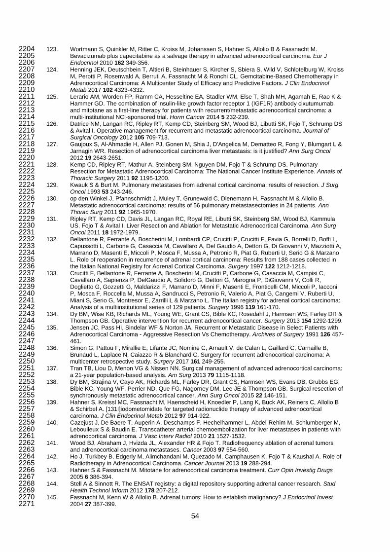
123. Wortmann S, Quinkler M, Ritter C, Kroiss M, Johanssen S, Hahner S, Allolio B & Fassnacht M. 2204 Bevacizumab plus capecitabine as a salvage therapy in advanced adrenocortical carcinoma. Eur J 2205 Endocrinol 2010 162 349-356. 2206
124. Henning JEK, Deutschbein T, Altieri B, Steinhauer S, Kircher S, Sbiera S, Wild V, Schlotelburg W, Kroiss 2207 M, Perotti P, Rosenwald A, Berruti A, Fassnacht M & Ronchi CL. Gemcitabine-Based Chemotherapy in 2208 Adrenocortical Carcinoma: A Multicenter Study of Efficacy and Predictive Factors. J Clin Endocrinol 2209 Metab 2017 102 4323-4332. 2210
125. Lerario AM, Worden FP, Ramm CA, Hesseltine EA, Stadler WM, Else T, Shah MH, Agamah E, Rao K & 2211 Hammer GD. The combination of insulin-like growth factor receptor 1 (IGF1R) antibody cixutumumab 2212 and mitotane as a first-line therapy for patients with recurrent/metastatic adrenocortical carcinoma: a 2213 multi-institutional NCI-sponsored trial. Horm Cancer 2014 5 232-239. 2214
126. Datrice NM, Langan RC, Ripley RT, Kemp CD, Steinberg SM, Wood BJ, Libutti SK, Fojo T, Schrump DS 2215 & Avital I. Operative management for recurrent and metastatic adrenocortical carcinoma. Journal of 2216 Surgical Oncology 2012 105 709-713. 2217
127. Gaujoux S, Al-Ahmadie H, Allen PJ, Gonen M, Shia J, D'Angelica M, Dematteo R, Fong Y, Blumgart L & 2218 Jarnagin WR. Resection of adrenocortical carcinoma liver metastasis: is it justified? Ann Surg Oncol 2219 2012 19 2643-2651. 2220
128. Kemp CD, Ripley RT, Mathur A, Steinberg SM, Nguyen DM, Fojo T & Schrump DS. Pulmonary 2221 Resection for Metastatic Adrenocortical Carcinoma: The National Cancer Institute Experience. Annals of 2222 Thoracic Surgery 2011 92 1195-1200. 2223
129. Kwauk S & Burt M. Pulmonary metastases from adrenal cortical carcinoma: results of resection. J Surg 2224 Oncol 1993 53 243-246. 2225
130. op den Winkel J, Pfannschmidt J, Muley T, Grunewald C, Dienemann H, Fassnacht M & Allolio B. 2226 Metastatic adrenocortical carcinoma: results of 56 pulmonary metastasectomies in 24 patients. Ann 2227 Thorac Surg 2011 92 1965-1970. 2228
131. Ripley RT, Kemp CD, Davis JL, Langan RC, Royal RE, Libutti SK, Steinberg SM, Wood BJ, Kammula 2229 US, Fojo T & Avital I. Liver Resection and Ablation for Metastatic Adrenocortical Carcinoma. Ann Surg 2230 Oncol 2011 18 1972-1979. 2231
132. Bellantone R, Ferrante A, Boscherini M, Lombardi CP, Crucitti P, Crucitti F, Favia G, Borrelli D, Boffi L, 2232 Capussotti L, Carbone G, Casaccia M, Cavallaro A, Del Gaudio A, Dettori G, Di Giovanni V, Mazziotti A, 2233 Marrano D, Masenti E, Miccoli P, Mosca F, Mussa A, Petronio R, Piat G, Ruberti U, Serio G & Marzano 2234 L. Role of reoperation in recurrence of adrenal cortical carcinoma: Results from 188 cases collected in 2235 the Italian National Registry for Adrenal Cortical Carcinoma. Surgery 1997 122 1212-1218. 2236
133. Crucitti F, Bellantone R, Ferrante A, Boscherini M, Crucitti P, Carbone G, Casaccia M, Campisi C, 2237 Cavallaro A, Sapienza P, DelGaudio A, Solidoro G, Dettori G, Marogna P, DiGiovanni V, Colli R, 2238 Doglietto G, Gozzetti G, Maldarizzi F, Marrano D, Minni F, Masenti E, Fronticelli CM, Miccoli P, Iacconi 2239 P, Mosca F, Roccella M, Mussa A, Sandrucci S, Petronio R, Valerio A, Piat G, Cangemi V, Ruberti U, 2240 Miani S, Serio G, Montresor E, Zarrilli L & Marzano L. The Italian registry for adrenal cortical carcinoma: 2241 Analysis of a multiinstitutional series of 129 patients. Surgery 1996 119 161-170. 2242
134. Dy BM, Wise KB, Richards ML, Young WE, Grant CS, Bible KC, Rosedahl J, Harmsen WS, Farley DR & 2243 Thompson GB. Operative intervention for recurrent adrenocortical cancer. Surgery 2013 154 1292-1299. 2244
135. Jensen JC, Pass HI, Sindelar WF & Norton JA. Recurrent or Metastatic Disease in Select Patients with 2245 Adrenocortical Carcinoma - Aggressive Resection Vs Chemotherapy. Archives of Surgery 1991 126 457-2246 461. 2247
136. Simon G, Pattou F, Mirallie E, Lifante JC, Nomine C, Arnault V, de Calan L, Gaillard C, Carnaille B, 2248 Brunaud L, Laplace N, Caiazzo R & Blanchard C. Surgery for recurrent adrenocortical carcinoma: A 2249 multicenter retrospective study. Surgery 2017 161 249-255. 2250
137. Tran TB, Liou D, Menon VG & Nissen NN. Surgical management of advanced adrenocortical carcinoma: 2251 a 21-year population-based analysis. Am Surg 2013 79 1115-1118. 2252
138. Dy BM, Strajina V, Cayo AK, Richards ML, Farley DR, Grant CS, Harmsen WS, Evans DB, Grubbs EG, 2253 Bible KC, Young WF, Perrier ND, Que FG, Nagorney DM, Lee JE & Thompson GB. Surgical resection of 2254 synchronously metastatic adrenocortical cancer. Ann Surg Oncol 2015 22 146-151. 2255
139. Hahner S, Kreissl MC, Fassnacht M, Haenscheid H, Knoedler P, Lang K, Buck AK, Reiners C, Allolio B 2256 & Schirbel A. [131I]iodometomidate for targeted radionuclide therapy of advanced adrenocortical 2257 carcinoma. J Clin Endocrinol Metab 2012 97 914-922. 2258
140. Cazejust J, De Baere T, Auperin A, Deschamps F, Hechelhammer L, Abdel-Rehim M, Schlumberger M, 2259 Leboulleux S & Baudin E. Transcatheter arterial chemoembolization for liver metastases in patients with 2260 adrenocortical carcinoma. J Vasc Interv Radiol 2010 21 1527-1532. 2261
141. Wood BJ, Abraham J, Hvizda JL, Alexander HR & Fojo T. Radiofrequency ablation of adrenal tumors 2262 and adrenocortical carcinoma metastases. Cancer 2003 97 554-560. 2263
142. Ho J, Turkbey B, Edgerly M, Alimchandani M, Quezado M, Camphausen K, Fojo T & Kaushal A. Role of 2264 Radiotherapy in Adrenocortical Carcinoma. Cancer Journal 2013 19 288-294. 2265
143. Hahner S & Fassnacht M. Mitotane for adrenocortical carcinoma treatment. Curr Opin Investig Drugs 2266 2005 6 386-394. 2267
144. Stell A & Sinnott R. The ENSAT registry: a digital repository supporting adrenal cancer research. Stud 2268 Health Technol Inform 2012 178 207-212. 2269
145. Fassnacht M, Kenn W & Allolio B. Adrenal tumors: How to establish malignancy? J Endocrinol Invest 2270 2004 27 387-399. 2271
54

146. Allolio B & Fassnacht M. Clinical review: Adrenocortical carcinoma: clinical update. J Clin Endocrinol 2272 Metab 2006 91 2027-2037. 2273
147. Nieman LK, Biller BM, Findling JW, Newell-Price J, Savage MO, Stewart PM & Montori VM. The 2274 diagnosis of Cushing's syndrome: an Endocrine Society Clinical Practice Guideline. J Clin Endocrinol 2275 Metab 2008 93 1526-1540. 2276
148. Libe R, Fratticci A & Bertherat J. Adrenocortical cancer: pathophysiology and clinical management. 2277 Endocrine-Related Cancer 2007 14 13-28. 2278
149. Legro RS, Arslanian SA, Ehrmann DA, Hoeger KM, Murad MH, Pasquali R & Welt CK. Diagnosis and 2279 treatment of polycystic ovary syndrome: an Endocrine Society clinical practice guideline. J Clin 2280 Endocrinol Metab 2013 98 4565-4592. 2281
150. Funder JW, Carey RM, Mantero F, Murad MH, Reincke M, Shibata H, Stowasser M & Young WF, Jr. 2282 The Management of Primary Aldosteronism: Case Detection, Diagnosis, and Treatment: An Endocrine 2283 Society Clinical Practice Guideline. J Clin Endocrinol Metab 2016 101 1889-1916. 2284
151. Dinnes J, Bancos I, Ferrante di Ruffano L, Chortis V, Davenport C, Bayliss S, Sahdev A, Guest P, 2285 Fassnacht M, Deeks JJ & Arlt W. MANAGEMENT OF ENDOCRINE DISEASE: Imaging for the diagnosis 2286 of malignancy in incidentally discovered adrenal masses: a systematic review and meta-analysis. Eur J 2287 Endocrinol 2016 175 R51-64. 2288
152. Peppercorn PD, Grossman AB & Reznek RH. Imaging of incidentally discovered adrenal masses. Clin 2289 Endocrinol (Oxf) 1998 48 379-388. 2290
153. Caoili EM, Korobkin M, Francis IR, Cohan RH, Platt JF, Dunnick NR & Raghupathi KI. Adrenal masses: 2291 characterization with combined unenhanced and delayed enhanced CT. Radiology 2002 222 629-633. 2292
154. Blake MA, Kalra MK, Sweeney AT, Lucey BC, Maher MM, Sahani DV, Halpern EF, Mueller PR, Hahn PF 2293 & Boland GW. Distinguishing benign from malignant adrenal masses: multi-detector row CT protocol with 2294 10-minute delay. Radiology 2006 238 578-585. 2295
155. Ilias I, Sahdev A, Reznek RH, Grossman AB & Pacak K. The optimal imaging of adrenal tumours: a 2296 comparison of different methods. Endocr Relat Cancer 2007 14 587-599. 2297
156. Mackie GC, Shulkin BL, Ribeiro RC, Worden FP, Gauger PG, Mody RJ, Connolly LP, Kunter G, 2298 Rodriguez-Galindo C, Wallis JW, Hurwitz CA & Schteingart DE. Use of [18F]fluorodeoxyglucose positron 2299 emission tomography in evaluating locally recurrent and metastatic adrenocortical carcinoma. J Clin 2300 Endocrinol Metab 2006 91 2665-2671. 2301
157. Groussin L, Bonardel G, Silvera S, Tissier F, Coste J, Abiven G, Libe R, Bienvenu M, Alberini JL, 2302 Salenave S, Bouchard P, Bertherat J, Dousset B, Legmann P, Richard B, Foehrenbach H, Bertagna X & 2303 Tenenbaum F. 18F-Fluorodeoxyglucose positron emission tomography for the diagnosis of 2304 adrenocortical tumors: a prospective study in 77 operated patients. The Journal of clinical endocrinology 2305 and metabolism 2009 94 1713-1722. 2306
158. Deandreis D, Leboulleux S, Caramella C, Schlumberger M & Baudin E. FDG PET in the management of 2307 patients with adrenal masses and adrenocortical carcinoma. Horm Cancer 2011 2 354-362. 2308
159. Cistaro A, Niccoli Asabella A, Coppolino P, Quartuccio N, Altini C, Cucinotta M, Alongi P, Balma M, 2309 Sanfilippo S, Buschiazzo A, Piccardo A, Fanelli M, Sambuceti G, Bomanji J, Baldari S, Bisi G, Fanti S & 2310 Rubini G. Diagnostic and prognostic value of 18F-FDG PET/CT in comparison with morphological 2311 imaging in primary adrenal gland malignancies - a multicenter experience. Hell J Nucl Med 2015 18 97-2312 102. 2313
160. Altinmakas E, Hobbs BP, Ye H, Grubbs EG, Perrier ND, Prieto VG, Lee JE & Ng CS. Diagnostic 2314 performance of (18-)F-FDG-PET-CT in adrenal lesions using histopathology as reference standard. 2315 Abdom Radiol (NY) 2017 42 577-584. 2316
161. Ciftci E, Turgut B, Cakmakcilar A & Erturk SA. Diagnostic importance of 18F-FDG PET/CT parameters 2317 and total lesion glycolysis in differentiating between benign and malignant adrenal lesions. Nucl Med 2318 Commun 2017 38 788-794. 2319
162. Bluemel C, Hahner S, Heinze B, Fassnacht M, Kroiss M, Bley TA, Wester HJ, Kropf S, Lapa C, Schirbel 2320 A, Buck AK & Herrmann K. Investigating the Chemokine Receptor 4 as Potential Theranostic Target in 2321 Adrenocortical Cancer Patients. Clin Nucl Med 2017 42 e29-e34. 2322
163. Werner RA, Kroiss M, Nakajo M, Mugge DO, Hahner S, Fassnacht M, Schirbel A, Bluemel C, Higuchi T, 2323 Papp L, Zsoter N, Buck AK, Bundschuh RA & Lapa C. Assessment of tumor heterogeneity in treatment-2324 naive adrenocortical cancer patients using <sup>18</sup>F-FDG positron emission tomography. 2325 Endocrine 2016 53 791-800. 2326
164. Wu YW & Tan CH. Determination of a cutoff attenuation value on single-phase contrast-enhanced CT for 2327 characterizing adrenal nodules via chemical shift MRI. Abdominal Radiology 2016 41 1170-1177. 2328
165. Nakajo M, Jinguji M, Shinaji T, Nakabeppu Y, Fukukura Y & Yoshiura T. Texture analysis of FDG 2329 PET/CT for differentiating between FDG-avid benign and metastatic adrenal tumors: efficacy of 2330 combining SUV and texture parameters. Abdom Radiol (NY) 2017 42 2882-2889. 2331
166. Guerin C, Pattou F, Brunaud L, Lifante JC, Mirallie E, Haissaguerre M, Huglo D, Olivier P, Houzard C, 2332 Ansquer C, Hindie E, Loundou A, Archange C, Tabarin A, Sebag F, Baumstarck K & Taieb D. 2333 Performance of 18F-FDG PET/CT in the Characterization of Adrenal Masses in Noncancer Patients: A 2334 Prospective Study. J Clin Endocrinol Metab 2017 102 2465-2472. 2335
167. Marty M, Gaye D, Perez P, Auder C, Nunes ML, Ferriere A, Haissaguerre M & Tabarin A. Diagnostic 2336 accuracy of computed tomography to identify adenomas among adrenal incidentalomas in an 2337 endocrinological population. Eur J Endocrinol 2018 178 439-446. 2338
55

168. Kim SJ, Lee SW, Pak K, Kim IJ & Kim K. Diagnostic accuracy of (18)F-FDG PET or PET/CT for the 2339 characterization of adrenal masses: a systematic review and meta-analysis. Br J Radiol 2018 20170520. 2340
169. Delivanis DA, Bancos I, Atwell TD, Schmit GD, Eiken PW, Natt N, Erickson D, Maraka S, Young WF & 2341 Nathan MA. Diagnostic performance of unenhanced computed tomography and (18) F-2342 fluorodeoxyglucose positron emission tomography in indeterminate adrenal tumours. Clin Endocrinol 2343 (Oxf) 2018 88 30-36. 2344
170. Romeo V, Maurea S, Cuocolo R, Petretta M, Mainenti PP, Verde F, Coppola M, Dell'Aversana S & 2345 Brunetti A. Characterization of Adrenal Lesions on Unenhanced MRI Using Texture Analysis: A Machine-2346 Learning Approach. J Magn Reson Imaging 2018. 2347
171. Thomas AJ, Habra MA, Bhosale PR, Qayyum AA, Ahmed K, Vicens R & Elsayes KM. Interobserver 2348 agreement in distinguishing large adrenal adenomas and adrenocortical carcinomas on computed 2349 tomography. Abdom Radiol (NY) 2018. 2350
172. Ng CS, Altinmakas E, Wei W, Ghosh P, Li X, Grubbs EG, Perrier NA, Prieto VG, Lee JE & Hobbs BP. 2351 Combining Washout and Noncontrast Data From Adrenal Protocol CT: Improving Diagnostic 2352 Performance. Acad Radiol 2018. 2353
173. Petersenn S, Richter PA, Broemel T, Ritter CO, Deutschbein T, Beil FU, Allolio B & Fassnacht M. 2354 Computed tomography criteria for discrimination of adrenal adenomas and adrenocortical carcinomas: 2355 analysis of the German ACC registry. European Journal of Endocrinology 2015 172 415-422. 2356
174. Bancos I, Tamhane S, Shah M, Delivanis DA, Alahdab F, Arlt W, Fassnacht M & Murad MH. 2357 DIAGNOSIS OF ENDOCRINE DISEASE: The diagnostic performance of adrenal biopsy: a systematic 2358 review and meta-analysis. Eur J Endocrinol 2016 175 R65-80. 2359
175. Williams AR, Hammer GD & Else T. Transcutaneous biopsy of adrenocortical carcinoma is rarely helpful 2360 in diagnosis, potentially harmful, but does not affect patient outcome. Eur J Endocrinol 2014 170 829-2361 835. 2362
176. Palazzo F, Dickinson A, Phillips B, Sahdev A, Bliss R, Rasheed A, Krukowski Z & Newell-Price J. 2363 Adrenal surgery in England: Better outcomes in high-volume practices. Clinical Endocrinology 2016 85 2364 17-20. 2365
177. Park HS, Roman SA & Sosa JA. Outcomes from 3144 adrenalectomies in the United States: which 2366 matters more, surgeon volume or specialty? Arch Surg 2009 144 1060-1067. 2367
178. Lindeman B, Hashimoto DA, Bababekov YJ, Stapleton SM, Chang DC, Hodin RA & Phitayakorn R. 2368 Fifteen years of adrenalectomies: impact of specialty training and operative volume. Surgery 2018 163 2369 150-156. 2370
179. Villar JM, Moreno P, Ortega J, Bollo E, Ramirez CP, Munoz N, Martinez C, Dominguez-Adame E, 2371 Sancho J, Del Pino JM, Couselo JM, Carrion A, Candel M, Caceres N, Octavio JM, Mateo F, Galan L, 2372 Ramia JM, Aguilo J & Herrera F. Results of adrenal surgery. Data of a Spanish National Survey. 2373 Langenbecks Arch Surg 2010 395 837-843. 2374
180. Gallagher SF, Wahi M, Haines KL, Baksh K, Enriquez J, Lee TM, Murr MM & Fabri PJ. Trends in 2375 adrenalectomy rates, indications, and physician volume: A statewide analysis of 1816 adrenalectomies. 2376 Surgery 2007 142 1011-1021; discussion 1011-1021. 2377
181. Lombardi CP, Raffaelli M, Boniardi M, De Toma G, Marzano LA, Miccoli P, Minni F, Morino M, Pelizzo 2378 MR, Pietrabissa A, Renda A, Valeri A, De Crea C & Bellantone R. Adrenocortical carcinoma: effect of 2379 hospital volume on patient outcome. Langenbecks Arch Surg 2012 397 201-207. 2380
182. Gratian L, Pura J, Dinan M, Reed S, Scheri R, Roman S & Sosa JA. Treatment Patterns and Outcomes 2381 for Patients with Adrenocortical Carcinoma Associated with Hospital Case Volume in the United States. 2382 Ann Surg Oncol 2014 21 3509-3514. 2383
183. Hermsen IG, Kerkhofs TM, den Butter G, Kievit J, van Eijck CH, Nieveen van Dijkum EJ & Haak HR. 2384 Surgery in adrenocortical carcinoma: Importance of national cooperation and centralized surgery. 2385 Surgery 2012 152 50-56. 2386
184. Kerkhofs TM, Verhoeven RH, Bonjer HJ, van Dijkum EJ, Vriens MR, De Vries J, Van Eijck CH, Bonsing 2387 BA, Van de Poll-Franse LV & Haak HR. Surgery for adrenocortical carcinoma in The Netherlands: 2388 analysis of the national cancer registry data. Eur J Endocrinol 2013 169 83-89. 2389
185. Gaujoux S & Mihai R. European Society of Endocrine Surgeons (ESES) and European Network for the 2390 Study of Adrenal Tumours (ENSAT) recommendations for the surgical management of adrenocortical 2391 carcinoma. British Journal of Surgery 2017 104 358-376. 2392
186. Gaujoux S & Brennan MF. Recommendation for standardized surgical management of primary 2393 adrenocortical carcinoma. Surgery 2012 152 123-132. 2394
187. Porpiglia F, Fiori C, Daffara FC, Zaggia B, Ardito A, Scarpa RM, Papotti M, Berruti A, Scagliotti GV & 2395 Terzolo M. Does nephrectomy during radical adrenalectomy for stage II adrenocortical cancer affect 2396 patient outcome? J Endocrinol Invest 2016 39 465-471. 2397
188. Donatini G, Caiazzo R, Do Cao C, Aubert S, Zerrweck C, El-Kathib Z, Gauthier T, Leteurtre E, Wemeau 2398 JL, Vantyghem MC, Carnaille B & Pattou F. Long-term survival after adrenalectomy for stage I/II 2399 adrenocortical carcinoma (ACC): a retrospective comparative cohort study of laparoscopic versus open 2400 approach. Ann Surg Oncol 2014 21 284-291. 2401
189. Sgourakis G, Lanitis S, Kouloura A, Zaphiriadou P, Karkoulias K, Raptis D, Anagnostara A & Caraliotas 2402 C. Laparoscopic versus Open Adrenalectomy for Stage I/II Adrenocortical Carcinoma: Meta-Analysis of 2403 Outcomes. J Invest Surg 2015 28 145-152. 2404
56

190. Autorino R, Bove P, De Sio M, Miano R, Micali S, Cindolo L, Greco F, Nicholas J, Fiori C, Bianchi G, Kim 2405 FJ & Porpiglia F. Open Versus Laparoscopic Adrenalectomy for Adrenocortical Carcinoma: A Meta-2406 analysis of Surgical and Oncological Outcomes. Ann Surg Oncol 2016 23 1195-1202. 2407
191. Langenhuijsen J, Birtle A, Klatte T, Porpiglia F & Timsit MO. Surgical Management of Adrenocortical 2408 Carcinoma: Impact of Laparoscopic Approach, Lymphadenectomy, and Surgical Volume on Outcomes-A 2409 Systematic Review and Meta-analysis of the Current Literature. Eur Urol Focus 2016 1 241-250. 2410
192. Lee CW, Salem AI, Schneider DF, Leverson GE, Tran TB, Poultsides GA, Postlewait LM, Maithel SK, 2411 Wang TS, Hatzaras I, Shenoy R, Phay JE, Shirley L, Fields RC, Jin LX, Pawlik TM, Prescott JD, Sicklick 2412 JK, Gad S, Yopp AC, Mansour JC, Duh QY, Seiser N, Solorzano CC, Kiernan CM, Votanopoulos KI, 2413 Levine EA & Weber SM. Minimally Invasive Resection of Adrenocortical Carcinoma: a Multi-Institutional 2414 Study of 201 Patients. J Gastrointest Surg 2017 21 352-362. 2415
193. Zheng GY, Li HZ, Deng JH, Zhang XB & Wu XC. Open adrenalectomy versus laparoscopic 2416 adrenalectomy for adrenocortical carcinoma: a retrospective comparative study on short-term oncologic 2417 prognosis. Onco Targets Ther 2018 11 1625-1632. 2418
194. Mpaili E, Moris D, Tsilimigras DI, Oikonomou D, Pawlik TM, Schizas D, Papalampros A, Felekouras E & 2419 Dimitroulis D. Laparoscopic Versus Open Adrenalectomy for Localized/Locally Advanced Primary 2420 Adrenocortical Carcinoma (ENSAT I-III) in Adults: Is Margin-Free Resection the Key Surgical Factor that 2421 Dictates Outcome? A Review of the Literature. J Laparoendosc Adv Surg Tech A 2018 28 408-414. 2422
195. Huynh KT, Lee DY, Lau BJ, Flaherty DC, Lee J & Goldfarb M. Impact of Laparoscopic Adrenalectomy on 2423 Overall Survival in Patients with Nonmetastatic Adrenocortical Carcinoma. Journal of the American 2424 College of Surgeons 2016 223 485-492. 2425
196. Barczynski M, Konturek A, Golkowski F, Cichon S, Huszno B, Peitgen K & Walz MK. Posterior 2426 retroperitoneoscopic adrenalectomy: a comparison between the initial experience in the invention phase 2427 and introductory phase of the new surgical technique. World J Surg 2007 31 65-71. 2428
197. Schreinemakers JM, Kiela GJ, Valk GD, Vriens MR & Rinkes IH. Retroperitoneal endoscopic 2429 adrenalectomy is safe and effective. Br J Surg 2010 97 1667-1672. 2430
198. Nilubol N, Patel D & Kebebew E. Does Lymphadenectomy Improve Survival in Patients with 2431 Adrenocortical Carcinoma? A Population-Based Study. World J Surg 2016 40 697-705. 2432
199. Saade N, Sadler C & Goldfarb M. Impact of Regional Lymph Node Dissection on Disease Specific 2433 Survival in Adrenal Cortical Carcinoma. Hormone and Metabolic Research 2015 47 820-825. 2434
200. Harrison LE, Gaudin PB & Brennan MF. Pathologic features of prognostic significance for adrenocortical 2435 carcinoma after curative resection. Arch Surg 1999 134 181-185. 2436
201. Panjwani S, Moore MD, Gray KD, Finnerty BM, Beninato T, Brunaud L, Fahey TJ, 3rd & Zarnegar R. 2437 The Impact of Nodal Dissection on Staging in Adrenocortical Carcinoma. Ann Surg Oncol 2017 24 3617-2438 3623. 2439
202. Gerry JM, Tran TB, Postlewait LM, Maithel SK, Prescott JD, Wang TS, Glenn JA, Phay JE, Keplinger K, 2440 Fields RC, Jin LX, Weber SM, Salem A, Sicklick JK, Gad S, Yopp AC, Mansour JC, Duh QY, Seiser N, 2441 Solorzano CC, Kiernan CM, Votanopoulos KI, Levine EA, Hatzaras I, Shenoy R, Pawlik TM, Norton JA & 2442 Poultsides GA. Lymphadenectomy for Adrenocortical Carcinoma: Is There a Therapeutic Benefit? Ann 2443 Surg Oncol 2016 23 708-713. 2444
203. Reibetanz J, Jurowich C, Erdogan I, Nies C, Rayes N, Dralle H, Behrend M, Allolio B & Fassnacht M. 2445 Impact of Lymphadenectomy on the Oncologic Outcome of Patients With Adrenocortical Carcinoma. 2446 Annals of Surgery 2012 255 363-369. 2447
204. Chiche L, Dousset B, Kieffer E & Chapuis Y. Adrenocortical carcinoma extending into the inferior vena 2448 cava: Presentation of a 15-patient series and review of the literature. Surgery 2006 139 15-27. 2449
205. Turbendian HK, Strong VE, Hsu M, Ghossein RA & Fahey TJ. Adrenocortical carcinoma: The influence 2450 of large vessel extension. Surgery 2010 148 1057-1064. 2451
206. Mihai R, Iacobone M, Makay O, Moreno P, Frilling A, Kraimps JL, Soriano A, Villar del Moral J, 2452 Barczynski M, Duran MC, Sadler GP, Niederle B, Dralle H, Harrison B & Carnaille B. Outcome of 2453 operation in patients with adrenocortical cancer invading the inferior vena cava--a European Society of 2454 Endocrine Surgeons (ESES) survey. Langenbecks Arch Surg 2012 397 225-231. 2455
207. Eller-Vainicher C, Morelli V, Salcuni AS, Battista C, Torlontano M, Coletti F, Iorio L, Cairoli E, Beck-2456 Peccoz P, Arosio M, Ambrosi B, Scillitani A & Chiodini I. Accuracy of several parameters of 2457 hypothalamic-pituitary-adrenal axis activity in predicting before surgery the metabolic effects of the 2458 removal of an adrenal incidentaloma. Eur J Endocrinol 2010 163 925-935. 2459
208. Bornstein SR, Allolio B, Arlt W, Barthel A, Don-Wauchope A, Hammer GD, Husebye ES, Merke DP, 2460 Murad MH, Stratakis CA & Torpy DJ. Diagnosis and Treatment of Primary Adrenal Insufficiency: An 2461 Endocrine Society Clinical Practice Guideline. J Clin Endocrinol Metab 2016 101 364-389. 2462
209. Duregon E, Volante M, Bollito E, Goia M, Buttigliero C, Zaggia B, Berruti A, Scagliotti GV & Papotti M. 2463 Pitfalls in the diagnosis of adrenocortical tumors: a lesson from 300 consultation cases. Hum Pathol 2464 2015 46 1799-1807. 2465
210. Sangoi AR, Fujiwara M, West RB, Montgomery KD, Bonventre JV, Higgins JP, Rouse RV, Gokden N & 2466 McKenney JK. Immunohistochemical distinction of primary adrenal cortical lesions from metastatic clear 2467 cell renal cell carcinoma: A study of 248 cases. American Journal of Surgical Pathology 2011 35 678-2468 686. 2469
211. Weissferdt A, Phan A, Suster S & Moran CA. Adrenocortical carcinoma: a comprehensive 2470 immunohistochemical study of 40 cases. Appl Immunohistochem Mol Morphol 2014 22 24-30. 2471
57

212. Tissier F, Aubert S, Leteurtre E, Alghuzlan A, Patey M, Decaussin M, Dousset L, Gobet F, Hoang C, 2472 Mazerolles C, Monges G, Sturm N, Renaudin K, Vacher-Lavenu MC, Viallon V, Baudin E, Bertagna X, 2473 Coste J & Libe R. Adrenocortical tumors (ACT): Evaluation and harmonization of the reading of the 2474 Weiss system criteria at the French level. Laboratory Investigation 2010 90 133A. 2475
213. Tissier F, Aubert S, Leteurtre E, Al Ghuzlan A, Patey M, Decaussin M, Doucet L, Gobet F, Hoang C, 2476 Mazerolles C, Monges G, Renaudin K, Sturm N, Trouette H, Vacher-Lavenu MC, Viallon V, Baudin E, 2477 Bertagna X, Coste J & Libe R. Adrenocortical Tumors: Improving the Practice of the Weiss System 2478 Through Virtual Microscopy A National Program of the French Network INCa-COMETE. American 2479 Journal of Surgical Pathology 2012 36 1194-1201. 2480
214. Weiss LM. Comparative histologic study of 43 metastasizing and nonmetastasizing adrenocortical 2481 tumors. Am J Surg Pathol 1984 8 163-169. 2482
215. Weiss LM, Medeiros LJ & Vickery AL, Jr. Pathologic features of prognostic significance in adrenocortical 2483 carcinoma. Am J Surg Pathol 1989 13 202-206. 2484
216. van Slooten H, Schaberg A, Smeenk D & Moolenaar AJ. Morphologic characteristics of benign and 2485 malignant adrenocortical tumors. Cancer 1985 55 766-773. 2486
217. Duregon E, Fassina A, Volante M, Nesi G, Santi R, Gatti G, Cappellesso R, Dalino Ciaramella P, 2487 Ventura L, Gambacorta M, Dei Tos AP, Loli P, Mannelli M, Mantero F, Berruti A, Terzolo M & Papotti M. 2488 The reticulin algorithm for adrenocortical tumor diagnosis: a multicentric validation study on 245 2489 unpublished cases. Am J Surg Pathol 2013 37 1433-1440. 2490
218. Bisceglia M, Ludovico O, Di Mattia A, Ben-Dor D, Sandbank J, Pasquinelli G, Lau SK & Weiss LM. 2491 Adrenocortical oncocytic tumors: Report of 10 cases and review of the literature. International Journal of 2492 Surgical Pathology 2004 12 231-243. 2493
219. Duregon E, Volante M, Cappia S, Cuccurullo A, Bisceglia M, Wong DD, Spagnolo DV, Szpak-Ulczok S, 2494 Bollito E, Daffara F, Berruti A, Terzolo M & Papotti M. Oncocytic Adrenocortical Tumors: Diagnostic 2495 Algorithm and Mitochondrial DNA Profile in 27 Cases. American Journal of Surgical Pathology 2011 35 2496 1882-1893. 2497
220. Wong DD, Spagnolo DV, Bisceglia M, Havlat M, McCallum D & Platten MA. Oncocytic adrenocortical 2498 neoplasms--a clinicopathologic study of 13 new cases emphasizing the importance of their recognition. 2499 Hum Pathol 2011 42 489-499. 2500
221. Fuhrman SA, Lasky LC & Limas C. Prognostic significance of morphologic parameters in renal cell 2501 carcinoma. Am J Surg Pathol 1982 6 655-663. 2502
222. Lu H, Papathomas TG, van Zessen D, Palli I, de Krijger RR, van der Spek PJ, Dinjens WN & Stubbs AP. 2503 Automated Selection of Hotspots (ASH): enhanced automated segmentation and adaptive step finding 2504 for Ki67 hotspot detection in adrenal cortical cancer. Diagn Pathol 2014 9 216. 2505
223. Papathomas TG, Pucci E, Giordano TJ, Lu H, Duregon E, Volante M, Papotti M, Lloyd RV, Tischler AS, 2506 van Nederveen FH, Nose V, Erickson L, Mete O, Asa SL, Turchini J, Gill AJ, Matias-Guiu X, Skordilis K, 2507 Stephenson TJ, Tissier F, Feelders RA, Smid M, Nigg A, Korpershoek E, van der Spek PJ, Dinjens 2508 WNM, Stubbs AP & de Krijger RR. An International Ki67 Reproducibility Study in Adrenal Cortical 2509 Carcinoma. American Journal of Surgical Pathology 2016 40 569-576. 2510
224. Morimoto R, Satoh F, Murakami O, Suzuki T, Abe T, Tanemoto M, Abe M, Uruno A, Ishidoya S, Arai Y, 2511 Takahashi K, Sasano H & Ito S. Immunohistochemistry of a proliferation marker Ki67/MIB1 in 2512 adrenocortical carcinomas: Ki67/MIB1 labeling index is a predictor for recurrence of adrenocortical 2513 carcinomas. Endocr J 2008 55 49-55. 2514
225. Abiven G, Coste J, Groussin L, Anract P, Tissier F, Legmann P, Dousset B, Bertagna X & Bertherat J. 2515 Clinical and biological features in the prognosis of adrenocortical cancer: Poor outcome of cortisol-2516 secreting tumors in a series of 202 consecutive patients. Journal of Clinical Endocrinology & Metabolism 2517 2006 91 2650-2655. 2518
226. Burotto M, Tageja N, Rosenberg A, Mahalingam S, Quezado M, Velarde M, Edgerly M & Fojo T. Brain 2519 metastasis in patients with adrenocortical carcinoma: a clinical series. J Clin Endocrinol Metab 2015 100 2520 331-336. 2521
227. Leboulleux S, Dromain C, Bonniaud G, Auperin A, Caillou B, Lumbroso J, Sigal R, Baudin E & 2522 Schlumberger M. Diagnostic and prognostic value of 18-fluorodeoxyglucose positron emission 2523 tomography in adrenocortical carcinoma: A prospective comparison with computed tomography. Journal 2524 of Clinical Endocrinology & Metabolism 2006 91 920-925. 2525
228. Ardito A, Massaglia C, Pelosi E, Zaggia B, Basile V, Brambilla R, Vigna-Taglianti F, Duregon E, Arena V, 2526 Perotti P, Penna D & Terzolo M. 18F-FDG PET/CT in the post-operative monitoring of patients with 2527 adrenocortical carcinoma. Eur J Endocrinol 2015 173 749-756. 2528
229. Berruti A, Fassnacht M, Baudin E, Hammer G, Haak H, Leboulleux S, Skogseid B, Allolio B & Terzolo M. 2529 Adjuvant therapy in patients with adrenocortical carcinoma: a position of an international panel. J Clin 2530 Oncol 2010 28 e401-402; author reply e403. 2531
230. Megerle F, Herrmann W, Schloetelburg W, Ronchi CL, Pulzer A, Quinkler M, Beuschlein F, Hahner S, 2532 Kroiss M & Fassnacht M. Mitotane monotherapy in patients with advanced adrenocortical carcinoma. J 2533 Clin Endocrinol Metab 2018. 2534
231. Volante M, Terzolo M, Fassnacht M, Rapa I, Germano A, Sbiera S, Daffara F, Sperone P, Scagliotti G, 2535 Allolio B, Papotti M & Berruti A. Ribonucleotide reductase large subunit (RRM1) gene expression may 2536 predict efficacy of adjuvant mitotane in adrenocortical cancer. Clin Cancer Res 2012 18 3452-3461. 2537
232. Ronchi CL, Sbiera S, Volante M, Steinhauer S, Scott-Wild V, Altieri B, Kroiss M, Bala M, Papotti M, 2538 Deutschbein T, Terzolo M, Fassnacht M & Allolio B. CYP2W1 Is Highly Expressed in Adrenal Glands 2539
58

and Is Positively Associated with the Response to Mitotane in Adrenocortical Carcinoma. Plos One 2014 2540 9. 2541
233. Terzolo M, Baudin AE, Ardito A, Kroiss M, Leboulleux S, Daffara F, Perotti P, Feelders RA, deVries JH, 2542 Zaggia B, De Francia S, Volante M, Haak HR, Allolio B, Al Ghuzlan A, Fassnacht M & Berruti A. 2543 Mitotane levels predict the outcome of patients with adrenocortical carcinoma treated adjuvantly 2544 following radical resection. European Journal of Endocrinology 2013 169 263-270. 2545
234. Terzolo M, Daffara F, Ardito A, Zaggia B, Basile V, Ferrari L & Berruti A. Management of adrenal cancer: 2546 a 2013 update. J Endocrinol Invest 2014 37 207-217. 2547
235. Huang H & Fojo T. Adjuvant mitotane for adrenocortical cancer--a recurring controversy. J Clin 2548 Endocrinol Metab 2008 93 3730-3732. 2549
236. Terzolo M, Fassnacht M, Ciccone G, Allolio B & Berruti A. Adjuvant mitotane for adrenocortical cancer - 2550 Working through uncertainty. Journal of Clinical Endocrinology and Metabolism 2009 94 1879-1880. 2551
237. Polat B, Fassnacht M, Pfreundner L, Guckenberger M, Bratengeier K, Johanssen S, Kenn W, Hahner S, 2552 Allolio B & Flentje M. Radiotherapy in Adrenocortical Carcinoma. Cancer 2009 115 2816-2823. 2553
238. Sabolch A, Else T, Jackson W, Williams A, Miller BS, Worden F, Hammer GD & Jolly S. Improved local 2554 control with adjuvant radiation therapy in localized adrenocortical carcinoma: A case-matched 2555 retrospective study. International Journal of Radiation Oncology Biology Physics 2013 1) S84. 2556
239. Nelson DW, Chang SC, Bandera BC, Fischer TD, Wollman R & Goldfarb M. Adjuvant Radiation is 2557 Associated with Improved Survival for Select Patients with Non-metastatic Adrenocortical Carcinoma. 2558 Ann Surg Oncol 2018. 2559
240. Cerquetti L, Bucci B, Marchese R, Misiti S, De Paula U, Miceli R, Muleti A, Amendola D, Piergrossi P, 2560 Brunetti E, Toscano V & Stigliano A. Mitotane increases the radiotherapy inhibitory effect and induces 2561 G2-arrest in combined treatment on both H295R and SW13 adrenocortical cell lines. Endocr Relat 2562 Cancer 2008 15 623-634. 2563
241. Cerquetti L, Sampaoli C, Amendola D, Bucci B, Misiti S, Raza G, De Paula U, Marchese R, Brunetti E, 2564 Toscano V & Stigliano A. Mitotane sensitizes adrenocortical cancer cells to ionizing radiations by 2565 involvement of the cyclin B1/CDK complex in G<inf>2</inf> arrest and mismatch repair enzymes 2566 modulation. International Journal of Oncology 2010 37 493-501. 2567
242. Khan TS, Imam H, Juhlin C, Skogseid B, Grondal S, Tibblin S, Wilander E, Oberg K & Eriksson B. 2568 Streptozocin and o,p'DDD in the treatment of adrenocortical cancer patients: long-term survival in its 2569 adjuvant use. Ann Oncol 2000 11 1281-1287. 2570
243. Hermsen IGC, Gelderblom H, Kievit J, Romijn JA & Haak HR. Extremely long survival in six patients 2571 despite recurrent and metastatic adrenal carcinoma. European Journal of Endocrinology 2008 158 911-2572 919. 2573
244. Bednarski BK, Habra MA, Phan A, Milton DR, Wood C, Vauthey N, Evans DB, Katz MH, Ng CS, Perrier 2574 ND, Lee JE & Grubbs EG. Borderline resectable adrenal cortical carcinoma: a potential role for 2575 preoperative chemotherapy. World J Surg 2014 38 1318-1327. 2576
245. Wangberg B, Khorram-Manesh A, Jansson S, Nilsson B, Nilsson O, Jakobsson CE, Lindstedt S, Oden A 2577 & Ahlman H. The long-term survival in adrenocortical carcinoma with active surgical management and 2578 use of monitored mitotane. Endocr Relat Cancer 2010 17 265-272. 2579
246. Rangel C, Scattolin G, Pais-Costa SR, Vieira E & Gaio E. Neoadjuvant chemotherapy and salvage 2580 surgery for an aldosterone-producing adrenal carcinoma with inferior vena cava thrombus: Case report 2581 and literature review. Asian Journal of Surgery 2013 36 134-136. 2582
247. Ronchi CL, Sbiera S, Kraus L, Wortmann S, Johanssen S, Adam P, Willenberg HS, Hahner S, Allolio B 2583 & Fassnacht M. Expression of excision repair cross complementing group 1 and prognosis in 2584 adrenocortical carcinoma patients treated with platinum-based chemotherapy. Endocr Relat Cancer 2585 2009 16 907-918. 2586
248. Malandrino P, Al Ghuzlan A, Castaing M, Young J, Caillou B, Travagli JP, Elias D, de Baere T, Dromain 2587 C, Paci A, Chanson P, Schlumberger M, Leboulleux S & Baudin E. Prognostic markers of survival after 2588 combined mitotane- and platinum-based chemotherapy in metastatic adrenocortical carcinoma. Endocr 2589 Relat Cancer 2010 17 797-807. 2590
249. Roca E, Berruti A, Sbiera S, Rapa I, Oneda E, Sperone P, Ronchi CL, Ferrari L, Grisanti S, Germano A, 2591 Zaggia B, Scagliotti G, Fassnacht M, Volante M, Terzolo M & Papotti M. Topoisomerase2alpha and 2592 thymidylate synthase expression in adrenocortical cancer. Endocr Relat Cancer 2017. 2593
250. Laufs V, Altieri B, Sbiera S, Kircher S, Steinhauer S, Beuschlein F, Quinkler M, Willenberg HS, 2594 Rosenwald A, Fassnacht M & Ronchi CL. ERCC1 as predictive biomarker to platinum-based 2595 chemotherapy in adrenocortical carcinomas. Eur J Endocrinol 2018 178 183-190. 2596
251. Bates SE, Shieh CY, Mickley LA, Dichek HL, Gazdar A, Loriaux DL & Fojo AT. Mitotane enhances 2597 cytotoxicity of chemotherapy in cell lines expressing a multidrug resistance gene (mdr-1/P-glycoprotein) 2598 which is also expressed by adrenocortical carcinomas. J Clin Endocrinol Metab 1991 73 18-29. 2599
252. Almeida MQ, Fragoso MC, Lotfi CF, Santos MG, Nishi MY, Costa MH, Lerario AM, Maciel CC, Mattos 2600 GE, Jorge AA, Mendonca BB & Latronico AC. Expression of IGF-II and its Receptor in Pediatric and 2601 Adult Adrenocortical Tumors. J Clin Endocrinol Metab 2008. 2602
253. Boulle N, Logie A, Gicquel C, Perin L & Le Bouc Y. Increased levels of insulin-like growth factor II (IGF-2603 II) and IGF-binding protein-2 are associated with malignancy in sporadic adrenocortical tumors. J Clin 2604 Endocrinol Metab 1998 83 1713-1720. 2605
59

254. Gicquel C, Bertagna X, Schneid H, Francillard-Leblond M, Luton JP, Girard F & Le Bouc Y. 2606 Rearrangements at the 11p15 locus and overexpression of insulin-like growth factor-II gene in sporadic 2607 adrenocortical tumors. J Clin Endocrinol Metab 1994 78 1444-1453. 2608
255. Giordano TJ, Thomas DG, Kuick R, Lizyness M, Misek DE, Smith AL, Sanders D, Aljundi RT, Gauger 2609 PG, Thompson NW, Taylor JM & Hanash SM. Distinct transcriptional profiles of adrenocortical tumors 2610 uncovered by DNA microarray analysis. Am J Pathol 2003 162 521-531. 2611
256. Weber MM, Fottner C & Wolf E. The role of the insulin-like growth factor system in adrenocortical 2612 tumourigenesis. Eur J Clin Invest 2000 30 Suppl 3 69-75. 2613
257. Jones RL, Kim ES, Nava-Parada P, Alam S, Johnson FM, Stephens AW, Simantov R, Poondru S, 2614 Gedrich R, Lippman SM, Kaye SB & Carden CP. Phase I Study of Intermittent Oral Dosing of the Insulin-2615 like Growth Factor-1 and Insulin Receptors Inhibitor OSI-906 in Patients With Advanced Solid Tumors. 2616 Clinical Cancer Research 2015 21 693-700. 2617
258. Naing A, Kurzrock R, Burger A, Gupta S, Lei X, Busaidy N, Hong D, Chen HX, Doyle LA, Heilbrun LK, 2618 Rohren E, Ng C, Chandhasin C & LoRusso P. Phase I trial of cixutumumab combined with temsirolimus 2619 in patients with advanced cancer. Clin Cancer Res 2011 17 6052-6060. 2620
259. Vezzosi D, Do Cao C, Hescot S, Bertherat J, Haissaguerre M, Bongard V, Drui D, De La Fouchardiere 2621 C, Illouz F, Borson-Chazot F, Djobo B, Berdelou A, Tabarin A, Schlumberger M, Briet C, Caron P, 2622 Leboulleux S, Libe R & Baudin E. Time Until Partial Response in Metastatic Adrenocortical Carcinoma 2623 Long-Term Survivors. Horm Cancer 2018 9 62-69. 2624
260. Kroiss M, Quinkler M, Lutz WK, Allolio B & Fassnacht M. Drug interactions with mitotane by induction of 2625 CYP3A4 metabolism in the clinical management of adrenocortical carcinoma. Clin Endocrinol (Oxf) 2011 2626 75 585-591. 2627
261. Faggiano A, Leboulleux S, Young J, Schlumberger M & Baudin E. Rapidly progressing high o,p'DDD 2628 doses shorten the time required to reach the therapeutic threshold with an acceptable tolerance: 2629 preliminary results. Clin Endocrinol (Oxf) 2006 64 110-113. 2630
262. Mauclere-Denost S, Leboulleux S, Borget I, Paci A, Young J, Al Ghuzlan A, Deandreis D, Drouard L, 2631 Tabarin A, Chanson P, Schlumberger M & Baudin E. High-dose mitotane strategy in adrenocortical 2632 carcinoma (ACC) : prospective analysis of plasma mitotane measurement during the first three months 2633 of follow-up. Eur J Endocrinol 2011. 2634
263. Terzolo M, Pia A, Berruti A, Osella G, Ali A, Carbone V, Testa E, Dogliotti L & Angeli A. Low-dose 2635 monitored mitotane treatment achieves the therapeutic range with manageable side effects in patients 2636 with adrenocortical cancer. J Clin Endocrinol Metab 2000 85 2234-2238. 2637
264. Terzolo M & Berruti A. Adjunctive treatment of adrenocortical carcinoma. Curr Opin Endocrinol Diabetes 2638 Obes 2008 15 221-226. 2639
265. Kerkhofs TM, Baudin E, Terzolo M, Allolio B, Chadarevian R, Mueller HH, Skogseid B, Leboulleux S, 2640 Mantero F, Haak HR & Fassnacht M. Comparison of two mitotane starting dose regimens in patients 2641 with advanced adrenocortical carcinoma. J Clin Endocrinol Metab 2013 98 4759-4767. 2642
266. Kerkhofs TM, Derijks LJ, Ettaieb MH, Eekhoff EM, Neef C, Gelderblom H, den Hartigh J, Guchelaar HJ & 2643 Haak HR. Short-term variation in plasma mitotane levels confirms the importance of trough level 2644 monitoring. Eur J Endocrinol 2014 171 677-683. 2645
267. Daffara F, De Francia S, Reimondo G, Zaggia B, Aroasio E, Porpiglia F, Volante M, Termine A, Di Carlo 2646 F, Dogliotti L, Angeli A, Berruti A & Terzolo M. Prospective evaluation of mitotane toxicity in 2647 adrenocortical cancer patients treated adjuvantly. Endocr Relat Cancer 2008 15 1043-1053. 2648
268. van Slooten H, Moolenaar AJ, van Seters AP & Smeenk D. The treatment of adrenocortical carcinoma 2649 with o,p'-DDD: prognostic implications of serum level monitoring. Eur J Cancer Clin Oncol 1984 20 47-2650 53. 2651
269. Chortis V, Taylor AE, Schneider P, Tomlinson JW, Hughes BA, O'Neil DM, Libé R, Allolio B, Bertagna X, 2652 Bertherat J, Beuschlein F, Fassnacht M, Karavitaki N, Mannelli M, Mantero F, Opocher G, Porfiri E, 2653 Quinkler M, Sherlock M, Terzolo M, Nightingale P, Shackleton CH, Stewart PM, Hahner S & Arlt W. 2654 Mitotane therapy in adrenocortical cancer induces CYP3A4 and inhibits 5?-reductase, explaining the 2655 need for personalized glucocorticoid and androgen replacement. In The Journal of clinical endocrinology 2656 and metabolism, pp 161-171, 2013. 2657
270. Reimondo G, Puglisi S, Zaggia B, Basile V, Saba L, Perotti P, De Francia S, Volante M, Zatelli MC, 2658 Cannavo S & Terzolo M. Effects of mitotane on the hypothalamic-pituitary-adrenal axis in patients with 2659 adrenocortical carcinoma. Eur J Endocrinol 2017 177 361-367. 2660
271. Kerkhofs TM, Derijks LJ, Ettaieb H, den Hartigh J, Neef K, Gelderblom H, Guchelaar HJ & Haak HR. 2661 Development of a pharmacokinetic model of mitotane: toward personalized dosing in adrenocortical 2662 carcinoma. Ther Drug Monit 2015 37 58-65. 2663
272. Alexandraki KI, Kaltsas GA, le Roux CW, Fassnacht M, Ajodha S, Christ-Crain M, Akker SA, Drake WM, 2664 Edwards R, Allolio B & Grossman AB. Assessment of serum-free cortisol levels in patients with 2665 adrenocortical carcinoma treated with mitotane: a pilot study. Clinical Endocrinology 2010 72 305-311. 2666
273. Russo M, Scollo C, Pellegriti G, Cotta OR, Squatrito S, Frasca F, Cannavo S & Gullo D. Mitotane 2667 treatment in patients with adrenocortical cancer causes central hypothyroidism. Clin Endocrinol (Oxf) 2668 2016 84 614-619. 2669
274. Tada H, Nohara A, Kawashiri MA, Inazu A, Mabuchi H & Yamagishi M. Marked transient 2670 hypercholesterolemia caused by low-dose mitotane as adjuvant chemotherapy for adrenocortical 2671 carcinoma. J Atheroscler Thromb 2014 21 1326-1329. 2672
60

275. Nieman LK, Biller BM, Findling JW, Murad MH, Newell-Price J, Savage MO & Tabarin A. Treatment of 2673 Cushing's Syndrome: An Endocrine Society Clinical Practice Guideline. J Clin Endocrinol Metab 2015 2674 100 2807-2831. 2675
276. Claps M, Cerri S, Grisanti S, Lazzari B, Ferrari V, Roca E, Perotti P, Terzolo M, Sigala S & Berruti A. 2676 Adding metyrapone to chemotherapy plus mitotane for Cushing's syndrome due to advanced 2677 adrenocortical carcinoma. Endocrine 2017. 2678
277. Castinetti F, Fassnacht M, Johanssen S, Terzolo M, Bouchard P, Chanson P, Do Cao C, Morange I, 2679 Pico A, Ouzounian S, Young J, Hahner S, Brue T, Allolio B & Conte-Devolx B. Merits and pitfalls of 2680 mifepristone in Cushing's syndrome. Eur J Endocrinol 2009 160 1003-1010. 2681
278. Chow E, Hoskin P, Mitera G, Zeng L, Lutz S, Roos D, Hahn C, van der Linden Y, Hartsell W & Kumar E. 2682 Update of the international consensus on palliative radiotherapy endpoints for future clinical trials in bone 2683 metastases. Int J Radiat Oncol Biol Phys 2012 82 1730-1737. 2684
279. Pin Y, Paix A, Le Fevre C, Antoni D, Blondet C & Noel G. A systematic review of palliative bone 2685 radiotherapy based on pain relief and retreatment rates. Crit Rev Oncol Hematol 2018 123 132-137. 2686
280. Ferrell BR, Temel JS, Temin S, Alesi ER, Balboni TA, Basch EM, Firn JI, Paice JA, Peppercorn JM, 2687 Phillips T, Stovall EL, Zimmermann C & Smith TJ. Integration of Palliative Care Into Standard Oncology 2688 Care: American Society of Clinical Oncology Clinical Practice Guideline Update. J Clin Oncol 2017 35 2689 96-112. 2690
281. Herrmann LJ, Heinze B, Fassnacht M, Willenberg HS, Quinkler M, Reisch N, Zink M, Allolio B & Hahner 2691 S. TP53 germline mutations in adult patients with adrenocortical carcinoma. J Clin Endocrinol Metab 2692 2012 97 E476-485. 2693
282. Raymond VM, Else T, Everett JN, Long JM, Gruber SB & Hammer GD. Prevalence of germline TP53 2694 mutations in a prospective series of unselected patients with adrenocortical carcinoma. J Clin Endocrinol 2695 Metab 2013 98 E119-125. 2696
283. Waldmann J, Patsalis N, Fendrich V, Langer P, Saeger W, Chaloupka B, Ramaswamy A, Fassnacht M, 2697 Bartsch DK & Slater EP. Clinical impact of TP53 alterations in adrenocortical carcinomas. Langenbecks 2698 Archives of Surgery 2012 397 209-216. 2699
284. Raymond VM, Everett JN, Furtado LV, Gustafson SL, Jungbluth CR, Gruber SB, Hammer GD, Stoffel 2700 EM, Greenson JK, Giordano TJ & Else T. Adrenocortical carcinoma is a lynch syndrome-associated 2701 cancer. J Clin Oncol 2013 31 3012-3018. 2702
285. Stoffel EM, Mangu PB, Gruber SB, Hamilton SR, Kalady MF, Lau MW, Lu KH, Roach N & Limburg PJ. 2703 Hereditary colorectal cancer syndromes: American Society of Clinical Oncology Clinical Practice 2704 Guideline endorsement of the familial risk-colorectal cancer: European Society for Medical Oncology 2705 Clinical Practice Guidelines. J Clin Oncol 2015 33 209-217. 2706
286. Daly MB, Pilarski R, Berry M, Buys SS, Farmer M, Friedman S, Garber JE, Kauff ND, Khan S, Klein C, 2707 Kohlmann W, Kurian A, Litton JK, Madlensky L, Merajver SD, Offit K, Pal T, Reiser G, Shannon KM, 2708 Swisher E, Vinayak S, Voian NC, Weitzel JN, Wick MJ, Wiesner GL, Dwyer M & Darlow S. NCCN 2709 Guidelines Insights: Genetic/Familial High-Risk Assessment: Breast and Ovarian, Version 2.2017. J Natl 2710 Compr Canc Netw 2017 15 9-20. 2711
287. Kratz CP, Achatz MI, Brugieres L, Frebourg T, Garber JE, Greer MC, Hansford JR, Janeway KA, 2712 Kohlmann WK, McGee R, Mullighan CG, Onel K, Pajtler KW, Pfister SM, Savage SA, Schiffman JD, 2713 Schneider KA, Strong LC, Evans DGR, Wasserman JD, Villani A & Malkin D. Cancer Screening 2714 Recommendations for Individuals with Li-Fraumeni Syndrome. Clin Cancer Res 2017 23 e38-e45. 2715
288. Ballinger ML, Best A, Mai PL, Khincha PP, Loud JT, Peters JA, Achatz MI, Chojniak R, Balieiro da Costa 2716 A, Santiago KM, Garber J, O'Neill AF, Eeles RA, Evans DG, Bleiker E, Sonke GS, Ruijs M, Loo C, 2717 Schiffman J, Naumer A, Kohlmann W, Strong LC, Bojadzieva J, Malkin D, Rednam SP, Stoffel EM, 2718 Koeppe E, Weitzel JN, Slavin TP, Nehoray B, Robson M, Walsh M, Manelli L, Villani A, Thomas DM & 2719 Savage SA. Baseline Surveillance in Li-Fraumeni Syndrome Using Whole-Body Magnetic Resonance 2720 Imaging: A Meta-analysis. JAMA Oncol 2017 3 1634-1639. 2721
289. Gupta S, Provenzale D, Regenbogen SE, Hampel H, Slavin TP, Jr., Hall MJ, Llor X, Chung DC, Ahnen 2722 DJ, Bray T, Cooper G, Early DS, Ford JM, Giardiello FM, Grady W, Halverson AL, Hamilton SR, 2723 Klapman JB, Larson DW, Lazenby AJ, Lynch PM, Markowitz AJ, Mayer RJ, Ness RM, Samadder NJ, 2724 Shike M, Sugandha S, Weiss JM, Dwyer MA & Ogba N. NCCN Guidelines Insights: Genetic/Familial 2725 High-Risk Assessment: Colorectal, Version 3.2017. J Natl Compr Canc Netw 2017 15 1465-1475. 2726
290. Stoffel EM, Mangu PB & Limburg PJ. Hereditary colorectal cancer syndromes: American Society of 2727 Clinical Oncology clinical practice guideline endorsement of the familial risk-colorectal cancer: European 2728 Society for Medical Oncology clinical practice guidelines. J Oncol Pract 2015 11 e437-441. 2729
291. Else T. Association of adrenocortical carcinoma with familial cancer susceptibility syndromes. Mol Cell 2730 Endocrinol 2012 351 66-70. 2731
292. McDonnell CM & Zacharin MR. Adrenal cortical tumours: 25 years' experience at the Royal Children's 2732 Hospital, Melbourne. J Paediatr Child Health 2003 39 682-685. 2733
293. Custodio G, Parise GA, Kiesel Filho N, Komechen H, Sabbaga CC, Rosati R, Grisa L, Parise IZ, 2734 Pianovski MA, Fiori CM, Ledesma JA, Barbosa JR, Figueiredo FR, Sade ER, Ibanez H, Arram SB, 2735 Stinghen ST, Mengarelli LR, Figueiredo MM, Carvalho DC, Avilla SG, Woiski TD, Poncio LC, Lima GF, 2736 Pontarolo R, Lalli E, Zhou Y, Zambetti GP, Ribeiro RC & Figueiredo BC. Impact of neonatal screening 2737 and surveillance for the TP53 R337H mutation on early detection of childhood adrenocortical tumors. J 2738 Clin Oncol 2013 31 2619-2626. 2739
61
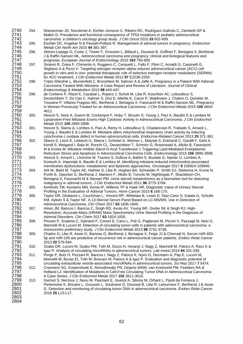
294. Wasserman JD, Novokmet A, Eichler-Jonsson C, Ribeiro RC, Rodriguez-Galindo C, Zambetti GP & 2740 Malkin D. Prevalence and functional consequence of TP53 mutations in pediatric adrenocortical 2741 carcinoma: a children's oncology group study. J Clin Oncol 2015 33 602-609. 2742
295. Eschler DC, Kogekar N & Pessah-Pollack R. Management of adrenal tumors in pregnancy. Endocrinol 2743 Metab Clin North Am 2015 44 381-397. 2744
296. Abiven-Lepage G, Coste J, Tissier F, Groussin L, Billaud L, Dousset B, Goffinet F, Bertagna X, Bertherat 2745 J & Raffin-Sanson ML. Adrenocortical carcinoma and pregnancy: clinical and biological features and 2746 prognosis. European Journal of Endocrinology 2010 163 793-800. 2747
297. Sirianni R, Zolea F, Chimento A, Ruggiero C, Cerquetti L, Fallo F, Pilon C, Arnaldi G, Carpinelli G, 2748 Stigliano A & Pezzi V. Targeting estrogen receptor-alpha reduces adrenocortical cancer (ACC) cell 2749 growth in vitro and in vivo: potential therapeutic role of selective estrogen receptor modulators (SERMs) 2750 for ACC treatment. J Clin Endocrinol Metab 2012 97 E2238-2250. 2751
298. Tripto-Shkolnik L, Blumenfeld Z, Bronshtein M, Salmon A & Jaffe A. Pregnancy in a Patient With Adrenal 2752 Carcinoma Treated With Mitotane: A Case Report and Review of Literature. Journal of Clinical 2753 Endocrinology & Metabolism 2013 98 443-447. 2754
299. de Corbiere P, Ritzel K, Cazabat L, Ropers J, Schott M, Libe R, Koschker AC, Leboulleux S, 2755 Deutschbein T, Do Cao C, Hahner S, Drui D, Miehle K, Caron P, Waldmann J, Chabre O, Quinkler M, 2756 Touraine P, Villares Fragoso MC, Bertherat J, Bertagna X, Fassnacht M & Raffin-Sanson ML. Pregnancy 2757 in Women Previously Treated for an Adrenocortical Carcinoma. J Clin Endocrinol Metab 2015 100 4604-2758 4611. 2759
300. Hescot S, Seck A, Guerin M, Cockenpot F, Huby T, Broutin S, Young J, Paci A, Baudin E & Lombes M. 2760 Lipoprotein-Free Mitotane Exerts High Cytotoxic Activity in Adrenocortical Carcinoma. J Clin Endocrinol 2761 Metab 2015 100 2890-2898. 2762
301. Hescot S, Slama A, Lombes A, Paci A, Remy H, Leboulleux S, Chadarevian R, Trabado S, Amazit L, 2763 Young J, Baudin E & Lombes M. Mitotane alters mitochondrial respiratory chain activity by inducing 2764 cytochrome c oxidase defect in human adrenocortical cells. Endocrine-Related Cancer 2013 20 371-381. 2765
302. Sbiera S, Leich E, Liebisch G, Sbiera I, Schirbel A, Wiemer L, Matysik S, Eckhardt C, Gardill F, Gehl A, 2766 Kendl S, Weigand I, Bala M, Ronchi CL, Deutschbein T, Schmitz G, Rosenwald A, Allolio B, Fassnacht 2767 M & Kroiss M. Mitotane Inhibits Sterol-O-Acyl Transferase 1 Triggering Lipid-Mediated Endoplasmic 2768 Reticulum Stress and Apoptosis in Adrenocortical Carcinoma Cells. Endocrinology 2015 156 3895-3908. 2769
303. Hescot S, Amazit L, Lhomme M, Travers S, DuBow A, Battini S, Boulate G, Namer IJ, Lombes A, 2770 Kontush A, Imperiale A, Baudin E & Lombes M. Identifying mitotane-induced mitochondria-associated 2771 membranes dysfunctions: metabolomic and lipidomic approaches. Oncotarget 2017 8 109924-109940. 2772
304. Arlt W, Biehl M, Taylor AE, Hahner S, Libe R, Hughes BA, Schneider P, Smith DJ, Stiekema H, Krone N, 2773 Porfiri E, Opocher G, Bertherat J, Mantero F, Allolio B, Terzolo M, Nightingale P, Shackleton CH, 2774 Bertagna X, Fassnacht M & Stewart PM. Urine steroid metabolomics as a biomarker tool for detecting 2775 malignancy in adrenal tumors. J Clin Endocrinol Metab 2011 96 3775-3784. 2776
305. Kerkhofs TM, Kerstens MN, Kema IP, Willems TP & Haak HR. Diagnostic Value of Urinary Steroid 2777 Profiling in the Evaluation of Adrenal Tumors. Horm Cancer 2015 6 168-175. 2778
306. Taylor DR, Ghataore L, Couchman L, Vincent RP, Whitelaw B, Lewis D, Diaz-Cano S, Galata G, Schulte 2779 KM, Aylwin S & Taylor NF. A 13-Steroid Serum Panel Based on LC-MS/MS: Use in Detection of 2780 Adrenocortical Carcinoma. Clin Chem 2017 63 1836-1846. 2781
307. Hines JM, Bancos I, Bancos C, Singh RD, Avula AV, Young WF, Grebe SK & Singh RJ. High-2782 Resolution, Accurate-Mass (HRAM) Mass Spectrometry Urine Steroid Profiling in the Diagnosis of 2783 Adrenal Disorders. Clin Chem 2017 63 1824-1835. 2784
308. Pinzani P, Scatena C, Salvianti F, Corsini E, Canu L, Poli G, Paglierani M, Piccini V, Pazzagli M, Nesi G, 2785 Mannelli M & Luconi M. Detection of circulating tumor cells in patients with adrenocortical carcinoma: a 2786 monocentric preliminary study. J Clin Endocrinol Metab 2013 98 3731-3738. 2787
309. Chabre O, Libe R, Assie G, Barreau O, Bertherat J, Bertagna X, Feige JJ & Cherradi N. Serum miR-483-2788 5p and miR-195 are predictive of recurrence risk in adrenocortical cancer patients. Endocr Relat Cancer 2789 2013 20 579-594. 2790
310. Szabo DR, Luconi M, Szabo PM, Toth M, Szucs N, Horanyi J, Nagy Z, Mannelli M, Patocs A, Racz K & 2791 Igaz P. Analysis of circulating microRNAs in adrenocortical tumors. Lab Invest 2014 94 331-339. 2792
311. Perge P, Butz H, Pezzani R, Bancos I, Nagy Z, Paloczi K, Nyiro G, Decmann A, Pap E, Luconi M, 2793 Mannelli M, Buzas EI, Toth M, Boscaro M, Patocs A & Igaz P. Evaluation and diagnostic potential of 2794 circulating extracellular vesicle-associated microRNAs in adrenocortical tumors. Sci Rep 2017 7 5474. 2795
312. Creemers SG, Korpershoek E, Atmodimedjo PN, Dinjens WNM, van Koetsveld PM, Feelders RA & 2796 Hofland LJ. Identification of Mutations in Cell-Free Circulating Tumor DNA in Adrenocortical Carcinoma: 2797 A Case Series. J Clin Endocrinol Metab 2017 102 3611-3615. 2798
313. Garinet S, Nectoux J, Neou M, Pasmant E, Jouinot A, Sibony M, Orhant L, Pipoli da Fonseca J, 2799 Perlemoine K, Bricaire L, Groussin L, Soubrane O, Dousset B, Libe R, Letourneur F, Bertherat J & Assie 2800 G. Detection and monitoring of circulating tumor DNA in adrenocortical carcinoma. Endocr Relat Cancer 2801 2018 25 L13-L17. 2802
2803 2804
62


