European Journal of Radiology Open - COREC.G. Kaiser et al. / European Journal of Radiology Open 3...
6
European Journal of Radiology Open 3 (2016) 117–122 Contents lists available at ScienceDirect European Journal of Radiology Open jou r n al hom epage: www.elsevier.com/locate/ejro Focal transitional mastitis in MR-Mammography: Preliminary findings Clemens G. Kaiser a,∗,1 , Matthias Dietzel b,c,1 , Pascal Baltzer d , Anna K. Kaiser e , Thomas Henzler a , Werner A. Kaiser c , Julia Knaudt a a Institute of Clinical Radiology and Nuclear Medicine, University Medical Centre Mannheim, Medical Faculty Mannheim-University of Heidelberg, Germany b Institute of Diagnostic Radiology, University of Erlangen-Nuremberg, Erlangen, Germany c Institute of Diagnostic and Interventional Radiology, University Hospital Jena, Jena, Germany d Department of Biomedical Imaging and Image-Guided Therapy, Medical University Vienna, Austria e School of Social Science, University of Mannheim, Germany a r t i c l e i n f o Article history: Received 28 April 2016 Accepted 3 May 2016 Available online 6 June 2016 a b s t r a c t Purpose: During clinical routine, we retrospectively discovered diagnostic criteria for “focal mastitis” in MR-Mammography (MRM). The aim of this study was to prospectively evaluate these criteria. Methods: 1975 consecutive patients were examined between 01/2010 and 12/2011. 29 patients fit the diagnostic criteria of focal mastitis. Results: In follow-up scans, 28 patients showed a complete remission of the previous findings. One patient was followed-up with persisting findings, which could histologically be correlated to an area of DCIS after biopsy. Conclusion: The morphologic, kinetic and follow-up criteria we discovered seem to be a reliable diagnostic indicator for focal mastitis. © 2016 The Author(s). Published by Elsevier Ltd. This is an open access article under the CC BY-NC-ND license (http://creativecommons.org/licenses/by-nc-nd/4.0/). 1. Introduction The role of MRI as an imaging modality of the breast (MR- Mammography: MRM) has evolved to be increasingly important over the last 20 years. So far current guidelines of the ACR list among the specific indications for breast MR mainly the following [1,2]: 1) patients after operation or radiation 2) preoperative staging 3) cancer of unknown primary (CUP Syndrome) 4) genetic disposition (BRCA1 or 2, etc.) The increasing use of MR-mammography (MRM) has ever since been accompanied by the discussion about its appropriate tech- nique and optimal accuracy, especially its specificity. For a long time MRM has been attributed a high sensitivity, yet a low specificity. The enhancement of a lesion strongly depends on the tumor biology (benign vs. malignant), the type and dose of contrast as well as the acquisition technique [3]. In order to reduce the number of ∗ Corresponding author at: University Medical Centre Mannheim, Medical Faculty Mannheim – University of Heidelberg, Theodor-Kutzer-Ufer 1-3, 68167 Mannheim, Germany. E-mail address: [email protected] (C.G. Kaiser). 1 Clemens G. Kaiser and Matthias Dietzel contributed equally to this work. false positive and false negative cases, more and more morpholog- ical and kinetic signs have been evaluated in the past (including e.g the Blooming sign, the Dark-T2w-TSE-Signal in the T2w-Turbo- Spin Echo sequences, the Hook sign, the perifocal edema sign, etc.) and need to be considered forming a diagnosis [4]. An additional finding in clinical routine over the last years was patients displaying a focal, non-malignant, non-mass enhancement in some areas, which had been included in the differential diagnosis of DCIS and often resulted in a benign histology, mostly including lymphocytic infiltration as a “side effect” (personal observations). This report describes the finding of “MR-only-Mastitis”, in which by use of follow-ups as reference standards and sometimes histol- ogy an accompanying area of transitional focal enhancement was discovered. These findings are described and the differential diag- nostic aspects for the differentiation between mastitis and DCIS and other malignant findings are listed. 2. Material and methods All patients gave their written informed consent for the exam- ination in this IRB approved study. As an additional indication we included “dense breasts” to the list of general MRM indication in an agreement with one of the major German insurance companies. The first patient to be discovered with focal mastitis was a high-risk patient and had a history of two previous operations, http://dx.doi.org/10.1016/j.ejro.2016.05.001 2352-0477/© 2016 The Author(s). Published by Elsevier Ltd. This is an open access article under the CC BY-NC-ND license (http://creativecommons.org/licenses/by-nc-nd/ 4.0/).
Transcript of European Journal of Radiology Open - COREC.G. Kaiser et al. / European Journal of Radiology Open 3...

F
CTa
b
c
d
e
a
ARAA
1
Mot
1234
bnM
ba
MG
h24
European Journal of Radiology Open 3 (2016) 117–122
Contents lists available at ScienceDirect
European Journal of Radiology Open
jou r n al hom epage: www.elsev ier .com/ locate /e j ro
ocal transitional mastitis in MR-Mammography: Preliminary findings
lemens G. Kaisera,∗,1, Matthias Dietzelb,c,1, Pascal Baltzerd, Anna K. Kaisere,homas Henzlera, Werner A. Kaiserc, Julia Knaudta
Institute of Clinical Radiology and Nuclear Medicine, University Medical Centre Mannheim, Medical Faculty Mannheim-University of Heidelberg, GermanyInstitute of Diagnostic Radiology, University of Erlangen-Nuremberg, Erlangen, GermanyInstitute of Diagnostic and Interventional Radiology, University Hospital Jena, Jena, GermanyDepartment of Biomedical Imaging and Image-Guided Therapy, Medical University Vienna, AustriaSchool of Social Science, University of Mannheim, Germany
r t i c l e i n f o
rticle history:eceived 28 April 2016ccepted 3 May 2016vailable online 6 June 2016
a b s t r a c t
Purpose: During clinical routine, we retrospectively discovered diagnostic criteria for “focal mastitis” inMR-Mammography (MRM). The aim of this study was to prospectively evaluate these criteria.Methods: 1975 consecutive patients were examined between 01/2010 and 12/2011. 29 patients fit thediagnostic criteria of focal mastitis.
Results: In follow-up scans, 28 patients showed a complete remission of the previous findings. One patientwas followed-up with persisting findings, which could histologically be correlated to an area of DCIS afterbiopsy.Conclusion: The morphologic, kinetic and follow-up criteria we discovered seem to be a reliable diagnosticindicator for focal mastitis.© 2016 The Author(s). Published by Elsevier Ltd. This is an open access article under the CC BY-NC-ND
. Introduction
The role of MRI as an imaging modality of the breast (MR-ammography: MRM) has evolved to be increasingly important
ver the last 20 years. So far current guidelines of the ACR list amonghe specific indications for breast MR mainly the following [1,2]:
) patients after operation or radiation) preoperative staging) cancer of unknown primary (CUP Syndrome)) genetic disposition (BRCA1 or 2, etc.)
The increasing use of MR-mammography (MRM) has ever sinceeen accompanied by the discussion about its appropriate tech-ique and optimal accuracy, especially its specificity. For a long time
RM has been attributed a high sensitivity, yet a low specificity.The enhancement of a lesion strongly depends on the tumoriology (benign vs. malignant), the type and dose of contrast as wells the acquisition technique [3]. In order to reduce the number of
∗ Corresponding author at: University Medical Centre Mannheim, Medical Facultyannheim – University of Heidelberg, Theodor-Kutzer-Ufer 1-3, 68167 Mannheim,ermany.
E-mail address: [email protected] (C.G. Kaiser).1 Clemens G. Kaiser and Matthias Dietzel contributed equally to this work.
ttp://dx.doi.org/10.1016/j.ejro.2016.05.001352-0477/© 2016 The Author(s). Published by Elsevier Ltd. This is an open access articl.0/).
license (http://creativecommons.org/licenses/by-nc-nd/4.0/).
false positive and false negative cases, more and more morpholog-ical and kinetic signs have been evaluated in the past (includinge.g the Blooming sign, the Dark-T2w-TSE-Signal in the T2w-Turbo-Spin Echo sequences, the Hook sign, the perifocal edema sign, etc.)and need to be considered forming a diagnosis [4].
An additional finding in clinical routine over the last years waspatients displaying a focal, non-malignant, non-mass enhancementin some areas, which had been included in the differential diagnosisof DCIS and often resulted in a benign histology, mostly includinglymphocytic infiltration as a “side effect” (personal observations).
This report describes the finding of “MR-only-Mastitis”, in whichby use of follow-ups as reference standards and sometimes histol-ogy an accompanying area of transitional focal enhancement wasdiscovered. These findings are described and the differential diag-nostic aspects for the differentiation between mastitis and DCIS andother malignant findings are listed.
2. Material and methods
All patients gave their written informed consent for the exam-ination in this IRB approved study. As an additional indication we
included “dense breasts” to the list of general MRM indication in anagreement with one of the major German insurance companies.The first patient to be discovered with focal mastitis was ahigh-risk patient and had a history of two previous operations,
e under the CC BY-NC-ND license (http://creativecommons.org/licenses/by-nc-nd/
118 C.G. Kaiser et al. / European Journal of Radiology Open 3 (2016) 117–122
Fig. 1. From left to right: T2w TSE, subtractions 1, 2 and 7 min post the injection of 0.1 mmol/kg contrast medium.51-year old patient with fibrocystic desease, presenting with a unilateral, patchy, non-focal area of benign enhancement in the outer aspect of the left breast in 11/2009 (bluearrows). Upon inquiry the patient reported an otitis of the right ear 3 months prior to the scan. In a follow-up examination after 6 months (06/2010) the area of enhancementcould no longer be detected. (For interpretation of the references to colour in this figure legend, the reader is referred to the web version of this article.)
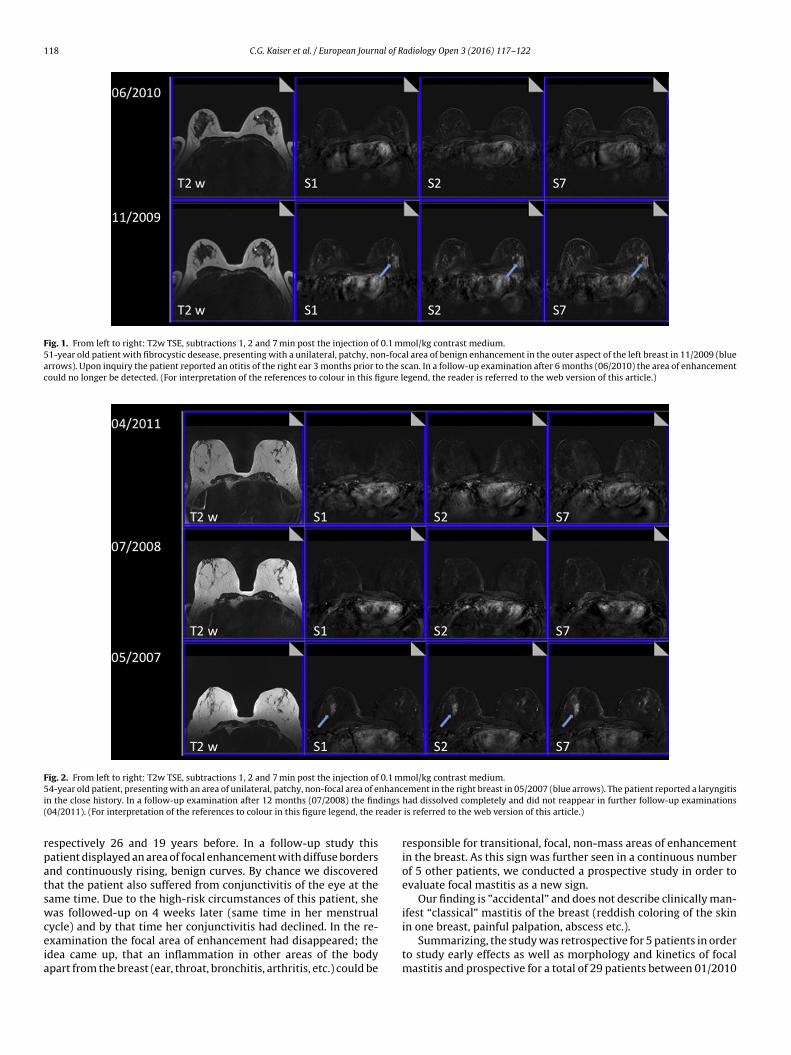
Fig. 2. From left to right: T2w TSE, subtractions 1, 2 and 7 min post the injection of 0.1 mmol/kg contrast medium.5 nhanci dings
( eader
rpatswceia
4-year old patient, presenting with an area of unilateral, patchy, non-focal area of en the close history. In a follow-up examination after 12 months (07/2008) the fin04/2011). (For interpretation of the references to colour in this figure legend, the r
espectively 26 and 19 years before. In a follow-up study thisatient displayed an area of focal enhancement with diffuse bordersnd continuously rising, benign curves. By chance we discoveredhat the patient also suffered from conjunctivitis of the eye at theame time. Due to the high-risk circumstances of this patient, sheas followed-up on 4 weeks later (same time in her menstrual
ycle) and by that time her conjunctivitis had declined. In the re-
xamination the focal area of enhancement had disappeared; thedea came up, that an inflammation in other areas of the bodypart from the breast (ear, throat, bronchitis, arthritis, etc.) could beement in the right breast in 05/2007 (blue arrows). The patient reported a laryngitishad dissolved completely and did not reappear in further follow-up examinationsis referred to the web version of this article.)
responsible for transitional, focal, non-mass areas of enhancementin the breast. As this sign was further seen in a continuous numberof 5 other patients, we conducted a prospective study in order toevaluate focal mastitis as a new sign.
Our finding is ”accidental” and does not describe clinically man-ifest “classical” mastitis of the breast (reddish coloring of the skinin one breast, painful palpation, abscess etc.).
Summarizing, the study was retrospective for 5 patients in orderto study early effects as well as morphology and kinetics of focalmastitis and prospective for a total of 29 patients between 01/2010

C.G. Kaiser et al. / European Journal of Radiology Open 3 (2016) 117–122 119
Fig. 3. From left to right: T2w TSE, subtractions 1, 2 and 7 min post the injection of 0.1 mmol/kg contrast medium.55-year old patient with fibrocystic desease, presenting with two small unilateral, patchy, non-focal areas of enhancement in the right breast in 06/2006 (blue arrows). Uponininquiry, the patient reported she had suffered from viral respiratory tract infection (common flew) a few weeks before the MR-mammogram. In a follow-up examinationafter 3 months (09/2006) the findings had dissolved completely and did not reappear in further follow-up examinations (09/2009 and 09/2011). (For interpretation of thereferences to colour in this figure legend, the reader is referred to the web version of this article.)
Table 1Examination protocol.
Sequence (Nr.) 1. Nat cor 2. Dynamica tra 3. CM cor 4. T2-TSE 5. STIRWeighting T1 T1 T1 T2 T2 (T1, 150 ms)Puls sequence FLASH FLASH FLASH TSE TSEOrientation cor transv cor transv transvTR (ms) 113 113 113 8900 8420TE (ms) 4,6 4,6 4,6 207 70Flip angle (◦) 80 80 80 191 180Slice thickness (mm) 3 3 3 3 3Gap (mm) 0 0 0 0 0Field of view (mm) 350 350 350 350 350Nr. of slices 44 44 44 44 44Matrix (Pixels) 230 × 256 307 × 384 230 × 256 435 × 512 326 × 384
d-DTPa
ac
1
2
a Connotation: dynamic study before and after the i.v. application of 0,1 mmol Gutomatic injector (Medrad, Spectris, Pittsburgh) with 3 ml/s .
nd 12/2011. These patients would usually not have a history oflinical mastitis, yet displayed the following findings:
. A slight, focal, unilateral non-mass and area of progredientenhancement without cancer corner or washout sign (see
Figs. 1–6).. When inquired about a recent history of inflammation or infec-tion, patients would report about otitis, arthritis, sinusitis,inflammatory processes of the skin, etc.
A per kg body weight within 10 s, followed by the injection of 30 ml saline via an
3. They would also describe a subtle discomfort they had registeredin the affected breast upon forceful palpation.
4. Dissolving or at least decline after a waiting period of at least 3months.
Since serological parameters (CRP) occurred to be unspecificin the retrospectively analyzed patients, CRP was therefore notincluded into the evaluation of the prospective study cases.

120 C.G. Kaiser et al. / European Journal of Radiology Open 3 (2016) 117–122
Fig. 4. From left to right: T2w TSE, subtractions 1, 2 and 7 min post the injection of 0.1 mmol/kg contrast medium.44-year old patient with fibrocystic desease, presenting with a large unilateral, patchy, non-focal area of enhancement in the right breast in April 2010 (blue arrows) after aninconspicuous MR-mammogram in 06/2007. Upon ininquiry, the patient reported she had suffered from a Hordeolum of the right eye two months prior to the examination.In a follow-up examination after 4 months (08/2010) the findings were regredient. In a further follow-up after 14 months (12/2011), the previous findings had dissolved.(For interpretation of the references to colour in this figure legend, the reader is referred to the web version of this article.)
Fig. 5. From left to right: T2w TSE, subtractions 1, 2 and 7 min post the injection of 0.1 mmol/kg contrast medium.44-year old patient, presenting with two areas of unilateral, patchy, benign enhancement in the lower aspects of the right breast in 05/2011 (blue arrows). With a patienthistory of tonsillitis 2 months prior to the MR-Mammogram, a follow-up examination after 6 months in 11/2011 was conducted. See Fig. 6 for a magnification of the rightbreast. (For interpretation of the references to colour in this figure legend, the reader is referred to the web version of this article.)

C.G. Kaiser et al. / European Journal of Radiology Open 3 (2016) 117–122 121
Fig. 6. Magnification of the right breast (Fig. 5). From left to right: T2w TSE, subtractions 1, 2 and 7 min post the injection of 0.1 mmol/kg contrast medium.44-year old patient, presenting with two areas of unilateral, patchy, benign enhancement in the lower aspects of the right breast in 05/2011 (blue arrows). With a patienth n aftea specto rsion
aadspnics
2
(a
3
bpfpafMwf
tef
4
bnp
istory of tonsillitis 2 months prior to the MR-Mammogram, a follow-up examinatiospect of the right breast was regredient in size, while the small focus in the inner af the references to colour in this figure legend, the reader is referred to the web ve
The standard reference was a follow-up MRM-evaluation after waiting period of 3–6 months: these diagnostic focal, non-massreas of enhancements as well the discomfort during palpationissolved or declined in the follow-up MRM examination afteruccessful treatment or self-healing of the reported inflammatoryrocess. In case of these MRM findings a “focal mastitis” was diag-osed (Fig. 1). In none of the cases of this study an accompanying
nflammatory cancer was found; no patient showed an increase (1ase of consistent enhancement) of either the MRM- or the clinicalymptoms.
.1. MR-Scanner
All MRM exams were performed with a 1.5 T-MR ScannerSiemens, Avanto) using the following standard protocol (Table 1),s described in other publications [5].
. Results
Between 07/2010 and 12/2011, 1975 breast patients haveeen examined in our University Hospital. Among these 1975atients the total amount of 29 patients showed signs of a distinctocal mastitis (unilateral enhancement, progredient enhancement,atchy-confluent enhancement, no corner sign, reversible afterntibiotic treatment or waiting period, slight unilateral discom-ort during forceful palpation). These patients were recommended
RM follow-up examination after 6 months in order to evaluate,hether or not the “lesion” had dissolved. In the follow-up the
ollowing results were received.28 patients showed a complete or nearly complete dissolving of
he previous findings. 1 patient was found to display a progredi-nt area of enhancement, which histologically proved to be a smallocus of DCIS after biopsy.
. Discussion
MRM is a highly sensitive technique to detect changes in thereast, induced by vascularization. Among these changes are malig-ant lesions, but also changed vascularization due to inflammatoryrocesses. To our knowledge, this study was the first to describe this
r 6 months in 11/2011 was conducted. The larger area of enhancement in the outer of the right breast stayed constant in morphology and kinetics. (For interpretationof this article.)
accompanying phenomenon, which are slight and distinct, but haveto be considered in the differential diagnosis of non-mass lesions.
In the literature the morphological and kinetic signs of non-puerperal mastitis have been known for some time, however mostof these reports have been severe granulomatous mastitis-casesand abscesses [6]. As a hallmark of “focal transitional mastitis”,we assessed a unilateral area of slight, non-mass and continu-ous enhancement without border sign (i.e. no separation betweensegments of the breast − an indication of DCIS), an inflamma-tory anamnesis as well as slight discomfort upon palpation of theaffected breast in contrast to the contralateral breast; in addition,the patient had reported about an infection in other parts of thebody upon questioning. Since this type of enhancement appearedto dissolve in follow-up studies it was considered the result of focalinflammation.
In our experience accompanying hormone- or menstrual effectsare furthermore unlikely to cause such enhancement, as they rathertend to affect both breasts and therefore are not relevant for thedifferential diagnosis of lesions imaged with our technique.
The detection of focal transitional mastitis in MR-Mammography is probably only possible due to the very highcontrast and sensitivity towards changes after contrast injection ofthe MR-technique. It is also an example how a medical diagnosis ismade by a combination of imaging findings, clinical examination,the evaluation of the patients history and results of follow-upexaminations. Only when these effects are not regredient infollow-up examinations, a biopsy should be recommended to testfor a malignant finding. The knowledge of this diagnosis (focaltransitional mastitis) seems to be important in order to reduce thenumber of unnecessary biopsies in the future.
5. Conclusion
Among the differential diagnosis of enhancing breast lesions, afocal area of transitional inflammation needs to be added to thelist of differential diagnosis. Typical signs are: continuous unilat-eral enhancement, lack of border signs, inflammatory anamnesis,
accompanying discomfort upon palpation of the affected breast.The patient should be followed-up on for correlation of focal inflam-mation after a time period of not longer than 6 months (eventuallyapplying antibiotic treatment), in order to rule out DCIS or a
1 al of R
gia
C
A
tS
R
[
[
[
[
[
[6] H.A.T. Al-Khawari, H.A. Al-Manfouhi, J.P. Madda, et al., Radiologic features ofgranulomatous mastitis, Breast J. 17 (6) (2011) 645–650.
[7] C.G. Kaiser, J. Krammer, S.O. Schönberg, W.A. Kaiser, K. Wasser, Focaltransitional mastitis in MR-mammography: preliminary findings, Eur. J. Radiol.81 (Suppl. 1) (2012) S72–73.
22 C.G. Kaiser et al. / European Journ
rowing malignant lesion. Further studies are necessary to clar-fy whether these findings are dependent on different technicalspects or varying patient groups.
onflict of interest
The authors declare that they have no conflict of interest.
cknowledgment
Published in part (only abstract) during the “6th Interna-ional Congress on MR-Mammography” in Jena, Germany (27–29eptember, 2012) [7].
eferences
1] E.S. Burnside, E.A. Sickles, L.W. Bassett, et al., The ACR BI-RADS® experience:learning from history, J. Am. Coll. Radiol. JACR 6 (12) (2009) 851–860.
adiology Open 3 (2016) 117–122
2] S.D. Edwards, J.A. Lipson, D.M. Ikeda, J.M. Lee, Updates and revisions to theBI-RADS magnetic resonance imaging lexicon, Magn. Reson. Imag. Clin. N. Am.21 (3) (2013) 483–493.
3] T. Pabst, W. Kenn, W.A. Kaiser, D. Hahn, Understanding why contrastenhancement in dynamic MRI is not reproducible: illustration with a simplephantom, Breast J. 7 (3) (2001) 166–170.
4] W.A. Kaiser, Signs in MR-Mammography, 1st ed., Springer, 2009, 2008. Corr.2nd printing, 400 p.
5] C. Kuhl, The current status of breast MR imaging Part I. Choice of technique,image interpretation, diagnostic accuracy, and transfer to clinical practice,Radiology 244 (2) (2007) 356–378.