European Conference on - Ruđer Bošković Instituteexrs2008.irb.hr/EXRS-programme.pdf · The EXRS...
36
Transcript of European Conference on - Ruđer Bošković Instituteexrs2008.irb.hr/EXRS-programme.pdf · The EXRS...

EXRS 2008
European Conference on X-Ray Spectrometry
Organized by: Ruđer Bošković Institute, Zagreb, Croatia
In cooperation with: Ministry of Science, Education and Sports, Croatia International Atomic Energy Agency European X-ray Spectrometry Association
Programme & General Information
16th – 20th June 2008 Cavtat, Dubrovnik, Croatia
1

2
Welcome The Local Organizing Committee of the 13th European Conference on X-ray Spectrometry (EXRS 2008) is pleased to welcome all participants of this conference. About 280 delegates from 41 countries will share opportunity to exchange ideas and knowledge related to X-ray Spectrometry, including fundamental aspects, technological developments, traditional and novel areas of applications and interdisciplinary research. We are greatefull to our sponsors and all people that helped us in the organisation of this event. We hope that you will enjoj in stimulating and friendly environment and that you will benefit from your participation at EXRS 2008. Stjepko Fazinić and Milko Jakšić EXRS Co-chairs, on behalf of the Local Organizing Comittee Conference organization The EXRS 2008 has been organized by the Ruđer Bošković Institute, Zagreb, Croatia, in cooperation with the Ministry of science, education and sports of Croatia, International Atomic Energy Agency and European X-ray Spectrometry Association. Local Organising Committee
International Advisory Committee
Co-chairs: Stjepko Fazinić* and Milko Jakšić* Members: Mladen Bogovac* Iva Božičević* Vladan Desnica*** Deša Jelavić* Marko Karlušić* Luka Mandić** Ivo Orlić** Željko Pastuović* Iva Bogdanović Radović* Mira Ristić* Zdravko Siketić* Natko Skukan* Darek Wegrzynek **** Ivana Zamboni* * Ruđer Bošković Institute, Zagreb ** University of Rijeka *** University of Zagreb **** International Atomic Energy Agency
Burkhard Beckhoff, Germany Maria Luisa Carvalho, Portugal Roberto Cesareo, Italy Jorge E. Fernandez, Italy Yohichi Gohshi, Japan Marie-Christine Lepy, France Marcelo Rubio, Argentina Eva Selin-Lindgren, Sweden Szabina Török, Hungary René Van Grieken, Belgium Peter Wobrauschek, Austria
Conference Secretariat Rudjer Bošković Institute Laboratory for Ion Beam Interactions Bijenička cesta 54 P.O. Box 180 Tel: +385 1 4571 254 HR-10002 Zagreb Fax: +385 1 4680 239 Croatia E-mail: [email protected]
3
Scientific programme The EXRS 2008 international conference will be 13th in a series of biennial conferences which bring together scientists from the various research fields of X-ray spectrometry, using photon beams, electrons or other energetic particles. The conference programme will consist of 11 invited lectures (30 min) from distinguished scientists, 98 oral presentations (20 min) and about 150 poster contributions, all divided among 14 sessions. Conference Sessions Session 1: Applications of XRS in archaeometry Session 2: Quantification methodology Session 3: Synchrotron XRS Session 4: PIXE and electron induced XRS Session 5: X-ray sources, optics and detectors Session 6: X-ray imaging and tomography Session 7: Applications – materials and nanoscience Session 8: X-ray absorption (EXAFS, XANES) Session 9: Applications – earth and environment sciences Session 10: WDXRS Session 11: Applications – life sciences Session 12: Interactions of X-rays with matter Session 13: Microbeam techniques Session 14: TXRF and related techniques Conference sponsors We are grateful to the following institutions for supporting the conference: Ministry of Science, Education and Sports, Croatia http://www.mzos.hr/ International Atomic Energy Agency http://www.iaea.org/ European X-ray Spectrometry Association http://www.exsa.hu/
4
Invited speakers David Cohen, Australia The latest directions and trends in ion beam analysis of fine particle air pollution
Tuesday 08:30 Hall 2 (Orlando, Ground floor)
Jean Claude Dousse, Switzerland Photo induced atomic inner-shell processes investigated by means of high-resolution XRS
Thursday 11:00 Hall 2 (Orlando, Ground floor)
Terrence Jach, USA The effect of chemical bonding on high-resolution X-ray spectroscopy
Thursday 09:00 Hall 1 (Bobara, First Floor)
Frank de Groot, The Netherlands The nature of X-ray absorption spectra: Novel theoretical and experimental tools
Wednesday 09:00 Hall 1 (Bobara, First Floor)
Maria Filomena Guerra, France Tracking gold forgeries with X-rays
Monday 10:00 Hall Ragusa (Ground Floor)
Wolfgang Malzer, Germany Quantification in XRF micro-analysis
Monday 14:00 Hall 2 (Orlando, Ground floor)
Philippe Moretto, France Biomedical applications of µ-PIXE
Thursday 14:00 Hall 1 (Bobara, First Floor)
Gyorgy Vanko, Hungary Spin-state studies with X-ray spectroscopy, and applications to Earth's lower mantle constituents
Wednesday 11:00 Hall 2 (Orlando, Ground floor)
Alex von Bohlen, Germany Nanoparticles observed under X-ray grazing incidence
Friday 11:00 Hall 2 (Orlando, Ground floor)
Kazuto Yamauchi, Japan Synchrotron-radiation-based hard X-ray nanobeam by Kirkpatrick-Baez mirrors
Friday 09:00 Hall 1 (Bobara, First Floor)
Darek Wegrzynek, IAEA X-ray phase contrast imaging and tomography of malaria transmitting mosquitoes
Tuesday 14:00 Hall 1 (Bobara, First Floor)
Activities Oral sessions will be held from Monday until Friday noon. During the three poster sessions organised on Monday, Tuesday and Thursday between 17:00 and 18:30, authors will have opportunity to discuss posters. Industrial exhibition will be held on Monday afternoon, Tuesday, Wednesday and Thursday. Sunday, 15 June. Registration 18:00-20:00. Welcome reception 20:00. Monday, 16 June. Registration. Opening at 9:00. Oral sessions. Poster session A. Industrial exhibition. Tuesday, 17 June. Oral sessions from 08:30. EXSA Meeting. Poster Session B. Wednesday, 18 June. Oral sessions from 09:00. Conference excursion. Thursday, 19 June. Oral sessions from 09:00. Poster session C. Conference dinner. Friday, 20 June. Oral sessions from 09:00. Concluding remarks.
5
Industrial exhibitors and sponsors The industrial exhibition will play a key role by presenting equipment and books related to X-ray spectroscopy and applications. The exhibition starts on Monday afternoon at 14:00. It will be opened until Thursday afternoon,18:30. The list of exhibitors and sponsors is as follows:
Amptek Inc. http://www.amptek.com/ Australian X-Ray Tubes Pty Limited, AXT http://www.axt.com.au/ Bruker AXS http://www.bruker-axs.de/ Cambridge Scientific http://www.cambridgescientific.net/ Canberra NV/SA http://www.canberra.com/ E2V Scientific Instruments http://www.e2vsi.com// Horiba Jobin Yvon http://www.jobinyvon.fr/ IFG http://www.ifg-adlershof.de/ KETEK GmbH http://www.ketek.net/ Moxtek, Inc. http://www.moxtek.com/ PNDetector GmbH http://www.pndetector.de/ PNSensor GmbH http://www.pnsensor.de/ Pulse Tor LLC http://www.pulsetor.com/ Rigaku http://www.rigaku.com/ Seiko Instruments GmbH Nano Technology http://www.seiko-instruments.de/ Spectro/Edax
http://www.spectro.com/http://www.edax.com/
Spex Certiprep Ltd. http://www.spexcertiprep.co.uk/ The European Physical Journal, Applied Physics http://www.epjap.org/ Thermo Fisher Scientific - Niton http://www.niton.com/Default.aspx Toivel http://www.toivel.com/ Wiley Blackwell http://eu.wiley.com/ XIA LLC http://www.xia.com/ XOS http://www.xos.com/index.php Proceedings The conference proceedings will be published on a CD-ROM for distribution among the participants after the conference. The conference proceedings will contain the submitted manuscripts and submitted presentations in pdf format. Contributions transmitted to the Conference Secretariat before July 4, 2008 will be automatically included, without any reviewing procedure. Here there are no specific instructions about size and shape: the manuscript will be included such as the authors will have prepared it, under their own responsibility. Authors of accepted contributions are invited to submit a manuscript for publication in a special issue of X-Ray Spectrometry (Wiley Intescience). Manuscripts should be prepared in a strict format of the XRS and will be reviewed in the normal review procedure of the Journal. Manuscripts can only be submitted online (http://www3.interscience.wiley.com/journal/1870/home/ForAuthors.html), mentioning the manuscript is intended for the special issue on EXRS2008. The on-line submission must be done before June 30, and a hard copy should preferably be left at the conference desk during EXRS2008.
6

Oral presentations Time attributed to each oral presentation is 15 minutes + 5 min for questions. Presenters will have on disposal a digital projector and a PC running Microsoft Powerpoint. Speakers are kindly asked to prepare Power Point presentations in the format compatible with the Microsoft Office 2003, and to load their file onto presentation PCs at least one session prior to the one in which they will make their presentation. Speakers should be present at least 15 minutes before the beginning of the Session in which they will give their presentation in order to introduce themselves to the Session Chair. Posters The size of the display area for a poster is 90 cm in width and 120 cm in height. Posters will be on display during one of the three poster sessions, planned for Monday, Tuesday and Thursday. Contributors to the Poster Session A are kindly asked to place their posters on Monday 16 June during the Lunch break (12:00 – 14:00), to keep them on display until Tuesday 17 June 13:00, and to be present and available for discussions during the Poster Session A (Monday 17:00 – 18:30). Contributors to the Poster Session B are kindly asked to place their posters on Tuesday 17 June between 13:00 and 14:00, to keep them on display until Thursday 19 June 13:00, and to be present and available for discussions during the Poster Session B (Tuesday 17:10 – 18:40). Contributors to the Poster Session C are kindly asked to place their posters on Thursday 19 June between 13:00 and 14:00, to keep them on display until Friday 10:30, and to be present and available for discussions during the Poster Session C (Thursday 17:00 – 18:30). Awards Award for young X-ray spectrometrist The award for young X-ray spectrometists will be delivered by the European X-ray Spectrometry Association (EXSA). The award ceremony will take place during the conference. Best poster awards The best poster award prize at EXRS 2008 will be a selection of Wiley-Blackwell books up to the value of 300 Euros. The award ceremony will take place during the conference dinner. Conference venue The EXRS-2008 conference will be held in hotel Croatia, which is a five star hotel that lies on a peninsula overlooking the Adriatic on one side and the picturesque bay and the old town of Cavtat on the other. The meeting will take place in a large meeting hall 'Ragusa' (Monday morning) and two smaller halls (hall 1 'Bobara' and hall 2 'Orlando'). Poster sessions and industrial exhibition will be organized in the vicinity of meeting hall 1 'Bobara' and terrace for coffee break time.
7
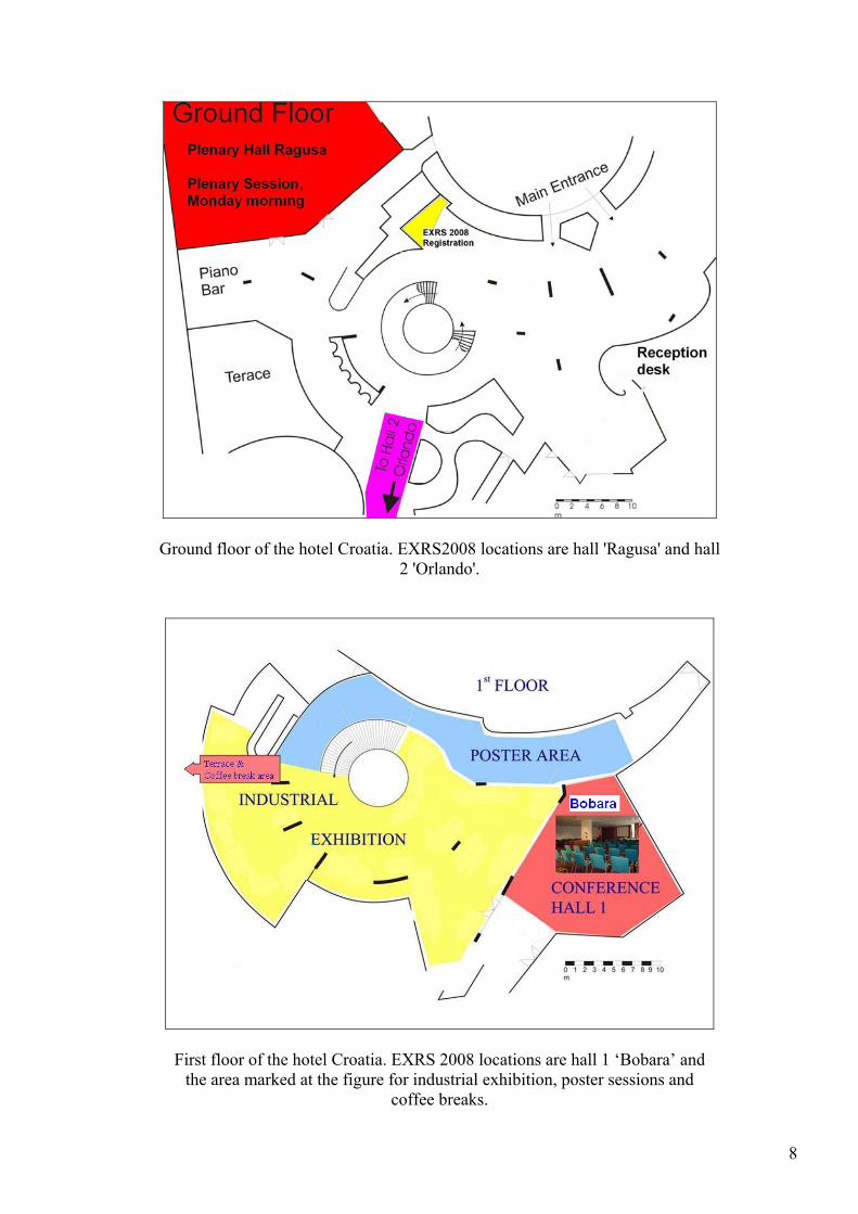
Ground floor of the hotel Croatia. EXRS2008 locations are hall 'Ragusa' and hall 2 'Orlando'.
First floor of the hotel Croatia. EXRS 2008 locations are hall 1 ‘Bobara’ and the area marked at the figure for industrial exhibition, poster sessions and
coffee breaks.
8
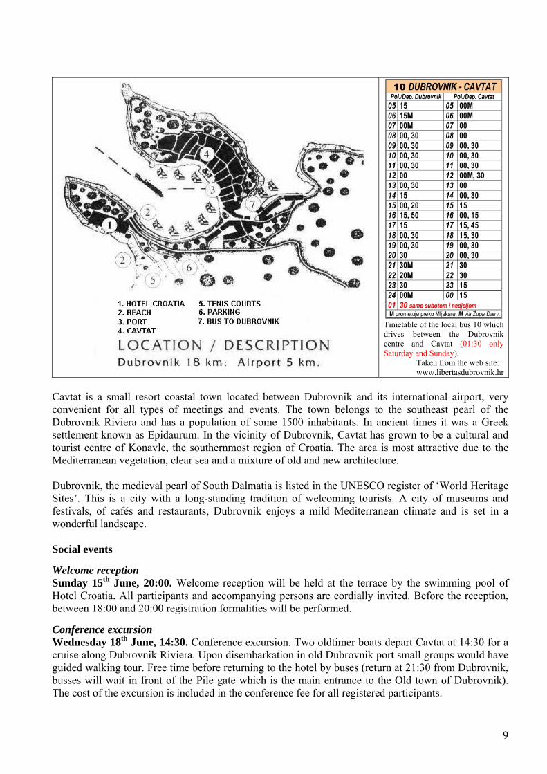
Timetable of the local bus 10 which drives between the Dubrovnik centre and Cavtat (01:30 only Saturday and Sunday). Taken from the web site: www.libertasdubrovnik.hr
Cavtat is a small resort coastal town located between Dubrovnik and its international airport, very convenient for all types of meetings and events. The town belongs to the southeast pearl of the Dubrovnik Riviera and has a population of some 1500 inhabitants. In ancient times it was a Greek settlement known as Epidaurum. In the vicinity of Dubrovnik, Cavtat has grown to be a cultural and tourist centre of Konavle, the southernmost region of Croatia. The area is most attractive due to the Mediterranean vegetation, clear sea and a mixture of old and new architecture. Dubrovnik, the medieval pearl of South Dalmatia is listed in the UNESCO register of ‘World Heritage Sites’. This is a city with a long-standing tradition of welcoming tourists. A city of museums and festivals, of cafés and restaurants, Dubrovnik enjoys a mild Mediterranean climate and is set in a wonderful landscape. Social events Welcome reception Sunday 15th June, 20:00. Welcome reception will be held at the terrace by the swimming pool of Hotel Croatia. All participants and accompanying persons are cordially invited. Before the reception, between 18:00 and 20:00 registration formalities will be performed. Conference excursion Wednesday 18th June, 14:30. Conference excursion. Two oldtimer boats depart Cavtat at 14:30 for a cruise along Dubrovnik Riviera. Upon disembarkation in old Dubrovnik port small groups would have guided walking tour. Free time before returning to the hotel by buses (return at 21:30 from Dubrovnik, busses will wait in front of the Pile gate which is the main entrance to the Old town of Dubrovnik). The cost of the excursion is included in the conference fee for all registered participants.
9

Conference dinner Thursday 19th June, 20:30. Conference dinner at the Hotel Croatia. The cost of the conference dinner is not included in the registration fee. Lunches Monday 16th at 12:00, and Tuesday 17th June at 12:30. Monday and Tuesday lunches are organised for all registered conference participants at the conference hotel during lunch breaks. The cost is included in the registration fee. Accompanying persons The accompanying persons fee includes welcome reception on Sunday, conference excursion on Wednesday and lunches at the conference hotel on Monday and Tuesday. Hotel Croatia and our official agent ATLAS offer various sightseeing tours on request. Please consult ATLAS representative on the spot or the hotel reception for details. Official photograph The conference photo will be taken on terrace of hotel Croatia on Wednesday 18 June at 12:45.
10

Conference Schedule
11
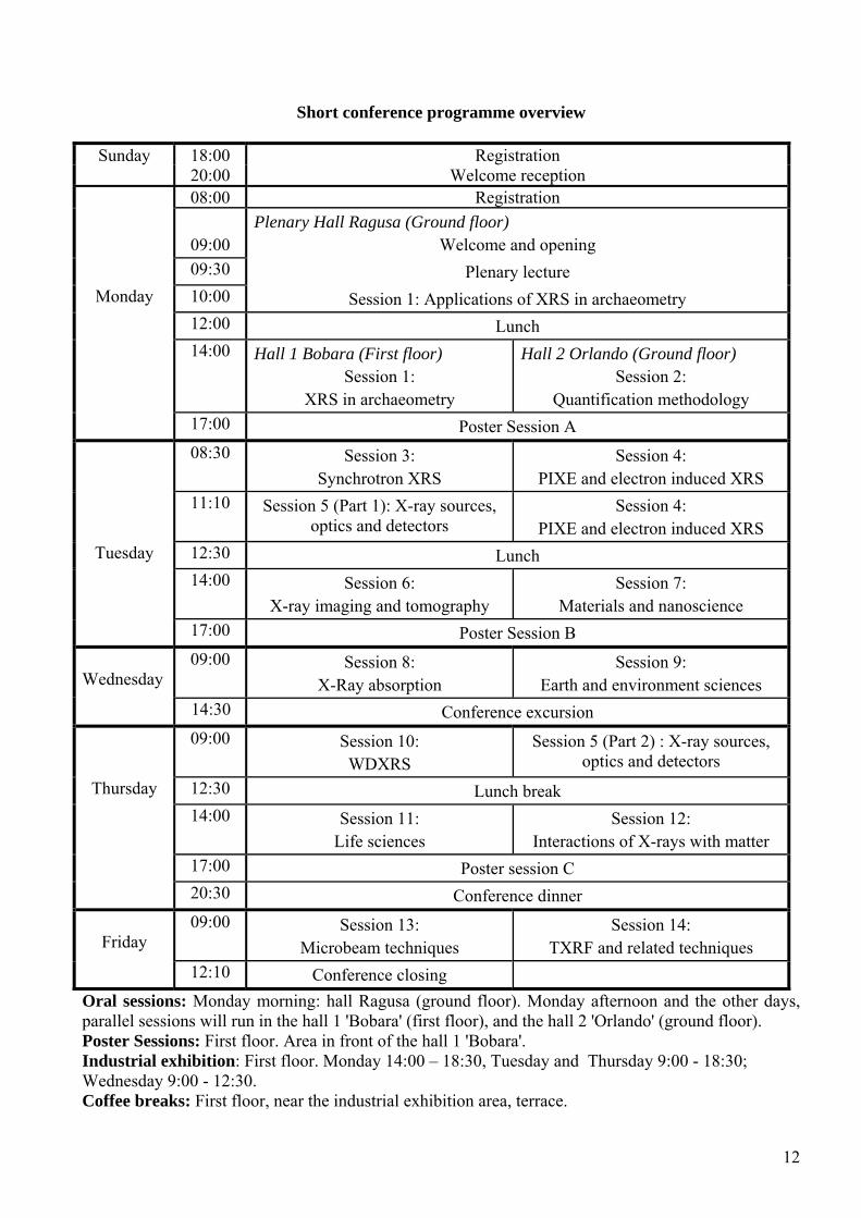
Short conference programme overview
Sunday 18:00 Registration 20:00 Welcome reception 08:00 Registration
09:00 Plenary Hall Ragusa (Ground floor)
Welcome and opening 09:30 Plenary lecture
Monday 10:00 Session 1: Applications of XRS in archaeometry 12:00 Lunch 14:00 Hall 1 Bobara (First floor)
Session 1: XRS in archaeometry
Hall 2 Orlando (Ground floor) Session 2:
Quantification methodology 17:00 Poster Session A 08:30 Session 3:
Synchrotron XRS Session 4:
PIXE and electron induced XRS 11:10 Session 5 (Part 1): X-ray sources,
optics and detectors Session 4:
PIXE and electron induced XRS Tuesday 12:30 Lunch
14:00 Session 6: X-ray imaging and tomography
Session 7: Materials and nanoscience
17:00 Poster Session B
Wednesday 09:00 Session 8:
X-Ray absorption Session 9:
Earth and environment sciences 14:30 Conference excursion 09:00 Session 10:
WDXRS Session 5 (Part 2) : X-ray sources,
optics and detectors Thursday 12:30 Lunch break
14:00 Session 11: Life sciences
Session 12: Interactions of X-rays with matter
17:00 Poster session C 20:30 Conference dinner
Friday 09:00 Session 13:
Microbeam techniques Session 14:
TXRF and related techniques 12:10 Conference closing
Oral sessions: Monday morning: hall Ragusa (ground floor). Monday afternoon and the other days, parallel sessions will run in the hall 1 'Bobara' (first floor), and the hall 2 'Orlando' (ground floor). Poster Sessions: First floor. Area in front of the hall 1 'Bobara'. Industrial exhibition: First floor. Monday 14:00 – 18:30, Tuesday and Thursday 9:00 - 18:30; Wednesday 9:00 - 12:30. Coffee breaks: First floor, near the industrial exhibition area, terrace.
12
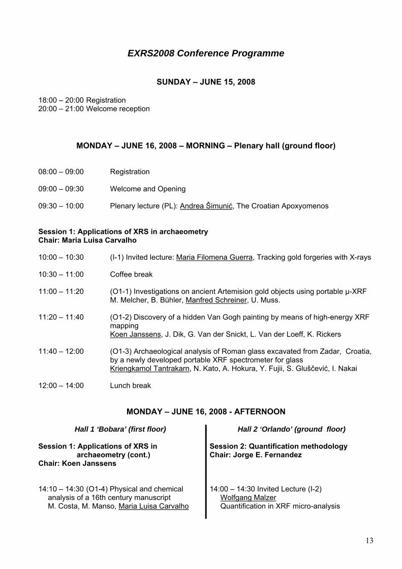
EXRS2008 Conference Programme
SUNDAY – JUNE 15, 2008 18:00 – 20:00 Registration 20:00 – 21:00 Welcome reception
MONDAY – JUNE 16, 2008 – MORNING – Plenary hall (ground floor) 08:00 – 09:00 Registration 09:00 – 09:30 Welcome and Opening 09:30 – 10:00 Plenary lecture (PL): Andrea Šimunić, The Croatian Apoxyomenos Session 1: Applications of XRS in archaeometry Chair: Maria Luisa Carvalho 10:00 – 10:30 (I-1) Invited lecture: Maria Filomena Guerra, Tracking gold forgeries with X-rays 10:30 – 11:00 Coffee break 11:00 – 11:20 (O1-1) Investigations on ancient Artemision gold objects using portable µ-XRF
M. Melcher, B. Bühler, Manfred Schreiner, U. Muss. 11:20 – 11:40 (O1-2) Discovery of a hidden Van Gogh painting by means of high-energy XRF
mapping Koen Janssens, J. Dik, G. Van der Snickt, L. Van der Loeff, K. Rickers
11:40 – 12:00 (O1-3) Archaeological analysis of Roman glass excavated from Zadar, Croatia, by a newly developed portable XRF spectrometer for glass Kriengkamol Tantrakarn, N. Kato, A. Hokura, Y. Fujii, S. Gluščević, I. Nakai 12:00 – 14:00 Lunch break
MONDAY – JUNE 16, 2008 - AFTERNOON
Hall 1 ‘Bobara’ (first floor) Hall 2 ‘Orlando’ (ground floor) Session 1: Applications of XRS in archaeometry (cont.) Chair: Koen Janssens
Session 2: Quantification methodology Chair: Jorge E. Fernandez
14:10 – 14:30 (O1-4) Physical and chemical analysis of a 16th century manuscript M. Costa, M. Manso, Maria Luisa Carvalho
14:00 – 14:30 Invited Lecture (I-2) Wolfgang Malzer Quantification in XRF micro-analysis
13
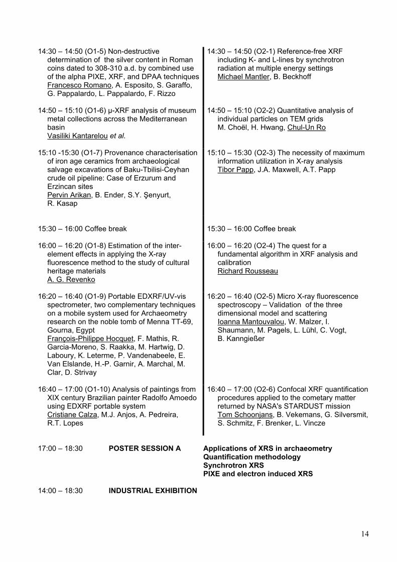
14:30 – 14:50 (O1-5) Non-destructive determination of the silver content in Roman coins dated to 308-310 a.d. by combined use of the alpha PIXE, XRF, and DPAA techniques Francesco Romano, A. Esposito, S. Garaffo, G. Pappalardo, L. Pappalardo, F. Rizzo
14:30 – 14:50 (O2-1) Reference-free XRF including K- and L-lines by synchrotron radiation at multiple energy settings Michael Mantler, B. Beckhoff
14:50 – 15:10 (O1-6) µ-XRF analysis of museum metal collections across the Mediterranean basin Vasiliki Kantarelou et al.
14:50 – 15:10 (O2-2) Quantitative analysis of individual particles on TEM grids M. Choël, H. Hwang, Chul-Un Ro
15:10 -15:30 (O1-7) Provenance characterisation of iron age ceramics from archaeological salvage excavations of Baku-Tbilisi-Ceyhan crude oil pipeline: Case of Erzurum and Erzincan sites Pervin Arikan, B. Ender, S.Y. Şenyurt, R. Kasap
15:10 – 15:30 (O2-3) The necessity of maximum information utilization in X-ray analysis Tibor Papp, J.A. Maxwell, A.T. Papp
15:30 – 16:00 Coffee break
15:30 – 16:00 Coffee break
16:00 – 16:20 (O1-8) Estimation of the inter- element effects in applying the X-ray fluorescence method to the study of cultural heritage materials A. G. Revenko
16:00 – 16:20 (O2-4) The quest for a fundamental algorithm in XRF analysis and calibration Richard Rousseau
16:20 – 16:40 (O1-9) Portable EDXRF/UV-vis spectrometer, two complementary techniques on a mobile system used for Archaeometry research on the noble tomb of Menna TT-69, Gourna, Egypt François-Philippe Hocquet, F. Mathis, R. Garcia-Moreno, S. Raakka, M. Hartwig, D. Laboury, K. Leterme, P. Vandenabeele, E. Van Elslande, H.-P. Garnir, A. Marchal, M. Clar, D. Strivay
16:20 – 16:40 (O2-5) Micro X-ray fluorescence spectroscopy – Validation of the three dimensional model and scattering Ioanna Mantouvalou, W. Malzer, I. Shaumann, M. Pagels, L. Lühl, C. Vogt, B. Kanngießer
16:40 – 17:00 (O1-10) Analysis of paintings from XIX century Brazilian painter Radolfo Amoedo using EDXRF portable system Cristiane Calza, M.J. Anjos, A. Pedreira, R.T. Lopes
16:40 – 17:00 (O2-6) Confocal XRF quantification procedures applied to the cometary matter returned by NASA's STARDUST mission Tom Schoonjans, B. Vekemans, G. Silversmit, S. Schmitz, F. Brenker, L. Vincze
17:00 – 18:30 POSTER SESSION A Applications of XRS in archaeometry Quantification methodology Synchrotron XRS PIXE and electron induced XRS 14:00 – 18:30 INDUSTRIAL EXHIBITION
14
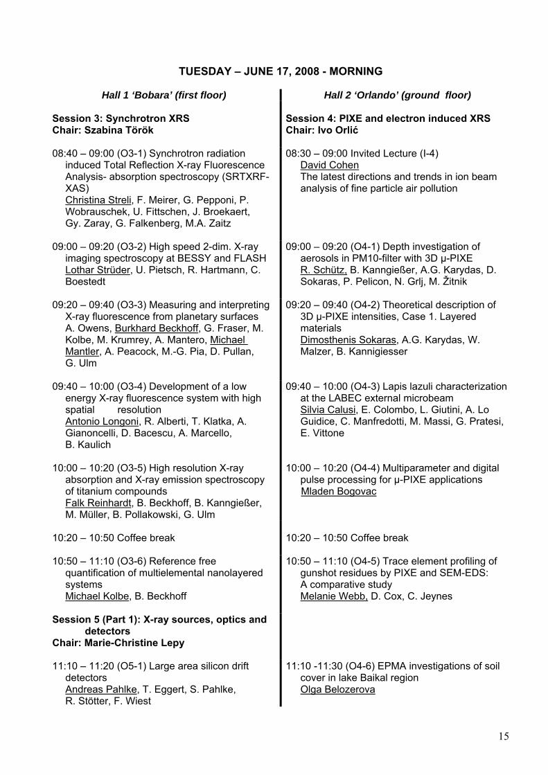
TUESDAY – JUNE 17, 2008 - MORNING
Hall 1 ‘Bobara’ (first floor) Hall 2 ‘Orlando’ (ground floor) Session 3: Synchrotron XRS Chair: Szabina Török
Session 4: PIXE and electron induced XRS Chair: Ivo Orlić
08:40 – 09:00 (O3-1) Synchrotron radiation induced Total Reflection X-ray Fluorescence Analysis- absorption spectroscopy (SRTXRF- XAS) Christina Streli, F. Meirer, G. Pepponi, P. Wobrauschek, U. Fittschen, J. Broekaert, Gy. Zaray, G. Falkenberg, M.A. Zaitz
08:30 – 09:00 Invited Lecture (I-4) David Cohen The latest directions and trends in ion beam analysis of fine particle air pollution
09:00 – 09:20 (O3-2) High speed 2-dim. X-ray imaging spectroscopy at BESSY and FLASH Lothar Strüder, U. Pietsch, R. Hartmann, C. Boestedt
09:00 – 09:20 (O4-1) Depth investigation of aerosols in PM10-filter with 3D µ-PIXE R. Schütz, B. Kanngießer, A.G. Karydas, D. Sokaras, P. Pelicon, N. Grlj, M. Žitnik
09:20 – 09:40 (O3-3) Measuring and interpreting X-ray fluorescence from planetary surfaces A. Owens, Burkhard Beckhoff, G. Fraser, M. Kolbe, M. Krumrey, A. Mantero, Michael Mantler, A. Peacock, M.-G. Pia, D. Pullan, G. Ulm
09:20 – 09:40 (O4-2) Theoretical description of 3D µ-PIXE intensities, Case 1. Layered materials Dimosthenis Sokaras, A.G. Karydas, W. Malzer, B. Kannigiesser
09:40 – 10:00 (O3-4) Development of a low energy X-ray fluorescence system with high spatial resolution Antonio Longoni, R. Alberti, T. Klatka, A. Gianoncelli, D. Bacescu, A. Marcello, B. Kaulich
09:40 – 10:00 (O4-3) Lapis lazuli characterization at the LABEC external microbeam Silvia Calusi, E. Colombo, L. Giutini, A. Lo Guidice, C. Manfredotti, M. Massi, G. Pratesi, E. Vittone
10:00 – 10:20 (O3-5) High resolution X-ray absorption and X-ray emission spectroscopy of titanium compounds Falk Reinhardt, B. Beckhoff, B. Kanngießer, M. Müller, B. Pollakowski, G. Ulm
10:00 – 10:20 (O4-4) Multiparameter and digital pulse processing for µ-PIXE applications Mladen Bogovac
10:20 – 10:50 Coffee break
10:20 – 10:50 Coffee break
10:50 – 11:10 (O3-6) Reference free quantification of multielemental nanolayered systems Michael Kolbe, B. Beckhoff
10:50 – 11:10 (O4-5) Trace element profiling of gunshot residues by PIXE and SEM-EDS: A comparative study Melanie Webb, D. Cox, C. Jeynes
Session 5 (Part 1): X-ray sources, optics and detectors Chair: Marie-Christine Lepy
11:10 – 11:20 (O5-1) Large area silicon drift detectors Andreas Pahlke, T. Eggert, S. Pahlke, R. Stötter, F. Wiest
11:10 -11:30 (O4-6) EPMA investigations of soil cover in lake Baikal region Olga Belozerova
15
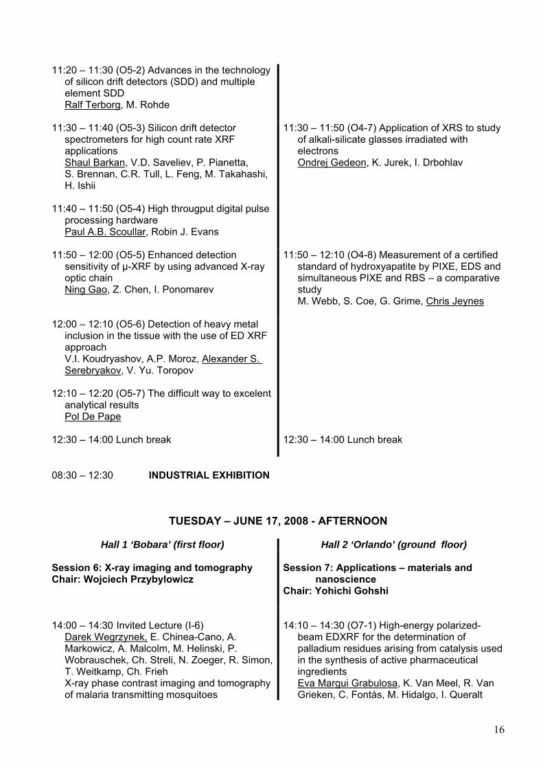
11:20 – 11:30 (O5-2) Advances in the technology of silicon drift detectors (SDD) and multiple element SDD Ralf Terborg, M. Rohde
11:30 – 11:40 (O5-3) Silicon drift detector spectrometers for high count rate XRF applications Shaul Barkan, V.D. Saveliev, P. Pianetta, S. Brennan, C.R. Tull, L. Feng, M. Takahashi, H. Ishii
11:30 – 11:50 (O4-7) Application of XRS to study of alkali-silicate glasses irradiated with electrons Ondrej Gedeon, K. Jurek, I. Drbohlav
11:40 – 11:50 (O5-4) High througput digital pulse processing hardware Paul A.B. Scoullar, Robin J. Evans
11:50 – 12:00 (O5-5) Enhanced detection sensitivity of µ-XRF by using advanced X-ray optic chain Ning Gao, Z. Chen, I. Ponomarev
11:50 – 12:10 (O4-8) Measurement of a certified standard of hydroxyapatite by PIXE, EDS and simultaneous PIXE and RBS – a comparative study M. Webb, S. Coe, G. Grime, Chris Jeynes
12:00 – 12:10 (O5-6) Detection of heavy metal inclusion in the tissue with the use of ED XRF approach V.I. Koudryashov, A.P. Moroz, Alexander S. Serebryakov, V. Yu. Toropov 12:10 – 12:20 (O5-7) The difficult way to excelent analytical results Pol De Pape
12:30 – 14:00 Lunch break
12:30 – 14:00 Lunch break
08:30 – 12:30 INDUSTRIAL EXHIBITION
TUESDAY – JUNE 17, 2008 - AFTERNOON
Hall 1 ‘Bobara’ (first floor) Hall 2 ‘Orlando’ (ground floor) Session 6: X-ray imaging and tomography Chair: Wojciech Przybylowicz
Session 7: Applications – materials and nanoscience Chair: Yohichi Gohshi
14:00 – 14:30 Invited Lecture (I-6) Darek Wegrzynek, E. Chinea-Cano, A. Markowicz, A. Malcolm, M. Helinski, P. Wobrauschek, Ch. Streli, N. Zoeger, R. Simon, T. Weitkamp, Ch. Frieh X-ray phase contrast imaging and tomography of malaria transmitting mosquitoes
14:10 – 14:30 (O7-1) High-energy polarized- beam EDXRF for the determination of palladium residues arising from catalysis used in the synthesis of active pharmaceutical ingredients Eva Margui Grabulosa, K. Van Meel, R. Van Grieken, C. Fontás, M. Hidalgo, I. Queralt
16
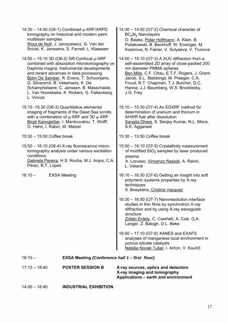
14:30 – 14:50 (O6-1) Combined µ-XRF/XRPD tomography on historical and modern paint multilayer samples Wout de Nolf, J. Jaroszewicz, G. Van der Snickt, K. Janssens, S. Farnell, L. Klaassen
14:30 – 14:50 (O7-2) Chemical character of BCxNy Nanolayers O. Baake, Peter Hoffmann, A. Klein, B. Pollakowski, B. Beckhoff, W. Ensinger, M. Kosinova, N. Fainer, V. Sulyaeva, V. Trunova
14:50 – 15:10 3D (O6-2) SR-Confocal µ-XRF combined with absorption microtomography on Daphnia magna: Instrumental developments and recent advances in data-processing Björn De Samber, R. Evens, T. Schoonjans, G. Silversmit, B. Vekemans, K. De Schamphelaere, C. Janssen, B. Masschaele, L. Van Hoorebeke, K. Rickers, G. Falkenberg, L. Vincze
14:50 – 15:10 (O7-3) A XUV diffraction from a self-assembled 2D array of close-packed 200 nm diameter PMMA spheres Ben Mills, C.F. Chau, E.T.F. Rogers, J. Grant- Jacob, S.L. Stebbings, M. Praeger, C.A. Froud, R.T. Chapman, T.J. Butcher, D.C. Hanna, J.J. Baumberg, W.S. Brocklesby, J.G. Frey
15:10 -15:30 (O6-3) Quantitative elemental imaging of fragments of the Dead Sea scrolls with a combination of µ-XRF and 3D µ-XRF Birgit Kanngießer, I. Mantouvalou, T. Wolff, O. Hahn, I. Rabin, W. Malzer
15:10 – 15:30 (O7-4) An EDXRF method for determination of uranium and thorium in AHWR fuel after dissolution Sangita Dhara, S. Sanjay Kumar, N.L. Misra, S.K. Aggarwal
15:30 – 15:50 Coffee break 15:30 – 15:50 Coffee break 15:50 – 16:10 (O6-4) X-ray fluorescence micro- tomography analysis under various excitation conditions Gabriela Pereira, H.S. Rocha, M.J. Anjos, C.A. Pérez, R.T. Lopes
15:50 – 16:10 (O7-5) Crystallinity measurement of modified SiO2 samples by laser produced plasma A. Lorusso, Vincenzo Nassisi, A. Rainó, L. Velardi
16:10 – EXSA Meeting 16:10 – 16:30 (O7-6) Getting an insight into soft polymeric systems properties by X-ray techniques S. Boeykens, Cristina Vazquez
16:30 – 16:50 (O7-7) Nanoresolution interface studies in thin films by synchrotron X-ray diffraction and by using X-ray waveguide structure Zoltán Erdely, C. Cserhéti, A. Csik, G.A. Langer, Z. Balogh, D.L. Beke
16:50 – 17:10 (O7-8) XANES and EXAFS analyses of manganese local environment in porous silicate catalysts Nataša Novak Tušar, I. Arčon, V. Kaučič
16:10 – EXSA Meeting (Conference hall 1 – first floor) 17:10 – 18:40 POSTER SESSION B X-ray sources, optics and detectors X-ray imaging and tomography Applications – earth and environment 14:00 – 18:40 INDUSTRIAL EXHIBITION
17
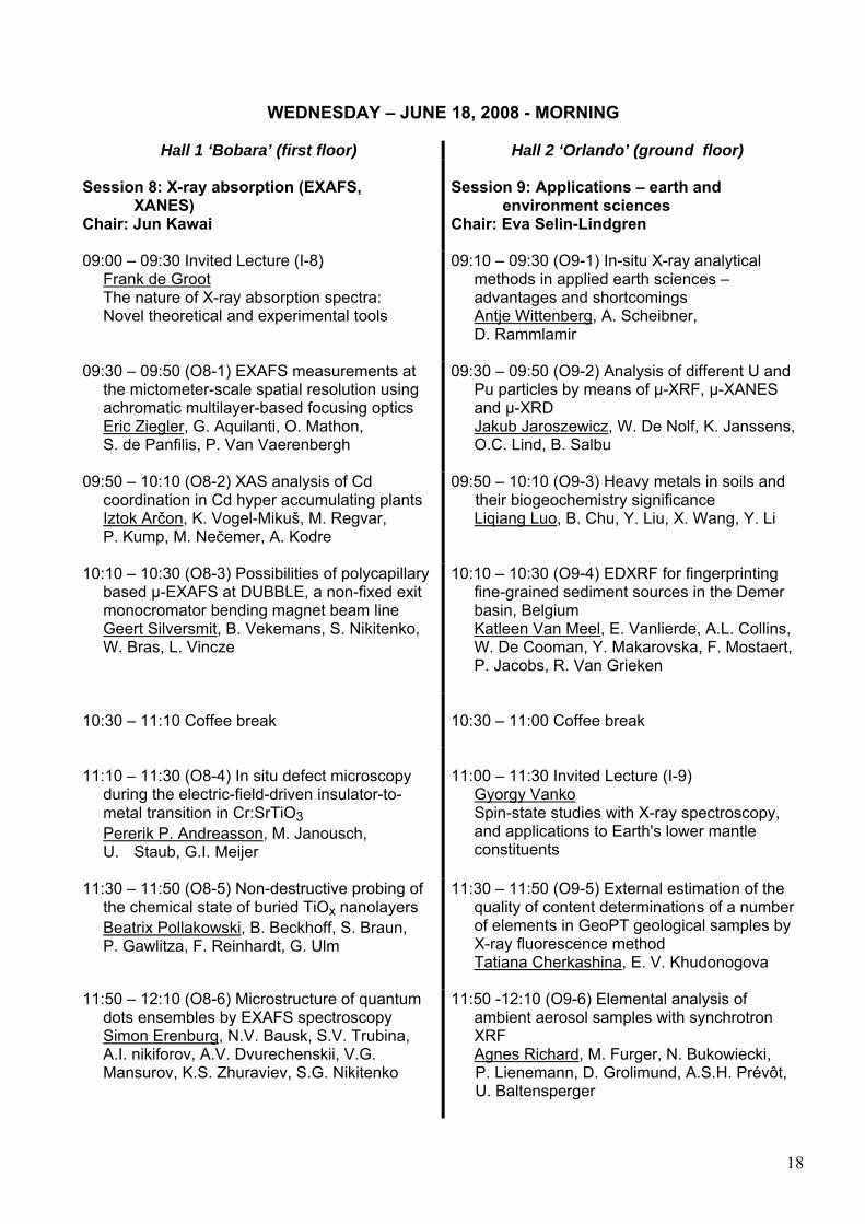
WEDNESDAY – JUNE 18, 2008 - MORNING
Hall 1 ‘Bobara’ (first floor) Hall 2 ‘Orlando’ (ground floor) Session 8: X-ray absorption (EXAFS, XANES) Chair: Jun Kawai
Session 9: Applications – earth and environment sciences Chair: Eva Selin-Lindgren
09:00 – 09:30 Invited Lecture (I-8) Frank de Groot The nature of X-ray absorption spectra: Novel theoretical and experimental tools
09:10 – 09:30 (O9-1) In-situ X-ray analytical methods in applied earth sciences – advantages and shortcomings Antje Wittenberg, A. Scheibner, D. Rammlamir
09:30 – 09:50 (O8-1) EXAFS measurements at the mictometer-scale spatial resolution using achromatic multilayer-based focusing optics Eric Ziegler, G. Aquilanti, O. Mathon, S. de Panfilis, P. Van Vaerenbergh
09:30 – 09:50 (O9-2) Analysis of different U and Pu particles by means of µ-XRF, µ-XANES and µ-XRD Jakub Jaroszewicz, W. De Nolf, K. Janssens, O.C. Lind, B. Salbu
09:50 – 10:10 (O8-2) XAS analysis of Cd coordination in Cd hyper accumulating plants Iztok Arčon, K. Vogel-Mikuš, M. Regvar, P. Kump, M. Nečemer, A. Kodre
09:50 – 10:10 (O9-3) Heavy metals in soils and their biogeochemistry significance Liqiang Luo, B. Chu, Y. Liu, X. Wang, Y. Li
10:10 – 10:30 (O8-3) Possibilities of polycapillary based µ-EXAFS at DUBBLE, a non-fixed exit monocromator bending magnet beam line Geert Silversmit, B. Vekemans, S. Nikitenko, W. Bras, L. Vincze
10:10 – 10:30 (O9-4) EDXRF for fingerprinting fine-grained sediment sources in the Demer basin, Belgium Katleen Van Meel, E. Vanlierde, A.L. Collins, W. De Cooman, Y. Makarovska, F. Mostaert, P. Jacobs, R. Van Grieken
10:30 – 11:10 Coffee break
10:30 – 11:00 Coffee break
11:10 – 11:30 (O8-4) In situ defect microscopy during the electric-field-driven insulator-to- metal transition in Cr:SrTiO3 Pererik P. Andreasson, M. Janousch, U. Staub, G.I. Meijer
11:00 – 11:30 Invited Lecture (I-9) Gyorgy Vanko Spin-state studies with X-ray spectroscopy, and applications to Earth's lower mantle constituents
11:30 – 11:50 (O8-5) Non-destructive probing of the chemical state of buried TiOx nanolayers Beatrix Pollakowski, B. Beckhoff, S. Braun, P. Gawlitza, F. Reinhardt, G. Ulm
11:30 – 11:50 (O9-5) External estimation of the quality of content determinations of a number of elements in GeoPT geological samples by X-ray fluorescence method Tatiana Cherkashina, E. V. Khudonogova
11:50 – 12:10 (O8-6) Microstructure of quantum dots ensembles by EXAFS spectroscopy Simon Erenburg, N.V. Bausk, S.V. Trubina, A.I. nikiforov, A.V. Dvurechenskii, V.G. Mansurov, K.S. Zhuraviev, S.G. Nikitenko
11:50 -12:10 (O9-6) Elemental analysis of ambient aerosol samples with synchrotron XRF Agnes Richard, M. Furger, N. Bukowiecki, P. Lienemann, D. Grolimund, A.S.H. Prévôt, U. Baltensperger
18
12:10 – 12:30 (O8-7) Electronic structure of copper films in aqueous solutions Sergei Butorin
09:00 – 12:30 INDUSTRIAL EXHIBITION
WEDNESDAY – JUNE 18, 2008 – AFTERNOON
14:30 Conference excursion, departure for old town of Dubrovnik by boat
THURSDAY – JUNE 19, 2008 - MORNING
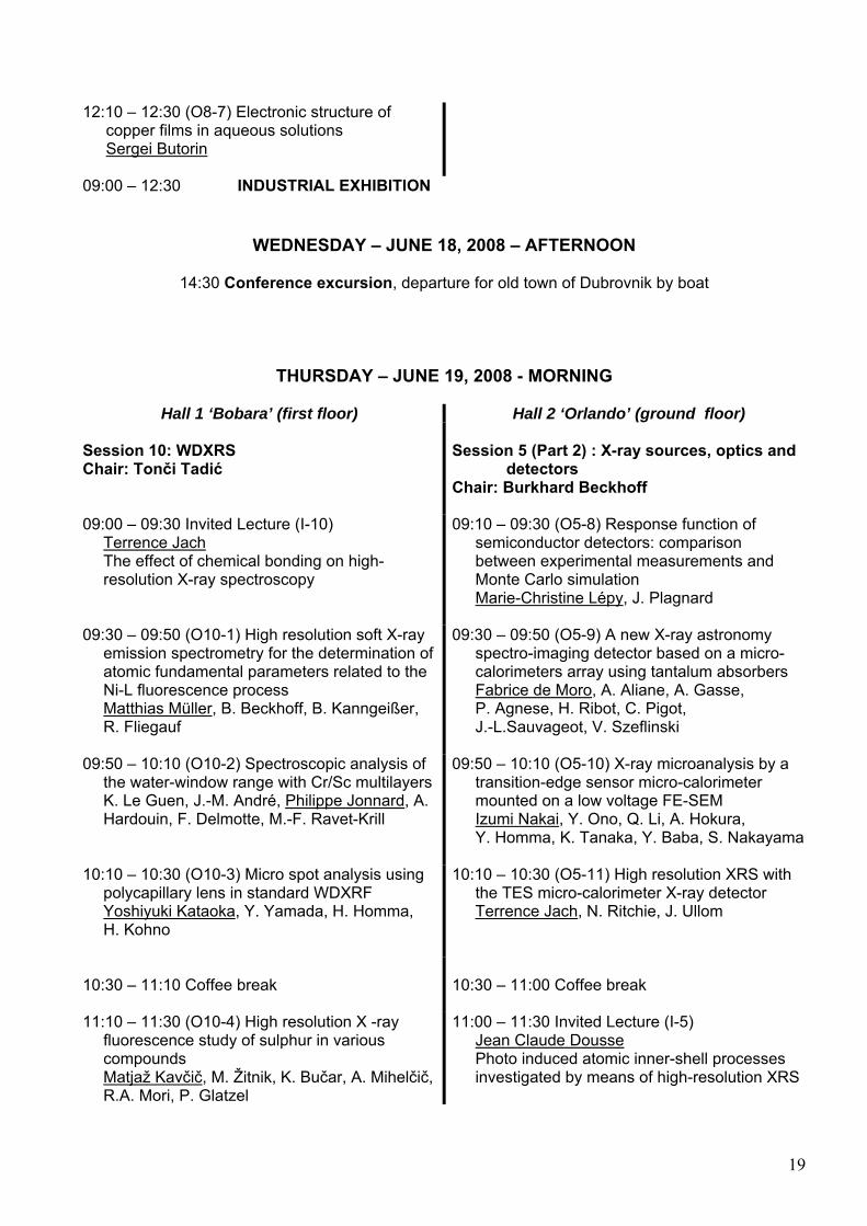
Hall 1 ‘Bobara’ (first floor) Hall 2 ‘Orlando’ (ground floor) Session 10: WDXRS Chair: Tonči Tadić
Session 5 (Part 2) : X-ray sources, optics and detectors Chair: Burkhard Beckhoff
09:00 – 09:30 Invited Lecture (I-10) Terrence Jach The effect of chemical bonding on high- resolution X-ray spectroscopy
09:10 – 09:30 (O5-8) Response function of semiconductor detectors: comparison between experimental measurements and Monte Carlo simulation Marie-Christine Lépy, J. Plagnard
09:30 – 09:50 (O10-1) High resolution soft X-ray emission spectrometry for the determination of atomic fundamental parameters related to the Ni-L fluorescence process Matthias Müller, B. Beckhoff, B. Kanngeißer, R. Fliegauf
09:30 – 09:50 (O5-9) A new X-ray astronomy spectro-imaging detector based on a micro- calorimeters array using tantalum absorbers Fabrice de Moro, A. Aliane, A. Gasse, P. Agnese, H. Ribot, C. Pigot, J.-L.Sauvageot, V. Szeflinski
09:50 – 10:10 (O10-2) Spectroscopic analysis of the water-window range with Cr/Sc multilayers K. Le Guen, J.-M. André, Philippe Jonnard, A. Hardouin, F. Delmotte, M.-F. Ravet-Krill
09:50 – 10:10 (O5-10) X-ray microanalysis by a transition-edge sensor micro-calorimeter mounted on a low voltage FE-SEM Izumi Nakai, Y. Ono, Q. Li, A. Hokura, Y. Homma, K. Tanaka, Y. Baba, S. Nakayama
10:10 – 10:30 (O10-3) Micro spot analysis using polycapillary lens in standard WDXRF Yoshiyuki Kataoka, Y. Yamada, H. Homma, H. Kohno
10:10 – 10:30 (O5-11) High resolution XRS with the TES micro-calorimeter X-ray detector Terrence Jach, N. Ritchie, J. Ullom
10:30 – 11:10 Coffee break
10:30 – 11:00 Coffee break
11:10 – 11:30 (O10-4) High resolution X -ray fluorescence study of sulphur in various compounds Matjaž Kavčič, M. Žitnik, K. Bučar, A. Mihelčič, R.A. Mori, P. Glatzel
11:00 – 11:30 Invited Lecture (I-5) Jean Claude Dousse Photo induced atomic inner-shell processes investigated by means of high-resolution XRS
19
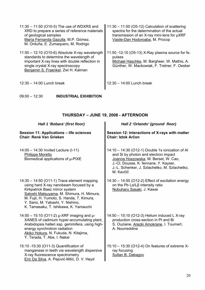
11:30 – 11:50 (O10-5) The use of WDXRS and XRD to prepare a series of reference materials of geological samples María Fernanda Gazulla, M.P. Gómez, M. Orduña, E. Zumaquero, M. Rodrigo
11:30 – 11:50 (O5-12) Calculation of scattering spectra for the determination of the actual transmission of an X-ray mini-lens for µXRF Vasile-Dan Hodoroaba, M. Procop
11:50 – 12:10 (O10-6) Absolute X-ray wavelength standards to determine the wavelength of important X-ray lines with double reflection in single crystal X-ray spectroscopy Benjamin S. Fraenkel, Zwi H. Kalman
11:50 -12:10 (O5-13) X-Ray plasma source for fs- pulses Michael Haschke, M. Bargheer, W. Mathis, A. Günther, W. Mackowiak, F. Tretner, F. Oesker
12:30 – 14:00 Lunch break
12:30 – 14:00 Lunch break
09:00 – 12:30 INDUSTRIAL EXHIBITION
THURSDAY – JUNE 19, 2008 - AFTERNOON
Hall 1 ‘Bobara’ (first floor) Hall 2 ‘Orlando’ (ground floor) Session 11: Applications – life sciences Chair: René Van Grieken
Session 12: Interactions of X-rays with matter Chair: Iztok Arčon
14:00 – 14:30 Invited Lecture (I-11) Philippe Moretto Biomedical applications of µ-PIXE
14:10 – 14:30 (O12-1) Double 1s ionization of Al and Si by photon and electron impact Joanna Hoszowska, M. Berset, W. Cao, J.-Cl. Dousse, K. fennane, Y. Kayser, J.-L. Schenker, J. Szlachetko, M. Szlachetko, M. Kavčič
14:30 – 14:50 (O11-1) Trace element mapping using hard X-ray nanobeam focused by a Kirkpatrick Baez mirror system Satoshi Matsuyama, M. Shimura, H. Mimura, M. Fujii, H. Yumoto, S. Handa, T. Kimura, Y. Sano, M. Yabashi, Y. Nishino, K. Tamasaku, T. Ishikawa, K. Yamauchi
14:30 – 14:50 (O12-2) Effect of excitation energy on the Pb Lα/Lβ intensity ratio Nobuharu Sasaki, J. Kawai
14:50 – 15:10 (O11-2) µ-XRF imaging and µ- XANES of cadmium hyper-accumulating plant, Arabidopsis halleri ssp. gemmifera, using high- energy synchrotron radiation Akiko Hokura, N. Fukuda, N. Kitajima, Y. Terada, T. Abe, I. Nakai
14:50 – 15:10 (O12-3) Helium induced L X-ray production cross-section in Pt and Bi S. Ouziane, Arezki Amokrane, I. Toumert, A. Nourreddine
15:10 -15:30 (O11-3) Quantification of manganese in teeth via wavelength dispersive X-ray fluorescence spectrometry Eric Da Silva, A. Pejović-Milić, D. V. Heyd
15:10 – 15:30 (O12-4) On features of extreme X- ray focusing Sultan B. Dabagov
20
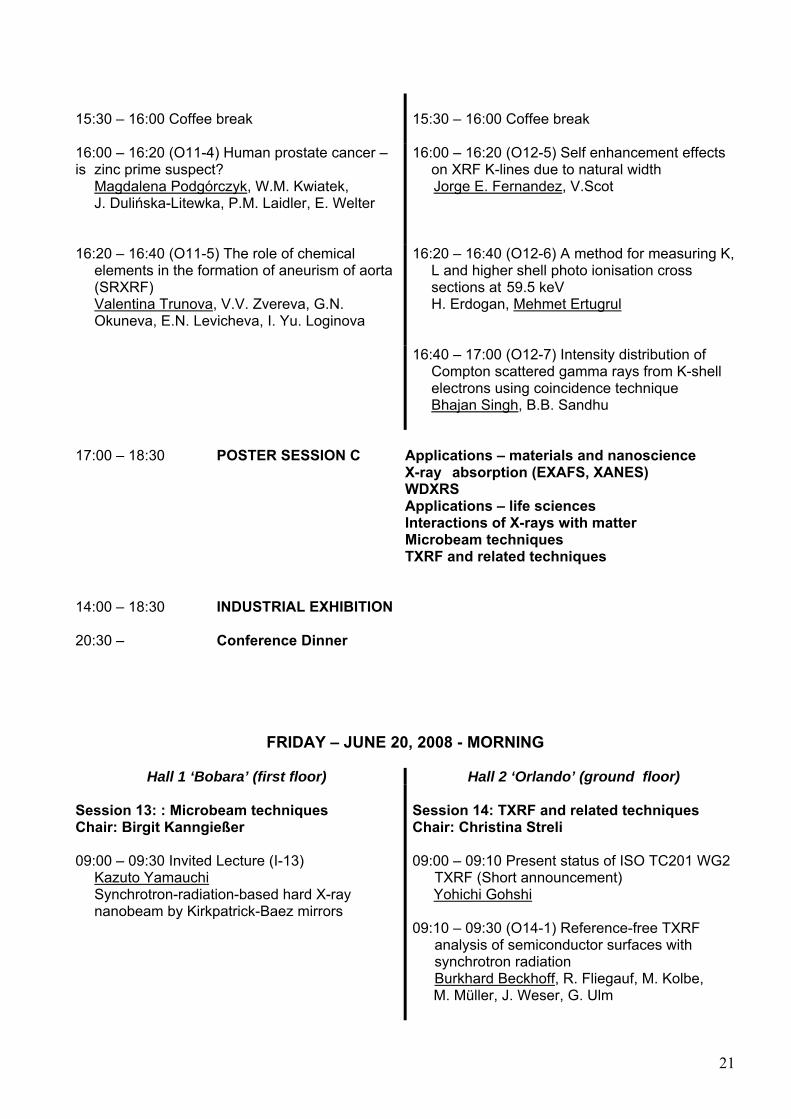
15:30 – 16:00 Coffee break
15:30 – 16:00 Coffee break
16:00 – 16:20 (O11-4) Human prostate cancer – is zinc prime suspect? Magdalena Podgórczyk, W.M. Kwiatek, J. Dulińska-Litewka, P.M. Laidler, E. Welter
16:00 – 16:20 (O12-5) Self enhancement effects on XRF K-lines due to natural width Jorge E. Fernandez, V.Scot
16:20 – 16:40 (O11-5) The role of chemical elements in the formation of aneurism of aorta (SRXRF) Valentina Trunova, V.V. Zvereva, G.N. Okuneva, E.N. Levicheva, I. Yu. Loginova
16:20 – 16:40 (O12-6) A method for measuring K, L and higher shell photo ionisation cross sections at 59.5 keV H. Erdogan, Mehmet Ertugrul
16:40 – 17:00 (O12-7) Intensity distribution of Compton scattered gamma rays from K-shell electrons using coincidence technique Bhajan Singh, B.B. Sandhu
17:00 – 18:30 POSTER SESSION C Applications – materials and nanoscience X-ray absorption (EXAFS, XANES) WDXRS Applications – life sciences Interactions of X-rays with matter Microbeam techniques TXRF and related techniques 14:00 – 18:30 INDUSTRIAL EXHIBITION 20:30 – Conference Dinner
FRIDAY – JUNE 20, 2008 - MORNING
Hall 1 ‘Bobara’ (first floor) Hall 2 ‘Orlando’ (ground floor) Session 13: : Microbeam techniques Chair: Birgit Kanngießer
Session 14: TXRF and related techniques Chair: Christina Streli
09:00 – 09:30 Invited Lecture (I-13) Kazuto Yamauchi Synchrotron-radiation-based hard X-ray nanobeam by Kirkpatrick-Baez mirrors
09:00 – 09:10 Present status of ISO TC201 WG2 TXRF (Short announcement) Yohichi Gohshi 09:10 – 09:30 (O14-1) Reference-free TXRF analysis of semiconductor surfaces with synchrotron radiation Burkhard Beckhoff, R. Fliegauf, M. Kolbe, M. Müller, J. Weser, G. Ulm
21
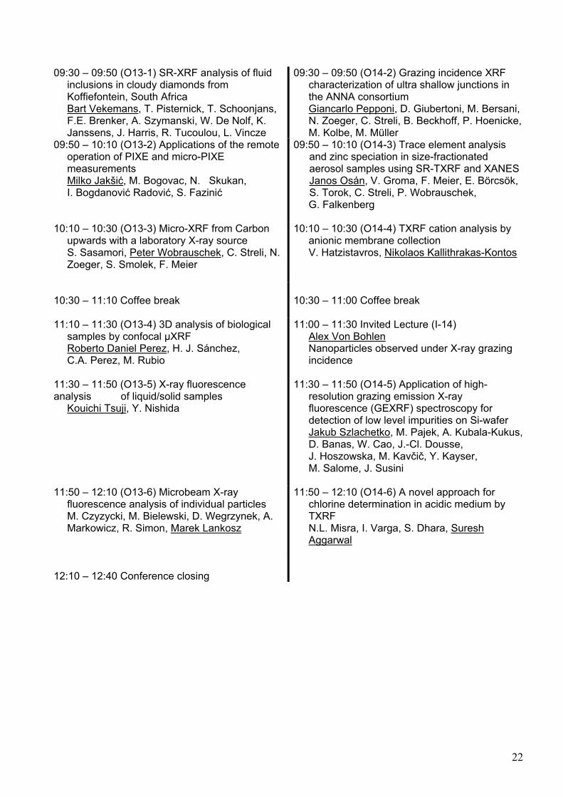
09:30 – 09:50 (O13-1) SR-XRF analysis of fluid inclusions in cloudy diamonds from Koffiefontein, South Africa Bart Vekemans, T. Pisternick, T. Schoonjans, F.E. Brenker, A. Szymanski, W. De Nolf, K. Janssens, J. Harris, R. Tucoulou, L. Vincze
09:30 – 09:50 (O14-2) Grazing incidence XRF characterization of ultra shallow junctions in the ANNA consortium Giancarlo Pepponi, D. Giubertoni, M. Bersani, N. Zoeger, C. Streli, B. Beckhoff, P. Hoenicke, M. Kolbe, M. Müller
09:50 – 10:10 (O13-2) Applications of the remote operation of PIXE and micro-PIXE measurements Milko Jakšić, M. Bogovac, N. Skukan, I. Bogdanović Radović, S. Fazinić
09:50 – 10:10 (O14-3) Trace element analysis and zinc speciation in size-fractionated aerosol samples using SR-TXRF and XANES Janos Osán, V. Groma, F. Meier, E. Börcsök, S. Torok, C. Streli, P. Wobrauschek, G. Falkenberg
10:10 – 10:30 (O13-3) Micro-XRF from Carbon upwards with a laboratory X-ray source S. Sasamori, Peter Wobrauschek, C. Streli, N. Zoeger, S. Smolek, F. Meier
10:10 – 10:30 (O14-4) TXRF cation analysis by anionic membrane collection V. Hatzistavros, Nikolaos Kallithrakas-Kontos
10:30 – 11:10 Coffee break
10:30 – 11:00 Coffee break
11:10 – 11:30 (O13-4) 3D analysis of biological samples by confocal µXRF Roberto Daniel Perez, H. J. Sánchez, C.A. Perez, M. Rubio
11:00 – 11:30 Invited Lecture (I-14) Alex Von Bohlen Nanoparticles observed under X-ray grazing incidence
11:30 – 11:50 (O13-5) X-ray fluorescence analysis of liquid/solid samples Kouichi Tsuji, Y. Nishida
11:30 – 11:50 (O14-5) Application of high- resolution grazing emission X-ray fluorescence (GEXRF) spectroscopy for detection of low level impurities on Si-wafer Jakub Szlachetko, M. Pajek, A. Kubala-Kukus, D. Banas, W. Cao, J.-Cl. Dousse, J. Hoszowska, M. Kavčič, Y. Kayser, M. Salome, J. Susini
11:50 – 12:10 (O13-6) Microbeam X-ray fluorescence analysis of individual particles M. Czyzycki, M. Bielewski, D. Wegrzynek, A. Markowicz, R. Simon, Marek Lankosz
11:50 – 12:10 (O14-6) A novel approach for chlorine determination in acidic medium by TXRF N.L. Misra, I. Varga, S. Dhara, Suresh Aggarwal
12:10 – 12:40 Conference closing
22

POSTER SESSION A – Monday 16 June, 17:00 – 18:30
Applications of XRS in archaeometry Quantification methodology Synchrotron XRS PIXE and electron induced XRS
PA-1 M.I. Báez, J. Ramírez-Castellanos, M.D. Gayo, J.L. Baldonedo, L. Vidal and M.J. García. Analytical characterization of the ground layer in paint section of Rubens’ paintings at “El Prado” National Museum PA-2 Geert Van der Snickt, J. Jaroszewicz, W. De Nolf, K. Janssens, J. Dik, L. Van der Loeff and L.
Klaassen. SR micro-XRD, SR micro-XRF and SR micro-XANES analyses on degraded cadmium sulphide paint samples
PA-3 S. Caglio, G. Poldi. EDXRF coupled with vis-RS to study iron-gall inks: laboratory tests and in situ analysis on Palladio’s drawings (WITHDRAWN)
PA-4 D. Mudronja, S. Fazinić, Ž. Pastuović, M. Jakšić, M. Braun, and M. Pustić. Analysis of pigments on paintings from two churches in Dubrovnik
PA-5 E. Da Silva, M. Robinson, A. Pejović-Milić, C. Evans, D. V. Heyd. Monitoring the process of
gilding, tarnishing and restoration of 21st century daguerreotypes by wavelength-dispersive X-ray fluorescence spectrometry
PA-6 C. Calza, D.F. Oliveira, H.S. Rocha, A. Pedreira, R.T. Lopes. Computed radiography of XIX century Brazilian paintings
PA-7 V. Desnica, K. Skaric, D. Mudronja, M. Bošnjak, S. Fazinić, Z. Pastuović. XRF and PIXE analysis of the wooden polychrome altars from the northern Croatian region
PA-8 D. Šatović, V. Desnica, L. Valek, S. Martinez, S. Fazinić, Ž. Pastuović. Homogeneity study of modern bronzes for artistic castings using PIXE and PLP
PA-9 D. Šatović, L.Valek, V. Desnica, S. Martinez, S. Fazinić, Ž. Pastuović. Contribution of tin compounds to protective properties of patina layers on statuary bronzes
PA-10 V. Andrić, M. Stojanović, S. Čupić, D. Jovanović. Physicochemical investigation of late bronze age artifacts using EDXRF Spectrometry
PA-11 K. Uhlir, M. Griesser, H. Hanzer, P. Wobrauschek, C. Streli, G. Buzanich, D. Wegrzynek, A. Markowicz, E. Chinea-Cano. Analysis of the enamel of Benvenuto Cellini’s “Saliera” using XRF, confocal XRF and SEM/EDX
PA-12 S. Varvara, B. Fabbri, S. Gualtieri, M. Gligor. Chemical and structural features of the neolithic pottery from Alba Iulia-Lumea Noua (Romania) archaeological settlement
PA-13 D. Krstić, M. Jakšić, I. Božičević, D. Mudronja. PIXE characterization of particle modified consolidant applied to quarried limestones
PA-14 W. Tanthanuch, W. Pattanasiriwisawa, W. Somphon, S. Srilomsak, Ban Chiang. Pottery from archaeological site of Thailand: study of firing technique in ancient periods
PA-15 Lin Cheng, Xunliang Ding, Rongwu Li, Guoxia Li. Micro-X-ray fluorescence and 3D confocal Micro-X-ray fluorescence analysis of Chinese ancient cultural heritages
23
PA-16 A. Kriznar, M.V. Muñoz, F. de la Paz, M. A. Respaldiza and M. Vega. XRF Analysis of two terracotta polychromated sculptures by Pietro Torrigiano
PA-17 L. Bonizzoni, A. Galli, M. Milazzo. Possibility of punctual XRF analysis for archaeological pottery classification (WITHDRAWN)
PA-18 V. Virgili, M.L. Vitobello, M.F. Guerra. Wires and granulation in gold jewellery pastiches
PA-19 I. Queralt, A.M. Jurado-Roldán, J.Pujol. The use of EDXRF in gold alloys analysis: Instrumental set-up and evaluation
PA-20 C. Vázquez, D. Elkin, S. Boeykens. TXRF and underwater archaeology: revealing the liquid into the bottle
PA-21 S. Cagno, V. Van der Linden, O. Schalm, K. Janssens, P. Cosyns, K. Nys. SEM-EDX analysis of black-appearing Roman glass
PA-22 I. Mantouvalou, T. Wolff, W. Malzer, M. Pagels, L. Lühl , O. Hahn, B. Kanngießer. Non-destructive, depth resolved investigation of corrosion layers of historical glass objects by 3D Micro X-Ray Fluorescence Analysis
PA-23 N. Kato, Y. Shindo, I. Nakai. Nondestructive on site analyses of Islamic glass in Egypt by using a portable X-ray Fluorescence Spectrometer
PA-24 V. Kantarelou, D. Sokaras, A. G. Karydas. A quantification approach for portable µ-XRF spectrometers
PA-25 T.Wolff, W. Malzer, I. Mantouvalou, O. Hahn, B. Kanngießer. Extended calibration procedure for the determination of Micro-XRF excitation spectra
PA-26 E. Cherkashin, M. Granin, T. Cherkashina. Intelligent computational infrastructure for XRF-based investigations
PA-27 R. Simon, G. Falkenberg, R. Dietsch, U. Fittschen, D. Weissbach. Thin film samples as reference samples for micro XRF
PA-28 D. Sokaras, M. Kolbe, M. Gerlach, A.G Karydas, B. Beckhoff, C. Zarkadas. X-ray fluorescence enhancement induced by Photo-Electrons Secondary Excitation
PA-29 R. M. Rousseau. New XRF software packages for the application of the fundamental algorithm in practice
PA-30 T. Trojek, D. Wegrzynek. Measurements and Monte Carlo calculations of X-ray fluorescence Kα/Kβ ratios for layered specimens
PA-31 K. Streib, T. Alford, J. Mayer. An algorithm to for calculation of atomic density factors used in facilitating calculation of x-ray production cross sections from raw data (WITHDRAWN)
PA-32 G. W. W. Wepukhulu, H. K. Angeyo, O. A. Mustapha, M. Mangala. Simulation of environmental aerosol loaded filters for ED-XRF analysis by partial least squares
PA-33 V. Rößiger, M. Hofmann, B. Nensel. Standardless material identification
PA-34 M.J. Farquharson, A. Al-Ebraheem, R.Leek, A.L.Harris, G. Falkenberg, R.Simon, D.A.Bradley, K.Rickers. Micro distribution of biologically important metals in primary invasive ductal carcinoma of breast
PA-35 I.Lima, M.J.Anjos, J.T.Assis, T.U.Pantaleão, V.M.Correa da Costa, M.L.Farias, E.Sales, R.T.Lopes. Biological study using X-Ray Micro-fluorescence image
24
PA-36 M. Radtke, H. Riesemeier, I. Reiche, M.F. Guerra. SR-XRF analysis of trace amounts of Pt in gold alloys
PA-37 M. Müller, M. Kolbe, B. Beckhoff, K. Feldrapp, S. Storm, A.-D. Weber. Absolute determination of cross sections for resonant Raman scattering on silicon carbide
PA-38 Koteswara Rao Veeranki, M. V. R. Murti, L. Mombasawala, S. S. Raju, B. Seetharami Reddy. A study of low energy Kα X – Ray satellites of Phosphorous and its compounds
PA-39 D. Sokaras, Ch. Zarkadas, R. Fliegauf, B. Beckhoff, A.G. Karydas. A novel multi-purpose PIXE induced XRF chamber
PA-40 T. Papp. On the accuracy of L-shell ionization cross sections II. The choice between experimental or theoretical
PA-41 M. Roumié, B. Nsouli. PIXE protocols for cluster analysis of archeological samples: the funny filter case
PA-42 M. Roumié, B. Nsouli, N. Saliba. PIXE identification of fine and coarse particles of aerosol samples from Beirut
PA-43 T. Tada, H. Fukuda, J. Hasegawa, Y. Oguri. In-situ observation of chemical state change of sulfur in solid targets during MeV-proton irradiation
PA-44 C. Figueroa, N. Nieva, S. P. Heluani. Approximate solution for the fraction of backscattered electrons using invariant imbedding method
PA-45 L.A. Pavlova, R.G. Kravtsova. EPMA determination of modes of gold and silver occurrence in litho chemical stream sediments exemplified by the Ducat gold-silver deposit in northeastern Russia
PA-46 L. Suvorova, N. Nemchinova. Investigation of phase composition of metallurgical silicon by EPMA
PA-47 V.P. Petukhov. X-Ray and electron emission during transporting of slow energy electrons through glass capillary optics
PA-48 R. Alberti, A. Bjeoumikhov, N. Grassi, C. Guazzoni, T. Klatka, A. Longoni, A. Quattrone. X-ray detection system for PIXE measurements based on a SDD coupled to polycapillary optics
PA-49 I. Orlić and M Bogovac. Sim-IBA - Computer software for simulation of PIXE and RBS spectra and elemental mapping
PA-50 C. M. Romo-Kröger. A methodology based on thickness dependent sample irradiation to evaluate the enhancement in K-shell ionization cross sections due to vacancies in the projectile
25

26
POSTER SESSION B – Tuesday 17 June, 17:10 – 18:40
X-ray sources, optics and detectors X-ray imaging and tomography Applications – materials and nanoscience
PB-1 A. Pahlke, T. Eggert, S. Pahlke, R. Stötter, F. Wiest. Large area silicon drift detectors
PB-2 N. Gao, Z. Chen, I. Ponomarev. Enhanced detection sensitivity of Micro X-ray Fluorescence by using advanced X-ray optic chain
PB-3 H. Stosnach. Ultratrace total reflection X-ray fluorescence (TXRF) analysis of platinum group elements applying nickel sulfide melt enrichment
PB-4 M. Procop, V.-D. Hodoroaba, A. Bjeoumikhov, R. Wedell, A. Warrikhoff. Improvements of the low-energy performance of a micro-focus X-ray source for XRF analysis with the SEM
PB-5 Y. Nose, N.Kawada, S.Maeo, T.Utaka, K.Taniguchi. Development of the analyzer for thickness and trace elements in multilayer using multi excitation sources
PB-6 A. Lorusso, V. Nassisi, M.V. Siciliano. Short soft X-Ray sources
PB-7 S. Maeo, M. Krämer, T.Utaka, K. Taniguchi. Development of laboratory µ-XRF spectrometer using multi monochromatic X-Ray sources
PB-8 R. Havlikova, V. Semencova, L. Sveda, L. Pina, A. Inneman, R. Hudec. Usability of industrial grade Si wafers for x-ray optical instruments
PB-9 T. Wolff, I. Mantouvalou, W. Malzer, J. Nissen, D. Berger, B. Kanngießer. Characterization of polycapillary half lenses in two directions
PB-10 S. Bjeoumikhova, A. Bjeoumikhov. X-Ray capillary optics: status and perspective
PB-11 A. Bjeoumikhov, S. Bjeoumikhova. New applications of capillary optics for XRF and XRD
PB-12 L. Luehl, C. Seim, T. Holz, R. Dietsch, B. Kanngießer. Characterisation of the Multilayer optic Astix 100
PB-13 Z.W. Chen, D. Li, S. Kemtekar, W. M. Gibson. Monochromatic X-beam for micro-beam EDXRF
PB-14 V.K. Egorov, E.V. Egorov, M.S. Afanas’ev. Comparative characterization of x-ray beams formed by complicated waveguide-resonance compositions
PB-15 C. Seim, L. Luehl, B. Kanngießer. The Performance of micro focus X-Ray Tubes: The influence of X- Ray Tube- settings
PB-16 A. Glushkov, O. Khetselius, Yu. Dubrovskaya. Discharge of meta-stable nuclei during muon capture and realization of the high power monochromatic γ radiation source
PB-17 O. Khetselius, A. Loboda. Search of the optimal plasma parameters for X-ray lasing on the basis of modeling the elementary processes in a collisionally pumped plasma
PB-18 A. Glushkov, O. Khetselius, E. Gurnitskaya, A. Loboda. Generation of ultra-short X-ray pulses in cluster system during ionization by femto-second optical pulse
27
PB-19 A. Tartari, G. Di Domenico, C. Bonifazzi, C. Baraldi. New measurements and observations on aged NaI detectors
PB-20 A.G. Touriyanski, R.A. Khmelnitski, M.A. Negodaev, T. Tschentscher. X-Ray prism spectrometer for ultra-fast single pulse spectral measurements
PB-21 W. De Nolf, J. Jaroszewicz, R. Terzano, O.C. Lind, B. Salbu, G. Falkenberg, K. Janssens. 2θ-resolution in synchrotron micro-XRPD: instrumental comparison at HASYLAB beam line L
PB-22 R. Alberti, L. Bombelli, C. Fiorini, T. Frizzi, A. Gola, C. Guazzoni, T. Klatka, A. Longoni, G. Membretti. High performance read-out electronics for silicon drift detectors
PB-23 G. Georgiev, I.Peev. The advantages of digital adaptive filtering in fast pulse processing
PB-24 F. Scholze, M. Procop. Modelling the response function of an EDS with a silicon detector
PB-25 S. Pahlke, C. Dietzinger, T. Eggert, A. Pahlke, R. Stötter, F. Wiest. Modular analytical X-Ray acquisition system (AXAS-M)
PB-26 I. Gomez-Morilla, H. Rahn, A. Waske, J. Herzen, S. Weise, S. Odenbach. Fast tomography for the study of time-dependent processes
PB-27 I. Gomez-Morilla, M.D. Ynsa, J.C. Millán, L.C. Alves, H. Rahn, S. Odenbach, T. Pinheiro. Tomography analysis of femoral bones from rats under osteoporosis preventive treatments
PB-28 W.J. Przybylowicz, T. Tyliszczak, A. Barnabas, J. Mesjasz-Przybylowicz. Ni mapping in Berkheya coddii by Micro-PIXE and NEXAFS
PB-29 M. Haschke, D. Grötzsch, A. Günther, I. Reiche, I. Mantouvalou, K. Lange, B. Kanngießer. A compact 3D Micro-XRF laboratory spectrometer for archeometric applications
PB-30 A. Dyszkiewicz, P. Połeć, Jakub Zajdel, D. Chachulski, B. Pawlus, Jan Szczegielniak. Collimation of non-axial x-rays spectrum by means of steering ferrofluid - way of reducing geometrical faults in x-ray-pictures
PB-31 A. Tartari, M. De Felici, M. Gambaccini, P. Querzoli, C. Bonifazzi, R Felici. X ray scattering tissue characterization in micro-beams based techniques
PB-32 T. Martinez, J. Lartigue, G. Zarazua, L. Carapio, P. Avila-Perez, S. Tejeda. Metals in aerosol particulate matter from the Metropolitan Zone of the Valley of Mexico
PB-33 I. J. Kwame Aboh, D. Henriksson, J. Laursen, M. Lundin, F. G. Ofosu, N. Pind, E. S. Lindgren, T. Wahnström. Identification of aerosol particle sources in semi-rural area of Kwabenya, near Accra, Ghana
PB-34 Y. Makarovska, A. Krata, R. Van Grieken, K. Van Meel, J. Vercauteren, E. Roekens. Determination of elements in PM10 by EDXRF in the air in 2006-2007 over Flanders, Belgium
PB-35 L. Samek, M. Lankosz. Study of the elemental concentration of air particulate matter by EDXRF method collected in Poland
PB-36 J. Boman, J. Lindén, S. Thorsson, B. Holmer, I. Eliasson. Analysis of urban and sub-urban fine particles (PM2.5) collected in Ouagadougou, Burkina Faso
PB-37 J. Boman, M. L. de Carvalho, M. B. Alizadeh, P. Rezaievar, A. Wagner. Elemental content of aerosol particles in an underground tram station
28
PB-38 M. Furger, A. Richard, N. Bukowiecki, P. Lienemann, D. Grolimund, A.S.H. Prévôt, U. Baltensperger. Trace elements in hourly ambient aerosol samples determined with synchrotron XRF
PB-39 S. Bamford, M. Jakšić, S. Fazinić, D. Wegrzynek, E. Chinea-Cano, K. Šega, I. Bešlić, A. Markowicz, P. Wobrauschek. PIXE and XRF analysis of Suspended particulates in an urban residential area of Zagreb, Croatia
PB-40 S. Bamford, K. Šega, I. Bešlić, R. Godec, P. Wobrauschek. Seasonal variations in particulate mass concentrations and elemental composition of urban aerosols in Zagreb, Croatia, using ED(P)XRF Analysis
PB-41 R. Moschochoritou, N. Kallithrakas-Kontos. Analysis of methyl bromide ozone depleting substance
PB-42 B. B. Naziriwo, S. O. Wandiga, M. J. G. Gatari, O. V. Madadi, P. J. Ssebuwufu. Determination of trace metal concentrations in waters of Nakivubo channel and Lake Victoria using Energy Dispersive X-ray Fluorescence Analysis
PB-43 O. Gonzalez-Fernandez, I. Queralt. Analysis of major and trace elements content of mining waters by means of wavelength dispersive X-ray fluorescence of dried residues
PB-44 N. Alov, R. Bulgachev, S. Dmitrienko. XRF Determination of metals in water using polyurethane foams
PB-45 D. Zaichick, V. Zaichick, S. Lyapunov, E. Schevchenko. WDXRS of minor and trace element contents in the soil of Moscow's park
PB-46 N. Civici, A. Tashko. Application of a Portable EDXRF Spectrometer for in-situ measurement of pollutants in overbank sediments of Mati river (northern Albania)
PB-47 E.V. Khudonogova, T.Yu. Cherkashina. Determination of Mo, Nb, Zr, Y, Sr, Rb, U, Th, Pb in different rocks by X-ray fluorescence analysis
PB-48 E. Marguí, K.Van Meel, R.Van Grieken, C.Fontàs, M.Hidalgo, I.Queralt. Application of high-energy polarised-beam EDXRF for the study of metal burden in mining waste samples
PB-49 B. Kovacevik, A. Wagner, J. Boman, J. Laursen and J. B. C. Pettersson. The seasonal variation of the elemental composition of particulate matter in Skopje, FYR of Macedonia
PB-50 M.J. G. Gatari and J. Boman. Design and development of an energy-dispersive X-ray spectrometer: A tool for environmental research in Kenya
29

30
POSTER SESSION C – Thursday 19 June, 17:00 – 18:30
Applications – materials and nanoscience X-ray absorption (EXAFS, XANES) WDXRS Applications – life sciences Interactions of X-rays with matter Microbeam techniques TXRF and related techniques
PC-1 I. N. Orbulov, Á. Németh, J. Dobránszky. XRD and EDS investigations of metal matrix composites and syntactic foams
PC-2 O. Adiguzel. X-Ray diffraction studies on crystallography of Martensite in two Copper based shape memory alloys
PC-3 G. Puchkovska, T. Bezrodna, A. Tolochko, E. Kotelnikova, N. Platonova. X-ray study of composite materials based on montmorillonite organoclay
PC-4 P. Hönicke, B. Beckhoff, M. Kolbe, T. Conard, S. List, H. Struyff. Depth dependant nanolayer analysis of vertical sidewalls on structured wafers
PC-5 J. Petrů, M. Hubičková, P.Sajdl. Corrosion layers on steel and their XPS analysis
PC-6 A. Styervoyedov, V. Farenik. XPS investigations of nanoscale Ti-O-N films on silicon
PC-7 M. Hubičková, P. Kučera, J. Petrů, P. Sajdl. Analysis of XPS spectra of metal oxides on corrosion surface
PC-8 U. Burkhardt, I. Veremchuk, R. Gumeniuk, A. Leithe-Jasper, Yu. Prots, W.Schnelle,Yu. Grin. Eu L3 XANES investigations of the valence instability of Eu4Pd29B8
PC-9 A. Alsecz, J. Osán, I. Sajó, S. Török, G. Falkenberg, K. Rickers. Chemical speciation of unknown uranium ore and mine tailings particles with µ-XANES and µ-XRD methods
PC-10 W. Pattanasiriwisawa, J. Siritapetawee, O. Patarapaiboolchai, W. Klysubun. Structural characterization of Sulfur vulcanized natural rubber using X-ray Absorption Near-Edge Spectroscopy
PC-11 S .B. Erenburg, N. V. Bausk, S. V. Trubina, B. L. Moroz, A. V. Kalinkin, V. I. Bukhtiyarov, S. G. Nikitenko. Structure peculiarities of catalytically active gold nanoparticles by EXAFS and XANES spectroscopy
PC-12 M.A.Kareem, Phanisree K.D. TImmaraju, N.V.Sitamahalakshmi, K.PremaChand. EXAFS effects around K-edge in Lanthanum
PC-13 B. Beckhoff, O. Hahn, M. Kolbe, J. Weser, M. Wilke. Environmental analyses by TXRF - NEXAFS and IR-Spectroscopy: Speciation of Bromine in organics and characterization of the organic matrix
PC-14 A. Cremonesi, G. Calestani, G. Antonioli, D. Bersani, P. P. Lottici. XAS on surfactant-based nanostructured V2O5 thin films: monitoring short and long range order from mesophase residual to crystalline network
31
PC-15 V.M. Chubarov, A.L. Finkelshtein. X-ray fluorescence determination of FeO/Fe2O3 total ratio in rocks
PC-16 T. Tadić, M. Jakšić, I. Božičević. X-ray tracing study of crystal spectrometers for WDXRS application
PC-17 L. Mandić, S. Fazinić, M. Jakšić, J. Dobrinić. Spectrum modeling and influence on chemical state analysis using WDS-PIXE system
PC-18 V. Starý, R. Havel, J. Kadlec, K. Jurek, V. Peřina, J. Fencl. DLC layers on biocompatible Ti alloys
PC-19 K. Watanabe, H. Homma, Y. Kataoka, Y. Yamada, K. Kansai, H. Kohno. Cement analysis for ASTM C114 by using bench-top WDXRF
PC-20 V. Zaichick, T. Sviridova, S. Zaichick. XRF application in the early prostate cancer diagnosis
PC-21 M. Simon, H. Seznec, P. Barberet, D. Bacqueville, A. Mavon, P. Moretto. The skin barrier function: a micro-PIXE study
PC-22 E. Da Silva, C. Heirwegh, A. Pejović-Milić, D. V. Heyd. Use of hydroxyapatite bone composites for the calibration of in vivo EDXRF-based systems for bone strontium quantification
PC-23 N. Uzunov, C. Furlan, S. Galassini, G.B. Girardello, P. Passi, G. Moschini, A. Zadro. Micro-PIXE and SEM study of endosseous implants surface and the implant material release in the surrounding bone tissue
PC-24 E.A. Preoteasa, E. Preoteasa, D.D. Marin, D. Gurban, A. Scafes. PIXE and PIGE assessment of in vivo elemental and physical changes of a composite from a dental filling
PC-25 E.A. Preoteasa, D.D. Marin, D. Gurban, A. Scafes, M. Gugiu, E. Preoteasa. Single detector PIGE and PIXE analysis of dental composites
PC-26 A. Brunetti, G. Piga, A. Santos Cubedo, A. Malgosa, S. Enzo. A study by XRF and XRD of fossilized dinosaur bones from Spain
PC-27 D. Guimarães, J. P. Santos, M. L. Carvalho, V. Geraldes, L. Silva-Carvalho, I. Rocha. Elemental concentration in lead contaminated rat kidneys by EDXRF analysis
PC-28 M. Pagels, W. Malzer, G. Weseloh, L. Lühl, B. Kanngießer. 3D Micro-XRF under cryogenic conditions: A pilot experiment for elemental imaging in biological specimens
PC-29 S.O. Olabanji, G.A. Osinkolua, D.A. Pelemoa, A.A. Abionaa, A.T. Oladeleb. EDXRF Analysis of Thaumatococcus Danielli in Osun State of Nigeria
PC-30 Jun Kawai, Ryosuke Shioi, Nobuharu Sasaki, Kenji Okada, Goro Kinugawa, Shinsuke Kunimura, Takashi Yamamoto. Why Lα:Lβ=1:1 in spite that the quantum mechanical intensity ratio is 2:1?
PC-31 Y. Ménesguen, M.-C. Lépy. New approach for measuring fluorescence yields using tunable monochromatic radiation
PC-32 P. Jonnard, K. Le Guen, J.-M. André. High-resolution X-ray analysis with multilayer gratings
PC-33 B.R. Kerur, Sharanabasappa, S. B. Kaginelli, S. Anilkumar. Post edge structure effects on mass attenuation coefficients
PC-34 J. Lartigue, A. Ramírez. Some cases of mineralogical effect due to the presence of crystals different from that one containing the fluorescent element
32
PC-35 O. Khetselius. On population dynamics of the atomic ensembles in a X-ray laser pulse: Optical bi-stability effect
PC-36 S. Pessanha, M.L. Carvalho, A. Guilherme. Study of the performance of two x-ray spectrometers in different matrix samples
PC-37 J. Bielecki, S. Bożek, J. Lekki, Z. Stachura, W.M. Kwiatek. Construction and applications of the Cracow X-ray microprobe
PC-38 S. Smolek, C. Streli, N. Zoeger, P. Wobrauschek, F. Meirer. Micro X-ray Fluorescence Spectrometer with low power tube for light element analysis
PC-39 S. Kunimura, J. Kawai. Portable TXRF spectrometer with a 40 kV X-ray tube
PC-40 N. Zoeger, D. Ingerle, C. Streli, F. Meirer, P. Wobrauschek, G. Pepponi. Tabletop spectrometer for grazing incidence XRF
PC-41 F. Meirer, G. Pepponi, C. Streli, P. Wobrauschek, N. Zoeger. A new grazing-exit-XRF setup at HASYLAB beam-line L
PC-42 C. Horntrich, F. Meirer, C. Streli, P. Kregsamer, G. Pepponi, N. Zoeger, P. Wobrauschek. Influence of the sample morphology on Total Reflection X-Ray Fluorescence Analysis
PC-43 K. Tsuji, S. Kawamata. Micro TXRF analyses of multiple residues on a flat substrate
PC-44 K. Tsuji, K. Nakano, K. Okubo, Y. Nishida. X-ray Fluorecenece analysis using X-ray transparent thin films for sample support
PC-45 C.L. Mota, N.G.V. Pinto, C.J.G. Pinheiro, D. Braz, R.C. Barroso, A.S. Melo Junior, S. Moreira. Study of alteration of elemental contents in human whole blood and Hemocomponents following irradiation injury by SR-TXRF
PC-46 J. Osán, S. Török, B. Beckhoff, F. Reinhardt. TXRF-NEXAFS study of carbon and nitrogen compounds in fine particulate matter deposited on silicon wafers
PC-47 L. A. Marins, R.F. Barbosa, E. F. O. de Jesus, M. J. Anjos, M. G. T. do Carmo, M. S. Rocha, R. T. Lopes. Effect of ethanol intake during lactation on brain mineral status of pup and young rats using X-ray total reflection spectrometry
PC-48 Y. Kayser, J-Cl. Dousse, J. Hoszowska, W. Cao, J.-L. Schenker, M. Pajek, A. Kubala-Kukus, P. Jagodzinski, M. Kavčič, J. Szlachetko. Depth profiling of Al-implanted Si by means of synchrotron radiation based high resolution grazing emission x-ray fluorescence
PC-49 M. J. G. Gatari, D. M. Maina, S. Bartilol, S. M. Gaita. Determination of trace metals in bottled commercial drinking water using TXRF in Nairobi, Kenya
PC-50 N. Alov, A. Volkov, A. Sokolov, E. Ishmetyev, A. Usherov. On-line X-Ray fluorescence analysis of iron-ore mixtures on a conveyor belt
PC-51 B. Nensel, V. Rößiger, M. Hofmann, S. Dill. The Application of Standardless Material Identification for the Analysis of precious Metal Alloys and RoHS Samples
PC-52 E. Blokhina, X. Dreißigacker, W. Klöck. Quality improvement of FP quantification for thickness and composition of alloy layers by XRF method
PC-53 C. Simons, C. Mans, S. Hanning, A. Janßen, D. Alber, M. Radtke, U. Reinholz, M. Kreyenschmidt. Development and characterization of new polymeric reference materials with the aid of XRF, LA-ICP-MS and Sy-XRF
33
PC-54 N. Vér, L. Matus, M. Kunstár, J. Osán, Z. Hózer. Delay of ruthenium escape in the presence of some fission product elements
PC-55 G. Wellenreuther, U.E.A. Fittschen, M. Achard, M. Salomone Stagni, A. Faust, X. Kreplin and W. Meyer-Klaucke. Total reflection X-ray fluorescence (TXRF): A future routine analysis tool in structural biology?
34