Tunicate: Northward spread, diversity, source, and impact of non
Upload
yohei-takahashiCategory
view
213download
0
Bioorganic & Medicinal Chemistry Letters 20 (2010) 4100–4103
Contents lists available at ScienceDirect
Bioorganic & Medicinal Chemistry Letters
journal homepage: www.elsevier .com/ locate/bmcl
Eudistomidin G, a new b-carboline alkaloid from the Okinawan marine tunicateEudistoma glaucus and structure revision of eudistomidin B
Yohei Takahashi, Haruaki Ishiyama, Takaaki Kubota, Jun’ichi Kobayashi *
Graduate School of Pharmaceutical Sciences, Hokkaido University, Sapporo 060-0812, Japan
a r t i c l e i n f o a b s t r a c t
Article history:Received 19 April 2010Revised 14 May 2010Accepted 18 May 2010Available online 10 June 2010
Keywords:b-Carboline alkaloidsTunicateEudistoma glaucusEudistomidins B and G
0960-894X/$ - see front matter � 2010 Elsevier Ltd.doi:10.1016/j.bmcl.2010.05.071
* Corresponding author. Tel.: +81 11 706 3239; faxE-mail address: [email protected] (J. Ko
A new b-carboline alkaloid, eudistomidin G (1), has been isolated from the Okinawan marine tunicateEudistoma glaucus, and the structure was elucidated from spectroscopic data. Furthermore, the structureof eudistomidin B (2), which has been isolated from the same tunicate, was revised from 2a to 2b bydetailed analyses of spectroscopic data. Asymmetric synthesis of the revised structure (2b) of eudistom-idin B (2) and its (1S,10S)-diastereomer (2c) has been accomplished with the Noyori catalytic asymmetrichydrogen-transfer reaction. The absolute configuration of eudistomidin B (2) was confirmed to be 2b pos-sessing (1R,10S)-configuration, from comparison of the 1H NMR data, CD spectra, [a]D values, and HPLCanalysis of 2b, 2c, and natural eudistomidin B.
� 2010 Elsevier Ltd. All rights reserved.
Marine tunicates are well known to be a rich source of second-ary metabolites with interesting structures and various biologicalactivities.1 Among them, many b-carboline alkaloids have been iso-lated from tunicates of the genus Eudistoma so far.2 In our searchfor new metabolites from marine tunicates, a new b-carbolinealkaloid, eudistomidin G (1), has been isolated from the Okinawantunicate Eudistoma glaucus together with a known related b-carbo-line alkaloid, eudistomidin B (2). In this Letter, we describe the iso-lation and structure elucidation of eudistomidin G (1) andstructure revision of eudistomidin B (2) from 2a to 2b as well as to-tal synthesis of 2b.
NMeNH
Br
H2N
Me
NMeNH
Br
MeHN
H H
S
S
R
S
proposed structure (2a) ofeudistomidin B (2)
revised structure (2b) ofeudistomidin B (2)
NMeNH
MeHN
H
R
S
eudistomidin G (1)
Br
1
6
1018
15
7
1
10
15
6
1
10
15
54
4a4b
99a 19
12
8 8a
According to the previous purification procedure,3 fractions
containing eudistomidins were purified by repeated reversed-phase HPLC to afford eudistomidins G (1) and B (2).All rights reserved.
: +81 11 706 4989.bayashi).
Eudistomidin G (1)4 was obtained as an optically active amor-phous solid. The ESIMS spectrum of eudistomidin G (1) showedthe pseudomolecular ion peaks at m/z 398 and 400 (1:1), indicatingthe presence of a bromine atom, and the molecular formula,C21H24N3
79Br, was established by HRESIMS data (m/z 398.1235[M+H]+, D +0.9 mmu). UV absorptions (kmax 296, 286, and 231 nm)of 1 implied the presence of tetrahydro-b-carboline ring, while theexistence of NH functionality was suggested by the IR absorptions(3211 and 3049 cm�1). The 1H NMR (Table 1) spectrum of 1 includedsignals due to one NH (dY 11.12), eight aromatic protons (dY 7.73–6.59), two sp3 methines (dY 5.14 and 4.13), six sp3 methylenes (dY3.28–2.42), and two N-methyls (dY 2.92 and 2.87), while the 13CNMR (Table 1) spectrum of 1 showed signals due to six sp2 quater-nary carbons, eight sp2 methines, two sp3 methines, three sp3 meth-ylenes, and two sp3 methyls.
Connectivities of C-1 to C-10, C-3 to C-4, C-5 to C-6, C-10 toC-11, and C-13 to C-17 were deduced from the 1H–1H COSY, TOCSY,and HMQC spectra of 1 (Fig. 1). HMBC correlations for H-5 to C-7and C-8a, H-6 to C-4b, H-8 to C-7, NH-9 to C-4b, and H-1 toC-9a, and NOESY correlations for H-1/NH-9, H-4/H-5, and H-8/NH-9 revealed the presence of a 1,2,7-trisubstituted tetrahydro-b-carboline ring. 13C chemical shifts of C-6–C-8 (dC 123.88,117.67, and 115.40, respectively) suggested that a bromine atomwas attached to C-7, while the connection of N-2 and C-19 was dis-closed by 1H and 13C chemical shifts for CH3-19 (dH 2.92; dC 39.79)and HMBC correlations for H-19 to C-1 and C-3. The HMBC cross-peak of H-18 to C-10 and 1H and 13C chemical shifts for CH-10(dH 4.13; dC 63.80) and CH3-18 (dH 2.87; dC 32.45) indicated thatCH3-18 was attached to C-10 via 10-NH. H-11 showed HMBC cor-relations for C-12 and C-13 (C-17), suggesting the connectivity of

Table 11H and 13C NMR data for eudistomidin G (1) in CDCl3
Position dC dH
1 63.83c CH 5.14 (d, 3.2)3 45.65 CH2 3.28 (dd, 13.0, 6.0)
2.95e (m)4 15.01 CH2 2.87e (m)
2.42 (dd, 16.3, 5.2)4a 105.98 C4b 123.82d C5 119.53 CH 7.23 (d, 8.5)6 123.88d CH 7.27 (dd, 8.5, 1.6)7 117.67 C8 115.40 CH 7.73 (d, 1.6)8a 137.53 C9 11.12 (s)9a 121.62 C10 63.80c CH 4.13 (m)11 33.06 CH2 3.24 (dd, 15.0, 4.8)
3.09 (dd, 15.0, 9.7)12 134.12 C13, 17 128.46 CH 6.59a (d, 7.5)14, 16 128.86 CH 7.04a (dd, 7.5, 7.5)15 127.77 CH 7.12 (dd, 7.5, 7.5)18 32.45 CH3 2.87b (s)19 39.79 CH3 2.92b (s)
a 2H.b 3H.
c,d Interchangeable.e J-values were not determined since overlapping with other signals.
NMeNH
MeHN
7
10
18
19
15
13 1H-1H COSY and TOCSYHMBCNOESY
Br
1
34
1
Figure 1. Selected 2D NMR correlations for eudistomidin G (1).
Table 21H and 13C NMR data for the revised structure (2b) of eudistomidin B (2) in CDCl3
Position dC dH
1 63.63 CH 5.07 (d, 2.9)3 45.85 CH2 3.30 (m)
2.94c (m)4 14.92 CH2 2.86c (m)
2.43 (br d, 10.5)4a 105.59 C4b 126.61 C5 121.06 CH 7.54 (s)6 113.71 C7 127.06 CH 7.41 (d, 8.5)8 113.82 CH 7.43 (d, 8.5)8a 135.53 C9 11.19 (s)9a 122.09 C10 63.86 CH 4.01 (m)11 33.37 CH2 3.22 (m)
3.07 (dd, 15.0, 9.4)12 133.82 C13, 17 128.46 CH 6.61a (d, 7.5)14, 16 128.93 CH 7.07a (dd, 7.5, 7.5)15 127.93 CH 7.15 (dd, 7.5, 7.5)18 32.54 CH3 2.85b (s)19 39.81 CH3 2.90b (s)
a 2H.b 3H.c Interchangeable.
NMeNH
Br
MeHN
6
10
18
19
15
131H-1H COSY and TOCSYHMBCNOESY
34
2b
Figure 2. Selected 2D NMR correlations for the revised structure (2b) of eudis-tomidin B (2).
Y. Takahashi et al. / Bioorg. Med. Chem. Lett. 20 (2010) 4100–4103 4101
C-11 and C-12. Thus, the gross structure of eudistomidin G waselucidated to be 1.
The relative stereochemistry of 1 was not able to be elucidatedfrom the 3JH-1/H-10 value (3.2 Hz) and NOESY correlations of H-1/H-10, H-10/H-19, and NH-9/H-11b. The absolute configuration at C-1in 1 was deduced as R from a positive Cotton effect at 228 nm (De+5.6) in the CD spectrum of 1.5
The structure of eudistomidin B (2) was reinvestigated, sincethe total synthesis of the proposed structure (2a) of eudistomidinB (2) revealed that the NMR data of the synthetic compound wereinconsistent with those of natural eudistomidin B (2).7 The molec-ular fomula of eudistomidin B (2)8 was shown to be C21H24N3
79Br(m/z 398.1235 [M+H]+, D +0.9 mmu), and UV and IR absorptions of2 were the same as those reported previously.3 The 1H NMR (Ta-ble 2) spectrum of 2 measured in CDCl3 showed signals due toone NH (dY 11.19), eight aromatic protons (dY 7.54–6.61), two sp3
methines (dY 5.07 and 4.01), six sp3 methylenes (dY 3.30–2.43),and two methyls (dY 2.90 and 2.85). The 13C NMR (Table 2) spec-trum of 2 included 19 signals derived from 21 carbons due to sixsp2 quaternary carbons, eight sp2 methines, two sp3 methines,three sp3 methylenes, and two sp3 methyls. Among them, twomethyls (dC 39.81, dY 2.90, and dC 32.54, dY 2.85) were ascribedto those bearing a nitrogen.
The 1H–1H COSY, TOCSY, and HMQC spectra of 2 revealed con-nectivities of C-1 to C-10, C-3 to C-4, C-7 to C-8, C-10 to C-11, and
C-13 to C-17. HMBC and NOESY correlations indicated the presenceof a 6-brominated 1,2,6-trisubstituted tetrahydro-b-carboline ring.The connectivity of C-11 and C-12 was suggested by HMBC corre-lations for H-11 to C-12 and C-13 (C-17). HMBC correlations forH3-19 to C-1 and C-3 disclosed that one methyl group (C-19) wasconnected to N-2, while the HMBC correlation for H3-18 to C-10 re-vealed that another methyl group (C-18) was not attached to C-15but attached to N-10. (Fig. 2). These data suggested that the grossstructure of eudistomidin B (2) was 2b but not 2a.9
The relative stereochemistry of revised structure (2b) of eudis-tomidin B (2) was not able to be assigned from the 3JH-1/H-10 value(2.9 Hz), and NOESY correlations for H-10/H-19, H-1/H-10, andNH-9/H-11b. The absolute configuration at C-1 in eudistomidin B(2) was deduced to be R from a positive Cotton effect at 231 nm(De +2.4) in the CD spectrum of 2.5,6
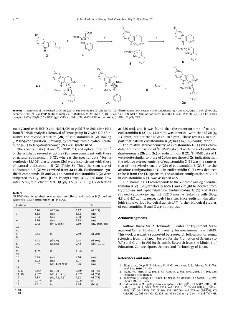
To elucidate the relative and absolute configurations of eudis-tomidin B, the revised structure (2b) of eudistomidin B (2) andits (1S,10S)-diastereomer (2c) were prepared in optical active formemploying the Noyori catalytic asymmetric hydrogen-transferreaction10 (Scheme 1). Treatment of 5-bromotryptamine 37 withcarboxylic acid 411 in the presence of EDC and HOBt providedamide 5. The Bischler–Napieralski reaction12 in benzene affordeddihydro-b-carboline (6). Noyori asymmetric hydrogen-transferreaction of 6 in DMF afforded tetrahydro-b-carboline, which was
CO2H
NMe
Fmoc
4
+
3
a
NH
NFmocMe
5
bN
NFmocMe
6
c, dNMeH
MeHN
2b
S
R1
10
O
6f, g, h
NMeH
MeHN
2c
S
S1
10NMeH
N S
R1
10e
7
MeFmoc
S
SSN
HNH2
NH
Br
Br
NH
Br
NH
Br
NH
Br
NH
Br
Scheme 1. Synthesis of the revised structure (2b) of eudistomidin B (2) and its (1S,10S)-diastereomer (2c). Reagents and conditions: (a) HOBt, EDC, CH2Cl2, 94%; (b) POCl3,benzene, 12%; (c) (S,S)-TsDPEN–Ru(II) complex, HCO2H/Et3N (5:2), DMF; (d) HCHO aq, NaBH3CN, MeCN, 89% for two steps; (e) DBU, CH2Cl2, 85%; (f) (R,R)-TsDPEN–Ru(II)complex, HCO2H/Et3N (5:2), DMF; (g) HCHO aq, NaBH3CN, MeCN, 92% for two steps; (h) DBU, CH2Cl2, 78%.
4102 Y. Takahashi et al. / Bioorg. Med. Chem. Lett. 20 (2010) 4100–4103
methylated with HCHO and NaBH3CN to yield 7 in 89% (dr >10:1from 1H NMR analysis). Removal of Fmoc group in 7 with DBU fur-nished the revised structure (2b) of eudistomidin B (2), having(1R,10S)-configuration. Similarly, by starting from dihydro-b-carb-oline (6), (1S,10S)-diastereomer (2c) was synthesized.
The spectral data (1H and 13C NMR, CD, and optical rotation)13
of the synthetic revised structure (2b) were coincident with thoseof natural eudistomidin B (2), whereas the spectral data14 for itssynthetic (1S,10S)-diastereomer (2c) were inconsistent with thoseof natural eudistomidin B (2) (Table 3). Thus, the structure ofeudistomidin B (2) was revised from 2a to 2b. Furthermore, syn-thetic compounds 2b and 2c, and natural eudistomidin B (2) weresubjected to C18 HPLC [Luna Phenyl-Hexyl, 4.6 � 250 mm; flowrate 0.5 mL/min: eluent; MeOH/H2O/TFA (80:20:0.1); UV detection
Table 31H NMR data for synthetic revised structure (2b) of eudistomidin B (2) and itssynthetic (1S,10S)-diastereomer (2c) in CDCl3
Position 2b 2c
1 5.10 (d, 2.0) 5.27 (d, 2.3)3 3.32 (m) 3.52 (m)
2.94 (m) 2.98 (m)4 2.86 (m) 2.88 (m)
2.42 (br d, 10.6) 2.30 (dd, 16.9, 4.9)4a4b5 7.53 (s) 7.49 (d, 2.0)67 7.43 (d, 8.6) 7.48 (d, 9.0)8 7.45 (d, 8.6) 7.42 (dd, 9.0, 2.0)8a9 11.04 (s) 11.27 (s)9a10 3.99 (m) 4.10 (m)11 3.22 (m) 3.27 (m)
3.07 (dd, 14.9, 9.3) 3.26 (m)1213, 17 6.56a (d, 7.3) 6.50a (d, 7.5)14, 16 7.07a (dd, 7.3, 7.3) 7.02a (d, 7.5)15 7.15 (dd, 7.3, 7.3) 7.12 (d, 7.5)18 2.87b (s) 2.92b (s)19 2.91b (s) 2.94b (br s)
a 2H.b 3H.
at 280 nm], and it was found that the retention time of naturaleudistomidin B (2) (tR 13.0 min) was identical with that of 2b (tR
13.0 min) but not that of 2c (tR 10.8 min). These results also sup-port that natural eudistomidin B (2) has (1R,10S)-configuration.
The relative stereochemistry of eudistomidin G (1) was eluci-dated from comparison of 1H NMR data of 1 with those of syntheticdiastereomers (2b and 2c) of eudistomidin B (2). 1H NMR data of 1were quite similar to those of 2b but not those of 2c, indicating thatthe relative stereochemistry of eudistomidin G (1) was the same asthat of the revised structure (2b) of eudistomidin B (2). Since theabsolute configuration at C-1 in eudistomidin G (1) was deducedto be R from the CD spectrum, the absolute configuration at C-10in eudistomidin G (1) was assigned as S.
Eudistomidin G (1) corresponds to the 7-bromo analog of eudis-tomidin B (2). Biosynthetically both 1 and 2 might be derived fromtryptophan and L-phenylalanine. Eudistomidins G (1) and B (2)showed cytotoxicity against L1210 murine leukemia cells (IC50,4.8 and 4.7 lg/mL, respectively) in vitro. Since eudistomidin alka-loids show various biological activity,3,15 further biological studiesof eudistomidins B and G are in progress.
Acknowledgments
Authors thank Ms. A. Tokumitsu, Center for Equipment Man-agement Center, Hokkaido University, for measurements of ESIMS.This work was partly supported by a research fellowship for youngscientists from the Japan Society for the Promotion of Science (toY.T.) and Grant-in-Aid for Scientific Research from the Ministry ofEducation, Culture, Sports, Science and Technology of Japan.
References and notes
1. Blunt, J. W.; Copp, B. R.; Munro, M. H. G.; Northcote, P. T.; Prinsep, M. R. Nat.Prod. Rep. 2010, 27, 165.
2. Wang, W.; Nam, S.-J.; Lee, B.-C.; Kang, H. J. Nat. Prod. 2008, 71, 163. andreferences cited therein.
3. Kobayashi, J.; Cheng, J.-F.; Ohta, T.; Nozoe, S.; Ohizumi, Y.; Sasaki, T. J. Org.Chem. 1990, 55, 3666.
4. Eudistomidin G (1): pale yellow amorphous solid; ½a�21D +6.4 (c 0.2, CHCl3); IR
(film) mmax 3211, 3049, 2922, 2851, and 1458 cm�1; UV (MeOH) kmax 366 (e200), 296 (sh 1870), 286 (2190), 231 (10,200), and 203 nm (10,400); CD(MeOH) kext 293 (De +0.11), 228 nm (+5.6), 213 nm (�2.5); 1H and 13C NMR

Y. Takahashi et al. / Bioorg. Med. Chem. Lett. 20 (2010) 4100–4103 4103
(CDCl3) see Table 1; ESIMS (pos.) m/z 398 and 400 ([M+H]+, 1:1); HRESIMS(pos.) m/z 398.1235 ([M+H]+, calcd for C21H25N3
79Br, 398.1226).5. Stöckigt, J.; Zenk, M. H. J. Chem. Soc., Chem. Commun. 1977, 646.6. Though we have previously reported3 that the absolute configuration at C-1 in
eudistomidin B (2) was assigned to be S from a negative Cotton effect at216 nm (De �14.4) in the CD spectrum5 of 2, the 1R configuration ofeudistomidin B (2) was deduced from a positive Cotton effect at 231 nm (De+2.4), considering red shift due to bromine substitution in the indole ring.
7. Ito, T.; Kitajima, M.; Takayama, H. Tetrahedron Lett. 2009, 50, 4506.8. Eudistomidin B (2): pale yellow amorphous solid; ½a�21
D +7.5 (c 0.5, CHCl3); IR(film) mmax 3211, 3029, 2916, 2855, and 1454 cm�1; UV (MeOH) kmax 364 (e170), 301 (sh 1950), 291 (2490), 284 (sh 2410), 230 (12,100), and 204 nm(11,500); CD (MeOH) kext 281 (De +0.39), 231 nm (+2.4), and 213 nm (�1.7); 1Hand 13C NMR (CDCl3) see Table 2; ESIMS (pos.) m/z 398 and 400 ([M+H]+, 1:1);HRESIMS (pos.) m/z 398.1235 ([M+H]+, calcd for C21H25N3
79Br, 398.1226).9. In this study, eudistomidin B reported previously3 has proved to be a (2:1)
mixture of eudistomidins B (2) and G (1).10. Uematsu, N.; Fujii, A.; Hashiguchi, S.; Ikariya, T.; Noyori, R. J. Am. Chem. Soc.
1996, 118, 4916.
11. Biron, E.; Chatterjee, J.; Ovadia, O.; Langenegger, D.; Brueggen, J.; Hoyer, D.;Schmid, H. A.; Jelinek, R.; Gilon, C.; Hoffman, A.; Kessler, H. Angew. Chem., Int.Ed. 2008, 47, 2595.
12. Bischler, A.; Napieralski, B. Chem. Ber. 1893, 26, 1891.13. Synthetic 2b: pale yellow amorphous solid; ½a�20
D +8.2 (c 0.2, CHCl3); UV (MeOH)kmax 361 (e 280), 301 (sh 1770), 293 (2320), 284 (sh 2700), 227 (12,700), and205 nm (12,800); CD (MeOH) kext 276 (De �0.6), 231 (+4.7), and 213 nm(�4.0); 1H NMR (CDCl3) see Table 3; ESIMS (pos.) m/z 398 and 400 ([M+H]+,1:1); HRESIMS (pos.) m/z 398.1230 ([M+H]+, calcd for C21H25N3
79Br, 398.1226);13C NMR (CDCl3) d:135.43 (C-8a), 133.93 (C-12), 128.95 (C-14,16), 128.41 (C-13,17), 127.94 (C-15), 127.15 (C-7), 126.52 (C-4b), 121.90 (C-9a), 121.00 (C-5),114.02 (C-8), 113.76 (C-6), 105.37 (C-4a), 64.22 (C-10), 64.03 (C-1), 45.61 (C-3),39.93 (C-19), 33.76 (C-11), 32.54 (C-18), 15.03 (C-4).
14. Synthetic 2c: pale yellow amorphous solid; ½a�20D �11.5 (c 0.5, CHCl3); UV
(MeOH) kmax 300 (e 1710), 290 (2180), 279 (2190), 227 (11,700), and 205 nm(11,100); CD (MeOH) kext 278 (De +0.7), 231 (�3.3), and 215 nm (+1.3); 1HNMR (CDCl3) see Table 3; ESIMS (pos.) m/z 398 and 400 ([M+H]+, 1:1);HRESIMS (pos.) m/z 398.1227 ([M+H]+, calcd for C21H25N3
79Br, 398.1226).15. Kobayashi, J.; Nakamura, H.; Ohizumi, Y.; Hirata, Y. Tetrahedron Lett. 1986, 27, 1191.