Etiopathogenesis of Gastric Cancer
6
Asian Pacific Journal of Cancer Prevention, Vol 17, 2016 2745 APJCP.2016.17.6.2745 Etiopathogenesis of Gastric Cancer Asian Pac J Cancer Prev, 17 (6), 2745-2750 Introduction There is a recent increase in the incidence and mortality rate of gastric cancers around the globe (Compare et al., 2010; Chung et al., 2014). The risk is higher in populations with poor socioeconomic status and disposition to gastric cancer is maintained in individuals who migrate from high-risk to low-risk areas. In most of the immigrants, the risk is comparable to that in children residents in the migrated area and these are listed in Table 1. In early life, environmental exposure and carcinogens in the diet affect gastric cancer development. Currently, frequency of proximal gastric cancers are increasing while distal cancers decreasing. Cardia cancers have poorer prognosis and are biologically more aggressive than distal cancers. Cardia cancers have a tendency for deep wall penetration, lymph node metastasis and lymphatic vein invasion. Whether there is a relation between Hp and preneoplastic atrophic gastritis/IM in proximal cancers is not fully know. According to gene expression studies, there are two molecular types of gastric cancers (Hu et al., 2012). 1) Diffuse type (undifferentiated) gastric cancer 2) Intestinal type (well differentiated) gastric cancer. The major cause in diffuse carcinoma is genetic events and the most important of these is loss of E-cadherin expression. E-cadherin plays the role of key cellular protein in ensuring intracellular links and epithelial tissue organization. Intestinal type gastric cancer is generally associated with H. pylori and its prognosis is better compared to diffuse type gastric cancer. Diffuse gastric cancer; are associated with more metastases and display rapid progression and poor prognosis. Diffuse gastric cancer invades the stomach wall more and sometimes invasion occurs also in the distal esophagus and duodenum, and occasionally results Department of Gastroenterology, Medical Faculty, Izmir University, Izmir, Turkey *For correspondence: [email protected] Abstract Gastric cancer is a multifactorial and complex malignant disease seen commonly worldwide. It is one of the few malignant conditions in which the etiology involves infectious agents (Helicobacter pylori), but there are many other risk factors incuding high salt intake. Its pathogenesis generally involves interactions between environmental factors and genetic disposition. It is currently onsidered that stem cells may play a central role in gastric cancer development. Keywords: Gastric cancer - Helicobacter pylori - oncogenes - stem cells MINI-REVIEW Etiopathogenesis of Gastric Cancer Vedat Goral in linitis plastica. As with intestinal cancers, it may be induced by Hp. However, preneoplastic changes seen with intestinal type gastric cancer are not observed in diffuse type gastric cancer (Figure 1). CDH1 gene mutation occurs at chromosome 16q22.1. Somatic mutations at CDH1 gene are observed in 40 to 83% of sporadic diffuse gastric cancer cases. A study found CDH1 changes (structural changes, e.g. mutations or heterozygosis loss and epigenetic changes) in 34% cases of diffuse cancers and in 26% of the cases in intestinal type cancer (Compare et al., 2010). The effect of CDH1 gene in gastric cancer development is as tumor suppressor gene and its inactivator. Intestinal type gastric cancer: It is more frequent in populations that are at high risk for gastric cancer and less frequent in low-risk populations. It is often sporadic and is associated with heredity and environmental factors including diet, smoking and alcohol use. Its incidence has decreased gradually over the last few decades. Although Hp is taken in infancy or early childhood, there is a cascade of events until the cancer develops in the 4th decade or later, namely chronic active non-atrophic gastritis g multifocal atrophic gastritis g intestinal metaplasia (generally complete) g dysplasia and invasive cancer (Diagram 2). Intestinal type cancers are usually in the form of an ulcerative mass located in incisura angularis and at adjacent antrum or corpus. Enviromental factors 1) Helicobacter pylori: Hp causes intestinal type and diffuse type gastric cancers. Hp-associated preneoplastic conditions usually lead to intestinal type gastric cancer. Hp causes cancer in about 1% of the cases (Ding et al., 2010; 2012). The
Transcript of Etiopathogenesis of Gastric Cancer
Asian Pacific Journal of Cancer Prevention, Vol 17, 2016 2745
APJCP.2016.17.6.2745Etiopathogenesis of Gastric Cancer
Asian Pac J Cancer Prev, 17 (6), 2745-2750
Introduction
There is a recent increase in the incidence and mortality rate of gastric cancers around the globe (Compare et al., 2010; Chung et al., 2014). The risk is higher in populations with poor socioeconomic status and disposition to gastric cancer is maintained in individuals who migrate from high-risk to low-risk areas. In most of the immigrants, the risk is comparable to that in children residents in the migrated area and these are listed in Table 1. In early life, environmental exposure and carcinogens in the diet affect gastric cancer development. Currently, frequency of proximal gastric cancers are increasing while distal cancers decreasing. Cardia cancers have poorer prognosis and are biologically more aggressive than distal cancers. Cardia cancers have a tendency for deep wall penetration, lymph node metastasis and lymphatic vein invasion. Whether there is a relation between Hp and preneoplastic atrophic gastritis/IM in proximal cancers is not fully know.
According to gene expression studies, there are two molecular types of gastric cancers (Hu et al., 2012). 1) Diffuse type (undifferentiated) gastric cancer 2) Intestinal type (well differentiated) gastric cancer. The major cause in diffuse carcinoma is genetic events and the most important of these is loss of E-cadherin expression. E-cadherin plays the role of key cellular protein in ensuring intracellular links and epithelial tissue organization. Intestinal type gastric cancer is generally associated with H. pylori and its prognosis is better compared to diffuse type gastric cancer.
Diffuse gastric cancer; are associated with more metastases and display rapid progression and poor prognosis. Diffuse gastric cancer invades the stomach wall more and sometimes invasion occurs also in the distal esophagus and duodenum, and occasionally results
Department of Gastroenterology, Medical Faculty, Izmir University, Izmir, Turkey *For correspondence: [email protected]
Abstract
Gastric cancer is a multifactorial and complex malignant disease seen commonly worldwide. It is one of the few malignant conditions in which the etiology involves infectious agents (Helicobacter pylori), but there are many other risk factors incuding high salt intake. Its pathogenesis generally involves interactions between environmental factors and genetic disposition. It is currently onsidered that stem cells may play a central role in gastric cancer development. Keywords: Gastric cancer - Helicobacter pylori - oncogenes - stem cells
MINI-REVIEW
Etiopathogenesis of Gastric Cancer
Vedat Goral
in linitis plastica. As with intestinal cancers, it may be induced by Hp. However, preneoplastic changes seen with intestinal type gastric cancer are not observed in diffuse type gastric cancer (Figure 1). CDH1 gene mutation occurs at chromosome 16q22.1. Somatic mutations at CDH1 gene are observed in 40 to 83% of sporadic diffuse gastric cancer cases. A study found CDH1 changes (structural changes, e.g. mutations or heterozygosis loss and epigenetic changes) in 34% cases of diffuse cancers and in 26% of the cases in intestinal type cancer (Compare et al., 2010). The effect of CDH1 gene in gastric cancer development is as tumor suppressor gene and its inactivator.
Intestinal type gastric cancer: It is more frequent in populations that are at high risk for gastric cancer and less frequent in low-risk populations. It is often sporadic and is associated with heredity and environmental factors including diet, smoking and alcohol use. Its incidence has decreased gradually over the last few decades. Although Hp is taken in infancy or early childhood, there is a cascade of events until the cancer develops in the 4th decade or later, namely chronic active non-atrophic gastritis g multifocal atrophic gastritis g intestinal metaplasia (generally complete) g dysplasia and invasive cancer (Diagram 2). Intestinal type cancers are usually in the form of an ulcerative mass located in incisura angularis and at adjacent antrum or corpus.
Enviromental factors
1) Helicobacter pylori: Hp causes intestinal type and diffuse type gastric
cancers. Hp-associated preneoplastic conditions usually lead to intestinal type gastric cancer. Hp causes cancer in about 1% of the cases (Ding et al., 2010; 2012). The
Vedat Göral
Asian Pacific Journal of Cancer Prevention, Vol 17, 20162746
likelihood of Hp causing cancer is 3 persons in 10.000 every year. Hp has 4 major virulence factors. a) CagA, b) cagPAI (cag-pathogenicity island), c) VacA and d) other membrane proteins (OMPs). CagA is an oncoprotein. EPIYA Eastern motive of CagA in vacAs1m1 and CagA gene are virulence factors.
Different Hp strains differ with respect to gastric cancer. CagPAI (pathogenicity island) is highly virulent. Risk of severe gastritis, precancerous lesions and cancer is higher with strains that carry vacA s1 and vacA m1 virulence factors and CagA. CagA causes proteolytic breakdown of E-cadherin. It negatively affects intestinal differentiation. In Hp presence, the cells converted from bone marrow in the stomach settle in stomach mucosa and are converted into gastric epithelial cells. Hp activates growth factor receptors via CagA, potentiates proliferation, inhibits apoptosis and increases angiogenesis and invasion (Ding et al., 2010; Zabaleta et al., 2012).
Trough urease-mediated myosin II activation, H. pylori releases urease, protease, phospholipase, ammonium, and acetaldehyde and thus disturbs gastric mucosal barrier. Hp induces reactive oxygen production and nitrogen strains. It suppresses the antioxidant defense mechanisms of the host and result in oxidative damage. It contributes to development of gastric carcinogenesis by increasing endogenous DNA damage, reducing nuclear and mitochondrial mutation and repair activity in DNA resulting in aberrant DNA methylation (Diagram 3 and 4).
NO is mutagenic and causes abnormalities in the DNA of epithelial cells. iNOS formation in Hp infection plays an important role in progression to cancer. iNOS was found in gastric cancer and in cytoplasm of dysplastic cells. Thus, oxidative, nitrosative stress is involved in progression to cancer. With MLST (multilocus sequence typing), 7 phenotypes were identified in Hp (European, North American, West African, South African, Asian-Indian, Bangladesh, Thailand, Malaysia), Australian and East Asian). These phenotypes have different behavioral patterns.
2) Diet: a) Salt and salted products: high salt intake, over-
salted fish, smoked meat and salted vegetables increase the risk of gastric cancer. Hp and salt have synergistic activity. Salt-rich food increases the risk of Hp infection
( Compare et al., 2010).b) NO: NO-nitroso compounds in diet, tobacco and
other environmental factors lead to increased precancerous gastric lesions in the stomach. In the EPIC trial, no association was found between intake of nitrate and nitrate compounds and gastric cancer, but a relationship was identified between endogenous N-nitroso compounds’ production and non-cardia cancer. In non-cardia gastric cancers, an increased risk was determined in Hp-positive individuals who consumed red and cured meat.
c) Vegetable and fruit consumption is protective against gastric cancer. Low amount of citrus fruits in diet is associated with high incidence of gastric cancer. Vitamin C in diet leads reduced carcinogenic N-nitroso compounds. Cooked vegetables are not as protective as uncooked vegetables. Fiber in diet prevents diffuse gastric cancer but not in intestinal type. There is contradicting information available in the literature regarding folate.
2) Obesity People with BMI≥ 25 kg/m2 are at an increased
risk of gastric cancer. There is a direct proportion with higher BMIs. Smoking represents a 1.53-fold higher risk, especially in men. Occupational exposure: People working in coal, tin, metal (steel, copper) and rubber industries are at risk of gastric cancer. Conflict results, however, are being reported.
3) Socioeconomic status Distal gastric carcinomas are more frequent in people
living in areas of low socioeconomic status while proximal gastric cancers are more frequent in people living in areas
Table 1. Risk factors in Gastric Cance
Genetic Factors Environmental factors OtherGender H. pylori Gastric adenomasFAP EBV Barrett’s esophagusLynch II Nitrites HamartomasGenetic Excessive alcohol consumption Menetrier’s disease(E-cadherin-CDH1 mutation) Chr. atrophic gastritisGenetic polymorphism Diet Pernicious anemiaIL1β Polymorphism Low fiber intake,Stem cells vegetable and fruit Gastric metaplasiaPeutz-Jegher syndrome Antioxidant consumption (ascorbic acid, High grade dysplasia
carotene, folate and tocopherol) Benign gastric ulcerSmoking (cardia adenocarcinoma) Fundal gland polyps
Hyperplasic polypsSubtotal gastrectomy(>20 years)
Table 2. Genetic Aberrations Detected in Gastric Cancer
Abnormality Gene FrequencyDNA aneuploidy 70%Tumor suppressor gene p53 60%(Allelic loss) APC 30%
MCC 30%DCC 25%
Microsatellite instability 25%Oncogene amplification K-sam 15%
C-metErb B-2
Oncogene mutations ras 5%
Asian Pacific Journal of Cancer Prevention, Vol 17, 2016 2747
APJCP.2016.17.6.2745Etiopathogenesis of Gastric Cancer
of high socioeconomic status.
4) Gastric surgeryThere is an increased risk of gastric cancer 15-20
years after surgery. In Billroth II surgeries performed in the past, the risk of gastric cancer is greater compared to Billroth I. Regurgitation of alkaline bile and pancreatic fluid in the stomach is held responsible. Gastric cancers are more common among radiotherapy receivers (testis cancer, Hodgkin’s lymphoma, childhood cancers).
5) Growth hormonesMay be protective in women
6) EBV Worldwide, 5-10% of gastric cancers are associated
with EBV infection. However, how EBV causes cancer is unknown. EBV-associated gastric cancer generally has some characteristics including occurrence in men, in gastric cardia or postoperative tissue, lymphocytic
infiltration, low lymph node metastasis and better prognosis (Lizasa et al., 2012). A number of genes associated with EBV (EBER-1, EBER2, EBNA1, LMP2A, BARF0 and BARF1) are expressed in gastric cancer. The role of EBV is not clear.
Factors associated with the host: a) Gastric cancer is known to be more frequent in blood
type A, but it is believed that this is not associated with blood type antigens but with genes.
b) There is a chance, though small, of gastric cancer in cases with relevant familial structure, Hp positivity, chronic atrophic gastritis, hereditary non-polyposis colorectal carcinoma (HNPCC), FAP, Li-Fraumeni syndrome, and hereditary diffuse gastric cancer.
c) Hereditary diffuse gastric cancer is a hereditary form of diffuse gastric cancer. It has poor prognosis, late presentation and high invasive tumor characteristics. It is usually not associated with Hp; 30-50% of such cases have CDH1 mutations.
Genetic: Genetic events in gastric cancer are very important and develop through multipstep processes (Grabsch et al., 2013; Nadalud et al., 2013; Jiang et al.,
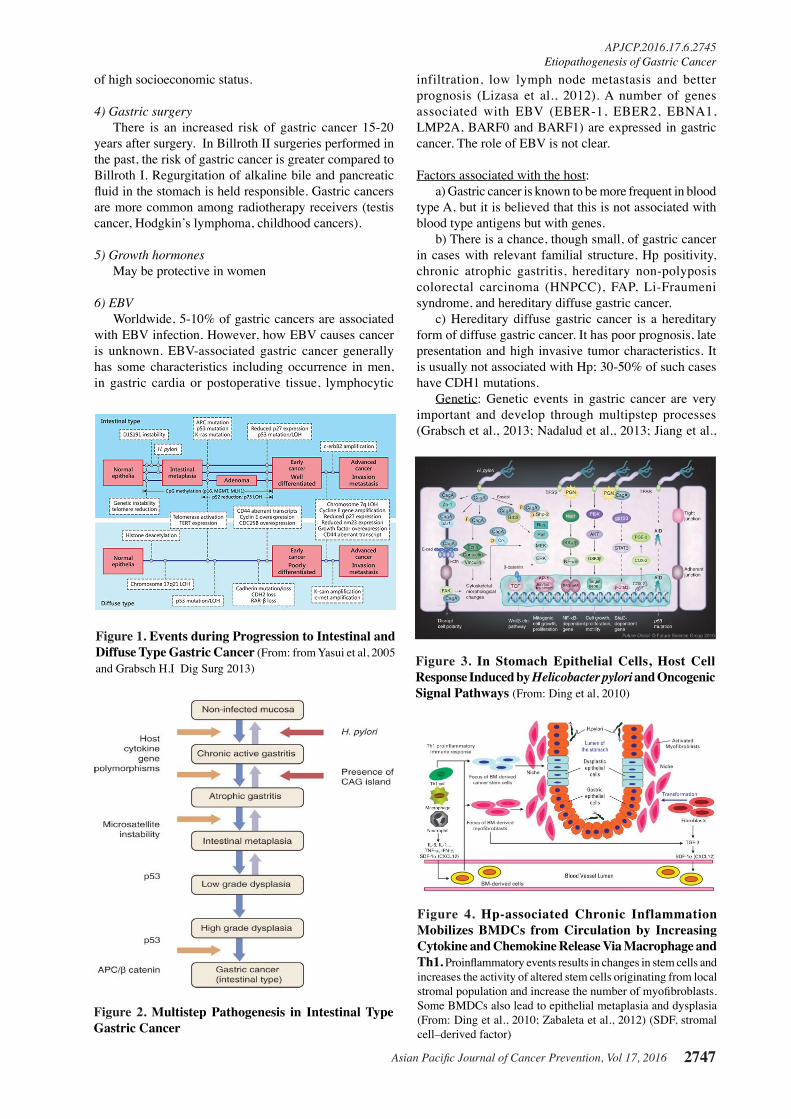
Figure 1. Events during Progression to Intestinal and Diffuse Type Gastric Cancer (From: from Yasui et al, 2005 and Grabsch H.I Dig Surg 2013)
Figure 2. Multistep Pathogenesis in Intestinal Type Gastric Cancer
Figure 3. In Stomach Epithelial Cells, Host Cell Response Induced by Helicobacter pylori and Oncogenic Signal Pathways (From: Ding et al, 2010)
Figure 4. Hp-associated Chronic Inflammation Mobilizes BMDCs from Circulation by Increasing Cytokine and Chemokine Release Via Macrophage and Th1. Proinflammatory events results in changes in stem cells and increases the activity of altered stem cells originating from local stromal population and increase the number of myofibroblasts. Some BMDCs also lead to epithelial metaplasia and dysplasia (From: Ding et al., 2010; Zabaleta et al., 2012) (SDF, stromal cell–derived factor)
Vedat Göral
Asian Pacific Journal of Cancer Prevention, Vol 17, 20162748
2014; Chung et al., 2014). The major genetic alternations that occur in the pathogenesis of gastric cancer are multiple genetic and epigenetic changes in oncogenes, tumor suppressor genes, DNA repair genes, cell cycle regulators and signal molecules (Table 2).
1) OncogenesSeveral oncogenes have been identified in different
stages of gastric cancer (Nadalud et al., 2013; Grabsch et al., 2013; Chung et al., 2014; Jiang et al., 2014)
A) Early K-ras mutations are seen in invasive gastric cancers, dysplasia and IM. c-met oncogenes (encodes hepatocyte growth factor), is found in 39% in diffuse type and in 19% in intestinal type. CagA (oncoprotein) modulates c-met receptor signal transduction which leads to gastric cancer onset and tumor progression. K-ras oncogene is mutated in intestinal gastric cancers and in its precursor lesions, in intestinal metaplasia and adenoma (codon-12). It has not been detected in diffuse gastric cancer. In intestinal type cancer, c-erbB2 (cellular surface receptor of tyrosine kinase family) is over-expressed, while c-met amplification (transmembrane tyrosine kinase receptor) and FGFR/ErbB3/PI3 kinase pathway aberrations are frequent in diffuse cancers. EZH2 (Enhancer of Zeste) is common in intestinal cancer and
causes distant metastasis.B) Tumor suppressor genes (TSG): Changes in tumor
suppressor genes such as TP53 (The p53 gene), TP73, APC (adenomatous polyposis coli), TFF (trefoil factor family), DCC, FHIT (fragile histidine triad) have been identified in about 50% of intestinal type gastric cancers. TP53 is an important regulator factor in cell cycle and loss of TP53 expression or mutational inactivation are the most common genetic changes in gastric cancer. It is found in 60% of invasive tumors. Likewise, abnormalities are found in Hp-associated chronic gastritis, IM and dysplasia. How p53 interacts with Hp is unknown. p53 is a key regulator molecule in response to microenvironmental chronic inflammatory stress (9). According to some investigators, p53 inactivation in gastric epithelial cells reduces progression to apoptosis in Hp-associated damage. Especially in intestinal type gastric cancer, changes in TSG have been identified in 50% on the cases (Nagini et al., 2012; Gomceli et al., 2012; McLean et al., 2014; Jaing et al., 2014). P53 gene is inactivated in gastric cancer. LOH, missense mutations, frameshift deletions, GC-AT transition in the P53 gene are common in diffuse type cancer and are induced by carcinogenic N-nitrosamines (dietary nitrite and amines). RUNX3 mutation in chromosome 1oq23.31 and LOH were found in gastric precancerous lesions and gastric cancer. Hypermethylation in nuclear retinoic acid receptor B was identified in intestinal type gastric cancer.
C) LOH (Loss of heterozygosity): It is a transcription factor and has a tumor suppressor gene-like function and may be detected in gastric carcinomas. LOH, 1p, 2q, 3p, 4p, 5q, 6p, 7p, 7q, 8p, 9p, 11q, 12q, 13q, 14q, 17p, 18q, 21q and 22q play an important role in gastric carcinogenesis. Expression loss in epigenetic mechanisms (promoter methylation) was shown in EBV-associated gastric carcinomas. p73 over-expression and oncogenic isoform DeltaNp73 suppress transcriptional and apoptotic activity of p73 in gastroepithelial cancers. This increases intracellular beta-catenin levels.
D) APC Mutations are more common in intestinal type cancers (33%, 13%). These mutations are also found in Hp-associated IM and dysplasia. High LOH frequency is found in APC, p53, nm23 and Rb locus. APC mutations
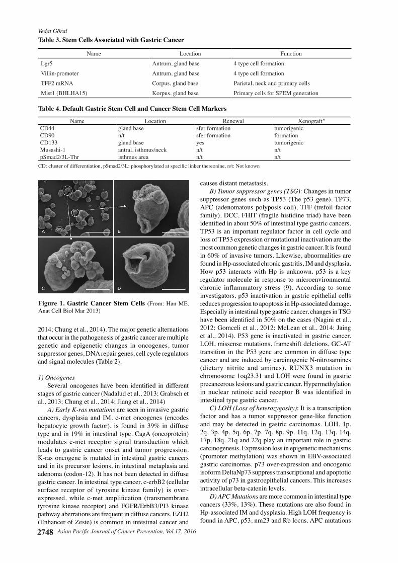
Figure 1. Gastric Cancer Stem Cells (From: Han ME. Anat Cell Biol Mar 2013)
Table 3. Stem Cells Associated with Gastric Cancer
Name Location FunctionLgr5 Antrum, gland base 4 type cell formationVillin-promoter Antrum, gland base 4 type cell formationTFF2 mRNA Corpus, gland base Parietal, neck and primary cellsMist1 (BHLHA15) Korpus, gland base Primary cells for SPEM generation
Table 4. Default Gastric Stem Cell and Cancer Stem Cell Markers
Name Location Renewal Xenograft"CD44 gland base sfer formation tumorigenicCD90 n/t sfer formation formationCD133 gland base yes tumorigenicMusashi-1 antral, isthmus/neck n/t n/tpSmad2/3L-Thr isthmus area n/t n/t
CD: cluster of differentiation, pSmad2/3L: phosphorylated at specific linker thereonine, n/t: Not known

Asian Pacific Journal of Cancer Prevention, Vol 17, 2016 2749
APJCP.2016.17.6.2745Etiopathogenesis of Gastric Cancer
modulates Wnt/catenin signal pathway.E) Cell cycle regulator molecules: Cyclin E and Cyclin
dependent kinase inhibitor 1B (CDKN1B, p27) are two important cell cycle regulators. Cyclin E overexpression is frequent in gastric carcinomas. It may be an indicator of malignant transformation of dysplasia and tumor aggressiveness in invasive cancer. Increased loss of CDKN1B expression leads to increased gastric cancer in Hp-infected mice. Cyclin E and CDK overexpression, p53 aberrant expression and p27 down-regulation are common in gastric cancer. TGFB1/2, TGFBR1, MYC and TP532 increases, SMAD4, CDKN1A, SMAD1/2/3, SMAD2/3 and CDKN1B decreases, and TGFBR2, SMAD7, RELA and CDC25A expression increases. IL-1B-511 T allele and IL-1 RN*2 VNTR polymorphism are common in intestinal cancer
F) Invasion and angiogenesis: E-cadherin plays an important role in cell motility, growth and cancer invasion. VEGF-A is involved in bone metastases of gastric cancer and VEGF-D plays a role in lymphatic metastases. Thus, metastases occur with VEGF.
G) micRNA (micro RNA): Recently, micRNAs have been shown to play an important role in development of many cancers. micRNAs are involved in proliferation, apoptosis, differentiation, angiogenesis, metastasis and immune response (Wang et al., 2012; Han et al., 2013; McLean et al., 2014). They have oncogene- and tumor suppressor gene-like functions. miR-21, miR-106a and miR-17 are upregulated (oncogenic effect) while miR-101, miR-181, miR-449, miR-486, let-7a are downregulated in gastric cancer (tumor suppressor effect).
2) Epigenetic mechanisms Occur as DNA methylation, histone methylation and
histone acetylation. Hypomethylation causes oncogenes’ activation and genomic instability while promoter hypermethylation causes tumor suppressor genes and transcriptional DNA mismatch repair genes suppression. Hipermethylation in promoter genes occur especially in Hp.
Epigenetic changes such as DNA methylation of gene promoters are common in intestinal type cancer and Hp-associated cancer (Nagini et al., 2012; McLean et al., 2014; Jiang et al., 2014). Hypermethylation of reprimo gene is seen in early gastric cancer. Urokinase plasminogen activator (uPAR) is a biomarker that is present in gastric cancer in invasive events and demonstrates cancer invasion. Beta catenin mutation is the most common cause of Wnt path activation in gastric cancer.
Genet ic polymorphism : There are cer ta in polymorphisms in gastric cancers. IL-1 beta (IL-1B) gene and IL-1 receptor antagonist gene (IL-1RN*2/*2) polymorphism, IFNGR1 gene, MTHF reductase polymorphism are associated with increased gastric cancer.
Chromosomal instability (CIN) is an increased potential of chromosomal abnormalities due to errors during replication, recombination, DNA repair, chromosome separation or at cell cycle checkpoints. Chromosomal instability is detected particularly in sporadic gastric cancers.
Microsatellite instability (MSI): Microsatellite instability (MSI) is not an inactivation mechanism of tumor suppressor genes (TSG); it is one of the caretaker genes and results from mutations in DNA repair genes such as MLH1, MSH2 which ensure genome stability by preventing mutations from occurring. Wrong base coupling during DNA replication, events causing replication errors such as insertion and deletion may be observed. This develops in microsatellite instability (MSI). Thus, MSI develops following damages in DNA replication and is found in 15-20% in gastric cancer and more frequently in familial gastric cancer. There is a high rate of epigenetic inactivation in mismatch repair (MMR) genes (hMLH1) in advanced invasive intestinal type gastric cancers.
Thus, genetic mechanisms in gastric cancer pathogenesis involve oncogenes activation, growth factors/receptors’ overexpression, tumor suppressor genes’, DNA repair genes’ and cell adhesion molecules’ inactivation, LOH (Loss of heterogenesity) and point mutations in tumor suppressor genes, and suppression of tumor suppressor genes via CpG island methylation.
Stem Cells
Normal stem cells (NSH) are found in many tissues, particularly bone marrow, brain and possibly GIS (Figure 1). NSHs are found in the gastric mucosa, in the proliferator area of the neck/isthmus. They demonstrate complex upward and downward bipolar migration to differentiate from normal epithelial cells. These cells are immature, poorly organized, multipotent stem cells. They are response for conversion of 4 major cells (Table 3, 4). It is assumed that oncogenesis causes NSHs’ conversion to CSH. Malignant stem cells have rapid proliferation and high metastasis potential while normal stem cells have lower growth and renewal capacity. Local microenvironment contributes to tumor development. Recent in vivo and in vitro studies have shown that gastric cancer could result from stem cells coming from normal stem cells or stem cells originating from bone marrow and that gastric cancer contained cancer stem cells (Nagini et al., 2012). This will provide guidance in planning gastric cancer treatment differently in the feature.
In conclusion, gastric cancer is a multistep event involving multiple genetic and molecular alternations and changes in cancer stem cells. Progression from preneoplastic lesions to cancer is associated with CSC (cancer stem cell) induction, increased cell proliferation and apoptosis.
References
Compare D, Rocco A, Nardone G (2010). Risk factors in gastric cancer. Eur Rev Med Pharmacol Sci, 14, 302-8.
Chung HW, Lim JB (2014). Role of the tumor microenvironment in the pathogenesis of gastric carcinoma. World J Gastroenterol, 21, 1667-80.
Ding SZ, Zheng PY (2012). Helicobacter pylori infection induced gastric cancer; advance in gastric stem cell research and the remaining challenges. Gut Pathog, 8,1-11.
Vedat Göral
Asian Pacific Journal of Cancer Prevention, Vol 17, 20162750
Ding SZ, Goldberg JB, Hatakeyama H (2010). Helicobacter pylori infection, oncogenic pathways and epigenetic mechanisms in gastric carcinogenesis. Future Oncol, 6, 851-862.
Fuccio L, Eusebi LH, Bazzoli F (2010). Gastric cancer, Helicobacter pylori infection and other risk Factors. World J Gastrointest Oncol, 15, 342-347.
Gomceli I, Demiriz B, Tez M (2012). Gastric carcinogenesis. World J Gastroenterol, 7, 5164-70.
Grabsch HI, Tan P (2013). Gastric cancer pathology and underlying molecular mechanisms. Dig Surg, 30, 150-8.
Han TS, Hur K, Xu G, et al (2015). MicroRNA-29c mediates initiation of gastric carcinogenesis by directly targeting ITGB1. Gut, 64, 203-214.
Hu B, El Hajj N, Sittler S, et al (2012). Gastric cancer: Classification, histology and application of molecular pathology. J Gastrointest Oncol, 3, 251-61.
Jiang HB, Yang TJ, Lu P, et al (2014). Ma YJ. Gene expression profiling of gastric cancer. Eur Rev Med Pharmacol Sci, 18, 2109-15.
Jiang HB, Yang TJ, Lu P, et al (2014). Gene expression profiling of gastric cancer. Eur Rev Med Pharmacol Sci, 18, 2109-15.
Iizasa H, Nanbo A, Nishikawa J, et al (2012). H. Epstein-Barr Virus (EBV)-associated gastric carcinoma. Viruses, 4, 3420-39.
McLean MH, El-Omar EM (2014). Genetic of gastric cancer. Nat Rev Gastroenterol Hepatol, 11, 664-74.
Nadalud LD, Ford JM (2013). Molecular profiling of gastric cancer: toward personalized cancer medicine. J Clin Oncol, 1, 838-9.
Nagini S (2012). Carcinoma of the stomach: A review of epidemiology, pathogenesis, molecular genetics and chemoprevention. World J Gastrointest Oncol, 15, 156-69.
Wang F, Sun GP, Zou YF, et al (2012). MicroRNAs as promising biomarkers for gastric cancer. Cancer Biomark, 11, 259-67.
Zabaleta J (2012). MicroRNA: A Bridge from H. pylori Infection to gastritis and gastric cancer development. Front Genet, 14, 3-294.


![[Ghiduri][Cancer]Gastric Cancer](https://static.fdocuments.net/doc/165x107/55cf9399550346f57b9de771/ghiduricancergastric-cancer.jpg)