Multinuclear NMR investigation of the anisotropic system NaDNA/Ethidium Bromide

Proc. Natd. Acad. Sci. USAVol. 89, pp. 6958-6962, August 1992Biochemistry
Ethidium bromide provides a simple tool for identifying genuineDNA-independent protein associations
(POU proteins/human autoantigen Ku/hmunoprepitation/protein-affllty chromatography/contaminating DNA)
JIANN-SHIUN LAI*t AND WINSHip HERR**Cold Spring Harbor Laboratory, Cold Spring Harbor, NY 11724; and tGenetics Program, State University of New York, Stony Brook, NY 11794
Communicated by Keith Yamamoto, April 23, 1992 (received for review February 4, 1992)
ABSTRACT DNA-dependent and DNA-independent asso-ciations of DNA-binding proteins are important in transcrip-tional regulation. The analysis of DNA-independent associa-tions frequently relies on assaying protein interaction in theabsence of target DNA sequences. We have found that con-taminating DNA in protein preparations can stabilize DNA-dependent associations that may appear DNA-independent.Three cellular proteins of 70, 85, and 110 kDa coimmunopre-cipitated with the octamer motif-binding protein Oct-2 becauseof the presence of contaminating DNA in the cell extracts. Inaddition, heterodimer formation between Oct-i (or Oct-2) andPit-i during protein-affinity chromatography was stabilized bythe contaminating DNA. In both instances, these DNA-dependent protein associations were selectively inhibited byethidlum bromide in the precipitation reaction without anyevident effect on DNA-independent protein associations. Thus,ethidlum bromide may serve as a simple and general indicatorofDNA-dependent and DNA-independent protein associations.
Transcriptional regulation depends not only on the interac-tions between sequence-specific DNA-binding proteins andtheir respective cis-regulatory elements but also on theinteractions among these proteins and with other componentsof the transcriptional machinery (reviewed in refs. 1-3). Theavailability of tools for studying sequence-specific transcrip-tion factors (e.g., cDNA clones and antibodies) has allowedmore detailed analysis of the mechanisms by which protein-protein interactions, both DNA-dependent and DNA-independent, regulate transcription. Current methods fordetermining DNA-independent protein-protein interactionsinclude coimmunoprecipitation and protein-affinity chroma-tography. We have used coimmunoprecipitation to identifyproteins that associate with the octamer motif-binding pro-teins Oct-1 (OTF-1, NFIII) and Oct-2 (OTF-2). These tran-scription factors are particularly interesting because theydisplay the same DNA-binding specificity (4) and share verysimilar DNA-binding POU domains (5, 6) but display quali-tatively different RNA polymerase II transcriptional activa-tion properties. Oct-1 is an effective activator of smallnuclear RNA-type promoters, whereas Oct-2 is an effectiveactivator of mRNA-type promoters (7). Therefore, it is likelythat Oct-1 and Oct-2 interact preferentially with differentcomponents of the transcriptional machinery.We identified four cellular proteins of 68, 70, 85, and 110
kDa that coimmunoprecipitated with Oct-2 from labeled cellextracts in what appeared to be a DNA-independent manner.We noticed, however, that the 70-, 85-, and 110-kDa proteinscould bind DNA on their own. We subsequently identifiedthe 70- and 85-kDa proteins as the two heterologous subunitsof the human autoantigen Ku, which possesses strong non-specific DNA-binding properties (ref. 8 and references there-
in). This observation suggested that the association of thesethree proteins with Oct-2 might be mediated by contaminat-ing DNA in the labeled cell extracts. Consistent with thishypothesis, these associations are sensitive to the presence ofthe DNA intercalator ethidium bromide (EtdBr) (9, 10), aninhibitor of DNA binding (11, 12), and are not detectedfollowing digestion with DNase. We subsequently showedthat association of Oct-1 (or Oct-2) with the related POU-domain protein Pit-1 (GHF-1) during protein-affinity chro-matography is also EtdBr-sensitive, suggesting that thisassociation is also stabilized by contaminating DNA. Thesestudies indicate that the sensitivity of protein associations toEtdBr is a good indicator of their dependence on DNA.
MATERIALS AND METHODSExpression Constructs and Antibodies. The mammalian
Oct-2 expression vector pCGoct-2 (13), and the in vitrotranslation vectors pBSoct-1, pCGoct-2, and pBSpit-1 (14)and pBSKRb (retinoblastoma gene product) (15) were asdescribed. The T7 RNA polymerase Escherichia coli expres-sion construct for the glutathione S-transferase (GST)-EiAfusion protein was pGEX-ElA 13S (16). The GST-Pit-1 POUdomain fusion protein was made from pETG-pit-i-POU,which contains the GST coding sequences fused to the 5' endof Pit-1 POU domain coding sequences in the E. coli expres-sion vector pETiic (G. Henry and W.H., unpublished re-sults). The two series of monoclonal antibodies raised againstOct-i (YL series) or Oct-2 (PT series) will be described indetail elsewhere; the anti-ElA monoclonal antibody was M73(17).
Cell Labeling, Immunoprecipitation, and DNA-Affinity Pre-cipitation. Cells were transfected and labeled as described(13). Labeled cells were washed with phosphate-bufferedsaline, collected, and lysed on ice for 1 hr in 1 ml of lysisbuffer containing 25 mM Hepes (pH 7.9), 200 mM KCI, 0.1%Nonidet P-40 (Sigma), 1 mM phenylmethylsulfonyl fluoride,leupeptin (1 ug/mi), aprotinin (1 ,ug/ml), 10 mM NaF, 0.1mM Na3VO4, 5 mM EDTA, and 5 mM dithiothreitol. Celldebris was removed, and the resulting lysate was preclearedwith normal rabbit serum and fixed and killed Staphylococ-cus aureus Cowan I (Zymed Laboratories) (18). Typically,for immunoprecipitation, 250 p.l of cleared lysate was incu-bated with monoclonal antibody for 1 hr on ice with inter-mittent, gentle mixing. Immune complexes were collectedwith recombinant protein G-agarose beads (GIBCO/BRL) byrocking at 40C for 1 hr, and the immunoprecipitates washedand prepared for SDS/PAGE (18).For DNA-affinity precipitation, DNAs containing multim-
erized copies ofthe octamer and heptamer Oct-2 binding sitesfrom the immunoglobulin heavy-chain promoter flanked byPvu II sites (5'-CAGCTGCCTCATGAGTATGCAAAT-CAGCTGC-3') covalently coupled to Sepharose beads (19)
Abbreviations: GST, glutathione S-transferase; EtdBr, ethidiumbromide.
6958
The publication costs of this article were defrayed in part by page chargepayment. This article must therefore be hereby marked "advertisement"in accordance with 18 U.S.C. §1734 solely to indicate this fact.

Proc. Natil. Acad. Sci. USA 89 (1992) 6959
were kindly provided by M. Tanaka (Cold Spring HarborLaboratory). We used the same procedure that was used forimmunoprecipitation except that the DNA Sepharose beadswere used in place of protein G-agarose beads.
EtdBr, Micrococcal Nuclease, and RNase Treatment ofCleared Lysates. EtdBr was added (10-400 gg/ml) and thelysates were incubated on ice for 30 min. Precipitates wereremoved by centrifugation for 5 min at 40C in a microcentri-fuge and the supernatant was transferred to a fresh tube. Theresulting lysate was then ready for immunoprecipitation orDNA-affinity precipitation. The original concentration ofEtdBr was maintained during the washing steps.For micrococcal nuclease treatment, the immunoprecipi-
tates or DNA-affinity precipitates bound to the beads afterfour washes were suspended in 50 t1 of digestion buffer (50mM NaCl/10 mM Tris, pH 7.0/4 mM CaCl2 with or without10 mM EGTA). This order of precipitation and subsequentdigestion allowed us to concentrate the sample and to transferit to a buffer appropriate for micrococcal nuclease treatment.After incubation at 370C for 1 hr with micrococcal nuclease(Worthington), the samples were washed twice with 1 ml ofdigestion buffer prior to SDS/PAGE. For RNase A and T1treatment, the samples were treated as for micrococcalnuclease treatment except that the RNase digestion bufferwas 300 mM NaCl/10 mM Tris, pH 7.5/5 mM EDTA and theincubation was at 30°C for 1 hr.
Purification of GST Fusion Proteins from E. coli. TheGST-E1A 13S and GST-Pit-1 POU domain fusion proteins,and GST alone were expressed in E. coli BL21(DE3) (20) andpurified from induced cultures (21). Glutathione-agarosebeads bound with GST or GST fusion protein were washedextensively with phosphate-buffered saline containing lyso-zyme (3 mg/ml), 0.1% Tween 20, and 0.1% Triton X-100 andwere stored at 4°C in the same buffer with 1 mM phenyl-methylsulfonyl fluoride, and 0.02% NaN3. The purified pro-teins thus treated remained stable for at least 1 month. Therelative amount of fusion protein bound to the beads wasdetermined by SDS/PAGE of proteins released from thebeads and staining of the gel with Coomassie blue. Beforeprotein-affinity chromatography, the beads were washedtwice in binding buffer [50 mM KCI/20 mM Hepes, pH 7.9/2mM EDTA/0.1% Nonidet P-40/5 mM dithiothreitol/10%(vol/vol) glycerol/0.5% nonfat milk]. Beads (150 ,u) bearing2 ,ug of each fusion protein were incubated on ice for 30 minin the absence or presence of EtdBr (10-400 ,ug/ml). 35S-labeled in vitro-translated proteins (30 ,ul) were then added tothe beads and incubated for 1 hr on ice with intermittent,gentle mixing. The beads were subsequently washed fourtimes with 1 ml of the binding buffer containing the sameconcentration of EtdBr used in the binding reaction. Boundlabeled proteins were resolved by SDS/PAGE.
RESULTSFour Cellular Proteins of 68, 70, 85, and 110 kDa Associate
with Oct-2 During Immunoprecipitation. Fig. 1 shows twoseries of immunoprecipitations from labeled 293 cell (humanembryonic kidney cell line) extracts containing or lackingectopically expressed Oct-2, with a panel of anti-Oct-1 andanti-Oct-2 monoclonal antibodies. Oct-2 was recovered fromthe Oct-2-containing extract with each of the six monoclonalantibodies directed against Oct-2 (lanes 2-7), but not in theabsence of antibody (lane 1) or with an Oct-i-specific anti-body (lane 8). The recovered Oct-2 molecules migrate as adoublet due to heterogeneous phosphorylation (13). In addi-tion to Oct-2, four other proteins of 68, 70, 85, and 110 kDawere coprecipitated by five of the six Oct-2 antibodies (lanes2-6). Except for a reduced level of the 68-kDa protein,neither Oct-2 nor the other cellular proteins were recoveredfrom the mock-transfected cell extracts (lanes 9-16). This
Oct-2tronsfectionI. 1
N NCN1CJ CLCL-M S VS A; 6 ;5 ;! 1!
200-
97.4-
68-amr
Mocktransfection
IN. cm JNcm JaD -'6 'd 16 5~ 5
-OCt-l..4O
]Oct-2
43- _
29-
184- _
2 3 4 5 6 7 8 9 10 11 12 13 14 15 16
FIG. 1. Four cellular proteins of 68, 70, 85, and 110 kDa copre-cipitate with ectopically expressed Oct-2 from 293 cells duringimmunoprecipitation. A panel of monoclonal antibodies specific forOct-Li Oct-2, or both proteins was used in immunoprecipitationreactions with 35S-labeled extracts from 293 cells transfected with anOct-2 expression vector (lanes 1-8) or mock transfected (lanes 9-16).Immunoprecipitated proteins were separated by SDS/10%o PAGEand stained for fluorography. Samples were precipitated either in theabsence ofantibody (lanes 1 and 9) or with monoclonal antibodies (a)directed against the N-terminal (PT1, lanes 2 and 10) or C-terminal(PT2, PT3, and PT7 in order in lanes 3-5 and 11-13) regions of Oct-2,the POU-specific regions (YL21, lanes 6 and 14) or POU home-odomains (YL123, lanes 7 and 15) of Oct-1 and Oct-2, or the uniquePOU-domain linker region of Oct-1 (YL15, lanes 8 and 16). Thepositions of Oct-1 and Oct-2 and the molecular size (kDa) of theprotein standards (lane M) are indicated to the right and left,respectively. Bands corresponding to the coprecipitated 68-, 70-, 85-,and 110-kDa cellular proteins are indicated by dots. Oct-2 is lessabundant in lane 6 because antibody YL21 has a low affinity for Oct-2(unpublished results).
result, together with the ability of multiple Oct-2 antibodiesto coprecipitate the four cellular proteins, indicated that thecoprecipitation was due to association with Oct-2 rather thandue to antibody crossreactions.One of the four Oct-2-associated proteins (68 kDa) can be
distinguished from the others because it is the only onerecovered with the monoclonal antibody directed against theOct-2 (and Oct-i) homeodomain (lane 7); high-resolutiontwo-dimensional gel electrophoresis (22) of Oct-2 immuno-precipitates indicates that this 68-kDa protein is the heatshock protein Hsp70 (data not shown). The inability torecover the 70-, 85-, and 110-kDa proteins with the Oct-2homeodomain antibody suggested that these proteins asso-ciate with the homeodomain. Consistent with this hypothe-sis, they can be recovered, albeit with reduced efficiency,when only the Oct-2 homeodomain is present (data notshown). Thus, these three proteins apparently associate withthe Oct-2 DNA-binding domain.The 70-, 85-, and 110-kDa Proteins Can Bind DNA in the
Absence of Oct-2. Protein association with POU domainsoccurs on DNA, as in the interaction between Oct-1 and theherpes simplex virus transactivator VP16 (23-25), or in theabsence of DNA, as in the interaction between the Drosoph-ila POU proteins I-POU and Cf1-a (26). Indeed, in the lattercase the interaction between I-POU and Cfl-a inhibits theability of Cfl-a to bind DNA (26). To test whether theassociation of Oct-2 and the 70-, 85-, and 110-kDa proteins iscompatible with Oct-2 DNA binding, we asked whether these
Biochemistry: Lai and Herr
Proc. Natl. Acad. Sci. USA 89 (1992)
three proteins could be recovered in a DNA-affinity precip-itation assay with multimerized copies of the octamer motifcovalently linked to Sepharose beads. Fig. 2 shows theresults of such an experiment using two different concentra-tions of DNA beads. The recovery of the Oct-2-associatedproteins was as efficient (or more efficient, for the 110-kDaprotein) in the DNA-affinity precipitation (lanes 2 and 3) asin an Oct-2 immunoprecipitation (lane 1), indicating thatthese three proteins can associate with Oct-2 bound to DNA.To our surprise, however, in a control experiment with a 293cell extract lacking Oct-2, the same three proteins were alsorecovered (lanes 4 and 5), indicating that these three proteinspossess Oct-2-independent DNA-binding activity. Consis-tent with this hypothesis, the 70- and 85-kDa proteins comi-grate with the 70- and 86-kDa subunits ofthe abundant humanautoantigen Ku, which possesses nonspecific DNA-bindingactivity (8) (data not shown); the size of the 110-kDa proteinsuggests that it is the 112- to 116-kDa poly(ADP-ribose)polymerase, which is also an abundant nonspecific DNA-binding protein (27, 28).
Association of the 70-, 85-, and 110-kDa Proteins with Oct-2Is Sensitive to EtdBr and Micrococcal Nuclease Digestion. Therealization that the 70-, 85-, and 1,10-kDa proteins can bindDNA in the absence of Oct-2 suggested that coimmunopre-cipitation of these three proteins with Oct-2 may not occurdirectly but rather may result from binding of Oct-2 and thesethree proteins to contaminating DNA in the protein extracts.To test this possibility we assayed the effect ofEtdBr, aDNAintercalator (9, 10) that interferes generally with protein-DNA interaction (11, 12).To demonstrate that EtdBr does not generally affect pro-
tein-protein interactions, we tested the effect of EtdBr onassociations between the adenovirus ElA proteins and cel-lular proteins (29, 30). In the absence of EtdBr (Fig. 3, lane1), the p300, p1O7/p1OSRb, and p60Cyclin-A ElA-associatedproteins (31-33) were recovered in an immunoprecipitation ofendogenous ElA from an 35S-labeled 293 cell extract; recov-
If. WCA
Z C
-------------1
OJc+-2ZIIt
2 i4
2 4
FIG. 2. The 70-, 85-, and 110-kDa Oct-2-associated proteins bindDNA in the absence ofOct-2. Extracts of35S-labeled 293 cells, eithertransfected with an Oct-2 expression vector (lanes 1-3) or mocktransfected (lanes 4 and 5), were used for immunoprecipitation (IP)(lane 1) with the N-terminal Oct-2-specific antibody PT1 or forDNA-affinity precipitation (DNAP) (lanes 2-5) with Sepharose beadslinked to multimerized copies of the immunoglobulin heavy-chainpromoter octamer and heptamer motifs (see Materials andMethods).Two different concentrations ofDNA beads were used, which couldbind either 0.5 pg (lanes 2 and 4) or 1.5 Ag (lanes 3 and 5) of E.coli-expressed Oct-2 as measured by Coomassie blue staining ofbound Oct-2 after electrophoresis. Dots identify the 70-, 85-, and110-kDa proteins.
:I.'TI.-.I_M i> .;
EtdBr
.mme
--w"-:k-
.AIE "
qW.,
I:ct2[Uw
_I
FIG. 3. Association of the 70-, 85-, and 11O-kDa proteins withOct-2 is sensitive to the presence of EtdBr. A 293 celi extractcontaining Oct-2 was immunoprecipitated (IP) with ElA-specific(M73, lanes 1-3) or Oct-2-specific (PT1, lanes 4-6) monoclonalantibody or used for DNA-affinity precipitation (DNAP) with mul-timerized octamer and heptamer motifs (lanes 7-9). The reactionswere done in the absence of EtdBr (lanes 1, 4, and 7) or in thepresence of EtdBr at 12.5 ,ug/ml (lanes 2, 5, and 8) or 50 .g/ml (lanes3, 6, and 9). Precipitated proteins were separated by SDS/10%PAGE and visualized by fluorography. Arrowheads indicate thepositions of the ElA-associated proteins p300, p107/p1i5Rb, andp6Cyln A. Dots indicate the positions of the three Oct-2-associatedproteins.
ery of the ElA-associated proteins was resistant to EtdBr at12.5 gg/ml (lane 2) or 50 ,Ag/ml (lane 3). The same concen-trations of EtdBr, however, did affect the recovery of Oct-2and the associated proteins in a DNA-affinity precipitationassay (compare lanes 8 and 9 with lane 7), showing that EtdBrinhibits protein-DNA interactions. As expected, in an Oct-2immunoprecipitation, addition of EtdBr did not affect themonoclonal antibody-Oct-2 interaction (lanes 4-6). Recov-ery of the Hsp70-like protein was not sensitive to EtdBr(lanes 4-6), but recovery ofthe 70-, 85-, and 110-kDa proteinswas disrupted by the addition of EtdBr (compare lanes 5 and6 with lane 4), suggesting that the association between Oct-2and these three cellular proteins is indeed DNA-dependent.To confirm the DNA-dependent association of Oct-2 with
the 70-, 85-, and 110-kDa proteins, we treated the Oct-2immunoprecipitates with micrococcal nuclease or RNaseafter immunoprecipitation from Oct-2-containing 293 cellextracts. Micrococcal nuclease treatment (Fig. 4, lane 4) hadthe same deleterious effect on association of Oct-2 with thethree cellular proteins as did addition of EtdBr (lane 2). Theeffect of micrococcal nuclease treatment was prevented bythe addition of EGTA (lane 6), which inhibits micrococcalnuclease activity (34). Treatment of the immunoprecipitateswith RNase had no effect on recovery of Oct-2 or the threeassociated proteins (lane 8), indicating that RNA could notfunction to tether Oct-2 and the 70-, 85-, and 110-kDaproteins.DNA Is Also Involved in Oct-i and Pit-iH1ete r
Formation. The unexpected finding that contaminating DNAcan stabilize associations between DNA-binding proteinsduring immunoprecipitation led us to test whether otherDNA-binding-protein associations thought to occur in theabsence of DNA might also be stabilized by contaminatingDNA. For example, in the absence of any added DNAbinding sites, in vitro-translated Oct-i was shown to associatewith a GST-Pit-1 POU domain fusion protein purified afterexpression in E. coli and bound to glutathione-agarose beads(21). We tested the sensitivity of this association to thepresence of EtdBr.
IMACAU7W Biochemistry: Lai and Herr

Proc. Natl. Acad. Sci. USA 89 (1992) 6961
MicrococcolNucleoseI I
EtdBr -EGTA +EGTA RNasem m m m1
M - - - + - +
Rb Oct-I ' I
GST- GST-o - EIA -g POUn 0 (13S) (g (Pit-l)
I I
EtdBr- - + - - - +. -
200-
974-_ -_ o
__ _
m8-_ss j ]Oct-2
43-Q 4 -NW
43- 40
29- _
2 3 4 5 6 7 8
FIG. 4. Association of the 70-, 85-, and 110-kDa proteins withOct-2 is sensitive to micrococcal nuclease but not to RNase diges-tion. In each sample, an 35S-labeled Oct-2-containing 293 cell extractwas used for immunoprecipitation with the Oct-2-specific monoclo-nal antibody PT1. For micrococcal nuclease treatment (lanes 3-6),the immunoprecipitates bound to protein G-agarose beads weresuspended in 50 1.l of digestion buffer containing 4 mM CaCl2 (lanes3 and 4) or additionally 10 mM EGTA (lanes 5 and 6) with (lanes 4and 6) or without (lanes 3 and 5) the addition of 0.4 unit ofmicrococcal nuclease. For RNase treatment, the immunoprecipi-tates were suspended in 50 1ul of RNase digestion buffer with (lane8) or without (lane 7) RNase A (80 ,ug/ml) and RNase T1 (4 ,ug/ml).Lanes 1 and 2, control immunoprecipitations in the absence orpresence of EtdBr (400 jg/ml).As a control to demonstrate that EtdBr does not generally
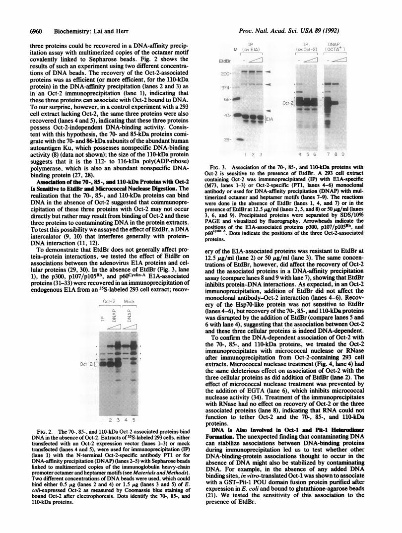
affect protein association in this assay, we tested its effect onthe association of a GST-EiA 13S fusion protein with invitro-translated piO5Rb (15, 16). As expected, in vitro-translated piOSRb (Fig. 5, lane 1) did not associate with GSTprotein alone (lane 2) but was recovered with the GST-EiA13S fusion protein (lane 4); this recovery was insensitive toEtdBr at 200 Ag/ml (lane 3). As described previously (21), invitro-translated Oct-i (lane 5) was recovered with the GST-Pit-1 POU domain fusion protein (lane 8) but not with GSTalone (lane 6), showing that the Pit-1 POU domain is requiredfor the association. However, this Oct-i-Pit-i associationwas very sensitive to EtdBr (compare lanes 7 and 8), sug-gesting that it was dependent on contaminating DNA in eitherthe purified GST-Pit-1 POU domain fusion protein prepara-tion or the in vitro-translated Oct-i. Consistent with thisresult, the association of Oct-i and the GST-Pit-1 POUdomain fusion protein was also sensitive to micrococcalnuclease treatment (data not shown).A similar association between Oct-2 and the Pit-1 POU
domain (lanes 9-12) was also disrupted by EtdBr. Curiously,however, although ethidium bromide had a large effect onPit-1 self-association, a residual level of self-association wasapparently resistant to EtdBr (compare lanes 15 and 16). Thislow level of association in the presence of EtdBr may besignificant because in a serial titration of EtdBr, the residualPit-1 self-association was resistant to as much as 400 Ag ofEtdBr per ml, whereas the majority of the self-associationwas disrupted by 10 ,g/ml (data not shown). This resistanceto EtdBr suggests that Pit-1 can form homodimers in theabsence of DNA.
DISCUSSIONWe have described two instances in which DNA contamina-tion apparently stabilized associations between DNA-
2 3 4 5 6 7 8 9 10 11 12 13 14 15 16
FIG. 5. Association of Oct-i, Oct-2, and Pit-1 with the Pit-1 POUdomain in protein-affinity chromatography is sensitive to EtdBr.Glutathione-agarose beads bearing the GST-ElA 13S fusion proteinwere incubated with in vitro-translated 35S-labeled p1o5Rb either inthe presence (lane 3) or absence (lane 4) of EtdBr (200 ,ug/ml). Beadsbearing the GST-Pit-i-POU domain fusion protein were incubatedwith 35S-labeled Oct-1 (lanes 7 and 8), Oct-2 (lanes 11 and 12), or Pit-1(lanes 15 and 16) in the presence (lanes 7, 11, and 15) or absence(lanes 8, 12, and 16) of EtdBr. The beads were then washed and theprecipitates were eluted for SDS/Ilo PAGE. Lanes 2, 6, 10, and 14,eluates of beads bearing GST proteins alone after incubation with35S-labeled p1O5Rb, Oct-i, Oct-2, and Pit-1, respectively. Lanes 1, 5,9, and 13, portions (5%) of reaction mixtures containing 35S-labeledprotein incubated with the POU domain and control beads, loadeddirectly onto the gel. Positions ofthe major p1O5Rb, Oct-i, Oct-2, andPit-1 in vitro translation products are indicated by arrowheads.
binding proteins that might otherwise be thought to occurindependently of DNA. (i) We found that three cellularproteins that coimmunoprecipitate with Oct-2 could bindDNA on their own. This finding led us to test the sensitivityof these associations to EtdBr, a general inhibitor of DNAbinding by proteins (11, 12), and micrococcal nuclease di-gestion, both of which disrupted the associations. (ii) Wefound that association between the POU-domain proteinsPit-1 and Oct-i in solution (21) was also dependent oncontaminating DNA, as indicated by sensitivity to EtdBr andmicrococcal nuclease digestion. Pit-1 POU domain self-association, however, revealed an EtdBr-resistant compo-nent (Fig. 5), which may reflect bona fide DNA-independentassociation. Oct-i also associates with itself in solution in aprotein-affinity assay similar to the one used to assay Pit-1-Oct-1 and Pit-1-Pit-1 association (35). It will be of interest todetermine whether this association is EtdBr-sensitive or not.
Addition ofEtdBr and digestion with micrococcal nucleaseare complementary methods for distinguishing DNA-dependent and DNA-independent protein association. BothEtdBr (36) and micrococcal nuclease (37) interact with DNAin a relatively nonspecific manner; therefore, the ability ofboth reagents to disrupt protein-DNA interactions is likely tobe reasonably indiscriminate. As a routine assay, however,addition of EtdBr provides several advantages over micro-coccal nuclease digestion. Digestion with micrococcal nucle-ase requires transfer of the sample to the appropriate diges-tion buffer and generally involves prolonged incubation
Oct-2
GST-o F POU-J ( (Pit -1)
+ _
Pit-I
GST--0 F- PO UJ 0 (Pit-I)- - -
-I,
Biochemistry: Lai and Herr

Proc. Nati. Acad. Sci. USA 89 (1992)
(about 1 hr) at elevated temperature (370C). Furthermore,unlike EtdBr, which has proved very effective for disruptingDNA-dependent protein association (Figs. 3 and 5), micro-coccal nuclease digestion is often incompletely effective(unpublished observations),perhaps because the enzyme can
be inhibited by contaminants or because it cannot access theprotein-bound DNA as readily as EtdBr. Thus, EtdBr isapparently an excellent, easy-to-use preliminary indicator ofDNA-dependent and DNA-independent protein association.EtdBr has been used previously to selectively disrupt
protein-DNA interactions. For example, Schroter et al. (12)used EtdBr, in addition to theDNA intercalators chloroquineand propidium iodide, to selectively elute DNA-bindingproteins from nuclei. We have also tested the efficacy ofchloroquine and propidium iodide to disrupt the DNA-dependent protein associations described here: althoughchloroquine was ineffective in our hands, propidium iodide at.15 ug/ml also disrupted these DNA-dependent proteinassociations (unpublished results). An unavoidable concernwith the use of any DNA intercalator to disrupt DNA-dependent association is that in some cases it may alsodisrupt direct protein-protein associations. In the case ofEtdBr such disruption is unlikely. We have assayed over 10different DNA-independent protein-protein associations(e.g., ElA with its associated proteins, and numerous anti-body-antigen interactions), none of which was affected bythe addition of EtdBr. Nevertheless, it is imperative to checkthe specificity of EtdBr disruption of protein associations byalso assaying the effects ofmicrococcal nuclease digestion orinclusion of propidium iodide.The association of the 70-, 80-, and iiO-kDa proteins with
Oct-2 in the coimmunoprecipitation assay is probably en-
hanced by the transient ectopic expression of Oct-2. Forexample, when endogenous Oct-2 is immunoprecipitatedfrom a B-cell extract, only the ii0-kDa protein is readilycoimmunoprecipitated (unpublished observation). Other ex-periments have shown that the 70-, 85-, and ii0-kDa proteinsalso coimmunoprecipitate with transiently expressed Oct-i,Pit-i, and an Oct-i variant carrying the Oct-3/4 POU domain(14). These associations are all dependent on the integrity ofthe DNA-binding domain, because they do not occur whenthe POU domain is deleted or with a mutant Oct-i proteincarrying three alanine substitutions in the homeodomain thatdisruptDNA binding (38). In an identical assay, however, thesame three cellular proteins do not associate with the serum
response factor SRF, the bovine papilloma virus transcrip-tion and replication factor E2, or the TATA box-bindingprotein TBP (unpublished results). Further, in a protein-affinity assay similar to the Pit--Oct-1 assay used here, theOct-1 POU domain does not associate with the adenovirusreplication factor NFI (35). The efficiency of associationsthat are dependent on contaminating DNA may reflect dif-ferences in the ability of these various DNA-binding proteinsto bind to the contaminating DNA or differences in bona fideprotein interactions when the proteins are bound to DNA.Many important associations between DNA-binding pro-
teins are dependent on DNA, as in the case of Oct-i and VP16(39-41). Inhibition ofDNA-dependent protein association byEtdBr does not discriminate between specific and nonspe-
cific associations stabilized by DNA. For example, Voss et
al. (21) have shown that Pit-1 and Oct-i can form het-erodimers on specific cis-regulatory targets. Thus, whereasour assays clearly show a DNA dependence for the interac-tion of Oct-i and Pit-i in solution, it is not clear whether thisassociation is stabilized by specific protein-protein contactsbetween DNA-bound Oct-i and Pit-1 molecules or simply bynonspecific tethering of the two proteins by the DNA. Ourstudies suggest that EtdBr can be an excellent reagent todiscriminate between protein associations that are entirely
independent of DNA and those that remain dependent onDNA, but further studies with other classes of DNA-bindingproteins will be required to establish its general utility.
We thank N. Dyson (pGEX-E1A 13S), Q. Hu (pBSKRb), R.Aurora (pBSpit-1), M. Tanaka (pCG-oct-2), and G. Henry (pETG-pit-1-POU) for plasmids; M. Tanaka for DNA-affinity Sepharosebeads; M. Mathews for Ku antisera; S. Bell, M. Cleary, N. Her-nandez, M. Tanaka, W. Tansey, and A. Wilson for critical readingsof the manuscript; J. Duffy and P. Renna for artwork; and J. Readerfor typing. This work was funded by National Cancer Institute GrantCA13106.
1. Lewin, B. (1990) Cell 61, 1161-1164.2. Ptashne, M. & Gann, A. (1990) Nature (London) 346, 329-331.3. Greenblatt, J. (1991) Cell 6, 1067-1070.4. Staudt, L. M., Singh, H., Sen, R., Wirth, T., Sharp, P. A. &
Baltimore, D. (1986) Nature (London) 323, 640-643.5. Sturm, R. A., Das, G. & Herr, W. (1988) Genes Dev. 2, 1582-1599.6. Clerc, R. G., Corcoran, L. M., LeBowitz, J. H., Baltimore, D. &
Sharp, P. A. (1988) Genes Dev. 2, 1570-1581.7. Tanaka, M., Lai, J.-S. & Herr, W. (1992) Cell 68, 755-767.8. Paillard, S. & Strauss, F. (1991) Nucleic Acids Res. 19, 5619-5624.9. Lerman, L. S. (1961) J. Mol. Biol. 3, 18-30.
10. Waring, M. (1970) J. Mol. Biol. 54, 247-279.11. Parker, R. C., Watson, R. M. & Vinograd, J. (1977) Proc. Natd.
Acad. Sci. USA 74, 851-855.12. Schr6ter, H., Maier, G., Ponstingl, H. & Nordheim, A. (1985)
EMBO J. 4, 3867-3872.13. Tanaka, M. & Herr, W. (1990) Cell 60, 375-386.14. Aurora, R. & Herr, W. (1992) Mol. Cell. Biol. 12, 455-467.15. Hu, Q., Bautista, C., Edwards, G. M., Defeo-Jones, D., Jones,
R. E. & Harlow, E. (1991) Mol. Cell. Biol. 11, 5792-5799.16. Rustgi, A. K., Dyson, N. & Bernards, R. (1991) Nature (London)
352, 541-544.17. Harlow, E., Franza, B. R., Jr., & Schley, C. (1985) J. Virol. 55,
533-546.18. Harlow, E. & Lane, D. (1988) Antibodies: A Laboratory Manual
(Cold Spring Harbor Lab., Cold Spring Harbor, NY) pp. 421-470.19. Kadonaga, J. T. & Tjian, R. (1986) Proc. Natl. Acad. Sci. USA 83,
5889-5893.20. Studier, F. W., Rosenberg, A., Dunn, J. & Dubendorff, J. (1990)
Methods Enzymol. 185, 60-89.21. Voss, J. W., Wilson, L. & Rosenfeld, M. G. (1991) Genes Dev. 5,
1309-1320.22. Garrels, J. I. (1983) Methods Enzymol. 100, 411-423.23. Gerster, T. & Roeder, R. G. (1988) Proc. Natl. Acad. Sci. USA 85,
6347-6351.24. Kristie, T. M., LeBowitz, J. H. & Sharp, P. A. (1989) EMBO J. 8,
4229-4238.25. Stern, S., Tanaka, M. & Herr, W. (1989) Nature (London) 341,
624-630.26. Treacy, M. N., He, X. & Rosenfeld, M. G. (1991) Nature (London)
35O, 577-584.27. Benjamin, R. C. & Gill, D. M. (1980) J. Biol. Chem. 255, 10502-
10508.28. Cherney, B. W., McBride, 0. W., Chen, D., Alkhatib, H., Bhatia,
K., Hensley, P. & Smulson, M. E. (1987) Proc. Natl. Acad. Sci.USA 84, 8370-8374.
29. Yee, S.-P. & Branton, P. (1985) Virology 147, 142-153.30. Harlow, E., Whyte, P., Franza, B. R., Jr., & Schley, C. (1986) Mol.
Cell. Biol. 6, 1579-1589.31. Whyte, P., Buchkovich, K. J., Horowitz, J. M., Friend, S. H.,
Raybuck, M., Weinberg, R. A. & Harlow, E. (1988) Nature (Lon-don) 334, 124-129.
32. Pine, J. & Hunter, T. (1990) Nature (London) 346, 760-763.33. Ewen, M. E., Xing, Y., Lawrence, J. B. & Livingston, D. M. (1991)
Cell 66, 1155-1164.34. Frank, J. J., Hawk, I. A. & Levy, C. C. (1975) Biochim. Biophys.
Acta 390, 117-124.35. Verrijzer, C. P., van Oosterhout, J. & van der Vliet, P. C. (1992)
Mol. Cell. Biol. 12, 542-551.36. Fox, K. R. & Waring, M. J. (1987) Nucleic Acids Res. 15, 491-507.37. Roberts, W. K., Dekker, C. A., Rushisky, G. W. & Knight, C. A.
(1962) Biochim. Biophys. Acta SS, 664-671.38. Sturm, R. A. & Herr, W. (1988) Nature (London) 336, 601-604.39. Xiao, P. & Capone, J. P. (1990) Mol. Cell. Biol. 10, 4974-4977.40. Kristie, T. M. & Sharp, P. A. (1990) Genes Dev. 4, 2383-23%.41. Stern, S. & Herr, W. (1991) Genes Dev. 5, 2555-2566.
Biochemistry: Lai and Herr