Estudo dos efeitos dos neuropeptídeos VIP e PACAP sobre a ... · Temerozo, Jairo Ramos. Estudo dos...
64
INSTITUTO OSWALDO CRUZ Mestrado em Biologia Celular e Molecular Estudo dos efeitos dos neuropeptídeos VIP e PACAP sobre a replicação do HIV-1 em macrófagos primários humanos. JAIRO RAMOS TEMEROZO Rio de Janeiro 2013
Transcript of Estudo dos efeitos dos neuropeptídeos VIP e PACAP sobre a ... · Temerozo, Jairo Ramos. Estudo dos...

INSTITUTO OSWALDO CRUZ
Mestrado em Biologia Celular e Molecular
Estudo dos efeitos dos neuropeptídeos VIP e PACAP sobre a replicação do
HIV-1 em macrófagos primários humanos.
JAIRO RAMOS TEMEROZO
Rio de Janeiro 2013

Temerozo, Jairo Ramos.
Estudo dos efeitos dos neuropeptídeos VIP e PACAP sobre a replicaçãodo HIV-1 em macrófagos primários humanos / Jairo Ramos Temerozo. - Rio dejaneiro, 2013. 53 f.; il.
Dissertação (Mestrado) – Instituto Oswaldo Cruz, Pós-Graduação emBiologia Celular e Molecular, 2013.
Orientador: Dumith Chequer Bou-Habib.
Bibliografia: Inclui Bibliografias.
1. HIV-1. 2. Macrófagos. 3. Neuropeptídeos. 4. VIP. I. Título.
Elaborada pelo Sistema de Geração Automática de Ficha Catalográfica da Biblioteca de Manguinhos/ICICT com os dadosfornecidos pelo(a) autor(a).

INSTITUTO OSWALDO CRUZ
Pós-Graduação em Biologia Celular e Molecular
Jairo Ramos Temerozo
Estudo dos efeitos dos neuropeptídeos VIP e PACAP sobre a replicação do HIV-1 em
macrófagos primários humanos.
Dissertação apresentada ao Instituto Oswaldo Cruz
como parte dos requisitos para obtenção do título de
Mestre em Biologia Celular e Molecular
Orientador: Prof. Dumith Chequer Bou-Habib
RIO DE JANEIRO
2013

ii
INSTITUTO OSWALDO CRUZ
Pós-Graduação em Biologia Celular e Molecular
AUTOR: JAIRO RAMOS TEMEROZO
ESTUDO DOS EFEITOS DOS NEUROPEPTÍDEOS VIP E PACAP SOBRE A
REPLICAÇÃO DO HIV-1 EM MACRÓFAGOS PRIMÁRIOS HUMANOS.
ORIENTADOR: Prof. Dumith Chequer Bou-Habib
Aprovada em: 26/11/2013
EXAMINADORES:
Prof. Vinícius de Frias Carvalho (Presidente)
Prof. Marcelo Alves Soares (Titular)
Prof. Pedro Muanis Persechini (Titular)
Prof. Thiago Moreno Lopes e Souza (Revisor/Suplente)
Prof. Daniella Arêas Mendes da Cruz (Suplente)
Rio de Janeiro, Novembro de 2013
iii
AGRADECIMENTOS
Ao longo do período do mestrado, muitas pessoas passaram por minha vida, deixando
marcas e lições para toda ela, proporcionando-me alegrias, conhecimento e crescimento
pessoal. Neste momento gostaria de agradecê-las, pois de forma direta ou indireta,
contribuíram para a conclusão desta etapa.
Aos meus grandes amigos Pedro Ferreira e Bruno Cister, que sempre estiveram
presentes e me ajudaram de diversas formas, e com os quais sempre dou boas risadas.
À Eduardo Régis (Edu), que iniciou os estudos com estes neuropeptídeos, e participou
da minha orientação durante a minha Iniciação Científica. Tenho seus conselhos, orientações
e apoio como parte da minha formação científica e pessoal.
Um especial agradecimento aos colegas e pesquisadores do LPT que sempre estiveram
dispostos a ajudar e discutir ciência durante esses anos.
Agradeço muito ao meu orientador, professor Dumith Chequer Bou-Habib, que tenho
como colega e amigo; sempre disposto a discutir e acrescentar, me encorajando e dando
espaço para exposição de opiniões. Minha formação é grande parte dependente dessa vivência
que me ensina muito.
Ao Instituto Oswaldo Cruz e a Coordenação do Programa de Pós-graduação em
Biologia Celular e Molecular, pelo auxílio financeiro e apoio para participação de eventos.
Finalmente e principalmente, agradeço a minha família, que são minha inspiração de
caráter, responsabilidade e carinho, e que sempre me apoiaram e me aconselharam em todos
os momentos.

iv
INSTITUTO OSWALDO CRUZ
Estudo dos efeitos dos neuropeptídeos VIP e PACAP sobre a replicação do HIV-1 em
macrófagos primários humanos.
RESUMO
DISSERTAÇÃO DE MESTRADO
Jairo Ramos Temerozo
O Peptídeo Intestinal Vasoativo (VIP) e o Peptídeo Ativador da Adenilato Ciclase
Pituitária (PACAP) regulam diversas funções fisiológicas por meio de três receptores
acoplados a proteína G (VPAC1, VPAC2 e PAC1). Tanto os receptores, quanto os ligantes,
possuem ampla distribuição orgânica, e no contexto do sistema imune são expressos por
células T e macrófagos. Trabalhos recentes descreveram que a ativação específica de
receptores de VIP e PACAP resulta em efeitos opostos sobre a replicação do HIV-1. Neste
estudo avaliamos a ação destes neuropeptídeos sobre a replicação do HIV-1 em macrófagos,
conhecidos reservatórios virais durante a infecção do HIV-1. Utilizamos macrófagos obtidos a
partir de células mononucleares de sangue periférico (PBMCs), infectados in vitro com HIV-1
e tratados com VIP ou PACAP e/ou agonistas ou antagonistas dos seus receptores.
Verificamos que ambos os neuropeptídeos inibem a replicação viral, podendo atuar de forma
sinergística ou aditiva, dependente da concentração empregada. A inibição por VIP depende
da ligação a VPAC1/2, e a inibição induzida por PACAP ocorre preferencialmente por PAC1,
podendo ocorrer também através dos receptores VPAC. A ativação isolada de VPAC2 e
PAC1 é capaz de inibir a replicação, e o estímulo de VPAC1 amplia a produção viral. A
associação dos agonistas de VPAC1 e VPAC2 mimetiza a ação de VIP, e a ação de PACAP
pode ser reproduzida pelo agonista de PAC1, da mesma forma que a associação de ambos os
três agonistas reproduz o efeito inibitório sobre o HIV-1 quando os dois neuropeptídeos estão
presentes. Ambos neuropeptídeos induzem β-quimiocinas e a citocina IL-10, como parte da
resposta destes neuropeptídeos sobre o HIV-1. Em suma, nossos resultados elucidam parte do
mecanismo de inibição VIP e PACAP sobre a replicação do HIV-1 em macrófagos,
contribuindo para a compreensão do papel destes neuropeptídeos e seus receptores na
infecção pelo HIV-1.
v
INSTITUTO OSWALDO CRUZ
Study of the effects of the neuropeptides VIP and PACAP on HIV-1 replication in
human primary macrophages.
ABSTRACT
MASTER DISSERTATION
Jairo Ramos Temerozo
The Vasoactive Intestinal Peptide (VIP) and the Pituitary Adenylate Cyclase
Activating Peptide (PACAP) regulate various physiological functions through three G
protein-coupled receptors (VPAC1 and VPAC2, PAC1). Both receptors and ligands have
wide organic distribution, and, in the immune system are expressed by T cells and
macrophages. Recent works described that activation of VIP and PACAP receptors resulted in
opposite effects on the replication of HIV-1. Here, we evaluated the action of these
neuropeptides on HIV-1 replication in macrophages, cells that act as viral reservoirs during
infection. Macrophages were obtained from peripheral blood mononuclear cells (PBMCs) of
healthy donors, infected with HIV-1 in vitro and treated with VIP and/or PACAP or with
agonists or antagonists of their receptors. We found that both neuropeptides inhibit viral
replication, and that both molecules can act in a concentration-dependent synergistic or
additive fashion. Inhibition by VIP is dependent of engagement of VPAC1/2, and PACAP-
induced inhibition occurs preferentially via PAC1. Isolated activation of VPAC2 or PAC1
inhibits HIV-1 replication, whereas the sole stimulation of VPAC1 induces viral production.
Association of VPAC1 and VPAC2 agonists mimics the inhibitory action of VIP, and
inhibition mediated by PACAP can be reproduced by PAC1 agonist, likewise, association of
the three agonists reproduces the neuropeptide inhibitory effect on HIV-1 replication. Both
neuropeptides induce β-chemokines and IL-10 production, and these mediators are implicated
on the inhibitory effect of these neuropeptides on HIV-1 replication. In summary, our results
demonstrate that VIP and PACAP increase macrophages resistance to HIV-1 replication,
through induction of β-chemokine and IL-10 production thus contributing to the
understanding of the role of these neuropeptides and their receptors on HIV-1 infection.

vi
ÍNDICE
AUTOR: Jairo Ramos Temerozo...................................................................................... II
AGRADECIMENTOS ................................................................................................... III
RESUMO ......................................................................................................................... IV
ABSTRACT ...................................................................................................................... V
Índice VI
1 Introdução ......................................................................................................................... 8
1.1 O HIV .................................................................................................................... 8
1.1.1 O Vírus da Imunodeficiência Humana (HIV) e a Síndrome da
Imunodeficiência Adquirida (AIDS). ................................................................................. 8
1.1.2 Aspectos genômicos e estruturais do HIV-1 ......................................... 11
1.1.3 Ciclo replicativo e a infecção do HIV-1 ............................................... 14
1.1.4 Imunopatogênese da infecção pelo HIV-1 ............................................ 15
1.1.5 Resposta imune à infecção pelo HIV-1 ................................................ 19
1.1.6 Imunomodulação da replicação do HIV-1 ............................................ 20
1.2 O Peptídeo Intestinal Vasoativo (VIP) e o Peptídeo Ativador da Adenilato Ciclase
Pituitária (PACAP) ................................................................................................................... 22
1.2.1 Caracterização, receptores e vias de sinalização. ................................. 22
1.2.2 Funções biológicas................................................................................ 25
1.2.3 VIP e PACAP no sistema imune .......................................................... 25
1.3 VIP, PACAP e a Infecção pelo HIV-1 .............................................................. 30
2 Justificativa e Hipótese ................................................................................................... 31
3 Objetivos .......................................................................................................................... 32
3.1 Objetivo Geral .................................................................................................... 32
3.2 Objetivos Específicos ......................................................................................... 32
4 Metodologia e Resultados ............................................................................................... 33
4.1 Efeito individual e associado de VIP e PACAP sobre a replicação do HIV-1 em
função do tempo de tratamento ............................................................................................... 34
5 Discussão .......................................................................................................................... 36
6 Conclusões ....................................................................................................................... 41
7 Perspectivas ..................................................................................................................... 42
8 Referências Bibliográficas .............................................................................................. 43
9 Apêndice .......................................................................................................................... 53

vii
Índice de Figuras
Figura 1 - Pessoas infectadas pelo HIV no mundo e prevalência em adultos. .................. 9
Figura 2 – Mapa global de distribuição dos subtipos e formas recombinantes do HIV-1.
.................................................................................................................................. 11
Figura 3 - Estrutura do vírion do HIV-1. .................................................................... 12
Figura 4 - Representação esquemática da organização genômica do HIV-1.................. 13
Figura 5 – Ciclo replicativo do HIV-1. ........................................................................ 15
Figura 6 - Curso clínico típico da infecção pelo HIV-1. ................................................ 16
Figura 7 - Gene e peptídeo precursor de VIP e PACAP. .............................................. 23
Figura 8 – Sinalização clássica ativada por VIP e PACAP. .......................................... 24
Figura 9 - Efeitos de VIP e PACAP sobre células do sistema imune. ............................ 28
Figura 10 - Efeitos de VIP e PACAP sobre macrófagos ativados. ................................. 29
Figura 11 - Efeito individual e associado de VIP e PACAP sobre a replicação do HIV-1
em função do tempo de tratamento. ............................................................................ 35
8
1 INTRODUÇÃO
1.1 O HIV
1.1.1 O Vírus da Imunodeficiência Humana (HIV) e a Síndrome da Imunodeficiência
Adquirida (AIDS).
O Vírus da Imunodeficiência Humana (HIV) é o agente etiológico da Síndrome da
Imunodeficiência Adquirida (AIDS), a qual é caracterizada por uma profunda
imunossupressão associada a infecções oportunistas, tumores malignos e degeneração do
sistema nervoso central (1). A AIDS consiste em uma pandemia, que é hoje considerada uma
preocupação global que tem exigido esforços conjuntos da comunidade científica, dos
governos e da sociedade em geral, para a sua efetiva prevenção e controle. De acordo com
dados do Programa das Nações Unidas para HIV e AIDS (Figura 1), estima-se que 34 milhões
(31,6 – 35,2 milhões) de indivíduos estejam infectados pelo HIV em todo o globo terrestre
(2). No Brasil, o Ministério da Saúde informou que os novos números de AIDS, atualizados
até junho de 2011, contabilizam 608.230 casos registrados desde 1980. Em relação a
disseminação, a taxa de incidência oscila em torno de 17,9 casos de AIDS por 100 mil
habitantes, e em 2009, foram notificados 34.218 casos da doença.

9
Figura 1 - Pessoas infectadas pelo HIV no mundo e prevalência em adultos. Modificada
de “Global epidemic (Powerpoint slides)” (http://www.who.int/hiv/data/en/) em outubro de
2013.
Dois tipos de HIV são hoje identificados, o tipo 1 e o tipo 2 (HIV-1 e HIV-2), que são
classificados em grupos e subtipos, com distribuição geográfica distinta, de acordo com suas
origens (3). O HIV-1 foi isolado por primeira vez em 1983 (4) e sua distribuição é irrestrita
pelo mundo. O HIV-2 foi isolado por primeira vez em 1986 (5) na África Ocidental.
Após o isolamento, clonagem molecular, e a classificação inicial do HIV-1, foram
descobertos vários Lentivirus geneticamente diferentes que infectavam primatas, e foram
determinadas suas relações filogenéticas com o HIV-1. A inoculação de espécies de macacos
asiáticos (por exemplo, os macacos rhesus) com estes agentes recém descobertos induziu uma
doença semelhante à AIDS (6), deste modo esses vírus foram nomeados vírus da
imunodeficiência símia (SIV) para distingui-los dos vírus humanos, o HIV-1 e HIV-2.
Filogeneticamente o HIV-2 é mais estreitamente relacionado com o SIVsmm (vírus selvagem
isolado de macacos sooty mangabey) do que o HIV-1 (7). Do mesmo modo o HIV-1 é mais
estreitamente relacionado com o SIVcpz (vírus selvagem isolado de chimpanzé) (8, 9). Devido
ao contato próximo entre humanos e macacos, que eram caçados para alimentação ou
mantidos como animais de estimação na África Ocidental, pensa-se atualmente que o HIV
representa uma transmissão zoonótica de SIV aos seres humanos (10).

10
As primeiras análises filogenéticas de isolados do HIV-1 foram realizadas em
amostras provenientes da Europa, América do Norte e África. A partir destas foram definidos
grupos genéticos ou “clades” do HIV-1, os quais podem ser classificados em: M (major); O
(outlier), N (não-M, não-O) e P (putative). O grupo M do HIV-1, que inclui mais dos 95%
dos vírus isolados, consiste em pelo menos nove subtipos ou subgrupos distintos (A, B, C, D,
F, G, H, J e K) e 51 formas recombinantes circulantes (CRF), as quais possuem segmentos
genômicos derivados de mais de um subtipo de HIV-1 (Figura 2) (11).
O subtipo C do HIV-1 é o subtipo viral mais prevalente em países com altas taxas de
infecção, como a Índia, China e África sub-saariana, sendo responsável por mais de 50% dos
casos de infecção no mundo. Os subtipos A, B, D e G são responsáveis por 12%, 11%, 2% e
5%, respectivamente, das infecções mundiais, enquanto que os subtipos F,H, J e K juntos
causam menos de 1% das infecões (Figura 2) (11).

11
Figura 2 – Mapa global de distribuição dos subtipos e formas recombinantes do HIV-1.
Extraída do portal IAVI Report – International AIDS Vaccine Initiative
(http://www.iavireport.org)
Vários fatores contribuem para a heterogeneidade genética extraordinária do HIV-1:
(a) a síntese do cDNA viral é propensa a erros durante a transcrição reversa, (b) altas taxas de
recombinação, a qual acontece durante a transcrição reversa e integração (c) os elevados
níveis de produção de vírus in vivo (109 partículas/dia, 150 a 300 ciclos de replicação/ano), e
(d) um grande número de indivíduos infectados (12-14), o permite que os eventos
probabilísticos de mutação ocorram com maior facilidade. Estima-se que em uma pessoa
infectada pelo HIV-1 a diversidade genética viral aumenta 1% por ano a partir da cepa do
primeiro vírus, durante a fase sintomática da infecção, na ausência de tratamento (15).
1.1.2 Aspectos genômicos e estruturais do HIV-1
O HIV é um vírus pertencente à família Retroviridae do gênero Lentivirus. Os
Retrovírus se distinguem por apresentar uma ou mais fitas simples de ácido ribonucleico
(RNA) de senso positivo como material genético e uma enzima DNA (ácido
desoxirribonucleico) polimerase dependente de RNA conhecida como Transcriptase reversa
(16, 17). Essa enzima é a responsável por converter o RNA de fita simples em cDNA de fita
dupla para a integração do genoma viral ao da célula hospedeira. Os Lentivirus são

12
principalmente caracterizados por apresentarem longos períodos de incubação, podendo
manter-se “silenciosos” por anos na célula infectada antes de iniciarem o processo de
replicação propriamente dito (17).
O vírion do HIV é constituído por um envelope lipoprotéico, um nucleocapsídeo
proteico que carreia o genoma viral e proteínas acessórias (Figura 3). O envelope lipoprotéico
é formado por uma bicamada fosfolipídica na qual ficam inseridas as proteínas do envelope
(gp120 e gp41) responsáveis pela ligação do vírus com a célula-alvo. O envelope viral é
derivado da própria membrana da célula na qual o vírion foi gerado, levando inclusive
consigo proteínas celulares tais como antígenos leucocitários humanos de classe 1 e 2.
Internamente se encontra uma matriz proteica constituída pela proteína viral p17. O núcleo
capsídeo, uma estrutura cônica proteica formada pela proteína p24, contém duas cópias de
RNA fita simples e proteínas não estruturais; Protease, Transcriptase reversa e Integrase (17).
Figura 3 - Estrutura do vírion do HIV-1. Extraída do Portal ViralZone do SIB Swiss Institute of
Bioinformatics (http://viralzone.expasy.org/all_by_species/7.html) em outubro de 2013.
O genoma do HIV-1 possui três fases de leitura que codificam nove genes, i.e.
gag, pol, env, tat, rev, nef, vif, vpr, vpu. Os genes gag, pol e env codificam proteínas
estruturais do vírion, são os maiores genes do genoma do HIV e são compartilhados com os
outros membros da família Retroviridae. Gag (antígeno específico do grupo) codifica uma
proteína precursora, p55, que após o processamento pela protease viral dá origem as proteínas
do capsídeo: p17 (matriz), p24 (Capsídeo), p7 (núcleo capsídeo), p6. Pol é a região genômica
que codifica para as enzimas virais Protease, Transcriptase reversa e Integrase. Essas enzimas
são produzidas pela proteína precursora Gag-Pol que é processada pela protease viral. Env

13
codifica as glicoproteínas do envelope viral gp120 e gp41, a partir de um precursor gp160 que
é clivado por proteases celulares. As glicoproteínas são encontradas envelope viral derivado
da membrana celular, elas estão ligadas não covalentemente e se arrumam de maneira a
formar trímeros que são as estruturas que se ligam ao CD4 celular e aos co-receptores para
promover a fusão e entrada do vírion na célula. Os outros seis genes codificam proteínas com
propriedades regulatórias que controlam a habilidade do HIV de infectar as células, estimulam
a replicação e combatem fatores endógenos que inibem a produção de novos vírions. Outro
elemento importante dentro do genoma proviral integrado são os LTRs (repetições terminais
longas) que flanqueiam o genoma e possuem regiões regulatórias, especialmente controlando
a iniciação da transcrição e a poliadenilação (Figura 4).
Figura 4 - Representação esquemática da organização genômica do HIV-1.
Extraída do portal HIV Databases
(http://www.hiv.lanl.gov/content/sequence/HIV/COMPENDIUM/2011/frontmatter.pdf)

14
1.1.3 Ciclo replicativo e a infecção do HIV-1
O HIV-1 infecta linfócitos T CD4+, macrófagos, células dendríticas e, no sistema
nervoso central, a microglia (18, 19), células que expressam a glicoproteína CD4 que serve
como receptor do HIV-1 e HIV-2. Essa via clássica começa com a adsorção das
glicoproteínas de sua superfície com o receptor na membrana das células alvo e a receptores
de quimiocinas; CCR5 e CXCR4. Como primeiro passo, a gp120 se liga ao receptor CD4,
essa ligação promove uma mudança conformacional na gp120 que permite que ela então se
ligue os co-receptores (CCR5 e CXCR4). Essa segunda ligação desencadeia uma mudança
conformacional ainda mais profunda que expõe a porção chamada heptad repeat 1 (HR1) da
gp41 que penetra na membrana. Posteriormente a porção heptad repeat 2 passa pelo último
rearranjo estrutural formando uma estrutura semelhante a um grampo que aproxima o
envelope da membrana forçando a fusão e permitindo a passagem do capsídeo para o
citoplasma (20, 21).
Após a entrada o material genético e as enzimas virais são liberados do capsídeo, e se
dá o processo de transcrição reversa. Esse consiste da ligação da enzima viral transcriptase
reversa à fita simples positiva de RNA (genoma viral) que vai transcrever reversamente uma
fita complementar de DNA (cDNA). A transcriptase reversa possui também atividade de
ribonuclease, degradando o RNA original durante a síntese do cDNA, e atividade DNA
polimerase dependente de DNA, que promove a criação da fita senso a partir do cDNA
antisenso. Ambas fitas se ligam formando um cDNA dupla fita viral que é transportado até o
núcleo da célula onde será incorporado ao genoma celular por meio da atividade da enzima
viral integrase. O cDNA viral integrado passa a ser chamado então de provírus (22).
O provírus pode permanecer silencioso durante a fase latente da infecção. Para ser
ativado, o pró-vírus necessita do auxilio de certos fatores de transcrição da célula, o mais
importante é o NF-κB, que se é regulado positivamente em células ativadas. Esse fator se liga
a região promotora do LTR viral induzindo a transcrição do provírus em RNA mensageiro
(mRNA) que passa pelo processo de “splicing” que o edita em fragmentos menores. Esses
fragmentos são transportados para o citoplasma e produzem as proteínas regulatórias Tat (que
amplifica regula positivamente a replicação) e Rev. As partículas de Rev então se acumulam
no núcleo onde se ligam ao mRNA não editado promovendo sua saída do núcleo, de onde eles
não sairiam até serem editados. Nesse ponto, as proteínas estruturais Gag e Env são traduzidas
no citoplasma a partir desses mRNAs não editados. Essas fitas são, na verdade, o RNA

15
genômico integral que vai se associar à proteína Gag para ser empacotado nas novas
partículas virais (22-24).
A formação de novas particulas virais ocorre na membrana celular através de uma
organização autônoma das poliproteínas estruturais precursoras Gag e Gag/Pol (Pr55 gag,
Pr155 Gag-Pol) junto com o RNA genômico. Após a formação desta ribonucleoproteína
(RNP) os novos vírions ainda imaturos são liberados da célula. Essas partículas ainda passam
por uma maturação morfológica e funcional, consequência da clivagem protéica das proteínas
precursoras. Essa clivagem ocorre durante a montagem do vírion ou logo após a liberação da
partícula imatura (Figura 5).
Figura 5 – Ciclo replicativo do HIV-1. Extraído de Alan Engelman & Peter Cherepanov,
2012 – Nature Reviews Microbiology.
1.1.4 Imunopatogênese da infecção pelo HIV-1
A infecção aguda ou primária é definida como o período inicial da infecção,
determinada entre a detecção do RNA viral no plasma de pacientes infectados pelo HIV, até a
formação de anticorpos específicos para o HIV, 3 a 4 semanas após a infecção. Quando a
infecção pelo HIV ocorre por transmissão sexual, existe uma fase inicial, antes da detecção de

16
RNA viral no plasma do paciente, que se caracteriza pela replicação do HIV no tecido
linfóide associado à mucosa vaginal ou retal (25).
Durante a infecção primária, a viremia aumenta atingindo seu ponto máximo após 21-
28 dias de infecção, juntamente com diminuição do número de células T CD4+
(Figura 6).
Embora a quantidade de células T circulantes retorne a um valor próximo ao normal, o
número de células T CD4+ no tecido linfóide associado ao intestino (GALT) permanece
reduzido (26). Essa perda é em grande parte irreversível e tem importantes consequências
imunológicas, como falha do sistema imune e progressão para a AIDS durante o transcurso da
infecção (27). No momento do pico da viremia, os pacientes desenvolvem sintomas gerais,
incluindo síndrome semelhante à gripe, com febre, dor de garganta, linfoadenopatias, e
exantema (28).
Figura 6 - Curso clínico típico da infecção pelo HIV-1. Extraído de Pantaleo G, 1993 –
New England Journal of Medicine.
Durante a fase crônica da infecção pelo HIV-1, respostas imunes celulares e humorais
são desencadeadas, mas podem não ser suficientes para conter a propagação viral e o
estabelecimento, mais tarde, do quadro de imunossupressão. Esta fase crônica da infecção
também se associa com uma severa depleção de células T CD4+ no tecido linfóide associado a
mucosas (MALT), principalmente as células T CD4+CCR5
+ que residem na lâmina própria.

17
A homeostase das células T CD4+ de memória efetoras residentes nas mucosas depende da
proliferação e migração de novas células, deste modo uma diminuição a nível sistêmico das
células T CD4+ de memória central resulta no déficit de células T de memória efetoras; este
processo está associado à progressão para a AIDS (29).
A ativação crônica do sistema imune em pacientes infectados pelo HIV-1 é uma
característica da progressão para AIDS. Neste contexto é observado um “turnover”
aumentado de células T, um maior número de células T ativadas, e níveis elevados de
quimiocinas e citocinas proinflamatórias no soro (30). O grau de ativação do sistema imune é
considerado por vários pesquisadores como melhor preditor da progressão da doença (31, 32).
A ativação imune na infecção pelo HIV-1 pode resultar em efeitos benéficos ou
nocivos para o paciente. Podemos mencionar algumas consequências benéficas, como a
restituição parcial (principalmente nas mucosas) do pool de células T CD4+ de memória
depletado e restabelecimento transitório da competência imune (33, 34). Porém, a longo
prazo, a ativação imune é deletéria. Alguns dos efeitos nocivos incluem a perpetuação da
replicação do HIV, e consequente destruição da arquitetura dos linfonodos e fibrose (35, 36),
retenção de células T efetoras nos linfonodos, perda da função tímica, drenagem de células
virgens para a circulação (31, 37, 38).
A ativação imune causa depleção de células T CD4+ durante a infecção pelo HIV, e
contribui com a morbidade relacionada à infecção, determinando a progressão para AIDS (39,
40). A ativação imune pode estar diretamente relacionada à replicação viral ou não, no
entanto se presume que um dos principais responsáveis pela ativação imune é o próprio vírus
(41-44). A Terapia Anti-Retroviral Altamente Ativa (HAART) diminui a carga viral e a
ativação do sistema imune (26, 45). Também contribui para a persistente ativação do sistema
imune a passagem de produtos microbianos do lúmen do intestino para a circulação, processo
conhecido como translocação microbiana, que ocorre durante o curso clínico da infecção (31,
46).
O aumento dos níveis séricos de citocinas proinflamatórias, liberadas pelo sistema
imune quando ativado pelas proteínas virais, ou pelo próprio vírion, é tanto uma causa quanto
uma consequência do processo da ativação imune crônica (47). Além disso, a infecção das
células T CD4+
reguladoras leva à sua própria depleção e pode agravar o estado de ativação
imune (48, 49). À exceção destes efeitos relacionados diretamente com o vírus, também
existem causas indiretas como, por exemplo, a acentuada destruição do tecido linfóide

18
associado à mucosa intestinal (GALT), a qual induz uma translocação aumentada da flora
intestinal para a circulação e uma subsequente ativação imune (31, 46, 50).
Estudos recentes mostram as consequências imunológicas da infecção pelo HIV-1 e
SIV nos tecidos linfóides associados às mucosas, os quais foram realizados principalmente
em macacos rhesus infectados com SIV. A principal conclusão destes estudos é que a
infecção aguda pelo HIV ou SIV é associada a uma rápida, pronunciada e irreversível
depleção de células T de memória na mucosa, principalmente aquelas que expressam o
coreceptor viral CCR5. Assim, a grande população de células T de memória/ativadas CD4+
CCR5+ que residem nas mucosas (principalmente na lâmina própria) representa um alvo
importante para a replicação viral. Este fenômeno não é observado no sangue periférico nem
nos linfonodos, onde as células T residentes são majoritariamente negativas para o coreceptor
CCR5, com fenótipo de células em repouso, näive ou de memória central. A depleção de
células T CD4+ do trato gastrointestinal é um processo multifatorial, tendo em conta que a
perda inicial de células T (após alguns dias de infecção) é provocada diretamente pela
infecção viral, e a subsequente perda das células T é causada pela morte induzida pela própria
resposta celular citotóxica do indivíduo (29).
Um fator adicional para a amplificação da replicação do HIV-1 e manutenção de carga
viral elevada é a alta taxa de expressão membranar do receptor de morte programada (PD-1).
Tais receptores, quando ativados pela ligação com o seu ligante PD-L1, induzem diminuição
da função celular, relacionada ao declínio da atividade citotóxica, baixa capacidade de
proliferar e de produzir citocinas (51). Assim, viu-se, em pacientes infectados pelo HIV-1,
que a expressão do receptor PD-1 está elevada em células T CD4+ e CD8+ específicas para
este vírus, e que este aumento está associado com o deficiente funcionamento destas células,
alta carga viral, baixo número de células T CD4+, e mais rápido progresso para AIDS (52).
Este mesmo grupo verificou que a expressão do marcador CTLA-4, uma molécula com
propriedades inibitórias sobre a resposta imune, está elevada em células T CD4+ específicas
para o HIV-1, e que este aumento também se correlaciona com o funcionamento celular
deficiente e acelerado progresso da doença (53).

19
1.1.5 Resposta imune à infecção pelo HIV-1
A resposta imune contra o HIV-1 envolve diferentes mecanismos efetores, entre estes
a produção de anticorpos neutralizantes e a resposta mediada por linfócitos T CD8+
citotóxicos (CTL) (41, 54). A resposta imune humoral tem um papel fundamental em muitas
infecções virais, porém não é sempre capaz de eliminar definitivamente o vírus. Foi observado
que os anticorpos presentes no soro de pacientes infectados pelo HIV-1 têm uma capacidade
de neutralização (in vitro) insuficiente para isolados primários de HIV-1 (55-58). Os primeiros
anticorpos neutralizantes encontrados em indivíduos infectados pelo HIV-1 são específicos
para a região hipervariável V3 da glicoproteína gp120 do envelope viral (“V3 loop”) (59).
Diversos estudos sugerem que em pacientes nos quais a infecção está bem estabelecida os
anticorpos neutralizantes têm uma contribuição mínima no controle da replicação do HIV-1,
pois são dirigidos principalmente para epítopos que não são expostos na partícula viral (55,
56, 60).
Em relação à resposta imune celular, reconhece-se a participação fundamental dos
linfócitos T citotóxicos CD8+ (LTCs) no controle da replicação do HIV-1 (61). Tanto nos
pacientes infectados pelo HIV-1 quanto em macacos infectados pelo SIV demonstrou-se a
existência de LTCs em número variado e em diversos compartimentos anatômicos, como por
exemplo, no sangue, espaço brônquio-alveolar, linfonodos, baço, pele, fluido cerebrospinal,
sêmen e tecidos de mucosa vaginal e gastrointestinal (54).
Os linfócitos T CD8+ citotóxicos (LTCs) inibem a replicação do HIV-1 in vitro, e
muitos mecanismos, tanto citotóxicos como não citotóxicos, têm sido associados com este
efeito antiviral (61-63). Os LTCs lisam as células infectadas pelo HIV-1 in vitro bloqueando
assim a propagação da infecção (64). Do mesmo modo estas células efetoras também
produzem fatores solúveis como, por exemplo, as β-quimiocinas CCL3 (MIP-1α), CCL4
(MIP-1β) e CCL5 (RANTES) que medeiam esse efeito (65-67). Durante os primeiros dias
após a infecção pelo HIV-1 há um controle da replicação viral que se correlaciona com o
aparecimento de uma resposta de LTCs específicos contra o HIV-1 (41). Este fenômeno foi
demonstrado pela associação entre o aparecimento de populações celulares efetoras capazes
de lisar células-alvo que expressam proteínas virais, e a diminuição do RNA viral plasmático
numa infecção primária pelo HIV-1 (64, 68).
Apesar das respostas imunes celulares e humorais serem induzidas após a infecção
pelo HIV-1, a replicação viral não é contida como um todo, e como consequência, é

20
observada uma progressiva supressão do sistema imune. Uma das causas do chamado “escape
imune” são as mutações nos epítopos virais, que são alvos das repostas celulares e humorais
(69). Dentre os mecanismos de escape para evadir a resposta humoral pode ser mencionada a
mudança nos carboidratos do envelope viral que protegem os sítios de ligação dos anticorpos,
que ocorrem com o curso da infecção (70). Um dos mecanismos de escape para evadir a
resposta dos LTCs são mutações de epítopos em sítios essenciais para o reconhecimento do
MHC de classe I ou do receptor da célula T (TCR), ou mutações nas regiões que as
flanqueiam, afetando o processamento antigênico (69, 71). Além disso a própria replicação
viral favorece o escape imune, por ocasionar a destruição e exaustão dos linfócitos T CD4+
que poderiam responder a infecção.
1.1.6 Imunomodulação da replicação do HIV-1
Os principais alvos da infecção do HIV-1 são as células que expressam moléculas de
CD4 em suas membranas. Essas células também expressam os co-receptores para a entrada do
HIV-1, receptores de quimiocinas. Em 1995, Cocchi e colaboradores identificaram as
quimiocinas CCL3, CCL4 e CCL5 como fatores supressores do HIV-1, esse fato foi sucedido
de uma série de publicações que demonstravam que essas quimiocinas eram antagonistas do
HIV-1 R5 trópico que competiam pelo seu co-receptor CCR5 (19). Em 1996, Bleul e
colaboradores demonstraram que CXCL12, ligante de CXCR4, bloqueava a entrada de uma
variante T-trópica de HIV-1 (72). Desde então o papel inibitório dessas quimiocinas sobre a
entrada do HIV-1 em suas células alvo foi bem estabelecido, contudo, outros estudos vieram a
demonstrar que as quimiocinas ligantes também podem atuar em macrófagos e monócitos
infectados induzindo um aumento da replicação viral. Este fenômeno está relacionado a
ativação da proteína G associada ao CCR5 que induz mecanismos intracelulares que estão
relacionados com a ativação celular e acabam por aumentar a replicação viral (73).
Assim como as quimiocinas, as citocinas também possuem uma ampla influência
sobre a modulação da replicação do HIV-1. Seus efeitos podem ser inibitórios, estimulatórios
ou ambos (73). Dentre as citocinas capazes de favorecer a replicação do HIV-1 encontram-se:
M-CSF, que estimula um aumento da expressão de CD4 e CCR5 em macrófagos favorecendo
sua infecção pelo HIV-1 (74); TNF-α, que estimula a transcrição de mRNA viral, efeito
parcialmente mediado por IFN-γ (75); IL-1, induz aumento da replicação viram mesmo em
células cronicamente infectadas (76); IL-6, potencializa o efeito do TNF-α sobre a replicação
do HIV por estimular a indução de NF-κB (77). As citocinas classicamente descritas por
inibirem a replicação do HIV-1 são: interferons do tipo 1, IFN-α e IFN-β, que possuem uma

21
grande atividade inibitória da replicação viral, IFN-α é capaz de inibir a transcrição reversa
assim como a transcrição de provirus integrados (78, 79), e IFN-β, se inibido com anticorpos
neutralizantes em células infectadas permite um aumento da produção de p24 (80); IL-10,
inibe a replicação viral mesmo em estágios precoces da infecção, inibe a expressão de mRNA
viral, e tem seu efeito relacionado a sua capacidade de modular negativamente IL-6 e TNF-α
(81-83); IL-13, possui efeito anti-HIV relacionado a diminuição da expressão de CD4, CCR5
e CXCR4 (84); IL-16, é um ligante natural de CD4 que compete pela ligação do receptor viral
impedindo a entrada do HIV (85).
Vários fatores celulares de restrição ao HIV-1, induzidos por Interferon, já foram
descritos atuando em diferentes etapas do processo de replicação viral, como o
APOBEC3G/3F (“Apolipoprotein B mRNA-editing enzyme catalytic polypeptide-like”),
BST2/CD317 (“tetherin/bone marrow stromal cell antigen 2”), TRIM5-α (“tripartite-motif-
containing 5α”), e outros mais recentes (86-88). A proteína BST-2 restringe o brotamento das
partículas maduras do HIV-1 mediante a retenção das mesmas na superfície da célula
infectada, efeito este que é contra-balanceado pela proteína viral Vpu (86). TRIM5α é uma
proteína que, mediante a formação de multímeros, tem a capacidade tanto de bloquear o
acúmulo de cDNA na célula infectada, como de impedir o transporte de cDNA ao núcleo da
mesma (87, 89). Vários membros da família de enzimas citidinas-deaminases, APOBEC3G,
APOBEC3F, APOBEC3A, têm sido descritos como potentes inibidores da replicação do
HIV-1. As proteínas APOBEC3G/3F são incorporadas à partícula viral em brotamento, e
provocam um grande número de mutações hiper-somáticas no DNA pró-viral durante o
processo de transcrição reversa, através da desaminação da desoxi-citosina, com consequente
formação da desoxi-uracila. Este acúmulo de mutações GA gera vírions não-infectivos,
impedindo com isso novos ciclos de infecção. A proteína viral Vif possibilita ao HIV-1 o
escape deste mecanismo celular, marcando a APOBEC3A, 3F e 3G para a degradação nos
proteassomas via ubiquitinização (87, 90).

22
1.2 O Peptídeo Intestinal Vasoativo (VIP) e o Peptídeo Ativador da Adenilato Ciclase
Pituitária (PACAP)
1.2.1 Caracterização, receptores e vias de sinalização.
O neuropeptídeo VIP (do inglês “Vasoactive Intestinal Peptide”; Peptídeo Intestinal
Vasoativo) (Figura 7) foi isolado de células do intestino delgado de suínos por Said e Mutt em
1970 (91), e caracterizado por estes estudiosos como uma molécula de 28 aminoácidos da
família secretina/glucagon dotada de potentes e distintos efeitos biológicos, capaz de provocar
vasodilatação sistêmica, aumento do débito cardíaco, hipotensão, estimulação respiratória e
hiperglicemia. Desde este estudo pioneiro, inúmeras outras propriedades desta molécula
foram relatadas, e hoje se sabe que este peptídeo está amplamente distribuido pelo organismo,
e que atua como neurotransmissor no sistema nervoso central e periférico, como um agente
anti-inflamatório endógeno, e como modulador de ações do sistema imunológico (92).
O neuropeptídeo PACAP (do inglês “Pituitary Adenylate Cyclase-activating
Polypeptide”; Polipeptídeo Ativador da Adenilato-ciclase Pituitária) (Figura 7) foi isolado e
identificado inicialmente no hipotálamo de ovinos por Miyata e colegas, em 1989, e recebeu
este nome em função da sua capacidade de ativar a enzima Adenilato Ciclase e induzir a
formação de AMP cíclico em células da pituitária anterior de ratos (93). Estes autores também
observaram que o PACAP aumenta a liberação dos Hormônios do Crescimento e
Luteinizante, Prolactina e Corticotrofina por estas mesmas células em doses muito baixas
(93). No estudo original, os autores descreveram que PACAP apresenta 68% de homologia na
seqüência de aminoácidos com o peptídeo VIP, e que a sua capacidade de ativar a enzima
Adenilato Ciclase é 1000 vezes maior que a de VIP (93). Um trabalho posterior identificou
duas isoformas de PACAP, uma de 38 aminoácidos (PACAP-38), e outra com 27
aminoácidos (PACAP-27) (Figura 7) (94). A isoforma PACAP-38 é a mais frequente nos
tecidos, (95, 96), e ambas apresentam as mesmas atividades biológicas. Atualmente se sabe
que o polipeptideo PACAP está amplamente expresso no sistema nervoso central e em tecidos
periféricos, e é capaz de exercer um elevado número de efeitos biológicos, como, entre outras,
atividade de neurotransmissão e de modulação do sistema imunológico (97).
Em relação a evolução dessas moléculas, ambos são bem conservados entre diversos
filos, fato que sugere uma forte pressão seletiva na manutenção das sequências destas
moléculas, ressaltando a importância biológica destes peptídeos nos organismos (98).

23
Figura 7 - Gene e peptídeo precursor de VIP e PACAP. Extraída de Dickson L &
Finlayson K, 2009 - Pharmacology & Therapeutics.
Os receptores de VIP e PACAP foram designados como VPAC1, VPAC2 e PAC1
pela União Internacional de Farmacologia (IUP) de acordo com a afinidade relativa pelos
respectivos ligantes (99). Com a clonagem de outros receptores de peptídeos da família
Secretina/Glucagon, uma nova subfamília de GPCRs foi descoberta, e a partir daí,
denominada de classe B de GPCR, ou classe II de GPCR, ou também família de receptores
“secretin-like” (100). Os receptores membros da subfamília B de GPCRs apresentam entre 25
e 50% de homologia no nível de aminoácidos e pouca homologia de sequência primária com
membros de outras classes de GPCRs, porém, como membros do grupo de receptores
acoplados a proteína G, compartilham a conformação típica de sete domínios transmembrana
(denominadas TM I até TM VII), interconectadas por alças intra e extracelulares (100).
Dois desses receptores, VPAC1 e VPAC2, reconhecem PACAP e VIP com igual
afinidade; o terceiro, PAC1, reconhece PACAP com maior afinidade (101-103). O mRNA de
PAC1 é alvo de “splicing” alternativo, e por isso diferentes variantes existem no organismo e
recrutam diferentes segundos mensageiros. São descritas cinco variantes resultantes do
processo de splicing na região codificante para a terceira alça intracelular do receptor PAC1
em ratos (104). Algumas dessas variantes foram caracterizadas pela ausência (short variant,
S), ou pela presença de um ou dois cassetes de 28 aminoácidos (hip ou hop1 variant), ou 27
aminoácidos (hop2 variant) (104). A presença do cassete hip diminui a ativação da Adenilato
Ciclase e impede a ativação da Fosfolipase C (PLC) (104). A quarta variante apresenta uma
deleção de 21 aminoácidos no domínio N-terminal (extracelular), que resulta em diferente
seletividade para as isoformas PACAP27 e PACAP38 (105). A quinta variante foi

24
caracterizada por apresentar diferenças no quarto domínio transmembrana (TM4) e foi
clonada a partir do cerebelo de ratos (106).
Cada um desses GPCRs é acoplado a sistemas de segundos mensageiros, sendo o
principal via proteína G heterotrimérica (Gαs) e Adenilato Ciclase para produção de AMP
cíclico e ativação da via de PKA (Figura 8) (100). Estes receptores podem também ativar
outros sistemas de mensageiros intracelulares em paralelo ou ao invés do descrito acima
(Figura 8). Entre as vias já descritas estão: via de óxido nítrico (107), Ativação de PLC via
Gαq ou Gαi, levando ao aumento da concentração intracellular de Ca2+ e ativação da via de
PKC, por conseguinte (104), via de PI3K (108), via de src (109), via de MAPKs (110, 111),
via de Jak/STAT e NF-kB (112, 113). Ampla capacidade dos receptores em ativar diversas
vias celulares, dependentes ou não de proteínas G, é condizente com os diversos efeitos
biológicos já descritos para VIP e PACAP.
Figura 8 – Sinalização clássica ativada por VIP e PACAP. Extraída de Dickson L &
Finlayson K, 2009 - Pharmacology & Therapeutics.
De fato alguns trabalhos demonstram que os receptores de VIP e PACAP são capazes
de interagir com outras proteínas acessórias (proteínas outras que não G) e transduzir um sinal
intracelular (114). Das proteínas desse grupo já foi descrito que VPAC1, porém não VPAC2,
é capaz de interagir com RAMPs (115, 116). E particularmente, por interagir com RAMP2,
VPAC1 induz a produção de inositol trifosfato e eleva a concentração intracelular de CA2+,
sem afetar o seu acoplamento com a Adenilato Ciclase (115). Outra proteína acessória já
descrita é a S-SCAM, que foi identificada ligando o C-terminal de VPAC1 através de seu
domínio PDZ, o que implica no recrutamento potencial de VPAC1 para os terminais
sinápticos (117), e, além disso, foi demonstrado que nessa condição ocorre inibição da
produção de AMP cíclico via VPAC1, e inibição da internalização do receptor após estímulo
com VIP (117). Além dessas proteínas acessórias, VPAC1 é capaz também de interagir

25
diretamente com a Calmodulina, porém a consequência dessa interação ainda é desconhecida
(118). E além da capacidade interagir com “proteínas acessórias não-G” VPAC1 é capaz de
dimerizar com VPAC2 e com os receptores de Secretina, sendo que a significância dessa
interação ainda não é conhecida (119).
1.2.2 Funções biológicas
VIP e PACAP, assim como os seus receptores são produzidos e estão expressos no
tecido nervoso central e em tecidos periféricos, os quais podem apresentar, simultaneamente,
tanto os peptídeos quanto os seus receptores. As principais fontes de VIP e PACAP são os
nervos periféricos e células do sitema imune, como células de Langerhans,
monócitos/macrófagos, e linfócitos T CD4+ do tipo Th2 (97, 120). Considerável progresso no
entendimento das atividades funcionais de VIP e PACAP foram obtidos nos últimos anos com
a realização de estudos em animais nocauteados para os seus receptores (121). Assim, a partir
destes estudos depreende-se que estes peptídeos regulam atividades metabólicas e endócrinas,
exercem efeitos sobre o tecido gastrointestinal, são agentes imunomoduladores e anti-
inflamatórios, participam do desenvolvimento neural e apresentam efeitos neuroprotetores. De
forma resumida, pode-se dizer que VIP e PACAP estimulam a secreção de Insulina e de
Glucagon pelas células β do pâncreas; regulam o metabolismo de lipídeos, apetite e o peso
corporal, e estimulam a motilidade gastrointestinal (peristaltismo intestinal) (97, 121). Efeitos
neuroprotetores de VIP e PACAP foram também definidos a partir de investigações realizadas
em diversos modelos experimentais, que mostraram que ambos os peptídeos protegem células
do tecido nervoso de lesões provocadas por agentes neurotóxicos, incluindo etanol, peróxido
de hidrogênio e a glicoproteina 120 (gp120) do vírus da imunodeficiência humana (HIV), e
reduziram o dano tecidual resultante de isquemia cerebral (122-126). VIP e PACAP podem
também reduzir os danos teciduais decorrentes de doenças neurodegenerativas, como Doenças
de Parkinson e de Alzheimer (127, 128).
1.2.3 VIP e PACAP no sistema imune
VIP e PACAP participam de uma variedade de funções do sistema imune e uma das
suas principais funções imunomodulatórias é atuar como uma citocina anti-inflamatória. VIP
e PACAP são produzidos por células T do tipo Th2, células T CD8+ e macrófagos,
principalmente (mas não somente) em condições inflamatórias (97, 129). O efeito anti-
inflamatório de VIP e PACAP está bem estabelecido, do ponto de vista experimental, em
diversas condições patológicas, como em doenças autoimunes (artrite reumatoide, diabete tipo

26
1, uveite e encefalite autoimunes, síndrome de Sjögren, doença inflamatória do intestino), e
infecciosas (choque séptico), assim como na prevenção da síndrome do enxerto versus
hospedeiro (130-134). Nestas condições, o efeito anti-inflamatório destes peptídeos é
marcante, e ocorre de acordo com a sua capacidade de modular a síntese e produção de
diversos mediadores pró ou anti-inflamatórios, como descrito acima. Os efeitos anti-
inflamatórios de VIP também podem ser exercidos através da sua capacidade de diminuir a
expressão celular dos receptores do tipo Toll (TLRs). VIP promove a redução da expressão de
TLR-2 e TLR-4 em células T CD4+ de animais portadores de colite experimental, e também
em células sinoviais obtidas de pacientes com artrite reumatoide, efeitos que possuem
potencial terapêutico. VIP também reduz a expressão destes receptores em macrófagos
humanos ativados pelos próprios agonistas de TLRs, evidência adicional do seu papel
imunoregulador e anti-inflamatório (135-139).
VIP e PACAP, junto com seus receptores, possuem papel importante em muitos
outros campos da resposta imune dependente de células T, como controle de infecções,
doenças auto-imunes e rejeição de transplantes. Em linfócitos T CD4 em repouso, VPAC1 é
expresso constitutivamente, e após ativação (por anticorpos anti-CD3 e anti-CD28) tem sua
expressão diminuída enquanto a expressão de VPAC2 é induzida (140). De forma curiosa,
mesmo com a diferenciação dos linfócitos T para os fenótipos “Th” sendo dependente
principalmente da interação com células apresentadoras de antígeno, alguns estudos mostram
que VIP e PACAP são capazes, em alguns casos, de induzir respostas Th2 diretamente, via
recrutamento de fatores de transcrição e indução de citocinas específicos desse fenótipo (141-
143). Os receptores VPAC1 e VPAC2 aparentam estar envolvidos nessas ações, como por
exemplo, em linfócitos T CD4 ativados VPAC2 é expresso durante a diferenciação para Th2,
indicando sua possível participação no processo (140). Paralelo a isso, a ativação de VPAC1
por VIP e PACAP diminui a produção, in vitro e in vivo, de CXCL10 e aumentam a produção
de CCL22, quimiocinas específicas, respectivamente, do perfil Th1 e Th2 (144). Tais ações
poderiam levar o recrutamento preferencial de células T com perfil Th2 para os sítios de
inflamação.
As células apresentadoras de antígenos regulam a diferenciação e proliferação de
linfócitos T, através de moléculas co-estimulatórias e citocinas específicas. Através dessa
interação, os linfócitos T diferenciam-se, após encontro com antígenos, em quatro principais
subtipos, denominados Th1, Th2, Th17 e Treg (145). Dentro desse contexto, estudos in vitro
demonstraram que VIP e PACAP são capazes de alterar, via VPAC1, a expressão de das
moléculas co-estimulatórias CD80 e CD86 em APCs (Figura 9) (146). VIP e PACAP também

27
aparentam regular a capacidade das células dendríticas em ativar linfócitos T (Figura9) (143).
Em células dendríticas derivadas da medula óssea, estes neuropeptídeos promovem o
aumento, via VPAC1, de CD86, e por conseguinte permitindo proliferação e diferenciação de
linfócitos com o perfil Th2, in vitro e in vivo (Figura 9) (143). Em contraste, VIP/PACAP
diminuem a expressão de CD80 e CD86 em macrófagos estimulados com LPS e dessa forma,
reduzindo a capacidade de estimular a proliferação de linfócitos T e também de liberar
citocinas do perfil Th1 e Th2 (Figura 9) (147). Em paralelo a essas funções, células
dendríticas tanto murinas, quanto humanas diferenciadas in vitro na presença de VIP exibem
um perfil “tolerogênico”, caracterizado pelo fenótipo CD11clow
CD45RBhigh
com ausência de
expressão de CD80, CD86 e CD40 e capacidade de liberar grandes quantidades de IL-10
após estímulo (Figura 9) (130). Estas células dendríticas diferenciadas na presença VIP se
mostram capazes de promover a diferenciação de linfócitos em Tregs tanto in vitro, quanto in
vivo (148, 149).
28
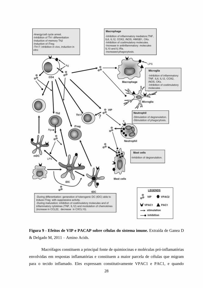
Figura 9 - Efeitos de VIP e PACAP sobre células do sistema imune. Extraída de Ganea D
& Delgado M, 2011 – Amino Acids.
Macrófagos constituem a principal fonte de quimiocinas e moléculas pró-inflamatórias
envolvidas em respostas inflamatórias e constituem a maior parcela de células que migram
para o tecido inflamado. Eles expressam constitutivamente VPAC1 e PAC1, e quando

29
expostos a estímulos inflamatórios passam a expressar também VPAC2 (Figura 9 e 10) (147,
150). As principais ações de VIP e PACAP sobre macrófagos são bem documentadas por
diversos autores; em macrófagos não estimulados, VIP e PACAP induzem a produção de IL-6
através da ativação de PKA e PKC (Figura 9 e 10) (151, 152). Em contraste, em macrófagos
estimulados (com LPS), VIP e PACAP inibem a produção das citocinas inflamatórias TNF-
alfa, IL-6 e IL-12 (Figura 9 e 10) (152-154). O efeito inibidor sobre a produção de TNF-alfa e
outros fatores inflamatórios produzidos por macrófagos aparenta ser mediado primariamente
por VPAC1, sendo que VPAC2 (induzido após estímulo) também poderia participar (155).
VIP e PACAP também se mostram capazes de induzir a síntese e liberação de moléculas anti-
inflamatórias, tais como a IL-10 e o antagonista endógeno do receptor de IL-1 (IL-1Ra),
promovendo assim a supressão de respostas inflamatórias (Figura 9 e 10) (156). Além disso, a
produção de diversas quimiocinas pode ser alterada por VIP e PACAP. Em macrófagos e na
micróglia estimulados com LPS, VIP e PACAP inibem a produção de MIP-2, IL-8, MIP-1α,
MIP-1β, MCP-1 e RANTES (157-159).
Figura 10 - Efeitos de VIP e PACAP sobre macrófagos ativados. Extraída de Ganea D &
Delgado M, 2002.

30
1.3 VIP, PACAP e a Infecção pelo HIV-1
Em 2002, Branch e colaboradores descreveram que a ativação de VPAC1 (um dos
receptores de VIP) com um ligante específico, induz um evento de sinalização que aumenta a
replicação da infecção pelo HIV-1 em linfócitos. Este estudo mostra que a ativação específica
de VPAC1 eleva a produtividade da infecção pelo HIV-1 em células primárias humanas
infectadas in vitro (160), mas não elucida os mecanismos envolvidos nesta facilitação da
infecção do HIV-1 mediada por este receptor.
Depois do trabalho de Branch e colaboradores (2002), outros estudos foram realizados
com destaque para o executado por Bokaei e colaboradores (2007), demonstrando que a
estimulação com agonistas específicos de outro receptor de VIP e PACAP, VPAC2, resulta na
inibição da integração do HIV-1 ao genoma de linfócitos, inibindo a infecção produtiva do
HIV-1 (161), embora sem definir os mecanismos responsáveis pela inibição da integração
viral. Estes resultados, portanto, demonstram uma interessante consequência da estimulação
do receptor VPAC2, resultando em um efeito inibitório na infecção por HIV-1. Este
fenômeno mostra-se contrário ao mostrado por Branch (2002), sugerindo que o VPAC1 e
VPAC2 desempenham funções opostas.
Estes achados despertam o interesse o efeito dos neuropeptídeos ligantes destes
receptores, VIP e PACAP na patogênese da infecção pelo HIV-1; mesmo assim, são poucos
os trabalhos disponíveis na literatura relacionados com o papel de VIP ou PACAP na infecção
pelo HIV-1.

31
2 JUSTIFICATIVA E HIPÓTESE
Os neuropeptídeos VIP e PACAP agem através de receptores acoplados a proteína G
(VPAC1, VPAC2 e PAC1), que, no contexto imune, são expressos por macrófagos e células
T (140, 150, 162-164). VIP e PACAP participam de uma variedade de funções do sistema
imume e uma das suas principais funções imunomodulatórias é atuarem como uma citocina
anti-inflamatória. Por conta das características imunomodulatórias VIP e PACAP têm sido
apontados como promissores alvos de estratégias terapêuticas em diversas patologias (165).
Em 2002, Branch e colaboradores descreveram que a ativação de VPAC1 com um
ligante específico, induz um evento de sinalização que aumenta a replicação do HIV-1 em
linfócitos. Este estudo mostra que a ativação específica de VPAC1 eleva a produtividade da
infecção pelo HIV-1 em células primárias humanas infectadas in vitro (160), mas não elucida
os mecanismos envolvidos nesta facilitação da infecção do HIV-1 mediada por este receptor.
Outro estudo, realizado por Bokaei e colaboradores (2007), demonstrou que a
estimulação de VPAC2 com agonistas específicos, resulta na inibição da integração do HIV-1
ao genoma de linfócitos, inibindo a infecção produtiva do HIV-1, embora sem definir os
mecanismos responsáveis pela inibição da integração viral. Este fenômeno mostra-se
contrário ao mostrado por Branch (2002), sugerindo que o VPAC1 e VPAC2 desempenham
funções opostas.
Estes achados despertam o interesse no efeito dos neuropeptídeos ligantes destes
receptores, VIP e PACAP na patogênese da infecção pelo HIV-1; mesmo assim, são poucos
os trabalhos disponíveis na literatura relacionados com o papel de VIP ou PACAP na infecção
pelo HIV-1. Considerando essa questão, nos propusemos a analisar a ação destas moléculas
sobre a replicação do HIV-1 em macrófagos, células quais que expressam os receptores de
VIP e PACAP, que são alvos primários do HIV-1, e que durante a infecção constituem um
importante reservatório viral no hospedeiro. Assim, neste estudo avaliamos a capacidade
inibitória dos neuropeptídeos VIP e PACAP sobre o HIV-1 em macrófagos, com a intenção
de descrever o fenômeno e identificar os receptores envolvidos nesta ação.
32
3 OBJETIVOS
3.1 Objetivo Geral
Avaliar a ação dos neuropeptídeos VIP e PACAP sobre a replicação do HIV-1 em
macrófagos, focando no entendimento da participação dos receptores VPAC1, VPAC2 e
PAC1 nessa ação, e buscando mecanismos que respondam pelo fenômeno.
3.2 Objetivos Específicos
Analisar o perfil de replicação do HIV-1 em macrófagos expostos a diferentes doses
de VIP e PACAP.
Analisar se VIP e PACAP poderiam atuar de forma sinergística ou aditiva para
promover a inibição do HIV-1.
Definir a contribuição individual dos receptores de VIP e PACAP no fenômeno
inibitório da replicação do HIV-1.
Verificar se VIP e PACAP modulariam a produção de β-quimiocinas e IL-10 em
macrófagos e se elas participariam do fenômeno de inibição da replicação do HIV-1
induzido por VIP e PACAP.

33
4 METODOLOGIA E RESULTADOS
A seção de metodologia e resultados está inserida no artigo anexado “Macrophage
Resistance to HIV-1 Infection Is Enhanced by the Neuropeptides VIP and PACAP”, cujo
conteúdo representa os objetivos citados na seção anterior dessa dissertação, a exceção de um
resultado obtido após a publicação deste artigo. O resultado citado está descrito na sub-seção
4.1, sob o título “Efeito individual e associado de VIP e PACAP sobre a replicação do HIV-1
em função do tempo de tratamento”.
Referência completa do artigo anexado:
Temerozo JR, Joaquim R, Regis EG, Savino W, Bou-Habib DC. Macrophage
Resistance to HIV-1 Infection Is Enhanced by the Neuropeptides VIP and PACAP.
PLoS One. 2013;8(6):e67701.

34
4.1 Efeito individual e associado de VIP e PACAP sobre a replicação do HIV-1 em
função do tempo de tratamento
Considerando que a concentração de 10 nM de VIP e PACAP isolados promove a
inibição da replicação do HIV-1 em macrófagos, e que a combinação de ambos em doses que
quando isolados eram não-funcionais (1 nM), promove o mesmo efeito inibitório, nos
perguntamos se poderíamos reproduzir a inibição da replicação do HIV-1 com essas doses
não-funcionais isoladas utilizando um protocolo de tratamento consecutivo; e se, com o
mesmo protocolo, poderíamos também observar uma maior inibição quando utilizando a
concentração ótima de VIP e PACAP (10 nM). Com base nessa hipótese, expusemos culturas
de macrófagos infectados pelo HIV-1 aos neuropeptídeos VIP e PACAP combinados e
isolados, nas suas doses ótimas e nas suas doses não-funcionais, para avaliar a inibição da
replicação do HIV-1; comparando o tratamento único e um protocolo de tratamento repetido
em dias não-consecutivos. Para melhor compreensão dos resultados, calculamos a área sob as
curvas de replicação oriundas das variáveis de tratamento (método descrito em Temerozo et
al, 2013, anexado a dissertação). Como mostra a Figura 11C, o protocolo de tratamento com
três exposições dos macrófagos aos neuropeptídeos (isolados, e na dose inicialmente não-
funcional de 1 nM), resultou na inibição da replicação do HIV-1 após 15 dias de infecção,
quando comparado com o tratamento em tempo único e de forma comparável com o
tratamento padrão de 10 nM e o tratamento combinado (ambos neuropeptídeos, 1 nM cada)
no mesmo protocolo. Em relação a possível ampliação do efeito inibitório de VIP e PACAP
quando em 10 nM e submetidos a esse protocolo de tratamento, não observamos incremento
na ação sobre o HIV-1, como também pode ser observado na Figura 11C. Assim,
evidenciamos que a inibição da replicação do HIV-1 pelos neuropeptídeos VIP e PACAP
pode ser obtida com o uso isolado dos mesmos em doses baixas; e que, no que tange ao nosso
modelo, existe a princípio, um platô máximo para a inibição da replicação do HIV-1 obtida
com o uso de VIP e PACAP.

35
Tratamento
0 5 10 150
1000
2000
3000
4000
5000
6000
7000
8000Controle
VIP 10 nM
PACAP 10 nM
VIP 1 nM
PACAP 1 nM
VIP/PACAP 1 nM
Dias pós-infecção
Rep
licação
do
HIV
-1 [
p24]
pg
/ml
Tratamento
0 5 10 150
1000
2000
3000
4000
5000
6000
7000
8000Controle
VIP 10 nM
PACAP 10 nM
VIP 1 nM
PACAP 1 nM
VIP/PACAP 1 nM
Dias pós-infecção
Rep
licação
do
HIV
-1 [
p24]
pg
/ml
Contr
ole
VIP 1
0 nM
PACAP 1
0 nM
VIP 1
nM
PACAP 1
nM
VIP/P
ACAP 1
nM
0
10000
20000
30000
40000
50000
Dose única
Dose tripla
*
*
Rep
licação
do
HIV
-1 (
AU
C)
A
B
C
Figura 11 - Efeito individual e associado de VIP e PACAP sobre a replicação do HIV-1
em função do tempo de tratamento. (A, B, C) Macrófagos foram infectados com o isolado
Ba-L (HIV-1 Subtipo B, CCR5 trópico) e tratados com diferentes concentrações de VIP e
PACAP no regime de dose única (Figura 10A, dia 0, seta indicativa) ou dose tripla (Figura
10B, dias 0, 5 e 10, seta indicativa). Foram colhidas amostras de sobrenadantes após 5, 10 e
15 dias de infecção, e a replicação viral foi avaliada por ELISA para o antígeno p24 do
capsídeo do HIV-1. (n=4) **, p<0.01

Macrophage Resistance to HIV-1 Infection Is Enhancedby the Neuropeptides VIP and PACAPJairo R. Temerozo1, Rafael Joaquim2, Eduardo G. Regis1, Wilson Savino1, Dumith Chequer Bou-Habib1*
1 Laboratory on Thymus Research, Oswaldo Cruz Institute/Fiocruz, Rio de Janeiro, Brazil, 2 Laboratory of Microbiology, Central Hospital of Maputo, Maputo, Mozambique
Abstract
It is well established that host factors can modulate HIV-1 replication in macrophages, critical cells in the pathogenesis ofHIV-1 infection due to their ability to continuously produce virus. The neuropeptides VIP and PACAP induce well-characterized effects on macrophages through binding to the G protein-coupled receptors VPAC1, VPAC2 and PAC1, buttheir influence on HIV-1 production by these cells has not been established. Here, we describe that VIP and PACAP reducemacrophage production of HIV-1, acting in a synergistic or additive manner to decrease viral growth. Using receptorantagonists, we detected that the HIV-1 inhibition promoted by VIP is dependent on its ligation to VPAC1/2, whereasPACAP decreases HIV-1 growth via activation of the VPAC1/2 and PAC1 receptors. Specific agonists of VPAC2 or PAC1decrease macrophage production of HIV-1, whereas sole activation of VPAC1 enhances viral growth. However, thecombination of specific agonists mimicking the receptor preference of the natural neuropeptides reproduces the ability ofVIP and PACAP to increase macrophage resistance to HIV-1 replication. VIP and PACAP up-regulated macrophage secretionof the b-chemokines CCL3 and CCL5 and the cytokine IL-10, whose neutralization reversed the neuropeptide-inducedinhibition of HIV-1 replication. Our results suggest that VIP and PACAP and the receptors VPAC2 and PAC1 could be used astargets for developing alternative therapeutic strategies for HIV-1 infection.
Citation: Temerozo JR, Joaquim R, Regis EG, Savino W, Bou-Habib DC (2013) Macrophage Resistance to HIV-1 Infection Is Enhanced by the Neuropeptides VIPand PACAP. PLoS ONE 8(6): e67701. doi:10.1371/journal.pone.0067701
Editor: Jialin Charles Zheng, University of Nebraska Medical Center, United States of America
Received December 6, 2012; Accepted May 22, 2013; Published June 20, 2013
Copyright: � 2013 Temerozo et al. This is an open-access article distributed under the terms of the Creative Commons Attribution License, which permitsunrestricted use, distribution, and reproduction in any medium, provided the original author and source are credited.
Funding: This work was supported by Conselho Nacional de Desenvolvimento Cientıfico e Tecnologico (CNPq; grant # 475958/2011-0) and Fundacao CarlosChagas Filho de Amparo a Pesquisa do Estado do Rio de Janeiro (Faperj; grant # E-26/102755/2008). The funders had no role in study design, data collection andanalysis, decision to publish, or preparation of the manuscript.
Competing Interests: The authors have declared that no competing interests exist.
* E-mail: [email protected]
Introduction
The neuropeptides Vasoactive Intestinal Peptide (VIP) and
Pituitary Adenylate Cyclase-activating Peptide (PACAP) belong to
the secretin/glucagon family of peptides and were initially
discovered due to their vasodilatation properties on the gastroin-
testinal tract and ability to activate rat pituitary adenylate cyclase,
respectively [1,2]. VIP and PACAP present a 68% homology in
their amino acid sequences, and share many biological properties
[3,4] through their interaction with the G protein-coupled
receptors VPAC1, VPAC2 and PAC1. PACAP binds to all three
receptors, with higher affinity to PAC1, while VIP interacts
preferentially with VPAC1 and VPAC2 [5–8]. VIP and PACAP
are produced by Th2 CD4+ and CD8+ T cells, and their receptors
are expressed by a variety of cell types, including T cells,
macrophages and dendritic cells [4].
VIP and PACAP have well-characterized effects on the immune
system and anti-inflammatory properties, including inhibition of
macrophage adherence and down-regulation of inflammatory
cytokines and reactive oxygen species [9,10–14]. Moreover, they
can induce production of the anti-inflammatory cytokine IL-10
[12,14]. Due to their immunomodulatory properties, both
neuropeptides have been considered as promising therapeutic
agents for a range of pathologies [15–17].
Macrophages play a central role in the pathogenesis of the
human immunodeficiency virus type 1 (HIV-1) infection due to
their ability to resist HIV-1-mediated cytopathic effects and to
continuously produce virus even in the presence of antiretrovirals
[18–20]. They function as an HIV-1 reservoir and contribute in
HIV-1 transmission to CD4+ T cells and virus propagation in
lymphoid tissues [21,22]. Considering that HIV-1 replication in
macrophages can be modulated by a variety of inflammatory
mediators and cytokines [23–25], identifying factors that influence
HIV-1 growth in these cells is essential to understand the
immunopathogenesis of HIV-1 infection and to design novel
strategies to control HIV-1 propagation. We recently reported that
the neuroimmunomodulatory molecule Nerve Growth Factor
(NGF) stimulates HIV-1 replication in primary monocyte-derived
macrophages [26], and we now address whether the immunosup-
pressive neuropeptides VIP and PACAP, which also regulate the
functioning of the neuro-immune-endocrine system, could also
affect HIV-1 production in those cells.
Few studies have addressed the biological effects of VIP and
PACAP during HIV-1 infection, which have mainly focused on
the repercussion of VIP and PACAP receptor ligation on HIV-1
production, describing that VPAC1 facilitates productive HIV-1
infection in CD4+ T cell lines [27], and that VPAC2 stimulation
diminishes HIV-1 production in peripheral blood mononuclear
cells (PBMCs) and in CD4+ T cell lines [28]. These findings
suggest that the sole activation of VPAC1 or VPAC2 receptors can
lead to opposite effects on HIV-1 replication, but the consequence
of the simultaneous ligation of these receptors and PAC1 by their
natural ligands on viral production is unknown. Therefore,
because the existent data regarding the influence of VIP and
PLOS ONE | www.plosone.org 1 June 2013 | Volume 8 | Issue 6 | e67701

PACAP receptors on HIV-1 infection were obtained in T cells
using selective receptor agonists, we analyzed whether these
neuropeptides could directly modulate the viral production in
HIV-1-infected monocyte-derived macrophages, a possibility that
has not been pursued thus far. We found that VIP and PACAP
increased macrophage resistance to HIV-1 replication by inducing
the synthesis of b-chemokines and IL-10 following preferential
activation of the receptors VPAC2 and PAC1.
Materials and Methods
Ethics StatementAll experimental procedures involving human cells were
performed with samples obtained after written informed consent
and were approved by the Research Ethics Committee of the
Oswaldo Cruz Foundation/Fiocruz (Rio de Janeiro, RJ, Brazil)
under the number 397-07.
HIV-1 isolates and reagentsThe CCR5-dependent isolate HIV-1Ba-L was obtained through
the AIDS Research and Reference Reagent Program (NIH,
Bethesda, MD). The neuropeptides VIP and PACAP and the VIP
antagonist, which blocks both the VPAC1 and VPAC2 receptors,
were from Anaspec (USA). The recombinant protein Maxadilan
(PAC1 agonist) and its truncated form MaxadilanD65 (M65;
PAC1 antagonist) were kindly donated by Dr. Ethan A. Lerner
(Department of Dermatology, Massachusetts General Hospital,
MA, USA). The VPAC1 and VPAC2 agonists Ala11,22,28-VIP and
Bay 55-9837, respectively, were obtained from Tocris Bioscience
(Bristol, UK). The neutralizing antibodies to CCL3, CCL4 and
CCL5 and to the IL-10 receptor were obtained from Peprotech
(NJ, USA) and Abcam (MA, USA), respectively. The endotoxin
levels in the VIP and PACAP preparations were below the lower
limit of detection (0.1 EU/mL), as measured by the Limulus
Amebocyte Lysate (LAL) assay (Lonza).
CellsHuman monocyte-derived macrophages were obtained from
PBMCs that had been isolated by density gradient centrifugation
(Ficoll-Paque Premium 1.077; GE Healthcare Biosciences, PA,
USA) from buffy coat preparations of blood from healthy donors,
through adherence onto plastic plates. Briefly, 1.5 – 2.06106
PBMCs were plated onto 48-well plates (Corning, MA, USA) in
Dulbecco’s modified Eagle’s medium (DMEM; LGC Bio, SP,
Brazil) containing 10% normal human serum (EMD Millipore,
MA, USA) and penicillin-streptomycin (LGC Bio, SP, Brazil).
Cells were maintained at 37uC in 5% CO2 for 6–7 days for
monocyte differentiation into macrophages. Non-adherent cells
were washed out, and the remaining macrophage layer was
maintained in DMEM with 5% human serum. Macrophage purity
was . 90%, as determined by flow cytometry (FACScan; Becton
Dickinson, NJ, USA) analysis using anti-CD3 (BD Biosciences
Pharmingen, CA, USA) and anti-CD68 (Southern Biotech, AL,
USA) monoclonal antibodies.
Macrophage production of b-chemokines and IL-10Uninfected macrophages were treated with VIP or PACAP
(10 nM), and concentrations of the b-chemokines CCL3 and
CCL5 and of the cytokine IL-10 in the culture supernatants were
measured using specific ELISA kits (R&D Systems, MN, USA,
and eBioscience Inc, CA, USA, respectively). The results are
shown as mass/volume and also by the area under curve (AUC)
transformation, which allows a global analysis of the induced
production of the mediators.
HIV-1 infectionMacrophages were exposed for 16–18 h to viral suspensions
containing 5–10 ng/mL of HIV-1 p24 antigen, as previously
described [29]. The infected cells were then washed, replenished
with fresh medium and maintained under standard culture
conditions. HIV-1 replication was evaluated in cell culture
supernatants after 12–14 days using an ELISA kit for HIV-1
p24 antigen (ZeptoMetrix Corp, NY, USA). Due to the common
donor-to-donor variation of HIV-1 replication in primary cells
[30], some HIV-1 inhibition results are presented normalized to
viral production by macrophages maintained only in culture
medium, with the absolute values shown in the legend of the
figure.
Effect of VIP and PACAP on HIV-1 replicationHIV-1-infected macrophages were treated either with VIP or
PACAP immediately after cell infection, and viral production was
measured as described above. After establishing that VIP and
PACAP decreased viral replication, we addressed the relative
contributions of the VIP and PACAP receptors using two different
approaches. Initially, acutely HIV-1-infected cells were exposed to
receptor antagonists for 15 min followed by the addition of VIP
and PACAP to cell preparations. In later experiments, infected
macrophages were treated with specific pharmacological agonists
of the VIP and PACAP receptors (as listed above). In another set
of experiments, HIV-1-infected macrophages were treated with
either neuropeptide five days after infection together with
neutralizing antibodies to the IL-10 receptor (1 mg/mL) or to
the b-chemokines CCL3, CCL4 and CCL5 (1 mg/mL each). HIV-
1 replication was evaluated as previously described.
Statistical AnalysisAll results presented in this study were prepared using
GraphPad Prism 5.0 software (CA, USA). Statistical analysis
calculation was performed using one-way ANOVA and the
Tukey-Kramer tests. The results are shown as the mean 6 SEM
(standard error of the mean), and the comparisons between values
were considered significantly different when the p value was less
than 0.05 (p#.05 = *; p#.01 = **; p#.001 = ***).
Results
VIP and PACAP treatment inhibited HIV-1 production inmacrophages
Because activation of the receptors VPAC1 and VPAC2 has
previously resulted in opposite effects during HIV-1 infection
[27,28], we initially investigated whether the neuropeptides VIP
and PACAP, the natural ligands of those receptors, would also
affect HIV-1 replication. To test this hypothesis, HIV-1-infected
monocyte-derived macrophages were treated with VIP or PACAP.
We first observed that both neuropeptides induced a significant
reduction in virus replication (Fig. 1). VIP and PACAP were each
individually able to decrease HIV-1 replication, achieving 33%
and 38% of viral inhibition at 5 nM and 62% and 58% at 10 nM
concentrations for VIP and PACAP, respectively. These results
suggest that both neuropeptides were similarly effective in their
ability to reduce HIV-1 production in macrophages. Higher
concentrations of VIP or PACAP did not inhibit virus production
and actually enhanced it (VIP at 100 nM), possibly due to receptor
desensitization or an inverse agonist effect, as discussed later.
Therefore, the next experiments were performed using the optimal
inhibitory concentration of 10 nM for both neuropeptides.
VIP and PACAP Inhibit HIV-1 Infection
PLOS ONE | www.plosone.org 2 June 2013 | Volume 8 | Issue 6 | e67701

VIP and PACAP present synergistic and additive effectson HIV-1 inhibition
As VIP and PACAP share receptors, we analyzed whether these
neuropeptides could cooperatively modulate HIV-1 replication by
exposing infected macrophages to combinations of sub-optimal or
optimal viral inhibitory concentrations of VIP and PACAP.
Combinations of 1 nM and 5 nM significantly potentiated
inhibition relative to their individual activities, while no increment
of HIV-1 inhibition occurred when both peptides were combined
at a concentration of 10 nM (Fig. 2). To accurately classify the
nature of this finding, we calculated the interaction coefficient of
VIP and PACAP at those concentrations by dividing the inhibition
percentages found when the peptides were associated by the sum
of the inhibition of each isolated peptide (Fig. 2D; an interaction
coefficient on the order of 1 indicates an additive phenomenon,
whereas values greater than 1 indicate a synergistic effect).
Therefore, VIP and PACAP synergize at 1 nM and act in an
additive manner on viral production at 5 nM. These results
suggest that combinations of small concentrations of VIP and
PACAP could result in potent pharmacological activity, which
could be physiologically relevant in situations where both peptides
are simultaneously present in the tissue microenvironment.
Receptor engagement in the VIP and PACAP modulationof HIV-1 replication
Because VIP preferentially activates the VPAC1 and VPAC2
receptors and PACAP binds the three receptors with high affinity
[5–8], we analyzed the neuropeptide dependence of these
receptors to inhibit HIV-1 production using two distinct assays.
Initially, we added specific antagonists of PAC1 or VPAC1/2 to
HIV-1-infected cells before treating them with VIP or PACAP. As
shown in Fig 3A, VIP-induced HIV-1 inhibition is largely
dependent on VPAC1/2, since blockade of both receptors
abrogated the VIP-mediated inhibition of HIV-1 production with
no significant changes following PAC1 blockade. We also observed
that PACAP could inhibit HIV-1 replication via activation of all
three receptors, as its ability to decrease viral growth was
maintained when VPAC1/2 or PAC1 was antagonized separately
but was abolished when all three receptors were blocked together
(Fig 3B).
We further studied the role of the individual receptors in the
VIP- and PACAP-mediated regulation of HIV-1 replication by
using specific agonists to VPAC1, VPAC2 and PAC1 (ALA-VIP,
Bay 55 and Maxadilan, respectively). We found that the VPAC1
agonist at 5 nM increased HIV-1 production by 48%, whereas the
VPAC2 agonist at 5 nM and 10 nM reduced viral growth by 31%
and 35%, respectively. The PAC1 agonist at 5 nM and 10 nM
decreased HIV-1 replication by 56% and 46%, respectively (Fig
4). Of note, the optimal concentrations of the receptor agonists
that significantly modulated viral production were similar to those
of the natural receptor ligands VIP and PACAP.
Due to the opposing effects of VPAC1 and VPAC2 or PAC1
engagement, we analyzed the outcome of agonist combinations on
viral production. For these assays we used the agonists at sub-
optimal concentrations to allow for comparison with non-
saturating doses of the natural ligands VIP and PACAP.
Combination treatment with VPAC1 and VPAC2 agonists
decreased HIV-1 replication by 48%, similar to the effect observed
with sub-optimal doses of VIP. Their combined usage with a
PAC1 agonist reduced viral growth by 68%, similar to the additive
effects observed during co-treatment with VIP and PACAP (Fig.
5). These results showed that combination treatment with agonists
of the different receptors mimicked the receptor preference of both
natural peptides in reproducing the individual or additive effects of
VIP and PACAP treatment on HIV-1 replication. Of note,
simultaneous activation of VPAC1 and PAC1 slightly increased
HIV-1 replication, whereas binding of VPAC2 plus PAC1 did not
change HIV-1 replication (data not shown). In conclusion, VIP
depended on the ligation of VPAC1/2 to increase macrophage
resistance to HIV-1 growth, and PACAP promoted the same
phenomenon either by activating PAC1 only or through activating
VPAC1/2 plus PAC1.
VIP and PACAP increase b-chemokine production bymacrophages
VIP and PACAP can induce the production of the b-
chemokines CCL3, CCL4 and CCL5 in microglial cells, an effect
associated with the prevention of HIV-1 gp120-induced apoptosis
[31,32]. Because b-chemokines are potent inhibitors of HIV-1
infection [33,34], we evaluated whether VIP and PACAP could
also induce macrophage secretion of these molecules. Indeed, both
neuropeptides enhanced macrophage release of CCL3 and CCL5,
with CCL3 production peaking 96 h after stimulation with either
peptide (Fig. S1A) and the maximum production of CCL5
occurring 24 h and 48 h after VIP or PACAP stimulation,
respectively (Fig. S1B). VIP and PACAP more than doubled
CCL3 and CCL5 production relative to untreated cells based on
measuring chemokine production by the area under the curve
(AUC) (Figs. 6A and 6B). Moreover, because the differences in
chemokine production induced by VIP or PACAP were not
significantly different (based on AUC), we can postulate that the
ability of either peptide to induce chemokine production is similar.
VIP and PACAP increase IL-10 production bymacrophages
The immunomodulatory activities of VIP and PACAP are at
least partially dependent of IL-10 [9], an anti-inflammatory
cytokine able to inhibit the HIV-1 replication [35,36]. We also
detected that VIP and PACAP increase macrophage production of
IL-10 (Fig. S1C), which peaked 48 h after the stimuli. Based on
Figure 1. VIP and PACAP inhibit HIV-1 replication. Macrophageswere infected with an R5-tropic HIV-1 isolate (Ba-L) and treated oncewith different concentrations of the neuropeptides, as indicated. Virusreplication was measured in the culture supernatants by an HIV-1 p24ELISA 12-14 days after infection. Data represent means 6 SEM of fiveindependent experiments for each peptide. *p#.05; ***p#.001.doi:10.1371/journal.pone.0067701.g001
VIP and PACAP Inhibit HIV-1 Infection
PLOS ONE | www.plosone.org 3 June 2013 | Volume 8 | Issue 6 | e67701

AUC analyses (Fig. 6C), VIP treatment doubled IL-10 production,
whereas PACAP treatment tripled macrophage release of this
cytokine.
Contribution of b-chemokines and IL-10 to the VIP- andPACAP- mediated inhibition of HIV-1 replication
We next evaluated whether these mediators were implicated in
the ability of VIP and PACAP to reduce HIV-1 growth by adding
the neuropeptides to infected macrophages together with neutral-
izing antibodies to CCL3, CCL4 and CCL5, or to the IL-10
receptor. This experiment was conducted five days after infection
because this is the approximate time when the infection becomes
productive, thus allowing the antibodies to neutralize the anti-
HIV-1 effector molecules when new rounds of infections were
occurring. Indeed, neutralization of those three b-chemokines and
blocking of the IL-10 receptor significantly reduced the inhibitory
effects of VIP and PACAP on HIV-1 replication (Fig. 7), showing
that CCL3, CCL4 and CCL5 and IL-10 are implicated in
neuropeptide-mediated inhibition of HIV-1 growth.
Discussion
VIP and PACAP are pleiotropic factors associated with a
number of physiological processes, such as endocrine, metabolic
and gastrointestinal effects, also including modulatory effects on
the immune system. The receptors for VIP and PACAP, the G
coupled-receptors VPAC1, VPAC2 and PAC1, are widely
distributed, a feature that allows VIP and PACAP to exert their
large range of effects. Here, we show that VIP and PACAP
treatment increased resistance to HIV-1 replication in primary
macrophages by inducing the production of the b-chemokines
CCL3 and CCL5 and the cytokine IL-10, molecules that are able
to reduce HIV-1 growth in vitro. Our study focused, for the first
time, on the ability of the natural peptides VIP and PACAP to
modulate HIV-1 infection in a primary target cell, in addition to
defining the relative contribution of each of their receptors to this
Figure 2. Effects of combined VIP and PACAP treatment on HIV replication. HIV-1-infected macrophages were simultaneously treated with1 nM, 5 nM or 10 nM of VIP and PACAP (A, B and C, respectively), and virus replication was measured as above. Data represent means 6 SEM of threeindependent experiments. (D) Equation used to calculate the interaction coefficient of VIP and PACAP at the indicated concentrations, based on thelevels of HIV-1 inhibition shown in A, B and C. *p#.05; **p#.01; ***p#.001.doi:10.1371/journal.pone.0067701.g002
VIP and PACAP Inhibit HIV-1 Infection
PLOS ONE | www.plosone.org 4 June 2013 | Volume 8 | Issue 6 | e67701
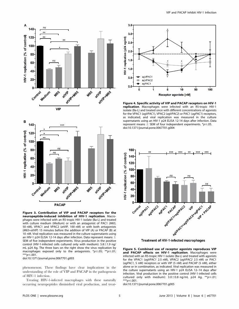
phenomenon. These findings have clear implications in the
understanding of the role of VIP and PACAP in the pathogenesis
of HIV-1 infection.
Treating HIV-1-infected macrophages with these naturally
occurring neuropeptides diminished viral production, and treat-
Figure 3. Contribution of VIP and PACAP receptors for theneuropeptide-induced inhibition of HIV-1 replication. Macro-phages were infected with an R5-tropic HIV-1 isolate (Ba-L) and treatedwith culture medium (Medium) or with an antagonist of PAC1 (M65,50 nM), VPAC1 and VPAC2 (atVIP, 100 nM) or with both antagonists(M65+atVIP) 15 minutes before the addition of VIP (A) or PACAP (B) at10 nM. Viral replication was measured in the culture supernatants usingan HIV-1 p24 ELISA 12-14 days after infection. Data represent means 6
SEM of five independent experiments. Virus production in the positivecontrol (HIV-1-infected cells cultured only with medium): 5.861.9 ng/mL p24 Ag. The three bars on the right show the virus replication bymacrophages exposed only to the antagonists. *p#.05; **p#.01;***p#.001.doi:10.1371/journal.pone.0067701.g003
Figure 4. Specific activity of VIP and PACAP receptors on HIV-1replication. Macrophages were infected with an R5-tropic HIV-1isolate (Ba-L) and treated once with different concentrations of agonistsfor the VPAC1 (agVPAC1), VPAC2 (agVPAC2) or PAC1 (agPAC1) receptors,as indicated, and viral replication was measured in the culturesupernatants using an HIV-1 p24 ELISA 12-14 days after infection. Datarepresent means 6 SEM of four independent experiments. *p#.05.doi:10.1371/journal.pone.0067701.g004
Figure 5. Combined use of receptor agonists reproduces VIPand PACAP effects on HIV-1 replication. Macrophages wereinfected with an R5-tropic HIV-1 isolate (Ba-L) and treated with agonistsfor the VPAC1 (agVPAC1 2.5 nM), VPAC2 (agVPAC2 2.5 nM) or PAC1(agPAC1, 5 nM) receptors or with VIP (5 nM) and PACAP (5 nM), eitheralone or in combination, as indicated. Viral replication was measured inthe culture supernatants using an HIV-1 p24 ELISA 12–14 days afterinfection. Viral production in the positive control (HIV-1-infected cellscultured only with medium): 3.060.8 ng/mL p24 Ag. **p#.01;***p#.001.doi:10.1371/journal.pone.0067701.g005
VIP and PACAP Inhibit HIV-1 Infection
PLOS ONE | www.plosone.org 5 June 2013 | Volume 8 | Issue 6 | e67701

ment with specific agonists of the neuropeptide receptors VPAC2
and PAC1 showed similar effects. The inhibitory effect on viral
replication was not dose-dependent within the range of tested
concentrations of VIP and PACAP between 1 nM and 100 nM,
with the optimal inhibitory concentration at 10 nM for both
peptides. The finding that VIP treatment at a concentration 5-fold
that of the optimal HIV-1 inhibitory concentration and PACAP
treatment at a 10-fold ratio did not influence viral production may
be explained by receptor desensitization. On the other hand, the
Figure 6. VIP and PACAP induce CCL3, CCL5 and IL-10production in macrophages. Figure shows the production of CCL3(A), CCL5 (B) and IL-10 (C) by area under the curve (AUC) analysis, whichwas calculated based on the respective concentrations measured byELISA (See Figure S1). Data represent means 6 SEM of six (CCL3) andfour (CCL5 and IL10) independent experiments. *p#.05; **p#.01;***p#.001.doi:10.1371/journal.pone.0067701.g006
Figure 7. b-chemokines and IL-10 are implicated in the VIP- andPACAP-induced inhibition of HIV-1 replication. Macrophageswere infected with an R5-tropic HIV-1 isolate (Ba-L), and after 5 days,were treated with VIP or PACAP plus anti-CCL3, CCL4 and CCL5antibodies (a-bC) (A), anti-IL-10 receptor antibodies (a-IL10R), or isotypecontrol antibodies. Viral replication was measured in the culturesupernatants using an HIV-1 p24 ELISA 12–14 days after infection. Datarepresent means 6 SEM of five (A) or four (B) independent experiments.Virus production in the positive control (HIV-1-infected cells culturedonly with medium): (A) 14.869.0 ng/mL and (B) 14.567.0 ng/mL p24Ag. *p#.05; **p#.01.doi:10.1371/journal.pone.0067701.g007
VIP and PACAP Inhibit HIV-1 Infection
PLOS ONE | www.plosone.org 6 June 2013 | Volume 8 | Issue 6 | e67701

observed increase in HIV-1 production induced by 100 nM VIP
could be explained by the property of inverse agonism that occurs
for some ligands at saturating concentrations, a phenomenon that
has already been described for a variety of G-coupled receptor
ligands [37]. Alternatively, taking into account that the sole
activation of VPAC1 facilitates HIV-1 infection in CD4+ T cell
lines (27), one could explain the HIV-1 promoting effect by VIP at
100 nM due to a possible preferential engagement of VPAC1 at
high VIP concentrations, although it has been described that the
affinity of VIP for its receptors VPAC1 and VPAC2 is similar [4–
7]. Importantly, the concentration of both neuropeptides that
effectively inhibited HIV-1 production (10 nM) also possesses a
variety of immunomodulatory roles [38–41], suggesting that the
ability of these peptides to influence HIV-1 replication in
macrophages is associated with their immunoregulatory activities
on these cells.
The ability of VIP and PACAP to down-regulate HIV-1
production became even more evident in light of the experiment
testing their additive and synergistic activity on infected macro-
phages. Both neuropeptides presented an additive effect at 5 nM
concentration, and showed potent synergy at 1 nM concentration,
and their combined effects at these concentrations were the same
as those observed following treatment with each molecule
individually at 10 nM concentration (Fig. 2). Furthermore, the
addition of sub-optimal concentrations of the receptor agonists to
HIV-1-infected macrophages restricted the viral growth in a
similar manner to treatment with equivalent doses of VIP and
PACAP (Fig. 5). Thus, even taking into account that the activation
of the VPAC1 receptor may favor HIV-1 replication, we believe
that the concomitant engagement of the three receptors by the
native neuropeptides, as presumably occurs in lymphoid tissues,
will indeed enhance the macrophage resistance to HIV-1 growth.
VIP promoted HIV-1 inhibition through stimulation of the
receptors VPAC1 and VPAC2 but not through stimulation of
PAC1. This effect likely occurred due to the high affinity of VIP
for VPAC1 and VPAC2 and its low affinity for PAC1. The ability
of PACAP to diminish HIV-1 replication, on the other hand,
resulted from its ligation of all three receptors because its effect was
only abrogated when all three receptors were blocked. Because the
PACAP affinity for PAC1 is higher than its affinity for VPAC1 or
VPAC2 [8], it is conceivable that it inhibits HIV-1 upon
preferential binding to PAC1, but can also exert an inhibitory
effect following ligation to VPAC1 and VPAC2 in the absence or
hindrance of PAC1, or in a situation of excess PACAP
concentration.
We defined the receptors preference for the HIV-1 inhibitory
activity of VIP and PACAP using specific receptor agonists. The
combination of these molecules not only mimicked the effects of
the natural neuropeptides but also established the receptors
VPAC2 and PAC1 as HIV-1 inhibitory mediators, and VPAC1
as an enhancer of HIV-1 production. Our findings are in
agreement with previous studies that reported that VPAC1
facilitates productive HIV-1 infection in CD4+ tumor cell lines
[27], and that VPAC2 activation inhibits HIV-1 integration and
viral production in CD4+ tumor cell lines and PBMCs [28]. In
addition, we defined the role of the PAC1 receptor during HIV-1
infection. Therefore, it is reasonable to assume that the
simultaneous activation of these receptors by their natural ligands
VIP and PACAP results in reduced HIV-1 replication in
macrophages, even under conditions in which the receptor
VPAC1 is present and activated, and that the HIV-1-enhancing
activity of the sole activation of VPAC1 activation is overcome by
the concomitant recruitment of the inhibitory receptors VPAC2
and PAC1.
VIP and PACAP induced macrophage release of CCL3 and
CCL5, which contributed to the HIV-1 inhibitory effect elicited by
both neuropeptides as evidenced by immunoneutralization of both
b-chemokines (and CCL4 as well). VIP and PACAP can inhibit b-
chemokine production upon pro-inflammatory induction [40,42],
but they are also able to stimulate the production of chemokines in
different settings [31,32,43–45], pointing out their ability to
regulate the chemokine axis. VIP and PACAP have been
suggested to confer a protective role against HIV-1 in the central
nervous system through CCL3 and CCL5 production [46], and
our results now imply that these neuropeptides can offer a systemic
protection against HIV-1 by boosting macrophage resistance to
viral growth by augmenting the secretion of these mediators.
A concurrent mechanism implicated in HIV-1 inhibition by
VIP and PACAP is their ability to induce macrophage secretion of
IL-10, a cytokine that inhibits HIV-1 by interfering in the reverse
transcription of viral RNA [35,47]. IL-10 is readily produced by
human and murine cells stimulated by VIP and PACAP and is a
critical mediator of the anti-inflammatory properties of these
neuropeptides. Here, we found thatIL-10 neutralization reduced
the ability of VIP and PACAP to inhibit HIV-1 replication,
indicating that IL-10, likewise the b-chemokines is required to
increase macrophage resistance following treatment with these
neuropeptides.
In conclusion, our study shows that VIP and PACAP, molecules
endowed with a large spectrum of physiological activities on the
neuro-immune-endocrine system, can increase macrophage resis-
tance to HIV-1 replication. It is remarkable to observe the
contrasting roles of neurotrophic molecules on the HIV-1 biology,
as we recently showed that Nerve Growth Factor (NGF) favors
viral replication in macrophages [26]. Our findings also open new
possibilities for therapeutic strategies, in which HIV-1 replication
could be controlled through activation of VPAC2 and PAC1
receptors through their natural ligands VIP and PACAP, or by
their specific agonists, in association with the present available
antiretroviral compounds.
Supporting Information
Figure S1 VIP and PACAP induce CCL3, CCL5 and IL-10production in macrophages. Macrophages were left untreated
(Nil) or treated with VIP or PACAP (10 nM), and supernatants
were collected at different time-points. The concentrations of
CCL3 (A), CCL5 (B) and IL-10 (C) were measured by ELISA.
Data represent means 6 SEM of six (CCL3) and four (CCL5 and
IL10) independent experiments.
(TIF)
Acknowledgments
We thank the Hemotherapy Service of the Hospital Clementino Fraga
Filho (Federal University of Rio de Janeiro, Brazil) for providing buffy-
coats, and the NIH AIDS Research and Reference Reagent Program
(Division of AIDS, NIAID, NIH, Bethesda, MD) for providing the HIV-1
isolate Ba-L. The recombinant protein Maxadilan and its truncated form
MaxadilanD65 (M65) were kindly donated to us by Dr. Ethan A. Lerner
(Department of Dermatology, Massachusetts General Hospital, MA, USA).
Author Contributions
Conceived and designed the experiments: JRT EGR WS DCBH.
Performed the experiments: JRT RJ EGR. Analyzed the data: JRT WS
DCBH. Wrote the paper: JRT WS DCBH.
VIP and PACAP Inhibit HIV-1 Infection
PLOS ONE | www.plosone.org 7 June 2013 | Volume 8 | Issue 6 | e67701

References
1. Said SI, Mutt V. (1970) Polypeptide with broad biological activity: isolation from
small intestine. Science; 169:1217–8.2. Miyata A, Arimura A, Dahl RR, Minamino N, Uehara A, et al. (1989) Isolation
of a novel 38 residue-hypothalamic polypeptide which stimulates adenylatecyclase in pituitary cells. Biochem Biophys Res Commun; 164:567–74.
3. Sherwood NM, Krueckl SL, McRory JE. (2000) The origin and function of the
pituitary adenylatecyclase-activating polypeptide.Endocr Rev; 21:619–70.4. Dickson L, Finlayson K. (2009) VPAC and PAC receptors: From ligands to
function. Pharmacol Ther; 121:294–316.5. Pisegna JR, Wank SA. (1993) Molecular cloning and functional expression of the
pituitary adenylate cyclase-activating polypeptide type I receptor. Proc Natl
Acad Sci USA; 90:6345–9.6. Ishihara T, Shigemoto R, Mori K, Takahashi K, Nagata S. (1992) Functional
expression and tissue distribution of a novel receptor for vasoactive intestinalpolypeptide. Neuron; 8:811–9.
7. Lutz EM, Sheward WJ, West KM, Morrow JA, Fink G, et al. (1993) The VIP2receptor: molecular characterization of a cDNA encoding a novel receptor for
vasoactive intestinal peptide. FEBS Lett; 334:3–8.
8. Langer I, Robberecht P. (2007) Molecular mechanisms involved in vasoactiveintestinal peptide receptor activation and regulation: current knowledge,
similarities to and differences from the A family of G-protein-coupled receptors.Biochem Soc Trans; 35:724–8.
9. Ganea D, Delgado M. (2002) Vasoactive intestinal peptide (VIP) and pituitary
adenylate cyclase-activating polypeptide (PACAP) as modulators of both innateand adaptive immunity. Crit Rev Oral Biol Med; 13:229–37.
10. Kim WK, Kan Y, Ganea D, Hart RP, Gozes I, et al. (2000) Vasoactive intestinalpeptide and pituitary adenylyl cyclase-activating polypeptide inhibit tumor
necrosis factor-alpha production in injured spinal cord and in activatedmicroglia via a cAMP-dependent pathway. J Neurosci; 20:3622–30.
11. Delgado M, Munoz-Elias EJ, Gomariz RP, Ganea D. (1999) VIP and PACAP
inhibit IL-12 production in LPS-stimulated macrophages. Subsequent effect onIFN gamma synthesis by T cells. J Neuroimmunol; 96:167–81.
12. Larocca L, Calafat M, Roca V, Franchi AM, Leiros CP. (2007) VIP limits LPS-induced nitric oxide production through IL-10 in NOD mice macrophages. Int
Immunopharmacol; 7:1343–9.
13. Gonzalez-Rey E, Delgado M. (2008) Vasoactive intestinal peptide inhibitscyclooxygenase-2 expression in activated macrophages, microglia, and dendritic
cells. Brain Behav Immun; 22:35–41.14. Delgado M, Munoz-Elias EJ, Martinez C, Gomariz RP, Ganea D. (1999) VIP
and PACAP38 modulate cytokine and nitric oxide production in peritonealmacrophages and macrophage cell lines. Ann N Y Acad Sci; 897:401–14.
15. Abad C, Waschek JA. (2011) Immunomodulatory roles of VIP and PACAP in
models of multiple sclerosis. Curr Pharm Des; 17:1025–35.16. Gonzalez-Rey E, Varela N, Chorny A, Delgado M. (2007) Therapeutical
approaches of vasoactive intestinal peptide as a pleiotropic immunomodulator.Curr Pharm Des; 13:1113–39.
17. Pozo D, Gonzalez-Rey E, Chorny A, Anderson P, Varela N, et al. (2007) Tuning
immune tolerance with vasoactive intestinal peptide: a new therapeuticapproach for immune disorders. Peptides; 28:1833–46.
18. Eisele E, Siliciano RF. (2012) Redefining the viral reservoirs that prevent HIV-1eradication. Immunity; 37:377–88.
19. Igarashi T, Brown CR, Endo Y, Buckler-White A, Plishka R, et al. (2001)Macrophage are the principal reservoir and sustain high virus loads in rhesus
macaques after the depletion of CD4+ T cells by a highly pathogenic simian
immunodeficiency virus/HIV type 1 chimera (SHIV): Implications for HIV-1infections of humans. Proc Natl Acad Sci USA; 98:658–63.
20. Kedzierska K, Crowe SM. (2002) The role of monocytes and macrophages inthe pathogenesis of HIV-1 infection. Curr Med Chem; 9:1893–903.
21. Brown A, Zhang H, Lopez P, Pardo CA, Gartner S. (2006) In vitro modeling of
the HIV-macrophage reservoir. J Leukoc Biol; 80:1127–35.22. Herbein G, Varin A. (2010) The macrophage in HIV-1 infection: from
activation to deactivation? Retrovirology; 7:33.23. Andreani G, Lodge R, Richard D, Tremblay MJ. (2012) Mechanisms of
interaction between protozoan parasites and HIV. Curr Opin HIV AIDS;
7:276–82.24. Pawlowski A, Jansson M, Skold M, Rottenberg ME, Kallenius G. (2012)
Tuberculosis and HIV co-infection. PLoS Pathog; 8:e1002464.25. Alfano M, Poli G. (2005) Role of cytokines and chemokines in the regulation of
innate immunity and HIV infection. Mol Immunol; 42:161–82.
26. Souza TM, Rodrigues DQ, Passaes CP, Barreto-de-Souza V, Aguiar RS, et al.
(2010) The nerve growth factor reduces APOBEC3G synthesis and enhances
HIV-1 transcription and replication in human primary macrophages. Blood;117:2944–52.
27. Branch DR, Valenta LJ, Yousefi S, Sakac D, Singla R, et al. (2002) VPAC1 is a
cellular neuroendocrine receptor expressed on T cells that actively facilitatesproductive HIV-1 infection. AIDS; 16:309–19.
28. Bokaei PB, Ma XZ, Sakac D, Branch DR. (2007) HIV-1 integration is inhibited
by stimulation of the VPAC2 neuroendocrine receptor. Virology; 362:38–49.
29. Lima RG, Van Weyenbergh J, Saraiva EM, Barral-Netto M, Galvao-Castro B,
et al. (2002) The replication of human immunodeficiency virus type 1 inmacrophages is enhanced after phagocytosis of apoptotic cells. J Infect Dis 185,
1561–6.
30. Bol SM, van Remmerden Y, Sietzema JG, Kootstra NA, Schuitemaker H, et al.
(2009) Donor variation in in vitro HIV-1 susceptibility of monocyte-derivedmacrophages. Virology; 390:205–11.
31. Brenneman DE, Hauser J, Spong CY, Phillips TM, Pert CB, et al. (1999) VIP
and D-ala-peptide T-amide release chemokines which prevent HIV-1 GP120-
induced neuronal death. Brain Res; 838:27–36.
32. Brenneman DE, Hauser JM, Spong C, Phillips TM. (2002) Chemokine release isassociated with the protective action of PACAP-38 against HIV envelope protein
neurotoxicity. Neuropeptides; 36:271–80.
33. Berger EA, Murphy PM, Farber JM. (1999) Chemokine receptors as HIV-1
coreceptors: roles in viral entry, tropism, and disease. Annu Ver Immunol;17:657–700.
34. Garzino-Demo A, DeVico AL, Conant KE, Gallo RC. (2000) The role of
chemokines in human immunodeficiency virus infection. Immunol Rev; 177:79–87.
35. Montaner LJ, Griffin P, Gordon S. (1994) Interleukin-10 inhibits initial reverse
transcription of human immunodeficiency virus type 1 and mediates a virostatic
latent state in primary blood-derived human macrophages in vitro. J Gen Virol;75 (n12):3393–400.
36. Weissman D, Poli G, Fauci AS. (1994) Interleukin 10 blocks HIV replication in
macrophages by inhibiting the autocrine loop of tumor necrosis factor alpha and
interleukin 6 induction of virus. AIDS Res Hum Retroviruses; 10:1199–206.
37. Gether U. (2000) Uncovering molecular mechanisms involved in activation of Gprotein-coupledReceptors.Endocr Rev; 21:90–113.
38. Martınez C, Delgado M, Pozo D, Leceta J, Calvo JR, et al. (1998) Vasoactive
intestinal peptide and pituitary adenylate cyclase-activating polypeptide
modulate endotoxin-induced IL-6 production by murine peritoneal macro-phages.J Leukoc Biol; 63:591–601.
39. Delgado M, Gonzalez-Rey E, Ganea D. (2004) VIP/PACAP preferentially
attract Th2 effectors through differential regulation of chemokine production bydendritic cells. Faseb J; 18:1453–5.
40. Delgado M, Ganea D. (2001) Inhibition of endotoxin-induced macrophagechemokine production by VIP and PACAPin vitro and in vivo. Arch Physiol
Biochem; 109:377–82.
41. Chorny A, Gonzalez-Rey E, Delgado M. (2006) Regulation of dendritic celldifferentiation by vasoactive intestinal peptide: therapeutic applications on
autoimmunity and transplantation. Ann N Y AcadSci; 1088:187–94.
42. Delgado M, Ganea D. (2001) Inhibition of endotoxin-induced macrophage
chemokine production by vasoactive intestinal peptide and pituitary adenylatecyclase-activating polypeptide in vitro and in vivo. J Immunol; 167:966–75.
43. Sanchez A, Tripathy D, Grammas P. (2009) RANTES release contributes to the
protective action of PACAP38 against sodium nitroprusside in cortical neurons.
Neuropeptides; 43:315–20.
44. Dejda A, Sokolowska P, Nowak JZ. (2005) Neuroprotective potential of threeneuropeptides PACAP, VIP and PHI. Pharmacol Rep; 57:307–20.
45. Kulka M, Sheen CH, Tancowny BP, Grammer LC, Schleimer RP. (2008)
Neuropeptides activate human mast cell degranulation and chemokine
production. Immunology; 123:398–410.
46. Brenneman DE. (2007) Neuroprotection: a comparative view of vasoactiveintestinal peptide and pituitary adenylate cyclase-activating polypeptide.
Peptides; 28:1720–6.
47. Naif HM, Chang J, Ho-Shon M, Li S, Cunningham AL. (1996) Inhibition ofhuman immunodeficiency virus replication in differentiating monocytes by
interleukin 10 occurs in parallel with inhibition of cellular RNA expression.AIDS
Res Hum Retroviruses ;12:1237–45.
VIP and PACAP Inhibit HIV-1 Infection
PLOS ONE | www.plosone.org 8 June 2013 | Volume 8 | Issue 6 | e67701


36
5 DISCUSSÃO
VIP e PACAP atuam como fatores neuroprotetores e neuromoduladores, porém seus
efeitos não são restritos ao sistema nervoso, estes neuropeptídeos são fatores pleiotrópicos
associados com diversos processos fisiológicos, tais como, regulação metabólica e
gastrointestinal, incluindo também, modulação do sistema imune. Suas funções são mediadas
por três receptores acoplados a proteína G, VPAC1, VPAC2 e PAC1, que são amplamente
distribuídos sistemicamente, incluindo em linfócitos e macrófagos, o que confere a VIP e
PACAP tamanha quantidade de ações. Neste trabalho, damos continuidade a estudos prévios
de nosso laboratório que mostram que VIP e PACAP aumentam a resistência de macrófagos a
replicação do HIV-1, e induzem a produção de β-quimiocinas e da citocina IL-10, moléculas
com capacidade de diminuir a replicação do HIV-1 in vitro. Demonstramos aqui que ambos
fatores induzidos por VIP e PACAP participam ativamente do fenômeno inibitório promovido
por estes neuropeptídeos, e também investigamos a participação e contribuição de cada um
dos seus receptores nessa ação. Dessa forma acreditamos que nosso estudo agrega
informações importantes para a melhor compreensão das interações vírus/hospedeiro que
ocorrem e determinam a patogênese na infecção pelo HIV-1.
Os estudos de imunopatogênese da infecção pelo HIV-1 são dificultados pelo fato do
HIV-1 só infectar células humanas, o que não permite o uso de modelos animais para estudos
in vivo. No hospedeiro humano, a replicação do HIV-1 ocorre basicamente em linfócitos T
CD4+ ativados e macrófagos presentes em tecidos linfóides. Embora linfócitos sejam os
maiores produtores de HIV-1 no organismo humano (166), os macrófagos são células
fundamentais na replicação do HIV-1. Ao contrário dos linfócitos, macrófagos são altamente
resistentes aos efeitos citopáticos produzidos em decorrência da replicação viral (167, 168) e
podem produzir vírus in vitro por até dois meses após infecção (167). In vivo, microglias
podem permanecer infectadas por alguns anos, sugerindo que estas células podem funcionar
como reservatórios virais (168). Além disso, Orenstein e colaboradores (1997) demonstraram
que os macrófagos são os principais produtores de HIV-1 em linfonodos de pacientes co-
infectados por Mycobacterium avium e HIV-1, ou co-infectados por Pneumocystis carinii e
HIV-1 (169). Em 2001, Igarashi e colaboradores reforçaram a importância dos macrófagos
como produtores e reservatórios contínuos de HIV-1 (170). Estes autores observaram que
continuava ocorrendo produção do isolado viral SHIV (uma quimera de HIV-1 com vírus da
imunodeficiência símia SIV) mesmo após a completa depleção de linfócitos T CD4+ dos
animais infectados experimentalmente. Em face ao exposto, acreditamos que os estudos de

37
replicação do HIV-1 em macrófagos podem contribuir significativamente para o
entendimento dos mecanismos de patogênese do HIV-1 nos hospedeiros humanos.
Os dados apresentados neste estudo mostram que os neuropeptídeos VIP e PACAP
inibem significativamente a replicação do HIV-1 em macrófagos primários humanos in vitro.
O efeito inibitório sugere uma curva dose não-dependente dentro do intervalo das
concentrações testadas (1 a 100 nM), com máximo efeito inibitório observado na
concentração de 10 nM tanto para VIP quanto para PACAP (Figura 1, artigo anexado). Como
nenhuma das concentrações testadas de PACAP induziu a replicação viral nestes macrófagos,
podemos sugerir a possibilidade de que seu efeito inibitório depende apenas de seu receptor
com maior afinidade, PAC1. No caso do VIP, cabe ressaltar que na concentração de 1 nM,
VIP apresenta uma tendência de aumento da replicação viral, e que na concentração de 100
nM promove o aumento da produção viral. Este é um fato interessante, pois indica a
possibilidade do efeito de VIP sobre o HIV-1 ser antagônico dependendo do receptor ativado
(VPAC1 ou VPAC2), tendo em vista que concentrações muito pequenas poderiam ativar
apenas um receptor com maior afinidade e/ou com maior expressão, enquanto que nas
maiores concentrações, por mais que o receptor com atividade inibitória seja ativado, o
excedente de ligante poderia se ligar a um segundo receptor com atividade indutora da
replicação. Já foi demonstrado que o estímulo de VPAC1 favorece a replicação do HIV-1
(160), ao passo que o estímulo de VPAC2 resulta numa forte inibição da infecção produtiva
do HIV-1 independente do tropismo viral (161); porém não há trabalhos que mostram o efeito
dos ligantes naturais desses receptores, no caso VIP e PACAP, sendo o nosso o primeiro
estudo a mostrar efeito desta moléculas sobre a replicação HIV-1. Portanto, é possível
especular que os efeitos de VIP e PACAP podem depender da concentração destes ligantes no
tecido onde o vírus está replicando. Entretanto, observamos que a concentração de 10 nM,
para ambos neuropeptídeos, reduz significativamente a produção viral, e que esta
concentração possui atividades modulatórias no sistema imune, de acordo com investigações
de Martinez (1996) e Delgado (1999) (171, 172). Logo, é possível imaginar que ocorra
inibição da replicação do HIV-1 associada ao efeito regulador destes peptídeos sobre uma
resposta imunológica no tecido infectado.
Na busca de mecanismos de ação envolvidos no efeito anti-HIV-1 de VIP e PACAP,
investigamos se estes neuropeptídeos induziriam a liberação de β-quimiocinas por
macrófagos, e vimos que o tratamento com VIP e PACAP induziu a liberação de CCL3 e
CCL5 por estas células. As β-quimiocinas CCL3, CCL4 e CCL5, ligantes naturais de CCR5,
foram inicialmente descritas por Cocchi (1995) (66) como potentes agentes inibidores da

38
infecção pelo HIV-1, e alguns estudos associam níveis altos das β-quimiocinas com infecção
assintomática pelo HIV-1, ilustrando a participação destas proteínas na resposta imune inata
contra este vírus (173). Estudos sobre o mecanismo de inibição das β-quimiocinas mostram
que estas substâncias afetam a entrada do HIV-1 através da modulação da expressão de
CCR5, e também através da competição direta com o HIV-1 pela ligação ao receptor CCR5
(174, 175). Estudos anteriores demonstraram que VIP e PACAP induzem a liberação de
CCL3 e CCL5, proteínas necessárias para a sobrevivência das células neurais em cultura e
potentes inibidores de diversas variantes de gp120 do HIV-1 em células neurais, conferindo
ao VIP e PACAP um papel protetor contra HIV-1 no sistema nervoso central (122, 123).
Nossos resultados sugerem que as β-quimiocinas RANTES (CCL5) e MIP-1α (CCL3)
participam do efeito inibitório do VIP/PACAP na replicação do HIV-1, pois o bloqueio destes
mediadores (por anticorpos neutralizantes) reverteu parcialmente a atividade anti-HIV-1 dos
neuropeptídeos.
Outro possível mecanismo avaliado pelo qual VIP e PACAP inibem a replicação do
HIV-1 em macrófagos foi a análise da sua capacidade de estimular a produção de IL-10. Os
nossos resultados mostram que o VIP e PACAP estimulam a produção de IL-10, achado que
está de acordo com os de Delgado e colaboradores (1999), que mostraram que VIP e PACAP
estimulam a produção de IL-10 em macrófagos murinos (156). IL-10 é uma citocina
produzida por macrófagos e linfócitos T ativados, está implicada no controle da inflamação e
na resposta imune frente a microorganismos patogênicos (176). Em relação aos efeitos da IL-
10 na infecção por HIV-1, vários autores já demonstraram a atividade antiretroviral desta
citocina (73), a qual reduz a replicação do HIV-1 nos estágios iniciais da infecção (82). E da
mesma forma que demonstramos para as β-quimiocinas, verificamos que o bloqueio do
receptor de IL-10 resultou num decréscimo do efeito inibitório desempenhado por
VIP/PACAP na produção do HIV-1.
Nós detectamos previamente que VIP e PACAP induzem a produção de β-quimocinas
por macrófagos, e determinamos que elas participam do fenômeno inibitório do HIV-1
promovido por VIP e PACAP através dos ensaios de neutralização, que significativamente
reduziu o grau de inibição da replicação viral promovido pelos neuropeptídeos. CCL3 e
CCL5, junto com CCL4, inibem a infecção pelo HIV-1 por meio da competição pelo receptor
CCR5, que é utilizado como co-receptor de entrada pelo HIV-1, podendo também inibir a
infecção em virtude da diminuição da expressão membranar induzida após ligação ao
receptor.
39
Outro mecanismo que demonstramos pelo qual VIP e PACAP inibem a replicação do
HIV-1, corresponde a capacidade destes neuropeptídeos induzirem a produção de IL-10 por
macrófagos. Conforme os experimentos de neutralização do receptor de IL-10 mostram, VIP
e PACAP dependem da indução desta citocina para promover a inibição ótima do HIV-1. No
contexto da infecção pelo HIV-1, a IL-10 é descrita como uma potente inibidora da
replicação, agindo através da interferência na transcrição reversa do RNA viral do HIV-1.
Considerando a dependência de receptores para a indução do efeito inibitório sobre o
HIV-1, observamos que a ação de VIP ocorre via estímulo dos seus receptores VPAC1 e
VPAC2, porém não através de PAC1. Estes resultados certamente derivam da alta afinidade
de VIP para VPAC1 e VPAC2, e de sua baixa afinidade para PAC1. A capacidade de PACAP
em diminuir a replicação do HIV-1, por outro lado, pode ser dependente dos seus três
receptores, desde que sua ação é anulada apenas quando os três receptores são bloqueados.
Em virtude da afinidade de PACAP por PAC1 ser muito maior do que a afinidade por VPAC1
e VPAC2, acreditamos que PACAP liga-se preferencialmente a PAC1, interagindo com os
outros receptores apenas em situações de ausência ou bloqueio do seu receptor primário, ou
também quando em concentrações muito altas. A noção da ação inibitória de VIP e PACAP,
dependente de receptores, sobre o HIV-1 foi consolidada com o uso de agonistas específicos.
Caracterizamos VPAC2 e PAC1 como receptores inibitórios da replicação do HIV-1,
enquanto que VPAC1 como um receptor favorável a replicação viral. Além disso,
determinamos que a ativação simultânea desses receptores por agonistas específicos, ou pelos
neuropeptídeos nativos, supera a ação pró-HIV-1 de VPAC1 quando ativado isoladamente.
Em outras palavras, o resultado final da ligação simultânea desses três receptores em
macrófagos infectados é a produção reduzida de partículas virais por estas células. A luz dos
dados aqui expostos, este estudo acrescenta informações que ajudam a melhor entender como
mecanismos de interações fisiológicas do hospedeiro podem participar e influenciar a
patogenia da infecção pelo HIV-1, e o discernimento das funções dos receptores destes
neuropeptídeos contribui para o possível desenvolvimento de novas estratégias
farmacológicas de controle da infecção pelo HIV-1, focando o hospedeiro.
A interação entre o HIV-1 e o hospedeiro é um mecanismo complexo e multifatorial,
dependente da virulência e patogenicidade do vírus, assim como da resposta imunológica do
hospedeiro frente a presença deste patógeno (177). Na ausência de tratamento antiretroviral, a
infecção por HIV-1 resulta no comprometimento do sistema imune do hospedeiro, com
progressiva depleção de linfócitos T CD4+ e o desenvolvimento da AIDS. Muitos
mecanismos que contribuem para a patogênese constituem desafios para o seu esclarecimento;

40
portanto, a identificação e caracterização de fatores endógenos que modulam a replicação do
HIV-1 contribuem de forma importante para uma melhor compreensão da infecção pelo HIV-
1. Além disso, um entendimento dos mecanismos que alteram a produção do vírus no corpo
humano permite a formulação de novas estratégias terapêuticas com vista ao controle da
infecção por este patógeno. Nessa perspectiva muitos estudos procuram elucidar a
participação dos diferentes fatores envolvidos na modulação da replicação viral no hospedeiro
(178).
Assim, entendemos que os resultados apresentados e discutidos na presente
dissertação possuem relevância dentro da linha de pesquisa de modulação da replicação do
HIV-1 por fatores do hospedeiro, trazendo novas perspectivas e possibilidades de estudos
complementares. Não podemos descartar efeitos diretos de VIP e PACAP sobre a transcrição
e tradução do vírus, tais como ativação ou inibição de vias de sinalização que atuem sobre o
HIV-1, incluindo tanto componentes citosólicos quanto fatores nucleares. A capacidade de
VIP e PACAP de induzir a síntese de β-quimiocinas e de IL-10, moléculas reconhecidas por
sua atividade antirretroviral, e a capacidade de inibir a replicação do HIV-1 em diferentes
doses e abordagens de tratamento mostram que estes neuropeptídeos podem desempenhar um
papel relevante na regulação da disseminação sistêmica e na multiplicação do HIV-1
principalmente nos macrófagos, considerados reservatórios virais e parcialmente refratários a
drogas antirretrovirais (179).

41
6 CONCLUSÕES
VIP e PACAP inibem a replicação do HIV-1 em macrófagos, e essa inibição pode ser
obtida com o uso combinado de ambos os neuropeptídeos em doses baixas, ou mesmo
isoladamente, quando em exposição repetida.
A ação de VIP ocorre através dos receptores VPAC1 e VPAC2, enquanto a de
PACAP ocorre primariamente via o receptor PAC1. Isoladamente, VPAC1 promove o
aumento da replicação viral, enquanto VPAC2 e PAC1 isolados atuam inibindo a produção de
HIV-1.
A exposição a VIP e PACAP resulta na produção de β-quimiocinas e de IL-10 por
macrófagos, e estas moléculas participam da inibição promovida por VIP e PACAP,
correspondendo à parte do mecanismo inibitório do HIV-1 por estes neuropeptídeos.

42
7 PERSPECTIVAS
Como objetivos futuros, pretendemos:
Verificar a atividade de componentes da via de sinalização de VIP e PACAP
em macrófagos infectados e tratados com estes neuropeptídeos.
Avaliar a expressão gênica e proteica de membros da família APOBEC3 em
macrófagos infectados pelo HIV-1 e expostos aos neuropeptídeos VIP e
PACAP.
Verificar se membros da família APOBEC3 são alvos de fosforilação por
componentes da via de sinalização de VIP e PACAP em macrófagos
infectados pelo HIV-1.
Avaliar a atividade enzimática dos membros da família APOBEC3 em células
infectadas por HIV-1 e tratadas com VIP e PACAP, com base na identificação
de hipermutações características no provírus integrado.
Averiguar se VIP e PACAP inibem a replicação do HIV-1 em macrófagos
expostos a estes neuropeptídeos antes da infecção.
Analisar a expressão gênica e proteica de fatores transcricionais e reguladores
do ciclo celular relacionados aos processos de transcrição e latência do HIV-1
em macrófagos tratados com VIP e PACAP antes e após a infecção.

43
8 REFERÊNCIAS BIBLIOGRÁFICAS
1. Pantaleo G, Fauci AS. New concepts in the immunopathogenesis of HIV infection.
Annual review of immunology. 1995;13:487-512.
2. UNAIDS. Global report: UNAIDS report on the global AIDS epidemic 2012. Geneva:
World Health Organization (WHO), 2012 Contract No.: ISBN 978-92-9173-996-7.
3. Essex M. Human immunodeficiency viruses in the developing world. Advances in
virus research. 1999;53:71-88.
4. Barre-Sinoussi F, Chermann JC, Rey F, Nugeyre MT, Chamaret S, Gruest J, et al.
Isolation of a T-lymphotropic retrovirus from a patient at risk for acquired immune deficiency
syndrome (AIDS). Science. 1983;220(4599):868-71.
5. Clavel F, Brun-Vezinet F, Guetard D, Chamaret S, Laurent A, Rouzioux C, et al.
[LAV type II: a second retrovirus associated with AIDS in West Africa]. Comptes rendus de
l'Academie des sciences Serie III, Sciences de la vie. 1986;302(13):485-8.
6. Daniel MD, Letvin NL, King NW, Kannagi M, Sehgal PK, Hunt RD, et al. Isolation
of T-cell tropic HTLV-III-like retrovirus from macaques. Science. 1985;228(4704):1201-4.
7. Hirsch VM, Olmsted RA, Murphey-Corb M, Purcell RH, Johnson PR. An African
primate lentivirus (SIVsm) closely related to HIV-2. Nature. 1989;339(6223):389-92.
8. Peeters M, Honore C, Huet T, Bedjabaga L, Ossari S, Bussi P, et al. Isolation and
partial characterization of an HIV-related virus occurring naturally in chimpanzees in Gabon.
AIDS. 1989;3(10):625-30.
9. Huet T, Cheynier R, Meyerhans A, Roelants G, Wain-Hobson S. Genetic organization
of a chimpanzee lentivirus related to HIV-1. Nature. 1990;345(6273):356-9.
10. Sharp PM, Hahn BH. The evolution of HIV-1 and the origin of AIDS. Philosophical
transactions of the Royal Society of London Series B, Biological sciences.
2010;365(1552):2487-94.
11. Hemelaar J, Gouws E, Ghys PD, Osmanov S. Global trends in molecular
epidemiology of HIV-1 during 2000-2007. AIDS. 2011;25(5):679-89.
12. Preston BD, Poiesz BJ, Loeb LA. Fidelity of HIV-1 reverse transcriptase. Science.
1988;242(4882):1168-71.
13. Perelson AS, Neumann AU, Markowitz M, Leonard JM, Ho DD. HIV-1 dynamics in
vivo: virion clearance rate, infected cell life-span, and viral generation time. Science.
1996;271(5255):1582-6.
14. Jetzt AE, Yu H, Klarmann GJ, Ron Y, Preston BD, Dougherty JP. High rate of
recombination throughout the human immunodeficiency virus type 1 genome. Journal of
virology. 2000;74(3):1234-40.
15. Shankarappa R, Margolick JB, Gange SJ, Rodrigo AG, Upchurch D, Farzadegan H, et
al. Consistent viral evolutionary changes associated with the progression of human
immunodeficiency virus type 1 infection. Journal of virology. 1999;73(12):10489-502.
16. Chiu IM, Yaniv A, Dahlberg JE, Gazit A, Skuntz SF, Tronick SR, et al. Nucleotide
sequence evidence for relationship of AIDS retrovirus to lentiviruses. Nature.
1985;317(6035):366-8.
17. Vogt PK. Historical Introduction to the General Properties of Retroviruses. In: Coffin
JM, Hughes SH, Varmus HE, editors. Retroviruses. Cold Spring Harbor (NY)1997.
18. Gartner S, Markovits P, Markovitz DM, Betts RF, Popovic M. Virus isolation from
and identification of HTLV-III/LAV-producing cells in brain tissue from a patient with AIDS.
JAMA : the journal of the American Medical Association. 1986;256(17):2365-71.
19. Stevenson M. HIV-1 pathogenesis. Nature medicine. 2003;9(7):853-60.
20. Chan DC, Kim PS. HIV entry and its inhibition. Cell. 1998;93(5):681-4.
21. Wyatt R, Sodroski J. The HIV-1 envelope glycoproteins: fusogens, antigens, and
immunogens. Science. 1998;280(5371):1884-8.

44
22. Zheng YH, Lovsin N, Peterlin BM. Newly identified host factors modulate HIV
replication. Immunology letters. 2005;97(2):225-34.
23. Pollard VW, Malim MH. The HIV-1 Rev protein. Annual review of microbiology.
1998;52:491-532.
24. Hiscott J, Kwon H, Genin P. Hostile takeovers: viral appropriation of the NF-kappaB
pathway. The Journal of clinical investigation. 2001;107(2):143-51.
25. McMichael AJ, Borrow P, Tomaras GD, Goonetilleke N, Haynes BF. The immune
response during acute HIV-1 infection: clues for vaccine development. Nat Rev Immunol.
2010;10(1):11-23.
26. Guadalupe M, Reay E, Sankaran S, Prindiville T, Flamm J, McNeil A, et al. Severe
CD4+ T-cell depletion in gut lymphoid tissue during primary human immunodeficiency virus
type 1 infection and substantial delay in restoration following highly active antiretroviral
therapy. J Virol. 2003;77(21):11708-17.
27. Picker LJ, Watkins DI. HIV pathogenesis: the first cut is the deepest. Nat Immunol. 6.
United States2005. p. 430-2.
28. Kahn JO, Walker BD. Acute human immunodeficiency virus type 1 infection. N Engl
J Med. 1998;339(1):33-9.
29. Paiardini M, Frank I, Pandrea I, Apetrei C, Silvestri G. Mucosal immune dysfunction
in AIDS pathogenesis. AIDS Rev. 2008;10(1):36-46.
30. Brenchley JM, Hill BJ, Ambrozak DR, Price DA, Guenaga FJ, Casazza JP, et al. T-
cell subsets that harbor human immunodeficiency virus (HIV) in vivo: implications for HIV
pathogenesis. J Virol. 2004;78(3):1160-8.
31. Brenchley JM, Price DA, Douek DC. HIV disease: fallout from a mucosal
catastrophe? Nat Immunol. 2006;7(3):235-9.
32. Brenchley JM, Price DA, Schacker TW, Asher TE, Silvestri G, Rao S, et al. Microbial
translocation is a cause of systemic immune activation in chronic HIV infection. Nat Med.
2006;12(12):1365-71.
33. Grossman Z, Meier-Schellersheim M, Sousa AE, Victorino RM, Paul WE. CD4+ T-
cell depletion in HIV infection: are we closer to understanding the cause? Nat Med.
2002;8(4):319-23.
34. Douek DC, Betts MR, Hill BJ, Little SJ, Lempicki R, Metcalf JA, et al. Evidence for
increased T cell turnover and decreased thymic output in HIV infection. J Immunol.
2001;167(11):6663-8.
35. Schacker TW, Nguyen PL, Beilman GJ, Wolinsky S, Larson M, Reilly C, et al.
Collagen deposition in HIV-1 infected lymphatic tissues and T cell homeostasis. J Clin Invest.
2002;110(8):1133-9.
36. Estes J, Baker JV, Brenchley JM, Khoruts A, Barthold JL, Bantle A, et al. Collagen
deposition limits immune reconstitution in the gut. J Infect Dis. 2008;198(4):456-64.
37. Hellerstein M, Hanley MB, Cesar D, Siler S, Papageorgopoulos C, Wieder E, et al.
Directly measured kinetics of circulating T lymphocytes in normal and HIV-1-infected
humans. Nat Med. 1999;5(1):83-9.
38. Dion ML, Poulin JF, Bordi R, Sylvestre M, Corsini R, Kettaf N, et al. HIV infection
rapidly induces and maintains a substantial suppression of thymocyte proliferation. Immunity.
2004;21(6):757-68.
39. Giorgi JV, Hultin LE, McKeating JA, Johnson TD, Owens B, Jacobson LP, et al.
Shorter survival in advanced human immunodeficiency virus type 1 infection is more closely
associated with T lymphocyte activation than with plasma virus burden or virus chemokine
coreceptor usage. J Infect Dis. 1999;179(4):859-70.
40. Moanna A, Dunham R, Paiardini M, Silvestri G. CD4+ T-cell depletion in HIV
infection: killed by friendly fire? Curr HIV/AIDS Rep. 2005;2(1):16-23.

45
41. Alter G, Teigen N, Ahern R, Streeck H, Meier A, Rosenberg ES, et al. Evolution of
innate and adaptive effector cell functions during acute HIV-1 infection. J Infect Dis.
2007;195(10):1452-60.
42. Meier A, Alter G, Frahm N, Sidhu H, Li B, Bagchi A, et al. MyD88-dependent
immune activation mediated by human immunodeficiency virus type 1-encoded Toll-like
receptor ligands. J Virol. 2007;81(15):8180-91.
43. Alter G, Suscovich TJ, Teigen N, Meier A, Streeck H, Brander C, et al. Single-
stranded RNA derived from HIV-1 serves as a potent activator of NK cells. J Immunol.
2007;178(12):7658-66.
44. Nazli A, Chan O, Dobson-Belaire WN, Ouellet M, Tremblay MJ, Gray-Owen SD, et
al. Exposure to HIV-1 directly impairs mucosal epithelial barrier integrity allowing microbial
translocation. PLoS Pathog. 2010;6(4):e1000852.
45. Benito JM, Lopez M, Martin JC, Lozano S, Martinez P, Gonzalez-Lahoz J, et al.
Differences in cellular activation and apoptosis in HIV-infected patients receiving protease
inhibitors or nonnucleoside reverse transcriptase inhibitors. AIDS Res Hum Retroviruses.
2002;18(18):1379-88.
46. Jiang W, Lederman MM, Hunt P, Sieg SF, Haley K, Rodriguez B, et al. Plasma levels
of bacterial DNA correlate with immune activation and the magnitude of immune restoration
in persons with antiretroviral-treated HIV infection. J Infect Dis. 2009;199(8):1177-85.
47. Chang JJ, Altfeld M. Innate immune activation in primary HIV-1 infection. J Infect
Dis. 2010;202 Suppl 2:S297-301.
48. Eggena MP, Barugahare B, Jones N, Okello M, Mutalya S, Kityo C, et al. Depletion
of regulatory T cells in HIV infection is associated with immune activation. J Immunol.
2005;174(7):4407-14.
49. Moreno-Fernandez ME, Zapata W, Blackard JT, Franchini G, Chougnet CA. Human
regulatory T cells are targets for human immunodeficiency Virus (HIV) infection, and their
susceptibility differs depending on the HIV type 1 strain. J Virol. 2009;83(24):12925-33.
50. Brenchley JM, Douek DC. The mucosal barrier and immune activation in HIV
pathogenesis. Curr Opin HIV AIDS. 2008;3(3):356-61.
51. Barber DL, Wherry EJ, Masopust D, Zhu B, Allison JP, Sharpe AH, et al. Restoring
function in exhausted CD8 T cells during chronic viral infection. Nature.
2006;439(7077):682-7.
52. Day CL, Kaufmann DE, Kiepiela P, Brown JA, Moodley ES, Reddy S, et al. PD-1
expression on HIV-specific T cells is associated with T-cell exhaustion and disease
progression. Nature. 2006;443(7109):350-4.
53. Kaufmann DE, Kavanagh DG, Pereyra F, Zaunders JJ, Mackey EW, Miura T, et al.
Upregulation of CTLA-4 by HIV-specific CD4+ T cells correlates with disease progression
and defines a reversible immune dysfunction. Nat Immunol. 2007;8(11):1246-54.
54. Letvin NL, Walker BD. Immunopathogenesis and immunotherapy in AIDS virus
infections. Nat Med. 2003;9(7):861-6.
55. Bou-Habib DC, Roderiquez G, Oravecz T, Berman PW, Lusso P, Norcross MA.
Cryptic nature of envelope V3 region epitopes protects primary monocytotropic human
immunodeficiency virus type 1 from antibody neutralization. J Virol. 1994;68(9):6006-13.
56. Moore JP, Trkola A, Korber B, Boots LJ, Kessler JA, 2nd, McCutchan FE, et al. A
human monoclonal antibody to a complex epitope in the V3 region of gp120 of human
immunodeficiency virus type 1 has broad reactivity within and outside clade B. J Virol.
1995;69(1):122-30.
57. Moog C, Fleury HJ, Pellegrin I, Kirn A, Aubertin AM. Autologous and heterologous
neutralizing antibody responses following initial seroconversion in human immunodeficiency
virus type 1-infected individuals. J Virol. 1997;71(5):3734-41.
58. Moog C, Spenlehauer C, Fleury H, Heshmati F, Saragosti S, Letourneur F, et al.
Neutralization of primary human immunodeficiency virus type 1 isolates: a study of

46
parameters implicated in neutralization in vitro. AIDS Res Hum Retroviruses. 1997;13(1):19-
27 "[
59. Javaherian K, Langlois AJ, McDanal C, Ross KL, Eckler LI, Jellis CL, et al. Principal
neutralizing domain of the human immunodeficiency virus type 1 envelope protein. Proc Natl
Acad Sci U S A. 1989;86(17):6768-72.
60. Richman DD, Wrin T, Little SJ, Petropoulos CJ. Rapid evolution of the neutralizing
antibody response to HIV type 1 infection. Proc Natl Acad Sci U S A. 2003;100(7):4144-9.
61. Saksena NK, Wu JQ, Potter SJ, Wilkinson J, Wang B. Human immunodeficiency
virus interactions with CD8+ T lymphocytes. Curr HIV Res. 2008;6(1):1-9.
62. Walker CM, Thomson-Honnebier GA, Hsueh FC, Erickson AL, Pan LZ, Levy JA.
CD8+ T cells from HIV-1-infected individuals inhibit acute infection by human and primate
immunodeficiency viruses. Cell Immunol. 1991;137(2):420-8.
63. Killian MS, Johnson C, Teque F, Fujimura S, Levy JA. Natural suppression of human
immunodeficiency virus type 1 replication is mediated by transitional memory CD8+ T cells.
J Virol. 2011;85(4):1696-705.
64. Koup RA, Safrit JT, Cao Y, Andrews CA, McLeod G, Borkowsky W, et al. Temporal
association of cellular immune responses with the initial control of viremia in primary human
immunodeficiency virus type 1 syndrome. J Virol. 1994;68(7):4650-5.
65. Verani A, Scarlatti G, Comar M, Tresoldi E, Polo S, Giacca M, et al. C-C chemokines
released by lipopolysaccharide (LPS)-stimulated human macrophages suppress HIV-1
infection in both macrophages and T cells. J Exp Med. 1997;185(5):805-16.
66. Cocchi F, DeVico AL, Garzino-Demo A, Arya SK, Gallo RC, Lusso P. Identification
of RANTES, MIP-1 alpha, and MIP-1 beta as the major HIV-suppressive factors produced by
CD8+ T cells. Science. 1995;270(5243):1811-5.
67. Nasr N, Maddocks S, Turville SG, Harman AN, Woolger N, Helbig KJ, et al. HIV-1
infection of human macrophages directly induces viperin which inhibits viral production.
Blood. 2012;120(4):778-88.
68. Borrow P, Lewicki H, Hahn BH, Shaw GM, Oldstone MB. Virus-specific CD8+
cytotoxic T-lymphocyte activity associated with control of viremia in primary human
immunodeficiency virus type 1 infection. J Virol. 1994;68(9):6103-10.
69. Lee JK, Stewart-Jones G, Dong T, Harlos K, Di Gleria K, Dorrell L, et al. T cell cross-
reactivity and conformational changes during TCR engagement. J Exp Med.
2004;200(11):1455-66.
70. Wei X, Decker JM, Wang S, Hui H, Kappes JC, Wu X, et al. Antibody neutralization
and escape by HIV-1. Nature. 2003;422(6929):307-12.
71. Brackenridge S, Evans EJ, Toebes M, Goonetilleke N, Liu MK, di Gleria K, et al. An
early HIV mutation within an HLA-B*57-restricted T cell epitope abrogates binding to the
killer inhibitory receptor 3DL1. J Virol. 2011;85(11):5415-22.
72. Bleul CC, Farzan M, Choe H, Parolin C, Clark-Lewis I, Sodroski J, et al. The
lymphocyte chemoattractant SDF-1 is a ligand for LESTR/fusin and blocks HIV-1 entry.
Nature. 1996;382(6594):829-33.
73. Kedzierska K, Crowe SM, Turville S, Cunningham AL. The influence of cytokines,
chemokines and their receptors on HIV-1 replication in monocytes and macrophages.
Reviews in medical virology. 2003;13(1):39-56.
74. Wang J, Roderiquez G, Oravecz T, Norcross MA. Cytokine regulation of human
immunodeficiency virus type 1 entry and replication in human monocytes/macrophages
through modulation of CCR5 expression. Journal of virology. 1998;72(9):7642-7.
75. Griffin GE, Leung K, Folks TM, Kunkel S, Nabel GJ. Induction of NF-kappa B
during monocyte differentiation is associated with activation of HIV-gene expression.
Research in virology. 1991;142(2-3):233-8.
76. Poli G, Kinter AL, Fauci AS. Interleukin 1 induces expression of the human
immunodeficiency virus alone and in synergy with interleukin 6 in chronically infected U1

47
cells: inhibition of inductive effects by the interleukin 1 receptor antagonist. Proceedings of
the National Academy of Sciences of the United States of America. 1994;91(1):108-12.
77. Poli G, Bressler P, Kinter A, Duh E, Timmer WC, Rabson A, et al. Interleukin 6
induces human immunodeficiency virus expression in infected monocytic cells alone and in
synergy with tumor necrosis factor alpha by transcriptional and post-transcriptional
mechanisms. The Journal of experimental medicine. 1990;172(1):151-8.
78. Shirazi Y, Pitha PM. Alpha interferon inhibits early stages of the human
immunodeficiency virus type 1 replication cycle. Journal of virology. 1992;66(3):1321-8.
79. Poli G, Biswas P, Fauci AS. Interferons in the pathogenesis and treatment of human
immunodeficiency virus infection. Antiviral research. 1994;24(2-3):221-33.
80. Gessani S, Puddu P, Varano B, Borghi P, Conti L, Fantuzzi L, et al. Role of
endogenous interferon-beta in the restriction of HIV replication in human
monocyte/macrophages. Journal of leukocyte biology. 1994;56(3):358-61.
81. Weissman D, Poli G, Fauci AS. Interleukin 10 blocks HIV replication in macrophages
by inhibiting the autocrine loop of tumor necrosis factor alpha and interleukin 6 induction of
virus. AIDS research and human retroviruses. 1994;10(10):1199-206.
82. Montaner LJ, Griffin P, Gordon S. Interleukin-10 inhibits initial reverse transcription
of human immunodeficiency virus type 1 and mediates a virostatic latent state in primary
blood-derived human macrophages in vitro. The Journal of general virology. 1994;75 ( Pt
12):3393-400.
83. Naif HM, Chang J, Ho-Shon M, Li S, Cunningham AL. Inhibition of human
immunodeficiency virus replication in differentiating monocytes by interleukin 10 occurs in
parallel with inhibition of cellular RNA expression. AIDS research and human retroviruses.
1996;12(13):1237-45.
84. Bailer RT, Lee B, Montaner LJ. IL-13 and TNF-alpha inhibit dual-tropic HIV-1 in
primary macrophages by reduction of surface expression of CD4, chemokine receptors CCR5,
CXCR4 and post-entry viral gene expression. European journal of immunology.
2000;30(5):1340-9.
85. Truong MJ, Darcissac EC, Hermann E, Dewulf J, Capron A, Bahr GM. Interleukin-16
inhibits human immunodeficiency virus type 1 entry and replication in macrophages and in
dendritic cells. Journal of virology. 1999;73(8):7008-13.
86. Neil S, Bieniasz P. Human immunodeficiency virus, restriction factors, and interferon.
J Interferon Cytokine Res. 2009;29(9):569-80.
87. Strebel K, Luban J, Jeang KT. Human cellular restriction factors that target HIV-1
replication. BMC Med. 2009;7:48.
88. Goujon C, Moncorge O, Bauby H, Doyle T, Ward CC, Schaller T, et al. Human MX2
is an interferon-induced post-entry inhibitor of HIV-1 infection. Nature. 2013;502(7472):559-
62.
89. Nakayama EE, Shioda T. Anti-retroviral activity of TRIM5 alpha. Rev Med Virol.
2010;20(2):77-92.
90. Romani B, Engelbrecht S, Glashoff RH. Antiviral roles of APOBEC proteins against
HIV-1 and suppression by Vif. Arch Virol. 2009;154(10):1579-88.
91. Said SI, Mutt V. Polypeptide with broad biological activity: isolation from small
intestine. Science. 1970;169(3951):1217-8.
92. Gonzalez-Rey E, Delgado M. Vasoactive intestinal peptide and regulatory T-cell
induction: a new mechanism and therapeutic potential for immune homeostasis. Trends Mol
Med. 2007;13(6):241-51.
93. Miyata A, Arimura A, Dahl RR, Minamino N, Uehara A, Jiang L, et al. Isolation of a
novel 38 residue-hypothalamic polypeptide which stimulates adenylate cyclase in pituitary
cells. Biochem Biophys Res Commun. 1989;164(1):567-74.
94. Miyata A, Jiang L, Dahl RD, Kitada C, Kubo K, Fujino M, et al. Isolation of a
neuropeptide corresponding to the N-terminal 27 residues of the pituitary adenylate cyclase

48
activating polypeptide with 38 residues (PACAP38). Biochem Biophys Res Commun.
1990;170(2):643-8.
95. Arimura A, Somogyvari-Vigh A, Miyata A, Mizuno K, Coy DH, Kitada C. Tissue
distribution of PACAP as determined by RIA: highly abundant in the rat brain and testes.
Endocrinology. 1991;129(5):2787-9.
96. Gaytan F, Martinez-Fuentes AJ, Garcia-Navarro F, Vaudry H, Aguilar E. Pituitary
adenylate cyclase-activating peptide (PACAP) immunolocalization in lymphoid tissues of the
rat. Cell Tissue Res. 1994;276(2):223-7.
97. Vaudry D, Falluel-Morel A, Bourgault S, Basille M, Burel D, Wurtz O, et al. Pituitary
adenylate cyclase-activating polypeptide and its receptors: 20 years the discovery. Pharmacol
Rev. 2009;61(3):283-357.
98. McRory J, Sherwood NM. Two protochordate genes encode pituitary adenylate
cyclase-activating polypeptide and related family members. Endocrinology.
1997;138(6):2380-90.
99. Harmar AJ, Arimura A, Gozes I, Journot L, Laburthe M, Pisegna JR, et al.
International Union of Pharmacology. XVIII. Nomenclature of receptors for vasoactive
intestinal peptide and pituitary adenylate cyclase-activating polypeptide. Pharmacol Rev.
1998;50(2):265-70.
100. Laburthe M, Couvineau A, Tan V. Class II G protein-coupled receptors for VIP and
PACAP: structure, models of activation and pharmacology. Peptides. 2007;28(9):1631-9.
101. Pisegna JR, Wank SA. Molecular cloning and functional expression of the pituitary
adenylate cyclase-activating polypeptide type I receptor. Proc Natl Acad Sci U S A.
1993;90(13):6345-9.
102. Ishihara T, Shigemoto R, Mori K, Takahashi K, Nagata S. Functional expression and
tissue distribution of a novel receptor for vasoactive intestinal polypeptide. Neuron.
1992;8(4):811-9.
103. Lutz EM, Sheward WJ, West KM, Morrow JA, Fink G, Harmar AJ. The VIP2
receptor: molecular characterisation of a cDNA encoding a novel receptor for vasoactive
intestinal peptide. FEBS Lett. 1993;334(1):3-8.
104. Spengler D, Waeber C, Pantaloni C, Holsboer F, Bockaert J, Seeburg PH, et al.
Differential signal transduction by five splice variants of the PACAP receptor. Nature.
1993;365(6442):170-5.
105. Dautzenberg FM, Mevenkamp G, Wille S, Hauger RL. N-terminal splice variants of
the type I PACAP receptor: isolation, characterization and ligand binding/selectivity
determinants. J Neuroendocrinol. 1999;11(12):941-9.
106. Chatterjee TK, Sharma RV, Fisher RA. Molecular cloning of a novel variant of the
pituitary adenylate cyclase-activating polypeptide (PACAP) receptor that stimulates calcium
influx by activation of L-type calcium channels. J Biol Chem. 1996;271(50):32226-32.
107. Murthy KS, Zhang KM, Jin JG, Grider JR, Makhlouf GM. VIP-mediated G protein-
coupled Ca2+ influx activates a constitutive NOS in dispersed gastric muscle cells. Am J
Physiol. 1993;265(4 Pt 1):G660-71.
108. Straub SG, Sharp GW. A wortmannin-sensitive signal transduction pathway is
involved in the stimulation of insulin release by vasoactive intestinal polypeptide and pituitary
adenylate cyclase-activating polypeptide. J Biol Chem. 1996;271(3):1660-8.
109. Koh SW. Signal transduction through the vasoactive intestinal peptide receptor
stimulates phosphorylation of the tyrosine kinase pp60c-src. Biochem Biophys Res Commun.
1991;174(2):452-8.
110. Barrie AP, Clohessy AM, Buensuceso CS, Rogers MV, Allen JM. Pituitary adenylyl
cyclase-activating peptide stimulates extracellular signal-regulated kinase 1 or 2 (ERK1/2)
activity in a Ras-independent, mitogen-activated protein Kinase/ERK kinase 1 or 2-dependent
manner in PC12 cells. J Biol Chem. 1997;272(32):19666-71.

49
111. Villalba M, Bockaert J, Journot L. Pituitary adenylate cyclase-activating polypeptide
(PACAP-38) protects cerebellar granule neurons from apoptosis by activating the mitogen-
activated protein kinase (MAP kinase) pathway. J Neurosci. 1997;17(1):83-90.
112. Delgado M, Ganea D. Vasoactive intestinal peptide and pituitary adenylate cyclase-
activating polypeptide inhibit interleukin-12 transcription by regulating nuclear factor kappaB
and Ets activation. J Biol Chem. 1999;274(45):31930-40.
113. Delgado M, Ganea D. Inhibition of IFN-gamma-induced janus kinase-1-STAT1
activation in macrophages by vasoactive intestinal peptide and pituitary adenylate cyclase-
activating polypeptide. J Immunol. 2000;165(6):3051-7.
114. Sun C, Song D, Davis-Taber RA, Barrett LW, Scott VE, Richardson PL, et al.
Solution structure and mutational analysis of pituitary adenylate cyclase-activating
polypeptide binding to the extracellular domain of PAC1-RS. Proc Natl Acad Sci U S A.
2007;104(19):7875-80.
115. Christopoulos A, Christopoulos G, Morfis M, Udawela M, Laburthe M, Couvineau A,
et al. Novel receptor partners and function of receptor activity-modifying proteins. J Biol
Chem. 2003;278(5):3293-7.
116. Sexton PM, Poyner DR, Simms J, Christopoulos A, Hay DL. Modulating receptor
function through RAMPs: can they represent drug targets in themselves? Drug Discov Today.
2009;14(7-8):413-9.
117. Gee HY, Kim YW, Jo MJ, Namkung W, Kim JY, Park HW, et al. Synaptic
scaffolding molecule binds to and regulates vasoactive intestinal polypeptide type-1 receptor
in epithelial cells. Gastroenterology. 2009;137(2):607-17, 17.e1-4.
118. Mahon MJ, Shimada M. Calmodulin interacts with the cytoplasmic tails of the
parathyroid hormone 1 receptor and a sub-set of class b G-protein coupled receptors. FEBS
Lett. 2005;579(3):803-7.
119. Harikumar KG, Morfis MM, Lisenbee CS, Sexton PM, Miller LJ. Constitutive
formation of oligomeric complexes between family B G protein-coupled vasoactive intestinal
polypeptide and secretin receptors. Mol Pharmacol. 2006;69(1):363-73.
120. Dickson L, Aramori I, McCulloch J, Sharkey J, Finlayson K. A systematic comparison
of intracellular cyclic AMP and calcium signalling highlights complexities in human
VPAC/PAC receptor pharmacology. Neuropharmacology. 2006;51(6):1086-98.
121. Moody TW, Ito T, Osefo N, Jensen RT. VIP and PACAP: recent insights into their
functions/roles in physiology and disease from molecular and genetic studies. Curr Opin
Endocrinol Diabetes Obes. 2011;18(1):61-7.
122. Brenneman DE, Hauser J, Spong CY, Phillips TM, Pert CB, Ruff M. VIP and D-ala-
peptide T-amide release chemokines which prevent HIV-1 GP120-induced neuronal death.
Brain Res. 1999;838(1-2):27-36.
123. Brenneman DE, Hauser JM, Spong C, Phillips TM. Chemokine release is associated
with the protective action of PACAP-38 against HIV envelope protein neurotoxicity.
Neuropeptides. 2002;36(4):271-80.
124. Botia B, Jolivel V, Burel D, Le Joncour V, Roy V, Naassila M, et al. Neuroprotective
effects of PACAP against ethanol-induced toxicity in the developing rat cerebellum. Neurotox
Res. 2011;19(3):423-34.
125. Fabian E, Reglodi D, Mester L, Szabo A, Szabadfi K, Tamas A, et al. Effects of
PACAP on intracellular signaling pathways in human retinal pigment epithelial cells exposed
to oxidative stress. J Mol Neurosci. 2012;48(3):493-500.
126. Masmoudi-Kouki O, Douiri S, Hamdi Y, Kaddour H, Bahdoudi S, Vaudry D, et al.
Pituitary adenylate cyclase-activating polypeptide protects astroglial cells against oxidative
stress-induced apoptosis. J Neurochem. 2011;117(3):403-11.
127. Reglodi D, Kiss P, Lubics A, Tamas A. Review on the protective effects of PACAP in
models of neurodegenerative diseases in vitro and in vivo. Curr Pharm Des. 2011;17(10):962-
72.

50
128. Morell M, Souza-Moreira L, Gonzalez-Rey E. VIP in neurological diseases: more than
a neuropeptide. Endocr Metab Immune Disord Drug Targets. 2012;12(4):323-32.
129. Dickson L, Finlayson K. VPAC and PAC receptors: From ligands to function.
Pharmacol Ther. 2009;121(3):294-316.
130. Delgado M, Chorny A, Ganea D, Gonzalez-Rey E. Vasoactive intestinal polypeptide
induces regulatory dendritic cells that prevent acute graft versus host disease and leukemia
relapse after bone marrow transplantation. Ann N Y Acad Sci. 2006;1070:226-32.
131. Delgado M, Ganea D. Vasoactive intestinal peptide: a neuropeptide with pleiotropic
immune functions. Amino Acids. 2011.
132. Sanlioglu AD, Karacay B, Balci MK, Griffith TS, Sanlioglu S. Therapeutic potential
of VIP vs PACAP in diabetes. J Mol Endocrinol. 2012;49(3):R157-67.
133. Yadav M, Huang MC, Goetzl EJ. VPAC1 (vasoactive intestinal peptide (VIP) receptor
type 1) G protein-coupled receptor mediation of VIP enhancement of murine experimental
colitis. Cell Immunol. 2011;267(2):124-32.
134. Kojima M, Ito T, Oono T, Hisano T, Igarashi H, Arita Y, et al. VIP attenuation of the
severity of experimental pancreatitis is due to VPAC1 receptor-mediated inhibition of
cytokine production. Pancreas. 2005;30(1):62-70.
135. Foster N, Lea SR, Preshaw PM, Taylor JJ. Pivotal advance: vasoactive intestinal
peptide inhibits up-regulation of human monocyte TLR2 and TLR4 by LPS and
differentiation of monocytes to macrophages. J Leukoc Biol. 2007;81(4):893-903.
136. Gutierrez-Canas I, Juarranz Y, Santiago B, Arranz A, Martinez C, Galindo M, et al.
VIP down-regulates TLR4 expression and TLR4-mediated chemokine production in human
rheumatoid synovial fibroblasts. Rheumatology (Oxford). 2006;45(5):527-32.
137. Jiang X, McClellan SA, Barrett RP, Zhang Y, Hazlett LD. Vasoactive intestinal
peptide downregulates proinflammatory TLRs while upregulating anti-inflammatory TLRs in
the infected cornea. J Immunol. 2012;189(1):269-78.
138. Arranz A, Abad C, Juarranz Y, Torroba M, Rosignoli F, Leceta J, et al. Effect of VIP
on TLR2 and TLR4 expression in lymph node immune cells during TNBS-induced colitis.
Ann N Y Acad Sci. 2006;1070:129-34.
139. Gomariz RP, Gutierrez-Canas I, Arranz A, Carrion M, Juarranz Y, Leceta J, et al.
Peptides targeting Toll-like receptor signalling pathways for novel immune therapeutics. Curr
Pharm Des. 2010;16(9):1063-80.
140. Lara-Marquez M, O'Dorisio M, O'Dorisio T, Shah M, Karacay B. Selective gene
expression and activation-dependent regulation of vasoactive intestinal peptide receptor type
1 and type 2 in human T cells. J Immunol. 2001;166(4):2522-30.
141. Voice J, Donnelly S, Dorsam G, Dolganov G, Paul S, Goetzl EJ. c-Maf and JunB
mediation of Th2 differentiation induced by the type 2 G protein-coupled receptor (VPAC2)
for vasoactive intestinal peptide. J Immunol. 2004;172(12):7289-96.
142. Delgado M, Leceta J, Ganea D. Vasoactive intestinal peptide and pituitary adenylate
cyclase-activating polypeptide promote in vivo generation of memory Th2 cells. Faseb j.
2002;16(13):1844-6.
143. Delgado M, Gonzalez-Rey E, Ganea D. VIP/PACAP preferentially attract Th2
effectors through differential regulation of chemokine production by dendritic cells. Faseb j.
2004;18(12):1453-5.
144. Jiang X, Jing H, Ganea D. VIP and PACAP down-regulate CXCL10 (IP-10) and up-
regulate CCL22 (MDC) in spleen cells. J Neuroimmunol. 2002;133(1-2):81-94.
145. Zhu J, Paul WE. CD4 T cells: fates, functions, and faults. Blood. 2008;112(5):1557-
69.
146. Delgado M, Reduta A, Sharma V, Ganea D. VIP/PACAP oppositely affects immature
and mature dendritic cell expression of CD80/CD86 and the stimulatory activity for CD4(+) T
cells. J Leukoc Biol. 2004;75(6):1122-30.

51
147. Ganea D, Delgado M. Neuropeptides as modulators of macrophage functions.
Regulation of cytokine production and antigen presentation by VIP and PACAP. Arch
Immunol Ther Exp (Warsz). 2001;49(2):101-10.
148. Pozo D, Gonzalez-Rey E, Chorny A, Anderson P, Varela N, Delgado M. Tuning
immune tolerance with vasoactive intestinal peptide: a new therapeutic approach for immune
disorders. Peptides. 2007;28(9):1833-46.
149. Gonzalez-Rey E, Delgado-Maroto V, Souza Moreira L, Delgado M. Neuropeptides as
Therapeutic Approach to Autoimmune Diseases. Curr Pharm Des. 2010;16(28):3158-72.
150. Delgado M, Pozo D, Martinez C, Garrido E, Leceta J, Calvo JR, et al. Characterization
of gene expression of VIP and VIP1-receptor in rat peritoneal lymphocytes and macrophages.
Regul Pept. 1996;62(2-3):161-6.
151. Martinez C, Delgado M, Pozo D, Leceta J, Calvo JR, Ganea D, et al. Vasoactive
intestinal peptide and pituitary adenylate cyclase-activating polypeptide modulate endotoxin-
induced IL-6 production by murine peritoneal macrophages. J Leukoc Biol. 1998;63(5):591-
601.
152. Martinez C, Delgado M, Pozo D, Leceta J, Calvo JR, Ganea D, et al. VIP and PACAP
enhance IL-6 release and mRNA levels in resting peritoneal macrophages: in vitro and in vivo
studies. J Neuroimmunol. 1998;85(2):155-67.
153. Delgado M, Munoz-Elias EJ, Gomariz RP, Ganea D. VIP and PACAP inhibit IL-12
production in LPS-stimulated macrophages. Subsequent effect on IFNgamma synthesis by T
cells. J Neuroimmunol. 1999;96(2):167-81.
154. Delgado M, Pozo D, Martinez C, Leceta J, Calvo JR, Ganea D, et al. Vasoactive
intestinal peptide and pituitary adenylate cyclase-activating polypeptide inhibit endotoxin-
induced TNF-alpha production by macrophages: in vitro and in vivo studies. J Immunol.
1999;162(4):2358-67.
155. Delgado M, Gomariz RP, Martinez C, Abad C, Leceta J. Anti-inflammatory properties
of the type 1 and type 2 vasoactive intestinal peptide receptors: role in lethal endotoxic shock.
Eur J Immunol. 2000;30(11):3236-46.
156. Delgado M, Munoz-Elias EJ, Gomariz RP, Ganea D. Vasoactive intestinal peptide and
pituitary adenylate cyclase-activating polypeptide enhance IL-10 production by murine
macrophages: in vitro and in vivo studies. J Immunol. 1999;162(3):1707-16.
157. Delgado M, Ganea D. Inhibition of endotoxin-induced macrophage chemokine
production by vasoactive intestinal peptide and pituitary adenylate cyclase-activating
polypeptide in vitro and in vivo. J Immunol. 2001;167(2):966-75.
158. Delgado M, Ganea D. Inhibition of endotoxin-induced macrophage chemokine
production by VIP and PACAP in vitro and in vivo. Arch Physiol Biochem. 2001;109(4):377-
82.
159. Delgado M, Jonakait GM, Ganea D. Vasoactive intestinal peptide and pituitary
adenylate cyclase-activating polypeptide inhibit chemokine production in activated microglia.
Glia. 2002;39(2):148-61.
160. Branch DR, Valenta LJ, Yousefi S, Sakac D, Singla R, Bali M, et al. VPAC1 is a
cellular neuroendocrine receptor expressed on T cells that actively facilitates productive HIV-
1 infection. Aids. 2002;16(3):309-19.
161. Bokaei PB, Ma XZ, Sakac D, Branch DR. HIV-1 integration is inhibited by
stimulation of the VPAC2 neuroendocrine receptor. Virology. 2007;362(1):38-49.
162. Delgado M, Martinez C, Johnson MC, Gomariz RP, Ganea D. Differential expression
of vasoactive intestinal peptide receptors 1 and 2 (VIP-R1 and VIP-R2) mRNA in murine
lymphocytes. J Neuroimmunol. 1996;68(1-2):27-38.
163. Johnson MC, McCormack RJ, Delgado M, Martinez C, Ganea D. Murine T-
lymphocytes express vasoactive intestinal peptide receptor 1 (VIP-R1) mRNA. J
Neuroimmunol. 1996;68(1-2):109-19.

52
164. Lara-Marquez ML, O'Dorisio MS, Karacay B. Vasoactive intestinal peptide (VIP)
receptor type 2 (VPAC2) is the predominant receptor expressed in human thymocytes. Ann N
Y Acad Sci. 2000;921:45-54.
165. Gonzalez-Rey E, Varela N, Chorny A, Delgado M. Therapeutical approaches of
vasoactive intestinal peptide as a pleiotropic immunomodulator. Curr Pharm Des.
2007;13(11):1113-39.
166. Cossarizza A. Apoptosis and HIV infection: about molecules and genes. Curr Pharm
Des. 2008;14(3):237-44.
167. Brown A, Zhang H, Lopez P, Pardo CA, Gartner S. In vitro modeling of the HIV-
macrophage reservoir. J Leukoc Biol. 2006;80(5):1127-35.
168. Cassol E, Alfano M, Biswas P, Poli G. Monocyte-derived macrophages and myeloid
cell lines as targets of HIV-1 replication and persistence. J Leukoc Biol. 2006;80(5):1018-30.
169. Orenstein JM, Fox C, Wahl SM. Macrophages as a source of HIV during opportunistic
infections. Science. 1997;276(5320):1857-61.
170. Igarashi T, Brown CR, Endo Y, Buckler-White A, Plishka R, Bischofberger N, et al.
Macrophage are the principal reservoir and sustain high virus loads in rhesus macaques after
the depletion of CD4+ T cells by a highly pathogenic simian immunodeficiency virus/HIV
type 1 chimera (SHIV): Implications for HIV-1 infections of humans. . Proc Natl Acad Sci U
S A. 2001;98(2):658-63.
171. Delgado M, Garrido E, Martinez C, Leceta J, Gomariz RP. Vasoactive intestinal
peptide and pituitary adenylate cyclase-activating polypeptides (PACAP27) and PACAP38)
protect CD4+CD8+ thymocytes from glucocorticoid-induced apoptosis. Blood.
1996;87(12):5152-61.
172. Delgado M, Munoz-Elias EJ, Martinez C, Gomariz RP, Ganea D. VIP and PACAP38
modulate cytokine and nitric oxide production in peritoneal macrophages and macrophage
cell lines. Ann N Y Acad Sci. 1999;897:401-14.
173. Garzino-Demo A, DeVico AL, Conant KE, Gallo RC. The role of chemokines in
human immunodeficiency virus infection. Immunol Rev. 2000;177:79-87.
174. Moriuchi H, Moriuchi M, Combadiere C, Murphy PM, Fauci AS. CD8+ T-cell-
derived soluble factor(s), but not beta-chemokines RANTES, MIP-1 alpha, and MIP-1 beta,
suppress HIV-1 replication in monocyte/macrophages. Proc Natl Acad Sci U S A.
1996;93(26):15341-5.
175. Lehner T, Wang Y, Doyle C, Tao L, Bergmeier LA, Mitchell E, et al. CD8+ T-cell-
derived soluble factor(s), but not beta-chemokines RANTES, MIP-1 alpha, and MIP-1 beta,
suppress HIV-1 replication in monocyte/macrophages. Eur J Immunol. 1999;29(8):2427-35.
176. Couper KN, Blount DG, Riley EM. IL-10: the master regulator of immunity to
infection. J Immunol. 2008;180(9):5771-7.
177. Wahl SM, Greenwell-Wild T, Hale-Donze H, Moutsopoulos N, Orenstein JM.
Permissive factors for HIV-1 infection of macrophages. J Leukoc Biol. 2000;68(3):303-10.
178. Wahl SM, Greenwell-Wild T, Peng G, Ma G, Orenstein JM, Vazquez N. Viral and
host cofactors facilitate HIV-1 replication in macrophages. J Leukoc Biol. 2003;74(5):726-35.
179. Blankson JN, Persaud D, Siliciano RF. The challenge of viral reservoirs in HIV-1
infection. Annu Rev Med. 2002;53:557-93.

53
9 APÊNDICE
Participação em eventos e cursos durante o período de mestrado:
XXXVII Congress of the Brazilian Society of Immunology – Campos do
Jordão/SP – 2012
VIII Curso Avançado de Patogênese do HIV – São Paulo/SP - 2013
XXXVIII Congress of the Brazilian Society of Immunology/ 11th
World
Congress on Inflammation – Natal/RN – 2013 (Honorable Mention of the SBI
Abstract Award on the category of Postgraduate (Master), IAIS/SBI)
Outras produções durante o período do mestrado:
Victoria S*, Temerozo JR*, Gobbo L, Pimenta-Inada HK, Bou-Habib DC.
Activation of Toll-like receptor 2 increases macrophage resistance to HIV-1
infection. Immunobiology. 2013 Dec;218(12):1529-36.
Souza TM*, Temerozo JR*, Araujo EG, Bou-Habib DC. The effects of
neurotrophins and the neuropeptides VIP and PACAP on HIV-1 infection:
histories with opposite ends. Neuroimmunomodulation. (Revisão aceita para
publicação)
*Contribuição igualitária