Establishment of mass spectrometric fingerprints of novel … · 2017-08-29 · Establishment of...
17
Establishment of Mass Spectrometric Fingerprints of Novel Synthetic Cholesteryl Neoglycolipids: The Presence of a Unique C-Glycoside Species During Electrospray Ionization and During Collision-Induced Dissociation Tandem Mass Spectrometry Anas El-Aneed Biochemistry Department, Memorial University of Newfoundland, St. John’s, Newfoundland, Canada Joseph Banoub* and Mariano Koen-Alonso Department of Fisheries and Oceans, Science Branch, St. John’s, Newfoundland, Canada Paul Boullanger and Dominique Lafont Laboratoire de Chimie Organique 2, CNRS UMR 5181, Université Claude Bernard Lyon 1, Villeurbanne, France In this study we evaluated the fragmentation pattern of 16 novel amphiphilic neoglyco- lipid cholesteryl derivatives that can be efficiently used to increase cationic liposomal stability and to enhance gene transfer ability. These neoglycolipids bear different sugar moieties, such as D-glucosamine, N-acetyl-D-glucosamine , N-trideuterioacetyl-D-glu- cosamine, N-acetyllactosamine, L-fucose, N-allyloxycarbonyl-D-glucosamine, and some of their per-O-acetylated derivatives. Regardless of the structure of the tested neoglycolipid, QqToF-MS analysis using electrospray ionization (ESI) source showed abundant proton- ated [MH] species. We also identified by both QqToF-MS and low-energy collision tandem mass spectrometry (CID-MS/MS) of the [MH] ion, the presence of specific common fingerprint fragment ions: [Cholestene] , sugar [oxonium] , [(Sugar-spacer- OH)H] , [oxonium-H 2 O] , and [(Cholesterol-spacer-OH)H] . In addition, we ob- served a unique ion that could not be rationally explained by the expected fragmentation of these amphiphilic molecules. The structure of this ion was tentatively proposed with that of a C-glycoside species formed by a chemical reaction between the sugar portion and the cholesterol. MS/MS analysis of this unique [C-glycoside] confirmed the validity of the proposed structure of this ion. The presence of an amino group at position C-2 and free hydroxyl groups of the sugar motif is crucial for the formation of a “reactive” sugar oxonium ion that can form the [C-glycoside] species. In summary, we precisely established the fragmentation patterns of the tested series of neoglycolipid cholesteryl derivatives and authenticated their structure as well; moreover, we speculated on the formation of a C-glycoside with the ESI source under atmospheric pressure and in the collision cell during MS/MS analysis. (J Am Soc Mass Spectrom 2007, 18, 294 –310) © 2007 American Society for Mass Spectrometry C ationic liposomes are one of the most prevalent nonviral gene delivery systems used in gene therapy [1]. Their safety, simplicity of prepara- tion, and high encapsulation capacity of foreign genetic materials make them an excellent candidate as an alternative to the immunogenic viral vectors that have been traditionally superior in terms of their gene trans- fer ability [2, 3]. One major limitation of the usage of cationic liposomes is their tendency to aggregate and their rapid clearance in the circulatory system. Many modifications have been introduced to counter and prevent these drawbacks. Liposomal formulations bear- ing polyethylene glycol (i.e., PEG-liposomes) were, for example, introduced [4, 5] and have proven to signifi- Published online November 7, 2006 Address reprint requests to Dr. Joseph H. Banoub, Fisheries and Oceans Canada, N.A.F.C., P.O. Box 5667, St. John’s, Newfoundland, Canada A1C 5X1. E-mail: [email protected] * Biochemistry Department, Memorial University of Newfoundland, St. John’s, Newfoundland, Canada. © 2007 American Society for Mass Spectrometry. Published by Elsevier Inc. Received June 14, 2006 1044-0305/07/$32.00 Revised September 25, 2006 doi:10.1016/j.jasms.2006.09.026 Accepted September 28, 2006
Transcript of Establishment of mass spectrometric fingerprints of novel … · 2017-08-29 · Establishment of...

Establishment of Mass SpectrometricFingerprints of Novel Synthetic CholesterylNeoglycolipids: The Presence of a UniqueC-Glycoside Species During ElectrosprayIonization and During Collision-InducedDissociation Tandem Mass Spectrometry
Anas El-AneedBiochemistry Department, Memorial University of Newfoundland, St. John’s, Newfoundland, Canada
Joseph Banoub* and Mariano Koen-AlonsoDepartment of Fisheries and Oceans, Science Branch, St. John’s, Newfoundland, Canada
Paul Boullanger and Dominique LafontLaboratoire de Chimie Organique 2, CNRS UMR 5181, Université Claude Bernard Lyon 1, Villeurbanne,France
In this study we evaluated the fragmentation pattern of 16 novel amphiphilic neoglyco-lipid cholesteryl derivatives that can be efficiently used to increase cationic liposomalstability and to enhance gene transfer ability. These neoglycolipids bear different sugarmoieties, such as D-glucosamine, N-acetyl-D-glucosamine, N-trideuterioacetyl-D-glu-cosamine, N-acetyllactosamine, L-fucose, N-allyloxycarbonyl-D-glucosamine, and some oftheir per-O-acetylated derivatives. Regardless of the structure of the tested neoglycolipid,QqToF-MS analysis using electrospray ionization (ESI) source showed abundant proton-ated [M�H]� species. We also identified by both QqToF-MS and low-energy collisiontandem mass spectrometry (CID-MS/MS) of the [M�H]� ion, the presence of specificcommon fingerprint fragment ions: [Cholestene]�, sugar [oxonium]�, [(Sugar-spacer-OH)�H]�, [oxonium-H2O]
�, and [(Cholesterol-spacer-OH)�H]�. In addition, we ob-served a unique ion that could not be rationally explained by the expected fragmentationof these amphiphilic molecules. The structure of this ion was tentatively proposed withthat of a C-glycoside species formed by a chemical reaction between the sugar portion andthe cholesterol. MS/MS analysis of this unique [C-glycoside]� confirmed the validity of theproposed structure of this ion. The presence of an amino group at position C-2 and freehydroxyl groups of the sugar motif is crucial for the formation of a “reactive” sugaroxonium ion that can form the [C-glycoside]� species. In summary, we preciselyestablished the fragmentation patterns of the tested series of neoglycolipid cholesterylderivatives and authenticated their structure as well; moreover, we speculated on theformation of a C-glycoside with the ESI source under atmospheric pressure and in thecollision cell during MS/MS analysis. (J Am Soc Mass Spectrom 2007, 18, 294–310) © 2007American Society for Mass Spectrometry
Cationic liposomes are one of the most prevalentnonviral gene delivery systems used in genetherapy [1]. Their safety, simplicity of prepara-
tion, and high encapsulation capacity of foreign genetic
materials make them an excellent candidate as analternative to the immunogenic viral vectors that havebeen traditionally superior in terms of their gene trans-fer ability [2, 3]. One major limitation of the usage ofcationic liposomes is their tendency to aggregate andtheir rapid clearance in the circulatory system. Manymodifications have been introduced to counter andprevent these drawbacks. Liposomal formulations bear-ing polyethylene glycol (i.e., PEG-liposomes) were, forexample, introduced [4, 5] and have proven to signifi-
Published online November 7, 2006Address reprint requests to Dr. Joseph H. Banoub, Fisheries and OceansCanada, N.A.F.C., P.O. Box 5667, St. John’s, Newfoundland, Canada A1C5X1. E-mail: [email protected]* Biochemistry Department, Memorial University of Newfoundland, St.John’s, Newfoundland, Canada.
© 2007 American Society for Mass Spectrometry. Published by Elsevier Inc. Received June 14, 20061044-0305/07/$32.00 Revised September 25, 2006doi:10.1016/j.jasms.2006.09.026 Accepted September 28, 2006

cantly prolong the half-life of liposomal formulationsby steric stabilization. More recently, synthetic neogly-colipids were tested as an alternative to PEG-liposomes,providing better formulations in terms of stability andgene transfer ability [6, 7].Structural conformation and purity of synthetic neogly-
colipids can be investigated with the aid of mass spec-trometry [8, 9]. In addition to structural studies, qualitycontrol and quality assurance of liposomal formulationsare pharmaceutical necessities for the production of anytherapeutically relevant liposomal-based gene carrier.Mass spectrometric analysis and fingerprint identification[through low-energy collision tandem mass spectrometry(CID-MS/MS) analysis] of different liposomal compo-nents can, with a great degree of sensitivity, assist inmonitoring various steps in the production process aswellas during any pharmacokinetic study [10, 11].We recently reported, for the first time, the fragmen-
tation pattern and the unique “in situ” formation of aC-glycoside species both within the electrospray ioniza-tion (ESI) interface and in the collision cell during thelow-energy collision CID-MS/MS analysis of novelamphiphilic neoglycolipid cholesteryl derivatives con-taining N-acetyl-D-glucosamine [12]. In this study, onlysix neoglycolipids were evaluated, providing evidencefor what we speculated was a C-glycosylation resultingfrom an ion-molecule reaction. This finding is of greatinterest in the field of carbohydrate chemistry because
the occurrence of a C-glycosylation is, by far, more diffi-cult than that of normal O- or N-glycosylation reactions.
C-Glycoside chemistry has grown over the years andnumerous published materials focus on the variousmethodologies of C-glycoside synthesis [13]. In addi-tion, many C-glycoside analogues have been used fortheir therapeutic values such as anticancer vaccines [14,15] and for protection against malaria as well as mela-nomametastases [16]. The formation of aryl C-glycosidewithin the mass spectrometry is a novel finding thatneeds to be explored and may allow interesting simplegas-phase C-glycoside reaction in contrast to tediousconventional bench-top chemistry.To further explore the phenomenon of C-glycoside
formation during mass spectrometric analysis, we syn-thesized a new series of cholesteryl neoglycolipidsbearing various carbohydrate moieties and these arepresented in Scheme 1. In addition, we performed thelow-energy CID-MS/MS analysis of these series ofnovel synthetic neoglycolipids and, subsequently, iden-tified the fingerprint fragment ions, which were com-mon among different structures.
ExperimentalSynthesis of the Neoglycolipid Derivatives
The synthesis of neoglycolipids 1–3 [17, 18], 4 [19], and6 [17, 18] were performed as reported earlier. The
OOH
OH
OHNH2
O-[spacer]-CholesterolO
OH
OH
OHNHAc
O-[spacer]-Cholesterol
OOH
OH
OHNHCOCD3
O-[spacer]-Cholesterol
OOH
OH
OHNHCOOCH2CH=CH2
O-[spacer]-Cholesterol
OO
OH
OHNHAc
O-[spacer]-Cholesterol
OOH OH
OHOH
O
HO OH
OH
O-[spacer]-Cholesterol
OO
OAc
AcONHAc
O-[spacer]-Cholesterol
OAcO OAc
AcOOAc
OAcO
OAc
AcONHCOOCH2CH=CH2
O-[spacer]-Cholesterol
(1)
(2)
(4)
(3)
(5)
(6)
[spacer] = none and (CH2CH2O)2
(CH2CH2O)n with n=0-3, and
[spacer] n=0,4
[spacer]=
(4) [spacer] =(CH2CH2O)3, acetylated form
(6)
[spacer] =(CH2CH2O)3
[spacer] =(CH2CH2O)3
[spacer] = none, acetylated form
[spacer] =(CH2CH2O)4
CD2CH2OCH2CH2OCH2CH2O,
CH2CH2OCH2CH2OCD2CH2O,
CH2CH2OCD2CH2OCH2CH2O
Scheme 1. The molecular structure of neoglycolipids.
295J Am Soc Mass Spectrom 2007, 18, 294–310 C-Glycoside Species during ESI and CID-MS/MS

syntheses of compounds 2 (with deuterated chains) and5 [20] will be reported in due course. In these amphiphi-lic neoglycolipid cholesteryl derivatives, the cholesteroland the carbohydrate moieties were attached either bymeans of a polyethoxy variable spacer or with nospacer, as in the case of simple O-glycoside. The chosencarbohydrate portion was: D-glucosamine (GlcNH2) 1,N-acetyl-D-glucosamine (GlcNAc) 2, N-trideuterioacetyl-D-glucosamine (GlcNHCOCD3) 3, N-acetyllactosamine(LacNAc) 4, L-fucose (Fuc) 5, N-allyloxycarbonyl-D-glucosamine (GlcNAloc) 6, and the corresponding per-O-acetylated derivatives of GlcNAloc and LacNAccarbohydrate residues (protected sugar species). Thegeneral molecular structure of these compounds ispresented in Scheme 1. The total number of neoglyco-lipids investigated in this study was 16.
Electrospray Quadrupole OrthogonalTime-of-Flight Mass Spectrometry
Mass spectra of the neoglycolipid derivatives wereacquired in the positive ion mode. All experiments wereperformed with an API QSTAR XL MS/MS quadrupoleorthogonal time-of-flight (QqToF-MS/MS) hybrid tan-dem mass spectrometer (Applied Biosystems, FosterCity, CA). Traces of the neoglycolipid derivatives weredissolved in methanol or methanol/ dichloromethane(1:1). Sample solution was then infused into the massspectrometer with an integrated Harvard syringe pump(Harvard Apparatus, Hollister, MA) at a rate of 5�L/min using the Turbo Ionspray Source. Formic acidwas added to increase the formation of the [M�H]�
protonated molecules.
Influence of Declustering Potential (DP)and Focusing Potential (FP) on Ion Formation
To study the influence of DP and FP on the ion count,the neoglycolipids were dissolved in methanol or meth-anol/ dichloromethane (1:1) such that the final concen-tration was 100 nmol/mL. Variable values of DP and FPwere generated using Microsoft Excel (Microsoft, Red-mond, WA). Ion count of different ions was measuredthree times for each condition. All other instrumentparameter were kept constant (ion source gas 1 � 25;ion source gas 2 � 0; curtain gas � 20; temperature � 0;and DP2 � 10).We randomly selected DP and FP values within the
instruments’ allowed ranges (DP [�10,350] and FP[0480]). The number of ion counts was performed foreach combination of DP and FP values. Then, thecorrelation between ion counts and DP or FP valueswere evaluated using the Spearman rank correlationcoefficient (rSpearman) [21]. After these initial test (seeResults and Discussion), it was clear that only DP hasinfluence on the ion count. Considering this result, wecompared the shape of the relationship between ioncount and DP obtained from the formation of the
C-glycoside with the ones obtained from the formationof other ions, [M�H]� and [(Sugar-Spacer)�H]�. Thesepairwise comparisons were made using the two sam-ples Kolmogorov–Smirnov Test [21]. Ion counts fromeach experimental run were normalized to its maxi-mum to ensure comparability among experiments (i.e.,we want to compare the shape of these distributions,not their absolute magnitudes).
Results and Discussion
QqToF-MS Analysis
Regardless of the structure of the tested neoglycolipid,abundant protonated [M�H]� species were observedduring ESI-QqToF-MS analysis; in most cases, the sodi-ated [M�Na]� along with the corresponding [M�K]�
adduct ions were also formed. Formation of the proton-ated singly charged ion was enhanced by the addition offormic acid, whereas the series of fragment ions obtainedin the ESI-MS were the same (i.e., with or without acid).QqToF-MS experiments also revealed the presence of aseries of 1,2-cyclic sugar oxonium ion: [GlcNAc]� at m/z204.08, [GlcNH2]
� at m/z 162.07, [GlcNHCOCD3]� at m/z
207.10, [Fuc]� at 147.01, [GlcNAloc]� at m/z 246.07, [Lac-NAc]� at m/z 366.11, and the corresponding per-O-acety-lated species of GlcNAloc and LacNAc at m/z 372.12 and618.20, respectively. Figure 1 represents the QqToF-MSspectra of four neoglycolipids bearing GlcNHCOCD3,GlcNAloc, GlcNH2, and GlcNAc (exact structures areshown within the figure) and illustrates the formation ofthe expected sugar [Oxonium]� ions. Note that this cyclic1,2-oxonium ion is in fact an [oxzazolinium]� ion, inwhich the positive charge is delocalized on the NOCOOatoms of this 1,2-cylic structure, and this will be desig-nated throughout this manuscript as the sugar [Oxon-ium]� ion. In addition, the cholesteryl cation that bears apositive charge, speculated to be localized on C-3=, wasformed, possibly by intact COC covalent bond cleavageandwas assigned as the [Cholestene]� ion observed atm/z369.35.Scheme 2 is a general representation of the fragment
ions observed during ESI-QqToF analysis of these seriesof neoglycolipids. In this schematic representation, andfor the sake of simplicity, the sugar oxonium portion(the 1,2-oxazolinium ion) was illustrated as a glu-cosamine derivative. As can be seen from Figure 1 andScheme 2, these neoglycolipds can fragment at either ofthe spacer’s ends, producing the two diagnostic frag-ment ions designated as [(Sugar-spacer-OH)�H]�
and/or [(Cholesterol-spacer-OH)�H]�. Obviously, thelength of the “Spacer-OH” corresponds with high accu-racy to the tested neoglycolipids. Evidently, there wereno “Spacer-OH” in the simple neoglycolipids withO-glycoside linkages and these were observed as pro-tonated reducing sugars bearing the anomeric hydroxylgroup. The occurrence of the [(Sugar-spacer-OH)�H]�
species was more frequently formed during the analysisthan the [(Cholesterol-spacer-OH)�H]� ion and was
296 EL-ANEED ET AL. J Am Soc Mass Spectrom 2007, 18, 294–310

Figure 1. Four examples of positive QqToF-MS scans of different amphiphilic neoglycolipids(structure shown within the figure).
297J Am Soc Mass Spectrom 2007, 18, 294–310 C-Glycoside Species during ESI and CID-MS/MS

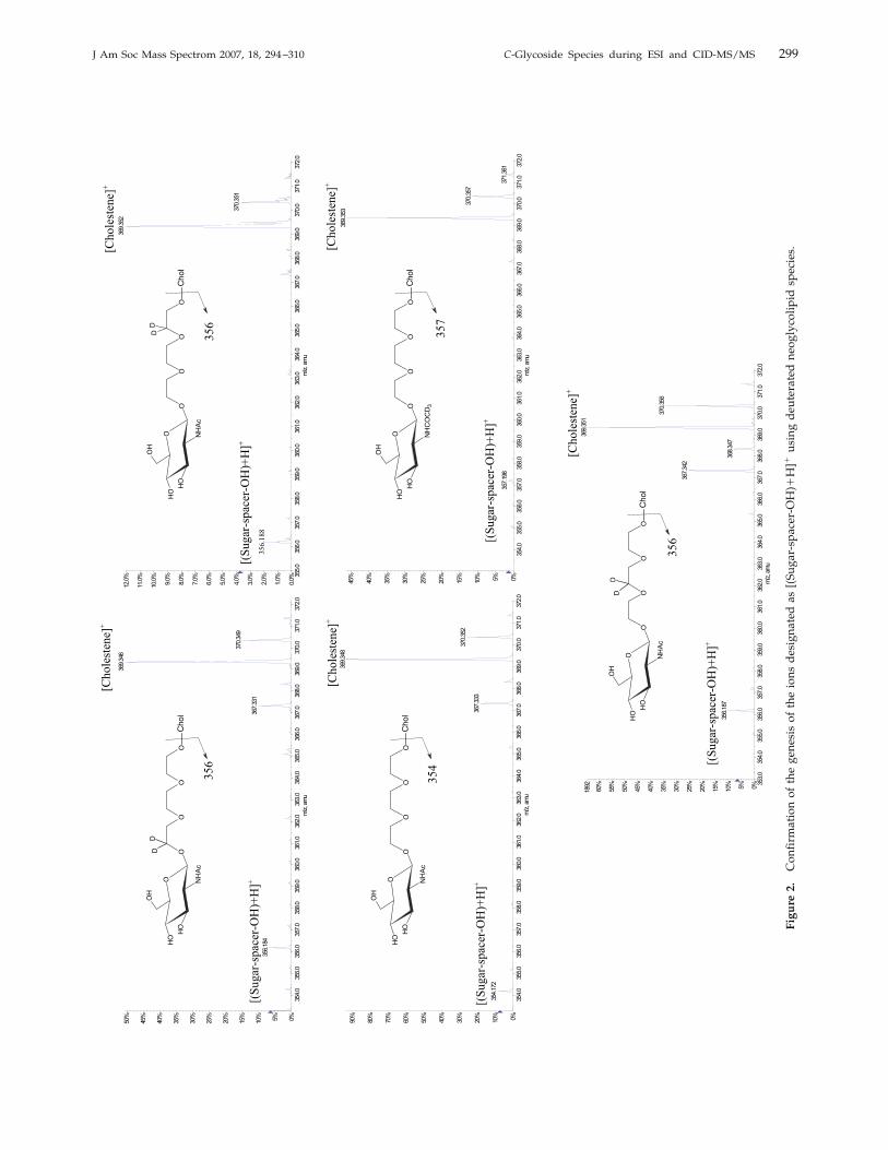
present in most of the studied neoglycolipids. Thegenesis of the [(Sugar-spacer-OH)�H]� fragment ionwas confirmed using four identical neoglycolipds thatbear GlcNAc and a spacer of (CH2CH2O)3, which in-cludes deuterated CH2 at varying positions of the chain.As expected, the nondeuterated species produced the[(Sugar-spacer-OH)�H]� fragment ion at m/z 354.17,whereas the m/z value for the corresponding deuteratedspecies was 356.18 (see Figure 2). Similarly, the presenceof GlcNHCOCD3 instead of GlcNAc resulted, as ex-
pected, in a shift in the m/z value of the [(Sugar-spacer-OH)�H]� ion from m/z 354.17 Th to m/z 357.19 Th (seeFigure 2).Additional elimination products, originating from
the sugar [Oxonium]� ions, were also observed. Thesecan be summarized as ions formed by the expected lossof one or two water molecules designated as [Oxonium-H2O]
� and [Oxonium-2H2O]�. The subsequent elimi-
nation of CO and CH2CO from these product ions wasalso noted, forming the ions assigned as [Oxonium-
+ H
O
OH
OH
OH
NH
R
CH3
CH3
CH3
CH3
CH3
[Spacer]
[M+H]+
+ HCH3
CH3
CH3
CH3
CH3
[Spacer]HO
[(Cholesterol-Spacer-OH)+H]+
O
OH
OH
OH
NH
R
[Spacer]
[(Sugar-Spacer-OH)+H]+
OH
+ H
O+
OH
OH
OH
NH
RCH
+
CH3
CH3
CH3
CH3
CH3
[Cholestene]+
m/z 369.35
[Oxonium]+
O+
OH
NH
R
OH
O+
CH2
NH
R
OH
O+
CH2
N
R
O
H
CH2
NH
C+
R
OH
O+
CH2
N
R
-H2O
-H2O
-CO
-CH2CO
++
+
[Oxonium - H2O]+
[Oxonium - 2H2O]+
[Oxonium - 2H2O - CO]+
[Oxonium - 2H2O - CH2CO]+
CH3
CH3
CH3
CH3
CH3
O
OH
OH
OH
NH
R
[C-glycoside]+
+ H
+
Scheme 2. General schematic representation of the proposed fragmentation pattern of the cho-lesteryl neoglycolipids observed in ToF-MS analysis. The sugar portion is illustrated as glucosaminederivative where R � H, COCH3, COCD3, or COOCH2CH¢CH2.
298 EL-ANEED ET AL. J Am Soc Mass Spectrom 2007, 18, 294–310
355.
035
6.0
357.
035
8.0
359.
036
0.0
361.
036
2.0
363.
036
4.0
365.
036
6.0
367.
036
8.0
369.
037
0.0
371.
037
2.0
m/z
, am
u
0.0%
1.0%
2.0%
3.0%
4.0%
5.0%
6.0%
7.0%
8.0%
9.0%
10.0
%
11.0
%
12.0
%36
9.35
2
370.
351
356.
188
O
OH
OH
OH
NH
Ac
OO
OO
DD
Cho
l
356
[Cho
lest
ene]
+
[(Su
gar-
spac
er-O
H)+
H]+
354.
035
5.0
356.
035
7.0
358.
035
9.0
360.
036
1.0
362.
036
3.0
364.
036
5.0
366.
036
7.0
368.
036
9.0
370.
037
1.0
372.
0m
/z, a
mu
0%5%10%
15%
20%
25%
30%
35%
40%
45%
50%
369.
346
370.
349
367.
331
356.
184
O
OH
OH
OH
NH
Ac
OO
OO
DD
Cho
l
[Cho
lest
ene]
+
[(Su
gar-
spac
er-O
H)+
H]+
356
Max
. 465
2.0
coun
ts.
354.
035
5.0
356.
035
7.0
358.
035
9.0
360.
036
1.0
362.
036
3.0
364.
036
5.0
366.
036
7.0
368.
036
9.0
370.
037
1.0
372.
0m
/z, a
mu
0%10%
20%
30%
40%
50%
60%
70%
80%
90%
369.
348
370.
352
367.
333
354.
172
O
OH
OH
OH
NH
Ac
OO
OO
Cho
l
[Cho
lest
ene]
+
[(Su
gar-
spac
er-O
H)+
H]+
354
+TO
F M
S: 2
0 M
CA
scan
s fro
m S
ampl
e 3
(Ful
l SC
AnD
P 13
0 _2
) of L
D21
(rec
alib
rate
d).w
iffa=
3.56
0919
0620
8459
780e
-004
, t0=
-1.8
2371
5493
3594
8660
e+00
0 R
;M
ax. 4
.9e4
cou
nts.
354.
035
5.0
356.
035
7.0
358.
035
9.0
360.
036
1.0
362.
036
3.0
364.
036
5.0
366.
036
7.0
368.
036
9.0
370.
037
1.0
372.
0m
/z, a
mu
0%5%10%
15%
20%
25%
30%
35%
40%
45%
369.
353
370.
357
371.
361
357.
198
O
OH
OH
OH
NH
CO
CD
3OO
OO
Cho
l
[Cho
lest
ene]
+
[(Su
gar-
spac
er-O
H)+
H]+
357
Max
. 293
6.0
coun
ts.
353.
035
4.0
355.
035
6.0
357.
035
8.0
359.
036
0.0
361.
036
2.0
363.
036
4.0
365.
036
6.0
367.
036
8.0
369.
037
0.0
371.
037
2.0
m/z
, am
u
0%5%10%
15%
20%
25%
30%
35%
40%
45%
50%
55%
60%
1892
369.
351
370.
356
367.
342
356.
187
368.
347
O
OH
OH
OH
NH
Ac
OO
OO
DD
Cho
l
356
[Cho
lest
ene]
+
[(Su
gar-
spac
er-O
H)+
H]+
Figu
re2.
Confirmationofthegenesisoftheionsdesignatedas[(Sugar-spacer-OH)�H]�usingdeuteratedneoglycolipidspecies.
299J Am Soc Mass Spectrom 2007, 18, 294–310 C-Glycoside Species during ESI and CID-MS/MS
2H2O-CO]� and [Oxonium-2H2O-CH2CO]
�. Figure 1and Scheme 2 show the proposed mechanism of theselosses.Such elimination products were absent in the case of
the sugar [Oxonium]� ions of the per-O-acetylatedneoglycolipids. Instead, elimination products that cor-respond to [Oxonium-HOAc]�, [Oxonium-2HOAc]�,and [Oxonium-3HOAc]� were observed (data notshown). The confirmation of the proposed structure ofthe various ions observed in the QqToF-MS was accom-plished by CID-MS/MS analysis, as described below.
Low-Energy Collision-Induced DissociationTandem Mass Spectrometric Analysis(CID-MS/MS)
The discussion in this section will be divided based onthe presence or absence of the acetylated hydroxylgroups within the sugar portion (i.e., protected versusnonprotected species).
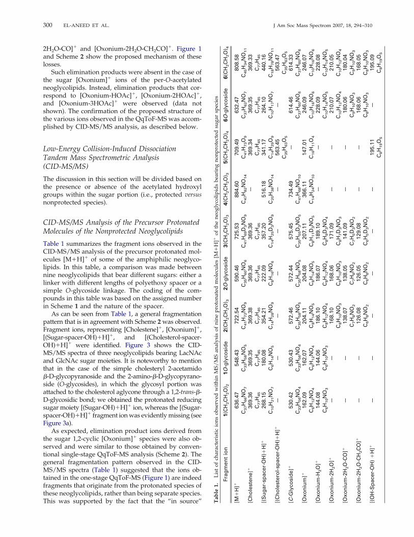
CID-MS/MS Analysis of the Precursor ProtonatedMolecules of the Nonprotected Neoglycolipids
Table 1 summarizes the fragment ions observed in theCID-MS/MS analysis of the precursor protonated mol-ecules [M�H]� of some of the amphiphilic neoglyco-lipids. In this table, a comparison was made betweennine neoglycolipids that bear different sugars: either alinker with different lengths of polyethoxy spacer or asimple O-glycoside linkage. The coding of the com-pounds in this table was based on the assigned numberin Scheme 1 and the nature of the spacer.As can be seen from Table 1, a general fragmentation
pattern that is in agreement with Scheme 2was observed.Fragment ions, representing [Cholestene]�, [Oxonium]�,[(Sugar-spacer-OH)�H]�, and [(Cholesterol-spacer-OH)�H]� were identified. Figure 3 shows the CID-MS/MS spectra of three neoglycolipids bearing LacNAcand GlcNAc sugar moieties. It is noteworthy to mentionthat in the case of the simple cholesteryl 2-acetamido�-D-glycopyranoside and the 2-amino-�-D-glycopyrano-side (O-glycosides), in which the glycosyl portion wasattached to the cholesterol aglycone through a 1,2-trans-�-D-glycosidic bond; we obtained the protonated reducingsugar moiety [(Sugar-OH)�H]� ion, whereas the [(Sugar-spacer-OH)�H]� fragment ionwas evidentlymissing (seeFigure 3a).As expected, elimination product ions derived from
the sugar 1,2-cyclic [Oxonium]� species were also ob-served and were similar to those obtained by conven-tional single-stage QqToF-MS analysis (Scheme 2). Thegeneral fragmentation pattern observed in the CID-MS/MS spectra (Table 1) suggested that the ions ob-tained in the one-stage QqToF-MS (Figure 1) are indeedfragments that originate from the protonated species ofthese neoglycolipids, rather than being separate species.This was supported by the fact that the “in source” T
able
1.ListofcharacteristicionsobservedwithinMS/MSanalysisofnineprotonatedmolecules[M
�H]�oftheneoglycolipidsbearingnonprotectedsugarspecies
Frag
men
tio
n1(C
H2C
H2O
) 21O
-gly
cosi
de
2(C
H2C
H2O
) 32O
-gly
cosi
de
3(C
H2C
H2O
) 34(C
H2C
H2O
) 35(C
H2C
H2O
) 46O
-gly
cosi
de
6(C
H2C
H2O
) 4
[M�
H]�
636.
47C
37H
66N
O7
548.
43C
33H
58N
O5
722.
54C
41H
72N
O9
590.
46C
35H
60N
O6
725.
53C
41H
69D
3N
O9
884.
60C
47H
82N
O14
709.
49C
41H
73O
9
632.
47C
37H
62N
O7
808.
58C
45H
34N
O11
[Ch
ole
sten
e]�
369.
36C
77H
45
369.
35C
77H
45
369.
38C
77H
45
369.
36C
77H
45
369.
36C
77H
45
—36
9.34
C77H
45
369.
35C
77H
45
369.
33C
77H
45
[(S
ug
ar-s
pac
er-O
H)�
H]�
268.
15C
10H
21N
O7
180.
08C
6H
14N
O5
354.
21C
14H
28N
O9
222.
09C
8H
16N
O6
357.
20C
14H
25D
3N
O9
516.
18C
20H
38N
O14
341.
17C
14H
29O
9
264.
10C
10H
17N
O7
440.
16C
18H
78N
O11
[(C
ho
lest
ero
l-sp
acer
-OH
)�H
]�—
——
——
—56
3.45
C35H
63O
5
—56
3.47
C35H
63O
5
[C-G
lyco
sid
e]�
530.
42C
33H
56N
O4
530.
43C
33H
56N
O4
572.
46C
35H
58N
O5
572.
44C
35H
58N
O5
575.
45C
35H
55D
3N
O5
734.
49C
41H
68N
O10
—61
4.46
C37H
60N
O6
614.
33C
37H
60N
O6
[Oxo
niu
m]�
162.
09C
6H
12N
O4
162.
07C
6H
12N
O4
204.
11C
8H
14N
O5
204.
08C
8H
14N
O5
207.
11C
8H
11D
3N
O5
366.
11C
14H
24N
O10
147.
01C
16H
11O
4
246.
09C
10H
16N
O6
246.
07C
10H
16N
O6
[Oxo
niu
m-H
2O
]�14
4.08
C6H
10N
O3
144.
06C
6H
10N
O3
186.
10C
8H
12N
O4
186.
07C
8H
12N
O4
189.
10C
8H
9D
3N
O4
——
228.
09C
10H
14N
O5
228.
08C
10H
14N
O5
[Oxo
niu
m-2
H2O
]�—
—16
8.10
C8H
10N
O3
168.
06C
8H
10N
O3
171.
09C
8H
7D
3N
O3
——
210.
07C
10H
12N
O4
210.
05C
10H
12N
O4
[Oxo
niu
m-2
H2O
-CO
]�—
—13
8.07
C7H
8N
O2
138.
05C
7H
8N
O2
141.
09C
7H
5D
3N
O2
——
180.
06C
9H
10N
O3
180.
04C
9H
10N
O3
[Oxo
niu
m-2
H2O
-CH
2C
O]�
——
126.
08C
6H
8N
O2
126.
05C
6H
8N
O2
129.
08C
6H
5D
3N
O2
——
168.
06C
8H
10N
O3
168.
05C
8H
10N
O3
[(O
H-S
pac
er-O
H)
�H
]�—
——
——
—19
5.11
C8H
19O
5
—19
5.09
C8H
19O
5
300 EL-ANEED ET AL. J Am Soc Mass Spectrom 2007, 18, 294–310

fragmentation was significantly repressed when manip-ulating the mass spectrometry parameters, that is, re-ducing the Declustering Potential (DP). It was alsoobserved that the presence of the [(Cholesterol-spacer-OH)�H]� and [(OH-Spacer-OH) �H]� fragment ionswere only associated with the neoglycolipids bearing apolyethoxy spacer of (CH2CH2O)4 (Table 1, Figure 3b).
In addition to the expected fragment ions, the recog-nized formation of the unique [C-Glycoside]� ion wasobserved during the CID-MS/MS only when the neo-glycolipids contained a 1,2-participating group, withthe exception of L-fucose. In fact, this ion was alsopresent in the conventional one-stage QqToF-MS; care-ful examination of the spectra shows that the [C-
50 100 150 200 250 300 350 400 450 500 550 600 650 700 750 800m/z, amu
0%
10%
20%
30%
40%
50%
60%
70%
80%
90%
100% 204.11
722.55
354.21572.47369.38126.08 186.10
[M+H]+
[C-Glycoside]+[Cholestene]+[(Sugar-spacer-OH)+H]+
[GlcNAc]+
[GlcNAc -H2O]+
[GlcNAc -2H2O-CH2CO]+
CH3
CH3
CH3
CH3
CH3
O
O
OH
OH
OH
NHAc
O O O
(b)
100 150 200 250 300 350 400 450 500 550 600 650 700 750 800 850 900m/z, amu
0%
10%
20%
30%
40%
50%
60%
70%
80%
90%
100% 366.11
884.60168.03 204.08 516.18 734.48138.03
[M+H]+
[C-Glycoside]+[(Sugar-spacer-OH)+H]+
[LacNAc]+
[GlcNAc]+
[GlcNAc-2H2O]+
[GlcNAc-2H2O-CO]+
CH3
CH3
OOOOO
OH
OH
NH
OO
OH
OH
OH
OH
Ac
(c)
150 200 250 300 350 400 450 500 550 600 650m/z, amu
0%
10%
20%
30%
40%
50%
60%
70%
80%
90%
100% 369.36
204.09
590.47
222.10
572.45186.08138.05126.05
[M+H]+
[C-Glycoside]+
[(Sugar-spacer-OH)+H]+
[Sugar+H]+
[GlcNAc]+
[Cholestene]+
[GlcNAc -H2O]+
[GlcNAc -2H2O-CH2CO]+
[GlcNAc-2H2O-CO]+
O
OH
OH
NH
OH
CH3
CH3
O
Ac
O
OH
OH
NH
OHO
Ac
H
+ H+
O+
OH
OH
NH
OH
Ac
+
(a)
Figure 3. MS/ MS experiments of neoglycolipids bearing (a) GlcNAc linked to the cholesterolportion by O-Glycoside; (b) GlcNAc linked to the cholesterol portion by (CH2CH2O)3 spacer; and (c)LacNAc linked to the cholesterol portion by (CH2CH2O)3 spacer.
301J Am Soc Mass Spectrom 2007, 18, 294–310 C-Glycoside Species during ESI and CID-MS/MS

Glycoside]� ion was also formed during ESI ionizationand it was not simply an “impurity,” as was initiallyspeculated when examining the QqToF-MS spectra (re-fer to Figure 1). We hypothesized that this ion resultsfrom a unique ion-molecule reaction between the neu-tral cholesterol on the sugar portion of the 1,2-cyclicoxonium ion species, from the �-face of this ion, thusproducing the 1,2-trans-C-Glycoside: specifically anelectrophilic addition of the conjugated diene of thecholesta-3,5-diene neutral molecule, on the electron-deficient 1,2-cyclic [oxonium]� ion (Lewis acid). The pres-ence of this [C-Glycoside]� species was originally noticedin the MS/MS analysis of six neoglycolipid cholesterylderivatives containing N-acetyl-D-glucosamine and wasconfirmed byMS/MS (referred to as quasi-MS3 analysis ofthis unique ion [12]. Further discussion on the structureand formation of this ion using the various neoglycolipidsevaluated in this study will be presented in the followingsections.Finally, the presence of LacNAc (�-D-Galp-(1¡4)-D-
GlcpNAc) resulted in the formation of additional sugarfragment ions beside those presented in Table 1 (Fig-ure 3C). These ions were formed from the diagnostic[LacNAc]� ion observed at m/z 366.11 [Oxonium]�) andinclude the [GlcNAc]� ion at m/z 204.08, the [GlcNAc-2H2O]
� ion at m/z 186.04, and the [GlcNAc-2H2O-CO]� ion at m/z 138.03 (all derived from the reducingend of the disaccharide). The presence of these addi-tional fragments is expected and they serve as diag-nostic product ions confirming the structure of thisneoglycolipid.To verify the authenticity of the fragment ions as-
signed based on the conventional single-stageQqToF-MS analysis and on the CID-MS/MS experi-ments of the precursor protonated species [M�H]�,additional CID-MS/MS analyses were obtained by in-creasing the “in source” fragmentation (also called “innozzle” fragmentation), which can be enhanced andoptimized for each tested species through manipulationof the Declustering Potential (DP) and the FocusingPotential (FP) values within the ionization source (i.e.,optimum potentials for each precursor ion, in terms ofion count and S/N ratio). Both DP and FP are electricalvoltages that have the greatest effects on the extent ofthe fragmentation within the orifice-skimmer region ofthe ESI source of the QStar XL hybrid tandem massspectrometer.Figure 4 represents the low-energy collision CID-
MS/MS spectra of the extracted protonated molecule[M�H]� of neoglycolipid 6 (Scheme 1) bearing(CH2CH2O)4 as a spacer (Figure 4a) as well as theCID-MS/MS of the extracted precursors: [(Cholesterol-spacer-OH)�H]� (Figure 4b) and the [(OH-Spacer-OH)�H]� fragment ions (Figure 4c). As can be seenin Figure 4, the CID-MS/MS analysis of the [M�H]�
ion followed the same pattern as discussed earlierand shown in Scheme 2 and Table 1. However, theCID-MS/MS of the [(Cholesterol-spacer-OH)�H]�
species at 563.47 produced two major product ions
corresponding to [Cholestene]� at m/z 369.33 andthe [(HOO(CH2CH2O)3OOH)� H]� at m/z 195.09.Similarly, the CID-MS/MS of the [(HO-(CH2CH2O)3OOH)�H]
� ion at 195.09 producedproduct ions corresponding to the proposed structureof this ion (see Figure 4C). It showed distinctive,consecutive losses of water molecules followed byelimination of an acetylene species (CH§CH).Similar analyses were also conducted on all the ions
presented in Table 1, confirming their proposed struc-tures, and these findings established the universal frag-mentation pattern illustrated in Scheme 2.
Formation of [C-Glycoside]�
As shown in Table 1 and Figure 3, the [C-Glycoside]�
species were observed first during the MS/MS experi-ment within the collision cell of the tandem massspectrometer. However, this ion was also present in theone-stage ESI-QqToF experiment (see Figure 1). Theformation of this unique ion cannot be explained basedon the molecular structure of these novel syntheticneoglycolipids (Scheme1). To confirm the proposedstructure of this ion, CID-MS/MSwas performed on theisolated [C-glycoside]�. This analysis tentativelyshowed that this ion can undergo two concerted cleav-ages, which occur simultaneously from the precursor[C-glycoside]� ion.The first involves the elimination of the axial H-2
hydrogen of the sugar moiety, with consecutive ruptureand migration of the anomeric C-1OC-3= bond and theconsequent formation of the neutral fragment 2-N-acetyl-2-deoxy-D-glucal. This is followed by formation ofthe C-3=-C-4= double bond andmigration of the endocyclicC-4=OC-5= double bond, to afford the protonated cholest-3,5-diene molecule assigned as [Cholesta-3,5-diene�H]�
at m/z 369.35.The second mechanism tentatively suggests the par-
ticipation of the lone pair of electrons on the sugar ringoxygen, to afford the stable sugar 1,2-oxonium ion[Oxonium]�, followed by an identical rupture of theanomeric C-1OC-3= bond and migration of the doublebonds, to produce the neutral choles-3,5-dienemolecule. Figure 5 shows the CID-MS/MS of two[C-Glycoside]� ions that contain LacNAc and GlcNAloc[extracted from the neoglycolipids 4 and 2, containingthe (OCH2CH2)3 spacer or simple O-glycoside, respec-tively] and illustrates clearly the presence of [Cholesta-3,5-diene�H]� at m/z 369.35 and [LacNAc]� at m/z366.13 and [GlcNAc]� at m/z 204.08. Elimination prod-ucts of the sugar [Oxonium]� were also present in thisMS/MS analysis. This MS/MS analysis authenticatedthe proposed structure of the [C-Glycoside]� ion.To explain the rationale beyond the formation of the
[C-glycoside]� ion, we proposed that a unique ion-molecule reaction occurs in the collision cell and withinthe ESI interface of the turbo ion spray source. It waspostulated that the parent [M�H]� ion of the completeneoglycolipid molecule undergoes covalent bond cleav-
302 EL-ANEED ET AL. J Am Soc Mass Spectrom 2007, 18, 294–310

ages, producing the 1,2-cyclic sugar [Oxonium]� ion aswell as the neutral cholesta-3,5-diene. This neutral mol-ecule by migration of the conjugated double bond,attached itself on the 1,2-[oxonium]� ion, which isconsidered to be the electrophilic center. This additionis initiated by the loss of an �-hydrogen atom, with
respect to the double bond of the cholesta-3,5-dieneportion, followed by attack on the positive charge thatis delocalized on NOCOO atoms of the 1,2-cyclic[oxazolinium]� ion, on the anomeric position, produc-ing the ion designated as the [C-Glycoside]� (seeScheme 3a and b for details). The presence of the amino
[Oxonium]+
[Oxonium-2H2O-CO]+
100 120 140 160 180 200 220 240 260 280 300 320 340 360 380 400 420 440 460 480 500 520 540 560 580 600m/z, amu
0%
10%
20%
30%
40%
50%
60%
70%
80%
90%
100% 369.33
195.09
563.48[(Cholesterol-spacer-OH)+H]+
[Cholestene]+
[(OH-Spacer-OH) +H]+
CH3
CH3
OOOOOH
+ H
+
369.33
195.09
m/z 563.48
O+OOOOH
H
HH
H H2O
O+
OOOH
H
H
HO+
OOOH
H
H
m/z 177.11m/z 151.10 m/z 195.12
C2H2
50 60 70 80 90 100 110 120 130 140 150 160 170 180 190 200m/z, amu
0%
10%
20%
30%
40%
50%
60%
70%
80%
90%
100% 89.06
133.09
195.12
177.11151.10107.07
195 -H2O
177 - C2H2
151 -H2O
133- C2H2
107 -H2O
89(c)
100 150 200 250 300 350 400 450 500 550 600 650 700 750 800 850 900m/z, amu
0%
10%
20%
30%
40%
50%
60%
70%
80%
90%
100% 246.07
563.47
195.09
369.33 808.58
440.16 614.33180.04168.05 228.08
[M+H]+
[C-Glycoside]+
[Cholestene]+
[Oxonium-H2O]+
[(OH-Spacer-OH) +H]+
[(Cholesterol-spacer-OH)+H
[Oxonium-2H2O-CH2CO]+
[(Sugarl-spacer-OH)+H]+
(a)
CH3
CH3
OOOOOO
OH
OH
NH
OH
C
O
OCH2CHCH2
(b)
Figure 4. (a) MS/MS (MS [2] scan of neoglycolipid bearing GlcNAloc linked to the cholesterolportion by (CH2CH2O)4 spacer. (b) MS/MS of the fragment ion designated as [(Cholesterol-spacer-OH)�H]�. (c) MS/MS of the fragment ion designated as [(OH-Spacer-OH) �H]�.
303J Am Soc Mass Spectrom 2007, 18, 294–310 C-Glycoside Species during ESI and CID-MS/MS

group at position C-2 of the sugar residue enhances theformation of a more “reactive species.” This species iscalled in the case of the C-2 acetamido group, the1,2-cyclic oxazolinium, whereas in the case of the2-amino group it is called the “1,2-cyclic-aziridinium.”Both oxonium ions containing a delocalized positivecharge and thus are electron acceptors (also calledLewis acids).In the event that the glycosyl portion of the neogly-
colipid, did not bear a participating group at positionC-2, the formation of the [C-Glycoside]� was absent, asin the case of neoglycolipid bearing the Fuc sugar
(Table 1). Despite the presence of the [Fuc]� oxoniumion at m/z 147.01, this ion failed to react with the neutralcholesta-3,5-diene molecule because it lacks the partic-ipating group at position C-2 (and thus does not formthe reactive 1,2-cyclic oxonium ion) and, as a result,cannot form the corresponding ion [C-Glycoside]�
(Scheme 3c).As can be seen from Scheme 3 and Figure 5, we
opted to illustrate that the tentative [C-Glycoside]� ionswere produced by the formation of a C-1OC-3= cova-lent bond between the sugar and the cholesterol por-tions. However, the presence of another [C-Glycoside]�
Max. 108.0 counts.
100 120 140 160 180 200 220 240 260 280 300 320 340 360 380 400 420 440 460 480 500 520 540 560 580 600m/z, amu
0%
10%
20%
30%
40%
50%
60%
70%
80%
90%
100% 369.35
572.44
204.08
168.06186.06
[C-Glycoside]+[Cholestdiene+H]+
[GlcNAc]+
[GlcNAc-2H2O]+
[GlcNAc-H2O]+
(a)
Max. 35.0 counts.
100 150 200 250 300 350 400 450 500 550 600 650 700 750 800m/z, amu
0%
10%
20%
30%
40%
50%
60%
70%
80%
90%
100% 734.50
204.05369.35
168.04
138.04
[GlcNAc-2H2O]+[LacNAc]+
[Cholestdiene+H]+[GlcNAc]+
[C-Glycoside]+
[GlcNAc-2H2O-CO]+
366.13
(b)
O+
NH
OHOH
OH
OCH3
+
HO
NH
OHOH
OHCH3
CH3
CH3 CH3
CH3
OCH3
CH3
CH3
CH3 CH3
CH3
H
CH3
CH3
CH3 CH3
CH3
H
O
NH
OHOH
OH
OCH3
+H
+
quasi MS3
+
[C-Glycoside]+ m/z 572
m/z 204 m/z 369
+ H
+
+
HO
NH
OHO
OHCH3
CH3
CH3 CH3
CH3
OCH3
O
OH
OH
OH
OH
CH3
CH3
CH3 CH3
CH3
H
CH3
CH3
CH3 CH3
CH3
H
+H
+
quasi MS3
+
[C-Glycoside]+ m/z 734
m/z 366 m/z 369
HO
+
NH
OHO
OH
OCH3
O
OH
OH
OH
OHO
NH
OHO
OH
OCH3
O
OH
OH
OH
OH
+ H
+
Figure 5. MS/MS of the ion identified as [C-glycoside]�, extracted from the neoglycolipids bearing(a) GlcNAc and linked to the cholesterol through O-glycoside linkage and (b) the LacNAc and linkedthe cholesterol through (CH2CH2)3 spacer.
304 EL-ANEED ET AL. J Am Soc Mass Spectrom 2007, 18, 294–310

O+
NH
OHO
OH
OR
R1
CH
3
CH
3
CH
3C
H3
CH
3
H
[Sp
ace
r]O
OH
OH
NH
O
CO
R
R1
CH
3
CH
3
CH
3C
H3
CH
3
[Spa
cer]
O
OH
OH
NH
2
OH
CH
3
CH
3
CH
3C
H3
CH
3
O
NH
OHO
OH
O
R
R1
+
O+
N
OHO
H
OH H
HC
H3
CH
3
CH
3C
H3
CH
3
HO N
+
OHO
H
OH H
H
+
O NH
OHO
OH
CH
3
CH
3
CH
3C
H3
CH
3
OR
R1
Noz
zle
'in s
itu' C
ID-M
S
[C-G
lyco
sid
e]+
Col
lisio
n ce
ll 'in
situ
' CID
-MS
/MS
O NH
2
OHO
H
OH
CH
3
CH
3
CH
3C
H3
CH
3
Noz
zle
'in s
itu' C
ID-M
SC
ollis
ion
cell
'in s
itu' C
ID-M
S/M
S
[C-G
lyco
sid
e]+
CH
3
CH
3
CH
3C
H3
CH
3
[Spa
cer]
CH
3
CH
3
CH
3C
H3
CH
3
OO
H
OH
OH
O+
OH
OH
OH
Ele
ctro
ph
ilic
att
ack
is n
ot
po
ssib
le
un
de
r th
ese
co
nd
itio
ns
(c)
(b)
(a)
1,2
Cyc
lic f
orm
ca
nnot
be
form
ed
+ H
+
+ H
+
Sch
eme
3.Schematicrepresentationofthemechanisticprocessthatleadstotheformationofthe[C-Glycoside]
�ionwithintheESIinterfaceandduringCID-MS/MSanalysis.(
a)Thesugarportionisaderivative
N-acylglucosaminewhereR
�CH3,CD3,orCOOCH2CH¢CH2;R1
�H,or
�-D-Gal.(
b)ThesugarportionisaGlcNH2species.(c)Thesugarportion
isFucspecies.
305J Am Soc Mass Spectrom 2007, 18, 294–310 C-Glycoside Species during ESI and CID-MS/MS

species that carry the C-1OC-5= covalent bond cannotbe excluded by the same mechanism, although it isimprobable because of the potential steric hindrance ofthis latter C-glycoside. Needless to say, there is no wayto tell for sure whether the formed C-glycoside pos-sesses the structure we proposed, wherever it has the�-D-or �-D-configurations, whereas it has an anomericmixture of both, and/or even may be a cyclic 1,2-orthoester carbohydrate derivative type in which theC-cholesteryl aglycone would attack the central carbonatom of the positively charged 1,2-oxazolinium ion.This latter 1,2 cyclic C-orthoester is very unlikely to beformed, given that it will rearrange instantaneously inthe presence of traces of acid into the correspondingproposed C-glycoside.This is beyond the scope of this article and cannot be
verified with the present instrumentation. Nevertheless,the structure we propose has tentatively been attributedto the �-D-configuration that is, without doubt, thepreferred configuration and the more thermodynami-cally stable [22].An argument in favor of our proposal is that the
C-glycoside formation was found to be concentrationdependent. When the CID-MS/MS of the precursor
protonated molecule [M�H]� was extracted from amore concentrated solution, the comparative abun-dance of the [C-glycoside]� species was augmentedrelative to the intensities of the other product ions.In addition to the absence of the [C-Glycoside]� in
the case of Fuc-derived neoglycolipids, this ion wasalso absent in the low-energy CID-MS/MS analysis ofall the sodiated adducts [M�Na]� of the neoglyco-lipid series evaluated in this study. For illustrationpurpose, Scheme 4 represents the fragmentation pat-tern of the product ion scan of the [M�Na]�, isolatedfrom the neoglycolipid designated as 3, containingthe spacer (CH2CH2O)3. The fact that both [Oxon-ium]� and [Cholestene]� ions were absent in theMS/MS of the [M�Na]� suggests that this adductbreaks in different fashion than the one observedwith [M�H]� and does not form the reactive speciesthat can result in the formation of the C-glycosidebond. All the fragment ions were actually sodiatedspecies and included a fragment ion that is generatedfrom inner sugar breakage, that is, 0,2X. Such innersugar fragments are common and widely observed inglycoconjugate analysis [23, 24], and it was observed
O
OH
OH
OH
NHAc
O O O OChol
[M+Na]+
m/z 744.36
NHAc
O O O OChol
+Na
[(Chol-Spacer)+Na]+
m/z 541.43
OH O O OChol
+Na
+Na
O
OH
OH
OH
NHAc
O O O OH
[(Sugar-Spacer)+Na]+
m/z 376.07
+NaO
OH
OH
OH
NHAc
[Sugar+Na]+
m/z 226.02
+Na
OH O O OH
[Spacer+Na]+
m/z 173.05
+Na
0,2X
[(0,2X-Spacer-Chol)+Na]+
m/z 624.48
Scheme 4. The proposed fragmentation pattern of the [M�Na]� of the neoglycolipid bearingGlcNAc with a (CH2CH2O)3 spacer.
306 EL-ANEED ET AL. J Am Soc Mass Spectrom 2007, 18, 294–310

in our study of the [M�H]�, that is, the ion desig-nated [OxoniumO2H2OOCH2CO]
� (see Scheme 2).
Influence of DP and FP on the Formationof C-Glycoside
Both DP and FP are electrical voltages that have thegreatest effect on the extent of fragmentation withinthe orifice-skimmer region of the ESI source. C-Glycoside ion formation was significantly correlatedwith the DP (Spearman’s correlation coefficient, P �
0.009), (Figure 6a) but not with the FP (P � 0.54)(Figure 6b). Therefore, the FP value was kept con-stant (FP � 220) while studying the influence of theDP on ion formation. The pattern between the ioncount (intensity) and the DP values was similar forthe formation of the C-glycoside species and thesugar [oxonium]� ions (two-sample Kolmogorov–Smirnov test, P � 0.34), but showed significantdifferences when the formation of the C-glycoside was compared to that of other ions such as[M�H]� and [(Sugar-spacer-OH)�H]� (P values of8.082e-08 and 2.2e-16, respectively). As shown inFigure 7, the [C-Glycoside]� ion formation (withrespect to DP values) followed the exact pattern ofthe sugar [oxonium]� species. This supports thetheory that the formation of the [C-Glycoside]� spe-cies was a product of a reaction between the sugar[oxonium]� ion and the neutral fragment cholesta-3,5-diene. Note that when comparing different spe-cies the ion count values were normalized to themaximum reading.
0
50
100
150
200
250
300
0 50 100 150 200 250 300 350
DP
Ion
Co
un
t
0
50
100
150
200
250
300
0 100 200 300 400 500
Ion Count
FP
(a)
(b)
Figure 6. The relationship between [C-glycoside]� ion count and(a) DP values; (b) FP values.
0
0.2
0.4
0.6
0.8
1
1.2
0 50 100 150 200 250 300 350
DP
Ion
Co
un
t/M
axim
um
C-Glycoside
Sugar
M+H
Sugar-Spacer
Figure 7. The influence of DP on the formation of [C-glycoside]�,[Oxonium]�, [M�H]�, and [(Sugar-spacer)�H]� ions within theESI interface of the Q-Star Machine.
Table 2. List of characteristic ions observed within MS/MS analysis of protonated molecules of the neoglycolipids bearing protectedsugar species
Fragment ion 4(CH2CH2O)3 6(CH2CH2O)4 6O-glycoside
[M�H]� 1136.66C59H94NO20
943.62C51H84NO14
758.51C43H68NO10
[Cholestene]� 369.34C77H45
— 369.37C77H45
[(Cholesterol-spacer-OH)�H]� — 563.47C35H63O5
—
[(Sugar-spacer-OH)�H]� 768.29C32H50NO20
566.25C24H40NO14
390.16C16H24NO10
[C-Glycoside]� — — —[Oxonium]� 618.20
C26H36NO16
372.12C16H22NO9
372.14C16H22NO9
[Oxonium-OAc]� 558.18C24H32NO14
— 312.10C14H18NO7
[Oxonium-2OAc]� 498.15C22H28NO12
252.08C12H14NO5
252.09C12H14NO5
[Oxonium-3OAc]� 438.09C20H24NO10
— 192.07C10H10NO3
307J Am Soc Mass Spectrom 2007, 18, 294–310 C-Glycoside Species during ESI and CID-MS/MS

CID-MS/MS Analysis of the Precursor ProtonatedMolecules of the Protected Per-O-AcetylatedNeoglycolipids
We opted to choose the per-O-acetylated neoglycolipidsbecause we anticipated that the formation of the morereactive 1,2-cyclic oxonium ion would enhance the rateof the ion-molecule reaction. This may result in moreefficient formation of the C-glycoside in the ESI sourceand in the collision cell of the hybrid tandem massspectrometer. Table 2 summarizes the fragmentationpattern of the protonated molecules [M�H]� of theper-O-acetylated neoglycolipids. The presence of thediagnostic fingerprint ions: [Cholestene]�, [(sugar-spacer-OH)�H]�, and [Oxonium]�—was observed.Similar to the nonprotected neoglycolipids, the pres-ence of [(cholesterol-spacer-OH)�H]� was associatedonly with the neoglycolipid bearing (CH2CHO)4 spacer.As expected, the elimination products originating fromthe [Oxonium]� ions were a result of one, two, or threelosses of acetic acid groups (see Table 2).The most striking finding, however, was that the
[C-glycoside]� ion was absent in the case of theseper-O-acetylated sugar derivatives (see Table 2). Thisobservation contradicts expectations based on carbo-hydrate chemistry foundation, given that the per-O-acetylated sugars are routinely used for C-glycosylationreactions in synthetic chemistry [13]. In fact, the per-O-acetylated neoglycolipids are not used for liposomalpreparations and they were synthesized to enhance toformation of C-glycoside. However, the fact that thisreaction occurs in the gas phase can introduce manynew factors that are different from the conditions thatrule solid and solution chemistry. One possible expla-nation is the overall reactivity of the per-O-acetylated-sugar oxonium ions. These oxonium ions, being ex-tremely reactive, preferentially eliminate molecules ofacetic acid and ketene, by concerted mechanisms, de-stabilizing the 1,2-cyclic oxazolinium ion. Therefore,this oxazolinium ion has no time to form the cyclicreactive species that can react with the electrophiliccholesta-3,5-diene molecule found in the collision cell.The CID-MS/MS of the [Oxonium]� ions that corre-sponds to neoglycolipids bearing the 2-N-allyloxycar-bonylamino-2-deoxy group (GlcNAloc), or the per-O-acetylated GlcNAloc sugar species with the(CH2CH2O)4 spacer, were compared. The nonprotected[GlcNAloc]� produced six fragment ions (data notshown) that are in accordance with Scheme 2 and Table1. The protected per-O-acetylated [GlcNAloc]� ion,however, produced 15 fragment ions (data not shown).This fact supports the speculation regarding the overallreactivity of the per-O-acetylated oxonium in compari-son to the nonprotected oxonium.Although this speculation can provide some expla-
nation for the absence of the [C-glycoside]�, otherpossible mechanisms such as steric hindrance cannot beexcluded. Various neoglycolipids that bear glucosaminederivatives that are blocked at one or two variable
positions will be synthesized and tested for the forma-tion of the [C-Glycoside]� and the outcome of this workwill be the subject of a future report. Other possibilitiesmay also unfold in the future with advances in the fieldof gas chemistry.
Conclusions
In this study, synthetic neoglycolipids that bear a mono-or disaccharide glycosyl moiety (hydrophilic) and acholesterol moiety (hydrophobic), which are linked by apolyethoxy spacer, were evaluated using QqToF-MS/MS hybrid instrumentation (ESI and CID-MS/MSanalysis). The results demonstrated clearly that, both inthe ESI source and within the collision cell of the massspectrometer, the neoglycolipids follow a universalfragmentation pattern, producing a series of productions that corresponded precisely to the theoretical struc-ture of these amphiphilic molecules (Scheme 2 andTable 1). Breakage can occur at either of the spacer’sends, producing most of the fragments observed in theESI-qQ-ToF and MS/MS analysis. In addition, elimina-tion products that are generated from the sugar portionwere also observed and include product ions thatare related to inner sugar fragmentation, such as the[oxonium-2H2O-CH2CO]
� ion. The genesis of theseproduct ions was further confirmed by MS/MS analysisas shown in Figure 4. This universal fragmentationpattern can be used to easily predict the fragmentationof new compounds that have the same general struc-tural backbone. In addition, single or multiple ionmonitor reactions can be performed for any quantitativestudies of liposomes bearing these novel neoglycolipidsas stabilizing components.In addition to the establishment of the mass spectro-
metric fingerprints of these compounds, we haveshown interesting evidence for the presence of anunprecedented C-glycosylation reaction during theESI-MS and MS/MS analysis (Figure 5 and Scheme 3).Such a reaction does not occur easily and requiresreasonable efforts on a bench top in a chemistry lab. Itoccurred, however, in a timeframe of millisecondswithin the mass spectrometry equipment. Ion-moleculereactions within mass spectrometry are old phenomenathat were first noticed as early as 1913 [25]. They aremainly observed and monitored within ion trap (IT)mass analyzers (quadrupole IT and Fourier TransformMS) [26–29]. It was suggested, for example, that poly-(ethylene/propylene glycol) under collisionally acti-vated dissociation using FT-ICR MS/MS resulted inmisleading rearrangements [28, 29]. Both triple- andpenta-quadrupoles were also used to evaluate ion-molecule reactions [30, 31]. We and others have shownthat these reactions can occur under atmospheric pres-sure in the ESI source [12, 32].In this study, a quadrupole time of flight mass
spectrometer was used, illustrating the provisional oc-currence of a unique C-glycosylation reaction in thecollision cell as well as under atmospheric pressure in
308 EL-ANEED ET AL. J Am Soc Mass Spectrom 2007, 18, 294–310

the ESI source. We have tentatively established that theproduction of a C-glycoside occurs from a reactionbetween the positively charged N-sugar oxoniom ionand a neutral choleta-3,5-diene species. When studyingthese compounds in the negative ion mode (data notshown), this reaction fails to occur, supporting themechanism presented in Scheme 3. The presence of anamino group at position C-2 and the free OH groups onthe C-3, C-4, and C-6 positions are both critical for theformation of this unique ion. This reaction is verydistinctive because the formation of the C-glycoside isvery difficult in comparison to O- or N-glycosylation.The McLafferty group has suggested that poly(eth-
ylene/propylene glycol) under collisionally activateddissociation, using FT-ICR MS/MS or other energeticmethods, inevitably issue in misleading rearrangements[28, 29]. Brull and collaborators reported that duringMS/MS analysis of underivatized and per-O-methyl-ated trisaccharides, with either high- or low-energy CIDusing FAB ionization, they observed losses of the inter-nal residue of 1¡6 substituted monosaccharide [33].This phenomenon of “internal residue loss,” which wascharacterized as an “internal rearrangement” processcatalyzed by a proton, was not observed in the CID-MS/MS analysis of sodium-cationized oligosaccharidescontaining N-acetyl-D-glucosamine [34]. Claeys andcollaborators evaluated the CID-MS/MS of O-flavonoidO-eutinosides and O-neohesperidosides, illustrating in-ternal glucose residue loss. They proposed mobilizationof the proton from the aglycone to the disaccharideportion [35]. Note that in the previous cases, theserearrangements reinstate the final formation of eitherO-ether or O-glycosidic linkages.By comparison with the work of McLafferty, Brull, and
Claeys groups [28, 29, 33–35], our work, presented in thisrationale, includes a series of amphiphilic molecules con-taining an amino sugar and cholesteryl moieties separatedby a polyethylene spacer, which are indeed quite differentfrom the products used in their respective rearrange-ments. Also the formation of the final compounds is notan O-glycoside but a C-Glycoside, the formation of whichrepresents a formidable synthetic task to be achieved in aMS instrument.We therefore cautiously propose that themechanism of
formation of the [C-Glycoside]� product ion, which occursin the collision cell of the tandem mass spectrometer,arises from the nucleophilic attack of the neutral fragmentcholest-3,5-diene molecule on the activated electrophilicintermediate product [GlcNAc]� oxonium ion. We hy-pothesize that these latter were formed in the collision cellby fragmentation of the precursor protonated molecule.Thus, the formation of this C-glycoside occurs from thenet result of breakage of two original covalent bondsseparated by the polyethoxy chain spacer in the precursorprotonated neoglycolipid. This [C-glycoside]� tentativelyreforms by a product ion-molecule reaction to produce anew C-1OC-3= covalent bond.Obviously the formation of a C-glycoside formed by
C-1OC-5= covalent bond is not to be excluded by the
same sort of mechanism, although improbable, result-ing from the more sterically hindered structure of thislatter C-glycoside. Unfortunately, we do not have anychemical means to distinguish between these two pos-sible diastereomers, or any other product that may beformed such as the most dubious C-1,2-orthoester, asthe formed C-glycoside exists only for a fraction of amoment and cannot be isolated.During the CID-MS/MS of the protonated neoglyco-
lipid molecules, the N-acetyl-2-deoxy-D-glucosaminyland the cholesteryl moieties are both O-linked and areseparated by a variable-length polyethoxy spacer arm.We proposed that the formation of the C-glycoside wasnot related to any specific conformation in the gasphase. This was verified by molecular modeling usingSYBIL 7.2.3 (SYBIL Molecular Modeling Software, Tri-pos, St. Louis, MO) using the Molecular Mechanic ForceField MM� under vacuum and in the same conditionsmimicked from the collision cell. This showed that thelowest energy minimized structure had the hydrophilicN-acetyl-D-glucosamine moiety very much far apartfrom the lipophilic cholesteryl moiety. This formationof a COC linkage by this type of ion-molecule reactionis quite unusual and rather unique, and could notinvolve, under any circumstances, a COO intramolec-ular rearrangement.It should be also noted that most gas-phase ion-
molecule reactions involve reactive atoms such as oxy-gen, nitrogen, and sulfur [36]. The formation of carbon–carbon bonds (in nonsugar structures) were, however,observed previously; for example, Kenttamaa andCooks [37] illustrated the formation of a characteristiccyclic product, resulting from a selective reaction be-tween ethyl vinyl ether and hydroxycarbonyl com-pounds.In summary, the exact structure and the fragmenta-
tion patterns of these synthetic neoglycolipids weretentatively established including the elucidation of aunique “in situ” C-glycosylation reaction. This fact willbe further explored in the future with the aid of thestate-of-the-art desorption electrospray ionization(DESI), and mass spectrometry may eventually serve asa reaction vessel that is capable of generating the finalproducts within a very short time frame.
AcknowledgmentJB acknowledges the financial support of the Natural Sciences andEngineering Research Council of Canada in the form of a Discov-ery Grant.
References1. El-Aneed, A. An Overview of Current Delivery Systems in Cancer GeneTherapy. J. Controlled Release 2004, 94, 1–14.
2. Katsube, K.; Bishop, A. T.; Friedrich, P. F. Transduction of RabbitSaphenous Artery: A Comparison of Naked DNA, Liposome Com-plexes, and Adenovirus Vectors. J. Orthop. Res. 2004, 22, 1290–1295.
3. Lundstrom, K.; Boulikas, T. Viral and Non-viral Vectors in GeneTherapy: Technology Development and Clinical Trials. Technol. CancerRes. Treat. 2003, 2, 471–86.
309J Am Soc Mass Spectrom 2007, 18, 294–310 C-Glycoside Species during ESI and CID-MS/MS

4. Klibanov, A. L.; Maruyama, K.; Torchilin, V. P.; Huang, L. AmphipathicPolyethyleneglycols Effectively Prolong the Circulation Time of Lipo-somes. FEBS Lett. 1990, 268, 235–237.
5. Klibanov, A. L.; Maruyama, K.; Beckerleg, A. M.; Torchilin, V. P.;Huang, L. Activity of Amphipathic Poly(ethylene glycol) 5000 toProlong the Circulation Time of Liposomes Depends on the LiposomeSize and Is Unfavorable for Immunoliposome Binding to Target.Biochim. Biophys. Acta 1991, 1062, 142–148.
6. Perouzel, E.; Jorgensen, M. R.; Keller, M.; Miller, A. D. Synthesis andFormulation of Neoglycolipids for the Functionalization of Liposomesand Lipoplexes. Bioconjug. Chem. 2003, 14, 884–898.
7. Xu, Z.; Jayaseharan, J.; Marchant, R. E. Synthesis and Characterizationof Oligomaltose-Grafted Lipids with Application to Liposomes. J.Colloid Interface Sci. 2002, 252, 57–65.
8. Routier, F. H.; Nikolaev, A. V.; Ferguson, M. A. The Preparation ofNeoglycoconjugates Containing Inter-saccharide Phosphodiester Link-ages as Potential Anti-Leishmania Vaccines. Glycoconj. J. 1999, 16,773–780.
9. Pohlentz, G.; Schlemm, S.; Klima, B.; Egge, H. Fast Atom BombardmentMass Spectrometry of N-Acetylated Neoglycolipids of the 1-Deoxy-1-Phosphatidylethanolamino-Lactitol-Type. Chem. Phys. Lipids 1994, 70,83–94.
10. Khan, S.; Ahmad, A.; Ahmad, I. A Sensitive and Rapid Liquid Chro-matography Tandem Mass Spectrometry Method for Quantitative De-termination of 7-Ethyl-10-hydroxycamptothecin (SN-38) in HumanPlasma Containing Liposome-based SN-38 (LE-SN38). Biomed. Chro-matogr. 2003, 17, 493–499.
11. Ng, A. W.; Lukic, T.; Pritchard, P. H.; Wasan, K. M. Development andCharacterization of Liposomal Disodium Ascorbyl Phytostanyl Phos-phates (FM-VP4). Drug Dev. Ind. Pharm. 2004, 30, 739–758.
12. Banoub, J.; Boullanger, P.; Lafont, D.; Cohen, A.; El-Aneed, A.; Row-lands, E. In Situ Formation of C-Glycosides during Electrospray Ioniza-tion Tandem Mass Spectrometry of a Series of Synthetic AmphiphilicCholesteryl Polyethoxy Neoglycolipids Containing N-Acetyl-D-glucosamine. J. Am. Soc. Mass Spectrom. 2005, 16, 565–570.
13. Postema, M. H. D. C-Glycoside Synthesis, 1st ed.; CRC Press: Boca Raton,FL, 1995.
14. Peri, F.; Cipolla, L.; Rescigno, M.; La Ferla, B.; Nicotra, F. Synthesis andBiological Evaluation of an Anticancer Vaccine Containing the C-Glycoside Analogue of the Tn Epitope. Bioconjug. Chem. 2001, 12,325–328.
15. Kuberan, B.; Sikkander, S. A.; Tomiyama, H.; Linhardt, R. J. Synthesis ofa C-Glycoside Analogue of sTn: An HIV- and Tumor-associated Anti-gen. Angew. Chem. Int. Ed. Engl. 2003, 42, 2073–2075.
16. Schmieg, J.; Yang, G.; Franck, R. W.; Tsuji, M. Superior Protectionagainst Malaria and Melanoma Metastases by a C-Glycoside Analogueof the Natural Killer T Cell Ligand alpha-Galactosylceramide. J. Exp.Med. 2003, 198, 1631–1641.
17. Boullanger, P.; Chevalier, Y.; Croizier, M. C.; Lafont, D.; Sancho, M. R.Synthesis and Surface-active Properties of Some Alkyl 2-Amino-2-deoxy-�-D-glucopyranosides. Carbohydr. Res. 1995, 278, 91–101.
18. Lafont, D.; Boullanger, P.; Chierici, S.; Gelhausen, M.; Roux, B. Cho-lesteryl Oligoethyeneglycols as D-Glucosamine Anchors into Phospho-lipid Bilayers. New J. Chem. 1996, 20, 1093–1101.
19. Lafont, D.; Boullanger, P.; Carvalho, F.; Vottero, P. A Convenient Accessto �-Glycosides of N-Acetyllactosamine. Carbohydr. Res. 1997, 297,117–126.
20. Bardonnet, P. L.; Faivre, V.; Pirot, F.; Boullanger, P.; Falson, F. Cho-lesteryl Oligoethyleneglycol Glycosides: Fluidizing Effect of Their Em-
bedment into Phospholipid Bilayers. Biochem. Biophys. Res. Commun.2005, 329, 1186–1192.
21. Conover, W. J. Practical Nonparametric Statistics, 3rd ed.; John Wiley &Sons: New York, 1999.
22. Banoub, J.; Boullanger, P.; Lafont, D. Synthesis of Oligosaccharides of2-Amino-2-deoxy Sugars. Chem. Rev. 1992, 92, 1167–1195.
23. Banoub, J.; El-Aneed, A.; Cohen, A.; Martin, P. Characterization of theO-4 Phosphorylated and O-5 Substituted Kdo Reducing End Group andSequencing of the Core Oligosaccharide of Aeromonas salmonicida sspSalmonicida Lipopolysaccharide Using Tandem Mass Spectrometry.Eur. J. Mass Spectrom. (Chichester) 2004, 10, 715–730.
24. El-Aneed, A.; Banoub, J. Elucidation of the Molecular Structure of LipidA Isolated from Both a Rough Mutant and a Wild Strain of Aeromonassalmonicida Lipopolysaccharides Using Electrospray Ionization Quadru-pole Time-of-Flight Tandem Mass Spectrometry. Rapid Commun. MassSpectrom. 2005, 19, 1683–1695.
25. Thomson, J. J. Rays of Positive Electricity and Their Applications to ChemicalAnalysis. Longmans, Greens and Co.: London, 1913.
26. Wojcik, L.; Markowski, A. Mass Spectrometric Study of Ion/MoleculeReaction in Methane and Ammonia Mixtures. Vacuum. 2005, 78, 235–240.
27. Ottens, A. K.; Arkin, C. R.; Griffin, T. P.; Palmer, P. T.; Harrison, W. W.Ion-Molecule Reactions in Quadrupole Ion Trap Mass Spectrometry:Implications for Lightweight Gas Analysis. Int. J. Mass Spectrom. 2005,243, 31–39.
28. Vidasky, I.; Chorush, R. A.; Longevialle, P.; McLafferty, F. W. Func-tional Group Migration in Ionized Long-Chain Compounds. J. Am.Chem. Soc. 1994, 116, 5865–5872.
29. Cerda, B. A.; Horn, D. H. M.; Breuker, K.; McLafferty, F. W. Sequencingof Specific Copolymer Oligomers by Electron Capture-DissociationMass Spectrometry. J. Am. Chem. Soc. 2002, 124, 9287–9291.
30. Meurer, E. C.; Sparrapan, R.; Tomazela, D. M.; Eberlin, M. N.; Augusti,R. Cyclization Reactions of Acylium and Thioacylium Ions with Isocya-nates and Isothiocyanates: Gas Phase Synthesis of 3,4-Dihydro-2,4-dioxo-2H-1,3,5-Oxadiazinium Ions. J. Am. Soc. Mass Spectrom. 2005, 16,1602–1607.
31. Chen, H.; Cooks, R. G.; Meurer, E. C.; Eberlin, M. N. Hydrogen/Chlorine Exchange Reactions of Gaseous Carbanions. J. Am. Soc. MassSpectrom. 2005, 16, 2045–2051.
32. Meurer, E. C.; Eberlin, M. N. The Atmospheric Pressure MeerweinReaction. J. Mass Spectrom. 2006, 41, 470–476.
33. Brull, L.; Heerma, W.; Thomas-Oates, J. E.; Haverkamp, J.; Kovácik, V.;Kovac, P. Loss of Internal 1¡6 Substituted Monosaccharide Residuesand Per-O-methylated Trisaccharides. J. Am. Soc. Mass Spectrom. 1997, 8,43–49.
34. Brull, L. P.; Kovacik, V.; Thomas-Oates, J. E.; Heerma, W.; Haverkamp,J. Sodium-Cationized Oligosaccharides Do Not Appear to Undergo“Internal Residue Loss” Rearrangement Processes on Tandem MassSpectrometry. Rapid Commun. Mass Spectrom. 1998, 12, 1520–1532.
35. Ma, Y. L.; Vedernikova, I.; Van den Heuvel, H.; Claeys, M. InternalGlucose Residue Loss in Protonated O-Diglycosyl Flavonoids uponLow-Energy Collision-Induced Dissociation. J. Am. Soc. Mass Spectrom.2000, 11, 136–144.
36. Eberlin, M. N. Structurally Diagnostic Ion/Molecule Reactions: Classand Functional-Group Identification by Mass Spectrometry. J. MassSpectrom. 2006, 41, 141–156.
37. Kenttamaa, H. I.; Cooks, R. G. Identification of Protonated B-Hydroxy-carbonyl Compounds by Reactive Collisions in Tandem Mass Spec-trometry. J. Am. Chem. Soc. 1989, 111, 4122–4123.
310 EL-ANEED ET AL. J Am Soc Mass Spectrom 2007, 18, 294–310