Establishing the Secondary Metabolite Profile of the ... · PDF fileof these secondary...
20
marine drugs Article Establishing the Secondary Metabolite Profile of the Marine Fungus: Tolypocladium geodes sp. MF458 and Subsequent Optimisation of Bioactive Secondary Metabolite Production Bethlehem Kebede 1 , Stephen K. Wrigley 1 , Anjali Prashar 1 , Janina Rahlff 2 , Markus Wolf 2 , Jeanette Reinshagen 2 , Philip Gribbon 2 , Johannes F. Imhoff 3 , Johanna Silber 3 , Antje Labes 4 and Bernhard Ellinger 2, * 1 Hypha Discovery Ltd., Russell Building, Brunel Science Park, Kingston Lane, Uxbridge, Middlesex UB8 3PQ, UK; [email protected] (B.K.); [email protected] (S.K.W.);[email protected] (A.P.) 2 Fraunhofer IME ScreeningPort, Schnackenburgallee 114, 22525 Hamburg, Germany; [email protected] (J.R.); [email protected] (M.W.); [email protected] (J.R.); [email protected] (P.G.) 3 Helmholtz Centre for Ocean Research (GEOMAR), Am Kiel-Kanal 44, 24106 Kiel, Germany; [email protected] (J.F.I.); [email protected] (J.S.) 4 Flensburg University of Applied Sciences, Kanzleistr. 91–93, 24943 Flensburg, Germany; antje.labes@hs-flensburg.de * Correspondence: [email protected]; Tel.: +49-40-303764248 Academic Editors: Russell Kerr and Paul Long Received: 23 December 2016; Accepted: 12 March 2017; Published: 23 March 2017 Abstract: As part of an international research project, the marine fungal strain collection of the Helmholtz Centre for Ocean Research (GEOMAR) research centre was analysed for secondary metabolite profiles associated with anticancer activity. Strain MF458 was identified as Tolypocladium geodes, by internal transcribed spacer region (ITS) sequence similarity and its natural product production profile. By using five different media in two conditions and two time points, we were able to identify eight natural products produced by MF458. As well as cyclosporin A (1), efrapeptin D (2), pyridoxatin (3), terricolin A (4), malettinins B and E (5 and 6), and tolypocladenols A1/A2 (8), we identified a new secondary metabolite which we termed tolypocladenol C (7). All compounds were analysed for their anticancer potential using a selection of the NCI60 cancer cell line panel, with malettinins B and E (5 and 6) being the most promising candidates. In order to obtain sufficient quantities of these compounds to start preclinical development, their production was transferred from a static flask culture to a stirred tank reactor, and fermentation medium development resulted in a nearly eight-fold increase in compound production. The strain MF458 is therefore a producer of a number of interesting and new secondary metabolites and their production levels can be readily improved to achieve higher yields. Keywords: Tolypocladium geodes; tolypocladenol C; NCI60; malettinin B; malettinin E; fermentation development; activity guided purification; anticancer 1. Introduction The urgent need for novel substances for the treatment of severe human diseases such as cancer, combined with the recognition that marine organisms provide a rich potential source of such substances, support the intensive exploration of new substances from marine organisms [1]. Oceans Mar. Drugs 2017, 15, 84; doi:10.3390/md15040084 www.mdpi.com/journal/marinedrugs
Transcript of Establishing the Secondary Metabolite Profile of the ... · PDF fileof these secondary...
marine drugs
Article
Establishing the Secondary Metabolite Profile of theMarine Fungus: Tolypocladium geodes sp. MF458 andSubsequent Optimisation of Bioactive SecondaryMetabolite Production
Bethlehem Kebede 1, Stephen K. Wrigley 1, Anjali Prashar 1, Janina Rahlff 2, Markus Wolf 2,Jeanette Reinshagen 2, Philip Gribbon 2, Johannes F. Imhoff 3, Johanna Silber 3, Antje Labes 4
and Bernhard Ellinger 2,*1 Hypha Discovery Ltd., Russell Building, Brunel Science Park, Kingston Lane, Uxbridge,
Middlesex UB8 3PQ, UK; [email protected] (B.K.);[email protected] (S.K.W.); [email protected] (A.P.)
2 Fraunhofer IME ScreeningPort, Schnackenburgallee 114, 22525 Hamburg, Germany;[email protected] (J.R.); [email protected] (M.W.);[email protected] (J.R.); [email protected] (P.G.)
3 Helmholtz Centre for Ocean Research (GEOMAR), Am Kiel-Kanal 44, 24106 Kiel, Germany;[email protected] (J.F.I.); [email protected] (J.S.)
4 Flensburg University of Applied Sciences, Kanzleistr. 91–93, 24943 Flensburg, Germany;[email protected]
* Correspondence: [email protected]; Tel.: +49-40-303764248
Academic Editors: Russell Kerr and Paul LongReceived: 23 December 2016; Accepted: 12 March 2017; Published: 23 March 2017
Abstract: As part of an international research project, the marine fungal strain collectionof the Helmholtz Centre for Ocean Research (GEOMAR) research centre was analysed forsecondary metabolite profiles associated with anticancer activity. Strain MF458 was identifiedas Tolypocladium geodes, by internal transcribed spacer region (ITS) sequence similarity and its naturalproduct production profile. By using five different media in two conditions and two time points,we were able to identify eight natural products produced by MF458. As well as cyclosporin A (1),efrapeptin D (2), pyridoxatin (3), terricolin A (4), malettinins B and E (5 and 6), and tolypocladenolsA1/A2 (8), we identified a new secondary metabolite which we termed tolypocladenol C (7).All compounds were analysed for their anticancer potential using a selection of the NCI60 cancercell line panel, with malettinins B and E (5 and 6) being the most promising candidates. In order toobtain sufficient quantities of these compounds to start preclinical development, their production wastransferred from a static flask culture to a stirred tank reactor, and fermentation medium developmentresulted in a nearly eight-fold increase in compound production. The strain MF458 is therefore aproducer of a number of interesting and new secondary metabolites and their production levels canbe readily improved to achieve higher yields.
Keywords: Tolypocladium geodes; tolypocladenol C; NCI60; malettinin B; malettinin E; fermentationdevelopment; activity guided purification; anticancer
1. Introduction
The urgent need for novel substances for the treatment of severe human diseases such ascancer, combined with the recognition that marine organisms provide a rich potential source of suchsubstances, support the intensive exploration of new substances from marine organisms [1]. Oceans
Mar. Drugs 2017, 15, 84; doi:10.3390/md15040084 www.mdpi.com/journal/marinedrugs
Mar. Drugs 2017, 15, 84 2 of 20
are sources of a large group of structurally unique natural products that are mainly accumulated inmarine macroorganisms such as invertebrates (e.g., sponges, soft corals, tunicates) and algae. Severalof these secondary metabolites have pronounced pharmacological activities [2]. Microorganismssuch as fungi and bacteria are truly prolific producers of bioactive molecules and an increasingnumber of studies support the hypothesis that many compounds originally thought to be produced bymacroorganisms actually come from associated microbes [3–5]. To adapt to, and survive in, the marineecosystem, characterized by very special conditions that differ from those found in other habitats,marine microorganisms sometimes produce structurally unique bioactive secondary metabolites notfound in terrestrial organisms [6].
Although intensive research is necessary to unravel the important resources of the ocean, marinebiotechnology has now started to develop integrated strategies. During past decades, one of the mostserious bottlenecks in developing natural products from marine sources has been the availabilityof biomass and/or of optimised cultivation conditions to gain sufficient amounts of substancesfor preclinical and clinical studies. The concentrations of the compounds of interest are oftenminute, sometimes accounting for less than 10−6 percent of the wet weight of the macroorganism [7].Exploitation is further complicated by the fact that most of these metabolites possess highly complexstructures, making an economical production via chemical synthesis difficult. Many unsuccessfulattempts have been made to extract these substances in sufficient amounts from invertebrates andalgae including harvesting from appropriate sites, aquaculture and even cell culture of the respectiveorganisms [8]. Prominent examples are the bryostatins, the halichondrins and other antitumoral oranti-inflammatory active substances from marine invertebrates. In these cases, the content in theanimals was very low (less than 1 g per ton of biomass) and it was not possible to harvest such largeamounts of organisms from nature without destroying their habitats, nor was it possible to cultivatethe organisms or cell cultures thereof in sufficient scale and appropriate time [9].
In contrast, the focus on microbes associated with marine macroorganisms is a highly sustainableapproach aiming to conserve natural habitats, since only small pieces of tissue of macroorganisms arerequired. These microbes are considered to be an excellent source of secondary metabolites involvedin the interspecies communication because they presumably evolved for specific functions such asprotecting the host and/or the producer against competitors and/or diseases. From a biotechnologicalpoint of view, many of these compounds have pharmaceutical properties, often antibiotic or cytotoxic,that may be useful as lead structures for the development of new drugs [10]. Therefore, the use ofmarine fungi, which can easily be grown in the laboratory and at large scale, and that are prolificproducers of bioactive compounds, including anti-tumoral substances, provides a solution for thesupply issue.
In all cases studied, marine fungi have been revealed to produce natural products (NP) fora variety of ecological purposes. Fungal natural products are often produced in response to aplethora of environmental cues, which might be also small molecules [11]. Despite their potential forsecondary metabolite production, marine fungi are still poorly characterised and underutilised forbiotechnological application [12]. A literature survey covering more than 23,000 bioactive microbialproducts, i.e., antifungal, antibacterial, antiviral, cytotoxic and immunosuppressive agents, shows thatthe producing organisms are mainly from the fungal kingdom. Hence, fungi represent one of the mostpromising sources of bioactive compounds [13,14]. Prominent examples of compounds isolated frommarine fungi comprise ulocladol, halimide, avrainvillamide, pestalone, and the halovirs A-E [15–17].However, the number of available strains from marine sources is limited and the knowledge of marinefungi in general is scarce. For example, there is a deficit in systematic research of the potential of marinefungi in different application fields and the availability of new structures of bioactive compounds,especially targeted against cancer, is still low [18].
Within the comprehensive screening approach of the EU FP7 project MARINE FUNGI, thepotential of secondary metabolites from fungi associated with marine macroorganisms was evaluatedto provide lead compounds for the development of cancer treatments. Extracts from fermentations of
Mar. Drugs 2017, 15, 84 3 of 20
fungi isolated from Mediterranean sponges, Indonesian corals and Chilean macroalgae were screenedagainst tumour cell lines [19].
Secondary metabolite biosynthesis has long been known to depend on environmental cues,including carbon and nitrogen sources, ambient temperature, light and pH [20]. As a consequence,changes to culture conditions can largely modify the spectrum and amounts of secondary metabolitesproduced by fungi [21]. A number of stimuli, such as culture media and cultivation condition changes,which are known to induce the production of secondary metabolites [22], were employed to inducevariation of the metabolite pattern of the isolated marine fungi.
In our investigation, extracts from fermentations of the sponge-associated strain MF458,subsequently identified as Tolypocladium geodes, were found to have potent anti-tumour effectsderived predominantly from anti-proliferative rather than overtly cytotoxic profiles. As small-scaleassay-guided fractionation did not reveal the obvious presence of compounds known to haveanti-tumour properties, we commissioned larger scale fermentations. Extracts of these fermentationswere rich in diverse secondary metabolites. The strain was cultivated in a variety of media in order tocharacterise the metabolite spectrum of this strain and its susceptibility to stimulation by changingcultivation conditions [21].
Tolypocladium spp. have attracted significant attention as producers of bioactive secondarymetabolites. Efrapeptins, pyridoxatin and terricolin have previously been reported as products ofterrestrial isolates of T. geodes, while production of the medicinally-significant cyclosporins is usuallyassociated with other Tolypocladium spp. [23]. Metabolites from marine Tolypocladium spp., such as thenew efrapeptin J, have also been reported [24].
2. Results
2.1. Classification of Strain MF458
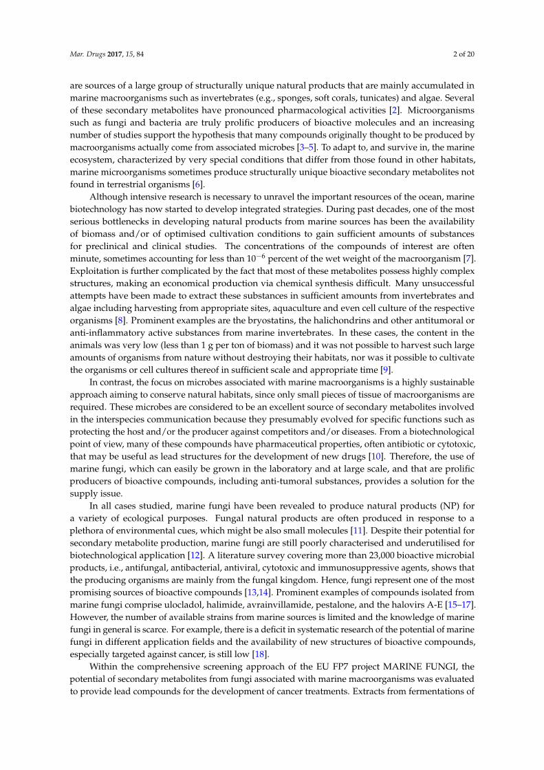
The fungal strain MF458 is part of the marine fungal strain collection of GEOMAR. On Wickerhamagar, the strain grew in whitish colonies (Figure 1), and produced Acremonium-like branchedconidophores. The sequences of the ITS rRNA genes comprised 501 nucleotides, which exhibited asimilarity of 93% for the ITS gene sequence of a Tolypocladium geodes. The 18S RNA gene sequence wasalso obtained, comprising 1827 nucleotides, and showed the highest degree of similarity (93%) to aCordyceps pleuricapitata.
Mar. Drugs 2017, 15, 84 3 of 3
Secondary metabolite biosynthesis has long been known to depend on environmental cues,
including carbon and nitrogen sources, ambient temperature, light and pH [20]. As a consequence,
changes to culture conditions can largely modify the spectrum and amounts of secondary metabolites
produced by fungi [21]. A number of stimuli, such as culture media and cultivation condition
changes, which are known to induce the production of secondary metabolites [22], were employed
to induce variation of the metabolite pattern of the isolated marine fungi.
In our investigation, extracts from fermentations of the sponge‐associated strain MF458,
subsequently identified as Tolypocladium geodes, were found to have potent anti‐tumour effects
derived predominantly from anti‐proliferative rather than overtly cytotoxic profiles. As small‐scale
assay‐guided fractionation did not reveal the obvious presence of compounds known to have anti‐
tumour properties, we commissioned larger scale fermentations. Extracts of these fermentations were
rich in diverse secondary metabolites. The strain was cultivated in a variety of media in order to
characterise the metabolite spectrum of this strain and its susceptibility to stimulation by changing
cultivation conditions [21].
Tolypocladium spp. have attracted significant attention as producers of bioactive secondary
metabolites. Efrapeptins, pyridoxatin and terricolin have previously been reported as products of
terrestrial isolates of T. geodes, while production of the medicinally‐significant cyclosporins is usually
associated with other Tolypocladium spp. [23]. Metabolites from marine Tolypocladium spp., such as
the new efrapeptin J, have also been reported [24].
2. Results
2.1. Classification of Strain MF458
The fungal strain MF458 is part of the marine fungal strain collection of GEOMAR. On
Wickerham agar, the strain grew in whitish colonies (Figure 1), and produced Acremonium‐like
branched conidophores. The sequences of the ITS rRNA genes comprised 501 nucleotides, which
exhibited a similarity of 93% for the ITS gene sequence of a Tolypocladium geodes. The 18S RNA gene
sequence was also obtained, comprising 1827 nucleotides, and showed the highest degree of
similarity (93%) to a Cordyceps pleuricapitata.
Figure 1. MF458; agar colony on WSP30 medium after 21 days of incubation at 22 °C. (a) Image taken
from above the plate; (b) Image taken from below the plate; (c) a liquid culture (non‐shaken) resulted
in white mycelium growing mainly subsurface.
2.2. Isolation and Identification of 1–6
The anti‐tumour activity of the ethyl acetate extracts of fermentations of MF458 were associated
with multiple chromatographic peaks that were separable by reversed phase HPLC (Figure 2) and
purification methods were developed accordingly (summarised in Appendix A Figure A1).
Figure 1. MF458; agar colony on WSP30 medium after 21 days of incubation at 22 ◦C. (a) Image takenfrom above the plate; (b) Image taken from below the plate; (c) a liquid culture (non-shaken) resultedin white mycelium growing mainly subsurface.
2.2. Isolation and Identification of 1–6
The anti-tumour activity of the ethyl acetate extracts of fermentations of MF458 were associatedwith multiple chromatographic peaks that were separable by reversed phase HPLC (Figure 2) andpurification methods were developed accordingly (summarised in Appendix A Figure A1).
Mar. Drugs 2017, 15, 84 4 of 20Mar. Drugs 2017, 15, 84 4 of 4
Figure 2. Reversed phase HPLC chromatogram of the whole culture ethyl acetate extract of a 10 L
MF458 fermentation analysed on a Symmetry Shield RP8 column with gradient elution (a) evaporative
light scattering detector chromatogram; (b) diode array UV‐visible (200–500 nm) chromatogram.
One major band of activity was associated with lipophilic components eluting from towards the
end of the reversed phase gradient employed for the separation illustrated in Figure 2a (retention
time 7.0 to 7.5 min). These components had very little UV absorbance and ESI‐MS indicated molecular
weights of 1188, 1202, 1216 and 1218. The major component from this band with a molecular weight
of 1202 ([MH]+ at m/z 1203, [MNH4]+ at m/z 1220, [M − H]− at m/z 1201, [M + formate]− at m/z 1247) was
purified and its 1H NMR spectrum in CDCl3 was identical with that of the known Tolypocladium spp.
metabolite cyclosporin A, 1 [25].
A more polar band of activity eluting from 5.0 to 5.5 min (Figure 2a) was also associated with
components of relatively high molecular weights. Again, these compounds had no significant UV
absorbance and molecular ions were detected in ESI+‐MS at m/z 1607, 1621, 1635, 1649 and 1663, which
are consistent with the presence of the known Tolypocladium spp. metabolites efrapeptins, being
positively charged linear peptides [26].
The major component of this band was isolated and had a molecular weight of 1621. Its 1H NMR
spectrum in CDCl3 was consistent with that expected for efrapeptin D, 2. Another major active
metabolite produced in the fermentation eluted just after the efrapeptins with a retention time of 5.6–
5.7 min (Figure 2b). This had a UV spectrum with maxima at 218 and 289 nm and ESI‐MS indicated
a molecular weight of 263 ([MH]+ at m/z 264 and [M − H]− at m/z 262). Its 1H and 13C NMR spectra
were identical with those reported for pyridoxatin, 3, first reported as an Acremonium sp. metabolite
[27]. Interestingly, a late‐eluting fraction in the chromatogram was found to be related to pyridoxatin.
Its ESI‐MS indicated a molecular weight of 842 ([MH]+ at m/z 843.6 and [M − H]− at m/z 841.7) and its
UV spectrum consisted of maxima at 221, 246, 287 and 388 nm. These characteristics match well with
those reported for terricolin, 4, a (pyridoxatin)3Fe complex reported as a product of Tolypocladium
terricola [28].
Two more polar compounds with significant anti‐tumour activity (eluting with retention times
of 4.3 and 4.5 min in Figure 2) were obviously closely related with the same molecular weight of 292
(deduced from ESI‐MS molecular ions at m/z 293 ([MH]+) and 291 ([M − H]−) in positive and negative
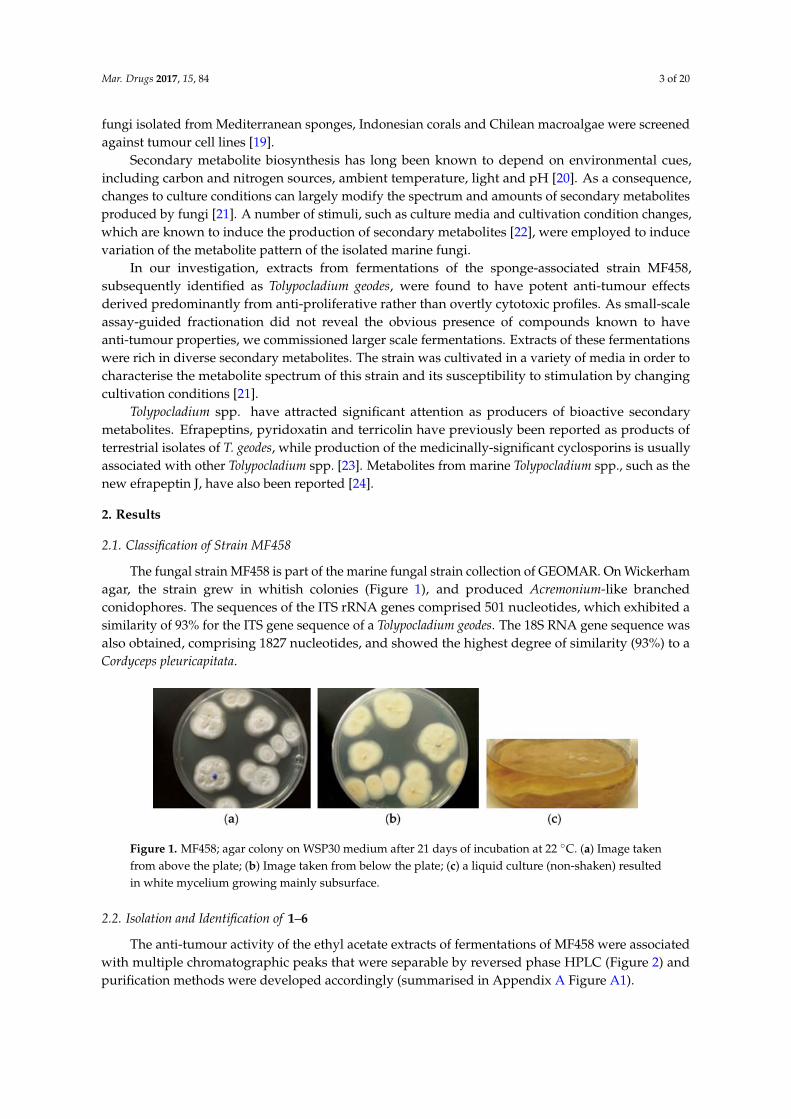
Figure 2. Reversed phase HPLC chromatogram of the whole culture ethyl acetate extract of a 10 LMF458 fermentation analysed on a Symmetry Shield RP8 column with gradient elution (a) evaporativelight scattering detector chromatogram; (b) diode array UV-visible (200–500 nm) chromatogram.
One major band of activity was associated with lipophilic components eluting from towards theend of the reversed phase gradient employed for the separation illustrated in Figure 2a (retention time7.0 to 7.5 min). These components had very little UV absorbance and ESI-MS indicated molecularweights of 1188, 1202, 1216 and 1218. The major component from this band with a molecular weightof 1202 ([MH]+ at m/z 1203, [MNH4]+ at m/z 1220, [M − H]− at m/z 1201, [M + formate]− atm/z 1247) was purified and its 1H NMR spectrum in CDCl3 was identical with that of the knownTolypocladium spp. metabolite cyclosporin A, 1 [25].
A more polar band of activity eluting from 5.0 to 5.5 min (Figure 2a) was also associated withcomponents of relatively high molecular weights. Again, these compounds had no significant UVabsorbance and molecular ions were detected in ESI+-MS at m/z 1607, 1621, 1635, 1649 and 1663,which are consistent with the presence of the known Tolypocladium spp. metabolites efrapeptins, beingpositively charged linear peptides [26].
The major component of this band was isolated and had a molecular weight of 1621. Its 1HNMR spectrum in CDCl3 was consistent with that expected for efrapeptin D, 2. Another major activemetabolite produced in the fermentation eluted just after the efrapeptins with a retention time of5.6–5.7 min (Figure 2b). This had a UV spectrum with maxima at 218 and 289 nm and ESI-MS indicateda molecular weight of 263 ([MH]+ at m/z 264 and [M − H]− at m/z 262). Its 1H and 13C NMRspectra were identical with those reported for pyridoxatin, 3, first reported as an Acremonium sp.metabolite [27]. Interestingly, a late-eluting fraction in the chromatogram was found to be related topyridoxatin. Its ESI-MS indicated a molecular weight of 842 ([MH]+ at m/z 843.6 and [M − H]− atm/z 841.7) and its UV spectrum consisted of maxima at 221, 246, 287 and 388 nm. These characteristicsmatch well with those reported for terricolin, 4, a (pyridoxatin)3Fe complex reported as a product ofTolypocladium terricola [28].
Two more polar compounds with significant anti-tumour activity (eluting with retention timesof 4.3 and 4.5 min in Figure 2) were obviously closely related with the same molecular weight of
Mar. Drugs 2017, 15, 84 5 of 20
292 (deduced from ESI-MS molecular ions at m/z 293 ([MH]+) and 291 ([M − H]−) in positive andnegative ionisation modes, respectively) and identical UV spectra with maxima at 253, 328sh and359 nm. This UV spectrum is characteristic of the presence of a tropolone moiety and these compoundswere identified by their 1H and 13C NMR spectra. The earlier eluting material was found to be identicalto the fungal metabolite malettinin B, 5, [29] while the second peak was found to correspond to therecently described malettinin E, 6 [30], and its identity was confirmed by a direct NMR comparisonwith authentic material of this compound. These malettinins have not previously been described asTolypocladium spp. metabolites. A polyketide origin has been proposed for the malettinins, with thetropolone unit perhaps arising via an aromatic ring expansion, as may be the case for other fungaltropolones [31].
2.3. Structure Elucidation of 7 and 8
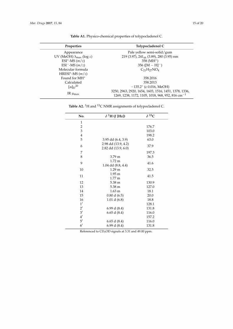
Compounds 7 and 8 are represented by the peaks eluting at 6.3 and 6.9 min in Figure 2.The physico-chemical properties, 1H and 13C NMR spectral data for compound 7 are summarized in theAppendix A Tables A1 and A2, respectively. The ESI-MS spectrum of compound 7 gave a molecular ion([MH]+) at m/z 358, indicating a molecular weight of 357, and accurate mass measurement on this ionwas consistent with the molecular formula C21H27NO5 (Appendix A Table A1). The 1H and 13C NMRdata for 7 are summarized in Appendix A Table A2. The 13C NMR spectrum confirmed the presenceof 21 carbon atoms and DEPT spectra indicated that these comprised 3 methyl, 3 methylene, 9 methineand 6 quaternary carbons. The 3 protons unaccounted for were not observed in the 1H spectrumin CD3OD and were inferred to be exchangeable. The presence of a p-substituted phenyl moietywas evident from the observation of four sp2 methine carbons as two coupled pairs of chemicallyequivalent protons C-2′/C-6′ (131.8/6.99 ppm) and C-3′/C-5′ (116.0/6.65 ppm). The presence of atetramic acid moiety was suggested by broad carbon signals at 198.2 (C-4), 197.3 (C-7), 176.7 (C-2), 103.0(C-3) and 63.0 (C-5) ppm. 1H coupling between methine H-5 at 3.95 ppm and methylene protons H-6at 2.98 and 2.82 ppm together with long range 1H-13C correlations observed in the HMBC spectrumbetween the methylene group at C-6 (37.9/2.98, 2.82 ppm) and carbon signals at C-4 (198.2 ppm),C-1′ (128.1 ppm) and C-2′/C-5′ (131.8 ppm) indicated that this methylene bridged between thep-substituted phenol and tetramic acid moieties. This conclusion was also consistent with the UVmaxima observed for 7 (Appendix A Table A1). COSY, HSQC and HMBC data were used to establishthe presence of a 5,7-dimethylheptan-2-enyl side chain. HMBC correlations between the H3-16 methylprotons at 1.01 ppm and H2-9 methylene protons at 1.72/1.04 ppm, and C-7 at 197.3 ppm indicatedthe point of attachment of this side chain to the tetramic acid ring to establish the structure of 7 as5-(4-hydroxybenzyl)-3-(2,4-dimethyl-1-hydroxyoct-6-en-1-ylidene)pyrrolidine-2,4-dione. Due to itsstructural similarity to the tolypocladenols from another Tolypocladium species, T. cylindrosporum, wepropose to assign this molecule to this family and name it tolypocladenol C.
The ESI-MS spectrum of compound 8 gave a molecular ion ([MH]+) at m/z 356, indicatinga molecular weight of 355, and accurate mass measurement on this ion was consistent withthe molecular formula C21H25NO5. It had a more extended UV spectrum than 7, indicatinga greater degree of conjugation. Its NMR spectra indicated a close structural relationshipto 7 and interpretation of the NMR data indicated that 8 is the 5,6-dehydro derivative of 7:5-(4-hydroxybenzylidene)-3-(2,4-dimethyl-1-hydroxyoct-6-en-1-ylidene)pyrrolidine-2,4-dione. Afterthis work was completed, compound 8 was reported as tolypocladenols A1/A2, isolated fromfermentations of the endolichenic fungus Tolypocladium cylindrosporum [32]. The NMR, UV and MSdata for 8 are consistent with those reported for tolypocladenols A1/A2 and are not reported herein.Tolypocladenols A1/A2 were identified as a mixture of enantiomers with RR and SS configurations atcarbons 8 and 10 and their structures were determined by single crystal X-ray diffraction. In additionto the tolpocladenols A1, A2 and B, 7 and 8 are structurally similar to a number of other fungalacyltetramic acids described in the literature. Examples would be militarinones B and C [33] andepicoccarines A and B [34], with 7, showing a high degree of structural similarity to the epicoccarines,
Mar. Drugs 2017, 15, 84 6 of 20
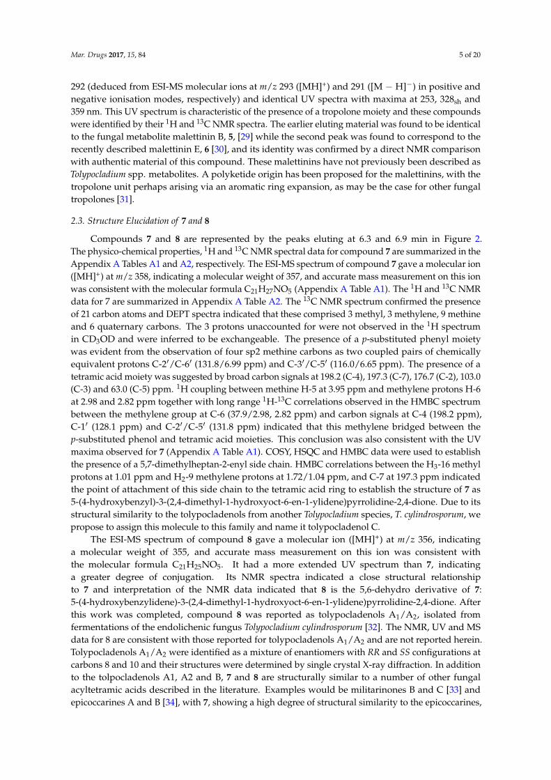
with their mostly saturated acyl chains. Due to the structural similarity between compound 8and 7 it can be assumed that the biosynthesis of both natural products is analogous to the hybridPKS-NRPS pathway proposed for a variety of tetramic acid based fungal natural products, suchas the epicoccarines A and B [32,35]. Figure 3 summarises the natural products isolated fromTolypocladium geodes MF458.
Mar. Drugs 2017, 15, 84 6 of 6
epicoccarines A and B [32,35]. Figure 3 summarises the natural products isolated from Tolypocladium
geodes MF458.
Figure 3. Natural products produced by Tolypocladium geodes sp. MF458: cyclosporin A (1); efrapeptin
D (2); pyridoxatin (3); terricolin (4); malletenin B (5); malletenin E (6); tolypocladenol C (7);
tolypocladenols A1 and A2 (8).
2.4. Biological Activities of 1–8
The biological activity of semi‐purified chromatography fractions was initially determined using
three sensitive cell lines at three concentrations for activity guided purification. The detailed
procedure was reported elsewhere [19] and showed a promising activity profile for a number of
fractions from MF458. Purified compounds were analysed using a panel of cell lines from the NCI60
panel, consisting of cell lines from 8 different tissues and a number of different cancer stages and
genetic characteristics. Terricolin (4), as an iron complex, was deprioritised due to unfavourable
chemical properties and not tested further. While no general trend with respect to tissue sensitivity
could be identified, clear differences between individual cell lines were apparent (see Appendix A
Table A3 for complete overview of inhibition values). The growth of the breast adenocarcinoma cell
line MCF‐7 was inhibited at least at the highest concentration by all the natural products evaluated,
while the growth of the ovarian carcinoma cell line OVCAR‐5 was reduced to at least 50% only by
four of the seven compounds (Figure 4).
Figure 3. Natural products produced by Tolypocladium geodes sp. MF458: cyclosporin A (1);efrapeptin D (2); pyridoxatin (3); terricolin (4); malletenin B (5); malletenin E (6); tolypocladenolC (7); tolypocladenols A1 and A2 (8).
2.4. Biological Activities of 1–8
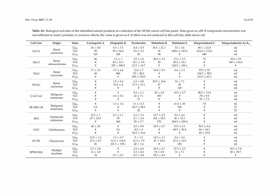
The biological activity of semi-purified chromatography fractions was initially determined usingthree sensitive cell lines at three concentrations for activity guided purification. The detailed procedurewas reported elsewhere [19] and showed a promising activity profile for a number of fractions fromMF458. Purified compounds were analysed using a panel of cell lines from the NCI60 panel, consistingof cell lines from 8 different tissues and a number of different cancer stages and genetic characteristics.Terricolin (4), as an iron complex, was deprioritised due to unfavourable chemical properties andnot tested further. While no general trend with respect to tissue sensitivity could be identified, cleardifferences between individual cell lines were apparent (see Appendix A Table A3 for completeoverview of inhibition values). The growth of the breast adenocarcinoma cell line MCF-7 was inhibited
Mar. Drugs 2017, 15, 84 7 of 20
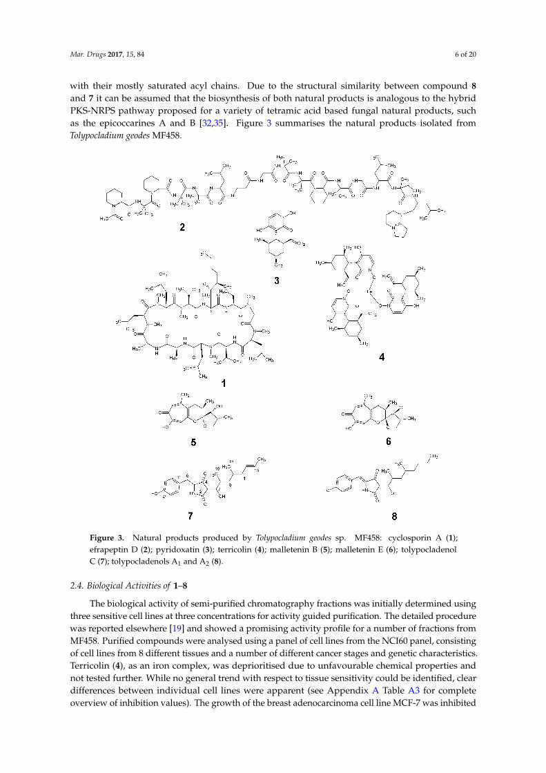
at least at the highest concentration by all the natural products evaluated, while the growth of theovarian carcinoma cell line OVCAR-5 was reduced to at least 50% only by four of the seven compounds(Figure 4).Mar. Drugs 2017, 15, 84 7 of 7
Figure 4. Activity of the isolated compounds in different cell lines. (a) Viability of MCF‐7 cell line after
48 h compound treatment. (b) Viability of OVCAR‐5 cell line after 48 h compound treatment;
cyclosporin A (blue), efrapeptin (violet), malettinin B (orange), pyridoxatin (red), malettinin E (dark
yellow), tolypocladenol C (green), tolypocladenol A1/A2 (brown).
The only compound with a similar efficiency in both cell lines is efrapeptin D (2), inhibiting the
growth of MCF‐7 and OVCAR‐5 at 7 μM and 12 μM, respectively. The effect of efrapeptin, allosteric
inhibition of the mitochondrial ATP synthase, explains its effectiveness in both cell lines and also the
broad activity profile of this predominantly cytostatic compound [36]. The malettinins B and E (5 and
6) are less studied and little is known about their biological effects [29,37]. This study represents
therefore the first description of their cytotoxic potential. The compounds show an activity in every
cell line evaluated, except melanoma cell line SK‐MEL‐28, which is not growth inhibited by malettinin
B. Tolypocladenol C (7) and also tolypocladenols A1/A2 (8), although less studied, appear to be not
active against the majority of cancer cell lines, reducing the relevance of these compounds in terms
of anticancer drug discovery. In line with previous reports, pyridoxatin (3) was identified as showing
a good toxicity profile against the cell line panel in the submicromolar and low micromolar range.
2.5. Secondary Metabolite Profile of MF458 Depending on Fermentation Conditions
Production of secondary metabolite extracts for screening and leading to the identification and
dereplication of the compounds 1–8 was achieved by fermentation in Erlenmeyer (EM) flasks at 1 L
scale under different conditions (Figure 5). Pre‐experiments (not shown) indicated that casamino
acids medium seems to be best for production of secondary metabolites by MF458. Therefore, the
casamino acids medium recipe was taken as a basis and adopted for optimisation of metabolite
production. Production of the metabolites depended on both the medium composition and the
shaking conditions.
Most of the compounds were produced best using standing conditions that allowed formation
of a mycelium cake. The conditions were used to produce higher amounts of the compounds for
purification and isolation purposes. Generally, cultivation had to be performed for >24 days (>550 h)
for obtaining maximum yield of the metabolites.
Figure 4. Activity of the isolated compounds in different cell lines. (a) Viability of MCF-7 cell lineafter 48 h compound treatment. (b) Viability of OVCAR-5 cell line after 48 h compound treatment;cyclosporin A (blue), efrapeptin (violet), malettinin B (orange), pyridoxatin (red), malettinin E(dark yellow), tolypocladenol C (green), tolypocladenol A1/A2 (brown).
The only compound with a similar efficiency in both cell lines is efrapeptin D (2), inhibiting thegrowth of MCF-7 and OVCAR-5 at 7 µM and 12 µM, respectively. The effect of efrapeptin, allostericinhibition of the mitochondrial ATP synthase, explains its effectiveness in both cell lines and alsothe broad activity profile of this predominantly cytostatic compound [36]. The malettinins B and E(5 and 6) are less studied and little is known about their biological effects [29,37]. This study representstherefore the first description of their cytotoxic potential. The compounds show an activity in everycell line evaluated, except melanoma cell line SK-MEL-28, which is not growth inhibited by malettininB. Tolypocladenol C (7) and also tolypocladenols A1/A2 (8), although less studied, appear to be notactive against the majority of cancer cell lines, reducing the relevance of these compounds in terms ofanticancer drug discovery. In line with previous reports, pyridoxatin (3) was identified as showing agood toxicity profile against the cell line panel in the submicromolar and low micromolar range.
2.5. Secondary Metabolite Profile of MF458 Depending on Fermentation Conditions
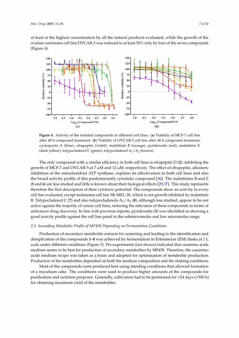
Production of secondary metabolite extracts for screening and leading to the identification anddereplication of the compounds 1–8 was achieved by fermentation in Erlenmeyer (EM) flasks at 1 Lscale under different conditions (Figure 5). Pre-experiments (not shown) indicated that casamino acidsmedium seems to be best for production of secondary metabolites by MF458. Therefore, the casaminoacids medium recipe was taken as a basis and adopted for optimisation of metabolite production.Production of the metabolites depended on both the medium composition and the shaking conditions.
Most of the compounds were produced best using standing conditions that allowed formationof a mycelium cake. The conditions were used to produce higher amounts of the compounds forpurification and isolation purposes. Generally, cultivation had to be performed for >24 days (>550 h)for obtaining maximum yield of the metabolites.
Mar. Drugs 2017, 15, 84 8 of 20
Mar. Drugs 2017, 15, 84 8 of 8
Figure 5. Comparison of production of MF458 natural product profiles in different media in
Erlenmeyer (EM) flasks. Peak area (ion count) is given as size and ranges from 0 to 10 M; values above
10 M are shown as maximum; B12: Casamino acid supplemented with B12, Casa: casamino acid
medium, half concentrated (½), double concentrated (2×), GM: casamino acid medium containing
glucose and malt, M: casamino acid medium supplemented with molasses, L, liquid shaken culture,
M. mycelium from standing culture, numbers indicate days of cultivation.
2.6. Optimisation of Production for Scaling‐Up the Production of the Malettinins
For a larger scale fermentation of malettinin E (6) and B (5) in bioreactors, the production had to
be transferred initially into liquid, shaken conditions, as the highest production in the initial screening
study was observed in standing cultures. Accordingly, casamino acids medium was supplemented
with substrates supporting stable mycelium formation under liquid shaken/stirred conditions, such
as crystalline cellulose, paper, gelatine, CaCO3, and agar. These pellet enhancers had different effects
on the different metabolites, e.g., the addition of CaCO3 increased especially the production of
tolypocladenol C (7) by a factor of 10. As the malettinins had the most interesting biological activity,
the optimisation strategy was, however, focused on optimal production conditions for the
malettinins. With respect to the pellet enhancers, addition of 10 g/L crystalline cellulose had the best
effect on malettinin production. Accordingly, casamino acids medium supplemented with crystalline
cellulose was chosen for a stirred tank reactor (STR) cultivation. STR cultures were conducted at the
10 L scale, controlling gas flow and stirrer speed but with an autonomous pH regime. Malettinin
production under STR conditions out‐competed the production of the other metabolites (Figure 6).
Figure 6. Optimisation of malettinin production. (a) Production of secondary metabolites in casamino
acid medium supplemented with crystalline cellulose under STR conditions as peak area [total ion
count (TIC)]; The color coding is: cyclosporin A (1, blue), efrapeptin (2, violet), malettinin B (5,
orange), pyridoxatin (3, red), malettinin E (6, dark yellow), tolypocladenol C (7, green),
tolypocladenol A1/A2 (8, brown). (b) Production of malettinin B (5): amount of malettinin B production
(star); pH (ranging from 5.50 to 4.18, shown as point); O2 saturation in % (diamond).
Figure 5. Comparison of production of MF458 natural product profiles in different media in Erlenmeyer(EM) flasks. Peak area (ion count) is given as size and ranges from 0 to 10 M; values above 10 M areshown as maximum; B12: Casamino acid supplemented with B12, Casa: casamino acid medium, halfconcentrated ( 1
2 ), double concentrated (2×), GM: casamino acid medium containing glucose and malt,M: casamino acid medium supplemented with molasses, L, liquid shaken culture, M. mycelium fromstanding culture, numbers indicate days of cultivation.
2.6. Optimisation of Production for Scaling-Up the Production of the Malettinins
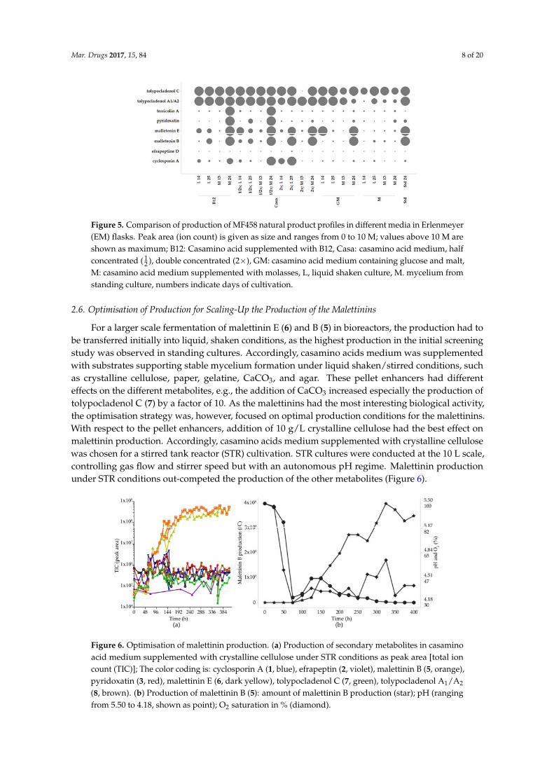
For a larger scale fermentation of malettinin E (6) and B (5) in bioreactors, the production had tobe transferred initially into liquid, shaken conditions, as the highest production in the initial screeningstudy was observed in standing cultures. Accordingly, casamino acids medium was supplementedwith substrates supporting stable mycelium formation under liquid shaken/stirred conditions, suchas crystalline cellulose, paper, gelatine, CaCO3, and agar. These pellet enhancers had differenteffects on the different metabolites, e.g., the addition of CaCO3 increased especially the production oftolypocladenol C (7) by a factor of 10. As the malettinins had the most interesting biological activity,the optimisation strategy was, however, focused on optimal production conditions for the malettinins.With respect to the pellet enhancers, addition of 10 g/L crystalline cellulose had the best effect onmalettinin production. Accordingly, casamino acids medium supplemented with crystalline cellulosewas chosen for a stirred tank reactor (STR) cultivation. STR cultures were conducted at the 10 L scale,controlling gas flow and stirrer speed but with an autonomous pH regime. Malettinin productionunder STR conditions out-competed the production of the other metabolites (Figure 6).
Mar. Drugs 2017, 15, 84 8 of 8
Figure 5. Comparison of production of MF458 natural product profiles in different media in
Erlenmeyer (EM) flasks. Peak area (ion count) is given as size and ranges from 0 to 10 M; values above
10 M are shown as maximum; B12: Casamino acid supplemented with B12, Casa: casamino acid
medium, half concentrated (½), double concentrated (2×), GM: casamino acid medium containing
glucose and malt, M: casamino acid medium supplemented with molasses, L, liquid shaken culture,
M. mycelium from standing culture, numbers indicate days of cultivation.
2.6. Optimisation of Production for Scaling‐Up the Production of the Malettinins
For a larger scale fermentation of malettinin E (6) and B (5) in bioreactors, the production had to
be transferred initially into liquid, shaken conditions, as the highest production in the initial screening
study was observed in standing cultures. Accordingly, casamino acids medium was supplemented
with substrates supporting stable mycelium formation under liquid shaken/stirred conditions, such
as crystalline cellulose, paper, gelatine, CaCO3, and agar. These pellet enhancers had different effects
on the different metabolites, e.g., the addition of CaCO3 increased especially the production of
tolypocladenol C (7) by a factor of 10. As the malettinins had the most interesting biological activity,
the optimisation strategy was, however, focused on optimal production conditions for the
malettinins. With respect to the pellet enhancers, addition of 10 g/L crystalline cellulose had the best
effect on malettinin production. Accordingly, casamino acids medium supplemented with crystalline
cellulose was chosen for a stirred tank reactor (STR) cultivation. STR cultures were conducted at the
10 L scale, controlling gas flow and stirrer speed but with an autonomous pH regime. Malettinin
production under STR conditions out‐competed the production of the other metabolites (Figure 6).
Figure 6. Optimisation of malettinin production. (a) Production of secondary metabolites in casamino
acid medium supplemented with crystalline cellulose under STR conditions as peak area [total ion
count (TIC)]; The color coding is: cyclosporin A (1, blue), efrapeptin (2, violet), malettinin B (5,
orange), pyridoxatin (3, red), malettinin E (6, dark yellow), tolypocladenol C (7, green),
tolypocladenol A1/A2 (8, brown). (b) Production of malettinin B (5): amount of malettinin B production
(star); pH (ranging from 5.50 to 4.18, shown as point); O2 saturation in % (diamond).
Figure 6. Optimisation of malettinin production. (a) Production of secondary metabolites in casaminoacid medium supplemented with crystalline cellulose under STR conditions as peak area [total ioncount (TIC)]; The color coding is: cyclosporin A (1, blue), efrapeptin (2, violet), malettinin B (5, orange),pyridoxatin (3, red), malettinin E (6, dark yellow), tolypocladenol C (7, green), tolypocladenol A1/A2
(8, brown). (b) Production of malettinin B (5): amount of malettinin B production (star); pH (rangingfrom 5.50 to 4.18, shown as point); O2 saturation in % (diamond).
Mar. Drugs 2017, 15, 84 9 of 20
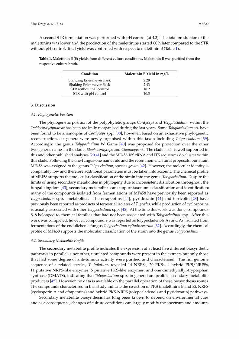
A second STR fermentation was performed with pH control (at 4.3). The total production of themalettinins was lower and the production of the malettinins started 60 h later compared to the STRwithout pH control. Total yield was confirmed with respect to malettinin B (Table 1).
Table 1. Malettinin B (5) yields from different culture conditions. Malettinin B was purified from therespective culture broth.
Condition Malettinin B Yield in mg/L
Standing Erlenmeyer flask 2.28Shaking Erlenmeyer flask 2.43STR without pH control 18.2
STR with pH control 10.3
3. Discussion
3.1. Phylogenetic Position
The phylogenetic position of the polyphyletic groups Cordyceps and Tolyplocladium within theOphiocordycipitaceae has been radically reorganised during the last years. Some Tolyplocladium sp. havebeen found to be anamorphs of Cordyceps spp. [38], however, based on an exhaustive phylogeneticreconstruction, six genera were newly organised within this taxon including Tolypocladium [39].Accordingly, the genus Tolypocladium W. Gams [40] was proposed for protection over the othertwo generic names in the clade, Elaphocordyceps and Chaunopycnis. The clade itself is well supported inthis and other published analyses [20,41] and the MF458 18S rRNA and ITS sequences do cluster withinthis clade. Following the one-fungus-one name rule and the recent nomenclatural proposals, our strainMF458 was assigned to the genus Tolypocladium, species geodes [42]. However, the molecular identity iscomparably low and therefore additional parameters must be taken into account. The chemical profileof MF458 supports the molecular classification of the strain into the genus Tolypocladium. Despite thelimits of using secondary metabolites in phylogeny due to inconsistent distribution throughout thefungal kingdom [43], secondary metabolites can support taxonomic classification and identification:many of the compounds isolated from fermentations of MF458 have previously been reported asTolypocladium spp. metabolites. The efrapeptins [44], pyridoxatin [44] and terricolin [28] havepreviously been reported as products of terrestrial isolates of T. geodes, while production of cyclosporinsis usually associated with other Tolypocladium spp. [45]. At the time this work was done, compounds5–8 belonged to chemical families that had not been associated with Tolypocladium spp. After thiswork was completed, however, compound 8 was reported as tolypocladenols A1 and A2, isolated fromfermentations of the endolichenic fungus Tolypocladium cylindrosporum [32]. Accordingly, the chemicalprofile of MF458 supports the molecular classification of the strain into the genus Tolypocladium.
3.2. Secondary Metabolite Profile
The secondary metabolite profile indicates the expression of at least five different biosyntheticpathways in parallel, since other, unrelated compounds were present in the extracts but only thosethat had some degree of anti-tumour activity were purified and characterised. The full genomesequence of a related species, T. inflatum, revealed 14 NRPSs, 20 PKSs, 4 hybrid PKS/NRPSs,11 putative NRPS-like enzymes, 5 putative PKS-like enzymes, and one dimethylallyl-tryptophansynthase (DMATS), indicating that Tolypocladium spp. in general are prolific secondary metaboliteproducers [45]. However, no data is available on the parallel operation of these biosynthesis routes.The compounds characterised in this study indicate the co-action of PKS (malettinins B and E), NRPS(cyclosporin A and efrapeptins) and hybrid PKS-NRPS (tolypocladenols and pyridoxatin) pathways.
Secondary metabolite biosynthesis has long been known to depend on environmental cuesand as a consequence, changes of culture conditions can largely modify the spectrum and amounts
Mar. Drugs 2017, 15, 84 10 of 20
of secondary metabolites produced [21]. Tolypocladium geodes MF 458 showed a wide spectrum ofmetabolites and reacted sensitively to changes of the culture conditions.
3.3. Biological Activity Profile
The eight identified natural products produced by MF458 show a diverse spectrum of activities.Cyclosporin A (1) is a well-known inhibitor of NFAT signalling and has been used since the 70s asan immunosuppressant in the clinic [46]. Efrapeptin D (2) binds to ATP synthase, also inhibiting theHSP90 chaperone function and the proteasome. These multiple targets prevent the development ofefrapeptin into a drug although the molecule is effective against MCF-7 induced breast cancer [47].The malettinins B and E (5 and 6) are less studied and little can be found about their biological activityother than that they exhibit weak antimicrobial activity [29,37]. However, their activity against all celllines evaluated and their generally low cytotoxicity make them interesting molecules for future targetdeconvolution strategies. Pyridoxatin (3) is similarly lacking a conclusive mode of action [48]. It is acompound with previously reported cytotoxicity against various cell lines and moderate inhibitoryactivity against gelatinase A, a matrix metalloprotease, but also lacking a conclusive cellular modeof action [48]. Instead pyridoxatin was shown to induce erythropoietin gene expression, although amore general effect on gene expression cannot be ruled out since it downregulated genes relevantfor ergosterol biosynthesis in Candida albicans, as well [49,50]. The compound is reported to be a freeradical scavenger, but it remains to be investigated whether this mode of action can explain its activityprofile [27]. Tolypocladenol C (7) and tolypocladenols A1/A2 (8) appear to be non-cytotoxic andterricolin (4) is an iron complex making it also unfavourable for further activity screening.
3.4. Fermentation Optimization
Lead optimisation and preclinical development for newly identified natural products is cruciallydependent on sufficient supply of material. For this purpose, chemical synthesis and biotechnologicalproduction have to be considered and compared in terms of feasibility, effort and costs. However,upscaling of the biotechnological production from, in most cases, Erlenmeyer flask cultures tocontrollable stirred tank reactors is a challenge, as the scaling itself is a change of cultivation conditionsthat may lead to a change of the secondary metabolite profile [21]. For MF458, pellet enhancers wereused to overcome the initial standing cultivation conditions and to expedite the transfer into STR.The phenomenon of production being restricted to cultures that grew on the surface of the culturebroth or at least under adherent conditions is well known for filamentous fungi. For instance, thesorbicillactone A produced by a marine Penicillium chrysogenum could be obtained only in surfacecultures [51,52]. Addition of pellet enhancers or application of very carefully regulated stirring regimesmight overcome this limitation to further scalability [53]. Application of amino acids based mediumsupplemented with crystalline cellulose enabled submerged cultures of MF458. This condition wasused for successful transfer into stirred tank reactor as the basis for upscaling of the production. To ourknowledge, this is the first report on STR conditions for T. geodes secondary metabolite production.
4. Methods
4.1. Isolation and Taxonomy of MF458
Strain MF458 was isolated by Dr. Karsten Schaumann from a sponge sample. DNA extraction,amplification of the internal transcribed spacer region (ITS) and the 18S rRNA gene, as well assequencing were performed as described by [30] with slight modifications, centrifugation of the DNAat 8000× g and 35 cycles of DNA amplification. The DNA sequences were deposited in GenBank underthe accession number KY696657, for the 18S rRNA sequence (MF458_18S), and KY696658, for the ITSregion (MF458_ITS1). Cryopreserved stock cultures of strain MF458 were kept at −100 ◦C using theMicrobank system (Microbank system, MAST DIAGNOSTIKA, Reinfeld, Germany). Closest relativeswere identified by sequence comparison with the NCBI Genbank database using BLAST (Basic Local
Mar. Drugs 2017, 15, 84 11 of 20
Alignment Search Tool) [54]. Sequence similarity values were determined with the “bl2seq” tool of theNCBI database [55].
4.2. Fermentation and Crude Extract Production
The fungal strain was cultivated in the following media: casamino acids glucose medium [56],casamino acids medium at half and double concentration, casamino acids supplemented with B12(5 g/L), GM medium (as modified by [57]), casamino acids supplemented with molasses (80 g/L)and modified Wickerham medium [58]. Cryopreserved stock cultures were activated on WSP agar(3% NaCl) at 28 ◦C in the dark for 7 days. Main cultures were done in 2 L EM flasks (containing1000 mL medium) using the different conditions. Flasks were inoculated using agar pieces fromprecultures (Ø = 2.6 cm) and incubated at 22 ◦C in the dark, at 120 rpm or using standing conditions.Crystalline cellulose (10 g/L), paper (40 g/L), gelatine (18 g/L), CaCO3 (15 g/L), and agar (3 g/L)were added as pellet enhancers.
For isolation of compounds, fermentation was done in multiple shake flasks containing casaminoacids medium, a temperature of 22 ◦C and standing conditions.
The cultivation was subsequently scaled up in stirred tank reactors in 10 L (Braun Biostat MD,glass tank), containing 8 L casamino acid medium supplemented with 10 g/L crystalline cellulose(Carl Roth, Karlsruhe, Germany). Precultures were established in the same medium and cultivated for20 days at 22 ◦C, 120 rpm. The inoculation volume was 100 mL (1.25%). The fermenter was run at25 ◦C with pH, oxygen, and stirring speed being measured and oxygen being controlled to a minimumof 30% air saturation (flow up to 400 L/min, stirrer speed up to 200 rpm). Foam formation was stoppedby addition of antifoam (Sigma, Taufkirchen, Germany). A second fermentation run was performedusing the same conditions but controlling the pH at 4.3 using 3 M NaOH.
All samples and final harvests of the fermentations were extracted using liquid-liquid extraction.2 volumes of ethyl acetate were added to one volume of culture, homogenized and separated by meansof separating funnels. The organic phase was washed using distilled water and dried in vacuo at 40 ◦C.Extracts were stored at −20 ◦C until use.
4.3. Anti-Tumour Assay-Guidance for Purification and Pure Compound Evaluation
Crude extracts and HPLC fractions were profiled at three concentrations against MCF-7, M14and 786-0 cell lines in a 96-well microtitre assay format using the neutral red cell viability protocol toidentify those containing potentially useful anticancer components [19]. The 100-fold concentratedmarine fungal fermentation extracts and HPLC fractions concentrated to dryness and re-dissolvedin DMSO-methanol (3:1) were tested for anti-tumour activity at three different dilutions: (A) 1/200,(B) 1/1000, and (C) 1/5000, to facilitate ranking of activities detected based on potency. This resulted,after two or three purification steps, in the isolation of single active compounds. These pure compoundswere then characterized in a wider range of cell lines from the NCI60 panel.
4.4. Purification and Identification of Compounds 1–8
The ethyl acetate extract of a 10 L fermentation in multiple shake flasks was dissolved inDMSO-methanol (3:1) and chromatographed in multiple injections on a Waters Xbridge phenyl column(19 × 100 mm) eluted with a linear water-acetonitrile gradient in the presence of 0.1% formic acid,increasing from 10% to 100% acetonitrile over a period of 8 min, holding at 100% acetonitrile for afurther 5 min before returning to the starting composition in 1 min and re-equilibrating for another6 min, at a flow rate of 17 mL/min. After the first minute, eluate fractions were collected every 35 s(24 fractions in total). These fractions were concentrated to dryness, redissolved in DMSO-methanol(3:1) and aliquots diluted 1/10 for assay against tumour cell lines as described above. Based on the assayresults and HPLC-MS analysis of the chemical contents of the active fractions, fractions were combinedand further purified as follows. The fractions (5–9) eluting between 3 and 5 min were concentratedand further purified by chromatography on a Waters Symmetry Shield RP8 column (19 × 100 mm)
Mar. Drugs 2017, 15, 84 12 of 20
eluted isocratically with 25% aqueous acetonitrile containing 10 mM ammonium formate and 0.1%formic acid at a flow rate of 17 mL/min with UV detection at 352 nm. The peaks eluting between 8–10and 11–13 min were separately collected and rotary evaporated to remove acetonitrile before applyingto Mega Bond Elut Plexa solid phase extraction (SPE) columns (Agilent Technologies, Santa Clara, CA,USA, 500 mg columns) that had been pre-conditioned with methanol followed by water, for de-salting.The columns were then washed with several column volumes of water and eluted with acetonitrile,and the acetonitrile eluates concentrated to dryness to yield compounds 5 (17 mg) and 6 (24 mg).
The Xbridge phenyl column fractions (10–13) eluting between 5 and 7 min were combined andfurther purified by chromatography on a Symmetry Shield RP8 column (19 × 100 mm) eluted with alinear gradient increasing from 35% to 65% acetonitrile in water in the presence of 10 mM ammoniumformate and 0.1% formic acid over a period of 15 min at a flow rate of 17 mL/min with UV detection at254 nm. Eluate collected from 6 to 8 min was concentrated to dryness and purified by chromatographyon the Symmetry Shield RP8 column eluted isocratically with 42% aqueous acetonitrile containing10 mM ammonium formate and 0.1% formic acid at a flow rate of 17 mL/min with UV detectionat 230 nm. The peak eluting between 5 and 6 min was collected, de-salted by SPE as above andconcentrated to dryness to yield 2 (1.3 mg). The peak eluting between 8 and 11 min was collected,concentrated to dryness and purified by chromatography on the Symmetry Shield RP8 column elutedisocratically with 35% aqueous acetonitrile containing 10 mM ammonium formate and 0.1% formicacid at a flow rate of 17 mL/min with UV detection at 250 nm. The peak eluting between 21 and23 min was collected, de-salted by SPE as above and concentrated to dryness to yield 3 (15 mg).The peak eluting between 11 and 14 min was concentrated to dryness and purified by chromatographyon the Symmetry Shield RP8 column eluted isocratically with 45% aqueous acetonitrile containing10 mM ammonium formate and 0.1% formic acid at a flow rate of 17 mL/min with UV detectionat 250 nm. The peak eluting between 7 and 8 min was collected, de-salted by SPE as above andconcentrated to dryness to yield 7 (9 mg). Eluate collected from 14 to 15.5 min was concentrated todryness and purified by chromatography on the Symmetry Shield RP8 column eluted isocratically with55% aqueous acetonitrile containing 10 mM ammonium formate and 0.1% formic acid at a flow rateof 17 mL/min with UV detection at 300 nm. The peak eluting between 12 and 13 min was collected,de-salted by SPE as above and concentrated to dryness to yield 8 (5 mg). Eluate eluting between 17and 18 min yielded 1 (2.4 mg).
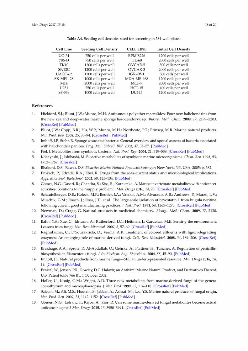
4.5. Assessment of Compound Cytotoxicity
Cell lines used were part of the NCI60 panel and maintained using RPMI-1640 medium(PAA, Hesse, Germany) containing 2 mM glutamine, 100 U/mL penicillin G, 100 mg/mL streptomycin,and 10% fetal calf serum (FCS). At about 80% confluence, cells were rinsed with Dulbecco’sphosphate-buffered saline (DPBS), trypsinised, suspended and counted in RPMI-1640 medium beforeseeding into 384-well plates. Cells were seeded at 20 µL per well in different densities (see Appendix ATable A4 for complete overview) in white, 384-well, PS, Cellstar plates (Greiner Bio-One GmbH,Frickenhausen, Germany) and incubated at 37 ◦C in the presence of 5% carbon dioxide. At 24 h postseeding, baseline growth was assessed using a control plate and CellTiter-Glo reagent (CTG reagent,Promega Inc., Madison, WI, USA). Briefly, 20 µL of CTG detection mix was added, and plates wereanalysed on an EnVision Multimode reader (PerkinElmer, Waltham, MA, USA) after 10 min incubationin the dark. In parallel assay plates were dosed with compounds in 11 point dose-response curvesand analysed after 48 h incubation at 37 ◦C in the presence of 5% carbon dioxide using CellTiter-Gloas described. Raw data were normalized to percent of cell growth by using the baseline growthand the corresponding high control (C), containing only the solvent DMSO (Carl Roth, Karlsruhe,Germany). The measured luminescence signal of a certain sample (S) was converted into percent ofcell growth compared to the average signal of the baseline control (B). In case of a sample signal higherthan the average baseline the following formula was used: percent effect = (S − B)/(C − B) × 100.In case of a sample signal lower than the average baseline the following formula was used: percent
Mar. Drugs 2017, 15, 84 13 of 20
effect = (S − B)/B × 100. This relative growth was used to calculate cell viability and proliferationparameters (GI50, LC50, and TGI) as described elsewhere [59]. Briefly the GI50 value correspondsto the concentration were growth is reduced to 50%, the TGI corresponds to full growth inhibition(100%) and the LC50 corresponds to 50% cell death compared to the baseline measurement. Dataanalysis was performed on a single plate level using ActivityBase software (IDBS Ltd, Guildford,UK). All data were recorded 3 times or more and average and estimated standard deviation of thecorresponding proliferation parameters (GI50, LC50, and TGI) were calculated using Microsoft Excel(Microsoft Inc., Redmond, WA, USA). Values are given without estimated standard deviation, in casethey were identical, resulting in a standard deviation of 0, or when only one of the three replicateswas reaching the specific proliferation parameter. For hit compounds, a counter assay was used todetermine the influence of compounds on CTG performance.
4.6. General
HPLC-mass spectrometry (HPLC-MS) to support compound purification and characterisationwas conducted on a system comprising a 2795 Alliance HT Separations Module (Waters, Milford,MA, USA), a 2996 Photodiode Array Detector (Waters, Milford, MA, USA) an Acquity SQdetector (Waters, Wexford, Ireland) and a PL-ELS 2100Ice Evaporative Light Scattering Detector(Polymer Laboratories, Church Stretton, UK). The standard analytical HPLC method employed aSymmetryShield RP8 column (3.5 µm; 4.6 × 75 mm) eluted with a linear gradient of 10%–95% MeCNin water, containing 10 mM ammonium formate + 0.1% formic acid, at a flow rate of 1 mL/min,held at 95% MeCN for 1.0 min before returning to initial conditions over 0.5 min; total run time12 min. Putative molecular weights, UV-Visible maxima and producing organism taxonomic datawere used to search two natural products databases to identify known compounds: Antibase andthe Chapman and Hall Dictionary of Natural Products. NMR spectra were recorded on a DRX500spectrometer (500 and 125 MHz for 1H and 13C NMR, respectively, Bruker, Karlsruhe, Germany) usingthe signals of the residual solvent protons and the solvent carbons as internal references (δH 3.31and δC 49.15 ppm for CH3OH-d4). High-resolution mass spectra were obtained on a micrOTOF IIspectrometer (Bruker Daltonics, Bremen, Germany) using an ESI ion source in negative mode. UVspectra were obtained with a SpectraMax Plus 384 Microplate Spectrophotometer (Molecular Devices,Sunnyvale, CA, USA). The optical rotation was determined using an AA10 Automatic Polarimeter(Optical Activity Ltd., Huntingdon, UK). IR spectra were measured using a Spectrum 100 FTIRspectrometer (Perkin Elmer, Bridgeport, CT, USA) by diffuse reflectance using a thin film.
Analytical HPLC-UV/MS to support fermentation development was conducted on aVWR-Hitachi LaChrom Elite system (pump L-2130, diode array detector L-2450, autosampler L-2200and column oven L-2300) (VWR, Darmstadt, Germany) with a Phenomenex Onxy Monolithic column(C18, 100 × 3.00 mm) (Phenomenex Inc., Aschaffenburg, Germany) applying a gradient of 0.1% formicacid in H2O (A) and 0.1% formic acid in acetonitrile (B): 0 min 5% B, 4 min 60% B, 6 min 100% B; flow2 mL·min−1. Coupling of the HPLC system to a Bruker esquire4000 ESI-ion trap (Bruker Daltonics,Bremen, Germany) allowed mass detection. Samples were solved in MeOH and filtered (0.2 µm) beforeapplication to the column.
5. Conclusions
In summary this report shows that the marine isolate of Tolypocladium geodes, analysed as partof the MARINE FUNGI FP7 program, is a very effective secondary metabolite producer. It thereforeshows that the investigation of new marine isolates of well-known terrestrial fungal species opens upthe possibility to discover new compounds with relevant biological activities. The reported malettininE and also the acyl tetramate tolypocladenol C were not previously described from terrestrial isolates.The assay-guided purification approach, which was used to characterise the extracts, resulted in thecharacterisation of molecules with potent anti-tumour effects and a predominantly anti-proliferativeactivity profile. The most potent activities found were due to compounds known to have anti-tumour
Mar. Drugs 2017, 15, 84 14 of 20
effects, namely efrapeptin D (2) and pyridoxatin (3), but also new active molecules were discovered.The acyltetramates tolypocladenol A1/A2 (8) as well as the novel tolypocladenol C (7) only had verymoderate anti-tumour potency, but the malettinins B and E (5 and 6) show more potent effects and arebeing evaluated further.
In analogy to reported biosynthesis pathways it can be assumed that the marine species ofTolypocladium expresses at least 5 biosynthetic pathways under conditions investigated to date.The compounds of most interest as potential anti-tumour leads were the malettinins, and productionof these was significantly improved by using tailored culture conditions. The impact of media andcultivation conditions on the secondary metabolite profile increases the relevance of MF458 as aproduction host and shows that marine isolates, even of well-studied terrestrial species, are definitelyworth exploring as serious sources for new and biologically active natural products.
Supplementary Materials: The following are available online at www.mdpi.com/1660-3397/15/4/84/s1,Supplementary Figure X1: 1H NMR spectrum (CD3OD) of tolypocladenol C (7), Supplementary Figure X2:13C NMR spectrum (CD3OD) of tolypocladenol C (7), Supplementary Figure X3: COSY NMR spectrum (CD3OD)of tolypocladenol C (7), Supplementary Figure X4: HSQC NMR spectrum (CD3OD) of tolypocladenol C (7),Supplementary Figure X5: HMBC NMR spectrum (CD3OD) of tolypocladenol C (7).
Acknowledgments: We thank Björn Ole Jacobsen and Karsten Schaumann for collecting and annotating MFU458.This work was funded by the European Union Seventh Framework Program (FP7/2007–2013 under grantagreement number 265926) within the research project MARINE FUNGI.
Author Contributions: Bethlehem Kebede was responsible for developing purification methods and purifying thecompounds with guidance from anti-tumour assays conducted by Anjali Prashar, Stephen K. Wrigley supervisedthe work at Hypha Discoveries Ltd. and interpreted spectroscopic data for compound identification, Janina Rahlffcultivated cells of the NCI60 panel, Markus Wolf and Jeanette Reinshagen supported by analysing cytotoxicitydata in ABase (idbs Ltd.), Bernhard Ellinger performed cytotoxicity assays, Philip Gribbon supervised the assaysat Fraunhofer IME, Johannes F. Imhoff supervised fermentation experiments, Antje Labes contributed to the STRexperiments, Johanna Silber was involved in compound dereplication in culture extracts. All authors contributedto writing and discussion of the paper.
Conflicts of Interest: The authors declare no conflict of interest.
Appendix A
Mar. Drugs 2017, 15, 84 14 of 14
In analogy to reported biosynthesis pathways it can be assumed that the marine species of
Tolypocladium expresses at least 5 biosynthetic pathways under conditions investigated to date. The
compounds of most interest as potential anti‐tumour leads were the malettinins, and production of
these was significantly improved by using tailored culture conditions. The impact of media and
cultivation conditions on the secondary metabolite profile increases the relevance of MF458 as a
production host and shows that marine isolates, even of well‐studied terrestrial species, are definitely
worth exploring as serious sources for new and biologically active natural products.
Supplementary Materials: The following are available online at www.mdpi.com/1660‐3397/15/4/84/s1, Supplementary Figure X1: 1H NMR spectrum (CD3OD) of tolypocladenol C (7), Supplementary Figure X2: 13C
NMR spectrum (CD3OD) of tolypocladenol C (7), Supplementary Figure X3: COSY NMR spectrum (CD3OD) of
tolypocladenol C (7), Supplementary Figure X4: HSQC NMR spectrum (CD3OD) of tolypocladenol C (7), Supplementary Figure X5: HMBC NMR spectrum (CD3OD) of tolypocladenol C (7).
Acknowledgments: We thank Björn Ole Jacobsen and Karsten Schaumann for collecting and annotating
MFU458. This work was funded by the European Union Seventh Framework Program (FP7/2007–2013 under
grant agreement number 265926) within the research project MARINE FUNGI.
Author Contributions: Bethlehem Kebede was responsible for developing purification methods and purifying
the compounds with guidance from anti‐tumour assays conducted by Anjali Prashar, Stephen K. Wrigley
supervised the work at Hypha Discoveries Ltd. and interpreted spectroscopic data for compound identification,
Janina Rahlff cultivated cells of the NCI60 panel, Markus Wolf and Jeanette Reinshagen supported by analysing
cytotoxicity data in ABase (idbs Ltd.), Bernhard Ellinger performed cytotoxicity assays, Philip Gribbon
supervised the assays at Fraunhofer IME, Johannes F. Imhoff supervised fermentation experiments, Antje Labes
contributed to the STR experiments, Johanna Silber was involved in compound dereplication in culture extracts.
All authors contributed to writing and discussion of the paper.
Conflicts of Interest: The authors declare no conflict of interest.
Appendix A
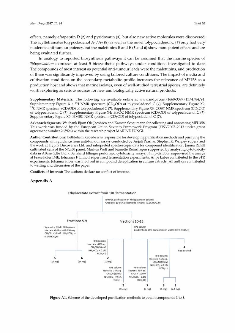
Figure A1. Scheme of the developed purification methods to obtain compounds 1 to 8.
Table A1. Physico‐chemical properties of tolypocladenol C.
Properties Tolypocladenol C
Appearance Pale yellow semi‐solid/gum
Figure A1. Scheme of the developed purification methods to obtain compounds 1 to 8.
Mar. Drugs 2017, 15, 84 15 of 20
Table A1. Physico-chemical properties of tolypocladenol C.
Properties Tolypocladenol C
Appearance Pale yellow semi-solid/gumUV (MeOH) λmax (log ε) 219 (3.97), 241sh (3.89), 280 (3.95) nm
ESI+-MS (m/z) 358 (MH+)ESI−-MS (m/z) 356 ([M − H]−)
Molecular formula C21H27NO4HRESI+-MS (m/z)
Found for MH+ 358.2016Calculated 358.2013
[α]D20 −135.2◦ (c 0.016, MeOH)
IR µmax3250, 2963, 2920, 1654, 1603, 1516, 1451, 1378, 1336,
1269, 1238, 1172, 1105, 1018, 968, 952, 816 cm−1
Table A2. 1H and 13C NMR assignments of tolypocladenol C.
No. δ 1H (J [Hz]) δ 13C
12 176.73 103.04 198.25 3.95 dd (6.4, 3.9) 63.0
6 2.98 dd (13.9, 4.2)2.82 dd (13.9, 6.0) 37.9
7 197.38 3.79 m 36.5
9 1.72 m1.04 dd (8.8, 4.4) 41.6
10 1.29 m 32.5
11 1.95 m1.77 m 41.5
12 5.38 m 130.913 5.38 m 127.014 1.63 m 18.115 0.80 d (6.5) 20.016 1.01 d (6.8) 18.81′ 128.12′ 6.99 d (8.4) 131.83′ 6.65 d (8.4) 116.04′ 157.25′ 6.65 d (8.4) 116.06′ 6.99 d (8.4) 131.8
Referenced to CD3OD signals at 3.31 and 49.00 ppm.
Mar. Drugs 2017, 15, 84 16 of 20
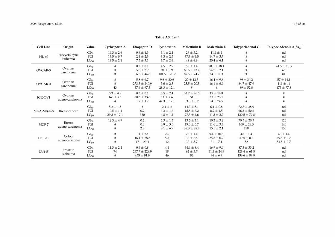
Table A3. Biological activities of the identified natural products in a selection of the NCI60 cancer cell line panel. Data given as µM. If compound concentration wasnot sufficient to reach cytostatic or cytotoxic effects, the value is given as #. If effect was not analysed in this cell line, table shows nd.
Cell Line Origin Value Cyclosporin A Efrapeptin D Pyridoxatin Malettinin B Malettinin E Tolypocladenol C Tolypocladenols A1/A2
UO-31 Renalcarcinoma
GI50 18 ± 0.8 6.5 ± 7.5 4.4 ± 0.5 34.5 ± 21.1 33 ± 3.6 49.1 ± 21.8 ndTGI 99 59 ± 56.6 9.6 ± 2.3 59 638.6 ± 143.5 214.4 ± 114.6 ndLC50 310 240 26 98 # 440 nd
786-O Renaladenocarinoma
GI50 46 1.6 ± 1 3.9 ± 1.6 20.4 ± 5.3 13.1 ± 2.3 75 19.6 ± 19TGI # 20 ± 21.1 10.1 ± 5.8 25 29.2 ± 10.1 # 149 ± 114.6LC50 # 325 ± 445.5 21.5 ± 14.7 # 142.5 ± 106.1 # #
TK10 Renalcarcinoma
GI50 17 ± 3.4 3.7 ± 4.4 12.2 ± 7 19.8 ± 5.9 6.6 ± 1.3 97.5 ± 55 ndTGI 49 800 74 ± 28.2 # # 104.5 ± 58.5 ndLC50 # # 250 ± 134.5 # # 110.5 ± 63.1 nd
SN12C Renalcarcinoma
GI50 # 1.9 ± 0.4 6.3 ± 0.8 20.5 ± 10.6 16 ± 7.1 # ndTGI # 10.2 ± 4 17.3 ± 15.1 57 45 # ndLC50 # # # # 120 # nd
UACC-62 Malignantmelanoma
GI50 # # 9.2 ± 1.1 38 ± 2.8 10.5 ± 0.7 58.5 ± 13.4 ndTGI # 0.4 ± 0.3 22 ± 7.1 450 # 78 ± 9.9 ndLC50 # # 51 # # 92 ± 7.2 nd
SK-MEL-28 Malignantmelanoma
GI50 # 1.3 ± 1.4 11 ± 11.3 # 61.5 ± 30 7.8 46TGI 2.9 11 62.7 ± 58.9 # 760 # 51LC50 # # 42 # # # 52
M14 Amelanoticmelanoma
GI50 12.5 ± 1 2.1 ± 1.1 2.2 ± 1.6 13.7 ± 2.5 9.6 ± 4.6 # ndTGI 67 ± 24.5 39 4.1 ± 2.4 102 ± 25.5 36 ± 10.1 # ndLC50 # 440 25 ± 5.7 370 244.5 ± 259.4 # nd
U251 GlioblastomaGI50 40 ± 24 # 4.5 ± 0.8 18.5 ± 0.7 13.5 ± 2.1 56.5 ± 6.4 ndTGI # 5.6 8.2 ± 4 # 48.5 ± 30.4 64 ± 14.1 ndLC50 # # 24.5 ± 10.6 # # 69 ± 19.8 nd
SF-539 GliosarcomaGI50 12.5 ± 1.3 1.2 ± 0.7 5 ± 1.2 14.5 ± 2.1 9.4 ± 2.6 # ndTGI 17.5 ± 0.7 61.6 ± 116.8 12.3 ± 7.5 47 ± 18.4 27.2 ± 10.9 # ndLC50 26 101.3 ± 155.1 40 ± 1.4 # 250 # nd
RPMI 8226Multiplemyeloma
GI50 7.5 ± 2.8 # 2.9 ± 0.4 49.5 ± 0.7 27.5 ± 3.5 # 19.5 ± 7.8TGI 12 ± 2.8 # 5.2 ± 0.6 53 ± 2.8 31 ± 7.1 # 38 ± 29.7LC50 34 0.7 ± 0.3 8.5 ± 0.8 55.5 ± 4.9 # # #
Mar. Drugs 2017, 15, 84 17 of 20
Table A3. Cont.
Cell Line Origin Value Cyclosporin A Efrapeptin D Pyridoxatin Malettinin B Malettinin E Tolypocladenol C Tolypocladenols A1/A2
HL-60 Procyelo-cyticleukemia
GI50 14.3 ± 2.6 0.9 ± 1.3 3.1 ± 2.4 29 ± 5.2 11.4 ± 4 # ndTGI 13.5 ± 0.7 2.1 ± 2.3 3.3 ± 2.5 37.3 ± 4.5 14.7 ± 3.7 # ndLC50 14.5 ± 2.1 7.5 ± 3.1 3.7 ± 2.6 48 ± 6.6 20.4 ± 6.1 # nd
OVCAR-5 Ovariancarcinoma
GI50 # 0.2 ± 0.1 4.5 ± 2.9 50 ± 1.4 20.5 ± 18.1 # 41.5 ± 16.3TGI # 5.8 ± 2.9 31 ± 9.9 60.5 ± 13.4 54.7 ± 2.1 # 68LC50 # 66.5 ± 44.8 101.5 ± 26.2 69.5 ± 24.7 64 ± 11.3 # 81
OVCAR-3 Ovariancarcinoma
GI50 # 5.8 ± 9.7 9.6 ± 20.6 22 ± 12.5 16.4 ± 9.6 69 ± 34.2 57 ± 14.1TGI # 273.3 ± 240.9 3.6 ± 2.3 25.5 ± 20.5 16.1 ± 6.9 84.7 ± 47.9 111 ± 41LC50 43 57.6 ± 97.3 28.3 ± 12.1 # # 89 ± 52.8 175 ± 77.8
IGR-OV1 Ovarianadeno-carcinoma
GI50 5.3 ± 4.8 0.3 ± 0.1 3.5 ± 2.4 32.7 ± 26.5 19 ± 18.8 # #TGI 145 ± 7.1 30.3 ± 33.6 11 ± 2.6 51 63 ± 23.1 # #LC50 # 1.7 ± 1.2 47.3 ± 17.1 53.5 ± 0.7 94 ± 74.5 # #
MDA-MB-468 Breast cancerGI50 5.2 ± 1.5 # 2.4 ± 2 14.3 ± 5.1 6.1 ± 0.8 72.8 ± 38.9 ndTGI 10.5 ± 1.1 0.2 3.3 ± 1.6 18.8 ± 3.2 8.2 ± 1.5 96.3 ± 50.6 ndLC50 29.3 ± 12.1 330 4.8 ± 1.1 27.3 ± 4.6 11.3 ± 2.7 120.5 ± 79.8 nd
MCF-7 Breastadeno-carcinoma
GI50 18.3 ± 4.9 0.3 2.3 ± 1.3 13.5 ± 2.1 10.2 ± 3.8 70.5 ± 20.5 120TGI # 0.8 4.8 ± 3.5 19.3 ± 6.7 11.6 ± 3.4 100 ± 28.3 140LC50 # 2.8 8.1 ± 6.9 38.3 ± 28.4 15.5 ± 2.1 150 150
HCT-15 Colonadenocarinoma
GI50 # 11 ± 22 2.6 28 ± 1.4 9.4 ± 10.8 42 ± 1.4 46 ± 1.4TGI # 16.4 ± 28.3 5.5 32 ± 2.8 25.5 ± 0.7 49.5 ± 0.7 49.5 ± 0.7LC50 # 17 ± 29.4 12 37 ± 5.7 31 ± 7.1 52 51.5 ± 0.7
DU145 Prostatecarinoma
GI50 11.3 ± 2.4 0.6 ± 0.8 4.1 34.4 ± 8.4 16.9 ± 9.4 87.3 ± 33.2 ndTGI 74 267.7 ± 229.9 18 62 ± 5.7 41.4 ± 24.6 123.4 ± 61.8 ndLC50 # 455 ± 91.9 46 86 94 ± 6.9 156.6 ± 89.9 nd
Mar. Drugs 2017, 15, 84 18 of 20
Table A4. Seeding cell densities used for screening in 384-well plates.
Cell Line Seeding Cell Density CELL LINE Initial Cell Density
UO-31 750 cells per well RPMI8226 1200 cells per well786-O 750 cells per well HL-60 2000 cells per wellTK10 1200 cells per well OVCAR-5 500 cells per well
SN12C 1200 cells per well OVCAR-3 2000 cells per wellUACC-62 1200 cells per well IGR-OV1 500 cells per well
SK-MEL-28 1000 cells per well MDA-MB-468 1200 cells per wellM14 2000 cells per well MCF-7 2000 cells per wellU251 750 cells per well HCT-15 400 cells per well
SF-539 1000 cells per well DU145 1200 cells per well
References
1. Hickford, S.J.; Blunt, J.W.; Munro, M.H. Antitumour polyether macrolides: Four new halichondrins fromthe new zealand deep-water marine sponge lissodendoryx sp. Bioorg. Med. Chem. 2009, 17, 2199–2203.[CrossRef] [PubMed]
2. Blunt, J.W.; Copp, B.R.; Hu, W.P.; Munro, M.H.; Northcote, P.T.; Prinsep, M.R. Marine natural products.Nat. Prod. Rep. 2008, 25, 35–94. [CrossRef] [PubMed]
3. Imhoff, J.F.; Stohr, R. Sponge-associated bacteria: General overview and special aspects of bacteria associatedwith halichondria panicea. Prog. Mol. Subcell. Biol. 2003, 37, 35–57. [PubMed]
4. Piel, J. Metabolites from symbiotic bacteria. Nat. Prod. Rep. 2004, 21, 519–538. [CrossRef] [PubMed]5. Kobayashi, J.; Ishibashi, M. Bioactive metabolites of symbiotic marine microorganisms. Chem. Rev. 1993, 93,
1753–1769. [CrossRef]6. Bhakuni, D.S.; Rawat, D.S. Bioactive Marine Natural Products; Springer: New York, NY, USA, 2005; p. 382.7. Proksch, P.; Edrada, R.A.; Ebel, R. Drugs from the seas–current status and microbiological implications.
Appl. Microbiol. Biotechnol. 2002, 59, 125–134. [PubMed]8. Gomes, N.G.; Dasari, R.; Chandra, S.; Kiss, R.; Kornienko, A. Marine invertebrate metabolites with anticancer
activities: Solutions to the “supply problem”. Mar. Drugs 2016, 14, 98. [CrossRef] [PubMed]9. Schaufelberger, D.E.; Koleck, M.P.; Beutler, J.A.; Vatakis, A.M.; Alvarado, A.B.; Andrews, P.; Marzo, L.V.;
Muschik, G.M.; Roach, J.; Ross, J.T.; et al. The large-scale isolation of bryostatin 1 from bugula neritinafollowing current good manufacturing practices. J. Nat. Prod. 1991, 54, 1265–1270. [CrossRef] [PubMed]
10. Newman, D.; Cragg, G. Natural products in medicinal chemistry. Bioorg. Med. Chem. 2009, 17, 2120.[CrossRef] [PubMed]
11. Bahn, Y.S.; Xue, C.; Idnurm, A.; Rutherford, J.C.; Heitman, J.; Cardenas, M.E. Sensing the environment:Lessons from fungi. Nat. Rev. Microbiol. 2007, 5, 57–69. [CrossRef] [PubMed]
12. Raghukumar, C.; D’Souza-Ticlo, D.; Verma, A.K. Treatment of colored effluents with lignin-degradingenzymes: An emerging role of marine-derived fungi. Crit. Rev. Microbiol. 2008, 34, 189–206. [CrossRef][PubMed]
13. Brakhage, A.A.; Sprote, P.; Al-Abdallah, Q.; Gehrke, A.; Plattner, H.; Tuncher, A. Regulation of penicillinbiosynthesis in filamentous fungi. Adv. Biochem. Eng. Biotechnol. 2004, 88, 45–90. [PubMed]
14. Imhoff, J.F. Natural products from marine fungi—Still an underrepresented resource. Mar. Drugs 2016, 14,19. [CrossRef] [PubMed]
15. Fenical, W.; Jensen, P.R.; Rowley, D.C. Halovir, an Antiviral Marine Natural Product, and Derivatives Thereof.U.S. Patent 6,458,766 B1, 1 October 2002.
16. Holler, U.; Konig, G.M.; Wright, A.D. Three new metabolites from marine-derived fungi of the generaconiothyrium and microsphaeropsis. J. Nat. Prod. 1999, 62, 114–118. [CrossRef] [PubMed]
17. Saleem, M.; Ali, M.S.; Hussain, S.; Jabbar, A.; Ashraf, M.; Lee, Y.S. Marine natural products of fungal origin.Nat. Prod. Rep. 2007, 24, 1142–1152. [CrossRef] [PubMed]
18. Gomes, N.G.; Lefranc, F.; Kijjoa, A.; Kiss, R. Can some marine-derived fungal metabolites become actualanticancer agents? Mar. Drugs 2015, 13, 3950–3991. [CrossRef] [PubMed]
Mar. Drugs 2017, 15, 84 19 of 20
19. Ellinger, B.; Silber, J.; Prashar, A.; Landskron, J.; Weber, J.; Rehermann, S.; Muller, F.J.; Smith, S.; Wrigley, S.;Tasken, K.; et al. A phenotypic screening approach to identify anticancer compounds derived from marinefungi. Assay Drug Dev. Technol. 2014, 12, 162–175. [PubMed]
20. Keller, N.P.; Turner, G.; Bennett, J.W. Fungal secondary metabolism—From biochemistry to genomics.Nat. Rev. Microbiol. 2005, 3, 937–947. [CrossRef] [PubMed]
21. Bode, H.B.; Bethe, B.; Hofs, R.; Zeeck, A. Big effects from small changes: Possible ways to explore nature’schemical diversity. Chembiochem 2002, 3, 619–627. [CrossRef]
22. Chiang, Y.M.; Lee, K.H.; Sanchez, J.F.; Keller, N.P.; Wang, C.C. Unlocking fungal cryptic natural products.Nat. Prod. Commun. 2009, 4, 1505–1510. [PubMed]
23. Survase, S.A.; Kagliwal, L.D.; Annapure, U.S.; Singhal, R.S. Cyclosporin a—A review on fermentativeproduction, downstream processing and pharmacological applications. Biotechnol. Adv. 2011, 29, 418–435.[CrossRef] [PubMed]
24. Bandani, A.R.; Khambay, B.P.S.; Faull, J.L.; Newton, R.; Deadman, M.; Butt, T.M. Production of efrapeptinsby tolypocladium species and evaluation of their insecticidal and antimicrobial properties. Mycol. Res. 2000,104, 537–544. [CrossRef]
25. Kessler, H.; Loosli, H.R.; Oschkinat, H. Peptide conformations. 30. Assignment of the h-1-nmr, c-13-nmr, andn-15-nmr spectra of cyclosporin-a in cdcl3 and c6d6 by a combination of homonuclear and heteronucleartwo-dimensional techniques. Helv. Chim. Acta 1985, 68, 661–681. [CrossRef]
26. Gupta, S.; Krasnoff, S.B.; Roberts, D.W.; Renwick, J.A.A.; Brinen, L.S.; Clardy, J. Structures of the efrapeptins:Potent inhibitors of mitochondrial atpase from the fungus tolypocladium-niveum. J. Am. Chem. Soc. 1991,113, 707–709. [CrossRef]
27. Teshima, Y.; Shinya, K.; Shimazu, A.; Furihata, K.; Chul, H.S.; Furihata, K.; Hayakawa, Y.; Nagai, K.; Seto, H.Isolation and structural elucidation of pyridoxatin, a free-radical scavenger of microbial origin. J. Antibiot.1991, 44, 685–687. [CrossRef] [PubMed]
28. Jegorov, A.; Matha, V.; Husak, M.; Kratochvil, B.; Stuchlik, J.; Sedmera, P.; Havlicek, V. Iron uptake system ofsome members of the genus tolypocladium–crystal-structure of the ligand and its iron(iii) complex. J. Chem.Soc. Dalton 1993, 28, 1287–1293. [CrossRef]
29. Angawi, R.F.; Swenson, D.C.; Gloer, J.B.; Wicklow, D.T. Malettinins b-d: New polyketide metabolites from anunidentified fungal colonist of hypoxylon stromata (nrrl 29110). J. Nat. Prod. 2005, 68, 212–216. [CrossRef][PubMed]
30. Silber, J.; Ohlendorf, B.; Labes, A.; Wenzel-Storjohann, A.; Näther, C.; Imhoff, J.F. Malettinin e, an antibacterialand antifungal tropolone produced by a marine cladosporium strain. Front. Mar. Sci. 2014, 1, 1–6. [CrossRef]
31. Turner, W.B.A.; Aldridge, D.C. Fungal Metabolites ii; Academic Press: New York, NY, USA, 1983.32. Li, X.B.; Li, L.; Zhu, R.X.; Li, W.; Chang, W.Q.; Zhang, L.L.; Wang, X.N.; Zhao, Z.T.; Lou, H.X. Tetramic acids
and pyridone alkaloids from the endolichenic fungus tolypocladium cylindrosporum. J. Nat. Prod. 2015, 78,2155–2160. [CrossRef] [PubMed]
33. Schmidt, K.; Riese, U.; Li, Z.Z.; Hamburger, M. Novel tetramic acids and pyridone alkaloids, militarinones b,c, and d, from the insect pathogenic fungus paecilomyces militaris. J. Nat. Prod. 2003, 66, 378–383. [CrossRef][PubMed]
34. Wangun, H.V.K.; Hertweck, C. Epicoccarines a, b and epipyridone: Tetramic acids and pyridone alkaloidsfrom an epicoccum sp associated with the tree fungus pholiota squarrosa. Org. Biomol. Chem. 2007, 5,1702–1705. [CrossRef] [PubMed]
35. Kakule, T.B.; Zhang, S.; Zhan, J.; Schmidt, E.W. Biosynthesis of the tetramic acids sch210971 and sch210972.Org. Lett. 2015, 17, 2295–2297. [CrossRef] [PubMed]
36. Gledhill, J.R.; Walker, J.E. Inhibitors of the catalytic domain of mitochondrial atp synthase. Biochem. Soc. Trans.2006, 34, 989–992. [CrossRef] [PubMed]
37. Angawi, R.F.; Swenson, D.C.; Gloer, J.B.; Wicklow, D.T. Malettinin a: A new antifungal tropolone froman unidentified fungal colonist of hypoxylon stromata (nrrl 29110). Tetrahedron Lett. 2003, 44, 7593–7596.[CrossRef]
38. Sung, G.H.; Spatafora, J.W.; Zare, R.; Hodge, K.T.; Gams, W. A revision of verticillium sect. Prostrata. Ii.Phylogenetic analyses of ssu and lsu nuclear rdna sequences from anamorphs and teleomorphs of theclavicipitaceae. Nova Hedwigia 2001, 72, 311–328.
Mar. Drugs 2017, 15, 84 20 of 20
39. Quandt, C.A.; Kepler, R.M.; Gams, W.; Araujo, J.P.; Ban, S.; Evans, H.C.; Hughes, D.; Humber, R.;Hywel-Jones, N.; Li, Z.; et al. Phylogenetic-based nomenclatural proposals for ophiocordycipitaceae(hypocreales) with new combinations in tolypocladium. IMA Fungus 2014, 5, 121–134. [CrossRef] [PubMed]
40. Gams, W. Cephalosporium-Artige Schimmelpilze (Hyphomycetes); G. Fischer: Stuttgart, Germany, 1971.41. Sung, G.H.; Hywel-Jones, N.L.; Sung, J.M.; Luangsa-Ard, J.J.; Shrestha, B.; Spatafora, J.W. Phylogenetic
classification of cordyceps and the clavicipitaceous fungi. Stud. Mycol. 2007, 57, 5–59. [CrossRef] [PubMed]42. Hawksworth, D.L. A new dawn for the naming of fungi: Impacts of decisions made in melbourne in july
2011 on the future publication and regulation of fungal names. Mycokeys 2011, 1, 7–20. [CrossRef]43. Kramer, A.; Labes, A.; Imhoff, J.F. Phylogenetic relationship and secondary metabolite production of marine
fungi producing the cyclodepsipeptides scopularide a and b. Mar. Biotechnol. 2016, 18, 466–474. [CrossRef][PubMed]
44. Silber, J.; Ohlendorf, B.; Erhard, A.; Labes, A.; Imhoff, J.F. Bioactive Metabolites of a Marine Calcarisporium sp.In Proceedings of the Research & Innovation-Inpact into Cancer, Shropshire, UK, 11 March 2014.
45. Bushley, K.E.; Raja, R.; Jaiswal, P.; Cumbie, J.S.; Nonogaki, M.; Boyd, A.E.; Owensby, C.A.; Knaus, B.J.;Elser, J.; Miller, D.; et al. The genome of tolypocladium inflatum: Evolution, organization, and expression ofthe cyclosporin biosynthetic gene cluster. PLoS Genet. 2013, 9, e1003496. [CrossRef] [PubMed]
46. Mascarell, L.; Truffa-Bachi, P. New aspects of cyclosporin a mode of action: From gene silencing to geneup-regulation. Mini Rev. Med. Chem. 2003, 3, 205–214. [CrossRef] [PubMed]
47. Papathanassiu, A.E.; MacDonald, N.J.; Emlet, D.R.; Vu, H.A. Antitumor activity of efrapeptins, alone orin combination with 2-deoxyglucose, in breast cancer in vitro and in vivo. Cell. Stress Chaperon 2011, 16,181–193. [CrossRef] [PubMed]
48. Lee, H.J.; Chung, M.C.; Lee, C.H.; Chun, H.K.; Kim, H.M.; Kho, Y.H. Pyridoxatin, an inhibitor of gelatinase awith cytotoxic activity. J. Microbiol. Biotechnol. 1996, 6, 445–450.
49. Cai, P.; Smith, D.; Cunningham, B.; Brown-Shimer, S.; Katz, B.; Pearce, C.; Venables, D.; Houck, D.8-methyl-pyridoxatin: A novel n-hydroxy pyridone from fungus os-f61800 that induces erythropoietinin human cells. J. Nat. Prod. 1999, 62, 397–399. [CrossRef] [PubMed]
50. Chang, W.; Zhang, M.; Li, Y.; Li, X.; Gao, Y.; Xie, Z.; Lou, H. Lichen endophyte derived pyridoxatin inactivatescandida growth by interfering with ergosterol biosynthesis. Biochim. Biophys. Acta 2015, 1850, 1762–1771.[CrossRef] [PubMed]
51. Bringmann, G.; Gulder, T.A.; Lang, G.; Schmitt, S.; Stohr, R.; Wiese, J.; Nagel, K.; Imhoff, J.F. Large-scalebiotechnological production of the antileukemic marine natural product sorbicillactone a. Mar. Drugs 2007,5, 23–30. [CrossRef] [PubMed]
52. Bringmann, G.; Lang, G.; Gulder, T.A.M.; Tsuruta, H.; Muhlbacher, J.; Maksimenka, K.; Steffens, S.;Schaumann, K.; Stohr, R.; Wiese, J.; et al. The first sorbicillinoid alkaloids, the antileukemic sorbicillactones aand b, from a sponge-derived penicillium chrysogenum strain. Tetrahedron 2005, 61, 7252–7265. [CrossRef]
53. Sears, M.A.; Gerhart, D.J.; Rittschof, D. Antifouling agents from marine spongelissodendoryx isodictyaliscarter. J. Chem. Ecol. 1990, 16, 791–799. [CrossRef] [PubMed]
54. Altschul, S.F.; Gish, W.; Miller, W.; Myers, E.W.; Lipman, D.J. Basic local alignment search tool. J. Mol. Biol.1990, 215, 403–410. [PubMed]
55. Tatusova, T.A.; Madden, T.L. Blast 2 sequences, a new tool for comparing protein and nucleotide sequences.FEMS Microbiol. Lett. 1999, 174, 247–250. [CrossRef] [PubMed]
56. Stevens, R.B. Mycology Guidebook; University of Washington Press: Seattle, WA, USA, 1974.57. Schneemann, I.; Nagel, K.; Kajahn, I.; Labes, A.; Wiese, J.; Imhoff, J.F. Comprehensive investigation of marine
actinobacteria associated with the sponge halichondria panicea. Appl. Environ. Microbiol. 2010, 76, 3702–3714.[CrossRef] [PubMed]
58. Wickerham, L.J. Taxonomy of Yeasts; U.S. Department of Agriculture: Washington, DC, USA, 1951.59. Shoemaker, R.H. The nci60 human tumour cell line anticancer drug screen. Nat. Rev. Cancer 2006, 6, 813–823.
[CrossRef] [PubMed]
© 2017 by the authors. Licensee MDPI, Basel, Switzerland. This article is an open accessarticle distributed under the terms and conditions of the Creative Commons Attribution(CC BY) license (http://creativecommons.org/licenses/by/4.0/).