Essentials of Human Anatomy & Physiology HCT II Muscular System.
37
Essentials of Human Anatomy & Physiology HCT II Muscular System
-
Upload
emery-davidson -
Category
Documents
-
view
232 -
download
0
Transcript of Essentials of Human Anatomy & Physiology HCT II Muscular System.
Essentials of Human Anatomy & Physiology
HCT II Muscular System
Characteristics of MusclesCharacteristics of Muscles
Muscle cells are elongated (muscle cell = muscle fiber)
Contraction of muscles is due to the movement of microfilaments
All muscles share some terminology
Prefix myo refers to muscle
Prefix mys refers to muscle
Prefix sarco refers to flesh
Skeletal Muscle CharacteristicsSkeletal Muscle Characteristics
Most are attached by tendons to bones
Cells are multinucleate
Striated – have visible banding
Voluntary – subject to conscious control
Cells are surrounded and bundled by connective tissue = great force, but tires easily
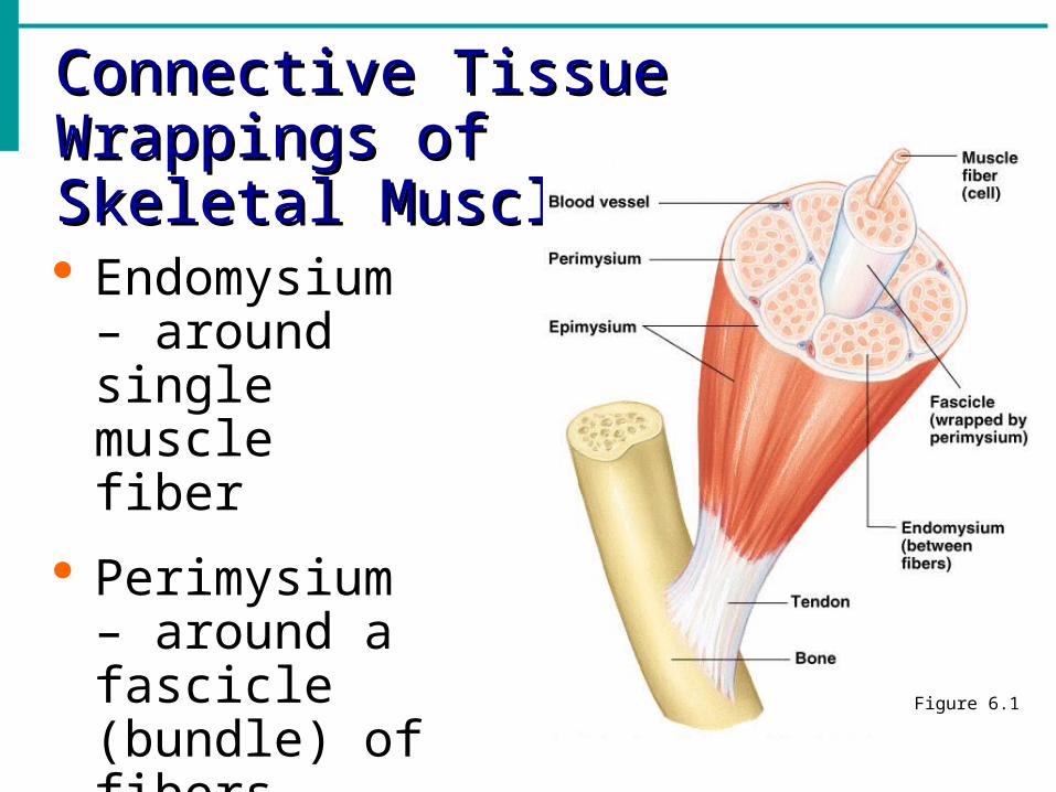
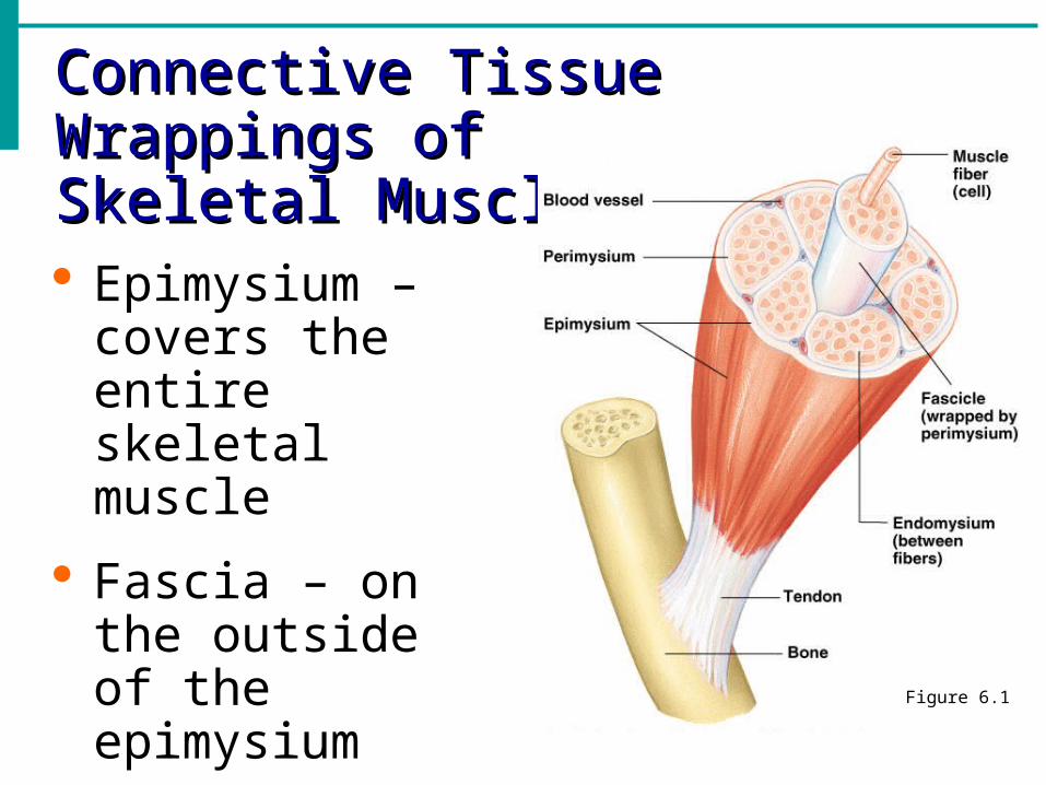
Connective Tissue Wrappings ofConnective Tissue Wrappings ofSkeletal MuscleSkeletal Muscle
Endomysium – around single muscle fiber
Perimysium – around a fascicle (bundle) of fibers Figure 6.1
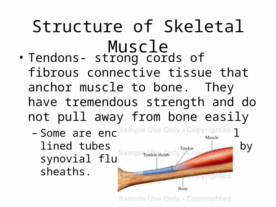
Structure of Skeletal Muscle• Tendons- strong cords of fibrous connective
tissue that anchor muscle to bone. They have tremendous strength and do not pull away from bone easily– Some are enclosed in a synovial lined tubes and
are lubricated by synovial fluid called tendon sheaths.
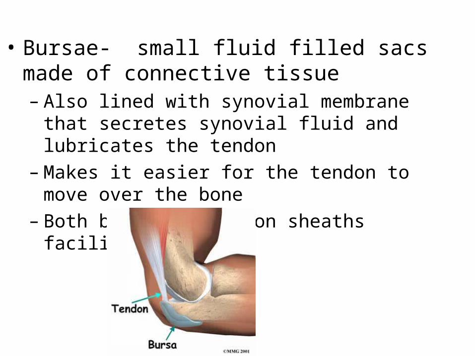
• Bursae- small fluid filled sacs made of connective tissue– Also lined with synovial membrane that secretes
synovial fluid and lubricates the tendon– Makes it easier for the tendon to move over the bone– Both bursa and tendon sheaths facilitate movement
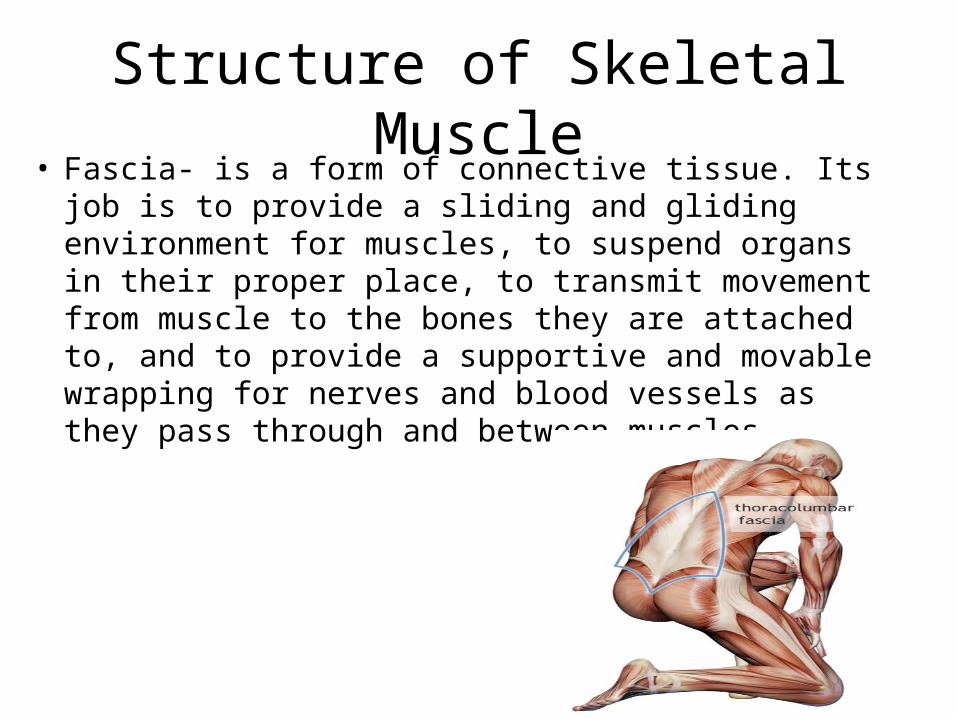
Structure of Skeletal Muscle• Fascia- is a form of connective tissue. Its job is to
provide a sliding and gliding environment for muscles, to suspend organs in their proper place, to transmit movement from muscle to the bones they are attached to, and to provide a supportive and movable wrapping for nerves and blood vessels as they pass through and between muscles.
Connective Tissue Wrappings ofConnective Tissue Wrappings ofSkeletal MuscleSkeletal Muscle
Epimysium – covers the entire skeletal muscle
Fascia – on the outside of the epimysium
Figure 6.1
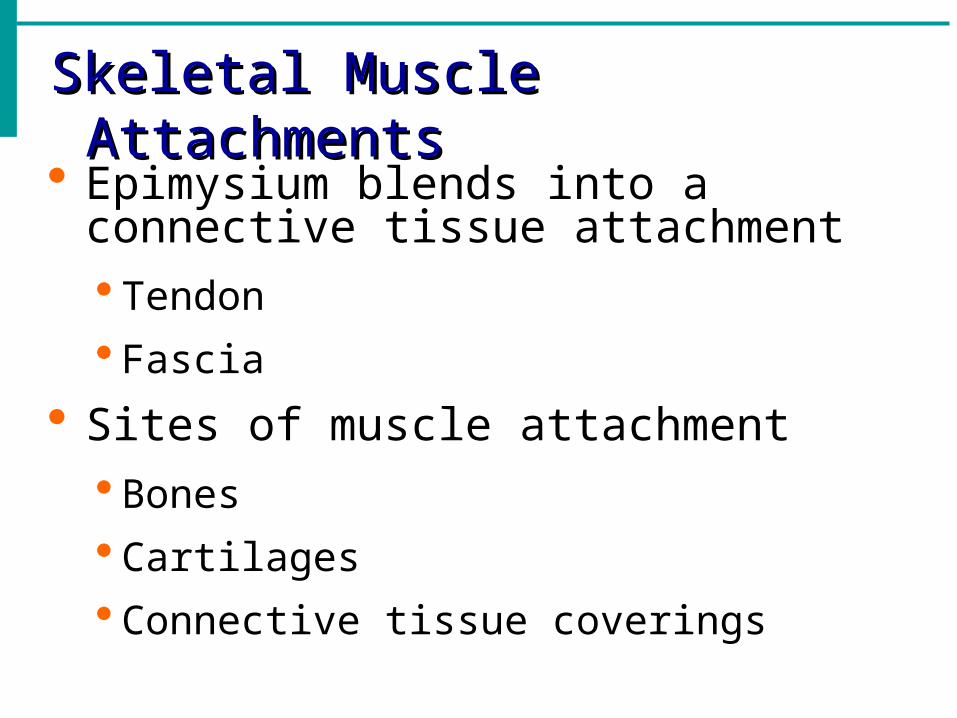
Skeletal Muscle AttachmentsSkeletal Muscle Attachments
Epimysium blends into a connective tissue attachment Tendon
Fascia
Sites of muscle attachment Bones
Cartilages
Connective tissue coverings
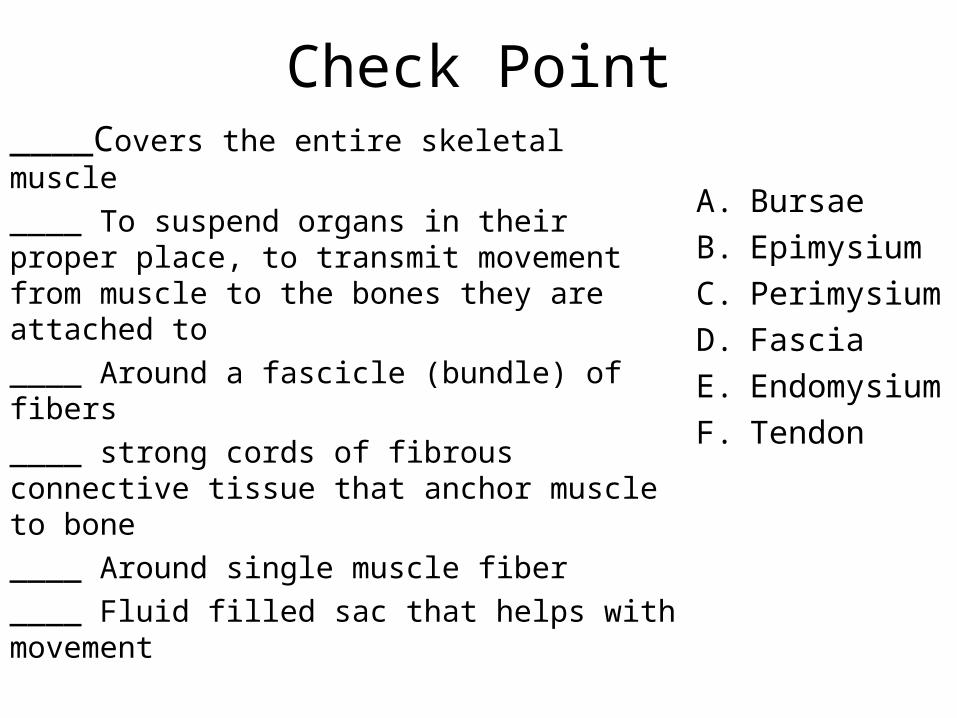
Check Point____Covers the entire skeletal muscle
____ To suspend organs in their proper place, to transmit movement from muscle to the bones they are attached to
____ Around a fascicle (bundle) of fibers
____ strong cords of fibrous connective tissue that anchor muscle to bone
____ Around single muscle fiber
____ Fluid filled sac that helps with movement
A. Bursae
B. Epimysium
C. Perimysium
D. Fascia
E. Endomysium
F. Tendon
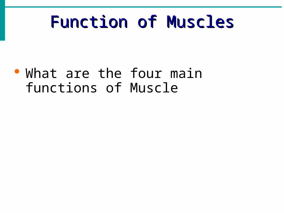
Function of MusclesFunction of Muscles
What are the four main functions of Muscle
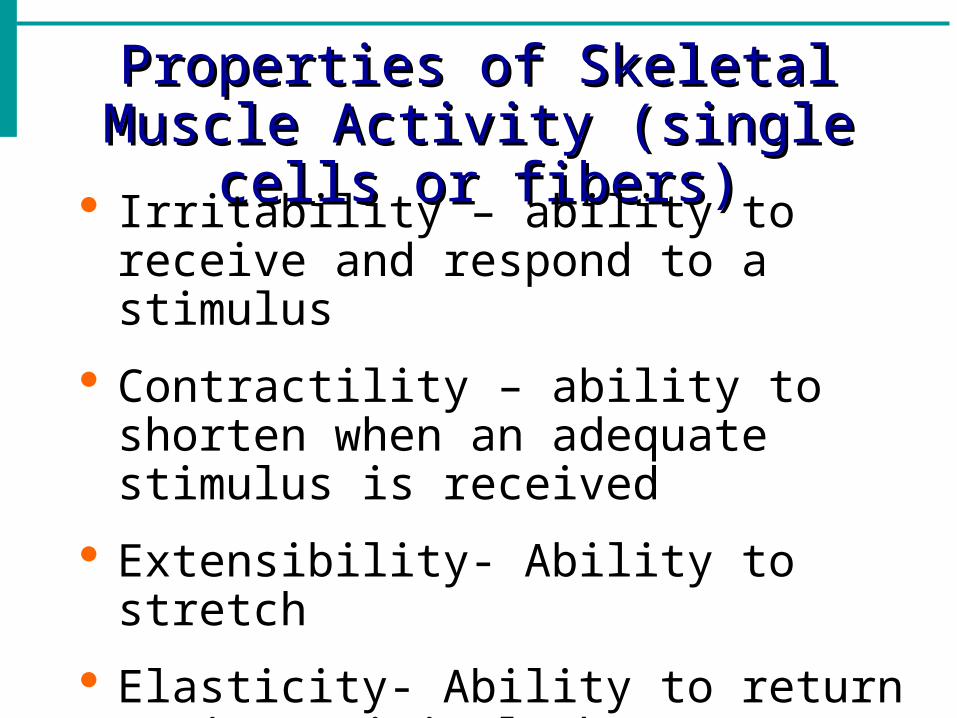
Properties of Skeletal Muscle Properties of Skeletal Muscle Activity (single cells or fibers)Activity (single cells or fibers)
Irritability – ability to receive and respond to a stimulus
Contractility – ability to shorten when an adequate stimulus is received
Extensibility- Ability to stretch
Elasticity- Ability to return to its original shape
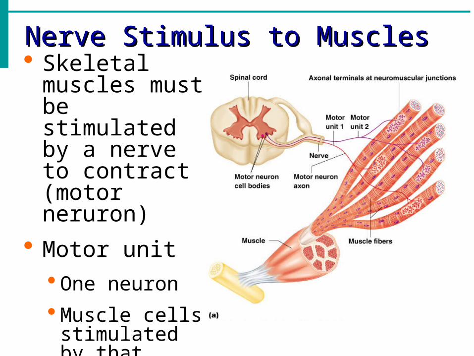
Nerve Stimulus to MusclesNerve Stimulus to Muscles Skeletal
muscles must be stimulated by a nerve to contract (motor neruron)
Motor unit One neuron
Muscle cells stimulated by that neuron
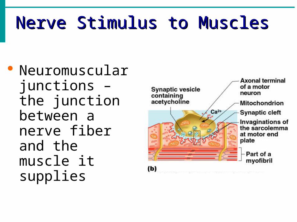
Nerve Stimulus to MusclesNerve Stimulus to Muscles
Neuromuscular junctions – the junction between a nerve fiber and the muscle it supplies

Nerve Stimulus to MusclesNerve Stimulus to Muscles
Synaptic cleft – gap between nerve and muscle Nerve and
muscle do not make contact
Area between nerve and muscle is filled with interstitial fluid Figure 6.5b
Transmission of Nerve Impulse to Transmission of Nerve Impulse to MuscleMuscle
Sodium rushing into the cell generates an action potential
Once started, muscle contraction cannot be stopped
Transmission of Nerve Impulse to Transmission of Nerve Impulse to MuscleMuscle
Neurotransmitter – chemical released by nerve upon arrival of nerve impulse
The neurotransmitter for skeletal muscle is acetylcholine
Neurotransmitter attaches to receptors on the sarcolemma
Sarcolemma becomes permeable to sodium (Na+)
Contraction of a Skeletal MuscleContraction of a Skeletal Muscle Muscle fiber contraction is “all or none”
Within a skeletal muscle, not all fibers may be stimulated during the same interval
Different combinations of muscle fiber contractions may give differing responses
Graded responses – different degrees of skeletal muscle shortening, rapid stimulus = constant contraction or tetanus
Muscle Response to Strong StimuliMuscle Response to Strong Stimuli
Muscle force depends upon the number of fibers stimulated
More fibers contracting results in greater muscle tension
Muscles can continue to contract unless they run out of energy
Energy for Muscle ContractionEnergy for Muscle Contraction
Initially, muscles used stored ATP for energy
Bonds of ATP are broken to release energy
Only 4-6 seconds worth of ATP is stored by muscles
After this initial time, other pathways must be utilized to produce ATP
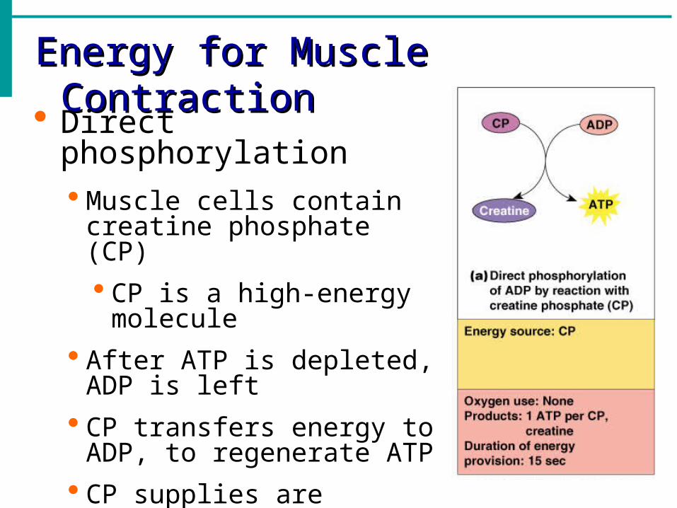
Energy for Muscle ContractionEnergy for Muscle Contraction
Direct phosphorylation Muscle cells contain creatine
phosphate (CP)
CP is a high-energy molecule
After ATP is depleted, ADP is left
CP transfers energy to ADP, to regenerate ATP
CP supplies are exhausted in about 20 seconds
Energy for Muscle ContractionEnergy for Muscle Contraction
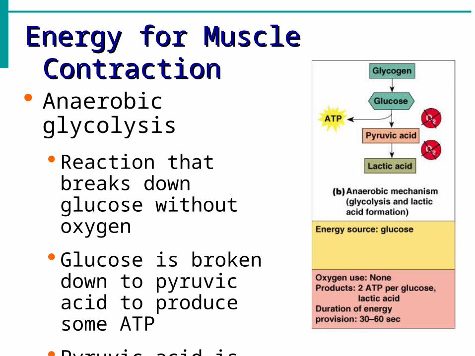
Anaerobic glycolysis
Reaction that breaks down glucose without oxygen
Glucose is broken down to pyruvic acid to produce some ATP
Pyruvic acid is converted to lactic acid
Energy for Muscle ContractionEnergy for Muscle Contraction
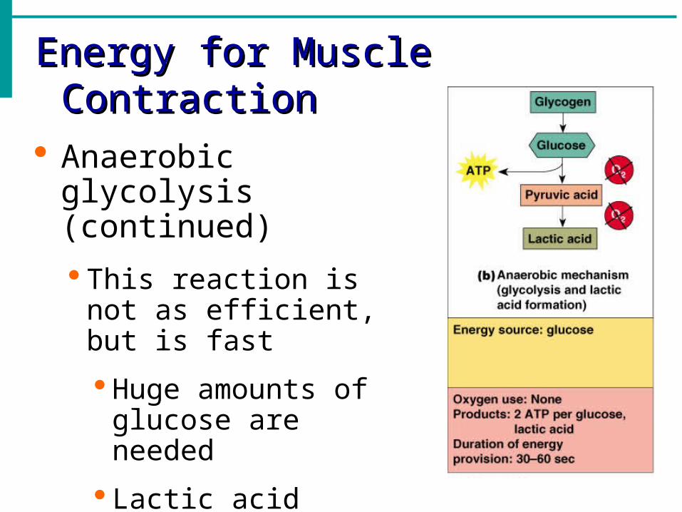
Anaerobic glycolysis (continued)
This reaction is not as efficient, but is fast
Huge amounts of glucose are needed
Lactic acid produces muscle fatigue
Energy for Muscle ContractionEnergy for Muscle Contraction
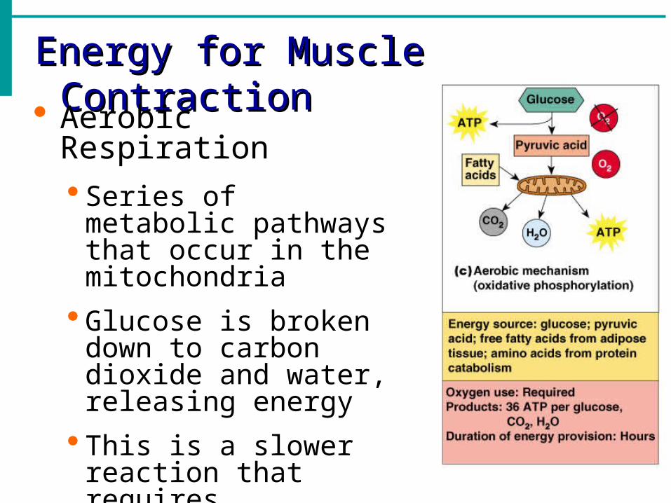
Aerobic Respiration Series of metabolic
pathways that occur in the mitochondria
Glucose is broken down to carbon dioxide and water, releasing energy
This is a slower reaction that requires continuous oxygen
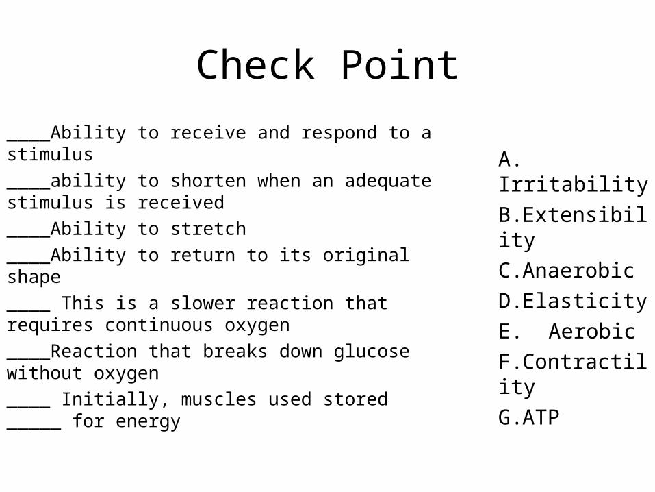
Check Point
____Ability to receive and respond to a stimulus
____ability to shorten when an adequate stimulus is received
____Ability to stretch
____Ability to return to its original shape
____ This is a slower reaction that requires continuous oxygen
____Reaction that breaks down glucose without oxygen
____ Initially, muscles used stored _____ for energy
A. Irritability
B.Extensibility
C.Anaerobic
D.Elasticity
E. Aerobic
F.Contractility
G.ATP
Muscle Fatigue and Oxygen DebtMuscle Fatigue and Oxygen Debt
When a muscle is fatigued, it is unable to contract
The common reason for muscle fatigue is oxygen debt Oxygen must be “repaid” to tissue to remove
oxygen debt
Oxygen is required to get rid of accumulated lactic acid
Increasing acidity (from lactic acid) and lack of ATP causes the muscle to contract less
Types of Muscle ContractionsTypes of Muscle Contractions
Isotonic contractions Myofilaments are able to slide past each
other during contractions
The muscle shortens
Isometric contractions Tension in the muscles increases
The muscle is unable to shorten
Muscle ToneMuscle Tone
Some fibers are contracted even in a relaxed muscle
Different fibers contract at different times to provide muscle tone
The process of stimulating various fibers is under involuntary control

Muscles and Body MovementsMuscles and Body Movements
Movement is attained due to a muscle moving an attached bone
Figure 6.12
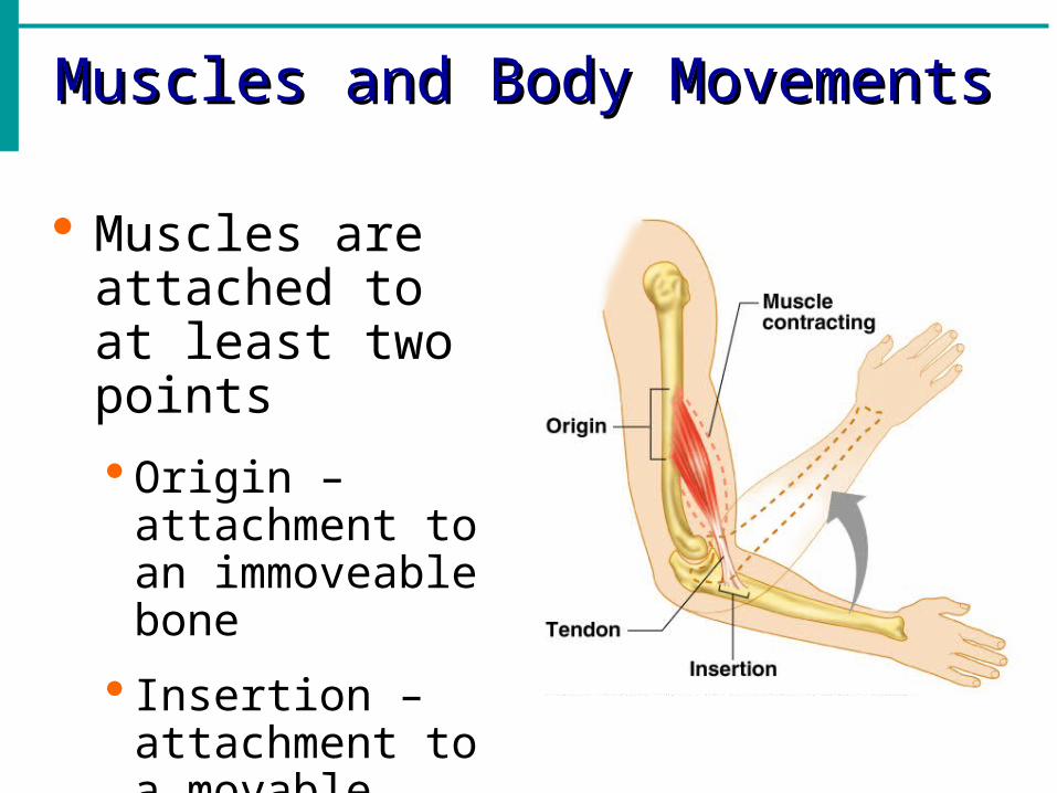
Muscles and Body MovementsMuscles and Body Movements
Muscles are attached to at least two points
Origin – attachment to an immoveable bone
Insertion – attachment to a movable bone
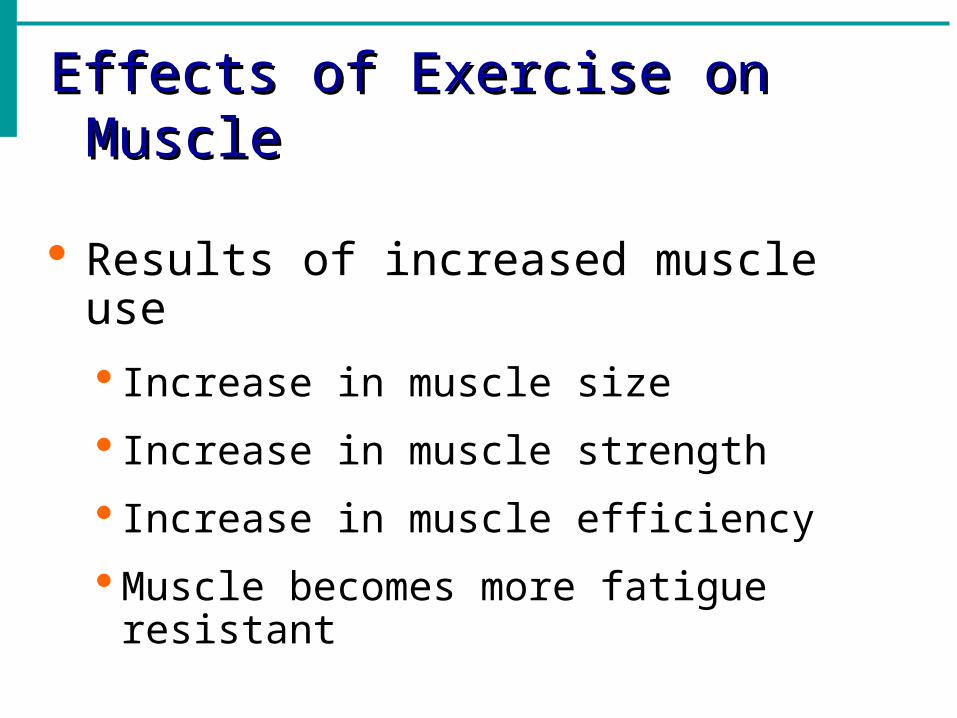
Effects of Exercise on MuscleEffects of Exercise on Muscle
Results of increased muscle use
Increase in muscle size
Increase in muscle strength
Increase in muscle efficiency
Muscle becomes more fatigue resistant
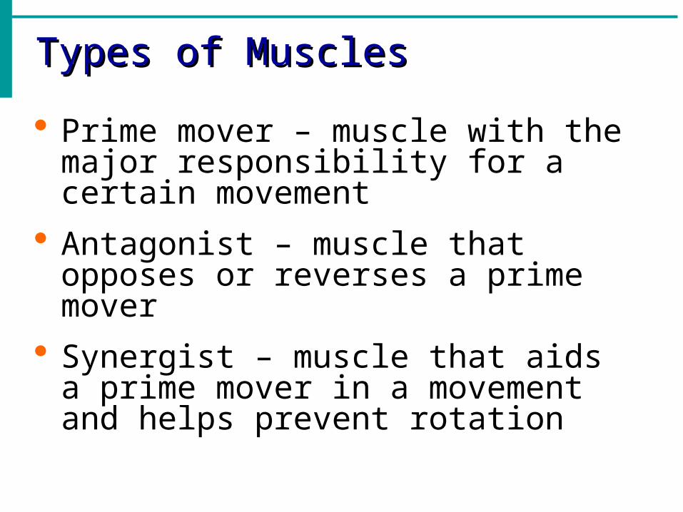
Types of MusclesTypes of Muscles
Prime mover – muscle with the major responsibility for a certain movement
Antagonist – muscle that opposes or reverses a prime mover
Synergist – muscle that aids a prime mover in a movement and helps prevent rotation

Movements Produced by Muscle Contractions• Flexion – movement reduces the angle
between two bones at their joint.• Extension – opposite of flexion – increases the
angle at a joint.• Abduction – moving a body part away from
the midline of the body.• Adduction – moving a body part toward the
midline of the body.• Rotation – movement of a body part around an
axis.
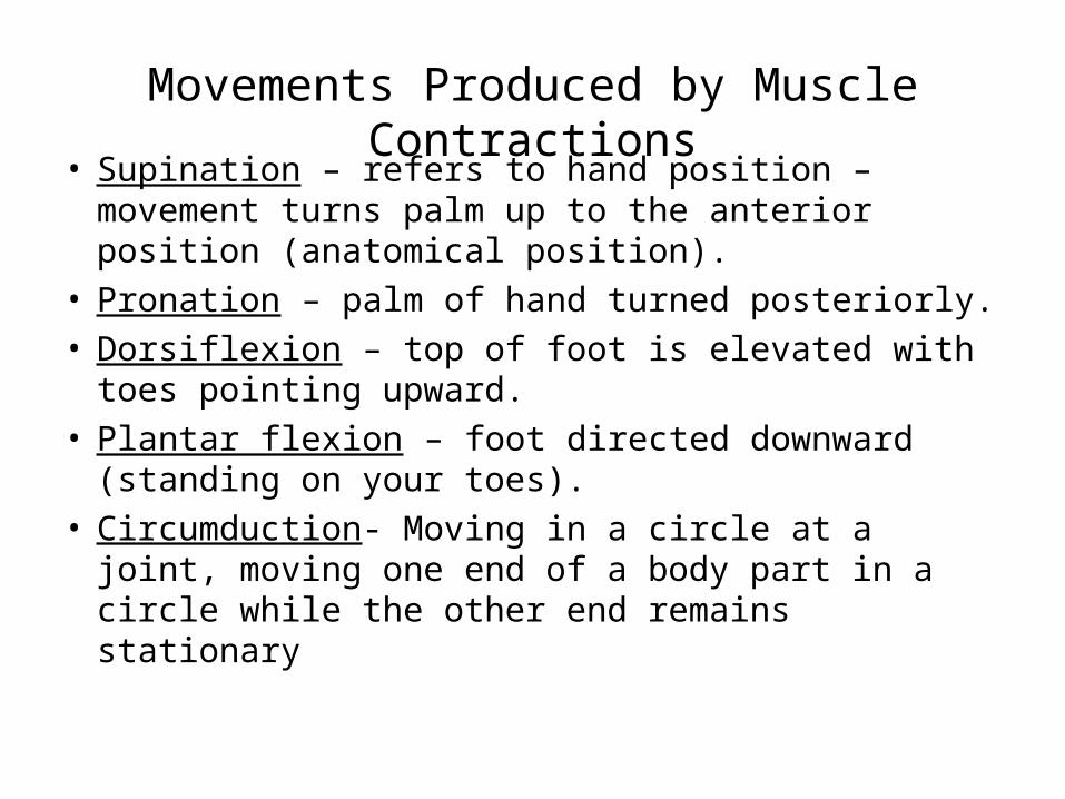
Movements Produced by Muscle Contractions• Supination – refers to hand position – movement
turns palm up to the anterior position (anatomical position).
• Pronation – palm of hand turned posteriorly.• Dorsiflexion – top of foot is elevated with toes
pointing upward.• Plantar flexion – foot directed downward (standing
on your toes).• Circumduction- Moving in a circle at a joint, moving
one end of a body part in a circle while the other end remains stationary
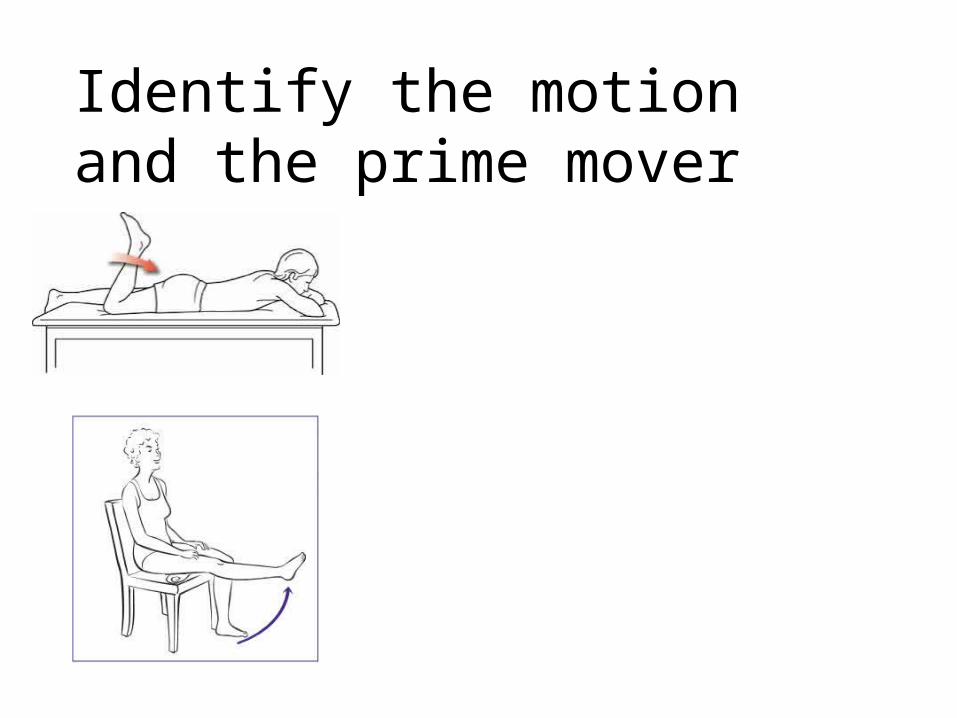
Identify the motion and the prime mover
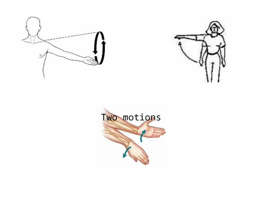
Two motions
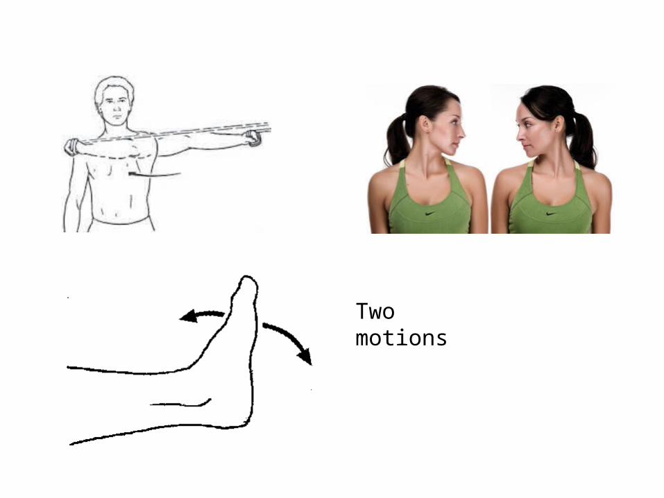
Two motions