
Esophageal Varices - pdfs.semanticscholar.org · Esophageal Varices esophageal varices are...
15
Esophageal Varices
Transcript of Esophageal Varices - pdfs.semanticscholar.org · Esophageal Varices esophageal varices are...
Esophageal Varices
Esophageal Varices
esophageal varices are extremely dilated sub-
mucosal veins in the lower third of the esophagus.
They are most often a consequence of portal
hypertension.
They are liable to rupture causing fatal bleeding
Resemble varicose veins that some people have in
their legs.
Esophageal Varices Causes
The causes of oesophageal varices are anything
that can cause portal hypertension. Some
examples are follow:
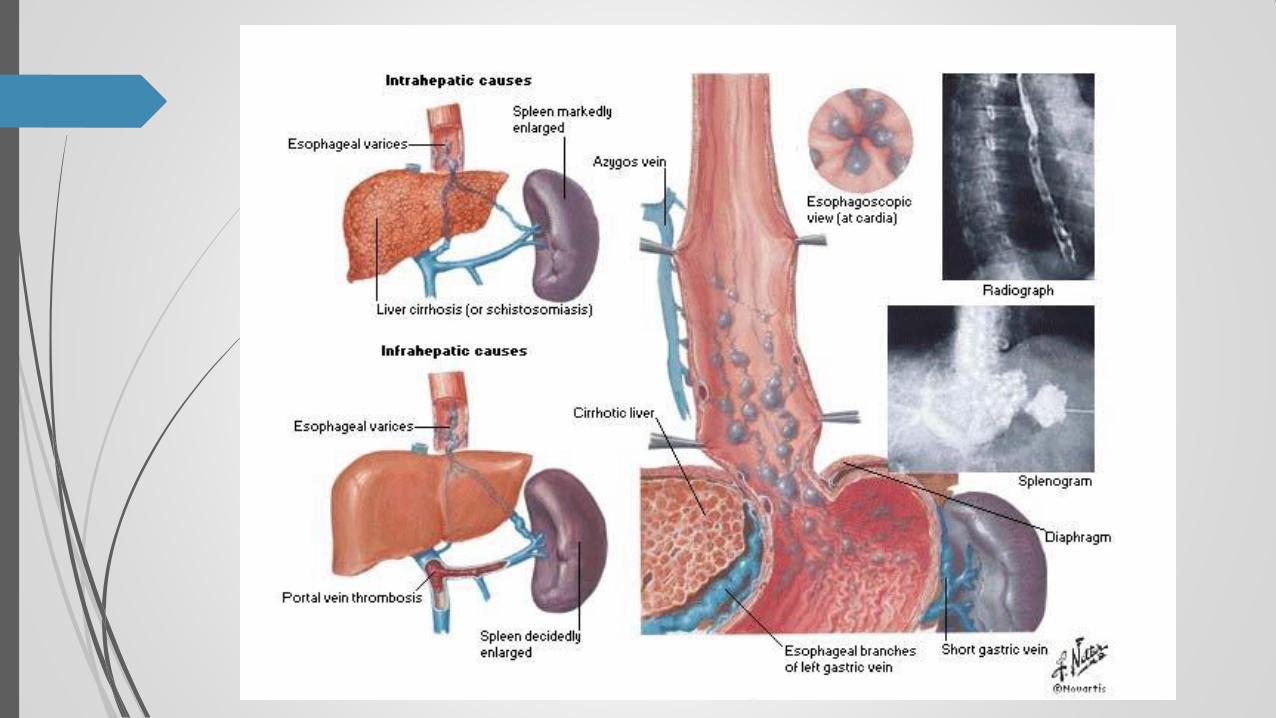
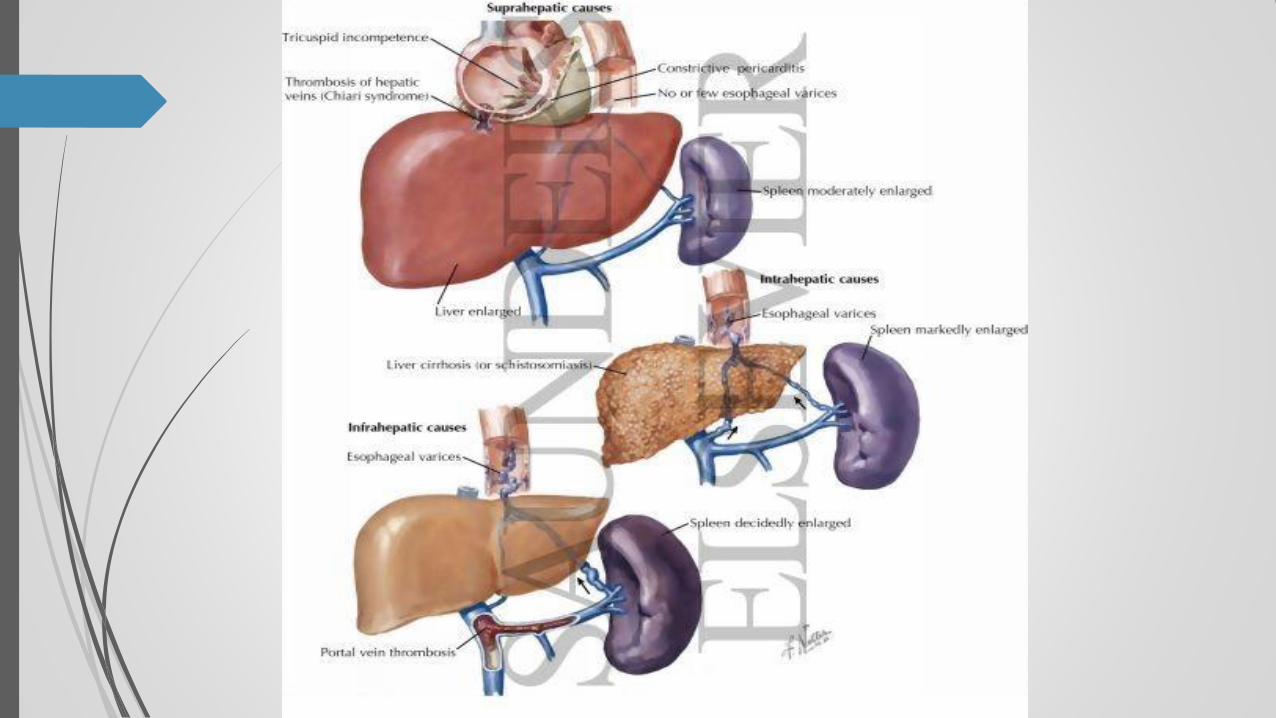
Pre-hepatic
• Portal vein thrombosis.
• Portal vein obstruction - congenital
atresia/stenosis.
• Increased portal blood flow - fistula.
• Increased splenic flow.
Esophageal Varices Causes
Intra-hepatic
• Cirrhosis due to various causes, including
alcoholic, chronic hepatitis (e.g viral or
autoimmune).
• Acute hepatitis (especially alcoholic).
• Schistosomiasis.
• Congenital hepatic fibrosis.
• Myelosclerosis.
Esophageal Varices Causes
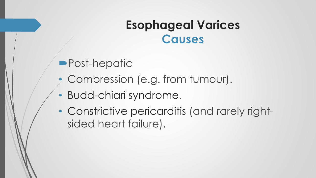
Post-hepatic
• Compression (e.g. from tumour).
• Budd-chiari syndrome.
• Constrictive pericarditis (and rarely right-
sided heart failure).
Esophageal Varices Pathogenesis
Portal hypertension results in the development
of collateral channels at sites where the portal
and caval systems communicate. Although
these collateral veins allow some drainage to
occur, they lead to development of a
congested subepithelial and submucosal
venous plexus within the distal esophagus.
These vessels, termed varices
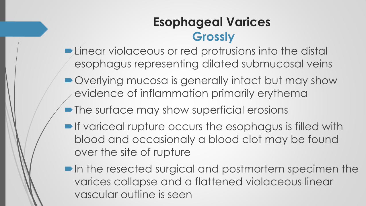
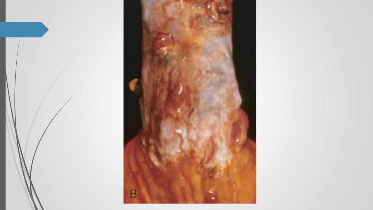
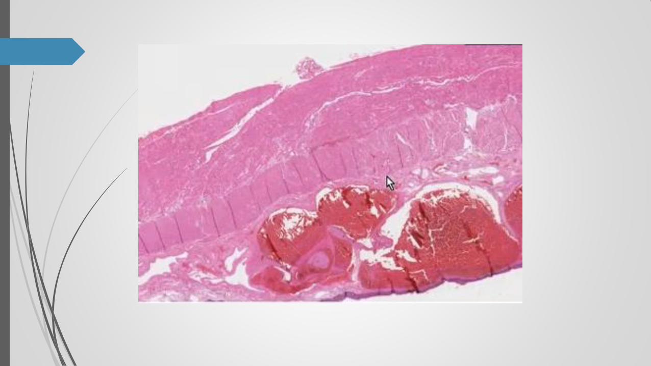
Esophageal Varices Grossly
Linear violaceous or red protrusions into the distal
esophagus representing dilated submucosal veins
Overlying mucosa is generally intact but may show
evidence of inflammation primarily erythema
The surface may show superficial erosions
If variceal rupture occurs the esophagus is filled with
blood and occasionaly a blood clot may be found
over the site of rupture
In the resected surgical and postmortem specimen the
varices collapse and a flattened violaceous linear
vascular outline is seen
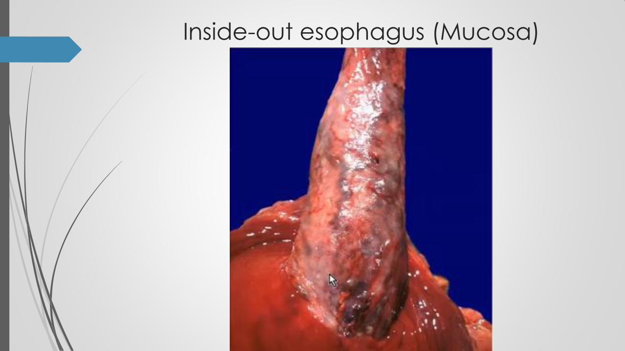
Inside-out esophagus (Mucosa)
Esophageal varices
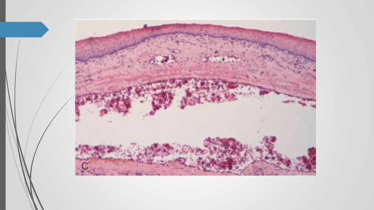
Microscopically
•Markedly dilated submucosal veins are present
•The overlying esophageal mucosa is generally
normal
•Partial thrombosis of varying age may be seen in
the varices

f


![Gastric varices: Classification, endoscopic and ...jrms.mui.ac.ir/files/journals/1/articles/10389/... · esophageal varices [Figure 2]. Thus, endoscopic findings of GV were classified](https://static.fdocuments.net/doc/165x107/609b5be24f2679079b73c086/gastric-varices-classification-endoscopic-and-jrmsmuiacirfilesjournals1articles10389.jpg)
![Small Esophageal Varices in Patients with Cirrhosis—Should ... · Varices of medium/large size (>5-mm diameter), or small varices with red spot signs [3†]. According to the](https://static.fdocuments.net/doc/165x107/5fa0286b8b7f711ce374a04d/small-esophageal-varices-in-patients-with-cirrhosisashould-varices-of-mediumlarge.jpg)


