Eriocaulon buergerianum extract protects PC12 cells and
10
RESEARCH Open Access Eriocaulon buergerianum extract protects PC12 cells and neurons in zebrafish against 6-hydroxydopamine-induced damage Meiwei Wang 1 , Zaijun Zhang 1 , Lorita Chi-Veng Cheang 1 , Zhixiu Lin 2 and Simon Ming-Yuen Lee 1* Abstract Background: Ericaulon buergerianum (Gujingcao) is an ophthalmic, anti-inflammatory and antimicrobial Chinese medicinal herb. This study aims to investigate the neuroprotective effects of Ericaulon buergerianum ethanol extract (EBE) and to elucidate its underlying action mechanism. Methods: The viability of dopaminergic (DA) neuron in zebrafish was examined by anti-tyrosine hydroxylase (TH) immunostaining. The locomotor activity of zebrafish was assessed with a digital video tracking system. The viability and cellular damage of the PC12 cells were determined by MTT and LDH assays respectively. The nuclear morphological changes in apoptotic cells were evaluated with DNA staining by Hoechst 33342 dye. Intracellular nitric oxide (NO) was quantified by DAF-FM diacetate staining. The expression of inducible nitric oxide synthase (iNOS) was determined by Western blot. Results: EBE inhibited the 6-OHDA-induced decrease in total distance of movement in zebrafish. Pretreatments of EBE (25, 50, 100 and 200 μg/ml) increased the viability of 6-OHDA-damaged PC12 cells in a dose dependent manner. Protection against 6-OHDA-induced nuclear fragmentation and accumulation of apoptotic bodies was also observed in EBE pretreated cells. Anti-oxidative (inhibition of NO production and iNOS expression in PC12 cells in vitro) activities of EBE are related to its neuroprotective effects in 6-OHDA-induced DA neuron damage. Conclusion: EBE exhibited significant neuroprotective activities in zebrafish, including recovery of dopaminergic neuron loss caused by 6-OHDA in a dose-dependent manner in vivo, inhibition of 6-OHDA-induced decrease of total distance in movement in zebrafish. The iNOS-NO pathway may be involved. Background A hydroxylated analogue of dopamine, namely 6-Hydro- xydopamine (6-OHDA) which induces damage of dopa- minergic neurons in vivo and in vitro, is commonly used in model systems to mimic Parkinson’s disease of which the main neuropathological feature is the loss of sub- stantia nigra pars compacta (SNpc) dopaminergic (DA) neurons. The toxic effects of 6-OHDA are mainly attrib- uted to the formation of free radicals, inflammatory pro- cesses and apoptosis [1,2]. Early studies indicated that NO participated in cellular signaling pathways regulating broad aspects of brain functions, such as synaptic plasticity, normal develop- ment and neuronal cell death [3]. NO is synthesized from L-arginine by nitric oxide synthase (NOS). Among the three major isoforms of NOS, inducible NOS (iNOS), a calcium-independent isoform, is regulated by oxidative stress and some inflammatory cytokines [4]. The implication of NO in PD pathogenesis is supported by the observations of unregulated iNOS expression in activated microglia [5,6]. NO induces neuronal cell damage by disrupting neuronal mitochondrial electron transport chain function [7,8]. As a result, the agents recovering the impaired mitochondrial function, sup- pressing neuroinflammation and the production of NO and iNOS may be beneficial for PD patients. Ericaulon buergerianum (Gujingcao), an aquatic plant native to Mainland China (eg Zhejiang, Guangdong and Fujian provinces), Taiwan and Japan, is used in Chinese * Correspondence: [email protected] 1 State Key Laboratory of Quality Research in Chinese Medicine, Institute of Chinese Medical Sciences, University of Macau, Av. Padre Tomás Pereira, Taipa, Macao, China Full list of author information is available at the end of the article Wang et al. Chinese Medicine 2011, 6:16 http://www.cmjournal.org/content/6/1/16 © 2011 Wang et al; licensee BioMed Central Ltd. This is an Open Access article distributed under the terms of the Creative Commons Attribution License (http://creativecommons.org/licenses/by/2.0), which permits unrestricted use, distribution, and reproduction in any medium, provided the original work is properly cited.
Transcript of Eriocaulon buergerianum extract protects PC12 cells and
RESEARCH Open Access
Eriocaulon buergerianum extract protects PC12cells and neurons in zebrafish against6-hydroxydopamine-induced damageMeiwei Wang1, Zaijun Zhang1, Lorita Chi-Veng Cheang1, Zhixiu Lin2 and Simon Ming-Yuen Lee1*
Abstract
Background: Ericaulon buergerianum (Gujingcao) is an ophthalmic, anti-inflammatory and antimicrobial Chinesemedicinal herb. This study aims to investigate the neuroprotective effects of Ericaulon buergerianum ethanol extract(EBE) and to elucidate its underlying action mechanism.
Methods: The viability of dopaminergic (DA) neuron in zebrafish was examined by anti-tyrosine hydroxylase (TH)immunostaining. The locomotor activity of zebrafish was assessed with a digital video tracking system. The viabilityand cellular damage of the PC12 cells were determined by MTT and LDH assays respectively. The nuclearmorphological changes in apoptotic cells were evaluated with DNA staining by Hoechst 33342 dye. Intracellularnitric oxide (NO) was quantified by DAF-FM diacetate staining. The expression of inducible nitric oxide synthase(iNOS) was determined by Western blot.
Results: EBE inhibited the 6-OHDA-induced decrease in total distance of movement in zebrafish. Pretreatments ofEBE (25, 50, 100 and 200 μg/ml) increased the viability of 6-OHDA-damaged PC12 cells in a dose dependentmanner. Protection against 6-OHDA-induced nuclear fragmentation and accumulation of apoptotic bodies was alsoobserved in EBE pretreated cells. Anti-oxidative (inhibition of NO production and iNOS expression in PC12 cells invitro) activities of EBE are related to its neuroprotective effects in 6-OHDA-induced DA neuron damage.
Conclusion: EBE exhibited significant neuroprotective activities in zebrafish, including recovery of dopaminergicneuron loss caused by 6-OHDA in a dose-dependent manner in vivo, inhibition of 6-OHDA-induced decrease oftotal distance in movement in zebrafish. The iNOS-NO pathway may be involved.
BackgroundA hydroxylated analogue of dopamine, namely 6-Hydro-xydopamine (6-OHDA) which induces damage of dopa-minergic neurons in vivo and in vitro, is commonly usedin model systems to mimic Parkinson’s disease of whichthe main neuropathological feature is the loss of sub-stantia nigra pars compacta (SNpc) dopaminergic (DA)neurons. The toxic effects of 6-OHDA are mainly attrib-uted to the formation of free radicals, inflammatory pro-cesses and apoptosis [1,2].Early studies indicated that NO participated in cellular
signaling pathways regulating broad aspects of brain
functions, such as synaptic plasticity, normal develop-ment and neuronal cell death [3]. NO is synthesizedfrom L-arginine by nitric oxide synthase (NOS). Amongthe three major isoforms of NOS, inducible NOS(iNOS), a calcium-independent isoform, is regulated byoxidative stress and some inflammatory cytokines [4].The implication of NO in PD pathogenesis is supportedby the observations of unregulated iNOS expression inactivated microglia [5,6]. NO induces neuronal celldamage by disrupting neuronal mitochondrial electrontransport chain function [7,8]. As a result, the agentsrecovering the impaired mitochondrial function, sup-pressing neuroinflammation and the production of NOand iNOS may be beneficial for PD patients.Ericaulon buergerianum (Gujingcao), an aquatic plant
native to Mainland China (eg Zhejiang, Guangdong andFujian provinces), Taiwan and Japan, is used in Chinese
* Correspondence: [email protected] Key Laboratory of Quality Research in Chinese Medicine, Institute ofChinese Medical Sciences, University of Macau, Av. Padre Tomás Pereira,Taipa, Macao, ChinaFull list of author information is available at the end of the article
Wang et al. Chinese Medicine 2011, 6:16http://www.cmjournal.org/content/6/1/16
© 2011 Wang et al; licensee BioMed Central Ltd. This is an Open Access article distributed under the terms of the Creative CommonsAttribution License (http://creativecommons.org/licenses/by/2.0), which permits unrestricted use, distribution, and reproduction inany medium, provided the original work is properly cited.
medicine as an anti-inflammatory and antimicrobialagent [9]. According to Chinese medicine theories, itexpels Feng (Wind), clears Re (Heat) and brightens theeyes. In Chinese Pharmacopoeia (2005), the capitulumof Ericaulon buergerianum is one of the most frequentlyused Chinese medicinal herbs, with flavonoids, volatileoils, anthraquinone, naphthopyranones, protocatechuicacid and c-tocopheryl acetate being the bioactive consti-tuents [10]. Flavonoids such as patuletin hispidulin,quercetin, quercetagetin and quercetagetin derivativesand volatile oil such as palmitic acid, (Z, Z)-9, 12-octa-cosane-dienoic acid are the two major classes of chemi-cals in Ericaulon buergerianum [9,11]. Water extract ofEricaulon buergerianum exhibits antimicrobial proper-ties [11]. Ericaulon buergerianum demonstrates signifi-cant therapeutic effects on headache, toothache,nasosinusitis, night blindness, glaucoma, retinochoroidi-tis, conjunctivitis and other eye diseases [12]. GuqingTang, a Chinese herbal formula consisting of Ericaulonbuergerianum and Celosia argentea (Qingxiangzi), isused to treat headache and eye diseases [13].The value of zebrafish (Danio rerio) for drug screen-
ing, target validation and toxicological studies is increas-ingly recognized in recent years [14,15]. A region in thezebrafish brain anatomically corresponding to the stria-tum was identified in the forebrain [16]. Zebrafish alsodisplay learning, sleeping, drug addiction and neurobe-havioral phenotypes that are quantifiable and related tothose in humans [17,18]. Therefore, it is an ideal modelto study the neuroprotective effect of herbal medicine invivo.The present study aims to investigate the neuroprotec-
tive effects of Ericaulon buergerianum in PC12 cells andzebrafish and elucidate the underlying mechanism of theprotective effects.
MethodsChemicals and reagentsHeat-inactivated horse serum, fetal bovine serum (FBS),penicillin and streptomycin were purchased from GibcoInvitrogen (USA). Nutrient Mixture F12 Ham Kaighn’sModification (F-12 K) growth medium, dimethyl sulfox-ide (DMSO) and 3-(4,5-dimethylthiazol-2-yl)-2,5-diphe-nyltetrazolium bromide (MTT) were purchased fromSigma (USA). Cytotoxicity Detection Kit was purchasedfrom Roche Applied Science (Germany). The fluorescentprobe 4-amino-5-methylamino- 2’,7’-difluorofluoresceindiacetate (DAF-FM diacetate) and Hoechst 33342 dyewere purchased from Molecular Probes(USA). RIPAlysis buffer, PMSF, protease inhibitor cocktail and BCAprotein assay kit were purchased from Pierce Biotech-nology (USA). Polyvinylidene difluoride (PVDF) mem-brane was purchased from Bio-Rad (USA). Anti-iNOSwas obtained from Cell Signaling Technology (USA).
ECL advanced western blotting detection kit was pur-chased from Amersham (UK). All other reagents used inthis study were obtained from Sigma.
Extraction procedureCrude herb of Ericaulon buergerianum was purchasedfrom ZhiXin Chinese Pharmaceutical Company in HongKong and authenticated in the School of Chinese Medi-cine, The Chinese University of Hong Kong accordingto appearance identification of raw material and com-parison of chemical constituents which have describedin Zhong-Yao-Zhi [19]. The crude herb was first groundinto powder with an electric grinder (Yongkang WeifengElectric Co. Ltd., China). The powder (100 g) was thenextracted with 80% ethanol for two hours, and theextract was filtered and dried. Ericaulon buergerianumethanol (EBE) extract (10.8 g) was stored at -20°C.
Chemical analysis of Ericaulon buergerianum ethanolextractEBE was dissolved in DMSO (100 mg/ml) and filteredwith a 0.45 μm membrane filter. Then the filtrate wasanalyzed on an Agilent 1100 series chromatographic sys-tem (Agilent, USA) consisting of a vacuum degasser, abinary pump, an auto-sampler, a column oven and adiode array detector (DAD). Chromatographic separationwas achieved on a GL Sciences Inertsil ODS-4 column, 5μm, 250 mm × 4.6 mm i.d. (GL Sciences, USA) at ambi-ent temperature. The flow rate was 0.8 mL/min. Themobile phase consisted of Milli-Q water (A) and aceton-trile (B). The gradient elution was as follows: 10-35% (B)in 0-40 min, 35-50% (B) in 40-50 min and 50-100% (B) in50-60 min. Detection wavelength was set at 280 nm.Injection volume was 10 μL. Evaluation of UV data wasperformed on an Agilent ChemStation A.09.03 (Agilent,USA) and DataAnalysis 2.2 (Bruker Daltonics, USA).
Fish maintenanceEmbryos of wild-type (AB strain) zebrafish were col-lected after natural spawning, staged according to stan-dard criteria [20], and synchronously raised at 28.5°C.Embryos were maintained in embryo medium (13.7 mMNaCl, 540 μM KCl, pH7.4, 25 μM Na2HPO4, 44 μMKH2PO4, 300 μM CaCl2, 100 μM MgSO4, 420 μMNaHCO3, pH7.4). Since embryos received nourishmentfrom the attached yolk sac, no additional maintenancewas required.
Cell cultureStock cultures of rat pheochromocytoma cells (PC12)(CRL-1721) were purchased from American Type Cul-ture Collection (ATCC, USA). They were cultured in F-12 K supplemented medium with 15% (v/v) heat-inacti-vated horse serum, 2.5% (v/v) FBS, penicillin (100 U/ml)
Wang et al. Chinese Medicine 2011, 6:16http://www.cmjournal.org/content/6/1/16
Page 2 of 10

and streptomycin (100 μg/ml) in a humidified atmo-sphere of 5% CO2 at 37°C. The medium was changedevery other day.
Experimental designFertilized eggs obtained from mating pairs of adult zeb-rafish were cultured in embryo medium. All experimentswere performed in 12-well plates with 20 embryos ineach well. Phenylthiourea (PTU, Sigma-Aldrich, USA)was added for a final concentration of 0.003% to preventpigmentation of embryos, up to 2 day post fertilization(dpf) and larvae (>2 dpf). 2 dpf zebrafish was exposed to250 μM 6-OHDA and different concentrations (25, 50,100 μg/ml) of EBE for 24 hours. Zebrafish co-treatedwith 6-OHDA and Nomifensin (DAT inhibitor)/L-nitroarginine methylester (L-NAME) were used as posi-tive controls. Anti-TH immunostaining was performedto evaluate the viability of dopaminergic neuron in thebrain of zebrafish.PC12 cells were plated at a density of 104 cells/100 μl/
well in 96-well plates. EBE of different concentrations(25, 50, 100 and 200 μg/ml) was added as pretreatmentto PC12 cells incubated in F-12 K medium supplemen-ted with 0.5% (v/v) heat-inactivated horse serum for 12hours at 37°C. Cells pretreated with L-NAME (250 μM)for 12 hours served as positive controls. The mediumswere then discarded, and the cells were incubated foranother 12 hours with 6-OHDA (1 mM) dissolved in0.5% (v/v) heat-inactivated horse serum at 37°C. A con-trol group of untreated normal cells was also included.The viability and cellular damage of the PC12 cells weredetected by the MTT and LDH assays respectively.
Anti-tyrosine hydroxylase (TH) whole mountimmunostainingZebrafish were fixed in 4% paraformaldehyde in phos-phate buffered saline (PBS) for five hours, rinsed andstored at -20°C in 100% EtOH. Fixed samples wereblocked (2% lamb serum in phosphate buffered salinecontaining 0.1% Tween-20 (PBS-T), 0.1% BSA) for onehour at room temperature. A mouse monoclonal anti-tyrosine hydroxylase antibody (1:200 diluted in blockingbuffer, MAB318, Millipore, USA) was used as the pri-mary antibody and incubated with the sample overnightat 4°C. Samples were then washed with PBS-T six times(30 min for each wash), followed by incubation withsecondary antibody according to the instruction of Vec-tastain ABC kit (Vector Laboratories, USA). After beingstained, zebrafish were flat mounted with 3.5% methyl-cellulose and photographed.
Testing of the locomotor activityFish behavior was analyzed with a digital video trackingsystem (Viewpoint, ZebraLab, France). The system
consists of a digital video camera connected to a computersystem running the analysis software ZebraLab Man Rev3.6B (ZebraLab, France). The locomotor activity of zebra-fish larvae was assessed in a 96 well plate filled with 200 μlembryo medium. At 7 dpf, the larvae were allowed tohabituate to the new environment for one hour and theirbehavior was recorded for two minutes with the ViewpointZebraLab system. Total distance of movement of each fishper group was calculated.
MTT assay3-(4, 5-dimethyl-2- thiazolyl) 2, 5-diphenyl-2H-tetrazo-lium bromide (MTT) is a tetrazolium salt that can bereduced to purple-colored formazan by live cells. For-mazan is dissolved and the resulting solution can bespectrophotometrically measured. Cells were incubatedfor four hours at 37°C with MTT solution (0.5 mg/ml)prepared in fresh 0.5% (v/v) heat-inactivated horseserum. The medium was then discarded, and 100 μl ofDMSO was applied to each well to dissolve the violetformazan crystals in intact cells. The absorbance wasmeasured at the wavelength of 490 nm by a multi-labelcounter (Wallac VICTOR3™V, Perkin Elmer, Nether-lands). Cell viability was expressed as a percentage ofthe control (untreated cells). All assays were performedin eight replicates and repeated at least three times.
LDH assayCell viability was determined by the activity of lactatedehydrogenase (LDH) released into the incubation med-ium when cellular membranes are damaged. Cells wereseeded in 96-well plates. After treatments, the releasedLDH was measured according to the specifications ofCytotoxicity Detection Kit (Roche, Germany). Briefly,100 μl of culture medium was collected from each well.The absorbance of the medium was measured at 490nm with 690 nm as a reference wavelength in an auto-matic microplate reader (Wallac VICTOR3TMV, perki-nelmer, USA). Results are shown as percentage versus6-OHDA group.
Hoechst 33342 stainingThe nuclear morphological changes in apoptotic cellswere evaluated by DNA staining with Hoechst 33342dye. Under fluorescent microscope, Hoechst 33342stains the condensed chromatin in apoptotic cells muchmore brightly than in normal cells. Cells were platedinto a 12-well plate at 105/well. Cells were pretreatedwith EBE (50, 100, 200 μg/ml) for 12 hours, then treatedwith 6-OHDA (1 mM) for 8 hours. Cells were thenstained with Hoechst 33342 (10 μg/ml) with RNase (5μg/ml) in PBS for 15 min at room temperature, followedby a 15-min fixation in 1% (w/v) paraformaldehyde.Images were recorded with a fluorescent microscope
Wang et al. Chinese Medicine 2011, 6:16http://www.cmjournal.org/content/6/1/16
Page 3 of 10
(Carl Zeiss, Axiovert 200, USA) with a mounted camera(Carl Zeiss, AxioCam HRc, USA).
Intracellular NO stainingIntracellular NO was evaluated by the fluorescent probe4-amino-5-methylamino- 2’,7’-difluorofluorescein diace-tate (DAF-FM diacetate). DAF-FM diacetate is cell-per-meant and passively diffuses across cellular membranes.Once inside cells, it is deacetylated by intracellularesterases to become DAF-FM. The cells were seeded in96-well black-bottom clear plates. After pretreated withEBE and 6-OHDA for 12 hours and one hour respec-tively, the cells were washed in PBS and incubated inmedium with PBS plus 2.5 μM DAF-FM diacetate for30 min at 37°C in darkness. Then the cells were washedtwice with PBS and the fluorescence was evaluated in amicroplate reader (Wallac VICTOR3TMV, perkinelmer,USA) at 495 nm (excitation) and 515 nm (emission).Meanwhile, images were recorded by a fluorescentmicroscope (Carl Zeiss, Axiovert 200, USA) with amounted camera (Carl Zeiss, AxioCam HRc, USA).
Western blot analysisAfter treatment, PC12 cells were washed three timeswith cold PBS and then incubated on ice with RIPAlysis buffer with 1% PMSF and 1% protease inhibitorcocktail for 30 min. Cell lysates were centrifuged at12,500 × g (Hitachi, Japan) for 20 min at 4°C. Thesupernatant was separated and the protein amount wasdetermined by the BCA protein assay kit. Sample buffer(10% SDS, 250 mM Tris-HCL 6.8, 50% glycerol, 8%DTT, 0.002% blue-bromophenol) was added and mixedwith protein at a ratio of 1:4, and boiled at 95°C for fiveminutes. Protein samples (40 μg) were separated by 10%SDS-poly-acrylamide gel electrophoresis and then trans-ferred to a polyvinylidene difluoride (PVDF) membrane(Bio-Rad, USA) for 90 min at 20 V. Subsequently, themembrane was blocked with 5% non-fat milk in PBScontaining 0.1% Tween20 (PBST) for one hour at roomtemperature. The blots were incubated overnight at 4°Cwith various primary antibodies (Cell Signaling, USA)including anti-iNOS (1:1000). After three washes withPBS-T, the membranes were incubated with horseradishperoxidase-conjugated secondary antibodies (1:2000) inPBS-T with 5% non-fat milk for one hour at room tem-perature. After repeated washes, proteins were visualizedwith an ECL advanced Western blotting detection kit(Amersham, UK) according to the manufacturer’s proto-col. Protein bands were photographed by a MolecularImager ChemiDoc XRS (Bio-Rad, USA).
Statistical analysisData are represented as mean ± standard deviation (SD).One-way ANOVA was used to detect significant
differences among concentration groups in the experi-ments. Newman-Keuls Multiple Comparison Test wasused to test the statistical significance of the differencebetween the concentration group and the control (vehi-cle) group. GraphPad Prism statistical software (Graph-Pad Software, USA) was used for all calculations. P <0.05 was considered as statistically significant.
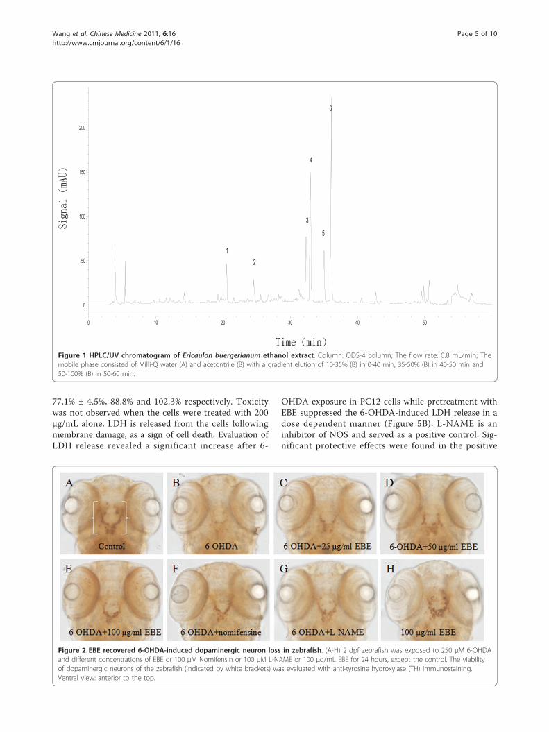
Results and DiscussionQuality control of EBEHigh-performance liquid chromatography (HPLC)coupled with (DAD) was used to generate the chemicalprofile of EBE (Figure 1). The HPLC fingerprinting maybe used as a reference for the purpose of quality assur-ance for any future experiments related to EBE.
Protection of 6-OHDA-induced dopaminergic neuron lossin zebrafishAll the tyrosine hydroxylase (TH)-positive neurons inzebrafish diencephalons are dopaminergic neurons [21].Anti-TH immunostaining was used to compare the via-bility of dopaminergic neurons in zebrafish receiving dif-ferent drug treatments (Figure 2). 2 dpf zebrafish wereexposed to 250 μM 6-OHDA for 24 hours, dopaminer-gic neurons in the ventral diencephalic clusters (indi-cated by white bracket) were significantly reduced whencompared to the control. Co-treatment with 6-OHDAand EBE for 24 hours recovered 6-OHDA-induceddopaminergic neuron loss in a dose-dependent manner(P = 0.046 at 25 μg/ml, *** P < 0.0001 at 25 and 50 μg/ml compared with 6-OHDA treatment alone) (Figure 3).Nomifensin (DAT inhibitor) and L-NAME were used aspositive controls, both significantly reversed the loss ofdopaminergic neurons caused by 6-OHDA (P < 0.0001).
Inhibition of 6-OHDA-induced decrease in total distanceof movement in zebrafishThe total distance of movement by 6-OHDA-lesionedfish was significantly decreased when compared with thecontrol group. EBE inhibited 6-OHDA-induced reduc-tion of total distance of movement in zebrafish in adose dependent manner (Figure 4).
Dose-dependent reduction of 6-OHDA-induced cell deathin PC12 cellsThe cell viability of PC12 cells exposed to 1 mM 6-OHDA for 12 hours was significantly decreased (46.2%± 9.2%; P < 0.0001) compared with the control group(Figure 5A). Pretreatment with EBE of various concen-trations (25, 50, 100 and 200 μg/ml) for 12 hours pro-tected PC 12 cells against 6-OHDA-induced cellulardamage in a dose-dependent manner. Compared withthe control, the survival rates of the EBE treatmentgroups (25, 50, 100, 200 μg/ml) were 63.4% ± 7.3%,
Wang et al. Chinese Medicine 2011, 6:16http://www.cmjournal.org/content/6/1/16
Page 4 of 10
77.1% ± 4.5%, 88.8% and 102.3% respectively. Toxicitywas not observed when the cells were treated with 200μg/mL alone. LDH is released from the cells followingmembrane damage, as a sign of cell death. Evaluation ofLDH release revealed a significant increase after 6-
OHDA exposure in PC12 cells while pretreatment withEBE suppressed the 6-OHDA-induced LDH release in adose dependent manner (Figure 5B). L-NAME is aninhibitor of NOS and served as a positive control. Sig-nificant protective effects were found in the positive
Figure 2 EBE recovered 6-OHDA-induced dopaminergic neuron loss in zebrafish. (A-H) 2 dpf zebrafish was exposed to 250 μM 6-OHDAand different concentrations of EBE or 100 μM Nomifensin or 100 μM L-NAME or 100 μg/mL EBE for 24 hours, except the control. The viabilityof dopaminergic neurons of the zebrafish (indicated by white brackets) was evaluated with anti-tyrosine hydroxylase (TH) immunostaining.Ventral view: anterior to the top.
0 10 20 30 40 50
0
50
100
150
200
12
3
4
5
6
Figure 1 HPLC/UV chromatogram of Ericaulon buergerianum ethanol extract. Column: ODS-4 column; The flow rate: 0.8 mL/min; Themobile phase consisted of Milli-Q water (A) and acetontrile (B) with a gradient elution of 10-35% (B) in 0-40 min, 35-50% (B) in 40-50 min and50-100% (B) in 50-60 min.
Wang et al. Chinese Medicine 2011, 6:16http://www.cmjournal.org/content/6/1/16
Page 5 of 10

control groups with both MTT and LDH assays (Figures5A and 5B).
Suppression of 6-OHDA-induced apoptosis in PC12 cellsApoptosis is morphologically characterized by cellshrinkage, chromatin condensation and nuclear frag-mentation. To identify whether EBE reverses 6-OHDA-induced PC12 cell apoptosis, we used DNA stainingwith Hoechst 33342 to evaluate nuclear condensation.Normal untreated cells appeared circle or ellipticalwhere no condensation of the nucleus was observable(Figure 6A). In contrast, bright condensed dots knownas apoptotic bodies (indicated by arrows in Figure 6B)were clearly identified after exposure to 1 mM 6-OHDAfor eight hours. Apoptotic bodies are generated whenchromatin fragments are packaged in apoptotic cells,and are commonly accepted as a marker of apoptosis.Reductions in colony density and cell size were alsoobservable when treated with 6-OHDA. These changesin nuclear characteristics of apoptosis were inhibitedwhen the cells were pretreated with EBE of differentconcentrations (25, 50, 100, 200 μg/ml) (Figures 6C-F).
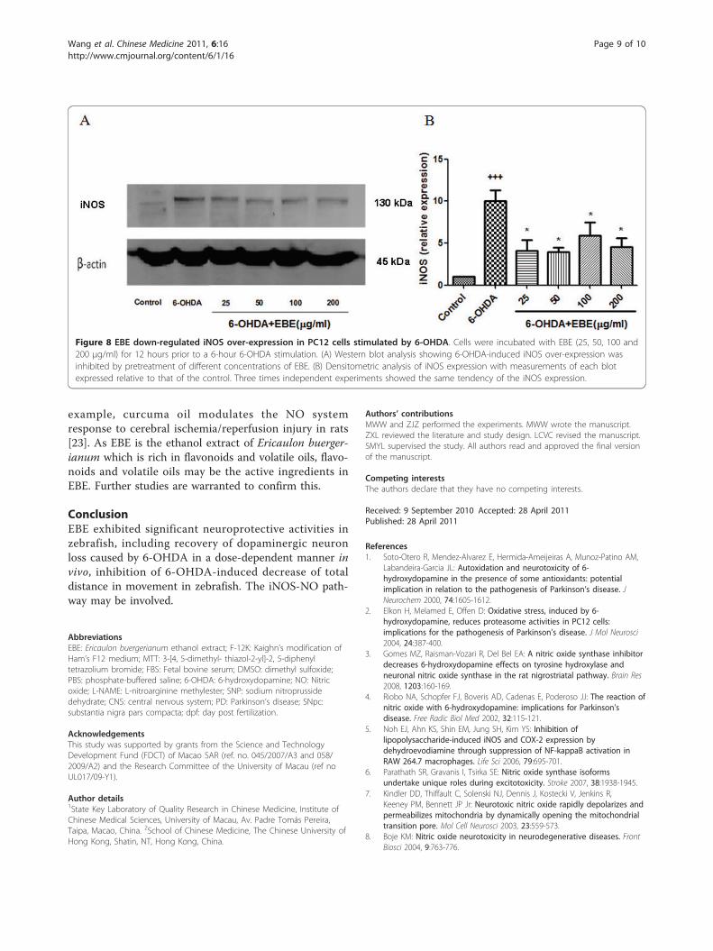
Inhibition of 6-OHDA-induced NO over-production anddown-regulated iNOS over-expressionFigure 7 shows that 6-OHDA exposure led to a roughly1.5-fold increase in NO production compared with thecontrol group; similar elevation was also observed whenSNP (sodium nitroprusside dehydrate, NO generator)was added. This increase in NO level was reduced in aconcentration-dependent manner by pretreatment withEBE in a 25-200 μg/mL range for 12 hours (Figure 7E-H). Pretreatment with 250 μM L-NAME for 12 hoursalso significantly (P = 0.044) reduced this 6-OHDA-induced NO over-production. Pretreatment with higherconcentrations (50, 100, 200 μg/ml) of EBE suppressedthe elevated NO production more efficiently than L-NAME (Figure 7D). Furthermore, DAF-FM diacetatestaining was photographed with a fluorescent micro-scope (Figure 7A-H). The expression of iNOS was up-regulated by 6-OHDA exposure in Western blot analysisand such up-regulation was prevented by pretreatmentwith EBE (Figure 8A).
Further studiesMany flavonoids, such as those derived from Vitis vini-fera (grape), Camellia sinensis (tea), Theobroma cacao(cocoa) and Vaccinium spp. (blueberry), exert their neu-roprotective actions via (1) modulating intracellular sig-naling cascades controlling neuronal survival, death anddifferentiation, (2) affecting gene expression and (3)interacting with mitochondria [22]. The neuroprotectiveeffects of volatile oils have also been reported; for
Figure 3 Quantitative analysis of area of TH+ neuron inzebrafish brain. All data expressed as percentage of control group,each bar represents mean ± SD. +++ P < 0.0001 versus controlgroup (without 6-OHDA treatment); * P = 0.046 versus 6-OHDA-treated group; *** P < 0.0001 versus 6-OHDA-treated group. Allexperiments were repeated 3 times.
Figure 4 EBE inhibited 6-OHDA-induced decrease of totaldistance of movement in zebrafish. Quantification of theswimming parameter of zebrafish: 4 dpf wild type zebrafish larvaewere treated with 6-OHDA (250 μM) and EBE (25 and 50 μg/mL) forthree days. The locomotor activity of zebrafish was assessed atseven dpf. All results were expressed as total distance of movementtraveled by the larvae, + P = 0.032 versus control group (without 6-OHDA treatment); * P = 0.041, ** P = 0.007 versus 6-OHDA-treatedgroup. All experiments were repeated 3 times.
Wang et al. Chinese Medicine 2011, 6:16http://www.cmjournal.org/content/6/1/16
Page 6 of 10
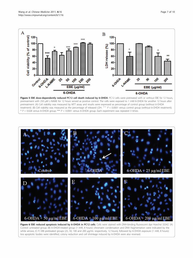
Figure 5 EBE dose-dependently reduced PC12 cell death induced by 6-OHDA. PC12 cells were pretreated with or without EBE for 12 hours,pretreatment with 250 μM L-NAME for 12 hours served as positive control. The cells were exposed to 1 mM 6-OHDA for another 12 hours afterpretreatment. (A) Cell viability was measured by MTT assay and results were expressed as percentage of control group (without 6-OHDAtreatment). (B) Cell viability was measured as the percentage of released LDH. +++ P < 0.0001 versus control group (without 6-OHDA treatment);* P = 0.028 versus 6-OHDA group; *** P < 0.0001 versus 6-OHDA group. Each experiment was repeated 3 times.
Figure 6 EBE reduced apoptosis induced by 6-OHDA in PC12 cells. Cells were stained with DNA-binding fluorescent dye Hoechst 33342. (A)Control: untreated group; (B) 6-OHDA-treated group (1 mM, 8 hours): chromatin condensation and DNA fragmentation were indicated by thewhite arrows; (C-F) EBE-pretreated groups (25, 50, 100 and 200 μg/mL respectively, 12 hours), followed by 6-OHDA exposure (1 mM, 8 hours):less apoptotic bodies were identified, colony reduction and cell shrinkage induced by 6-OHDA were also reversed.
Wang et al. Chinese Medicine 2011, 6:16http://www.cmjournal.org/content/6/1/16
Page 7 of 10
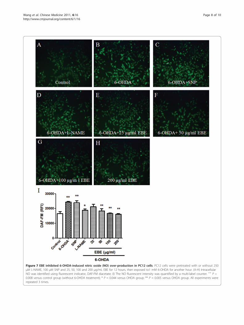
Figure 7 EBE inhibited 6-OHDA-induced nitric oxide (NO) over-production in PC12 cells. PC12 cells were pretreated with or without 250μM L-NAME, 100 μM SNP and 25, 50, 100 and 200 μg/mL EBE for 12 hours, then exposed to1 mM 6-OHDA for another hour. (A-H) IntracellularNO was identified using fluorescent indicator, DAF-FM diacetate; (I) The NO fluorescent intensity was quantified by a multi-label counter. ++ P =0.008 versus control group (without 6-OHDA treatment); * P = 0.044 versus OHDA group; ** P = 0.005 versus OHDA group. All experiments wererepeated 3 times.
Wang et al. Chinese Medicine 2011, 6:16http://www.cmjournal.org/content/6/1/16
Page 8 of 10
example, curcuma oil modulates the NO systemresponse to cerebral ischemia/reperfusion injury in rats[23]. As EBE is the ethanol extract of Ericaulon buerger-ianum which is rich in flavonoids and volatile oils, flavo-noids and volatile oils may be the active ingredients inEBE. Further studies are warranted to confirm this.
ConclusionEBE exhibited significant neuroprotective activities inzebrafish, including recovery of dopaminergic neuronloss caused by 6-OHDA in a dose-dependent manner invivo, inhibition of 6-OHDA-induced decrease of totaldistance in movement in zebrafish. The iNOS-NO path-way may be involved.
AbbreviationsEBE: Ericaulon buergerianum ethanol extract; F-12K: Kaighn’s modification ofHam’s F12 medium; MTT: 3-[4, 5-dimethyl- thiazol-2-yl]-2, 5-diphenyltetrazolium bromide; FBS: Fetal bovine serum; DMSO: dimethyl sulfoxide;PBS: phosphate-buffered saline; 6-OHDA: 6-hydroxydopamine; NO: Nitricoxide; L-NAME: L-nitroarginine methylester; SNP: sodium nitroprussidedehydrate; CNS: central nervous system; PD: Parkinson’s disease; SNpc:substantia nigra pars compacta; dpf: day post fertilization.
AcknowledgementsThis study was supported by grants from the Science and TechnologyDevelopment Fund (FDCT) of Macao SAR (ref. no. 045/2007/A3 and 058/2009/A2) and the Research Committee of the University of Macau (ref noUL017/09-Y1).
Author details1State Key Laboratory of Quality Research in Chinese Medicine, Institute ofChinese Medical Sciences, University of Macau, Av. Padre Tomás Pereira,Taipa, Macao, China. 2School of Chinese Medicine, The Chinese University ofHong Kong, Shatin, NT, Hong Kong, China.
Authors’ contributionsMWW and ZJZ performed the experiments. MWW wrote the manuscript.ZXL reviewed the literature and study design. LCVC revised the manuscript.SMYL supervised the study. All authors read and approved the final versionof the manuscript.
Competing interestsThe authors declare that they have no competing interests.
Received: 9 September 2010 Accepted: 28 April 2011Published: 28 April 2011
References1. Soto-Otero R, Mendez-Alvarez E, Hermida-Ameijeiras A, Munoz-Patino AM,
Labandeira-Garcia JL: Autoxidation and neurotoxicity of 6-hydroxydopamine in the presence of some antioxidants: potentialimplication in relation to the pathogenesis of Parkinson’s disease. JNeurochem 2000, 74:1605-1612.
2. Elkon H, Melamed E, Offen D: Oxidative stress, induced by 6-hydroxydopamine, reduces proteasome activities in PC12 cells:implications for the pathogenesis of Parkinson’s disease. J Mol Neurosci2004, 24:387-400.
3. Gomes MZ, Raisman-Vozari R, Del Bel EA: A nitric oxide synthase inhibitordecreases 6-hydroxydopamine effects on tyrosine hydroxylase andneuronal nitric oxide synthase in the rat nigrostriatal pathway. Brain Res2008, 1203:160-169.
4. Riobo NA, Schopfer FJ, Boveris AD, Cadenas E, Poderoso JJ: The reaction ofnitric oxide with 6-hydroxydopamine: implications for Parkinson’sdisease. Free Radic Biol Med 2002, 32:115-121.
5. Noh EJ, Ahn KS, Shin EM, Jung SH, Kim YS: Inhibition oflipopolysaccharide-induced iNOS and COX-2 expression bydehydroevodiamine through suppression of NF-kappaB activation inRAW 264.7 macrophages. Life Sci 2006, 79:695-701.
6. Parathath SR, Gravanis I, Tsirka SE: Nitric oxide synthase isoformsundertake unique roles during excitotoxicity. Stroke 2007, 38:1938-1945.
7. Kindler DD, Thiffault C, Solenski NJ, Dennis J, Kostecki V, Jenkins R,Keeney PM, Bennett JP Jr: Neurotoxic nitric oxide rapidly depolarizes andpermeabilizes mitochondria by dynamically opening the mitochondrialtransition pore. Mol Cell Neurosci 2003, 23:559-573.
8. Boje KM: Nitric oxide neurotoxicity in neurodegenerative diseases. FrontBiosci 2004, 9:763-776.
Figure 8 EBE down-regulated iNOS over-expression in PC12 cells stimulated by 6-OHDA. Cells were incubated with EBE (25, 50, 100 and200 μg/ml) for 12 hours prior to a 6-hour 6-OHDA stimulation. (A) Western blot analysis showing 6-OHDA-induced iNOS over-expression wasinhibited by pretreatment of different concentrations of EBE. (B) Densitometric analysis of iNOS expression with measurements of each blotexpressed relative to that of the control. Three times independent experiments showed the same tendency of the iNOS expression.
Wang et al. Chinese Medicine 2011, 6:16http://www.cmjournal.org/content/6/1/16
Page 9 of 10
9. Ho JC, Chen CM: Flavonoids from the aquatic plant Eriocaulonbuergerianum. Phytochemistry 2002, 61:405-408.
10. Chinese Pharmacopoeia Commission: In Pharmacopoeia of the People’sRepublic of China. Volume 1. Beijing: Chemical Industry Press; 2005.
11. Fang JJ, Ye G, Chen WL, Zhao WM: Antibacterial phenolic componentsfrom Eriocaulon buergerianum. Phytochemistry 2008, 69:1279-1286.
12. Yang WC, Liu H, Chen QL: Gujingcaoke yaoxue yanjiu gaikuang. Acta ChinMedicine and Pharmacol 2009, 37:92-93.
13. Sun YX, Zhang DF: Guqingtang jianjie. HeNan Tradit Chin Med 2001, 21:31.14. Parng C, Roy NM, Ton C, Lin Y, McGrath P: Neurotoxicity assessment using
zebrafish. J Pharmacol Toxicol Methods 2007, 55:103-112.15. McKinley ET, Baranowski TC, Blavo DO, Cato C, Doan TN, Rubinstein AL:
Neuroprotection of MPTP-induced toxicity in zebrafish dopaminergicneurons. Brain Res Mol Brain Res 2005, 141:128-137.
16. Rink E, Wullimann MF: Connections of the ventral telencephalon andtyrosine hydroxylase distribution in the zebrafish brain (Danio rerio) leadto identification of an ascending dopaminergic system in a teleost. BrainRes Bull 2002, 57:385-387.
17. Anichtchik OV, Kaslin J, Peitsaro N, Scheinin M, Panula P: Neurochemicaland behavioural changes in zebrafish Danio rerio after systemicadministration of 6-hydroxydopamine and 1-methyl-4-phenyl-1,2,3,6-tetrahydropyridine. J Neurochem 2004, 88:443-453.
18. Panula P, Sallinen V, Sundvik M, Kolehmainen J, Torkko V, Tiittula A,Moshnyakov M, Podlasz P: Modulatory neurotransmitter systems andbehavior: towards zebrafish models of neurodegenerative diseases.Zebrafish 2006, 3:235-247.
19. Institute of Materia Medica CAoMS: Zhong Yao Zhi Beijing: People’s MedicalPublishing House; 1995.
20. Westerfield M: The Zebrafish Book: A Guide for the Laboratory Use ofZebrafish (Danio rerio). 5 edition. Eugene: University of Oregon Press; 2007.
21. Schweitzer J, Driever W: Development of the dopamine systems inzebrafish. Adv Exp Med and Biol 2009, 651:1-14.
22. Spencer JP: Flavonoids: modulators of brain function? Br J Nutr 2008,99E(Suppl 1):ES60-77.
23. Dohare P, Varma S, Ray M: Curcuma oil modulates the nitric oxide systemresponse to cerebral ischemia/reperfusion injury. Nitric Oxide 2008,19:1-11.
doi:10.1186/1749-8546-6-16Cite this article as: Wang et al.: Eriocaulon buergerianum extract protectsPC12 cells and neurons in zebrafish against 6-hydroxydopamine-induced damage. Chinese Medicine 2011 6:16.
Submit your next manuscript to BioMed Centraland take full advantage of:
• Convenient online submission
• Thorough peer review
• No space constraints or color figure charges
• Immediate publication on acceptance
• Inclusion in PubMed, CAS, Scopus and Google Scholar
• Research which is freely available for redistribution
Submit your manuscript at www.biomedcentral.com/submit
Wang et al. Chinese Medicine 2011, 6:16http://www.cmjournal.org/content/6/1/16
Page 10 of 10


![Glucagon-Like Peptide-1 Receptor Agonist Protects Dorsal ...downloads.hindawi.com/journals/jdr/2019/9426014.pdf · 11] and antagonize apoptosis of PNS neurons or PC12 cells [12].](https://static.fdocuments.net/doc/165x107/5fa237beceb2131c3f440106/glucagon-like-peptide-1-receptor-agonist-protects-dorsal-11-and-antagonize.jpg)