
ErgoTM Imaging System - Digirad collimators simple and fast. An optional collimator cart provides...
8
Ergo TM Imaging System
Transcript of ErgoTM Imaging System - Digirad collimators simple and fast. An optional collimator cart provides...

ErgoTM Imaging System
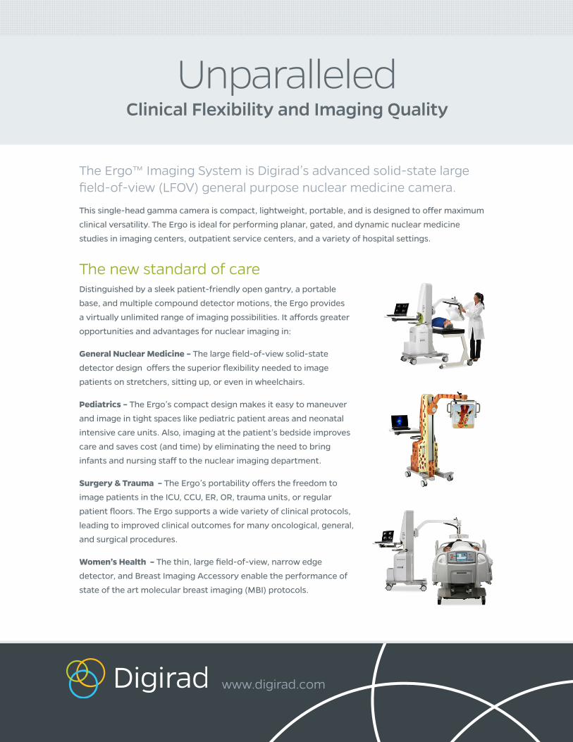
UnparalleledClinical Flexibility and Imaging Quality
The Ergo™ Imaging System is Digirad’s advanced solid-state large field-of-view (LFOV) general purpose nuclear medicine camera.
This single-head gamma camera is compact, lightweight, portable, and is designed to offer maximum
clinical versatility. The Ergo is ideal for performing planar, gated, and dynamic nuclear medicine
studies in imaging centers, outpatient service centers, and a variety of hospital settings.
The new standard of careDistinguished by a sleek patient-friendly open gantry, a portable
base, and multiple compound detector motions, the Ergo provides
a virtually unlimited range of imaging possibilities. It affords greater
opportunities and advantages for nuclear imaging in:
General Nuclear Medicine – The large field-of-view solid-state
detector design offers the superior flexibility needed to image
patients on stretchers, sitting up, or even in wheelchairs.
Pediatrics – The Ergo’s compact design makes it easy to maneuver
and image in tight spaces like pediatric patient areas and neonatal
intensive care units. Also, imaging at the patient’s bedside improves
care and saves cost (and time) by eliminating the need to bring
infants and nursing staff to the nuclear imaging department.
Surgery & Trauma – The Ergo’s portability offers the freedom to
image patients in the ICU, CCU, ER, OR, trauma units, or regular
patient floors. The Ergo supports a wide variety of clinical protocols,
leading to improved clinical outcomes for many oncological, general,
and surgical procedures.
Women’s Health – The thin, large field-of-view, narrow edge
detector, and Breast Imaging Accessory enable the performance of
state of the art molecular breast imaging (MBI) protocols.
www.digirad.com

Compact Open Gantry
The small gantry footprint combined with a thinner, compact, and lighter detector create a less
intimidating system, making it ideal for imaging claustrophobic and/or pediatric patients. The design’s
caudal and cephalic detector tilt capability further enhances the Ergo’s imaging flexibility to easily image
patients sitting up or lying down.
In addition to the Ergo’s™revolutionary
large field-of-view solid-state detector and superior positional flexibility, it offers
an array of conveniences that support the delivery of high-quality clinical results.
Lightweight Portable Design
The lightweight portable design and low profile
wheelbase make it easy to maneuver. A narrow
27” width allows for easy entry into 30” doorways
and simple movement through small hallways
and around patient beds.
Swivel Acquisition + Viewing Workstation
A dedicated laptop acquisition workstation
specially configured for Digirad imaging is
mounted on a swivel base, allowing operation
from either side of the camera. The operator
control console provides comprehensive
acquisition and viewing functionality and
supports Modality Worklist compatibility. The
user friendly interface also allows technologists
to easily create and customize imaging protocols.
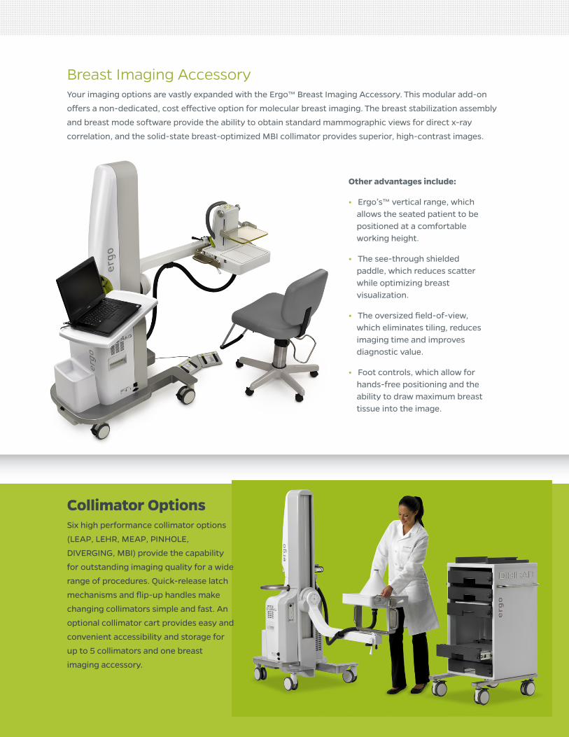
Other advantages include:
• Ergo’s™ vertical range, which allows the seated patient to be positioned at a comfortable working height.
• The see-through shielded paddle, which reduces scatter while optimizing breast visualization.
• The oversized field-of-view, which eliminates tiling, reduces imaging time and improves diagnostic value.
• Foot controls, which allow for hands-free positioning and the ability to draw maximum breast tissue into the image.
Breast Imaging AccessoryYour imaging options are vastly expanded with the Ergo™ Breast Imaging Accessory. This modular add-on
offers a non-dedicated, cost effective option for molecular breast imaging. The breast stabilization assembly
and breast mode software provide the ability to obtain standard mammographic views for direct x-ray
correlation, and the solid-state breast-optimized MBI collimator provides superior, high-contrast images.
Collimator OptionsSix high performance collimator options
(LEAP, LEHR, MEAP, PINHOLE,
DIVERGING, MBI) provide the capability
for outstanding imaging quality for a wide
range of procedures. Quick-release latch
mechanisms and flip-up handles make
changing collimators simple and fast. An
optional collimator cart provides easy and
convenient accessibility and storage for
up to 5 collimators and one breast
imaging accessory.

The heart of the Ergo™ is its large 12.25” x 15.5” field of view utilizing the most advanced
solid-state detector technology in the industry. The system delivers unsurpassed performance specifications
for general imaging with intrinsic spatial resolution of 3.25 mm, energy resolution of 7.9%, and count rate
capabilities greater than 5 Mcps. The versatility of the Ergo is unmatched by other general imaging systems.
It has six collimator options for use in various nuclear medicine applications including:
Bone Spot
3 Phase Bone (2 of 3 Phases)
Molecular Breast Imaging
White Blood Cell
Gallbladder
Thyroid
Liver
Brain Flow(Flow and Static Images)
Lung Perfusion
Planar Gated
Salivary Gland
Miscellaneous Studies: Parathyroid, Renal, Gastric Emptying, Indium, Gallium

As we enter this new era of nuclear medicine, we’re challenged to find innovative ways to improve quality, increase productivity, and reduce costs. Digirad has developed powerful solid-state detector technology that has transformed nuclear camera design by substantially raising overall performance and significantly lowering total lifecycle costs.
The Ergo™ large field-of-view imager represents an exciting new
genre of general-purpose nuclear medicine cameras designed
with a level of positional flexibility, clinical utility, and patient
friendliness that is unmatched by any other general imaging
system. We’ve taken breakthrough technology and applied it to
achieve measurable improvements that positively impact every
component of your healthcare system.
A New Era in Nuclear Medicine

DETECTOR/YOKE MOTIONrotation
detector 180°
yoke 360°
arm rotation 390°
vertical position
maximum 152.4 cm [60 in]
minimum 45.7cm [18 in]
maximum reach
parallel to the floor (looking up/down) 92.2 cm [36.3 in]
perpendicular to the floor (looking back) 66 cm [26 in]
DETECTOR SPECIFICATIONSdetector technology solid state, segmented CsI (Tl) / silicon photodiode
exterior dimensions 42.1 x 38.4 x 10.2 cm [16.6 x 15.1 x 4.0 in]
useful FOV 39.6 x 31.1 cm [15.6 x 12.2 in]
detector element size 3 x 3 mm
energy range 50-350 keV
collimator options LEAP, LEHR, MEAP, PINHOLE, DIVERGING, MBI
COLLIMATOR SPECIFICATIONS LEAP LEHR MEAP PINHOLE DIVERGING
isotope Tc-99m Tc-99m In-111, Ga-67 Tc-99m, I-123 Tc-99m
useful energy range (keV) 50-170 50-170 50-350 60-160 50-170
hole shape hex hex hex round hex
septal thickness (mm) 0.2 0.2 1.0 n/a 0.3
hole diameter (mm) 1.5 1.5 2.3 4, 6, and 8 1.9
hole length (mm) 23 30 30 218 30
sensitivity @ 10cm (cpm/uCi)2 250* 132* 153^ 210* 106*
system spatial resolution @ 10cm 10.3 7.4 11.2 10.0 10.8w/o scatter (mm)2
focal length @ exit surface (mm) n/a n/a n/a n/a 350
diameter at base of cone (mm) n/a n/a n/a 285 n/a
weight (lbs) 26 34 68 38 40
type parallel parallel parallel pinhole diverging
ERGOTM
IMAGING SYSTEM
SYSTEM PERFORMANCEsimultaneous dual isotope acquisition yes
acquisition matrix 64 x 64, 128 x 128, 256 x 256, 512 x 512
maximum emission count rate, cps 1 > 5M
intrinsic spatial resolution 3.3 mm
intrinsic energy resolution, FWHM, 140 keV 7.9%
intrinsic spatial linearity 0.3 mm (absolute)
1 The maximum count rate reached with 34mCi Tc-99m source in the open window was 5M cps. The fold-over point was not yet reached.
2 All spatial resolution and sensitivity numbers are typical values. Sensitivity numbers within +/-7% of spec are acceptable.* Sensitivity measured using 16% Tc-99m window.^ MEAP sensitivity calculated using Ga-67 with 86-100 keV and 170-200 keV windows.
Technical Specifications

BREAST IMAGING ACCESSORY MBI
isotope Tc-99m
useful energy range (keV) 130-152
hole shape hex
septal thickness (mm) 0.3
hole diameter (mm) 1.9
hole length (mm) 22
sensitivity @ 10cm (cpm/uCi)3 466
system spatial resolution @ 6 cm w/o scatter (mm) 2 8.9
focal length @ exit surface (mm) n/a
collimator weight (lbs) 30
accessory weight (lbs) 36
type parallel
accessory compression range 2-19 cm [0.8-7.5 in]
stabilization pressure 8 lbs
2 All spatial resolution and sensitivity numbers are typical values. Sensitivity numbers within +/-7% of spec are acceptable.
3 Sensitivity measured using 130-152keV Tc-99m window.
ENVIRONMENTAL/OPERATION REQUIREMENTSsystem weight 320 kg [705 lbs]
height 179.1cm [70.5 in]
width 74 cm [29 in]
length 170 cm [67 in]
minimum room size 8 x 8 ft
floor clearance 8.9 cm [3.5 in]
power requirements 15A @ 120VAC, 60Hz, 7.5A @ 240VAC, 50/60Hz
operating temperature range 18-29° C [65-84° F]
relative operating humidity 30-75%
architectural modifications none required
environmental storage 5-50° C [41-122° F]
storage humidity 10-90% (non-condensing)
COLLIMATOR CART (OPTION)length 83.8 cm [33 in]
width 58.4 cm [23 in]
height 119.4 cm [47 in] without accessories
weight 116.6 kg [257 lbs] without collimators or accessories
storage capacity five collimators plus one breast imaging accessory
13100 Gregg Street, Suite APoway, CA 92064800.947.6134 | www.digirad.com
ERGOTM
IMAGING SYSTEM
Technical Specifications


![[2] Basic Applications of Multileaf Collimators](https://static.fdocuments.net/doc/165x107/5535c8b455034686768b4718/2-basic-applications-of-multileaf-collimators.jpg)

