ErbB3 Is Involved in Activation of Phosphatidylinositol 3
9
MOLECULAR AND CELLULAR BIOLOGY, June 1994, p. 3550-3558 Vol. 14, No. 6 0270-7306/94/$04.00+0 Copyright C 1994, American Society for Microbiology ErbB3 Is Involved in Activation of Phosphatidylinositol 3-Kinase by Epidermal Growth Factor STEPHEN P. SOLTOFF,l* KERMIT L. CARRAWAY III,1 S. A. PRIGENT,2 W. G. GULLICK,2 AND LEWIS C. CANTLEY' Division of Signal Transduction, Department of Medicine, Beth Israel Hospital, Boston, Massachusetts 02115,1 and Molecular Oncology Laboratory, ICRF Oncology Group, Hammersmith Hospital, London W12 OHS, United Kingdom2 Received 11 October 1993/Returned for modification 11 November 1993/Accepted 24 February 1994 Conflicting results concerning the ability of the epidermal growth factor (EGF) receptor to associate with and/or activate phosphatidylinositol (Ptdlns) 3-kinase have been published. Despite the ability of EGF to stimulate the production of Ptdlns 3-kinase products and to cause the appearance of PtdIns 3-kinase activity in antiphosphotyrosine immunoprecipitates in several cell lines, we did not detect EGF-stimulated Ptdlns 3-kinase activity in anti-EGF receptor immunoprecipitates. This result is consistent with the lack of a phosphorylated Tyr-X-X-Met motif, the p85 Src homology 2 (SH2) domain recognition sequence, in this receptor sequence. The EGF receptor homolog, ErbB2 protein, also lacks this motif. However, the ErbB3 protein has seven repeats of the Tyr-X-X-Met motif in the carboxy-terminal unique domain. Here we show that in A431 cells, which express both the EGF receptor and ErbB3, Ptdlns 3-kinase coprecipitates with the ErbB3 protein (pl80eR3) in response to EGF. p180B3 is also shown to be tyrosine phosphorylated in response to EGF. In contrast, a different mechanism for the activation of Ptdlns 3-kinase in response to EGF occurs in certain cells (PC12 and A549 cells). Thus, we show for the first time that ErbB3 can mediate EGF responses in cells expressing both ErbB3 and the EGF receptor. Phosphatidylinositol (Ptdlns) 3-kinase is a cytosolic protein that is recruited to activated protein tyrosine kinases in re- sponse to a variety of factors that activate cells. It is a heterodimeric protein consisting of 85- and 110-kDa subunits (4, 26). The 85-kDa subunit (p85) contains two Src homology 2 (SH2) groups that bind to tyrosine-phosphorylated amino acids that have a consensus Tyr-X-X-Met motif. The 1 10-kDa subunit contains the catalytic site (13). Ptdlns 3-kinase is activated by various growth factors, including epidermal growth factor (EGF), platelet-derived growth factor, colony- stimulating factor 1, and insulin (reviewed in reference 3). An increase in Ptdlns 3-kinase activity also occurs in response to nerve growth factor (6, 35, 38), a factor that initiates cellular differentiation. PtdIns 3-kinase activity is also activated by addition of fMet-Leu-Phe to neutrophils (43) and thrombin to platelets (21, 25). Ptdlns 3-kinase has been shown to bind directly to activated receptors at domains that are autophos- phorylated on tyrosine and contain the Tyr-X-X-Met motif (3). The activation of PtdIns 3-kinase by insulin involves the association of p85 with an intermediary cytosolic protein, IRS-1, that is tyrosine phosphorylated in response to insulin and contains multiple Tyr-X-X-Met motifs (41). The activation of B lymphocytes by antigen binding to tyrosine-phosphory- lated membrane immunoglobulin M results in the recruitment of Ptdlns 3-kinase activity to tyrosine-phosphorylated CD19, a membrane protein that can be coligated with membrane immunoglobulin M but which does not have intrinsic protein- tyrosine kinase activity (44). Thus, in some cases the activation of Ptdlns 3-kinase involves its association with a tyrosine kinase substrate that is distinct from the receptor to which the activating factor binds. The precise role of the EGF receptor (EGFR) in the * Corresponding author. Mailing address: Division of Signal Trans- duction, Beth Israel Hospital, Department of Medicine, Alpert Build- ing, Room 148, 200 Longwood Ave., Boston, MA 02115. Phone: (617) 278-3093. Fax: (617) 278-3131. activation of Ptdlns 3-kinase by EGF remains unclear. EGF produces various effects on cells (reviewed in reference 5). EGF stimulates the canonical Ptdlns turnover pathway, which involves the phospholipase C--yl-mediated hydrolysis of Ptdlns 4,5-bisphosphate into inositol-1,4,5-trisphosphate (Ins-1,4,5- P3) and diacylglycerol. Consistent with this, EGF has been reported to phosphorylate phospholipase C--yl (22, 23, 45), increase Ins-1,4,5-P3 production (12, 31), release Ca2+ from intracellular stores and elevate intracellular free Ca2+ concen- tration (15), and activate Ca2+-sensitive ion channels (29). In A431 cells, phosphoinositide kinase activities were found to copurify with the EGFR (42), and EGF stimulated a Ptdlns kinase activity that was independent from the production of Ins-1,4,5-P3 (31). Subsequently, Ptdlns 4-kinase and PtdIns4P 5-ki- nase activities, enzymes in the canonical phosphoinositol turn- over pathway, were shown to be stimulated by EGF and to associate with the EGFR (7, 27). However, EGF also has been shown to stimulate the appearance of lipid products of PtdIns 3-kinase in a Leydig tumor cell line (30) and in PC12 cells (6, 35). EGF-stimulated PtdIns 3-kinase activity was immunopre- cipitated with anti-P-Tyr (6, 24, 35) and anti-EGFR (2) antibodies. However, the EGFR lacks a tyrosine-phosphory- lated Tyr-X-X-Met motif and binds p85 very weakly compared with the platelet-derived growth factor receptor (14). A number of proteins related to the EGFR make up an extended family of type I receptor tyrosine kinases that share sequence homologies that include two cysteine-rich extracel- lular domains, a single membrane-spanning transmembrane domain, and a cytoplasmic domain that has tyrosine kinase homology. In addition to the EGFR, other family members include ErbB2 (pl8serbB2/neu) (reviewed in reference 47), ErbB3 (pl8OerbB3) (20, 33), and ErbB4 (p180erbB4) (32). ErbB3 is of particular interest in regard to signalling since it contains seven copies of the Tyr-X-X-Met motif in its carboxy-terminal cytosolic domain (39). This observation and reports that ErbB2 may be a substrate for the EGFR (16, 18) suggest that the EGFR interacts with other similar proteins in this class. Thus, 3550 Downloaded from https://journals.asm.org/journal/mcb on 20 November 2021 by 210.103.96.219.
Transcript of ErbB3 Is Involved in Activation of Phosphatidylinositol 3

MOLECULAR AND CELLULAR BIOLOGY, June 1994, p. 3550-3558 Vol. 14, No. 60270-7306/94/$04.00+0Copyright C 1994, American Society for Microbiology
ErbB3 Is Involved in Activation of Phosphatidylinositol3-Kinase by Epidermal Growth Factor
STEPHEN P. SOLTOFF,l* KERMIT L. CARRAWAY III,1 S. A. PRIGENT,2W. G. GULLICK,2 AND LEWIS C. CANTLEY'
Division of Signal Transduction, Department of Medicine, Beth Israel Hospital, Boston, Massachusetts 02115,1 andMolecular Oncology Laboratory, ICRF Oncology Group, Hammersmith Hospital, London W12 OHS, United Kingdom2
Received 11 October 1993/Returned for modification 11 November 1993/Accepted 24 February 1994
Conflicting results concerning the ability of the epidermal growth factor (EGF) receptor to associate withand/or activate phosphatidylinositol (Ptdlns) 3-kinase have been published. Despite the ability of EGF tostimulate the production of Ptdlns 3-kinase products and to cause the appearance of PtdIns 3-kinase activityin antiphosphotyrosine immunoprecipitates in several cell lines, we did not detect EGF-stimulated Ptdlns3-kinase activity in anti-EGF receptor immunoprecipitates. This result is consistent with the lack of aphosphorylated Tyr-X-X-Met motif, the p85 Src homology 2 (SH2) domain recognition sequence, in thisreceptor sequence. The EGF receptor homolog, ErbB2 protein, also lacks this motif. However, the ErbB3protein has seven repeats of the Tyr-X-X-Met motif in the carboxy-terminal unique domain. Here we show thatin A431 cells, which express both the EGF receptor and ErbB3, Ptdlns 3-kinase coprecipitates with the ErbB3protein (pl80eR3) in response to EGF. p180B3 is also shown to be tyrosine phosphorylated in response toEGF. In contrast, a different mechanism for the activation of Ptdlns 3-kinase in response to EGF occurs incertain cells (PC12 and A549 cells). Thus, we show for the first time that ErbB3 can mediate EGF responsesin cells expressing both ErbB3 and the EGF receptor.
Phosphatidylinositol (Ptdlns) 3-kinase is a cytosolic proteinthat is recruited to activated protein tyrosine kinases in re-sponse to a variety of factors that activate cells. It is aheterodimeric protein consisting of 85- and 110-kDa subunits(4, 26). The 85-kDa subunit (p85) contains two Src homology2 (SH2) groups that bind to tyrosine-phosphorylated aminoacids that have a consensus Tyr-X-X-Met motif. The 1 10-kDasubunit contains the catalytic site (13). Ptdlns 3-kinase isactivated by various growth factors, including epidermalgrowth factor (EGF), platelet-derived growth factor, colony-stimulating factor 1, and insulin (reviewed in reference 3). Anincrease in Ptdlns 3-kinase activity also occurs in response tonerve growth factor (6, 35, 38), a factor that initiates cellulardifferentiation. PtdIns 3-kinase activity is also activated byaddition of fMet-Leu-Phe to neutrophils (43) and thrombin toplatelets (21, 25). Ptdlns 3-kinase has been shown to binddirectly to activated receptors at domains that are autophos-phorylated on tyrosine and contain the Tyr-X-X-Met motif (3).The activation of PtdIns 3-kinase by insulin involves theassociation of p85 with an intermediary cytosolic protein,IRS-1, that is tyrosine phosphorylated in response to insulinand contains multiple Tyr-X-X-Met motifs (41). The activationof B lymphocytes by antigen binding to tyrosine-phosphory-lated membrane immunoglobulin M results in the recruitmentof Ptdlns 3-kinase activity to tyrosine-phosphorylated CD19, amembrane protein that can be coligated with membraneimmunoglobulin M but which does not have intrinsic protein-tyrosine kinase activity (44). Thus, in some cases the activationof Ptdlns 3-kinase involves its association with a tyrosinekinase substrate that is distinct from the receptor to which theactivating factor binds.The precise role of the EGF receptor (EGFR) in the
* Corresponding author. Mailing address: Division of Signal Trans-duction, Beth Israel Hospital, Department of Medicine, Alpert Build-ing, Room 148, 200 Longwood Ave., Boston, MA 02115. Phone: (617)278-3093. Fax: (617) 278-3131.
activation of Ptdlns 3-kinase by EGF remains unclear. EGFproduces various effects on cells (reviewed in reference 5).EGF stimulates the canonical Ptdlns turnover pathway, whichinvolves the phospholipase C--yl-mediated hydrolysis of Ptdlns4,5-bisphosphate into inositol-1,4,5-trisphosphate (Ins-1,4,5-P3) and diacylglycerol. Consistent with this, EGF has beenreported to phosphorylate phospholipase C--yl (22, 23, 45),increase Ins-1,4,5-P3 production (12, 31), release Ca2+ fromintracellular stores and elevate intracellular free Ca2+ concen-tration (15), and activate Ca2+-sensitive ion channels (29). InA431 cells, phosphoinositide kinase activities were found tocopurify with the EGFR (42), and EGF stimulated a Ptdlnskinase activity that was independent from the production ofIns-1,4,5-P3 (31). Subsequently, Ptdlns 4-kinase and PtdIns4P 5-ki-nase activities, enzymes in the canonical phosphoinositol turn-over pathway, were shown to be stimulated by EGF and toassociate with the EGFR (7, 27). However, EGF also has beenshown to stimulate the appearance of lipid products of PtdIns3-kinase in a Leydig tumor cell line (30) and in PC12 cells (6,35). EGF-stimulated PtdIns 3-kinase activity was immunopre-cipitated with anti-P-Tyr (6, 24, 35) and anti-EGFR (2)antibodies. However, the EGFR lacks a tyrosine-phosphory-lated Tyr-X-X-Met motif and binds p85 very weakly comparedwith the platelet-derived growth factor receptor (14).A number of proteins related to the EGFR make up an
extended family of type I receptor tyrosine kinases that sharesequence homologies that include two cysteine-rich extracel-lular domains, a single membrane-spanning transmembranedomain, and a cytoplasmic domain that has tyrosine kinasehomology. In addition to the EGFR, other family membersinclude ErbB2 (pl8serbB2/neu) (reviewed in reference 47),ErbB3 (pl8OerbB3) (20, 33), and ErbB4 (p180erbB4) (32). ErbB3is of particular interest in regard to signalling since it containsseven copies of the Tyr-X-X-Met motif in its carboxy-terminalcytosolic domain (39). This observation and reports that ErbB2may be a substrate for the EGFR (16, 18) suggest that theEGFR interacts with other similar proteins in this class. Thus,
3550
Dow
nloa
ded
from
http
s://j
ourn
als.
asm
.org
/jour
nal/m
cb o
n 20
Nov
embe
r 20
21 b
y 21
0.10
3.96
.219
.

ErbB3 IS INVOLVED IN ACTIVATION OF Ptdlns BY EGF 3551
we hypothesized that EGF activates Ptdlns 3-kinase by aninteraction between EGFR and ErbB3.
In these studies we compared the effects of EGF on threedifferent cell lines. In all three cell lines, there is an increasein anti-P-Tyr-immunoprecipitable PtdIns 3-kinase activity inEGF-treated cells, but this is not observed in immunoprecipi-tates collected with anti-EGFR antibody. However, there is anincrease in anti-ErbB3-immunoprecipitable Ptdlns 3-kinaseactivity in EGF-treated A431 cells but not in PC12 or A549cells. Tyrosine-phosphorylated ErbB3 associated with anti-Ptdlns 3-kinase (otp85) antibody and recombinant p85 inEGF-treated A431 cells but not in the other cells. Our studiesindicate that EGF stimulates the association of PtdIns 3-kinasewith ErbB3 in A431 cells but not in PC12 cells or A549 cells.
MATERIALS AND METHODS
Chemicals. All chemicals were reagent grade or better.[32P]ATP (specific activity, 3,000 Ci/mmol) was purchasedfrom New England Nuclear (Boston, Mass.). Dulbecco's mod-ified Eagle medium was obtained from GIBCO Laboratories.EGF (catalog number 01-107) was purchased from UpstateBiochemicals Inc. (Lake Placid, N.Y.).
Antibodies. The anti-phosphotyrosine antibody (anti-P-Tyr)was a murine monoclonal antibody and was generously sup-plied by Brian Drucker (Dana-Farber Cancer Institute, Bos-ton, Mass.). Antibody (anti-p85) to the 85-kDa subunit ofPtdlns 3-kinase was raised in rabbits by Brian Schauffhausen(Tufts University, Boston, Mass.) and is commercially availablefrom Upstate Biochemicals (catalog number 06-195). This is a1:1 mixture of rabbit antibodies directed against a glutathioneS-transferase (GST) fusion protein containing the N-terminalSH2 (NSH2) domain of the p85 subunit of Ptdlns 3-kinase andagainst a GST fusion protein containing the full-length p85subunit. When noted, some experiments were performed withthe antibody raised against the NSH2 domain. The followingmouse monoclonal anti-EGFR antibodies were used: 13A9(Genentech, South San Francisco, Calif.), 291-3A and 291-4A(Randy Schatzman, Syntex), clone 29.1 (Sigma Chemical Co.,St. Louis, Mo.), and SC-120 (Santa Cruz Biotechnology, Inc.,Santa Cruz, Calif.). Anti-ErbB3 antibodies 49-3 (34) and 3183were raised in rabbits to a synthetic peptide containing cyto-plasmic sequence (ELEPELDLDLDLE), and 61-3 (34) and3185 were raised to a peptide sequence (AMRRYLERGESIE) from the juxtamembrane domain; these antibodies wereaffinity purified. A rabbit polyclonal antibody to the extracel-lular domain of human breast ErbB3 was purchased fromTransduction Laboratories (Lexington, Ky.). The anti-Neu(Randy Schatzman, Syntex) and Ki anti-ErbB3 (John Koland,University of Iowa) antibodies were raised in rabbits.
Cell culture. A431 cells and A549 cells were obtained fromthe American Type Culture Collection, and PC12 cells wereobtained from Larry Feig (Tufts University). Unless statedotherwise, cells used in lipid kinase assays were grown in100-mm-diameter dishes, and cells used in Western blotting(immunoblotting) were grown in 150-mm-diameter dishes. Allcells were grown in Dulbecco's modified Eagle medium plusserum at 37°C in a 95% air-5% CO2 mixture. PC12 cells weregrown in 5% horse serum plus 5% calf serum, and A431 cellsand A549 cells were grown in 10% fetal bovine serum. All cellswere used when confluent or nearly confluent. Where noted,cells were switched to serum-free medium (Dulbecco's modi-fied Eagle medium plus 0.1% bovine serum albumin [BSA])overnight prior to exposure to growth factors.Assay of Ptdlns 3-kinase activity. Growth factors were
added for the indicated times at 37°C to cells that were placed
in serum-free medium overnight. The cells were washed twicewith ice-cold buffer A (137 mM NaCl, 20 mM Tris, 1 mMMgCl2, 1 mM CaCl2, 0.2 mM vanadate [pH 7.5]), and werelysed in lysis buffer (buffer A plus 10% [vol/vol] glycerol, 1%[vol/vol] Nonidet P-40 [NP-40], and 1 mM phenylmethylsulfo-nyl fluoride). The lysates were vortexed and centrifuged at16,000 x g (Eppendorf 5414 microcentrifuge). The clearedsupernatants were transferred to fresh microcentrifuge tubesand incubated with anti-P-Tyr (6.6 ,ug/ml) or other antibodies(see figure legends) for 2 h at 4°C. Protein A-Sepharose (4mg/ml of lysate) was then added to the lysates for 2 h at 4°C.In some experiments, antibodies and protein A-Sepharosewere added simultaneously to the lysates for 3 h.The immunoprecipitates were pelleted, washed three times
in phosphate-buffered saline (PBS) (137 mM NaCl, 15.7 mMNaH2PO4, 1.47 mM KH2PO4, 2.68 mM KCl [pH 7.4])-1%NP-40, two times in 0.1 M Tris (pH 7.5)-0.5 M LiCl, and twotimes in TNE (10 mM Tris, 100 mM NaCl, 1 mM EDTA [pH7.5]). All wash solutions contained 200 ,uM vanadate. To assaythe PtdIns 3-kinase activity of the immunoprecipitates, soni-cated PtdIns [0.2 mg/ml (final concentration) in 10 mMN-2-hydroxyethylpiperazine-N'-2-ethanesulfonic acid (HEPES)-1mM ethylene glycol-bis(,3-aminoethyl ether)-N,N,N',N'-tet-raacetic acid (EGTA) (pH 7.5)] and [-y-32P]ATP (10 to 20 ,uCiper sample) were added to the immunoprecipitates for 10 minat room temperature. When noted, 0.5% (vol/vol) NP-40 alsowas present in the reaction mixture. The reaction was stoppedby the addition of 80 pl of 1 M HCl and 160 pAl of methanol-chloroform (1:1 [vol/vol] mixture). The lipid-containing or-ganic phase was resolved on oxalate-coated thin-layer chroma-tography plates (Silica Gel 60; MCB Reagents, Merck,Rahway, N.J.) developed in chloroform-methanol-water-am-monium hydroxide (60:40:11.3:2, vol/vol). Radiolabelled spotscorresponding to authentic Ptdlns-4-P were excised and quan-tified by scintillation counting or Cerenkov radiation. In someexperiments, the PtdIns phosphate (PtdlnsP) spot was deacy-lated and subjected to high-pressure liquid chromatography(HPLC) analysis to determine lipid identity and quantification,as previously described (1).
Identification of proteins by Western blot assays. Lysatesfrom confluent or nearly confluent cells grown in a 150-mm-diameter culture dish were prepared as described above.Cleared lysates were immunoprecipitated with various anti-bodies plus protein A-Sepharose for 3 to 5 h at 4°C. Antibodieswere used as designated in figure legends. The Sepharosebeads were pelleted, washed (three times with PBS-1% NP-40,twice with 0.1 M Tris-0.5 M LiCl, and twice with TNE), dilutedwith an equivalent volume of 2x sample buffer, and boiled for5 min. The supernatant was subjected to electrophoresis on asodium dodecyl sulfate (SDS)-7% polyacrylamide separatinggel with a 3% stacking gel. Proteins were transferred to0.2-,um-pore-size nitrocellulose filters, and the filters wereblocked with TBS (20 mM Tris, 137 mM NaCl [pH 7.6])-2%BSA for 1 h. The filters were washed in TBS-0.2% Tween 20(lTFBS) three times. To identify tyrosine-phosphorylated pro-teins, the blots were exposed to anti-P-Tyr (1 ,ug/ml) inTT7BS-1% BSA for 16 h at 4°C. The identification of proteinswith other antibodies is as reported in figure legends. Thefilters were washed three times in TTBS and exposed tosecondary antibody (anti-mouse horseradish peroxidase oranti-rabbit horseradish peroxidase [Boehringer-Mannheim]) ata 1:10,000 dilution in TFBS-l% BSA for 1 h. Filters werewashed three times with TT'BS and twice with TBS and werevisualized with a chemiluminescence system (ECL, Amer-sham). In some experiments, the filters were stripped byexposing them to 62.5 mM Tris (pH 6.8)-p3-mercaptoethanol
VOL. 14, 1994
Dow
nloa
ded
from
http
s://j
ourn
als.
asm
.org
/jour
nal/m
cb o
n 20
Nov
embe
r 20
21 b
y 21
0.10
3.96
.219
.

3552 SOLTOFF ET AL.
A.4PIP
-o-PIP
EGF: - + + + + +
B.
I.P. Ab: aEGFR aErbB2 cxErbB3 oaP-Tyr ap85
FIG. 1. Immunoprecipitation of Ptdlns 3-kinase activity from EGF-stimulated PC12 and A431 cells. Serum-starved PC12 cells (A) or A431 cells(B) were left untreated (-) or were treated for 5 min with EGF (100 ng/ml) (+). Lysates were prepared in 1 ml of lysis buffer, and Ptdlns 3-kinaseactivity was measured in immunoprecipitates (I.P.) of the cleared lysates (see Materials and Methods). Ptdlns 3-kinase activity was measured byusing exogenous Ptdlns as a substrate. The following amounts of antibodies (Ab) were added to 1 ml of cleared lysate: anti-EGFR (29.1), 5 Ul;anti-ErbB2, 5 [LI; anti-ErbB3 (K1), 5 pAl; anti-P-Tyr, 2 pig/ml; and anti-p85, 2 p.l. The dark area below the PtdlnsP (PIP) spot is radioactivity at theorigin and represents ATP or related products.
(0.1 M)-2% SDS at 70°C for 30 min. The stripped filters werewashed several times in TBS, blocked with TBS-2% BSA, andreprobed with other antibodies as described above or in figurelegends.
Expression of proteins in insect cells. The baculovirusexpression system was used to infect Sf9 insect cells withcDNAs encoding human EGFR and bovine p180erbB3. This willbe described in a forthcoming paper (10a).GST fusion proteins. GST fusion proteins of the intact p85
were produced as described previously (48).Data. Data are given as means ± standard errors of the
mean, with the number of determinations (n) representingseparate experiments.
RESULTS
Effects of EGF on Ptdlns 3-kinase activity in anti-P-Tyrimmunoprecipitates. Since the association of Ptdlns 3-kinaseactivity with the EGFR has been controversial (see Introduc-tion), we examined the activation of PtdIns 3-kinase in anti-P-Tyr immunoprecipitates of several cell lines that responded toEGF. After a 5-min exposure to EGF (100 ng/ml), the Ptdlns3-kinase activity was increased to 11.2 + 2.4 (n = 16) and 37.2+ 4.3 (n = 24) times the basal (unstimulated) activity in A431and PC12 cells, respectively. To confirm that the product of thePtdlns kinase assay was PtdIns-3-P, the product of Ptdlns3-kinase, the PtdlnsP identified via thin-layer chromatographywas deacylated and analyzed by HPLC. In growth factor-treated cells, Ptdlns-3-P made up more than 85% of thePtdlnsP radioactivity (not shown).
Association of Ptdlns 3-kinase activity with EGFR, ErbB2,and ErbB3. To examine whether the enhanced Ptdlns 3-kinaseactivity in EGF-treated cells involved the association of Ptdlns3-kinase with the EGFR or with other members of the type I(EGFR-like) family of growth factor receptors, we measuredPtdlns 3-kinase activity in immunoprecipitates by using anti-bodies to the EGFR (ErbBl), ErbB2, and ErbB3 proteins, aswell as with anti-P-Tyr and anti-p85 antibodies. For eachexperiment with a particular cell line, the Ptdlns 3-kinase
assays were performed at the same time with cells of the samepassage to facilitate a direct comparison of these five antibod-ies. In PC12 cells, EGF-stimulated Ptdlns 3-kinase activity wasreadily detected in anti-P-Tyr immunoprecipitates, but not inanti-EGFR (29.1 or 13A9), anti-ErbB2, or anti-ErbB3 (KI)immunoprecipitates (Fig. IA and Table 1). In contrast, inA431 cells a significant amount of EGF-stimulated Ptdlns3-kinase activity was found in anti-ErbB3 (Kl) immunoprecipi-tates as well as in anti-P-Tyr immunoprecipitates, but not inanti-EGFR (29.1) or anti-ErbB2 immunoprecipitates (Fig. 1Band Table 1). Treatment of A431 cells with EGF increased thelipid kinase activity in anti-ErbB3 (Kl) immunoprecipitates to6.2 ± 1.0 (n = 7) times the basal level. Increases in immuno-precipitable Ptdlns 3-kinase activity from EGF-treated A431cells were also found with other anti-ErbB3 antibodies, asfollows: 61-3, 5.3 + 2.1 (n = 4) times the basal activity; 3183,3.2 ± 0.6 (n = 7); 3185, 3.1 + 1.1 (n = 3); and Transduction,5.5 ± 1.3 (n = 3). Notably, in experiments in which bothanti-ErbB3 and anti-P-Tyr immunoprecipitates were examinedat the same time, the amount of EGF-stimulated Ptdlns3-kinase activity in the anti-ErbB3 immunoprecipitates was49.5% ± 24.7% (n = 5, with K1). The activity in anti-ErbB3(Kl) immunoprecipitates was blocked by >95% (n = 2) by thepresence of 0.5% NP-40, which inhibits Ptdlns 3-kinase activity(46) in the lipid kinase assay. These results suggest that ErbB3is involved in the EGF-promoted activation of Ptdlns 3-kinasein A431 cells but not in PC12 cells and that tyrosine phosphor-ylation was involved in the activation.
Since other investigators had reported the association ofPtdlns 3-kinase activity with the EGFR, we examined severalother EGFR antibodies for their abilities to immunoprecipi-tate EGF-stimulated Ptdlns 3-kinase activity from A431 cells.First, we compared the abilities of different antibodies toimmunoprecipitate the EGFR. Three antibodies (29.1, 13A9,and SC-120) immunoprecipitated relatively greater amounts ofEGFR from A431 cells compared with two other antibodies(291-3A and 291-4A) (Fig. 2A). However, no EGF-stimulatedPtdlns kinase activity was measured in immunoprecipitatesobtained with these three antibodies (Fig. 2B). In these
MOL. CELL. BIOL.
AdIOL.
Dow
nloa
ded
from
http
s://j
ourn
als.
asm
.org
/jour
nal/m
cb o
n 20
Nov
embe
r 20
21 b
y 21
0.10
3.96
.219
.

ErbB3 IS INVOLVED IN ACTIVATION OF Ptdlns BY EGF 3553
TABLE 1. Immunoprecipitation of Ptdlns 3-kinase activity withantibodies to different type I receptor tyrosine kinases
Cells and Presence No. of cpmaantibody of EGF Expt 1 Expt 2
A431XtEGFR - 43 454
+ 41 604
atErbB2 - 49 580+ 68 510
aErbB3 - 180 1,390+ 1,589 7,903
aP-Tyr - 141 1,670+ 3,849 6,198
ap85 (NSH2) - 16,487 352,828+ 22,489 389,534
PC12xEGFR - 26 26*
+ 36 27*
atErbB2 - 28 7*+ 34 32*
aErbB3 - 67 90*+ 48 63*
aP-Tyr - 101 101*+ 3,528 4,337*
ap85 (NSH2) - 43,186 115,583*+ 70,756 83,464*
a All values are Cerenkov counts, except those marked by an asterisk, whichare scintillation counts.
A.
I.P. Ab: 291-3A 13A9 29.1 SC-120 291.4AEGF: - + - + - + - + -+
.ffflfles
Lysat
Blotting Ab: a EGFRB.
I.P. Ab: 29.1EGF: - +
PIP w *
A3A9 SC-120 __aP-T- + - + - +
_
Originn* ** *@*
FIG. 2. Relative effects of different anti-EGFR antibodies in im-munoprecipitating EGFR (A) or Ptdlns kinase (B) activity from A431cells. Cells were serum starved overnight and left untreated (-) or
treated with EGF (100 ng/ml) for 5 min (+). (A) Lysates of cells grownin 100-mm-diameter dishes were prepared in 1 ml of lysis buffer, andthe following antibodies (Ab) were added to part (400 ,u1) of thecleared lysates: 291-3A (3 pul), 13A9 (2 pu1), 29.1 (3 pul), SC-120 (3 R.l),and 291-4A (3 p.l). The immunoprecipitates (I.P.) were washed,subjected to SDS-PAGE, transferred to nitrocellulose filters, andprobed overnight at 4°C with anti-EGFR (291-3A, 1:400 dilution). Aportion (40 p.1) of the cleared lysate was also subjected to SDS-PAGE.(B) Ptdlns kinase assays were performed as previously described (seeMaterials and Methods) on immunoprecipitates from A431 cells. Thefollowing antibodies were added to 1 ml of cleared lysate: 29.1 (3 p.1),13A9 (2 ,ul), SC-120 (3 p.1), and anti-P-Tyr (6.6 p.g/ml). PIP, PtdlnsP.
experiments, we extracted the PtdlnsP spots from the thin-layer chromatography and subjected them to HPLC analysis toconfirm that Ptdlns-3-P did not increase in response to EGF inthe anti-EGFR immunoprecipitates (not shown).
Since A431 cells are a human cell line and PC12 cells arederived from rat cells, we examined another human cell line todetermine if Ptdlns 3-kinase activity was also present inanti-ErbB3 immunoprecipitates. A549 cells are derived from ahuman lung carcinoma. Treatment of these cells with EGF(100 ng/ml, for 5 min) increased the anti-P-Tyr immunopre-cipitable Ptdlns 3-kinase activity to 15.7 ± 5.8 (n = 3) timesthat of unstimulated cells, but EGF did not produce an
increase in anti-ErbB3 (K1) immunoprecipitates or in anti-EGFR (29.1 or 13A9) immunoprecipitates (data not shown).As indicated in Table 1 and in other experiments, the Ptdlns
3-kinase activity of anti-P-Tyr immunoprecipitates of growthfactor-treated A431 and PC12 cells was much less (generallyless than 10%) than the Ptdlns 3-kinase activity that was
immunoprecipitated with anti-p85. These results indicate thata large amount of Ptdlns 3-kinase activity does not associatewith receptors or is not tyrosine phosphorylated.
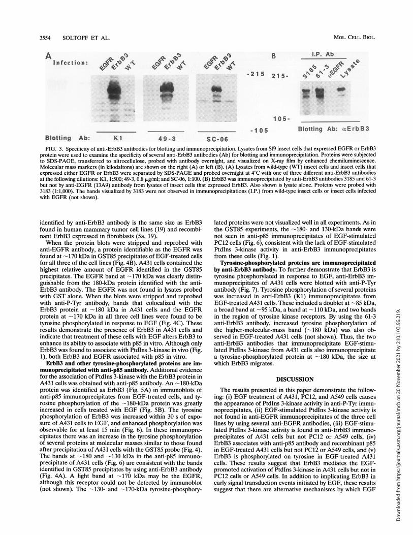
Association of ErbB3 with GST85 and its phosphorylationon tyrosine. The ability of different anti-ErbB3 antibodies toblot ErbB3 expressed in insect cells is shown in Fig. 3A. Threeantibodies (Ki, 49-3, and SC-06) identified a major band at 170to 175 kDa and a smaller band at 155 to 160 kDa. Two otherantibodies, 3183 and 3185, identified the same bands (notshown). These proteins were not observed in wild-type insect
cells or in insect cells that expressed the EGFR. ErbB3expressed in insect cells was immunoprecipitated with anti-ErbB3 antibody (3185 and 61-3) but not anti-EGFR antibody(13A9) (Fig. 3B).
Initial attempts to detect ErbB3 in A431 cells by immuno-blotting failed because of the low abundance of this protein.However, ErbB3 could be detected in association with exog-enously added recombinant p85. In these studies, we comparedthe effects of EGF and insulin, both of which have been foundto promote the phosphorylation of a number of proteins ontyrosine. Lysates of EGF- and insulin-treated A431, PC12, andA549 cells were probed with a GST fusion protein (GST85) ofthe p85 subunit of Ptdlns 3-kinase. As a control, cells were alsoprobed with GST alone. The precipitates were washed underthe stringent conditions, including high salt concentration, thatwere used in lipid kinase assays (see Materials and Methods).The precipitated proteins were subjected to SDS-polyacryl-amide gel electrophoresis (PAGE) transferred to nitrocellu-lose, and blotted with anti-ErbB3 (49-3) antibody. Two pro-teins, one at -180 kDa and one at -130 kDa, were observedin GST85 precipitates from A431 cells exposed to EGF (Fig.4A). These proteins were not seen in parallel experimentsconducted with PC12 cells or A549 cells probed with GST85 orin experiments with any of the cell lines probed with GSTalone (Fig. 4A). Since anti-ErbB3-immunoprecipitable Ptdlns3-kinase activity was found in A431 cells-but not in PC12 orA549 cells-treated with EGF, these results suggest that thereis an association between ErbB3 and p85 in A431 cells but notin the two other cell lines. The -180-kDa size of the protein
VOL. 14, 1994
Dow
nloa
ded
from
http
s://j
ourn
als.
asm
.org
/jour
nal/m
cb o
n 20
Nov
embe
r 20
21 b
y 21
0.10
3.96
.219
.
3554 SOLTOFF ET AL.
AInfection:
lb 0 qq .4bRl 4 'oA, ..k.4e A. 4 le 0 -41. lee4k
B l.P. Abl%b lb% e54
-2. t d...- 2 1 5 21 5 - ,Ab' S *
m
1 05-
Blotting Ab: Kl 4 9 - 3-1 05 Blotting Ab: aErbB3
Sc-06FIG. 3. Specificity of anti-ErbB3 antibodies for blotting and immunoprecipitation. Lysates from Sf9 insect cells that expressed EGFR or ErbB3
protein were used to examine the specificity of several anti-ErbB3 antibodies (Ab) for blotting and immunoprecipitation. Proteins were subjectedto SDS-PAGE, transferred to nitrocellulose, probed with antibody overnight, and visualized on X-ray film by enhanced chemiluminescence.Molecular mass markers (in kilodaltons) are shown on the right (A) or left (B). (A) Lysates from wild-type (WT) insect cells and insect cells thatexpressed either EGFR or ErbB3 were separated by SDS-PAGE and probed overnight at 4°C with one of three different anti-ErbB3 antibodiesat the following dilutions: Kl, 1:500; 49-3, 0.8 ,ug/ml; and SC-06, 1:100. (B) ErbB3 was immunoprecipitated by anti-ErbB3 antibodies 3185 and 61-3but not by anti-EGFR (13A9) antibody from lysates of insect cells that expressed ErbB3. Also shown is lysate alone. Proteins were probed with3183 (1:1,000). The bands visualized by 3183 were not observed in immunoprecipitations (I.P.) from wild-type insect cells or insect cells infectedwith EGFR (not shown).
identified by anti-ErbB3 antibody is the same size as ErbB3found in human mammary tumor cell lines (19) and recombi-nant ErbB3 expressed in fibroblasts (5a, 19).When the protein blots were stripped and reprobed with
anti-EGFR antibody, a protein identifiable as the EGFR wasfound at --170 kDa in GST85 precipitates of EGF-treated cellsfor all three of the cell lines (Fig. 4B). A431 cells contained thehighest relative amount of EGFR identified in the GST85precipitates. The EGFR band at -170 kDa was clearly distin-guishable from the 180-kDa protein identified with the anti-ErbB3 antibody. The EGFR was not found in lysates probedwith GST alone. When the blots were stripped and reprobedwith anti-P-Tyr antibody, bands that colocalized with theErbB3 protein at -180 kDa in A431 cells and the EGFRprotein at -170 kDa in all three cell lines were found to betyrosine phosphorylated in response to EGF (Fig. 4C). Theseresults demonstrate the presence of ErbB3 in A431 cells andindicate that treatment of these cells with EGF alters ErbB3 toenhance its ability to associate with p85 in vitro. Although onlyErbB3 was found to associate with Ptdlns 3-kinase in vivo (Fig.1), both ErbB3 and EGFR associated with p85 in vitro.ErbB3 and other tyrosine-phosphorylated proteins are im-
munoprecipitated with anti-p85 antibody. Additional evidencefor the association of Ptdlns 3-kinase with the ErbB3 protein inA431 cells was obtained with anti-p85 antibody. An - 180-kDaprotein was identified as ErbB3 (Fig. 5A) in immunoblots ofanti-p85 immunoprecipitates from EGF-treated cells, and ty-rosine phosphorylation of the -180-kDa protein was greatlyincreased in cells treated with EGF (Fig. SB). The tyrosinephosphorylation of ErbB3 was increased within 30 s of expo-sure of A431 cells to EGF, and enhanced phosphorylation wasobservable for at least 15 min (Fig. 6). In these immunopre-cipitates there was an increase in the tyrosine phosphorylationof several proteins at molecular masses similar to those foundafter precipitation of A431 cells with the GST85 probe (Fig. 4).The bands at -180 and -130 kDa in the anti-p85 immuno-precipitate of A431 cells (Fig. 6) are consistent with the bandsidentified in GST85 precipitates by using anti-ErbB3 antibody(Fig. 4A). A light band at -170 kDa may be the EGFR,although this receptor could not be detected by immunoblot(not shown). The -130- and -170-kDa tyrosine-phosphory-
lated proteins were not visualized well in all experiments. As inthe GST85 experiments, the -180- and 130-kDa bands werenot seen in anti-p85 immunoprecipitates of EGF-stimulatedPC12 cells (Fig. 6), consistent with the lack of EGF-stimulatedPtdlns 3-kinase activity in anti-ErbB3 immunoprecipitatesfrom these cells (Fig. 1).
Tyrosine-phosphorylated proteins are immunoprecipitatedby anti-ErbB3 antibody. To further demonstrate that ErbB3 istyrosine phosphorylated in response to EGF, anti-ErbB3 im-munoprecipitates of A431 cells were blotted with anti-P-Tyrantibody (Fig. 7). Tyrosine phosphorylation of several proteinswas increased in anti-ErbB3 (Kl) immunoprecipitates fromEGF-treated A431 cells. These included a doublet at -85 kDa,a broad band at --95 kDa, a band at --110 kDa, and two bandsin the region of tyrosine kinase receptors. By using the 61-3anti-ErbB3 antibody, increased tyrosine phosphorylation ofthe higher-molecular-mass band (--180 kDa) was also ob-served in EGF-treated A431 cells (not shown). Thus, the twoanti-ErbB3 antibodies that immunoprecipitate EGF-stimu-lated Ptdlns 3-kinase from A431 cells also immunoprecipitatea tyrosine-phosphorylated protein at --180 kDa, the size atwhich ErbB3 migrates.
DISCUSSION
The results presented in this paper demonstrate the follow-ing: (i) EGF treatment of A431, PC12, and A549 cells causesthe appearance of Ptdlns 3-kinase activity in anti-P-Tyr immu-noprecipitates, (ii) EGF-stimulated Ptdlns 3-kinase activity isnot found in anti-EGFR immunoprecipitates of the three celllines by using several anti-EGFR antibodies, (iii) EGF-stimu-lated Ptdlns 3-kinase activity is found in anti-ErbB3 immuno-precipitates of A431 cells but not PC12 or A549 cells, (iv)ErbB3 associates with anti-p85 antibody and recombinant p85in EGF-treated A431 cells but not PC12 or A549 cells, and (v)ErbB3 is phosphorylated on tyrosine in EGF-treated A431cells. These results suggest that ErbB3 mediates the EGF-promoted activation of Ptdlns 3-kinase in A431 cells but not inPC12 cells or A549 cells. In addition to implicating ErbB3 inearly signal transduction events initiated by EGF, these resultssuggest that there are alternative mechanisms by which EGF
MOL. CELL. BIOL.
t
Dow
nloa
ded
from
http
s://j
ourn
als.
asm
.org
/jour
nal/m
cb o
n 20
Nov
embe
r 20
21 b
y 21
0.10
3.96
.219
.

A431 PC12GST GST85
- EGF INS - EGF INSGST _ GST
- EGF INS - EGF INSGFI -GST85
- EGF INS - EGF INS
Blotting Ab: a ErbB3
A431
GST GSTIa- EGF INS - EGF INS
PC12. GST GST85
- EGF INS - EGF INS
A542GST GST85
- EGF INS - EGF INS
S
105-.
70-
Blotting Ab: a EGFR
A431
GST GST85- EGF INS - EGF INS
in 0
A
_
Pci 2
GST GST85- EGF INS - EGF INS
ItS a%*S.td
AS49GST GST85
- EGF INS - EGF INS
Yp
Blotting Ab: a PTyrFIG. 4. GST85 precipitates ErbB3 from EGF-treated A431 cells but not from PC12 or A549 cells. Cells were untreated (-) or were treated
for 5 min with EGF (100 nglml) or insulin (INS; 100 nM). The cleared supernatants were probed with GST or GST85, subjected to SDS-PAGE,and transferred to nitrocellulose filters. Molecular mass markers are indicated in kilodaltons on the left. The upper arrow indicates 180 kDa, thesize at which recombinant ErbB3 migrates when expressed in fibroblasts, and the lower arrow indicates 170 kDa, the size of the EGFR. Proteinswere visualized on X-ray film by enhanced chemiluminescence. (A) Filters were probed with anti-ErbB3 (49-3; 0.8 ,ug/ml) at 4°C overnight; (B)filters were stripped and reprobed with anti-EGFR (291-3A, 1:400 dilution) at 4°C overnight; (C) filters were stripped and reprobed with anti-P-Tyrantibody (1 ,ug/ml) overnight at 4°C.
3555
A.
215-
A549
a
a105-
70-
B.
215-
C.
215-
105-
70-
Dow
nloa
ded
from
http
s://j
ourn
als.
asm
.org
/jour
nal/m
cb o
n 20
Nov
embe
r 20
21 b
y 21
0.10
3.96
.219
.

3556 SOLTOFF ET AL.
cc p85 I.P - B- EGF21
Twl. 21 5
,4 ..i__k
Blotting Ab: a ErbB3105-
a pt3.5...L.P.- EGF
ur21 5-
Blotting Ab: a P-Tyr
FIG. 5. Tyrosine-phosphorylated ErbB3 is immunoprecipitatedfrom EGF-treated A431 cells with anti-p85 antibody. Cells grown in100-mm-diameter dishes were untreated (-) or were treated withEGF (100 ng/ml) for 5 min. The cleared supernatants were immuno-precipitated (I.P.) with anti-p85 (2 ,ul of NSH2), subjected to SDS-PAGE, transferred to nitrocellulose filters, and sequentially probedovernight with 3183 (1:1,000) (A) and anti-P-Tyr (B) antibodies.Proteins were visualized on X-ray film by enhanced chemilumines-cence. Arrows indicate -180 kDa, the size at which recombinantErbB3 migrates when expressed in fibroblasts. The blots were strippedbetween probes as described in the text.
can activate Ptdlns 3-kinase in different cell lines and thatPtdlns 3-kinase preferentially associates with ErbB3 comparedwith the EGFR.A number of studies have demonstrated that EGF activates
a Ptdlns kinase in A431 cells. This activity copurified with theEGFR (42) and a recent study identified two phosphoinositidekinases, Ptdlns 4-kinase and Ptdlns-4-P 5-kinase, that associ-ated with the EGFR (7). These studies are consistent withothers that demonstrated the production of Ptdlns productsthat were labelled in the D-4 position in isotopically labelledA431 cells (31) or A431 plasma membranes that were treatedwith EGF (27).
Other studies reported that EGF also activated Ptdlns3-kinase activity in a number of cells or cell lines. An increasein D-3-phosphorylated lipids in response to EGF was observedin Leydig tumor cells labelled with various radiolabelledprecursors (30). EGF-treated Leydig cells and A431 cells both
anB5 I.P.
21 5-
1 05-
PC 12- 0.5 1 5 15
A431- 0.5 1 5 15
Blotting Ab: aP-Tyr
FIG. 6. EGF increases the tyrosine phosphorylation of an anti-p85-immunoprecipitable -180-kDa protein in A431 cells but not PC12cells. Cells were untreated (-) or were treated with EGF (100 ng/ml)for 0.5 to 15 min as indicated above each lane. The cleared superna-
tants were immunoprecipitated (I.P.) with anti-p85 (2 p.l of NSH2),subjected to SDS-PAGE, transferred to nitrocellulose filters, andprobed with anti-P-Tyr antibody (1 jig/ml) overnight at 4°C. Molecularmass markers are indicated to the left and right in kilodaltons. Thearrow on the right indicates the -180-kDa protein, the size at whichrecombinant ErbB3 migrates when expressed in fibroblasts. Proteinswere visualized on X-ray film by enhanced chemiluminescence.
70-
Blotting Ab: oaP-Tyr
FIG. 7. EGF increases the tyrosine phosphorylation of an -180-
kDa band and other proteins that are immunoprecipitated from A431
cells by using anti-ErbB3 antibody. Cells were untreated (-) or were
treated for 5 min with EGF (100 ng/ml) (+). Lysates were prepared in
1 ml of lysis buffer with ZnCl2 (2 mM) and vanadate (1.2 mM) and
cleared, and proteins were immunoprecipitated (I.P.) with anti-ErbB3
antibody (Ab; Kl, 5 p.l/ml). Proteins were separated by SDS-PAGE,
transferred to nitrocellulose filters, and probed with anti-P-Tyr (1
p.gIml). Proteins were visualized on X-ray film by using enhanced
chemiluminescence. Molecular mass markers (in kilodaltons) are
indicated on the left. The -180-kDa band is indicated by an arrow on
the right.
displayed an increase in Ptdlns 3-kinase activity in anti-P-Tyrimmunoprecipitates (24). EGF-stimulated Ptdlns 3-kinase ac-
tivity was found in anti-EGFR immunoprecipitates of mouse
fibroblasts transfected with the human EGFR (2). These cellsexpressed a high density of EGFR (-106 per cell), like thatfound in A431 cells. Similar to the results shown here (Fig. 1,Table 1, and text), an enhanced Ptdlns 3-kinase activity was
measured in anti-P-Tyr immunoprecipitates of EGF-treatedPC12 cells (6, 35).
Anti-P-Tyr and anti-EGFR antibodies were used to immu-noprecipitate p85 from EGF-treated NIH 3T3 (HER14) cellsthat overexpress the human EGFR, and the EGFR was
immunoprecipitated with GST fusion proteins of either theNSH2 or C-terminal SH2 domain of p85 (14). By using an
anti-EGFR antibody, p85 was immunoprecipitated from ly-sates of insect cells that coexpressed the EGFR and p85, even
though cells were not treated with EGF (10). These experi-ments suggest that p85 can associate with the EGFR undersome conditions. However, in our experiments, we were unableto demonstrate EGF-stimulated Ptdlns 3-kinase activity inanti-EGFR immunoprecipitates of EGF-stimulated PC12,A431, or A549 cells, although a substantial activity was foundin anti-P-Tyr immunoprecipitates of all of these cells. Thissuggested that other members of the EGFR family contributeto the EGF-promoted activation of Ptdlns 3-kinase activity.We investigated the possibility that the ErbB2 or Neu
protein (pl85erbB2/neu) mediated the activation of Ptdlns 3-ki-nase in response to EGF. In NIH 3T3 cells that expressed achimera of the extracellular domain of the EGFR and thetransmembrane and cytoplasmic domain of Neu, other inves-tigators found that EGF-stimulated Ptdlns 3-kinase activitywas immunoprecipitated with anti-EGFR or anti-P-Tyr anti-
aErbB3 .1 P.- +
MOL. CELL. BIOL.
Dow
nloa
ded
from
http
s://j
ourn
als.
asm
.org
/jour
nal/m
cb o
n 20
Nov
embe
r 20
21 b
y 21
0.10
3.96
.219
.

ErbB3 IS INVOLVED IN ACTIVATION OF Ptdlns BY EGF 3557
bodies (28). This suggests that the ErbB2 protein contains abinding site for Ptdlns 3-kinase. Other studies have suggestedthat the EGFR and ErbB2 proteins interact and that ligandsacting through the EGFR phosphorylate ErbB2 (16, 18).However, we did not detect Ptdlns 3-kinase activity in anti-ErbB2 immunoprecipitates from EGF-stimulated A431, PC12,or A549 cells.We detected ErbB3 in A431 cells that were probed with
GST85. In these experiments, we identified proteins to whichthe p85 subunit could bind, and a protein identified by using ananti-ErbB3 antibody was found at -180 kDa in EGF-treatedA431 cells but not in PC12 or A549 cells (Fig. 4A). Tyrosinephosphorylation of a protein at -180 kDa was also observed inanti-p85 immunoprecipitates (Fig. 6) and anti-ErbB3 immuno-precipitates (Fig. 7) of EGF-stimulated A431 cells. This pro-tein was identified as ErbB3 in the anti-p85 immunoprecipi-tates. Consistent with these results, expression of the humanerbB3 gene in NIH 3T3 fibroblasts resulted in the productionof a 180-kDa glycoprotein (19). This protein was constitutivelytyrosine phosphorylated, and EGF stimulated the tyrosinephosphorylation of a chimera of the extracellular domain ofthe EGFR and the intracellular domain of ErbB3 expressed inNIH 3T3 cells (19). In a paper that was published while themanuscript of this paper was being revised, EGF was found tostimulate the association of Ptdlns 3-kinase with a chimericEGFR/ErbB3 receptor consisting of the extracellular andtransmembrane domain of EGFR with the cytosolic domain ofErbB3 (9). While that report and our data demonstrate theassociation of Ptdlns 3-kinase with the cytosolic domain ofErbB3, suggesting that ErbB3 can serve to recruit Ptdlns3-kinase to the plasma membrane of A431 cells, our resultsadditionally suggest that the EGFR is involved in this processunder in vivo physiological conditions. In this model, EGFactivates the intrinsic tyrosine kinase activity of the EGFR,resulting in the tyrosine phosphorylation of ErbB3 on theappropriate residue(s) to which Ptdlns 3-kinase is recruited.As mentioned in the Introduction, ErbB3 has seven Tyr-X-X-Met sites that are potential Ptdlns 3-kinase binding motifs.ErbB3 is expressed in other human mammary tumor cell lines(20, 33), and our current studies are investigating its role in theactivation of these cells by EGF and the recruitment of othersignalling proteins to ErbB3.These results suggest that the relationship between ErbB3
and the EGFR is analogous to that between IRS-1 and theinsulin receptor or insulin-like growth factor I receptor. Insulinpromotes the tyrosine phosphorylation of the ,B subunit of theinsulin receptor (reviewed in reference 36). In response toinsulin, IRS-1 associates with the insulin receptor, and IRS-1itself becomes phosphorylated on tyrosine (40). Subsequently,Ptdlns 3-kinase is recruited to IRS-1 (17, 40). There are ninepotential p85 binding sites on rat IRS-1 (39). In insulin-treatedcells, Ptdlns 3-kinase activity is greatly enhanced in anti-IRS-1immunoprecipitates or anti-P-Tyr immunoprecipitates (40,41), but much less stimulation is observed in anti-insulinreceptor immunoprecipitates (8, 37). Moreover, it has beendifficult to detect the insulin receptor in anti-p85 immunopre-cipitates of insulin-treated cells (11), although the insulinreceptor can be detected in anti-IRS-1 immunoprecipitates ofcells that overexpressed IRS-1 (41). Thus, IRS-1 functions asan accessory protein to the insulin receptor for the recruitmentof Ptdlns 3-kinase and activation of Ptdlns 3-kinase activity.We suggest that ErbB3 serves the same function for theEGFR. One difference between these two proteins is thatIRS-1 is a cytosolic protein, while ErbB3 is a transmembraneprotein. However, CD19, a membrane protein found in Blymphocytes, also has a Tyr-X-X-Met motif, and Ptdlns 3-ki-
nase bound to CD19 that was cross-linked to membraneimmunoglobulin M (43).
In conclusion, our results indicate that ErbB3 is involved inthe activation of Ptdlns 3-kinase activity by EGF in A431 cellsbut not in two other cell lines. In addition to implicating theErbB3 protein in early events in EGF-initiated signal trans-duction, it appears that the EGF-promoted signal transductionpathway involved in mitogenic responses is different in differ-ent types of cells. Presently, since there are four members ofthe type I receptor kinase family (EGFR, ErbB2, ErbB3, andErbB4), the possibility remains that ErbB3 associates withother members of this family and serves as the Ptdlns 3-kinasebinding protein. Levels of the EGFR are much higher in A431cells than in PC12 or A549 cells. The involvement of ErbB3 inthe EGF-stimulated activation of PtdIns 3-kinase may be aconsequence of the high receptor number. If so, this may playa role in tumor formation or progression, processes that areassociated with the overexpression of members of the class Ireceptor family.
ACKNOWLEDGMENTS
This work was supported in part by the Whitaker Foundation(S.P.S.), National Institutes of Health grant GM-41890 (L.C.C.), theMarkey Foundation (L.C.C.), and American Cancer Society grantPF-3964 (K.L.C.).
REFERENCES1. Auger, K. R., L. A. Serunian, S. P. Soltoff, P. Libby, and L. C.
Cantley. 1989. PDGF-dependent tyrosine phosphorylation stimu-lates production of novel polyphosphoinositides in intact cells.Cell 57:167-175.
2. Bjorge, J. D., T.-O. Chan, M. Antczak, H.-J. Kung, and D. J.Fujita. 1990. Activated type I phosphatidylinositol kinase is asso-ciated with the epidermal growth factor (EGF) receptor followingEGF stimulation. Proc. Natl. Acad. Sci. USA 87:3816-3820.
3. Cantley, L. C., K. R. Auger, C. Carpenter, B. Duckworth, A.Graziani, R. Kapeller, and S. Soltoff. 1991. Oncogenes and signaltransduction. Cell 64:281-302.
4. Carpenter, C. L., B. C. Duckworth, K. R. Auger, B. Cohen, B. S.Schaffhausen, and L. C. Cantley. 1990. Purification and charac-terization of phosphoinositide 3-kinase from rat liver. J. Biol.Chem. 265:19704-19711.
5. Carpenter, G., and S. Cohen. 1990. Epidermal growth factor. J.Biol. Chem. 265:7709-7712.
5a.Carraway, K. L., III, M. X. Sliwkowski, R. Akita, J. V. Platko,P. M. Guy, A. Nuijens, A. J. Diamonti, R. L. Vandlen, L. C.Cantley, and R. A. Cerione. The erbB3 gene product is a receptorfor heregulin. J. Biol. Chem., in press.
6. Carter, A. N., and C. P. Downes. 1992. Phosphatidylinositol3-kinase is activated by nerve growth factor and epidermal growthfactor in PC12 cells. J. Biol. Chem. 267:14563-14567.
7. Cochet, C., 0. Filhol, B. Payrastre, T. Hunter, and G. N. Gill. 1991.Interaction between epidermal growth factor receptor and phos-phoinositide kinases. J. Biol. Chem. 266:637-644.
8. Endemann, G., K. Yonezawa, and R. A. Roth. 1990. Phosphatidyl-inositol kinase or an associated protein is a substrate for theinsulin receptor tyrosine kinase. J. Biol. Chem. 265:396-400.
9. Fedi, P., J. H. Pierce, P. P. Di Fiore, and M. H. Kraus. 1994.Efficient coupling with phosphatidylinositol 3-kinase, but notphospholipase Cy or GTPase-activating protein, distinguishesErbB-3 signaling from that of other ErbB/EGFR family members.Mol. Cell. Biol. 14:492-500.
10. Gout, I., R. Dhand, G. Panayotou, M. J. Fry, I. Hiles, M. Otsu, andM. D. Waterfield. 1992. Expression and characterization of the p85subunit of the phosphatidylinositol 3-kinase complex and a relatedp85 P protein by using the baculovirus expression system. Biochem.J. 288:395-405.
10a.Guy, P. M., et al. Submitted for publication.11. Hayashi, H., S. Kamohara, Y. Nishioka, F. Kanai, N. Miyake, Y.
Fukui, F. Shibasaki, T. Takenawa, and Y. Ebina. 1992. Insulin
VOL. 14, 1994
Dow
nloa
ded
from
http
s://j
ourn
als.
asm
.org
/jour
nal/m
cb o
n 20
Nov
embe
r 20
21 b
y 21
0.10
3.96
.219
.

3558 SOLTOFF ET AL.
treatment stimulates the tyrosine phosphorylation of the a-type85-kDa subunit of phosphatidylinositol 3-kinase in vivo. J. Biol.Chem. 267:22575-22580.
12. Hepler, J. R., N. Nakahata, T. W. Lovenberg, J. DiGuiseppi, B.Herman, H. S. Earp, and T. K. Harden. 1987. Epidermal growthfactor stimulates the rapid accumulation of inositol (1,4,5)-trisphosphate and a rise in cytosolic calcium mobilized fromintracellular stores in A431 cells. J. Biol. Chem. 262:2951-2956.
13. Hiles, I. D., M. Otsu, S. Volinia, M. J. Fry, I. Gout, R. Dhand, G.Panayotou, F. Ruiz-Larrea, A. Thompson, N. F. Totty, J. J. Hsuan,S. A. Courtneidge, P. J. Parker, and M. D. Waterfield. 1992.Phosphatidylinositol 3-kinase: structure and expression of the 110kd catalytic subunit. Cell 70:419-429.
14. Hu, P., B. Margolis, E. Y. Skolnik, R. Lammers, A. Ullrich, and J.Schlessenger. 1992. Interaction of phosphatidylinositol 3-kinase-associated p85 with epidermal growth factor and platelet-derivedgrowth factor. Mol. Cell. Biol. 12:981-990.
15. Hughes, A. R., G. S. J. Bird, J. F. Obie, 0. Thastrup, and J. W.Putney, Jr. 1991. Role of inositol (1,4,5)trisphosphate in epider-mal growth factor-induced Ca2" signaling in A431 cells. Mol.Pharmacol. 40:254-262.
16. Johnson, G. R., B. Kannan, M. Shoyab, and K. Stromberg. 1993.Amphiregulin induces tyrosine phosphorylation of the epidermalgrowth factor receptor and p185crhB2. J. Biol. Chem. 268:2924-2931.
17. Kelly, K. L., and N. B. Ruderman. 1993. Insulin-stimulatedphosphatidylinositol 3-kinase. J. Biol. Chem. 268:4391-4398.
18. King, C. R., I. Borrello, F. Bellot, P. Comoglio, and J. Schles-singer. 1988. EGF binding to its receptor triggers a rapid tyrosinephosphorylation of the erbB-2 protein in the mammalian tumorcell line SK-BR-3. EMBO J. 7:1647-1651.
19. Kraus, M. H., P. Fedi, V. Starks, R. Muraro, and S. A. Aaronson.1993. Demonstration of ligand-dependent signaling by the erbB-3tyrosine kinase and its constitutive activation in human breasttumor cells. Proc. Natl. Acad. Sci. USA 90:2900-2904.
20. Kraus, M. H., W. Issing, T. Miki, N. C. Popescu, and S. A.Aaronson. 1989. Isolation and characterization of ERBB3, a thirdmember of the ERBB/epidermal growth factor receptor family:evidence for overexpression in a subset of human mammarytumors. Proc. Natl. Acad. Sci. USA 86:9193-9197.
21. Kucera, G. L., and S. E. Rittenhouse. 1990. Human platelets form3-phosphorylated phosphoinositides in response to a-thrombin,U46619, or GTPyS. J. Biol. Chem. 265:5345-5348.
22. Margolis, B., S. G. Rhee, S. Felder, M. Mervic, R. Lyall, A. Levizki,A. Ullrich, A. Zilberstein, and J. Schlessinger. 1989. EGF inducestyrosine phosphorylation of phospholipase C-II: a potential mech-anism for EGF receptor signaling. Cell 57:1101-1107.
23. Meisenhelder, J., P. G. Suh, S. G. Rhee, and T. Hunter. 1989.Phospholipase C-y is a substrate for the PDGF and EGF receptorprotein-tyrosine kinases in vivo and in vitro. Cell 57:1109-1122.
24. Miller, E. S., and M. Ascoli. 1990. Anti-phosphotyrosine immuno-precipitation of phosphatidylinositol 3' kinase activity in differentcell types after exposure to epidermal growth factor. Biochem.Biophys. Res. Commun. 173:289-295.
25. Nolan, R. D., and E. G. Lapetina. 1990. Thrombin stimulates theproduction of a novel polyphosphoinositide in human platelets. J.Biol. Chem. 265:2441-2445.
26. Otsu, M., I. Hiles, I. Gout, M. J. Fry, F. Ruiz-Larrea, G.Panayotou, A. Thompson, R. Dhand, J. Hsuan, N. Totty, A. D.Smith, S. J. Morgan, S. A. Courtneidge, P. A. Parker, and M. D.Waterfield. 1991. Characterization of two 85 kd proteins thatassociate with receptor tyrosine kinases, middle T/ppc60-src com-plexes, and P13-kinase. Cell 65:91-101.
27. Payrastre, B., M. Plantavid, M. Breton, E. Chambaz, and H. Chap.1990. Relationship between phosphoinositide kinase activities andprotein tyrosine phosphorylation in plasma membranes from A431cells. Biochem. J. 272:665-670.
28. Peles, E., R. Lamprecht, R. Ben-Levy, E. Tzahar, and Y. Yarden.1992. Regulated coupling of the Neu receptor to phosphatidylino-sitol 3'-kinase and its release by oncogenic action. J. Biol. Chem.267:12266-12274.
29. Peppelenbosch, M. P., L. G. J. Tertoolen, and S. W. de Laat. 1991.Epidermal growth factor-activated calcium and potassium chan-
nels. J. Biol. Chem. 266:19938-19944.30. Pignataro, 0. P., and M. Ascoli. 1990. Epidermal growth factor
increases the labeling of phosphatidylinositol-3,4-bisphosphate inMA-10 Leydig tumor cells. 265:1718-1723.
31. Pike, L. J., and A. T. Eakes. 1987. Epidermal growth factorstimulates the production of phosphatidylinositol monophosphateand the breakdown of polyphosphoinositides in A431 cells. J. Biol.Chem. 262:1644-1651.
32. Plowman, G. D., J. M. Culouscou, G. S. Whitney, J. M. Green,G. W. Carlton, L. Foy, M. G. Neubauer, and M. Shoyab. 1993.Ligand-specific activation of HER4/pl8OerbB4, a fourth memberof the epidermal growth factor receptor family. Proc. Natl. Acad.Sci. USA 90:1746-1750.
33. Plowman, G. D., G. S. Whitney, M. G. Neubauer, J. M. Green,V. L. McDonald, G. J. Todaro, and M. Shoyab. 1990. Molecularcloning and expression of an additional epidermal growth factorreceptor-related gene. Proc. Natl. Acad. Sci. USA 87:4905-4909.
34. Prigent, S. A., N. R. Lemoine, C. M. Hughes, G. D. Plowman, C.Selden, and W. J. Gullick. 1992. Expression of the c-erbB-3protein in normal human adult and fetal tissues. Oncogene7:1273-1278.
35. Raffioni, S., and R. A. Bradshaw. 1992. Activation of phosphati-dylinositol 3-kinase by epidermal growth factor, basic fibroblastgrowth factor, and nerve growth factor in PC12 pheochromocy-toma cells. Proc. Natl. Acad. Sci. USA 89:9121-9125.
36. Rosen, 0. M. 1987. After insulin binds. Science 237:1452-1458.37. Ruderman, N. B., R. Kapeller, M. F. White, and L. C. Cantley.
1990. Activation of phosphatidylinositol 3-kinase by insulin. Proc.Natl. Acad. Sci. USA 87:1411-1415.
38. Soltoff, S. P., S. L. Rabin, L. C. Cantley, and D. R. Kaplan. 1992.Nerve growth factor promotes the activation of phosphatidylino-sitol 3-kinase and its association with the trk tyrosine kinase. J.Biol. Chem. 267:14472-14477.
39. Songyang, Z., S. E. Shoelson, M. Chadhuri, G. Gish, T. Pawson,W. G. Haser, F. King, T. Roberts, S. Ratnofsky, R. J. Lechleider,B. G. Neel, R. B. Birge, J. E. Fajardo, M. M. Chou, H. Hanafusa,B. Schaffhausen, and L. C. Cantley. 1993. SH2 domains recognizespecific phosphopeptide sequences. Cell 72:767-778.
40. Sun, X. J., M. Miralpeix, M. G. Myers, Jr., E. M. Glasheen, J. M.Backer, C. R. Kahn, and M. F. White. 1992. Expression andfunction of IRS-1 in insulin signal transmission. J. Biol. Chem.267:22662-22672.
41. Sun, X. J., P. Rothenberg, C. R. Kahn, J. M. Backer, E. Araki, P. A.Wilden, D. A. Cahill, B. J. Goldstein, and M. F. White. 1991.Structure of the insulin receptor substrate IRS-1 defines a uniquesignal transduction protein. J. Biol. Chem. 352:73-77.
42. Thompson, D. M., C. Cochet, E. M. Chambaz, and G. N. Gill.1985. Separation and characterization of a phosphatidylinositolkinase activity that co-purifies with the epidermal growth factorreceptor. J. Biol. Chem. 260:8824-8830.
43. Traynor-Kaplan, A. E., B. L. Thompson, A. L. Harris, P. Taylor,G. M. Omann, and L. A. Sklar. 1989. Transient increase inphosphatidylinositol 3,4-bisphosphate and phosphatidylinositoltrisphosphate during activation of human neutrophils. J. Biol.Chem. 264:15668-15673.
44. Tuveson, D. A., R. H. Carter, S. P. Soltoff, and D. T. Fearon. 1993.CD19 of B cells as a surrogate kinase insert region to bindphosphatidylinositol 3-kinase. Science 260:986-989.
45. Wahl, M. I., S. Nishibe, P.-G. Suh, S. G. Rhee, and G. Carpenter.1989. Epidermal growth factor stimulates tyrosine phosphoryla-tion of phospholipase C-Il independently of receptor internaliza-tion and extracellular calcium. Proc. Natl. Acad. Sci. USA 36:1568-1572.
46. Whitman, M., D. Kaplan, T. Roberts, and L. Cantley. 1987.Evidence for two distinct phosphatidylinositol kinases in fibro-blasts. Biochem. J. 247:165-174.
47. Yarden, Y., and A. Ullrich. 1988. Growth factor receptor tyrosinekinases. Annu. Rev. Biochem. 57:443-478.
48. Yoakim, M., W. Hou, Y. Liu, C. L. Carpenter, R. Kapeller, andB. S. Schaffausen. 1992. Interactions of polyomavirus middle Twith the SH2 domains of the pp85 subunit of phosphatidylinositol3-kinase. J. Virol. 66:5485-5491.
MOL. CELL. BIOL.
Dow
nloa
ded
from
http
s://j
ourn
als.
asm
.org
/jour
nal/m
cb o
n 20
Nov
embe
r 20
21 b
y 21
0.10
3.96
.219
.