Equilibrative Nucleoside Transporter Family Members … · LdNT1.1 and LdNT1.2 exhibit different...
40
Equilibrative Nucleoside Transporter Family Members from Leishmania donovani are Electrogenic Proton Symporters Alex Stein‡||, Gayatri Vaseduvan¶, Nicola S. Carter§, Buddy Ullman§, Scott M. Landfear¶‡‡, and Michael Kavanaugh‡† From the ‡Vollum Institute, ¶Department of Molecular Microbiology and Immunology and §Department of Biochemistry and Molecular Biology, Oregon Health & Science University, Portland, OR 97239-3098 Running Title: Kinetics and Mechanism of L. donovani Proton/Nucleoside Cotransporters || Current Address: Department of Physiology and Biophysics, University of Washington, Seattle, WA 98195 † Current Address: Center for Structural and Functional Neuroscience, University of Montana, Missoula, MT 59812 ‡‡ To whom correspondence should be addressed: Department of Molecular Microbiology and Immunology, Oregon Health & Science University, Portland, OR 97239-3098 Tel: (503) 494-2426; Fax: (503) 494-6862; e-mail: [email protected]. * This work was supported in part by Grants NS 033270 (M.K.), AI 44138 (S.M.L.), and AI 23682 (B.U) from the National Institutes of Health. The publication costs of this article were Copyright 2003 by The American Society for Biochemistry and Molecular Biology, Inc. JBC Papers in Press. Published on June 30, 2003 as Manuscript M306188200 by guest on August 31, 2018 http://www.jbc.org/ Downloaded from
Transcript of Equilibrative Nucleoside Transporter Family Members … · LdNT1.1 and LdNT1.2 exhibit different...

Equilibrative Nucleoside Transporter Family Members from
Leishmania donovani are Electrogenic Proton Symporters
Alex Stein‡||, Gayatri Vaseduvan¶, Nicola S. Carter§, Buddy Ullman§, Scott M.
Landfear¶‡‡, and Michael Kavanaugh‡†
From the ‡Vollum Institute, ¶Department of Molecular Microbiology and Immunology and
§Department of Biochemistry and Molecular Biology, Oregon Health & Science University, Portland,
OR 97239-3098
Running Title: Kinetics and Mechanism of L. donovani Proton/Nucleoside
Cotransporters
|| Current Address: Department of Physiology and Biophysics, University of Washington,Seattle, WA 98195
† Current Address: Center for Structural and Functional Neuroscience, University ofMontana, Missoula, MT 59812
‡‡ To whom correspondence should be addressed: Department of Molecular Microbiologyand Immunology, Oregon Health & Science University, Portland, OR 97239-3098 Tel: (503)494-2426; Fax: (503) 494-6862; e-mail: [email protected].
* This work was supported in part by Grants NS 033270 (M.K.), AI 44138 (S.M.L.), and AI23682 (B.U) from the National Institutes of Health. The publication costs of this article were
Copyright 2003 by The American Society for Biochemistry and Molecular Biology, Inc.
JBC Papers in Press. Published on June 30, 2003 as Manuscript M306188200 by guest on A
ugust 31, 2018http://w
ww
.jbc.org/D
ownloaded from

2
defrayed in part by page charge payment. This article must therefore by hereby marked"advertisement" in accordance with 18 U.S.C. €1734 solely to indicate this fact.
by guest on August 31, 2018
http://ww
w.jbc.org/
Dow
nloaded from
3
Leishmania donovani express two members of the equilibrative
nucleoside transporter family; LdNT1 encoded by two closely related and
linked genes, LdNT1.1 and LdNT1.2, that transport adenosine and pyrimidine
nucleosides and LdNT2 that transports inosine and guanosine exclusively.
LdNT1.1, LdNT1.2, and LdNT2 have been expressed in Xenopus laevis
oocytes and found to be electrogenic in the presence of nucleoside ligands
for which they mediate transport. Further analysis revealed that ligand
uptake and transport currents through LdNT1-type transporters are proton-
dependent. In addition to the flux of protons that is coupled to the transport
reaction, LdNT1 transporters mediate a variable constitutive proton
conductance that is blocked by substrates and dipyridamole. Surprisingly,
LdNT1.1 and LdNT1.2 exhibit different electrogenic properties, despite their
close sequence homology. This electrophysiological study provides the first
demonstration that members of the equilibrative nucleoside transporter
family can be electrogenic and establishes that these three permeases,
unlike their mammalian counterparts, are probably concentrative rather
than facilitative transporters.
by guest on August 31, 2018
http://ww
w.jbc.org/
Dow
nloaded from

4
INTRODUCTION
Leishmania donovani is a protozoan parasite that causes visceral
leishmaniasis, a devastating and often fatal disease in humans. The parasite
exhibits a digenetic life cycle; the extracellular, flagellated, and motile
promastigote that resides within the insect vector, members of the
phlebotomine sandfly family, and the intracellular, aflagellar, and nonmotile
amastigote that exists within the phagolysosome of macrophages and other
reticuloendothelial cells of the infected mammalian host. The drugs used to
treat visceral leishmaniasis have been empirically derived and are toxic,
require prolonged and multiple administrations, and are often ineffective. The
toxicity can be ascribed to the lack of target specificity within the parasite. Thus,
the need for more efficacious and specific drugs is acute.
The design of selective antiparasitic drugs depends on the exploitation
of fundamental biochemical differences between parasite and host. Perhaps
the most remarkable metabolic discrepancy between protozoan parasites and
their human host is that the former are incapable of synthesizing the purine ring
de novo (1). Thus, all protozoan parasites studied to date have evolved a
unique series of purine salvage enzymes that enable parasites to scavenge
purines from their host. Purine acquisition by the parasite is initiated by the
translocation of extracellular purines across the parasite cell surface
membranes, a process that is mediated by nutritionally indispensable
nucleoside and nucleobase transporters.
Genetic and biochemical investigations (2,3) have demonstrated that L.
donovani express two nucleoside transporter activities of nonoverlapping
ligand specificities; LdNT1, which recognizes adenosine and pyrimidine
nucleosides, and LdNT2, which mediates the transport of inosine and
guanosine. Subsequently, the genes encoding LdNT1 (4) and LdNT2 (5) were
by guest on August 31, 2018
http://ww
w.jbc.org/
Dow
nloaded from
5
isolated by functional rescue of nucleoside transport-deficient L. donovani.
The LdNT1 locus encompasses two closely related genes, LdNT1.1 and
LdNT1.2, and although both are functional after expression in either Xenopus
laevis oocytes or L. donovani, only LdNT1.1 transcript is detected by Northern
blot analysis of promastigote mRNA (6). Predicted amino acid sequences and
membrane topologies of LdNT1.1, LdNT1.2, and LdNT2 reveal that all three
transporter proteins are members of the equilibrative nucleoside transporter1
(ENT) family (7). Mammalian cells and other eukaryotes also express a battery
of ENTs, as well as sodium-dependent concentrative nucleoside transporters,
which are unrelated in sequence to the ENTs (8).
Leishmania parasites maintain a large proton electrochemical gradient
across the plasma membrane with a resting potential near -100 mV (9,10), and
it has been conjectured that these organisms generally exploit this
electrochemical gradient to drive concentrative uptake of nutrients into the
parasite (11). One example of proton driven transport emerged from studies
on the proton/myo-inositol co-transporter of L. donovani (12,13). In order to
determine whether nucleoside uptake into Leishmania could utilize this proton
gradient, LdNT1.1, LdNT1.2, and LdNT2 cRNAs were expressed into X. laevis
oocytes. Two-electrode voltage clamp measurements on these oocytes
revealed that all three carriers were electrogenic. It was further shown that
LdNT1.1 and LdNT1.2 mediate two pharmacologically separable ion
permeation mechanisms, a proton-dependent, inwardly rectifying transport
current, and a tonic, linear, and reversible current carried by protons,
respectively. From these results, we conclude that these three nucleoside
1 The abbreviations used are: DPA, dipyridamole; ENT, equilibrative nucleoside transporter;Imax, maximum current; Km, Michaelis constant; pC, picoCoulombs; RMP, resting membranepotential; TMD, transmembrane domain; Vm, membrane potential; Vmax, maximum transportvelocity; Vrev, reversal potential.
by guest on August 31, 2018
http://ww
w.jbc.org/
Dow
nloaded from

6
permeases are likely concentrative proton symporters and that some members
of the ENT family are thus active transporters.
by guest on August 31, 2018
http://ww
w.jbc.org/
Dow
nloaded from
7
EXPERIMENTAL PROCEDURES
Nucleoside Transporter cRNA Expression in Oocytes – Defolliculated
stage V-VI X. laevis oocytes were microinjected with ~50 ng of capped mRNA
that was transcribed with T7 RNA polymerase (GibcoBRL, Rockville, MD) from
linearized pL2-5 plasmids (14) containing either LdNT1.1, LdNT1.2, or LdNT2
using a nanoliter injector from World Precision Instruments (Sarasota, FL).
Oocytes were stored at 16° C in frog Ringer's solution containing 96 mM NaCl,
2 mM KCl, 1.8 mM CaCl2, 1 mM MgCl2, 5 mM Na-HEPES, pH 7.5 and 1.5 %
heat inactivated horse serum. Electrophysiological and radiolabel uptake
measurements were performed 4-7 days after cRNA injection.
Electrophysiological recording - Unless otherwise indicated, recording
solutions contained 96 mM NaCl, 2 mM KCl, 1.8 mM CaCl2, 1 mM MgCl2, and
a buffer of either 10 mM HEPES-Tris, pH 7.5, 10 mM MES-Tris, pH 5.5, 10mM
MES-Tris, pH 6.5, or 10 mM Tris-HEPES, pH 8.5. In experiments where Na+
was varied or replaced, equimolar concentrations of choline were substituted
for the Na+. Two-microelectrode voltage-clamp recordings were performed at
room temperature using a Gene Clamp 500 interfaced to an IBM compatible
PC-AT using a Digidata 1200 A/D controlled by the pCLAMP program suite
(version 6.0.3; Axon Instruments). A MacLab/2e analog/digital converter
(ADInstruments) was used to continuously monitor currents. Microelectrodes
were filled with a 3 M KCl solution and had resistances of less than 1.5 MΩ.
Substrates and antagonists were introduced by gravity flow into a bath that was
continuously perfused with Ringer’s solution. Dipyridamole (DPA) was
dissolved in 0.1%DMSO, which did not induce a current itself in either LdNT-
injected or uninjected oocytes (data not shown). Current-voltage
measurements were made during 250 ms voltage pulses to a series of
command potentials. Current data were were digitized at 1 kHz. The normalized
by guest on August 31, 2018
http://ww
w.jbc.org/
Dow
nloaded from

8
mean concentration response of currents induced by substrate was fitted by
least squares to the Michaelis-Menten equation: I = Imax ([S] / ([S] + Km), where
S represents either nucleoside or protons. Unless otherwise indicated, Km
values are expressed as mean ± S.E. from fits to individual oocytes. Pre-
steady-state current measurements were recorded at the lowest gain possible
to avoid saturation of the amplifier response during the peak of the capacitance
transient. For each oocyte, the charge movements carried by the capacitive
transient currents were calculated by time integration of the substrate-
dependent current after subtraction of steady state current, which was defined
as the current recorded during the last 10 ms of the voltage pulse (16). Charge
movements were plotted versus voltage and fitted by least squares to the
Boltzmann equation: Qtot = (1 + exp[e0zδ(Vm – V0.5) / kT]) + Qoffset, where Qtot is the
total charge movement, V0.5 is the midpoint of the charge movement, Vm is the
membrane potential, zδ is the product of the valence of the charge and
apparent fraction of the field sensed by that charge, Qoffset is the offset that
depended on the holding potential, e0 is the elementary charge, k is the
Boltzmann constant, and T is the absolute temperature. For comparisons
among oocytes, charge movements were offset vertically by Qoffset and
normalized to the Qtot in the same oocytes.
Radiolabeled nucleoside flux - Oocytes expressing LdNT1.1 or LdNT1.2
were incubated in wells containing either 10 µM [3H]adenosine or [3H]uridine
(~3 x 10⁻⁵ Ci/mmol; Amersham Pharmacia Biotech, Piscataway, NJ) for 30 min.
Oocytes were then washed for 20 sec with Ringer’s buffer, transferred into a
scintillation tube, solubilized in 1% SDS and counted by liquid scintillation
spectrometry.
by guest on August 31, 2018
http://ww
w.jbc.org/
Dow
nloaded from
9
RESULTS
Electrogenic nucleoside transport – Inward currents were observed in
oocytes that were voltage-clamped at -100 mV and injected with LdNT1.2 cRNA
after application of adenosine, AMP, uridine, thymidine, or cytidine at a
concentration of 10 µM (Fig. 1A). No current was observed when the same
concentration of inosine, guanosine, adenine, ADP, or ATP, was added (Fig.
1A). In contrast, oocytes microinjected with LdNT1.1 cRNA exhibited an
outward current in response to either adenosine or AMP but an inward current
when the pyrimidine nucleosides uridine, cytidine, and thymidine were applied
(Fig. 1B). No currents were observed in uninjected oocytes in response to the
application of any substrate of LdNT1 permeases (Fig. 1C). When DPA (0.1 %
DMSO), a potent inhibitor of mammalian nucleoside transporters (7), was
superfused over oocytes expressing LdNT1.1 or LdNT1.2 cRNA, an outward
current similar to that observed with adenosine or AMP for LdNT1.1 was
observed (Fig. 1A and 1B). In contrast, DPA application to uninjected oocytes
induced a small inward current (~20 % of the outward current observed after
injection with LdNT1.2 cRNA, Fig. 1B). In summary, the known substrates of
LdNT1.1 and LdNT1.2, as well as AMP and DPA, induced currents in each
permease.
Although LdNT1.1 and LdNT1.2 differ in sequence by only 6 residues (4),
an interesting distinction in the adenosine and AMP response currents of these
two transporters was observed. At Vm = -100 mV, adenosine and AMP generate
outward currents similar to that observed with DPA when applied to LdNT1.1
(Fig. 1B). In LdNT1.2, however, a significant inward current developed in
response to 10 µM adenosine and AMP (Fig 1A). In order to correlate the
differences in the electrical properties of LdNT1.1 and LdNT1.2 with the
potential function of these proteins as concentrative transporters, the elicited
by guest on August 31, 2018
http://ww
w.jbc.org/
Dow
nloaded from
10
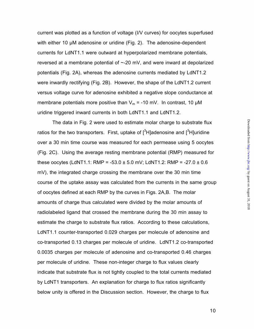
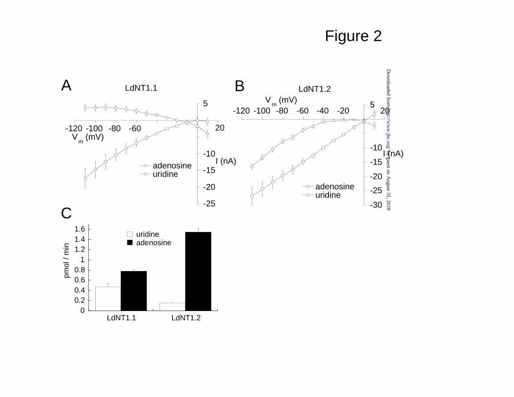
current was plotted as a function of voltage (I/V curves) for oocytes superfused
with either 10 µM adenosine or uridine (Fig. 2). The adenosine-dependent
currents for LdNT1.1 were outward at hyperpolarized membrane potentials,
reversed at a membrane potential of ~-20 mV, and were inward at depolarized
potentials (Fig. 2A), whereas the adenosine currents mediated by LdNT1.2
were inwardly rectifying (Fig. 2B). However, the shape of the LdNT1.2 current
versus voltage curve for adenosine exhibited a negative slope conductance at
membrane potentials more positive than Vm = -10 mV. In contrast, 10 µM
uridine triggered inward currents in both LdNT1.1 and LdNT1.2.
The data in Fig. 2 were used to estimate molar charge to substrate flux
ratios for the two transporters. First, uptake of [3H]adenosine and [3H]uridine
over a 30 min time course was measured for each permease using 5 oocytes
(Fig. 2C). Using the average resting membrane potential (RMP) measured for
these oocytes (LdNT1.1: RMP = -53.0 ± 5.0 mV; LdNT1.2: RMP = -27.0 ± 0.6
mV), the integrated charge crossing the membrane over the 30 min time
course of the uptake assay was calculated from the currents in the same group
of oocytes defined at each RMP by the curves in Figs. 2A,B. The molar
amounts of charge thus calculated were divided by the molar amounts of
radiolabeled ligand that crossed the membrane during the 30 min assay to
estimate the charge to substrate flux ratios. According to these calculations,
LdNT1.1 counter-transported 0.029 charges per molecule of adenosine and
co-transported 0.13 charges per molecule of uridine. LdNT1.2 co-transported
0.0035 charges per molecule of adenosine and co-transported 0.46 charges
per molecule of uridine. These non-integer charge to flux values clearly
indicate that substrate flux is not tightly coupled to the total currents mediated
by LdNT1 transporters. An explanation for charge to flux ratios significantly
below unity is offered in the Discussion section. However, the charge to flux
by guest on August 31, 2018
http://ww
w.jbc.org/
Dow
nloaded from

11
ratio of 0.46 for the uridine-induced currents in LdNT1.2 suggests that there is
a substantial coupling of transmembrane charge movement to substrate
import.
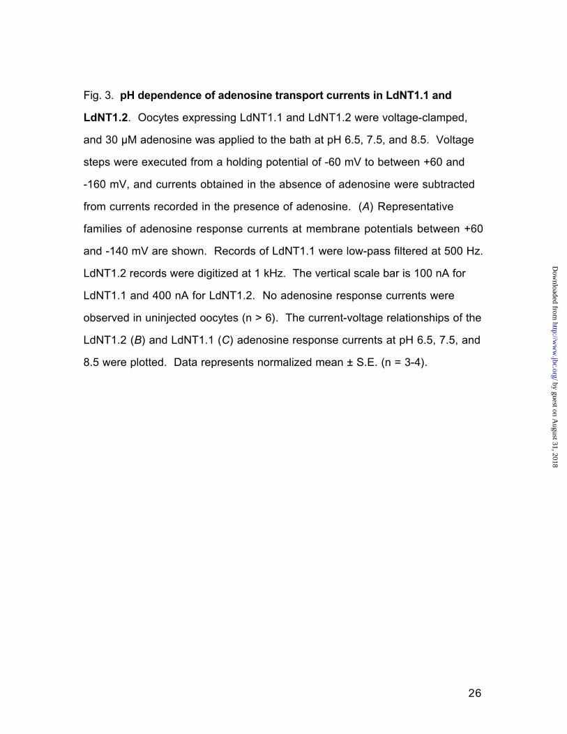
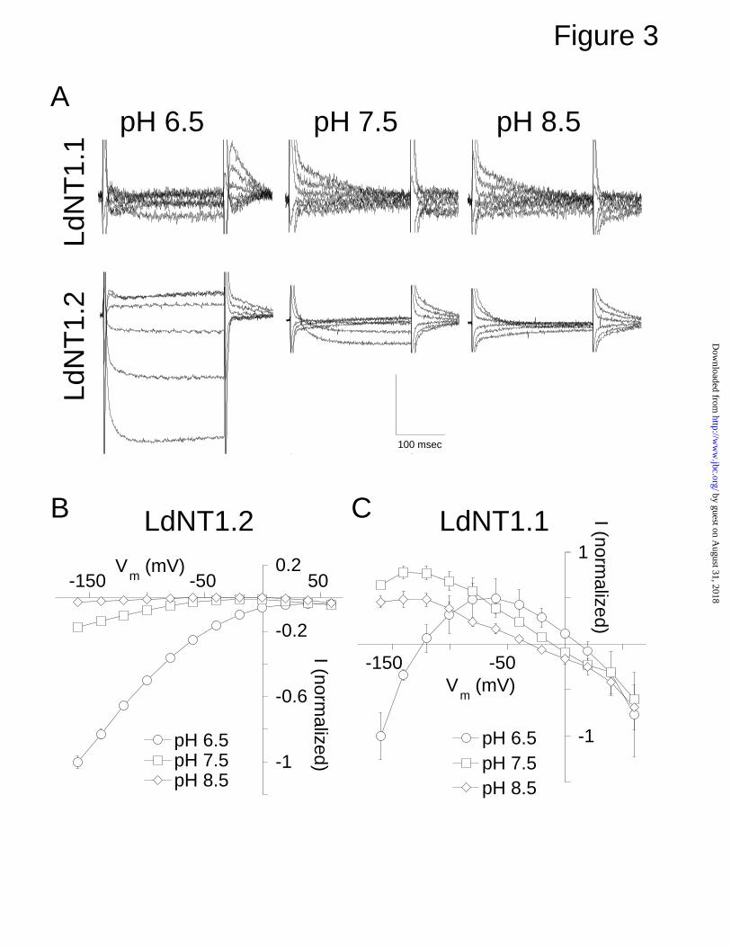
pH Dependence of Adenosine-Induced Steady State Currents—To
further investigate the origin of the currents observed in Figs. 1 and 2, and to
determine whether they might represent proton symport, the pH dependence of
the steady-state adenosine-response currents was examined for LdNT1.1 and
LdNT1.2. Oocytes expressing each permease were exposed to voltage jumps
from a holding potential of –60 mV to a range of command potentials. Currents
elicited in the absence of adenosine were then subtracted from those recorded
in the presence of 30 µM adenosine, resulting in the current versus time traces
shown in Fig. 3A. At pH 6.5, 7.5, and 8.5 LdNT1.1 and LdNT1.2-expressing
oocytes revealed an initial transient capacitive or pre-steady state current that
then decayed to a steady state value. No currents were observed in uninjected
oocytes (data not shown, n>5).
The steady-state adenosine-response currents for LdNT1.2 were plotted
as a current-voltage curve (Fig. 3B). Adenosine induced an inward rectifying
current at pH 6.5, which is consistent with an inward flux of protons that is
coupled to import of adenosine. Notably, the currents at pH 7.5 and 8.5 were
considerably smaller than at pH 6.5, again consistent with a coupled flux of
protons that experiences a smaller driving force at increased external pH
values. There were some differences however in the shapes of the curves,
with negative slope conductances at positive potentials at both pH 7.5 and 8.5.
In contrast, the shapes of the steady state current-voltage curves for
LdNT1.1 (Fig. 3C) were more complex than those for LdNT1.2. One notable
difference between the two permeases is that the adenosine-elicited currents
for LdNT1.1 generally exhibited negative slope conductances and reversal
by guest on August 31, 2018
http://ww
w.jbc.org/
Dow
nloaded from

12
potentials that were shifted approximately 25 mV per unit change in external
pH, in contrast to LdNT1.2. These data suggest that a primary action of
adenosine in LdNT1.1 is to block a membrane conductance that is carried in
part by protons. It is notable that in contrast to the results at higher pH values,
the outward current induced by LdNT1.1 at pH 6.5 decreased again at
potentials more negative that –60 mV and again became inward at potentials
more negative than ~-120 mV, resulting in an inverted U-shaped curve (Fig.
3C). These results suggest that at low pH, LdNT1.1, like LdNT1.2, mediates
an adenosine-coupled proton symport process that gives rise to an inward
current. This substrate-coupled import of protons would be of the largest
magnitude at pH 6.5 and at the most polarized membrane potentials, as
demonstrated in Fig. 3B for the corresponding current for LdNT1.2. The
inverted U-shaped curve for LdNT1.1 at pH 6.5 could thus be explained by the
additive combination of the blockage of a proton leak current by adenosine,
which predominates above a membrane potential of –60 mV, and an
adenosine-coupled proton symport current that becomes apparent below a
membrane potential of –60 mV.
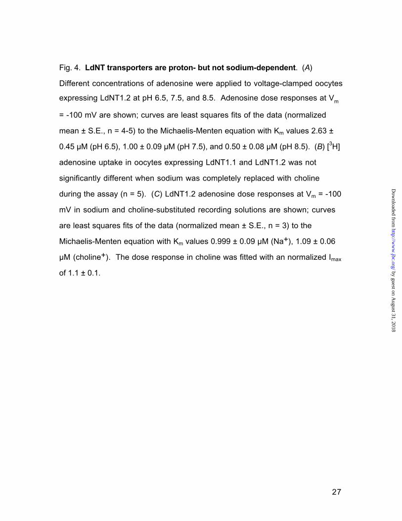
To further define the proton-dependence of LdNT1.2 steady state
currents, the adenosine concentration dependence of these response currents
was examined at Vm = -100 mV and at pH 6.5, 7.5, and 8.5 (Fig 4A). The
magnitudes of the currents elicited by adenosine concentrations ranging from
0.1 – 100 µM adenosine were greatest at pH 6.5 and lowest at pH 8.5. The
transporter affinity decreased and the maximal transport rate increased with
increasing proton concentrations. Thus from the currents observed, a Km value
of 1.00 ± 0.09 µM could be calculated for LdNT1.2 at pH 7.5. At pH 6.5, the
transporter exhibited a Km value of 2.63 ± 0.45 µM and an Imax 600 ± 26%
greater than the Imax at pH 7.5. At pH 8.5, the Km was 0.50 ± 0.08 µM, and the
by guest on August 31, 2018
http://ww
w.jbc.org/
Dow
nloaded from
13
Imax was 5.2 ± 0.5% of its value at pH 7.5. In summary, the increase of
adenosine-elicited currents in LdNT1.2 at lower pH values further supports
proton symport as a mechanism for transport.
Lack of sodium dependence of adenosine uptake or adenosine-induced
currents—The observation of electrogenic transport for these protozoan
nucleoside permeases contrasts with other members of the ENT family, which
are equilibrative permeases and not electrogenic (7). In contrast another family
of nucleoside transporters, the CNTs, are sodium-dependent electrogenic
active transporters (15). Consequently, whether LdNT1.1- or LdNT1.2-mediated
adenosine uptake might be sodium dependent was also determined.
Replacement of sodium in the bath with equimolar choline did not significantly
affect either LdNT1.1- or LdNT1.2-mediated adenosine uptake (Fig. 4B).
Furthermore, the LdNT1.2 transporter exhibited no significant difference in
either the affinity or maximal current when sodium was replaced with choline
(Fig. 4C). Indeed, replacement of sodium with choline did not affect
adenosine-induced currents for LdNT1.2 at any adenosine concentration,
indicating that LdNT1.2 is not a sodium symporter.
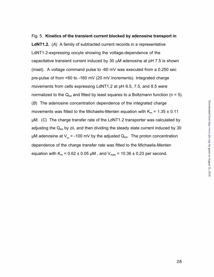
Capacitive gating currents – As noted above (Fig. 3), application of
voltage jumps to oocytes expressing LdNT1.1 or LdNT1.2 induced adenosine-
dependent pre-steady state or transient currents that decayed with exponential
time constants of <100 msec. For some transporters, such transient currents
have been linked to binding of a co-transported ion to the permease (16). To
determine whether the adenosine-induced transient currents could be
explained by binding of protons to the permeases, and could thus be useful for
probing interaction of protons with the transporters, the kinetic properties of
these pre-steady state currents were examined at several proton
concentrations by time integration of the currents following the return of the
by guest on August 31, 2018
http://ww
w.jbc.org/
Dow
nloaded from

14
clamped membrane to the holding potential from a voltage step (Fig. 5). The
transient charge movement obeyed similar voltage-dependencies at pH 6.5,
7.5, and 8.5 (Fig. 5A, n = 4). The total charge movement induced by adenosine
was similar at these pHs (Qtot, pH 6.5 = 35033 ± 3980 pC; Qtot, pH 7.5 = 44771
± 2883 pC; Qtot, pH 8.5 = 39179 ± 1771 pC). The midpoint of the integrated
current voltage-dependence is a measurement of the transporter's affinity for
the translocated charge (16). The midpoint of the integrated transient current
mediated by 30 µM adenosine was also found to be essentially independent of
pH (V0.5, pH 6.5 = 3.31 ± 3.23 mV; V0.5, pH 7.5 = -9.53 ± 3.33 mV; V0.5, pH 8.5 = -
4.93 ± 2.53 mV). The transient current elicited by 30 µM adenosine also
exhibited a similar slope to its voltage-dependence at the three pHs examined,
which implies that the translocated charge experiences the same fraction of the
membrane electric field under these conditions (slope, pH 6.5 = 60.90 ± 2.70
mV-1; slope, pH 7.5 = 81.88 ± 3.59 mV-1; slope, pH 8.5 = 74.40 ± 4.00 mV-1)
(17). The insensitivity of midpoint and slope values to changes in pH implies
that the capacitive transient current is not due to a proton binding event. Rather,
it may be a charge-moving conformational change in the transport protein.
To verify that the transient current elicited by adenosine was associated
with transporter function, the adenosine concentration dependence of the
integrated capacitive current was examined (Fig 5B). The magnitude of the
capacitive current was dependent upon adenosine concentration in a dose-
dependent manner, and the apparent affinity of the transporter for adenosine
(Km = 1.35 ± 0.11 µM) when measured in this way was similar to that derived
from analysis of the steady state currents (Km = 1.00 ± 0.09 µM, Fig. 4). These
results imply that the pre-steady state currents reflect transporter function.
It is possible to estimate the charge transfer rate of the transporter by
dividing the steady state current through the transporter (I, pC/sec) by the total
by guest on August 31, 2018
http://ww
w.jbc.org/
Dow
nloaded from
15
number of elementary charges blocked by adenosine (Q / zδ, pC) and
assuming a charge to flux stoichiometry of 1:1 (16). The charge transfer rate
was calculated at pHs between 5.5 and 8.5 (Fig 5C). Performing this
calculation at pH 7.5 revealed that the transporter moves 0.7 ± 0.05 times per
second through the membrane at -100 mV. The proton dose dependence of
the transporter's charge transfer rate was fitted with a proton affinity of 0.62 ±
0.05 µM , and the maximal cycling rate was found to be 10.4 ± 0.2 per sec.
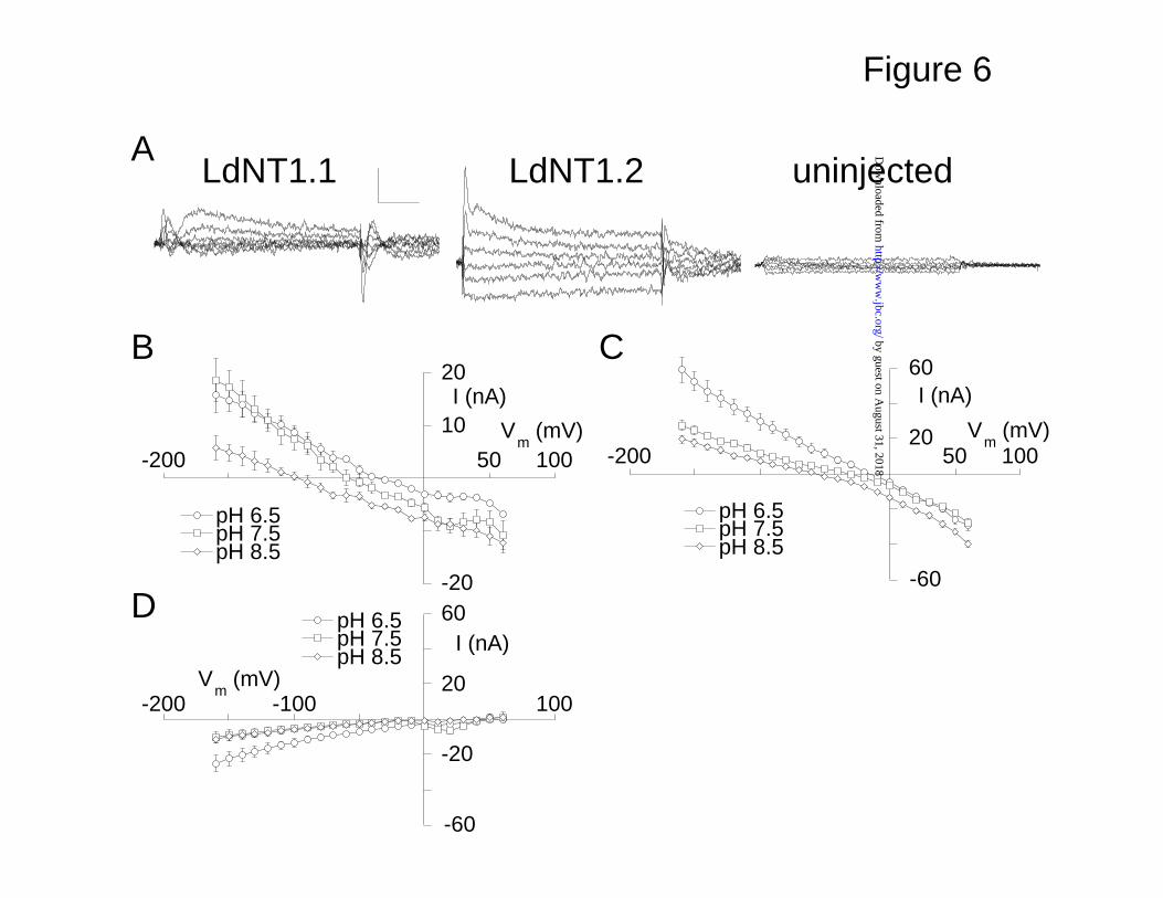
DPA blocks a tonic proton leak through LdNT1 transporters- DPA is a
high-affinity antagonist of mammalian adenosine transporters (7) but does not
significantly inhibit nucleoside transporters from parasitic protozoa (1). Despite
its failure to significantly inhibit the Leishmania permeases, DPA activates a
current in LdNT-expressing oocytes (Fig. 1 A,B). To further characterize the
effect of DPA on these transporters, the transmembrane currents recorded
prior to DPA application were subtracted from currents recorded during DPA
application revealing a current in LdNT1-expressing cells that was not
observed in uninjected oocytes (Fig. 6A). The DPA-induced currents in
LdNT1.1 and LdNT1.2 were linear and reversible, and they were outward at
more negative membrane potentials (Fig. 6B and C). The LdNT1-mediated
currents saturated with respect to the DPA concentration (Km = 35 ± 10 µM in
LdNT1.2 at Vm = -100 mV; data not shown, n = 4). In contrast, the smaller
current that was present in uninjected oocytes was inward and did not reverse
(Fig. 6D),
Similar to the behavior of the transporter current blocked by adenosine,
the reversal potential of the DPA-elicted currents was observed to shift to more
positive values as the extracellular concentration of protons was increased,
suggesting that this current is carried substantially by protons (Fig. 6B and C).
by guest on August 31, 2018
http://ww
w.jbc.org/
Dow
nloaded from
16
A quantitative analysis of the reversal potential pH dependence is precluded by
the DPA-induced current in uninjected oocytes (Fig. 6D).
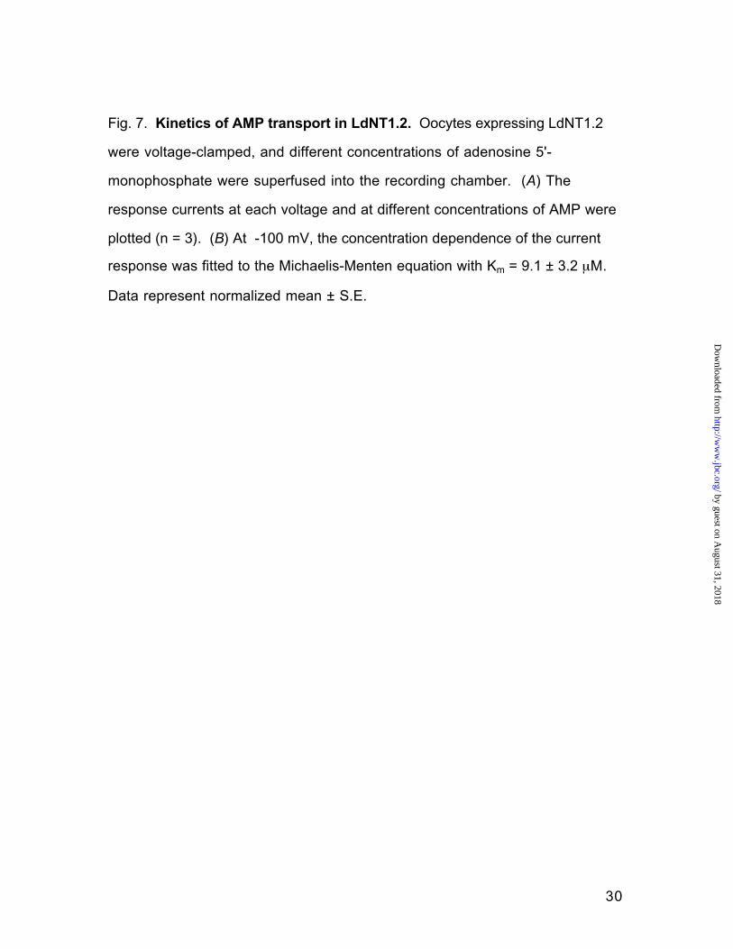
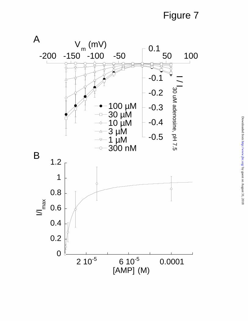
AMP induces currents for LdNT1.1 and LdNT1.2—The observation that
AMP was able to elicit currents in oocytes microinjected with either LdNT1.1 or
LdNT1.2 cRNA (Fig. 1) was surprising in view of the negative charge of the
nucleotide and the fact that nucleotides typically are not taken up by intact
eukaryotic cells. To further investigate this phenomenon, we measured
current-voltage curves for AMP-induced currents for LdNT1.2 (Fig. 7), the
permease that elicited inward directed currents with this ligand. For LdNT1.2
the maximal AMP-induced current at Vm = -100 mV was 20.1% of the maximal
current induced by a saturating (30 µM) concentration of adenosine in the same
oocytes (Fig. 7A). The AMP response current in LdNT1.2 was saturable with
respect to AMP, and its concentration-dependence exhibited an apparent affinity
of 9 ± 3 µM (Fig. 7B). The shapes of the AMP and adenosine response current-
voltage relationships are qualitatively similar at pH 7.5 (compare Fig. 7A to Fig.
3B). It does not appear that the AMP-induced current could be ascribed to
adenosine contamination because the purity of the AMP was verified by high
performance liquid and thin layer chromatography (data not shown).
Furthermore, when [3H]AMP was applied to LdNT1.2-expressing oocytes, the
radiolabel was taken up by the oocytes, but no significant amount of
[3H]adenosine was liberated into the supernatant (unpublished data, S.M.
Landfear). Hence, the AMP-induced currents cannot be ascribed to hydrolysis
of AMP that liberates free adenosine into the medium followed by uptake of this
liberated adenosine by the permease. The origin of these AMP-induced
currents is currently under investigation.
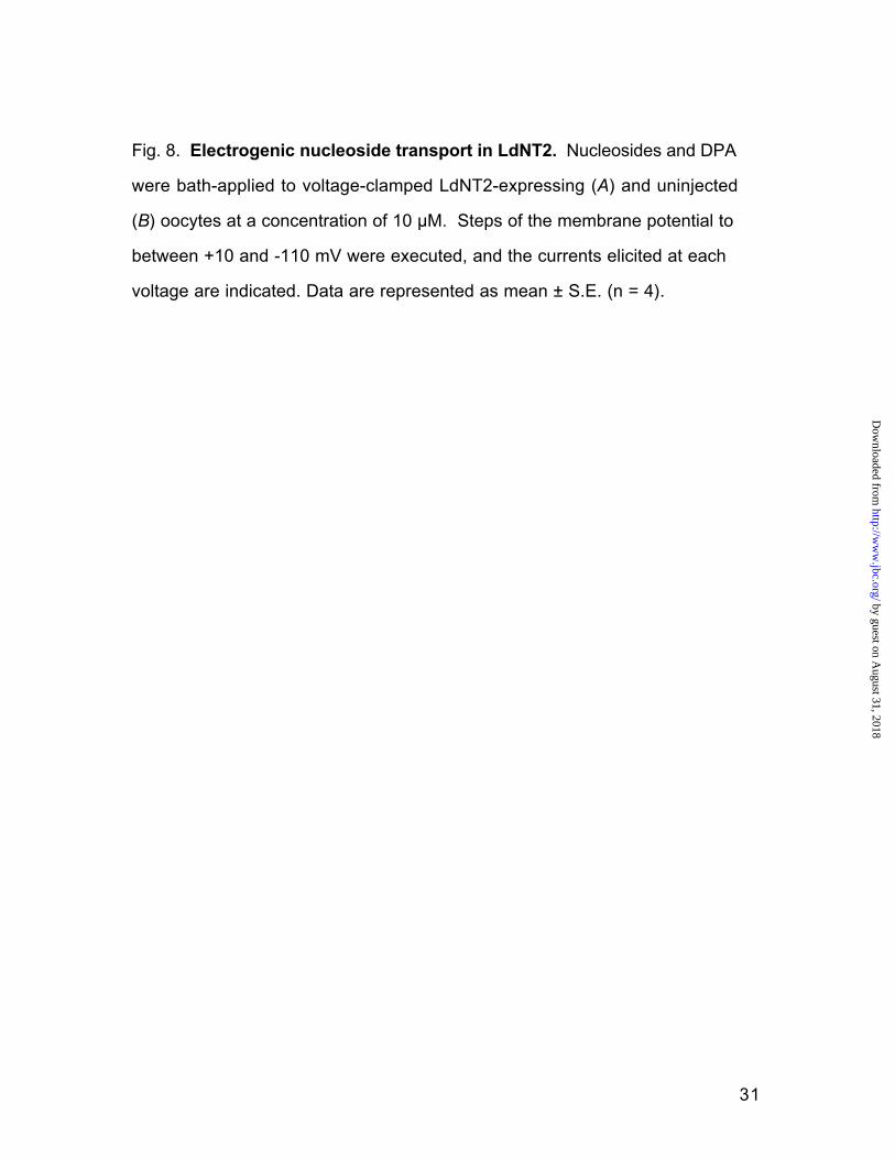
The LdNT2 inosine-guanosine permease is also electrogenic—The
other nucleoside transporter of L. donovani, LdNT2, mediates the uptake of
by guest on August 31, 2018
http://ww
w.jbc.org/
Dow
nloaded from
17
inosine and guanosine, is also a member of the ENT family, and exhibits 33%
sequence identity to LdNT1.1 (5). Oocytes injected with LdNT2 cRNA were also
shown to elicit inward response currents to the application of ligands. Inosine
and guanosine, but not adenosine or thymidine, mediated inward rectifying
currents in a voltage-dependent fashion (Fig. 8A). LdNT2 also mediated a
reversible DPA-induced outward current at negative membrane potentials,
similar to that observed for LdNT1.1 and LdNT1.2. Neither inosine, guanosine,
adenosine, nor thymidine induced a current in uninjected oocytes, although 10
µM DPA elicited a small inward current (Fig. 8B). The induction of inward
rectifying currents specifically by substrates of the LdNT2 permease implies
that this transporter also couples the symport of protons to nucleosides, much
as demonstrated above for the LdNT1.2 carrier. The outward directed currents
elicited by DPA suggest that this compound blocks a constitutive leak current in
LdNT2, in a manner similar to that observed for LdNT1.1 and LdNT1.2 (Fig. 6).
by guest on August 31, 2018
http://ww
w.jbc.org/
Dow
nloaded from

18
DISCUSSION
We have shown that adenosine/pyrimidine nucleoside transport through
the LdNT1 transporters is associated with the activation of trans-membrane
currents. The magnitudes of the currents mediated by LdNT1.2 and the
substrate fluxes mediated by both LdNT1.1 and LdNT1.2 were dependent on
the proton, but not the sodium, gradient, strongly suggesting that these
proteins are proton/nucleoside symporters. Unlike the Leishmania
proton/myo-inositol symporter MIT, which transports myo-inositol with a charge
to flux ratio of 1 (13), LdNT1 did not mediate substrate flux that was tightly
coupled to total charge translocation. Although LdNT1.1 and LdNT1.2 are very
similar in sequence, they exhibit striking differences with respect to their
electrogenic properties, most notably that adenosine strongly blocks a leak
current in LdNT1.1, whereas adenosine induces an inward proton current in
LdNT1.2. It is likely that the adenosine blockage of the leak current in LdNT1.1
obscures an inward directed adenosine-coupled proton current similar to that
observed in LdNT1.2. In fact, the inward directed current observed for LdNT1.1
at pH 6.5 below a transmembrane voltage of –120 mV (Fig. 3C) is likely to be
such a transport-coupled current that overcomes the block of the leak current at
this lower pH value and more negative membrane potential. Thus the
essential difference between the two permeases may be that adenosine
effects a more robust block of the leak current in LdNT1.1 compared to
LdNT1.2.
We have also shown that LdNT1 transporters mediate a constitutive
proton conductance that is blocked by the compound DPA. Unlike the
mammalian ENTs, the Leishmania nucleoside transporters are not
significantly inhibited by DPA at concentrations that obliterate transport by
by guest on August 31, 2018
http://ww
w.jbc.org/
Dow
nloaded from
19
mammalian ENTs (2), and thus the precise nature of the interaction between
this compound and the LdNTs is not clear. Nonetheless the ability of DPA,
which is very lipophilic and known to affect the entry of many structurally
unrelated compounds into mammalian cells (18,19), to block the constitutive
proton leak is instructive, as it likely reflects a similar block of this leak current
that is induced by interaction of substrates with the LdNT1 permeases.
We hypothesize that part of the reason charge translocation appears to
be loosely coupled to substrate flux in LdNT1.1 and LdNT1.2 is that adenosine
transport blocks the constitutive proton conductance in a similar manner to
DPA. By summing a linear current with a pH-dependent reversal potential such
as the one observed upon application of DPA (Fig. 6B and C) and a pH-
dependent inwardly rectifying transport current such as that observed after
application of adenosine to LdNT1.2 at pH 6.5 (Fig. 3B), it is possible to
generate current-voltage curves with shapes corresponding to the adenosine
response current-voltage curves. This hypothesis is supported by the
observation that, while lower pH increases the relative contribution of inward
adenosine-response current at negative potentials in LdNT1.1, it also shifts the
reversal potential of the current-voltage curve to more positive potentials (Fig.
3C), as would be expected for a current that represents blockage of a proton
leak. The presence of this blocked proton conductance would cause our
calculations of the charge to flux ratio to be significant underestimates of the
actual value, especially when the measurements are made at the resting
potentials of the oocytes (between –53 and – 27 mV) rather than at highly
polarized membrane potentials that favor the influx of protons that are coupled
to adenosine transport. In summary, the blockage of a constitutive proton leak
by substrates complicates the estimation of charge to substrate flux ratios for
these protozoan nucleoside transporters. However, this constitutive leak is
by guest on August 31, 2018
http://ww
w.jbc.org/
Dow
nloaded from
20
blocked less efficiently in LdNT1.2 than in LdNT1.1, and uridine blocks this leak
less effectively in both transporters compared to adenosine (Fig. 2).
Consequently, the charge to flux ratio calculated for uridine in LdNT1.2 (0.46
charges per molecule) is probably the estimate that most closely reflects the
value for proton symport coupled to a substrate. Hence, substrates appear to
mediate the import of at least ~0.5 charge units per molecule.
LdNT1.1 and LdNT1.2 differ by only 6 amino acid residues in sequence,
and three of these polymorphisms are located on the extreme carboxy termini
of the proteins. Despite the high degree of identity, these transporters exhibit
different affinities for the substrates adenosine and uridine (4) and mediate
different currents during adenosine and AMP transport. The COOH-terminal
residues are the uncharged ATY in LdNT1.1 compared to the highly charged
ERH in LdNT1.2. While these sequences could be important in determining
the distinct electrogenic properties of the proteins, perhaps a more likely
residue for the observed differences is P43 in LdNT1.1. This proline is a
serine in LdNT1.2, and it is the only polymorphism that lies within a predicted
transmembrane domain (TMD1). The other polymorphisms include M107I
(between TMD2 and TMD3) and T160A (between TMD4 and TMD5), which are
located within predicted hydrophilic loops of the permeases. Mutational
analysis outside the scope of this manuscript will be needed to evaluate the
role of each polymorphism on the kinetic and electrophysiological differences
between LdNT1.1 and LdNT1.2.
Our analysis of the transporters responsible for nucleoside transport in
L. donovani, LdNT1.1, LdNT1.2, and LdNT2, has demonstrated that these
proteins couple the large proton electrochemical gradient available to the
parasites to nucleoside uptake. Purine uptake is vital to the survival of the
parasites, as they are incapable of purine biosynthesis, and the ability of the
by guest on August 31, 2018
http://ww
w.jbc.org/
Dow
nloaded from

21
parasite to concentrate essential purines from the extracellular medium may
thus provide an evolutionary advantage by promoting its survival within both the
insect vector and/or the vertebrate host. The parasite nucleoside transporters
studied here exhibit approximately 30% identity at the amino acid sequence
level with members of the mammalian equilibrative nucleoside transporter
family. Since LdNT1.1, LdNT1.2 and LdNT2 are apparently proton symporters,
it now appears that some members of the ENT family are concentrative
permeases. This conclusion is also consistent with previous studies
performed on transport of nucleosides by intact procyclic forms of the related
protozoan Trypanosoma brucei, in which the dependence of nucleoside
transport upon the proton electrochemical gradient across the parasite plasma
membrane strongly suggested proton symport as a mechanism of active
transport (20). It is possible that relatively few changes in amino acid
sequence determine whether a nucleoside transporter of this family functions
as a concentrative or equilibrative permease. However, the extensive
differences in sequence between equilibrative and concentrative members of
the ENT family make it difficult to predict which residues may be required for
proton symport and suggest that extensive structure-function studies will likely
be required to elucidate determinants of active transport.
The ability of the nucleotide AMP to induce currents in LdNT1.1 and
LdNT1.2 that are very similar to those induced by adenosine is intriguing, as
these anionic metabolites are not thought to be transported across the plasma
membranes of eukaryotic cells. We are currently pursuing further studies to
determine the nature of the interaction of AMP with the LdNT1.1 and LdNT1.2
permeases.
by guest on August 31, 2018
http://ww
w.jbc.org/
Dow
nloaded from
22
REFERERENCES
1. Carter, N. S., Rager, N., and Ullman, B. (2003) in Molecular and Medical
Parasitology (Komuniecki, R., ed), pp. 197-223, Academic Press,
London
2. Aronow, B., Kaur, K., McCartan, K., and Ullman, B. (1987) Mol. Biochem.
Parasitol. 22, 29-37
3. Iovannisci, D. M., Kaur, K., Young, L., and Ullman, B. (1984) Mol. Cell.
Biol. 4, 1013-1019
4. Vasudevan, G., Carter, N. S., Drew, M. E., Beverley, S. M., Sanchez, M. A.,
Seyfang, A., Ullman, B., and Landfear, S. M. (1998) Proc. Natl. Acad. Sci.
U.S.A. 95, 9873-9878
5. Carter, N. S., Drew, M. E., Sanchez, M., Vasudevan, G., Landfear, S. M.,
and Ullman, B. (2000) J. Biol. Chem. 275, 20935-20941
6. Vasudevan, G., Ullman, B., and Landfear, S. M. (2001) Proc. Natl. Acad.
Sci. U. S. A. 98, 6092-6097
7. Hyde, R. J., Cass, C. E., Young, J. D., and Baldwin, S. A. (2001) Mol.
Membrane Biol. 18, 53-63
8. Baldwin, S. A., Mackey, J. R., Cass, C. E., and Young, J. D. (1999) Mol.
Med. Today 5, 216-224
9. Zilberstein, D., Philosoph, H., and Gepstein, A. (1989) Mol. Biochem.
Parasitol. 36, 109-118
by guest on August 31, 2018
http://ww
w.jbc.org/
Dow
nloaded from

23
10. Vieira, L., Slotik, I., and Cabantchik, Z. I. (1995) J. Biol. Chem. 270, 5299-
5304
11. Zilberstein, D. (1993) Adv. Parasitol. 32, 261-291
12. Drew, M. E., Langford, C. K., Klamo, E. M., Russell, D. G., Kavanaugh, M.
P., and Landfear, S. M. (1995) Mol. Cell. Biol. 15, 5508-5515
13. Klamo, E. M., Drew, M. E., Landfear, S. M., and Kavanaugh, M. P. (1996)
J. Biol. Chem. 271, 14937-14943
14. Arriza, J. L., Kavanaugh, M. P., Fairman, W. A., Wu, Y.-N., Murdoch, G. H.,
North, R. A., and Amara, S. G. (1993) J. Biol. Chem. 268, 15329-15332
15. Yao, S. Y., Ng, A. M., Loewen, S. K., Cass, C. E., Baldwin, S. A., and
Young, J. D. (2002) Am J Physiol Cell Physiol 283, C155-168
16. Wadiche, J. I., Arriza, J. L., Amara, S. G., and Kavanaugh, M. P. (1995)
Neuron 14, 1019-1027
17. Woodhull, A. M. (1973) J. Gen. Physiol. 61, 687-708
18. Kessel, D., and Dodd, D. C. (1972) Biochim. Bipphys. Acta 288, 190-194
19. Graff, J. C., Wohlhueter, R. M., and Plagemann, P. G. (1977) J. Biol.
Chem. 252, 4185-4190
20. de Koning, H. P., Watson, C. J., and Jarvis, S. M. (1998) J. Biol. Chem.
273, 9486-9494
by guest on August 31, 2018
http://ww
w.jbc.org/
Dow
nloaded from

24
Fig. 1. Substrate selectivity of the LdNT1 transport system. Oocytes were
voltage-clamped at Vm = -100 mV, and substrates (10 µM) of the LdNT1
transport system or other compounds were applied to the bath for the duration
indicated by the bar. In each case, current recordings from one oocyte are
shown, but the experiment was repeated with a total of n oocytes. Currents
from oocytes expressing LdNT1.2 (A, n > 6) and LdNT1.1 (B, n > 6) or
uninjected oocytes (C, n=5) are shown. Adenine, inosine, guanosine, ADP,
and ATP did not generate a current (A) in oocytes injected with LdNT1.1 or
LdNT1.2 cRNA (data not shown, n = 4).
by guest on August 31, 2018
http://ww
w.jbc.org/
Dow
nloaded from

25
Fig. 2. LdNT1.1 and LdNT1.2 exhibit different electrogenic properties.
Currents induced by adenosine and uridine were measured for LdNT1.1 (A,
n=3) and LdNT1.2 (B, n=3). Uptake of [3H] adenosine and [3H] uridine was also
measured over a 30 min time course for LdNT1.1 and LdNT1.2 (C, n = 5). The
uptake observed in LdNT1.1- and LdNT1.2-injected oocytes was between 21-
and 177-fold over the background uptake in uninjected oocytes. Data shown
are LdNT-specific uptake values.
by guest on August 31, 2018
http://ww
w.jbc.org/
Dow
nloaded from
26
Fig. 3. pH dependence of adenosine transport currents in LdNT1.1 and
LdNT1.2. Oocytes expressing LdNT1.1 and LdNT1.2 were voltage-clamped,
and 30 µM adenosine was applied to the bath at pH 6.5, 7.5, and 8.5. Voltage
steps were executed from a holding potential of -60 mV to between +60 and
-160 mV, and currents obtained in the absence of adenosine were subtracted
from currents recorded in the presence of adenosine. (A) Representative
families of adenosine response currents at membrane potentials between +60
and -140 mV are shown. Records of LdNT1.1 were low-pass filtered at 500 Hz.
LdNT1.2 records were digitized at 1 kHz. The vertical scale bar is 100 nA for
LdNT1.1 and 400 nA for LdNT1.2. No adenosine response currents were
observed in uninjected oocytes (n > 6). The current-voltage relationships of the
LdNT1.2 (B) and LdNT1.1 (C) adenosine response currents at pH 6.5, 7.5, and
8.5 were plotted. Data represents normalized mean ± S.E. (n = 3-4).
by guest on August 31, 2018
http://ww
w.jbc.org/
Dow
nloaded from
27
Fig. 4. LdNT transporters are proton- but not sodium-dependent. (A)
Different concentrations of adenosine were applied to voltage-clamped oocytes
expressing LdNT1.2 at pH 6.5, 7.5, and 8.5. Adenosine dose responses at Vm
= -100 mV are shown; curves are least squares fits of the data (normalized
mean ± S.E., n = 4-5) to the Michaelis-Menten equation with Km values 2.63 ±
0.45 µM (pH 6.5), 1.00 ± 0.09 µM (pH 7.5), and 0.50 ± 0.08 µM (pH 8.5). (B) [3H]
adenosine uptake in oocytes expressing LdNT1.1 and LdNT1.2 was not
significantly different when sodium was completely replaced with choline
during the assay (n = 5). (C) LdNT1.2 adenosine dose responses at Vm = -100
mV in sodium and choline-substituted recording solutions are shown; curves
are least squares fits of the data (normalized mean ± S.E., n = 3) to the
Michaelis-Menten equation with Km values 0.999 ± 0.09 µM (Na+), 1.09 ± 0.06
µM (choline+). The dose response in choline was fitted with an normalized Imax
of 1.1 ± 0.1.
by guest on August 31, 2018
http://ww
w.jbc.org/
Dow
nloaded from
28
Fig. 5. Kinetics of the transient current blocked by adenosine transport in
LdNT1.2. (A) A family of subtracted current records in a representative
LdNT1.2-expressing oocyte showing the voltage-dependence of the
capacitative transient current induced by 30 µM adenosine at pH 7.5 is shown
(inset). A voltage command pulse to -60 mV was executed from a 0.250 sec
pre-pulse of from +60 to -160 mV (20 mV increments). Integrated charge
movements from cells expressing LdNT1.2 at pH 6.5, 7.5, and 8.5 were
normalized to the Qtot and fitted by least squares to a Boltzmann function (n = 5).
(B) The adenosine concentration dependence of the integrated charge
movements was fitted to the Michaelis-Menten equation with Km = 1.35 ± 0.11
µM. (C) The charge transfer rate of the LdNT1.2 transporter was calculated by
adjusting the Qtot by zδ, and then dividing the steady state current induced by 30
µM adenosine at Vm = -100 mV by the adjusted Qtot. The proton concentration
dependence of the charge transfer rate was fitted to the Michaelis-Menten
equation with Km = 0.62 ± 0.05 µM , and Vmax = 10.36 ± 0.23 per second.
by guest on August 31, 2018
http://ww
w.jbc.org/
Dow
nloaded from

29
Fig. 6. DPA blocks a proton leak in LdNT1.1 and LdNT1.2. 30 µM DPA was
applied to oocytes expressing LdNT1.1 or LdNT1.2, and uninjected oocytes at
pH 6.5, 7.5, and 8.5. (A) Representative current families of the DPA response
current at pH 7.5 are shown. Cells were voltage-clamped at -50 mV, and the
membrane potential was stepped to between +60 and -140 mV. Data are
subtractions of control currents from currents recorded during dipyridamole
application. (B) Current-voltage relations for the LdNT1.1-mediated current
induced by dipyridamole at pH 6.5, 7.5, and 8.5 are shown. The reversal
potential (Vrev) of the response current is pH-dependent (n = 3). Vrev = -35 ± 6
mV (pH 6.5), -55 ± 8 mV (pH 7.5), -96 ± 7 mV (pH 8.5). (C) Current-voltage
relations for LdTN1.2-mediated currents (n = 3). Vrev = -16 ± 3 mV (pH 6.5), -34
± 3 mV (pH 7.5), -57 ± 3 mV (pH 8.5). (D) DPA response currents in uninjected
oocytes (n = 3).
by guest on August 31, 2018
http://ww
w.jbc.org/
Dow
nloaded from
30
Fig. 7. Kinetics of AMP transport in LdNT1.2. Oocytes expressing LdNT1.2
were voltage-clamped, and different concentrations of adenosine 5'-
monophosphate were superfused into the recording chamber. (A) The
response currents at each voltage and at different concentrations of AMP were
plotted (n = 3). (B) At -100 mV, the concentration dependence of the current
response was fitted to the Michaelis-Menten equation with Km = 9.1 ± 3.2 µM.
Data represent normalized mean ± S.E.
by guest on August 31, 2018
http://ww
w.jbc.org/
Dow
nloaded from
31
Fig. 8. Electrogenic nucleoside transport in LdNT2. Nucleosides and DPA
were bath-applied to voltage-clamped LdNT2-expressing (A) and uninjected
(B) oocytes at a concentration of 10 µM. Steps of the membrane potential to
between +10 and -110 mV were executed, and the currents elicited at each
voltage are indicated. Data are represented as mean ± S.E. (n = 4).
by guest on August 31, 2018
http://ww
w.jbc.org/
Dow
nloaded from

3 min
10 nA
adenosine thymidine uridine cytidine dipyridamole AMP
adenosine AMP adenine ATP dipyridamoleADP
20 nA
70 sec
adenosine uridine thymidine inosine guanosinecytidine
adenosine uridine thymidine AMPdipyridamolecytidine
A
B C
Figure 1
by guest on August 31, 2018
http://ww
w.jbc.org/
Dow
nloaded from
-25
-20
-15
-10
5
-120 -100 -80 -60 20
adenosineuridine
I (nA)
Vm
(mV)
LdNT1.1
-30
-25
-20
-15
-10
5-120 -100 -80 -60 -40 -20 20
LdNT1.2
adenosineuridine
I (nA)
Vm
(mV)
00.20.40.60.8
11.21.41.6
LdNT1.1 LdNT1.2
uridineadenosine
pmol
/ m
in
A B
C
Figure 2
by guest on August 31, 2018
http://ww
w.jbc.org/
Dow
nloaded from
100 msec
pH 6.5 pH 7.5 pH 8.5
-1
1
-150 -50
pH 6.5pH 7.5pH 8.5
I (normalized)
Vm (mV)
-1
-0.6
-0.2
0.2-150 -50 50
pH 6.5pH 7.5pH 8.5
I (normalized)
Vm
(mV)
A
B C
LdN
T1.
1Ld
NT
1.2
LdNT1.1LdNT1.2
Figure 3 by guest on A
ugust 31, 2018http://w
ww
.jbc.org/D
ownloaded from

0
1
2
3
4
5
6
7
10-7 10-6 10-5 10-4
pH 6.5pH 7.5pH 8.5
I/I30
µM
, pH
7.5
[adenosine] (M)0
0.5
1
1.5
2
2.5
LdNT1.1 LdNT1.2
Na+
choline+
pmol
/ m
in
0
0.2
0.4
0.6
0.8
1
1.2
0.0001 0.001 0.01 0.1
Na+
choline+I/Im
ax, N
a
[adenosine] (M)
A B
C
Figure 4
by guest on August 31, 2018
http://ww
w.jbc.org/
Dow
nloaded from

0.1
1
-200 -150 -100 -50 0 50 100
pH 6.5pH 7.5pH 8.5
log
(Q /
Qm
ax,
30 µ
M a
deno
sine
)
Vm
(mV)
1 x 10⁴
2 x 10⁴
3 x 10⁴
4 x 10⁴
5 x 10⁴
10-8 10-7 10-6 10-5 10-4
Q (
pC)
[adenosine] (M)
0
2
4
6
8
10
12
10-9 10-8 10-7 10-6 10-5 10-4
char
ge tr
ansf
er r
ate
(sec
-1)
[H+] (M)
1 µA
100 msec
A B
C
Figure 5
by guest on August 31, 2018
http://ww
w.jbc.org/
Dow
nloaded from
-20
10
20
-200 50 100
pH 6.5pH 7.5pH 8.5
I (nA)
Vm (mV)
-200 50 100
pH 6.5pH 7.5pH 8.5
I (nA)
Vm (mV)
-60
20
60
-20
20
60
-200 -100 100
pH 6.5pH 7.5pH 8.5
I (nA)
Vm (mV)
-60
LdNT1.1 LdNT1.2 uninjectedA
B C
D
Figure 6
by guest on August 31, 2018
http://ww
w.jbc.org/
Dow
nloaded from
-0.5
-0.4
-0.3
-0.2
-0.1
0.1-200 -150 -100 -50 50 100
100 µM30 µM10 µM3 µM1 µM300 nM
I / I30 uM adenosine, pH
7.5
Vm
(mV)
0
0.2
0.4
0.6
0.8
1
1.2
2 10-5 6 10-5 0.0001
I/Im
ax
[AMP] (M)
A
B
Figure 7 by guest on A
ugust 31, 2018http://w
ww
.jbc.org/D
ownloaded from

-15
-10
5
10
-120 -40 -20 20
inosineguanosinedipyridamoleadenosinethymidine
I (nA)
Vm (mV)
-15
-10
-5
5
10
-100 -60 -20 20
inosineguanosinedipyridamoleadenosinethymidine
I (nA)
Vm (mV)
A
B
Figure 8
by guest on August 31, 2018
http://ww
w.jbc.org/
Dow
nloaded from

Michael KavanaughAlex Stein, Gayatri Vasudevan, Nicola S. Carter, Buddy Ullman, Scott M. Landfear and
electrogenic proton symportersbEquilibrative nucleoside transporter family members from Leishmania donovani are
published online June 30, 2003J. Biol. Chem.
10.1074/jbc.M306188200Access the most updated version of this article at doi:
Alerts:
When a correction for this article is posted•
When this article is cited•
to choose from all of JBC's e-mail alertsClick here
by guest on August 31, 2018
http://ww
w.jbc.org/
Dow
nloaded from