EpiOcular Eye Irritation Test (OCL-200-EIT) - MatTek …€¦ · · 2017-07-28Protocol:...
47
MatTek Corporation MatTek In Vitro Life Science Laboratories 200 Homer Avenue, Ashland, MA - USA www.mattek.com Mlynské Nivy 73, Bratislava - Slovakia +1-508-881-6771 [email protected] +421-2-3260-7401 Page 1 of 47 Protocol MK-24-007-0055 06/29/2015 EpiOcular ™ Eye Irritation Test (OCL-200-EIT) For the prediction of acute ocular irritation of chemicals For use with MatTek Corporation’s Reconstructed Human EpiOcular™ Model This protocol was submitted to the OECD in 2014 and approved as the new OECD TG 492 at the 2015 meeting of the OECD Working Group of the National Coordinators for the Test Guidelines Programme. Publication of the Guideline is pending.
Transcript of EpiOcular Eye Irritation Test (OCL-200-EIT) - MatTek …€¦ · · 2017-07-28Protocol:...

MatTek Corporation MatTek In Vitro Life Science Laboratories 200 Homer Avenue, Ashland, MA - USA www.mattek.com Mlynské Nivy 73, Bratislava - Slovakia +1-508-881-6771 [email protected] +421-2-3260-7401
Page 1 of 47
Protocol
MK-24-007-0055 06/29/2015
EpiOcular™ Eye Irritation Test (OCL-200-EIT) For the prediction of acute ocular irritation of chemicals
For use with MatTek Corporation’s Reconstructed Human EpiOcular™ Model
This protocol was submitted to the OECD in 2014 and approved as the new
OECD TG 492 at the 2015 meeting of the OECD Working Group of the National Coordinators for the Test Guidelines Programme.
Publication of the Guideline is pending.

Protocol: EpiOcular™
Eye Irritation Test (OCL-200-EIT)
MatTek Corporation MatTek In Vitro Life Science Laboratories 200 Homer Avenue, Ashland, MA - USA www.mattek.com Mlynské Nivy 73, Bratislava - Slovakia +1-508-881-6771 [email protected] +421-2-3260-7401
Page 2 of 47 MK-24-007-0055 06/29/2015
Table of Contents A
Protocol Overview 3 A.1. Objectives & Applications 3 A.2. Basis of the Method 3 A.3. Experimental Description 4 A.4. Data Analysis / Prediction Model 4 A.5. Test Compounds & Results Summary 5 A.6. Modifications of the Method 5 A.7. Discussion 5 A.8. Status 5
A.9. Health and Safety Issues 5 A.10. Contact Information 6
B
Technical Description 7 B.1. Cell / Test System 7 B.2. Materials provided by MatTek Corporation 7
B.2.1. EpiOcular-EIT kit components 7 B.2.1.1. Standard assay kit components 8 B.2.1.2. MTT-100 assay kit components 8
B.3. Equipment and materials 9 B.4. Preparations 10
B.4.1. MTT Solution 10 B.4.2. Phosphate Buffered Saline (PBS) 10 B.4.3. Test Compounds 10 B.4.4. Positive Control 10 B.4.5. Negative Control 10 B.4.6. Microtiter Plates 10
B.5. Method 10 B.5.1. Test System Procurement 10 B.5.2. Assessment of Direct Test Article Reduction by MTT 10 B.5.3. Assessment of Colored or Staining Materials 11 B.5.4. Preparation of EpiOcular Tissues for Treatment 12 B.5.5. Controls 12 B.5.6. Test Material Exposure Procedures 13
B.5.6.1. Treatment of liquid test articles 13 B.5.6.2. Treatment of solid test articles 14
B.5.7. MTT Assay 16 B.5.8.
Killed Controls for Assessment of Residual Test Article Reduction by MTT 18
B.5.9. .6.
Colorant Controls for Assessment of Colored or Staining Test Articles 19 B.6. Acceptance Criteria 19 B.7. Data Analysis / Calculation of Results 19
B.7.1. General Calculations 19 B.7.2. Calculations for Viability Tests only 20 B.7.3. Calculations for Viability plus Killed Control Tests 20 B.7.4. Calculations for Viability plus Colorant Control Tests 21 B.7.5. Calculations for Viability plus Killed and Colorant Control Tests 21
B.8. Prediction Model 22 B.9. Bibliographic References / Reports 22
B.10. Annexes 22

Protocol: EpiOcular™
Eye Irritation Test (OCL-200-EIT)
MatTek Corporation MatTek In Vitro Life Science Laboratories 200 Homer Avenue, Ashland, MA - USA www.mattek.com Mlynské Nivy 73, Bratislava - Slovakia +1-508-881-6771 [email protected] +421-2-3260-7401
Page 3 of 47 MK-24-007-0055 06/29/2015
A. Protocol overview The EpiOcular
™ Eye Irritation Test (EIT) predicts the acute eye irritation potential of a chemical by measurement of its
cytotoxic effect on the EpiOcular™
cornea epithelial model.
A.1. Objectives & Applications Type of Testing: Replacement Level of Toxicity Assessment: Hazard Purpose of Testing: Classification and labeling of chemicals concerning their eye irritation potential Context of Use: Regulatory purposes as a test within a test battery/integrated testing strategy (replacement of the Draize Eye Irritation Test according to Method B.5 of Annex V to Directive 67/548/EEC or OECD TG 405). Approved as new OECD TG 492. Applicability Domain: Full spectrum chemicals, no differentiation between cat.1 and cat.2 Additional Information: This test method and the prediction model have been developed by MatTek Corporation and was introduced to and used for in pre-validation and validation projects of the Eye Irritation Project Team of COLIPA (European Cosmetics Association). The original workbook and the first SOP version were produced by the Institute for In Vitro Sciences, Inc. (IIVS) under contract to Mary Kay Inc.
A.2. Basis of the Method In a series of studies conducted in the late 1990s the cellular basis for cornea damage induced by a wide range of chemistries (Maurer et al., 2002) were examined. These studies used confocal microscopy and traditional histology to measure the depth of injury, the cell involved and the eventual recovery (or not) of the cornea. Their conclusions provide the basis for current approaches to the development of in vitro assays for the prediction of eye irritation. They observed that initial depth of injury to the cornea was highly predictive of the overall degree and duration of the eye irritation (Jester et al., 2001 and 2006). Eye irritation includes more than just the corneal response. The conjunctiva and iris are also scored in the standard rabbit eye irritation assay. However, the cornea damage is central to chronic loss of visual acuity or frank blindness so changes in this tissue are of particular concern. The EpiOcular tissue construct is a nonkeratinized epithelium prepared from normal human keratinocytes (MatTek). It models the cornea epithelium with progressively stratified, but not cornified cells. These cells are not transformed or transfected with genes to induce an extended life span in culture. The “tissue” is prepared in inserts with a porous membrane through which the nutrients pass to the cells. A cell suspension is seeded into the insert in specialized medium. After an initial period of submerged culture, the medium is removed from the top of the tissue so that the epithelial surface is in direct contact with the air. This allows the test material to be directly applied to the epithelial surface in a fashion similar to how the corneal epithelium would be exposed in vivo. The ability to expose the tissue topically is essential to model the same kind of progressive injury expected in vivo. It also allows both solid and liquid test materials to be applied directly to the tissue.

Protocol: EpiOcular™
Eye Irritation Test (OCL-200-EIT)
MatTek Corporation MatTek In Vitro Life Science Laboratories 200 Homer Avenue, Ashland, MA - USA www.mattek.com Mlynské Nivy 73, Bratislava - Slovakia +1-508-881-6771 [email protected] +421-2-3260-7401
Page 4 of 47 MK-24-007-0055 06/29/2015
In the standard in vivo rabbit assay, damage to the corneal epithelium is measured indirectly by changes in corneal clarity (increased opacity) as water is able to enter the hydroscopic stroma and makes it swell. The viability of the cells within the tissue construct is measured through the continued production of ATP through oxidative phosphorylation. Certain vital dyes are able to capture some of the electrons from this process and are themselves reduced. One such dye is MTT (3-[4,5 - dimethylthiazol-2-yl] - 2,5 - diphenyltetrazolium bromide) and it is used to measure the continued redox cycling of the population of cells within each tissue construct. Viability of the tissue is determined by the amount of dye reduced within a specific period (in this case three hours) under conditions where the amount of dye is not limiting and the cells are provided with glucose to maintain metabolic activity. The relative viability of the test article-treated tissue is measured by comparing the amount of dye reduced by the treated tissue with the amount of dye reduced by the negative control-treated tissues. The oxidized dye does not absorb in the same spectral region as the reduced dye so it does not interfere with the measurements. One does need to account for possible chemical reduction of the dye by the test material and provide the appropriate controls. A pre-screen for direct reduction is used to identify such test materials. Based on the depth of injury model, the EpiOcular EIT is intended to differentiate those materials that are nonirritants (would not require a warning label in the European chemical classification systems) from those that would require labeling as either GHS 1 or 2. That is, it would identify those materials that would induce no damage or damage limited into the corneal epithelium from those that would damage/destroy the epithelium and continue damage into the stroma. The current assay is not intended to differentiate between GHS class 1 and GHS class 2 or R36 and R41 (degree of stromal damage). As proposed by the ECVAM workshop of February 2005, this differentiation would be left to a second portion of a testing tier (Scott et al.: A proposed eye irritation testing strategy to reduce and replace in vivo studies using Bottom-up and Top-down approaches. Toxicology in vitro, 24, 1-9, 2010).
A.3. Experimental description Endpoint & Endpoint Detection: Relative tissue viability by photometrical measurement of formazan production by enzymatic reduction of MTT (MTT assay) Endpoint Value: Relative tissue viability [%] Test System: EpiOcular™ (MatTek Corporation) Basic Procedure: The EpiOcular EIT is based on the depth of injury model of Maurer and Jester (Jester et al., 2001; Maurer et al., 2002; Jester, 2006). In this assay, the test article is applied to the surface of the cornea epithelial construct for a fixed period, removed, and the tissue allowed to express the resulting damage. Liquids and solids are treated with different exposure and post-exposure incubations. Two construct tissues are used for each treatment and control group. Relative tissue viability is determined against the negative control-treated constructs by the reduction of the vital dye MTT (3-[4,5 - dimethylthiazol-2-yl] - 2,5 - diphenyltetrazolium bromide). A concurrent positive control is used with each assay (Kaluzhny et al, 2011).
A.4. Data Analysis/ Prediction Model For each test chemical, the mean optical density (OD) of two treated tissues is determined and expressed as relative percentage of viability of the negative control. Tissues treated with eye irritants (GHS class 1 or 2) have relative viability of ≤ 60.0% while treatment with non-irritants show viability >60.0%. This prediction model is applicable to all chemicals and can be used for the classification of all classes of chemicals.
The EpiOcular EIT has been pre-validated in a multicenter international study at 7 different laboratories (Pfannenbecker et al, 2013). Pre-validation studies have served to optimize protocols and refine a prediction model to insure high degree of sensitivity and specificity (Kaluzhny et al, 2015). A prospective validation study of

Protocol: EpiOcular™
Eye Irritation Test (OCL-200-EIT)
MatTek Corporation MatTek In Vitro Life Science Laboratories 200 Homer Avenue, Ashland, MA - USA www.mattek.com Mlynské Nivy 73, Bratislava - Slovakia +1-508-881-6771 [email protected] +421-2-3260-7401
Page 5 of 47 MK-24-007-0055 06/29/2015
the in vitro EpiOcular-EIT for the detection of chemical induced serious eye damage/eye irritation, has been conducted by the European Union Reference Laboratory for Alternatives to Animal Testing (EURL ECVAM) and Cosmetics Europe - The Personal Care Association.
A.5. Test Compounds & Results Summary In the method development and pre-validation, a total of 94 chemicals (20 solids and 74 liquids) of various chemical classes were tested. Among the liquids, 15 test samples were dilutions of chemicals. During the EURL ECVAM/Cosmetic Europe validation study, 113 coded test articles (53 liquids and 60 solids, substances and chemical mixtures), for which quality assured in vivo reference data (Draize eye irritation data) are available, were tested. The testing in the participating laboratories was conducted in compliance to GLP principles.
A.6. Modifications of the Method
This protocol, which discriminates only between non-irritant and irritant chemicals, is different to the time-to-toxicity protocol originally developed by Avon and also used by Colgate-Palmolive that measures the exposure time required to reduce tissue viability to 50% of the control treated tissue, which allows discrimination between irritation sub-classes depending on the specific prediction model used.
A.7. Discussion The Draize rabbit eye irritation test (OECD Test Guideline 405) has been the globally accepted regulatory method for assessing eye irritation potential of chemicals. This test has been criticized for animal welfare reasons since many years. These discussions have lead to a ban of in vivo eye irritation tests using animals on ingredients for cosmetic products within the European Union after March 2009. The EpiOcular EIT can be used to differentiate irritant chemicals which have to be classified as R36 or R41 according to the former EU classification based on the Dangerous Substances Directive (DSD) or as Cat 1 or Cat 2 according to UN GHS classification and the new EU classification based on the Classification, Labeling and Packaging (CLP) Regulation (implementing UN GHS in the EU) from those chemicals which have not to be classified. Together with an adjunct test which can differentiate between R36 vs. R41 or GHS/CLP Cat 1 vs. GHS/CLP Cat 2 (like the validated BCOP and ICE tests) this EpiOcular EIT can be used for the classification of chemicals without using animals. To perform this assay, no special equipment besides usual cell culture equipment is needed. To establish the method in a naïve laboratory, training at an experienced laboratory of two days is recommended. Also, testing of a set of five to ten reference chemicals is advisable. For the testing of 10 chemicals, one EpiOcular
™ EIT kit with 24 tissues is required.
A.8. Status This procedure was validated by EURL-ECVAM and Cosmetics Europe and adopted by the OECD in 2015 (Test Guideline 492) to be used for identifying chemicals not requiring classification and labeling for serious eye damage/eye irritation according to UN GHS and contribute as part of a testing strategy to the full replacement of the in vivo Draize eye irritation test.
A.9.Health & Safety Issues Although the cells used for the EpiOcular tissues are screened and are negative for HIV, hepatitis B and hepatitis C the tissues should be handled at biosafety level 2. It is therefore recommended to wear gloves during all experiments with the tissues and all kit components. All test chemicals should be handled according to the recommendations in the material safety data sheets. Unknown or coded test materials with no risk and safety

Protocol: EpiOcular™
Eye Irritation Test (OCL-200-EIT)
MatTek Corporation MatTek In Vitro Life Science Laboratories 200 Homer Avenue, Ashland, MA - USA www.mattek.com Mlynské Nivy 73, Bratislava - Slovakia +1-508-881-6771 [email protected] +421-2-3260-7401
Page 6 of 47 MK-24-007-0055 06/29/2015
information provided must be handled as if they were irritating and toxic. Work must be performed in accordance with chemical safety guidelines (use ventilated cabinet, wear gloves, protect eyes and face). The chemicals should be stored according to the recommendations. Due to the long post-incubation period at least for solids, it is necessary to perform the test under aseptic conditions in the microbiologically safety cabinet (laminar flow hood).
A.10. Contacts
USA and North America: Yulia Kaluzhny, Ph.D. Full Address: MatTek Corporation Tel: 1-508-881-6771 200 Homer Avenue Fax: 1-508-879-1532 Ashland, MA. 01721 E-mail: [email protected] USA. EUROPE AND ASIA: Helena Kandarova, Ph.D. Full Address: MaTek In Vitro Life Tel: +421-2-3260-7401 Science Laboratories Fax: +421-2-3260-7404 Mlynske Nivy 73 E-mail: [email protected] Bratislava, 821 05 SLOVAK REPUBLIC

Protocol: EpiOcular™
Eye Irritation Test (OCL-200-EIT)
MatTek Corporation MatTek In Vitro Life Science Laboratories 200 Homer Avenue, Ashland, MA - USA www.mattek.com Mlynské Nivy 73, Bratislava - Slovakia +1-508-881-6771 [email protected] +421-2-3260-7401
Page 7 of 47 MK-24-007-0055 06/29/2015
B. Technical Description B.1. Cell / Test system The EpiOcular
™ human cell construct (MatTek Corporation) is used in the assay. The use of EpiOcular
™ cultures
offers features appropriate for a model of ocular irritation. First, the model is composed of stratified human keratinocytes in a three-dimensional structure. Next, test materials can be applied topically to the model so that water insoluble materials may be tested. Prior to use, each plate (6, 12, and 24-well) and its cover will be uniquely identified in permanent marker by a plate number, the test article number, and the test phase. The toxicity of the test article (and thus the ocular irritation potential) is evaluated by the relative viability of the treated tissues relative to the negative control-treated tissues. Viability is determined by the NAD(P)H-dependent microsomal enzyme reduction of MTT (and to a lesser extent, by the succinate dehydrogenase reduction of MTT) in control and test article-treated cultures (Berridge, et al., 1996). Data are presented in the form of relative survival (relative MTT conversion). The protocol was designed by MatTek Corporation for testing 10 test articles plus one negative and one positive control article in one kit of EpiOcular
™ (24 OCL-200 tissues). A prediction model has been developed by MatTek
Corporation to determine the ocular irritation classification as follows:
If the test article-treated tissue viability is > 60.0% relative to negative control-treated tissue viability, the test article is labeled non-irritant.
If the test article-treated tissue viability is 60.0% relative to negative control-treated tissue viability, the test article is labeled irritant.
NOTE: This protocol defines two specific exposure and post-exposure procedures, dependent upon the physical nature of the test article. Protocol for Liquid Test Articles: Liquid test articles are tested by applying 50 µl of test article topically on cultures for 30 minutes, followed by a 12-minute post-treatment immersion, and a 120-minute post-treatment incubation, prior to the MTT endpoint. Protocol for Solid Test Articles: solid test articles are tested by applying a leveled spoonful (designed to hold approximately 50 mg) of test article topically on cultures for 6 hours, followed by a 25-minute post-treatment immersion, and 18-hour post-treatment incubation, prior to the MTT endpoint. A separate Method Documentation Sheet (MDS) is suggested for recording the materials and procedures used to perform this study.
B.2. Materials Provided By MatTek Corporation
B.2.1. EpiOcular™ EIT Kit Components EpiOcular-eit kits are shipped from Boston each Monday. Upon receipt of the EpiOcular tissues follow directions as indicated in B.5.4.

Protocol: EpiOcular™
Eye Irritation Test (OCL-200-EIT)
MatTek Corporation MatTek In Vitro Life Science Laboratories 200 Homer Avenue, Ashland, MA - USA www.mattek.com Mlynské Nivy 73, Bratislava - Slovakia +1-508-881-6771 [email protected] +421-2-3260-7401
Page 8 of 47 MK-24-007-0055 06/29/2015
B.2.1.1. Standard Assay Kit Components (part # OCL-200-EIT)
Amount Reagent Conditions Source Description Shelf Life
1
Sealed 24-well plate of
EpiOcular™ tissues (OCL-200)
2-8°C MatTek
Contains 24 tissues of cell culture
inserts, package on agarose
72 hours
1 bottle, 200 ml
EpiOcular™ Assay Medium (OCL-200-ASY)
2-8ºC MatTek DMEM based
medium 21 days
1 bottle, 100 ml
Ca++
Mg++
-Free D-PBS
RT Sigma-Aldrich,
D5652, or equiv.
Used for rinsing inserts
1 year
4 6-Well Plates RT Falcon
Used for maintaining tissues
during assay protocol
NA
2 12-Well Plates RT Falcon Used during assay
protocol NA
2 24-Well Plates RT Falcon Used to perform
MTT assay NA
1 vial, 0.5 ml
Methyl Acetate (CAS#79-20-9)
RT Sigma-Aldrich, Cat# 186325
Used as PC in the assay
1 month
B.2.1.2 MTT-100 Assay Kit Components (MTT-100 must be ordered separately)
Amount Reagent Conditions Source Description Shelf Life
1 vial, 2 ml
MTT Concentrate
(MTT-100-CON)
Protected from light (-20ºC)
MatTek Frozen MTT concentrate
2 months
1 vial, 8 ml
MTT diluent 2-8ºC MatTek
For diluting MTT concentrate prior to use in the MTT
assay
2 months
1 bottle, 60 ml
Isopropanol (CAS #67-63-0)
RT Sigma-Aldrich
extractant solution NA
The individual shelf life of the reagents has to be respected.
Protocol: EpiOcular™
Eye Irritation Test (OCL-200-EIT)
MatTek Corporation MatTek In Vitro Life Science Laboratories 200 Homer Avenue, Ashland, MA - USA www.mattek.com Mlynské Nivy 73, Bratislava - Slovakia +1-508-881-6771 [email protected] +421-2-3260-7401
Page 9 of 47 MK-24-007-0055 06/29/2015
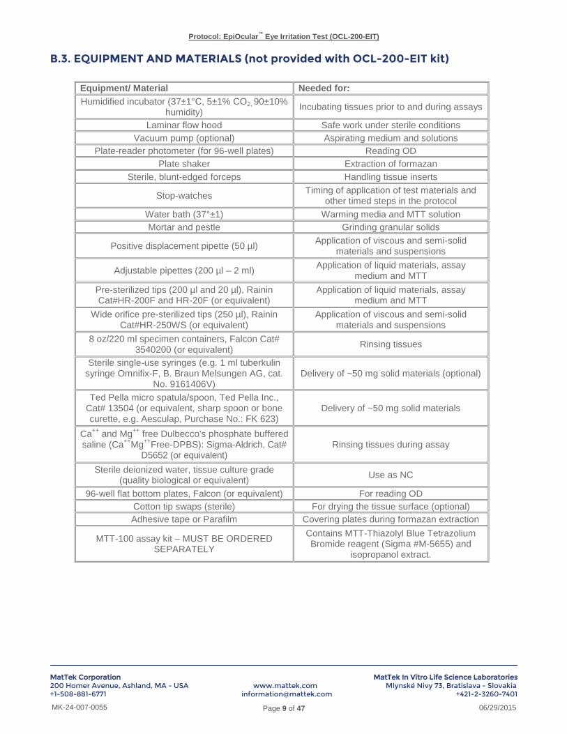
B.3. EQUIPMENT AND MATERIALS (not provided with OCL-200-EIT kit)
Equipment/ Material Needed for:
Humidified incubator (37±1°C, 5±1% CO2, 90±10% humidity)
Incubating tissues prior to and during assays
Laminar flow hood Safe work under sterile conditions
Vacuum pump (optional) Aspirating medium and solutions
Plate-reader photometer (for 96-well plates) Reading OD
Plate shaker Extraction of formazan
Sterile, blunt-edged forceps Handling tissue inserts
Stop-watches Timing of application of test materials and
other timed steps in the protocol
Water bath (37°±1) Warming media and MTT solution
Mortar and pestle Grinding granular solids
Positive displacement pipette (50 µl) Application of viscous and semi-solid
materials and suspensions
Adjustable pipettes (200 µl – 2 ml) Application of liquid materials, assay
medium and MTT
Pre-sterilized tips (200 µl and 20 µl), Rainin Cat#HR-200F and HR-20F (or equivalent)
Application of liquid materials, assay medium and MTT
Wide orifice pre-sterilized tips (250 µl), Rainin Cat#HR-250WS (or equivalent)
Application of viscous and semi-solid materials and suspensions
8 oz/220 ml specimen containers, Falcon Cat# 3540200 (or equivalent)
Rinsing tissues
Sterile single-use syringes (e.g. 1 ml tuberkulin syringe Omnifix-F, B. Braun Melsungen AG, cat.
No. 9161406V) Delivery of ~50 mg solid materials (optional)
Ted Pella micro spatula/spoon, Ted Pella Inc., Cat# 13504 (or equivalent, sharp spoon or bone curette, e.g. Aesculap, Purchase No.: FK 623)
Delivery of ~50 mg solid materials
Ca++
and Mg++
free Dulbecco's phosphate buffered saline (Ca
++Mg
++Free-DPBS): Sigma-Aldrich, Cat#
D5652 (or equivalent) Rinsing tissues during assay
Sterile deionized water, tissue culture grade (quality biological or equivalent)
Use as NC
96-well flat bottom plates, Falcon (or equivalent) For reading OD
Cotton tip swaps (sterile) For drying the tissue surface (optional)
Adhesive tape or Parafilm Covering plates during formazan extraction
MTT-100 assay kit – MUST BE ORDERED SEPARATELY
Contains MTT-Thiazolyl Blue Tetrazolium Bromide reagent (Sigma #M-5655) and
isopropanol extract.

Protocol: EpiOcular™
Eye Irritation Test (OCL-200-EIT)
MatTek Corporation MatTek In Vitro Life Science Laboratories 200 Homer Avenue, Ashland, MA - USA www.mattek.com Mlynské Nivy 73, Bratislava - Slovakia +1-508-881-6771 [email protected] +421-2-3260-7401
Page 10 of 47 MK-24-007-0055 06/29/2015
B.4. Preparations
B.4.1. MTT Solution
MTT solution and MTT diluent together with the extractant solution are ordered as MTT test kit (MTT-100, MatTek). For one OCL-200-EIT kit (24 tissues) one MTT-100 kit is needed.
A 1.0 mg/ml MTT solution is prepared no more than 2 hours before use and is stored at 2-8°C. MTT Concentrate is thawed and diluted in MTT Diluent (2 ml of MTT Concentrate to 8 ml of MTT Diluent) to produce a 1.0 mg/ml solution. MTT solution (e.g. pipetted in 24 well plates) should not be stored longer than one hour at 37°C.
If the MTT-100 is not used, proceed according to the following example: Per 24 tissues, dissolve 12.5 mg MTT (e.g. Sigma # M5655) in 2.5 ml PBS and thoroughly mix this stock-solution. After filtration (using a sterile 0.45 µm filter), add 2 ml of the stock-solution to 8 ml MTT-Assay Medium (final concentration: 1 mg MTT / ml medium). Keep the MTT medium in the dark.
B.4.2. Phosphate Buffered Saline (PBS) If powdered salts are used, prepare the PBS solution (without Ca
++ and Mg
++) according to the preparation
instructions of the supplier. Before use, the PBS solution should be sterilized by autoclaving or sterile filtration.
B.4.3. Test Compounds Test chemicals are administered neat by topical application onto the construct. Testing of dilutions is also possible and can be addressed.
B.4.4. Positive Control Neat methyl acetate is used as positive control (PC).
B.4.5. Negative Control Sterile deionized water is used as negative control (NC).
B.4.6 Microtiter plates Prior to use, each plate (6, 12, and 24-well) and its cover should be uniquely identified in permanent marker by a plate number, the test article number, and the test phase.
B.5. Method
B.5.1. Test System Procurement The EpiOcular
™ tissues have to be ordered from MatTek three weeks prior to scheduled test. Upon receipt of the
EpiOcular™
EIT test kit; the solutions should be stored as indicated by the manufacturer. The tissues are stored at 2-8ºC until use.
B.5.2. Assessment of Direct Test Article Reduction by MTT Test articles may have the ability to directly reduce MTT and to form a blue/purple reaction product which could have an impact on the quantitative MTT measurement. Therefore, it is necessary to assess this ability for each test article prior to conducting any assays with viable tissues. For this purpose a 1.0 mg/ml MTT solution

Protocol: EpiOcular™
Eye Irritation Test (OCL-200-EIT)
MatTek Corporation MatTek In Vitro Life Science Laboratories 200 Homer Avenue, Ashland, MA - USA www.mattek.com Mlynské Nivy 73, Bratislava - Slovakia +1-508-881-6771 [email protected] +421-2-3260-7401
Page 11 of 47 MK-24-007-0055 06/29/2015
(in DMEM) is prepared as described in § B.4.1. 50 µl (liquid test articles, see B.5.6.1.) or approximately 50 mg (solid test articles, see B.5.6.2.) are added to 1 ml of the MTT solution in a 6-well plate and the mixture is
incubated in the dark at 37 1°C in a humidified atmosphere of 5 1% CO2 in air (standard culture conditions, SCC) for approximately three hours. A negative control (50 µl of sterile deionized water) should be run concurrently. If the MTT solution colour turns blue/purple, the test article is presumed to have reduced the MTT. Water insoluble test materials may show direct reduction (darkening) only at the interface between the test article and the medium. In cases where the test article is shown to reduce MTT, a functional check using freeze-killed tissue controls (killed controls = KC) should be performed in at least one definitive assay to evaluate whether the test material is not binding to the tissue and leading to a false MTT reduction signal (see B.5.8 Method, Killed Controls for Assessment of Residual Test Article Reduction of MTT). Blue, dark purple and black test articles should also be tested on killed controls because it may not be possible to assess their potential to directly reduce MTT. It is recommended that the results are documented by taking photos.
B.5.3. Assessment of Colored or Staining Materials Colored test articles or test articles which become colored after application to the tissues may interfere with the quantitative photometric MTT measurement if the colorant binds to the tissue and is extracted together with MTT. Therefore, each test article has to be checked for its colorant properties. In this context, chemicals which absorb light and appear red, yellow, green, and blue should be considered as intrinsic colorants. Also test articles which look black may absorb light and should be considered as colorants. Blue, dark purple and black test articles may be directly tested on colorant controls without further tests because it is obvious that they can interfere with the blue MTT product. Such test articles should also be tested on killed controls because it may not be possible to assess their potential to directly reduce MTT. All other intrinsically colored test articles (e.g. red, yellow, green colorants) have to be tested for their ability to absorb significantly light at the wavelength used for the MTT determination (perform tests according to step 1 in isopropanol). The maximum amount of each test article, 50 µl for liquids and approximately 50 mg for solids, are added to 2 ml isopropanol, the same amount as used for MTT extraction, incubated in 6-well plates, and placed on an orbital plate shaker for 2 to 3 hours at room temperature. Two 200 µl aliquots of isopropanol solutions and of pure isopropanol are transferred to a 96-well plate and the absorbance is measured with a plate reader at the MTT measurement wavelength. Materials not completely soluble in isopropanol should be centrifuged if necessary (e.g. in 1 ml Eppendorf tubes for 15-30 s at 16,000 g) and 200 µL aliquots of the supernatant should be used for measurement. If, after subtraction of the OD for isopropanol, the OD of the test article solution is > 0.08 (approximately 5% of the mean viability of the negative control) the material has to be considered as possibly interacting with the MTT measurement and an additional test on colorant controls has to be performed (see § B.5.9.). For non-colored test articles additional tests have to be performed to assess if they become colorants after contact with water or isopropanol (perform tests according to steps 2 and 3 a described below). For this purpose 50 µl (liquid test articles, see B.5.6.1.) or approximately 50 mg (solid test articles, see B.5.6.2.) of each test article
are added to 1.0 ml of water in a 6-well plate and the mixture is incubated in the dark at 37 1°C in a humidified
atmosphere of 5 1% CO2 in air (standard culture conditions) for at least one hour. Furthermore, approximately 50 mg of solids are added to 2 ml isopropanol, the same amount as used for MTT extraction, and are incubated in 6-well plates for 2 to 3 hours at room temperature.
If the test article becomes colored either in water or isopropanol, it has to be considered as possibly interacting with the MTT measurement and an additional test on colorant controls has to be performed (see § B.5.9.).

Protocol: EpiOcular™
Eye Irritation Test (OCL-200-EIT)
MatTek Corporation MatTek In Vitro Life Science Laboratories 200 Homer Avenue, Ashland, MA - USA www.mattek.com Mlynské Nivy 73, Bratislava - Slovakia +1-508-881-6771 [email protected] +421-2-3260-7401
Page 12 of 47 MK-24-007-0055 06/29/2015
B.5.4. Preparation of EpiOcular Tissues for Treatment 1) On the day of receipt, normally on Tuesday, the tissues should be equilibrated (in its 24-well shipping container) to
room temperature for about 15 minutes.
2) An appropriate volume of EpiOcular™ Assay Medium should be warmed to approximately 37ºC. One (1.0) ml of Assay Medium is aliquoted into the appropriate wells of pre-labeled 6-well plates. The 6-well plates should be labeled with the test article or control codes and exposure times.
3) Each 24-well shipping container is removed from its plastic bag under sterile conditions and its surface disinfected by wiping with 70% isopropanol- or ethanol-soaked tissue paper. The sterile gauze is removed (see Fig. 1, left) and each tissue should be inspected for air bubbles between the agarose gel and insert (see Fig. 1, right). Cultures with air bubbles under the insert covering greater than 50% of the insert area should not be used. Any unusual observation should be noted separately. The tissues should be carefully removed from the 24-well shipping containers using sterile forceps. Any agarose adhering to the inserts should be removed by gentle blotting on sterile filter paper or gauze. The insert is then transferred aseptically into the 6-well plates and pre-incubated at standard culture conditions for one hour in the Assay Medium. After one hour, the Assay Medium should be replaced by 1 ml of fresh Assay Medium at 37°C and the EpiOcular™ tissues should be incubated at standard culture conditions overnight (16 - 24 hours).
Figure 1: Removal of the gauze and visual inspection of the epidermal tissues.
Alternatively, if the tissues arrive in the laboratory late Tuesday afternoon so that it is not possible to perform the 1 hour pre-incubation, the EpiOcular
™ tissues should be incubated at standard culture conditions overnight (16 - 24
hours) without medium exchange.
In case the delivery of the tissues is delayed until Wednesday morning, the transfer of the EpiOcular™
tissues, followed by the same procedure with pre-incubation as described above for normal receipt, should be started at an appropriate time so that the experiments can begin 16 - 24 hours later on Thursday.
The time of receipt and the pre-incubation procedure should be documented in the Method Documentation Sheet.
B.5.5. Controls The negative control (NC) is sterile deionized water. For the liquid protocol, 50 µL of the negative control is applied to the tissues exactly as for the liquid test articles for a 30 ± 2 minute exposure. For the solid protocol, 50 µL of the negative control are applied to the tissues (as described for the liquid test articles) for a 6 hr ± 15 minute exposure.
The positive control (PC) is methyl acetate. For the liquid protocol, 50 µl of the positive control is applied to the tissues exactly as for the liquid test articles for a 30 ± 2 minute exposure. For the solid protocol, 50 µl of the positive control are applied to the tissues (as described for the liquid test articles) for a 6 hr ± 15 minute exposure.

Protocol: EpiOcular™
Eye Irritation Test (OCL-200-EIT)
MatTek Corporation MatTek In Vitro Life Science Laboratories 200 Homer Avenue, Ashland, MA - USA www.mattek.com Mlynské Nivy 73, Bratislava - Slovakia +1-508-881-6771 [email protected] +421-2-3260-7401
Page 13 of 47 MK-24-007-0055 06/29/2015
Both, negative and positive controls are tested concurrently with the test articles in every run performed by a single technician.
B.5.6. Test Material Exposure Procedures Each test article and control is tested by treating two tissues (n=2). The test article dosing procedure is different for liquids and solids, as described later. Generally, the negative control and positive control is dosed first, followed by the test articles.
Liquids are fluid, pipetteable substances, such as liquids, gels, and creams. Solids are non-pipetteable substances like powders, resinous or waxy materials etc.
If the physical state of test articles is not easy to determine, follow these considerations: Test chemicals that can be pipetted at 37°C or lower temperatures (using a positive displacement pipette, if needed) should be treated as liquids. Viscous, waxy, resinous and gel-like test chemicals with unclear physical state should be incubated at 37±1°C for 15±1 minutes before deciding which treatment protocol to use. If such test chemicals become pipettable after this incubation period, they should be treated as liquids and should be applied to the tissues directly from the water bath (at 37±1°C), otherwise they should be treated as solids.
B.5.6.1. Treatment of liquid test articles
1) Pre-Treatment: After the overnight incubation, the tissues are pre-wetted with 20 µL of Ca++
Mg++
Free-DPBS. If the Ca
++Mg
++Free-DPBS does not spread across the tissues, the plate may be tapped to assure that the entire
tissue surface is wetted. The tissues are incubated at standard culture conditions for 30 ± 2 minutes.
2) Test article exposure: After the 30 ± 2 minute Ca++
Mg++
Free-DPBS pre-treatment, each liquid test and control article is tested by applying 50 µl topically on the EpiOcular™ tissues. The tissues are incubated at standard culture conditions for 30 ± 2 minutes.
Dosing details: 50 µl of the test article are applied directly on the tissue so as to cover the upper surface (see Fig. 2, left). The narrow points of the pipette tips may be cut off to widen the orifice for viscous materials. If positive displacement pipettes (see Fig. 2, right) are used for particularly viscous materials, the test article may first be transferred to a syringe to aid in filling the pipette. The pipette tip of the positive displacement pipette should be inserted into the dispensing tip of the syringe so that the material can be loaded into the displacement tip under pressure. Simultaneously, the syringe plunger is depressed as the pipette piston is drawn upwards. If air bubbles appear in the pipette tip, the test article should be removed (expelled) and the process repeated until the tip is filled without air bubbles. For very viscous liquids a dosing device (a flat headed cylinder of slightly less diameter than the inner diameter of the tissue insert or a plastic pushpin) may be placed over the test article to assure even spreading, if required.
Figure 2: Application of normal liquids with a standard pipette (left) and viscous liquids with a positive displacement pipette (right).

Protocol: EpiOcular™
Eye Irritation Test (OCL-200-EIT)
MatTek Corporation MatTek In Vitro Life Science Laboratories 200 Homer Avenue, Ashland, MA - USA www.mattek.com Mlynské Nivy 73, Bratislava - Slovakia +1-508-881-6771 [email protected] +421-2-3260-7401
Page 14 of 47 MK-24-007-0055 06/29/2015
3) Rinsing: At the end of the 30 ± 2 minutes treatment time, the test articles are removed by extensively rinsing the tissues with Ca
++Mg
++-free D-PBS (brought to room temperature), as described in detail below. If a dosing
device is used, it should be removed and discarded. Three clean beakers (glass or plastic with minimal 150 ml capacity), containing a minimum of 100 ml each of Ca
++Mg
++-free D-PBS should be used per test article (8
oz/220 ml disposable specimen containers from Falcon, Cat# 354020 are suggested). Each test article utilizes a different set of three beakers. The inserts containing the tissue be lifted out of the medium by grasping the upper edge of the plastic "collar" with fine forceps. Use of curved forceps facilitates handling and decanting. To assure throughput, the tissues should be rinsed two at a time by holding replicate inserts together by their collars using forceps. Be careful not to damage the tissues by the forceps. The test or control articles should be decanted from the tissue surface onto a clean absorbent material (paper towel, gauze, etc.) and the cultures dipped into the first beaker of D-PBS, swirled in a circular motion in the liquid for approximately 2 seconds (see Fig. 3, left), lifted out so that the inserts are mostly filled with D-PBS, and the liquid will be decanted back into the container. This process should be performed two additional times in the first beaker. The culture should then be rinsed in the second and third beakers of D-PBS three times each in the same fashion. Finally, any remaining liquid should be decanted onto the absorbent material. Decanting is most efficiently performed by rotating the insert to approximately a 45° angle (open end down, see Fig. 3, right) and touching the upper lip to the absorbent material (to break the surface tension).
Figure 3: Rinsing of two inserts (left) and decanting (right).
Note: If it is not possible to remove all of the visible test material, this should be noted in the notebook and under remarks in the spreadsheet. No further rinsing should be done. It is recommended to document any rinsing problems by taking photos, if possible.
4) Post-Soak: After rinsing, the tissues are immediately transferred to and immersed in 5 ml of previously warmed Assay Medium (room temperature) in a pre-labeled 12-well plate for a 12 ± 2 minute immersion incubation (Post-Soak) at room temperature. This incubation in Assay Medium is intended to remove any test article absorbed into the tissue.
5) Post-incubation: At the end of the Post-Soak immersion, each insert is removed from the Assay Medium, the medium is decanted off the tissue, and the insert is blotted on absorbent material, and transferred to the appropriate well of the pre-labeled 6-well plate containing 1 ml of warm Assay Medium. The tissues are incubated for 120 ± 15 minutes at standard culture conditions (Post-treatment Incubation).
B.5.6.2. Treatment of solid test articles
1) Pre-Treatment: After the overnight incubation, the tissues are pre-wetted with 20 µL of Ca++
Mg++
Free-DPBS. If the Ca
++Mg
++Free-DPBS does not spread across the tissues, the plate may be tapped to assure that the entire
tissue surface is wetted. The tissues should be incubated at standard culture conditions for 30 ± 2 minutes.
2) Test article exposure: After the 30 ± 2 minute Ca++
Mg++
Free-DPBS pre-treatment, the negative and positive controls are tested by applying 50 µl topically on the EpiOcular™ tissues. Each solid test article is tested by applying one leveled spoonful of the test article or by the means of an alternative tool (approximately 50 mg)

Protocol: EpiOcular™
Eye Irritation Test (OCL-200-EIT)
MatTek Corporation MatTek In Vitro Life Science Laboratories 200 Homer Avenue, Ashland, MA - USA www.mattek.com Mlynské Nivy 73, Bratislava - Slovakia +1-508-881-6771 [email protected] +421-2-3260-7401
Page 15 of 47 MK-24-007-0055 06/29/2015
topically on the EpiOcular™ tissues. To avoid that test article is spilled into the medium under the tissue inserts, the tissue insert should be removed from the medium, placed onto a sterile surface (e.g. the lid of a microtiter plate) and dosed by pouring the solid test article onto the tissue surface so that the surface of the tissue is completely covered by the test article. The tissue should be placed back into the culture medium after dosing and incubated at standard culture conditions for 6 hr ± 15 minutes. If the outer wall of the insert is contaminated by e.g. powders it may be necessary to wipe the particles off with cellulose tissue. Crystalline powders should be ground with a mortar and pestle to guarantee better contact to the tissue, if needed. Powders can be placed directly onto the culture at approximately 50 mg / culture by the means of a 1 ml syringe with head cut off (see Fig. 4, left). Powders are stuffed in the syringe when the plunger is drawn back and then applied by pressing the plunger down. With the help of the scale on the syringe, the amount of test article can be applied more reproducible after having weighed the specific chemical (see Fig. 4, right).
Figure 4: Syringe with head cut off (left) and application of a powder with the stuffed syringe (right).
As an alternative, powders can be placed by measuring one leveled spoonful of the test article (using a Ted Pella micro spatula/spoon, Cat# 13504, designed to measure approximately 50 mg) and applying the solids directly onto the tissue surface (see Fig. 5).
Figure 5: Application of solids with the Ted Pella spatula. For testing solids, at the time of dosing, the tissue insert will be removed from the medium, placed onto a sterile
surface (e.g. the lid of a microtiter plate) and dosed by pouring the solid test article onto the tissue surface. Shake insert gently from side to side to ensure that tissue is completely covered by the test article. The tissue will

Protocol: EpiOcular™
Eye Irritation Test (OCL-200-EIT)
MatTek Corporation MatTek In Vitro Life Science Laboratories 200 Homer Avenue, Ashland, MA - USA www.mattek.com Mlynské Nivy 73, Bratislava - Slovakia +1-508-881-6771 [email protected] +421-2-3260-7401
Page 16 of 47 MK-24-007-0055 06/29/2015
be placed back into the culture medium after dosing. The goal of this procedure is to eliminate the chance that test article is spilled into the medium under the tissue inserts.
3) Rinsing: At the end of the 6 hr ± 15 minutes treatment time, the test articles should be removed by extensively rinsing the tissues with Ca
++Mg
++-free D-PBS brought to room temperature, according to the same procedure
as described in B.5.6.1 for liquid test articles.
NOTE: If it is not possible to remove all of the visible test material, this should be noted under remarks in the spreadsheet. It is recommended to document any rinsing problems by taking photos, if possible.
4) Post-Soak: After rinsing, the tissue are immediately transferred to and immersed in 5 ml of previously- warmed Assay Medium (room temperature) in a pre-labeled 12-well plate for a 25 ± 2 minute immersion incubation (Post-Soak) at room temperature. This incubation in Assay Medium is intended to remove any test article absorbed into the tissue.
5) Post-incubation: At the end of the Post-Soak immersion, each insert is removed from the Assay Medium, the medium is decanted off the tissue, and the insert is blotted on absorbent material and transferred to the appropriate well of the pre-labeled 6-well plate containing 1 ml of warm Assay Medium. The tissues should be incubated for 18 ± 0.25 hours at standard culture conditions (Post-treatment Incubation).
B.5.7. MTT Assay
After the Post-treatment Incubation of 120 ± 15 minutes for liquids and 18 ± 0.25 hours for solids, respectively, the MTT assay is performed.
1) A 1.0 mg/ml MTT solution is prepared as described in B.4. “Preparations”: 300 µL of the MTT solution is added to each designated well of a pre-labeled 24-well plate.
2) At the end of the Post-treatment Incubation, each insert is removed from the 6-well plate and gently blotted on absorbent material. The tissues are placed into the 24-well plate containing 0.3 ml of MTT solution. Once all the tissues are placed into the 24-well plate, the plate is incubated for 180 ± 10 minutes at Standard Culture Conditions.
3) For liquids (except colorants), each insert is removed from the 24-well plate after 180 ± 10 minutes, the bottom of the insert is blotted on absorbent material, and then transferred to a pre-labeled 24-well plate containing 2.0 ml of isopropanol in each designated well so that isopropanol is flowing into the insert on the tissue surface. The plates are sealed with parafilm (between the plate cover and upper edge of the wells) or a standard plate sealer, and are either stored overnight at 2-8°C in the dark or immediately extracted. To extract the MTT, the plates are placed on an orbital plate shaker and shaken for 2 to 3 hours at room temperature. At the end of the extraction period, the tissue is pierced and the liquid within each insert is decanted into the well from which it was taken.
For solids and liquid colorants a different procedure should be used by extracting only from beneath since they might remain on the tissue and could contaminate the isopropanol extraction solution. Inserts are removed from the 24-well plate after 180 ± 10 minutes; the bottom of the insert is blotted on absorbent material, and then transferred to a pre-labeled 6-well plate containing 2 ml isopropanol in each well so that no isopropanol is flowing into the insert. The plates are handled as described above except that at the end of the extraction period, the tissues should not be pierced. The corresponding negative, positive, and colorant controls have to be treated identically without piercing. For this procedure it is necessary to seal the plates particularly thorough since a higher evaporation rate has to be expected due to the larger surface of wells in 6-well plates.
4) The extract solution is mixed and two 200 µl aliquots are transferred to the appropriate wells of a pre-labeled 96-well plate(s) according to the plate configuration given below (see Fig. 6).

Protocol: EpiOcular™
Eye Irritation Test (OCL-200-EIT)
MatTek Corporation MatTek In Vitro Life Science Laboratories 200 Homer Avenue, Ashland, MA - USA www.mattek.com Mlynské Nivy 73, Bratislava - Slovakia +1-508-881-6771 [email protected] +421-2-3260-7401
Page 17 of 47 MK-24-007-0055 06/29/2015
Figure 6: Standard plate configuration for viability tests.
In case of turbid extract solutions, caused by insoluble solids which have passed the insert membrane and
which may lead to increased OD values, the solutions should be centrifuged (e.g. in 1 ml Eppendorf tubes for 15-30 sec at 16,000 x g and 200 µl aliquots should be taken from the supernatant. 200 µL of isopropanol are added to the wells designated as blanks. The absorbance at 570 nm (OD570 or simply OD) of each well is measured with a Molecular Devices Vmax (or similar) plate reader. Since the absorption spectrum of the extracted dye builds a plateau (see Fig. 7) with the maximum at approximately 570 nm, a range of 550 to 590 nm is acceptable but should be consistent within a laboratory's data set. No reference wavelength measurement should be used.
Figure 7: Absorption spectrum of Formazan.
Since the absorbance value of the negative control can be already ~2.0, the additional absorbance of colorants and/or MTT reduction can lead to even higher absorbance values than 2.0. Therefore, the range of linearity of the photometer used may be exceeded. In this case the easiest way to avoid irregular viability results calculated from absorbance values above a linearity limit is to repeat the measurement of the entire experiment by measuring 100 µl aliquots (instead of 200 µl) in the same way as described above. All samples of the same test should be treated identically. Note: The range of linearity for the photometer used can be assessed by measuring a calibration curve of e.g. Trypan Blue solution. Prepare a 0.04% Trypan Blue solution (e.g. Trypan Blue 0.4%, Sigma T-8151, diluted 1:10 v/v with water). From this stock solution transfer different volumes from 50 µL up to 800 µL to the corresponding wells of a 24-well plate and fill these volumes up with water to 1 ml (concentration range from 50 to 800 µL/ml with an increment of 50 µL/ml). Shake the plate and transfer two 200 µL aliquots of each dilution to a 96-well

Protocol: EpiOcular™
Eye Irritation Test (OCL-200-EIT)
MatTek Corporation MatTek In Vitro Life Science Laboratories 200 Homer Avenue, Ashland, MA - USA www.mattek.com Mlynské Nivy 73, Bratislava - Slovakia +1-508-881-6771 [email protected] +421-2-3260-7401
Page 18 of 47 MK-24-007-0055 06/29/2015
microtiter plate. Use two 200 µL aliquots of water as blanks. Measure the absorbance of these dilutions at 570 nm with the plate reader used for the MTT assay, subtract the blank and draw a graph of the results (concentrations on the x-axis and absorbance on the y-axis). The linearity is considered as sufficient if the R² value of the OD measurement is > 0.999 (example see Fig. 8). It is recommended to repeat the linearity check every three months.
Figure 8: Example result of the photometer linearity test.
B.5.8. Killed Controls for Assessment of Residual Test Article Reduction of MTT In cases where the test article is shown to reduce MTT, only test articles that remain bound to the tissue after rinsing, resulting in a false MTT reduction signal, present a problem. To demonstrate that residual test article is not acting to directly reduce the MTT, a functional check on killed controls (KC) is performed in the definitive assay to show that the test material is not binding to the tissue and leading to a false MTT reduction signal. To determine whether residual test article is acting to directly reduce the MTT, two freeze-killed control tissues per test article are used. Freeze killed tissues are prepared by placing untreated EpiOcular™ constructs (in a 24 well plate) in the –20°C freezer overnight, thawing to room temperature, and then refreezing (two freeze-thaw cycles). Once frozen, the tissue may be stored indefinitely in the freezer. To test for residual test article reduction, two killed tissues are treated with the test article in the normal fashion. All assay procedures are performed as for the viable tissue. A killed control treated with sterile deionized water (negative killed control) is tested in parallel since a small amount of MTT reduction is expected from the residual NADH and associated enzymes within the killed tissue. If little or no MTT reduction is observed in the test article-treated killed control, the MTT reduction observed in the test article-treated viable tissue may be ascribed to the viable cells. If there is appreciable MTT reduction in the treated killed control (relative to the amount in the treated viable tissue), additional steps must be taken to account for the chemical reduction, or the test article may be considered un-testable in this system. The OD values of the killed controls are analyzed as described in B.7. “Data Analysis / Calculation of Results”.
If the direct reduction of MTT by the test article is > 60% of the viable negative control, the results obtained should be taken with caution as this is the cut-off used in EpiOcular™ EIT to distinguish classified from not classified chemicals.
It may not be necessary to perform a test on killed controls for those chemicals classified as irritants due to a viability being lower than the cut-off of 60% because any MTT reduction would not change the classification for such chemicals.

Protocol: EpiOcular™
Eye Irritation Test (OCL-200-EIT)
MatTek Corporation MatTek In Vitro Life Science Laboratories 200 Homer Avenue, Ashland, MA - USA www.mattek.com Mlynské Nivy 73, Bratislava - Slovakia +1-508-881-6771 [email protected] +421-2-3260-7401
Page 19 of 47 MK-24-007-0055 06/29/2015
If the uncorrected ODs of the tissue extracts obtained with the test article fall outside of the linear range of the spectrophotometer, the test article may be considered as not testable.
B.5.9. Colorant Controls for Assessment of Colored or Staining Test Articles
In cases where a test article has shown to have or to develop relevant color which can interact with the MTT measurement (for criteria and screening tests to be performed see B.5.3), an additional test has to be performed to determine the amount of color bound to and extracted from the tissues. For this purpose each colored test article is applied to two additional tissues, the colorant controls (CC), and is treated in the same way as described for liquids and solids in B.5.6.1. and B.5.6.2., respectively. In contrast to the normal viability test, no MTT assay is performed. The bound color is extracted and the absorbance of the isopropanol extracts is measured identically as in the MTT assay for colored test articles (according to B.5.7., as described for the MTT assay with 2 ml extraction solution in 6-well plates without piercing the tissue, and beginning from step 2 but starting with a 180 ± 10 min incubation in medium instead of MTT solution). The amount of extracted color will be subtracted from the results of the viability assay according to B.7. “Data Analysis / Calculation of Results”.
If the colorant control result is > 60% of the viable negative control, the results obtained with standard OD measurement should be taken with caution as this is the cut-off used in EpiOcular™ EIT to distinguish classified from not classified chemicals.
It may not be necessary to perform a test on colorant controls for those chemicals classified as irritants due to a viability being lower than the cut-off of 60% because any color interference would not change the classification for such chemicals.
If tissues are stained by a colorant and remain stained even after isopropanol extraction, this should be documented in the remarks and by taking photographs.
If the uncorrected ODs of the tissue extracts obtained with the test article fall outside of the linear range of the spectrophotometer, the test article may be considered as not testable using standard OD measurement.
If tissues are stained by a colorant and remained stained even after isopropanol extraction, this should be documented in the remarks and by taking photographs.
B.5.10. Non-Specific Killed Control (NSK) Controls for Assessment of Colored and Direct MTT Test Articles
In cases where the test article was identified as producing both colour interference and direct MTT reduction, a third set of controls will need to be performed. This is usually the case with darkly coloured test chemicals absorbing light in the range of 570±30 nm (e.g., blue, purple, black) because their intrinsic colour impedes the assessment of their capacity to directly reduce MTT. This force by default the use of both controls, KC and CC, which can lead to double correction for color interference. In NSKC, the test chemical is applied on two killed tissue replicates, which undergo the entire testing procedure but are incubated with medium instead of MTT solution during the MTT incubation step. NSKC should be performed concurrently to the KC control and with the same tissue batch. To calculate true tissue viability the percent non-specific colour obtained with NSKC will be added to relative tissue viability (B.7.5). Note: For colorants, it may be necessary to determine the amount of the MTT product using an HPLC/UPLC-spectrophotometry, since the HPLC/UPLC system allows for the separation of the MTT formazan from the chemical before its quantification (Alepee N, et al, 2015). Another method to overcome problems with colorants is to measure the absorption spectra of the isopropanol extracts of the viability assay and the colorant control test. In case of high absorbance differences of dyes between tissues of the viability and the colorant control test, the absorbance spectra may indicate the impact of bound color more exactly as the colorant controls measured at a single wavelength. Please contact MatTek Technical Assistance for more help in this area.

Protocol: EpiOcular™
Eye Irritation Test (OCL-200-EIT)
MatTek Corporation MatTek In Vitro Life Science Laboratories 200 Homer Avenue, Ashland, MA - USA www.mattek.com Mlynské Nivy 73, Bratislava - Slovakia +1-508-881-6771 [email protected] +421-2-3260-7401
Page 20 of 47 MK-24-007-0055 06/29/2015
B.6. ACCEPTANCE CRITERIA Acceptance Criteria for Test Results The results are acceptable if: 1) The negative control OD > 0.8 and < 2.5, 2) The mean relative viability of the positive control is: a) 30 minute exposure: below 50% of control viability b) 6 hr exposure: below 50% of control viability 3) The difference of viability between the two relating tissues of a single chemical is < 20% in the same run (for positive and negative control tissues and tissues of single chemicals). This applies also to the killed controls (single chemicals and negative killed control) and the colorant controls which are calculated as percent values related to the viability of the relating negative control.
B.7. DATA ANALYSIS / CALCULATION OF RESULTS The measured OD values are processed according to the following rules and calculations. An Excel spreadsheet is provided which is used for the calculation of the results. The Excel file contains five different sheets (Samples Sheet, Import Sheet, Remarks Sheet, Calculation Sheet, and Summary Sheet). For use details of the spreadsheet see Annex 2.
B.7.1. General Calculations 1) Calculate the mean OD value of the blank control wells (ODBlk) for each experiment. 2) Subtract ODBlk from each OD value of the same experiment (blank corrected values). 3) Calculate the mean value of the two aliquots for each tissue (= corrected test article OD). 4) Calculate the mean value of the two relating tissues for each control and test article (= corrected mean OD).
For further calculations only the corrected mean negative control OD value is needed and in case of the kil led controls the negative killed control OD value (ODNC_KC).
5) The corrected OD value of the negative control corresponds to 100% viability. Corrected negative control OD = Negative control OD - ODBlk = 100% Viability
B.7.2. Calculations for Viability Tests only 1) Calculate the percent viability of each of the two relating tissues for each control and test article relative to the
negative control (100% control). corrected test article OD
Viability [%] = x 100 corrected mean negative control OD
2) Calculate the difference of the viability. If the difference is >20% the test is considered as non-qualified. 3) Calculate the mean test article viability (TA viability) and classify the test article according to the prediction
model.
B.7.3. Calculations for Viability plus Killed Control Tests 1) Calculate the OD values of the killed control experiment as described in B.7.1. Step 1 to 3 = corrected OD test
article KC. 2) Subtract the mean negative control killed control value (ODNC_KC) from the test article-treated killed controls to
determine the individual killed control values for the test articles (ODKC).
OD killed control = Corrected OD test article KC - ODNC_KC

Protocol: EpiOcular™
Eye Irritation Test (OCL-200-EIT)
MatTek Corporation MatTek In Vitro Life Science Laboratories 200 Homer Avenue, Ashland, MA - USA www.mattek.com Mlynské Nivy 73, Bratislava - Slovakia +1-508-881-6771 [email protected] +421-2-3260-7401
Page 21 of 47 MK-24-007-0055 06/29/2015
The net OD killed control represents the amount of reduced MTT due to direct reduction by test article residues.
3) Transfer the killed control OD for the two relating tissues to a percentage value relative to the viability scale (killed control “viability”) by comparison to the corrected viability negative control OD of the relating experiment in which the test article viability test is performed.
corrected test article OD Killed control “viability” [%] = x 100 corrected mean negative control OD
4) Calculate the difference of the viability of the two tissues. If the difference is >20%, the killed control test is
considered as non-qualified. 5) Calculate the mean viability for the killed control and subtract this mean killed control “viability” value (KC
“viability”) from the relating mean viability of the same test article (TA viability) to determine the killed control corrected viability (KC corrected viability).
KC corrected viability = TA viability – KC”viability”
6) Classify the test article regarding the killed control corrected viability according to the prediction model.
B.7.4. Calculation for Viability plus Colorant Control Tests 1) Calculate the OD values of the colorant control test as described in B.7.1. Points 1 to 4. 2) Transfer the colorant control OD for the two relating tissues to a percentage value relative to the viability scale
(colorant control “viability”) by comparison to the corrected viability negative control of the same experiment. OD colorant control Colorant control “viability” [%] = x 100 corrected mean negative control OD 3) Calculate the difference of the viability of the two tissues. If the difference is >20%, the colorant control test is
considered as non-qualified. 4) Calculate the mean viability for the colorant control and subtract this mean colorant control “viability” value (CC
“viability”) from the relating mean viability of the same test article (TA viability) to determine the colorant control corrected viability (CC corrected viability).
CC corrected viability = TA viability – CC “Viability”
5) Classify the test article regarding the colorant control corrected viability according to the prediction model.
B.7.5. Calculations for Viability plus Killed Control, Colorant Control, and Non-Specific Killed Control Tests 1) Calculate the OD values as well as the test article viability (TA viability), the killed control “viability” (KC
“viability”), the colorant control “viability” (CC “viability”), and the non-specific killed control “viability” (NSKC “viability”) values of a specific test article according to the rules given in B.7.2., B.7.3. and B.7.4.
2) Subtract the KC “viability” and the CC “viability” from the TA viability, then add NSKC “viability to determine the killed, colorant, and non-specific killed control corrected viability (KC+CC+NSKC corrected viability).
KC+ CC + NSKC corrected viability = TA viability –KC “viability – CC “Viability” + NSCK “viability”
3) Classify the test article regarding the killed, colorant, and non-specific killed control corrected viability according to the prediction model.

Protocol: EpiOcular™
Eye Irritation Test (OCL-200-EIT)
MatTek Corporation MatTek In Vitro Life Science Laboratories 200 Homer Avenue, Ashland, MA - USA www.mattek.com Mlynské Nivy 73, Bratislava - Slovakia +1-508-881-6771 [email protected] +421-2-3260-7401
Page 22 of 47 MK-24-007-0055 06/29/2015
B.8. PREDICTION MODEL If the test article-treated tissue viability is > 60.0% relative to negative control-treated tissue viability, the test article is labeled non-irritant.
If the test article-treated tissue viability is 60.0% relative to negative control-treated tissue viability, the test article is labeled irritant.
In vitro result In vivo prediction
mean tissue viability 60.0% Irritant (I)
mean tissue viability > 60.0% non-irritant (NI)
B.9. BIBLIOGRAPHIC REFERENCES/REPORTS
- Test Submission Template (TST) for ECVAM submissions: Ocular Irritation Assay for Chemicals Using the EpiOcular™ Human Cell Construct (revised submission September 11, 2008)
- Ocular Irritation REACH Protocol (DRAFT), MatTek Corporation (workbook) - Berridge, M.V., Tan, A.S., McCoy, K.D., Wang, R. (1996): The Biochemical and Cellular Basis of Cell Proliferation
Assays That Use Tetrazolium Salts. Biochemica 4, 14-19. - Jester J. V., et al. (2001). Extent of initial corneal injury as a basis for alternative eye irritation tests. Toxicology In
Vitro 15, 115-30. - Jester J. V. (2006). Extent of corneal injury as a biomarker for hazard assessment and the development of
alternative models to the Draize rabbit eye test. Cutan Ocul Toxicol. 25, 41-54. - Maurer J. K., et al. (1997). Confocal microscopic characterization of initial corneal changes of surfactant-induced
eye irritation in the rabbit. Toxicol Appl Pharmacol. 143, 291-300. - Maurer J. K., et al., (2002): Extent of initial corneal injury as the mechanistic basis for ocular irritation: key
findings and recommendations for the development of alternative assays. Regul Toxicol Pharmacol. 36, 106-17.
- Scott L. et al. (2010): A proposed eye irritation testing strategy to reduce and replace in vivo studies using Bottom up and Top-down approaches. Toxicology in vitro 24, 1-9.
- Kaluzhny Y, et al (2011) “Development of the EpiOcularTM
Eye Irritation Test (EpiOcular-EIT) for Hazard Identification and Labeling of Eye Irritating Chemicals in Response to the Requirements of the Cosmetics Directive and REACH Legislation”, ATLA 39, 1-26, 2011.
- Kolle, S.N., Kandarova, H., Wareing, B., van Ravenzwaay, B., and Landsiedel, R. (2011). In-house validation of the EpiOcular™ eye irritation test and its combination with the bovine corneal opacity and permeability test for the assessment of ocular irritation. ATLA 39, 365–387.
- Pfannenbecker, U., et al., “Cosmetics Europe multi-laboratory pre-validation of the EpiOcular™ reconstituted human tissue test method for the prediction of eye irritation,” Toxicol In Vitro 27(2), 619-626 (2013).
- Alépée, N., et al., (2015) Use of HPLC/UPLC-spectrophotometry for detection of formazan in in vitro Reconstructed human Tissue (RhT)-based test methods employing the MTT-reduction assay to expand their applicability to strongly coloured test chemicals. Toxicol In Vitro 29(4):741-761.
- Kaluzhny, Y., et al, (2015). EpiOcular™ Eye Irritation Test (EIT) for hazard identification and labeling of eye irritating chemicals: protocol optimization for solid materials and extended shipment times. Altern Lab Anim. 2015 May;43(2):101-27.
B.10. ANNEXES Annex 1: Example Time Schedules for Liquids and Solids
Annex 2: Excel Spreadsheets
Annex 3: Method Documentation Sheet (MDS)
Annex 4: Characterization of Test Substances

Protocol: EpiOcular™
Eye Irritation Test (OCL-200-EIT)
MatTek Corporation MatTek In Vitro Life Science Laboratories 200 Homer Avenue, Ashland, MA - USA www.mattek.com Mlynské Nivy 73, Bratislava - Slovakia +1-508-881-6771 [email protected] +421-2-3260-7401
Page 23 of 47 MK-24-007-0055 06/29/2015
Annex 1a: Sample Time Schedule for Testing of Liquids

Protocol: EpiOcular™
Eye Irritation Test (OCL-200-EIT)
MatTek Corporation MatTek In Vitro Life Science Laboratories 200 Homer Avenue, Ashland, MA - USA www.mattek.com Mlynské Nivy 73, Bratislava - Slovakia +1-508-881-6771 [email protected] +421-2-3260-7401
Page 24 of 47 MK-24-007-0055 06/29/2015
Annex 1b: Sample time schedule for testing of solids Example: Test Schdule for Solids + Controls
Start time of the different work steps (daywork for one technician)
Each line corresponds to one pair of tissues
1 to 12 = order of work steps
1st day 2nd day
From 8:00 to 9:00: preparations
1 2 3 4 9 11
Liquids:
PBS Treatment Post-Soak Post-Incub. MTT Extract.
30 min 6 hours 25 min 18 hours 180min over night
NC 9.00 9.30 15.30 15.55 10.55 13.55
PC 9.02 9.32 15.32 15.57 10.56 13.56 12
1 9.04 9.34 15.34 15.59 10.57 13.57 Measure
2 9.06 9.36 15.36 16.01 10.58 13.58 between
3 9.08 9.38 15.38 16.03 10.59 13.59 8:00 to
4 9.10 9.40 15.40 16.05 11.00 14.00 10:00
5 9.12 9.42 15.42 16.07 11.01 14.01
6 9.14 9.44 15.44 16.09 11.02 14.02
7 9.16 9.46 15.46 16.11 11.03 14.03
8 9.18 9.48 15.48 16.13 11.04 14.04
9 9.20 9.50 15.50 16.15 11.05 14.05
10 9.22 9.52 15.52 16.17 11.06 14.06

Protocol: EpiOcular™
Eye Irritation Test (OCL-200-EIT)
MatTek Corporation MatTek In Vitro Life Science Laboratories 200 Homer Avenue, Ashland, MA - USA www.mattek.com Mlynské Nivy 73, Bratislava - Slovakia +1-508-881-6771 [email protected] +421-2-3260-7401
Page 25 of 47 MK-24-007-0055 06/29/2015
Annex 2: Excel Spreadsheets for Data Input and Calculations (*) (*) Spreadsheets were originally developed by Dr. Uwe Pfannenbecker, Beiersdorf AG, Germany and available from MatTek Corporation. 1. Open “1-Samples Sheet” and select from the drop-down menu (cell B10) whether your experiment is a viability test (+ colorant controls if relevant) or a killed control test.
Note: This choice has a strong impact on all other sheets! 2. Fill in or select from drop-down menus between different options in the requested fields.

Protocol: EpiOcular™
Eye Irritation Test (OCL-200-EIT)
MatTek Corporation MatTek In Vitro Life Science Laboratories 200 Homer Avenue, Ashland, MA - USA www.mattek.com Mlynské Nivy 73, Bratislava - Slovakia +1-508-881-6771 [email protected] +421-2-3260-7401
Page 26 of 47 MK-24-007-0055 06/29/2015
3. Select for each test article whether it is a MTT reducer or not (not necessary for Killed Control Tests).
4. Select for all test articles tested whether it is a colorant or not (not necessary for Killed Control Tests).
In the case when colorants have been tested and “Yes” has been selected, the spreadsheet is expecting that the following test article is the corresponding colorant control to the test article mentioned in the row before. This will be visualized in the column “Test Substance” (adding ‘-CC’ to code number). In a mixed experiment with test articles which have to be tested with colorant controls and with test articles without colorant controls, take those test articles with colorant controls always as the first test articles in the list. Note: This has to be taken into account for the measurement plate configuration 5. Fill in the run number of the specific test article (not necessary for Killed Control Tests if only one run is performed).

Protocol: EpiOcular™
Eye Irritation Test (OCL-200-EIT)
MatTek Corporation MatTek In Vitro Life Science Laboratories 200 Homer Avenue, Ashland, MA - USA www.mattek.com Mlynské Nivy 73, Bratislava - Slovakia +1-508-881-6771 [email protected] +421-2-3260-7401
Page 27 of 47 MK-24-007-0055 06/29/2015
If “Killed Control” is selected the order of test articles will be changed since no positive control is needed. In this case it is not necessary to select the options for “MTT interaction” and “Colorant”.

Protocol: EpiOcular™
Eye Irritation Test (OCL-200-EIT)
MatTek Corporation MatTek In Vitro Life Science Laboratories 200 Homer Avenue, Ashland, MA - USA www.mattek.com Mlynské Nivy 73, Bratislava - Slovakia +1-508-881-6771 [email protected] +421-2-3260-7401
Page 28 of 47 MK-24-007-0055 06/29/2015
Import Sheet: The 96-well microtiter plate has to be configured according to the plate configuration indicated in this sheet (different for Viability/CC Test and Killed Control Test!). The raw data (OD values) have to be copied into the marked cells (area from B29 to M36).
For Killed Control Tests no Positive Control will be tested and therefore the wells for test articles TA1 to TA 10 are switched one column to the left. Wells A12 to D12 are kept empty.

Protocol: EpiOcular™
Eye Irritation Test (OCL-200-EIT)
MatTek Corporation MatTek In Vitro Life Science Laboratories 200 Homer Avenue, Ashland, MA - USA www.mattek.com Mlynské Nivy 73, Bratislava - Slovakia +1-508-881-6771 [email protected] +421-2-3260-7401
Page 29 of 47 MK-24-007-0055 06/29/2015
Remarks Sheet: Record any relevant observation made during the experiments. The sheet has space for 20 test articles.
Calculation Sheet(s): Different Calculation Sheets are used for Viability/CC Tests and Killed Control Tests. The sheets will be filled out automatically and the data calculated according to the information in the Samples Sheet and the Import Sheet (rules for calculation are explained in the SOP). No entries have to be made in these sheets.
4a_Calc_Sheet Viability_CC:

Protocol: EpiOcular™
Eye Irritation Test (OCL-200-EIT)
MatTek Corporation MatTek In Vitro Life Science Laboratories 200 Homer Avenue, Ashland, MA - USA www.mattek.com Mlynské Nivy 73, Bratislava - Slovakia +1-508-881-6771 [email protected] +421-2-3260-7401
Page 30 of 47 MK-24-007-0055 06/29/2015
1. The spreadsheet controls if the negative control and positive control fulfill the acceptance criteria.
Non-qualified run: OD NC < 0.8 or > 2.5
Non-qualified run: PC > 50%
2. In case a colorant control is tested, no classification will be given. 3. If a test article does not fulfill the acceptance criterion regarding the difference of viability (D<20), this will be highlighted in red. Otherwise the test is qualified. In case of a Viability/CC Test, the calculation sheet for the Killed Control Tests will be empty and vice versa.

Protocol: EpiOcular™
Eye Irritation Test (OCL-200-EIT)
MatTek Corporation MatTek In Vitro Life Science Laboratories 200 Homer Avenue, Ashland, MA - USA www.mattek.com Mlynské Nivy 73, Bratislava - Slovakia +1-508-881-6771 [email protected] +421-2-3260-7401
Page 31 of 47 MK-24-007-0055 06/29/2015
4b_Calc Sheet_KC This spreadsheet looks differently than the spreadsheet for Viability/CC Tests.
Additionally to the blank subtraction the negative killed control value is subtracted from the OD values. No viability is calculated. As well, classification and check for qualification are not performed here (for this see Summary Sheet).
Note: To get the final calculation for the killed controls and the check for qualification, the corrected killed control values for a specific test article have to be copied into the corresponding Summary Sheets (of three or more runs) of this particular test article.

Protocol: EpiOcular™
Eye Irritation Test (OCL-200-EIT)
MatTek Corporation MatTek In Vitro Life Science Laboratories 200 Homer Avenue, Ashland, MA - USA www.mattek.com Mlynské Nivy 73, Bratislava - Slovakia +1-508-881-6771 [email protected] +421-2-3260-7401
Page 32 of 47 MK-24-007-0055 06/29/2015
Summary Sheet: In the Summary Sheet the results for the different possible parts of testing (viability, colorant control, and killed control) will be put together and a final classification will be calculated. For this purpose the corresponding killed control results of the test articles (if a killed control test is necessary) have to be copied or filled in the corresponding cells of columns I, J and K (1.). The calculation of percent values for the killed control data in relation to the OD of the viable negative control is performed here. Additionally, it is checked if the KC result fulfills the acceptance criteria regarding the difference of percentage values.
Remarks: All sheets (for Viability Tests: sheets 1, 2, 3, 4a and 5; for Killed Control: sheets 1, 2, 3 and 4b) have to be printed out and signed for the documentation. Also, a QC check by a second person has to be performed. Note: This Excel file has been produced by using Microsoft Office Excel 2007. If a different version of Excel is being used, the data may be presented differently than described here (e.g. highlighting ‘D>20’ in red in the Calculation Sheet, column ‘Qualified?”). Note: A complete set of Annex 2 spreadsheets is available from Uwe Pfannenbecker (Beiersdorf AG, Hamburg, Germany, [email protected]). A simplified version of it is available from MatTek Corporation (Ashland, MA, US).

Protocol: EpiOcular™
Eye Irritation Test (OCL-200-EIT)
MatTek Corporation MatTek In Vitro Life Science Laboratories 200 Homer Avenue, Ashland, MA - USA www.mattek.com Mlynské Nivy 73, Bratislava - Slovakia +1-508-881-6771 [email protected] +421-2-3260-7401
Page 33 of 47 MK-24-007-0055 06/29/2015
Annex 3: Method Documentation Sheet (MDS)

Protocol: EpiOcular™
Eye Irritation Test (OCL-200-EIT)
MatTek Corporation MatTek In Vitro Life Science Laboratories 200 Homer Avenue, Ashland, MA - USA www.mattek.com Mlynské Nivy 73, Bratislava - Slovakia +1-508-881-6771 [email protected] +421-2-3260-7401
Page 34 of 47 MK-24-007-0055 06/29/2015

Protocol: EpiOcular™
Eye Irritation Test (OCL-200-EIT)
MatTek Corporation MatTek In Vitro Life Science Laboratories 200 Homer Avenue, Ashland, MA - USA www.mattek.com Mlynské Nivy 73, Bratislava - Slovakia +1-508-881-6771 [email protected] +421-2-3260-7401
Page 35 of 47 MK-24-007-0055 06/29/2015

Protocol: EpiOcular™
Eye Irritation Test (OCL-200-EIT)
MatTek Corporation MatTek In Vitro Life Science Laboratories 200 Homer Avenue, Ashland, MA - USA www.mattek.com Mlynské Nivy 73, Bratislava - Slovakia +1-508-881-6771 [email protected] +421-2-3260-7401
Page 36 of 47 MK-24-007-0055 06/29/2015

Protocol: EpiOcular™
Eye Irritation Test (OCL-200-EIT)
MatTek Corporation MatTek In Vitro Life Science Laboratories 200 Homer Avenue, Ashland, MA - USA www.mattek.com Mlynské Nivy 73, Bratislava - Slovakia +1-508-881-6771 [email protected] +421-2-3260-7401
Page 37 of 47 MK-24-007-0055 06/29/2015

Protocol: EpiOcular™
Eye Irritation Test (OCL-200-EIT)
MatTek Corporation MatTek In Vitro Life Science Laboratories 200 Homer Avenue, Ashland, MA - USA www.mattek.com Mlynské Nivy 73, Bratislava - Slovakia +1-508-881-6771 [email protected] +421-2-3260-7401
Page 38 of 47 MK-24-007-0055 06/29/2015

Protocol: EpiOcular™
Eye Irritation Test (OCL-200-EIT)
MatTek Corporation MatTek In Vitro Life Science Laboratories 200 Homer Avenue, Ashland, MA - USA www.mattek.com Mlynské Nivy 73, Bratislava - Slovakia +1-508-881-6771 [email protected] +421-2-3260-7401
Page 39 of 47 MK-24-007-0055 06/29/2015

Protocol: EpiOcular™
Eye Irritation Test (OCL-200-EIT)
MatTek Corporation MatTek In Vitro Life Science Laboratories 200 Homer Avenue, Ashland, MA - USA www.mattek.com Mlynské Nivy 73, Bratislava - Slovakia +1-508-881-6771 [email protected] +421-2-3260-7401
Page 40 of 47 MK-24-007-0055 06/29/2015
DOSING TIMETABLE – SOLID TEST ARTICLES Table 9: Dosing timetable for solids (including colorant controls)
Test Article
Pre-incubation Main Experiment
MTT assay
Initial 60 ±5 min
Over- night 16-24 h
D-PBS Addition
(20 µl) 30±2 min
Treatment
(50 mg) 6 ± 0.25 hours
Treatment Term. / Post Soak Initiation 25 ± 5 min
Post Soak Term. / Post Inc. Start
18 ± 0.25 hours
Post Inc Term. / MTT Addition
180 ± 10 min
MTT Termi- nation
Remarks
Negative Control
Positive Control

Protocol: EpiOcular™
Eye Irritation Test (OCL-200-EIT)
MatTek Corporation MatTek In Vitro Life Science Laboratories 200 Homer Avenue, Ashland, MA - USA www.mattek.com Mlynské Nivy 73, Bratislava - Slovakia +1-508-881-6771 [email protected] +421-2-3260-7401
Page 41 of 47 MK-24-007-0055 06/29/2015
DOSING TIME TABLE – SOLID TEST ARTICLES-KILLED CONTROL TISSUES Table 10: Dosing timetable for solids using killed controls
Test Article
Pre-incubation Main Experiment
MTT assay
Initial 60 ±5 min
Over- night 16-24 h
D-PBS Addition
(20 µl) 30±2 min
Treatment
(50 mg) 6 ± 0.25 hours
Treatment Term. / Post Soak Initiation 25 ± 5 min
Post Soak Term. / Post Inc. Start
18 ± 0.25 hours
Post Inc Term. / MTT Addition
180 ± 10 min
MTT Termi- nation
Remarks
Negative Control
Positive Control

Protocol: EpiOcular™
Eye Irritation Test (OCL-200-EIT)
MatTek Corporation MatTek In Vitro Life Science Laboratories 200 Homer Avenue, Ashland, MA - USA www.mattek.com Mlynské Nivy 73, Bratislava - Slovakia +1-508-881-6771 [email protected] +421-2-3260-7401
Page 42 of 47 MK-24-007-0055 06/29/2015

Protocol: EpiOcular™
Eye Irritation Test (OCL-200-EIT)
MatTek Corporation MatTek In Vitro Life Science Laboratories 200 Homer Avenue, Ashland, MA - USA www.mattek.com Mlynské Nivy 73, Bratislava - Slovakia +1-508-881-6771 [email protected] +421-2-3260-7401
Page 43 of 47 MK-24-007-0055 06/29/2015

Protocol: EpiOcular™
Eye Irritation Test (OCL-200-EIT)
MatTek Corporation MatTek In Vitro Life Science Laboratories 200 Homer Avenue, Ashland, MA - USA www.mattek.com Mlynské Nivy 73, Bratislava - Slovakia +1-508-881-6771 [email protected] +421-2-3260-7401
Page 44 of 47 MK-24-007-0055 06/29/2015

Protocol: EpiOcular™
Eye Irritation Test (OCL-200-EIT)
MatTek Corporation MatTek In Vitro Life Science Laboratories 200 Homer Avenue, Ashland, MA - USA www.mattek.com Mlynské Nivy 73, Bratislava - Slovakia +1-508-881-6771 [email protected] +421-2-3260-7401
Page 45 of 47 MK-24-007-0055 06/29/2015
Annex 4: Characterization of Test Substances

Protocol: EpiOcular™
Eye Irritation Test (OCL-200-EIT)
MatTek Corporation MatTek In Vitro Life Science Laboratories 200 Homer Avenue, Ashland, MA - USA www.mattek.com Mlynské Nivy 73, Bratislava - Slovakia +1-508-881-6771 [email protected] +421-2-3260-7401
Page 46 of 47 MK-24-007-0055 06/29/2015

Protocol: EpiOcular™
Eye Irritation Test (OCL-200-EIT)
MatTek Corporation MatTek In Vitro Life Science Laboratories 200 Homer Avenue, Ashland, MA - USA www.mattek.com Mlynské Nivy 73, Bratislava - Slovakia +1-508-881-6771 [email protected] +421-2-3260-7401
Page 47 of 47 MK-24-007-0055 06/29/2015