Epileptic Syndromes in Neonates and...
39
Epileptic Syndromes in Neonates and Infants Chaiyos Khongkhatithum,MD Lunliya Thampratankul, MD Division of Neurology, Department of Pediatrics Ramathibodi Hospital, Mahidol University
Transcript of Epileptic Syndromes in Neonates and...
Epileptic Syndromes in Neonates and Infants
Chaiyos Khongkhatithum,MD
Lunliya Thampratankul, MD Division of Neurology, Department of Pediatrics
Ramathibodi Hospital, Mahidol University
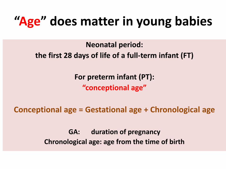
“Age” does matter in young babies
Neonatal period:
the first 28 days of life of a full-term infant (FT)
For preterm infant (PT):
“conceptional age”
Conceptional age = Gestational age + Chronological age
GA: duration of pregnancy
Chronological age: age from the time of birth

Neonatal seizures
• Most vulnerable period of life for developing sz
• Greatest occurrence in the first week
• Incidence 3: 1,000 live births
• Very high incidence in PT 57-132: 1,000
• May be short lived event
• Acute symptomatic >> epilepsy
• Often signify serious malfunction/damage to the immature brain
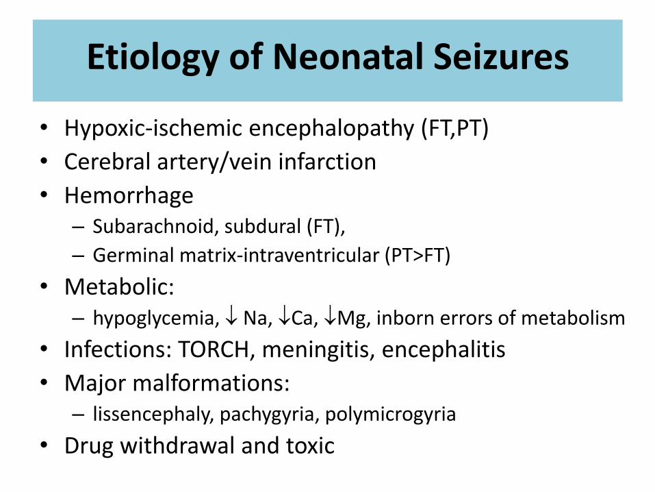
Etiology of Neonatal Seizures
• Hypoxic-ischemic encephalopathy (FT,PT)
• Cerebral artery/vein infarction
• Hemorrhage – Subarachnoid, subdural (FT),
– Germinal matrix-intraventricular (PT>FT)
• Metabolic: – hypoglycemia, Na, Ca, Mg, inborn errors of metabolism
• Infections: TORCH, meningitis, encephalitis
• Major malformations: – lissencephaly, pachygyria, polymicrogyria
• Drug withdrawal and toxic

Clinical Manifestations of Neonatal Seizures
• Paroxysmal, repetitive, stereotype
• Usually clinical subtle, inconspicuous and difficult to recognise from the normal behaviors or physiologic phenomena
• No recognisable post-ictal state.
• Frequently be overlooked
??????????????????????????????????????????

Classification of Neonatal Seizures
1. Subtle 50%
2. Clonic (focal, multifocal) 25%
3. Tonic (focal, generalized) 5%
4. Myoclonic (focal, multifocal, generalized) 20%
5. Non-paroxysmal repetitive behaviors
J. Volpe, Neurology of the Newborn 2008, p.203-237
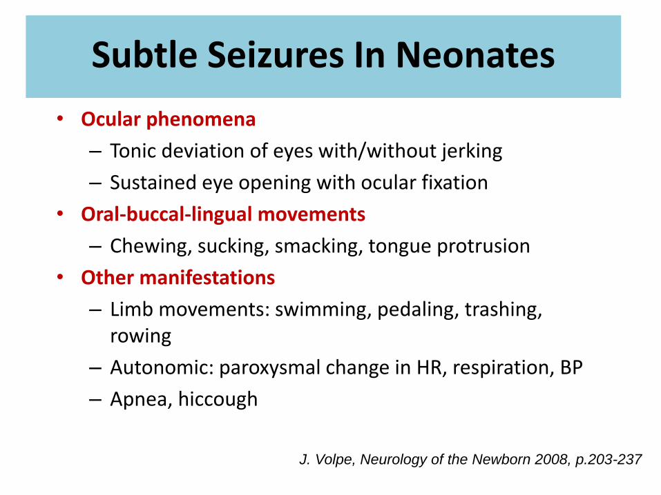
Subtle Seizures In Neonates
• Ocular phenomena
– Tonic deviation of eyes with/without jerking
– Sustained eye opening with ocular fixation
• Oral-buccal-lingual movements
– Chewing, sucking, smacking, tongue protrusion
• Other manifestations
– Limb movements: swimming, pedaling, trashing, rowing
– Autonomic: paroxysmal change in HR, respiration, BP
– Apnea, hiccough
J. Volpe, Neurology of the Newborn 2008, p.203-237
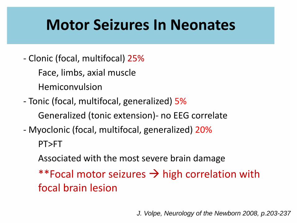
Motor Seizures In Neonates
- Clonic (focal, multifocal) 25%
Face, limbs, axial muscle
Hemiconvulsion
- Tonic (focal, multifocal, generalized) 5%
Generalized (tonic extension)- no EEG correlate
- Myoclonic (focal, multifocal, generalized) 20%
PT>FT
Associated with the most severe brain damage
**Focal motor seizures high correlation with focal brain lesion
J. Volpe, Neurology of the Newborn 2008, p.203-237
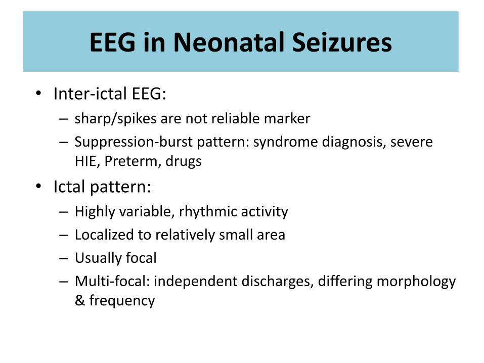
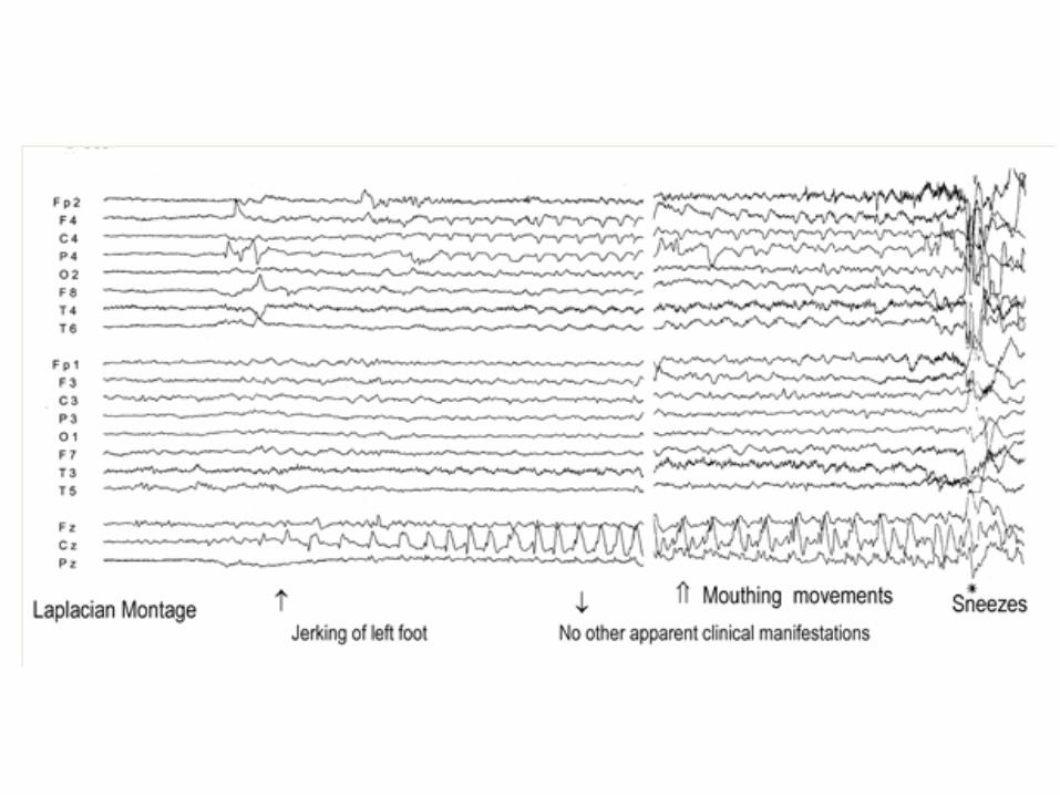
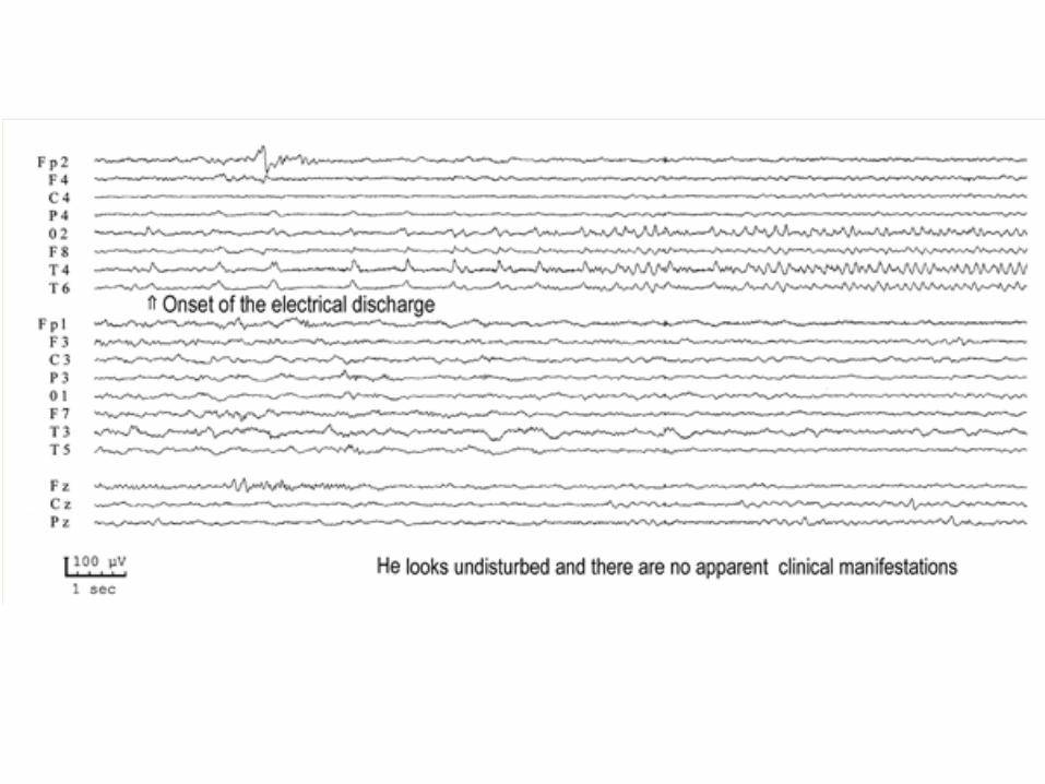
EEG in Neonatal Seizures
• Inter-ictal EEG:
– sharp/spikes are not reliable marker
– Suppression-burst pattern: syndrome diagnosis, severe HIE, Preterm, drugs
• Ictal pattern:
– Highly variable, rhythmic activity
– Localized to relatively small area
– Usually focal
– Multi-focal: independent discharges, differing morphology & frequency

Diagnosis of Neonatal Seizures
• Differentiate between non-epileptic vs epileptic:
– Suppressed by restrain or repositioning?
– Elicited by tactile stimulation?
• Detailed Hx of risk/etiology:
– Pregnancy -> birth –> afterbirth, Family Hx
• Physical exam
– Altered mental status, AF, focal neuro signs
– Dysmorphic features, neurocutaneous stigmata
– Peculiar odours
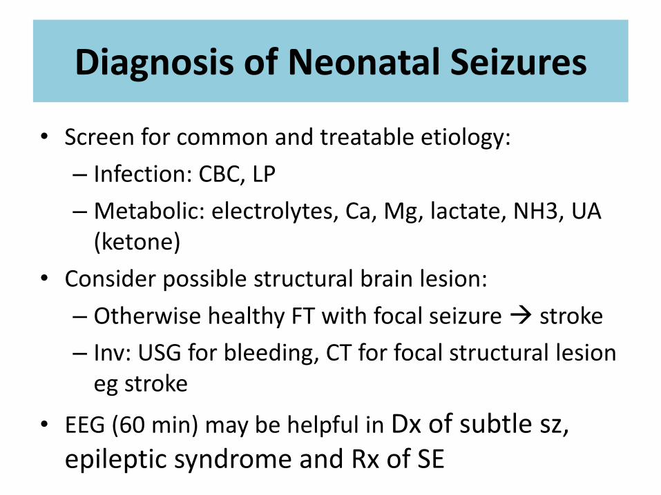
Diagnosis of Neonatal Seizures
• Screen for common and treatable etiology:
– Infection: CBC, LP
– Metabolic: electrolytes, Ca, Mg, lactate, NH3, UA (ketone)
• Consider possible structural brain lesion:
– Otherwise healthy FT with focal seizure stroke
– Inv: USG for bleeding, CT for focal structural lesion eg stroke
• EEG (60 min) may be helpful in Dx of subtle sz, epileptic syndrome and Rx of SE
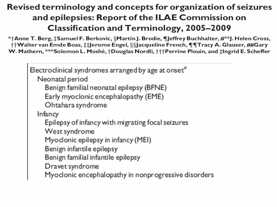

Epileptic Syndrome in Neonate
• Benign familial neonatal epilepsy (BFNC)
• Early Myoclonic Encephaopathy (EME)
• Ohtahara syndrome (Early Infantile Epileptic Encephalopathy, EIEE)
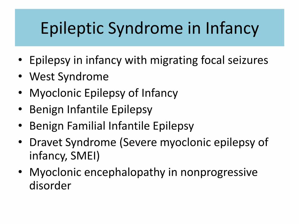
Epileptic Syndrome in Infancy
• Epilepsy in infancy with migrating focal seizures
• West Syndrome
• Myoclonic Epilepsy of Infancy
• Benign Infantile Epilepsy
• Benign Familial Infantile Epilepsy
• Dravet Syndrome (Severe myoclonic epilepsy of infancy, SMEI)
• Myoclonic encephalopathy in nonprogressive disorder

Benign Neonatal Convulsions
Benign Familial Neonatal Seizures (BFNC)
Benign Idiopathic (non-familial) Neonatal Seizures
(BINC)
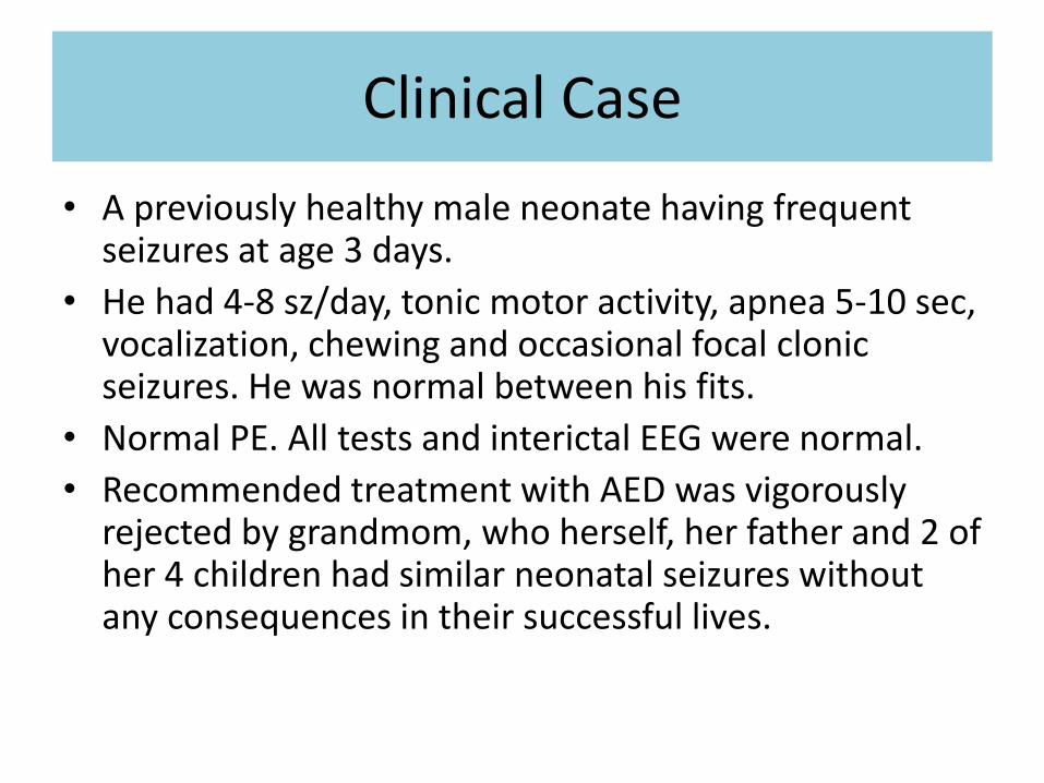
Clinical Case
• A previously healthy male neonate having frequent seizures at age 3 days.
• He had 4-8 sz/day, tonic motor activity, apnea 5-10 sec, vocalization, chewing and occasional focal clonic seizures. He was normal between his fits.
• Normal PE. All tests and interictal EEG were normal.
• Recommended treatment with AED was vigorously rejected by grandmom, who herself, her father and 2 of her 4 children had similar neonatal seizures without any consequences in their successful lives.
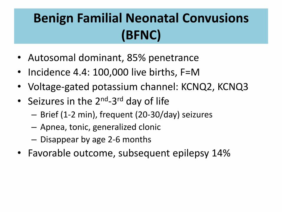
Benign Familial Neonatal Convusions (BFNC)
• Autosomal dominant, 85% penetrance
• Incidence 4.4: 100,000 live births, F=M
• Voltage-gated potassium channel: KCNQ2, KCNQ3
• Seizures in the 2nd-3rd day of life – Brief (1-2 min), frequent (20-30/day) seizures
– Apnea, tonic, generalized clonic
– Disappear by age 2-6 months
• Favorable outcome, subsequent epilepsy 14%
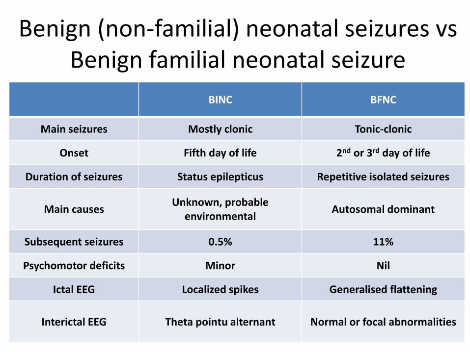
Benign (non-familial) neonatal seizures vs Benign familial neonatal seizure
BINC BFNC
Main seizures Mostly clonic Tonic-clonic
Onset Fifth day of life 2nd or 3rd day of life
Duration of seizures Status epilepticus Repetitive isolated seizures
Main causes Unknown, probable
environmental Autosomal dominant
Subsequent seizures 0.5% 11%
Psychomotor deficits Minor Nil
Ictal EEG Localized spikes Generalised flattening
Interictal EEG Theta pointu alternant Normal or focal abnormalities
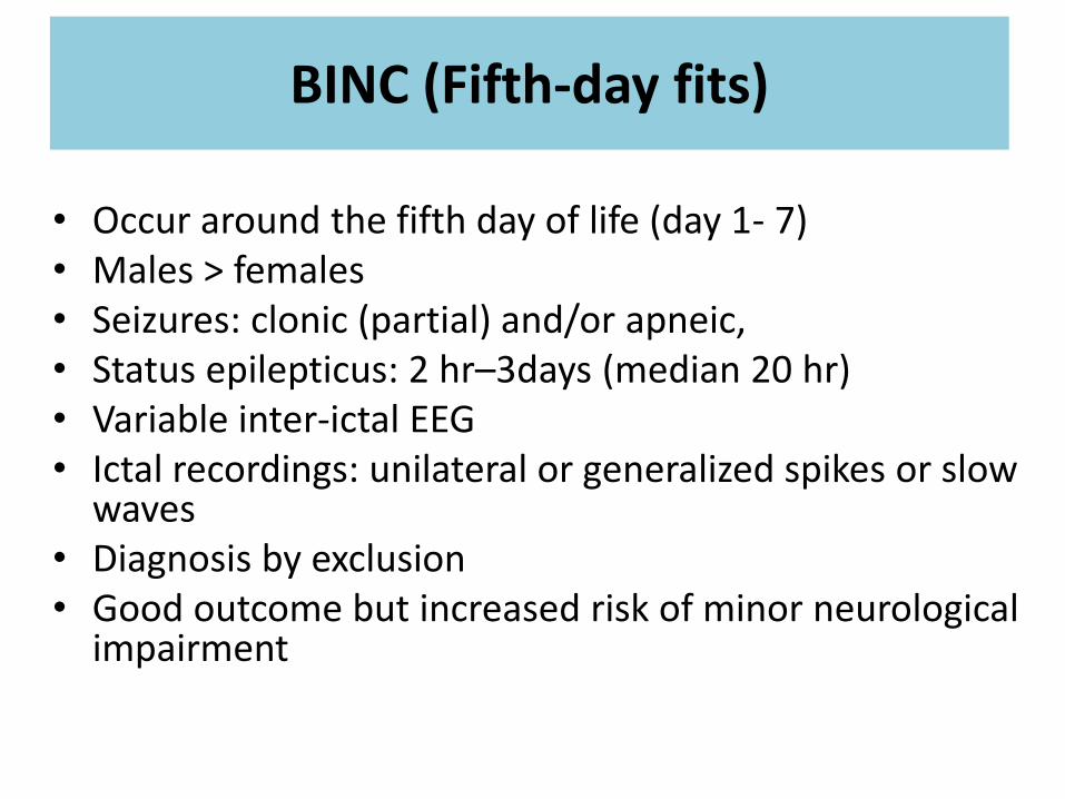
BINC (Fifth-day fits)
• Occur around the fifth day of life (day 1- 7) • Males > females • Seizures: clonic (partial) and/or apneic, • Status epilepticus: 2 hr–3days (median 20 hr) • Variable inter-ictal EEG • Ictal recordings: unilateral or generalized spikes or slow
waves • Diagnosis by exclusion • Good outcome but increased risk of minor neurological
impairment

Early Infantile Epileptic
Encephalopathies
with Suppression-Burst
Early Infantile Epileptic Encephalopathy
(EIEE: Ohtahara syndrome)
Early Myoclonic Encephalopathy (EME)
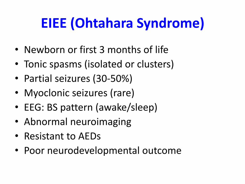
EIEE (Ohtahara Syndrome)
• Newborn or first 3 months of life
• Tonic spasms (isolated or clusters)
• Partial seizures (30-50%)
• Myoclonic seizures (rare)
• EEG: BS pattern (awake/sleep)
• Abnormal neuroimaging
• Resistant to AEDs
• Poor neurodevelopmental outcome
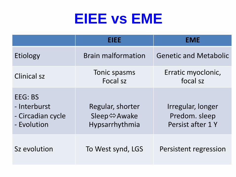
EIEE vs EME EIEE EME
Etiology Brain malformation Genetic and Metabolic
Clinical sz Tonic spasms Focal sz
Erratic myoclonic, focal sz
EEG: BS - Interburst - Circadian cycle - Evolution
Regular, shorter SleepAwake
Hypsarrhythmia
Irregular, longer Predom. sleep
Persist after 1 Y
Sz evolution To West synd, LGS Persistent regression
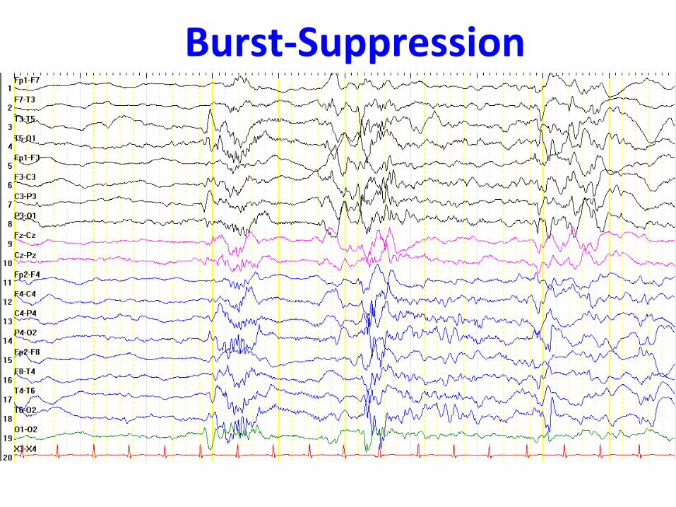
Burst-Suppression
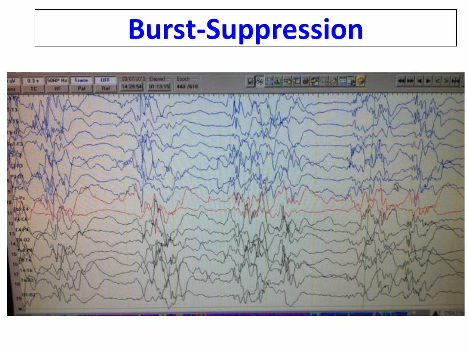
Burst-Suppression
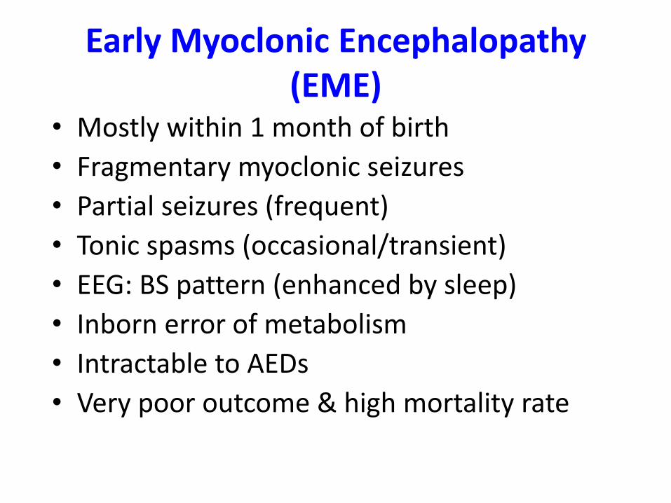
Early Myoclonic Encephalopathy (EME)
• Mostly within 1 month of birth
• Fragmentary myoclonic seizures
• Partial seizures (frequent)
• Tonic spasms (occasional/transient)
• EEG: BS pattern (enhanced by sleep)
• Inborn error of metabolism
• Intractable to AEDs
• Very poor outcome & high mortality rate

Epileptic Spasms
• Sustained contraction of axial muscles
• Flexion of neck & trunk with abduction and elevation of both arms
• “Salaam position”
• Initial movement relatively fast (myoclonic-like)
• Remain in the Salaam position for few seconds before relaxation
• Duration of each spasm: milliseconds to 5-10 seconds
• In CLUSTER
• Ictal EEG: Diffuse high-voltage slow followed by background attenuation

West Syndrome
• Epileptic(infantile) spasms
• Delayed development
• EEG- Hypsarrhythmia

West Syndrome
• Onset 3-7 months of age
• 1.6-4.3: 10,000 live births
• Spasms- flexor, extensor or mixed
• In clusters, frequently during drowsiness/arousal
• EEG: Hypsarrhythmia
(chaotic, irregular, diffuse asymmetric, high-voltage, interspersed with sharp waves and spikes)
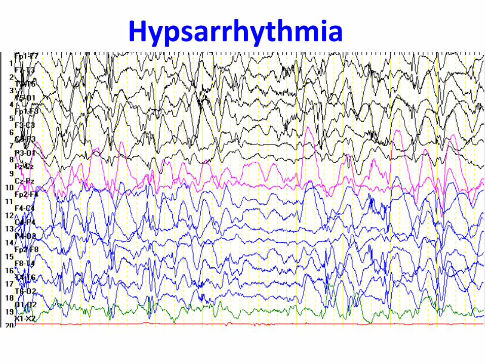
Hypsarrhythmia

Ictal EEG and EMG during Spasms
Federico Vigevano, Brain & Development 23 (2001) 467–472
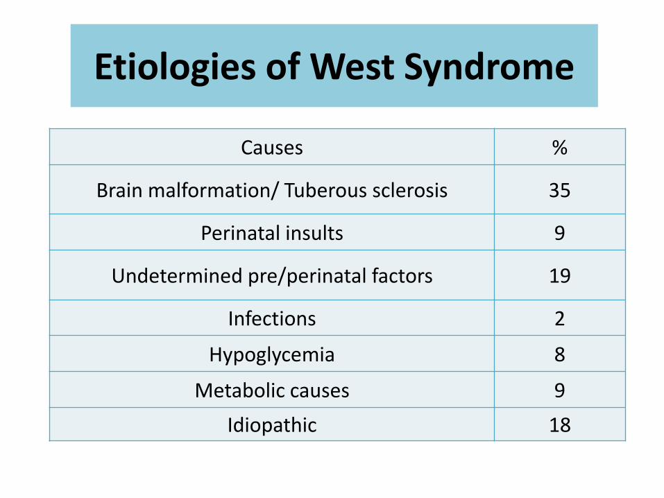
Causes %
Brain malformation/ Tuberous sclerosis 35
Perinatal insults 9
Undetermined pre/perinatal factors 19
Infections 2
Hypoglycemia 8
Metabolic causes 9
Idiopathic 18
Etiologies of West Syndrome
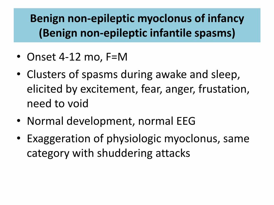
Benign non-epileptic myoclonus of infancy (Benign non-epileptic infantile spasms)
• Onset 4-12 mo, F=M
• Clusters of spasms during awake and sleep, elicited by excitement, fear, anger, frustation, need to void
• Normal development, normal EEG
• Exaggeration of physiologic myoclonus, same category with shuddering attacks
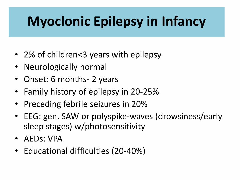
Myoclonic Epilepsy in Infancy
• 2% of children<3 years with epilepsy
• Neurologically normal
• Onset: 6 months- 2 years
• Family history of epilepsy in 20-25%
• Preceding febrile seizures in 20%
• EEG: gen. SAW or polyspike-waves (drowsiness/early sleep stages) w/photosensitivity
• AEDs: VPA
• Educational difficulties (20-40%)
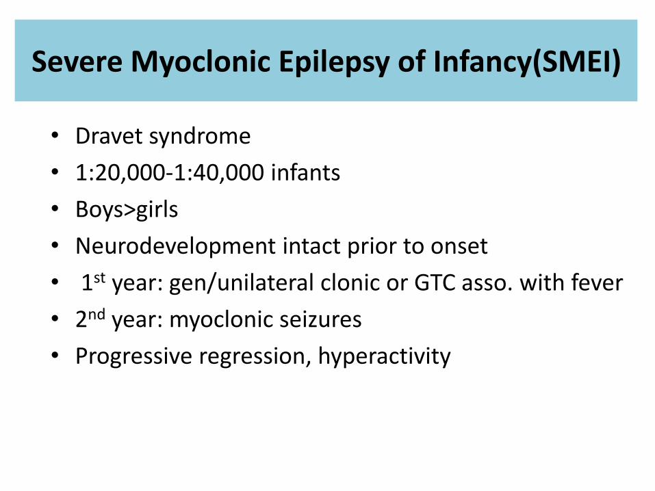
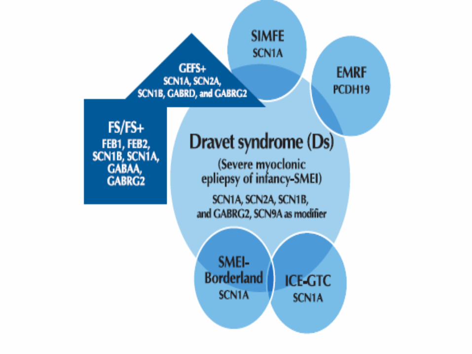
Severe Myoclonic Epilepsy of Infancy(SMEI)
• Dravet syndrome
• 1:20,000-1:40,000 infants
• Boys>girls
• Neurodevelopment intact prior to onset
• 1st year: gen/unilateral clonic or GTC asso. with fever
• 2nd year: myoclonic seizures
• Progressive regression, hyperactivity

Severe Myoclonic Epilepsy of Infancy(SMEI)
• Highly intractable to AEDs
– Useful AEDs: VPA, Benzodiazepines(CNZ, Clobazam, Lorazepam), Stiripentol
– Alternative: ZNS, VGB, Ketogenic diet
– Avoid: LTG?, PHT, CBZ
• Prognosis is very poor

Thank You for Your Attention