Epilepsy and Seizures Part 2: Introduction to Two Epilepsy Syndromes and Therapeutics Jacquelyne S....
47
Epilepsy and Seizures Part 2: Introduction to Two Epilepsy Syndromes and Therapeutics Jacquelyne S. Cios, MD MS Assistant Professor Department of Neurology, Division of Epilepsy [email protected] “It is thus with regard to the disease called Sacred: it appears to me to be nowise more divine nor more sacred than other diseases, but has a natural cause like other affections.” - Hippocrates, 400 BC
-
Upload
maud-pierce -
Category
Documents
-
view
216 -
download
0
Transcript of Epilepsy and Seizures Part 2: Introduction to Two Epilepsy Syndromes and Therapeutics Jacquelyne S....

Epilepsy and SeizuresPart 2: Introduction to Two Epilepsy Syndromes and Therapeutics
Jacquelyne S. Cios, MD MSAssistant Professor
Department of Neurology, Division of Epilepsy
“It is thus with regard to the disease called Sacred: it appears to me to be nowise more divine nor more sacred than other
diseases, but has a natural cause like other affections.”
- Hippocrates, 400 BC

Learning Objectives
Be able to describe some clinical, pathological, electrographic and radiographic features of the syndromes of Mesial Temporal Lobe Epilepsy and Childhood Absence Epilepsy.
Be familiar with a the availability of a range of epilepsy therapeutics including pharmacological and procedural based options.
Be able to recognize status epilepticus and be familiar with the basic treatment algorithm.

Part 2: Outline
Treatment Anti-epileptic drugs – mechanism and side effects
First generation Second generation
Vagal nerve stimulator Surgery Experimental: RNS Ketogenic diet
Role of the epilepsy monitoring unit (EMU) Status epilepticus
A closer look at two important syndromes
Mesial temporal lobe epilepsy vs. childhood absence epilepsy
Both commonly encountered in the adult population Illustrates partial vs. generalized dichotomy
Both have events of staring and unresponsiveness Both have events of tonic-clonic seizures But MTLE - partial onset seizures; CAE – generalized onset
seizures They differ in the focus or origin of the seizures and pattern of
spread
Let’s look at their clinical, electrographic, and radiographic features

Mesial temporal lobe epilepsy: clinical presentation
Risk factors include: History of childhood febrile seizures (81%) - this is a strong predictor Meningoencephalitis at presentation (10%) Head trauma (10%) No apparent risk factor (7%) Birth trauma (3%)
Histopathologically, many have mesial temporal sclerosis There is an interval between cerebral insult and onset of seizures (>95%, mean 7 years) Mean age at seizure onset – 9 years Some have only complex partial events (50%) without Gen tonic-clonic events
GTC is not the primary (most frequent) sz type Convulsive status epilepticus (repeated seizures constituting a medical emergency) is
uncommon(4%) Auras are common (96%)
Abdominal visceral sensation is most common Evolution of the disease over time
A silent interval of mean 6.7 years after onset of epilepsy Seizure phenomenology becoming more elaborate over time

Auras
Simple partial seizures that often precede a seizure Can last for seconds up to 1-2 minutes Common auras
Viscerosensory – “rising epigastric sensation” Altered perception - Déjà vu or jamais vu Unpleasant or metallic taste
Less common Visual and auditory illusions or hallucinations Tactile sensation

Impaired consciousness
Following the aura, the patient has altered consciousness
May still have upright posture, with eyes open, and still be talking in some cases
Commonly a behavioral arrest with staring for 30-60 seconds
Patients have amnesia for the event May also have variable amount of retrograde amnesia, including
the aura Due to bilateral hippocampal impairment of function during the
seizure

Automatisms
The behavioral arrest commonly includes involuntary, purposeless, repetitive motor activities
These are stereotypic (look identical when compared between seizures) Patient is amnestic to these Involving hands (Fumbling, picking, fidgeting) Or mouth (chewing, swallowing, lipsmacking)
These usually help localize the onset
May be de novo Doing something that is triggered after seizure onset , ie, chewing something placed in
patient’s mouth These include release phenomena- actions that are normally inhibited by frontal lobe
function ie, socially unacceptable behaviors
May be preservative (continuing to do something that patient was engaged in previous to sx onset)
Have localizing value Generally ipsilateral to the side of the seizure onset (the lesion)

Quiz - 06 Seizures and Epilepsy

Less common seizure manifestations
Leaving Running away, out of the room (cursive)
Vocalizations Ictal speech (or sign language) Affective behaviors (out of fear)
There is commonly a fearful look on the patient’s face Stems from involvement of the uncus, hence the early term for this
sz type, “uncinate fits”
Dacrystic (crying) Gelastic (laughing) – associated with hypothalamic
hamartoma Whistling

Spread to the motor areas
As the temporal lobe discharge recruits more territory, it marches across the brain
Contralateral limb dystonia is often seen There may be an early, nonforced head turn toward the side of the
sz onset There may be a forced head turn (version) later in the sz away from
the side of onset May be associated with gaze deviation also contralateral to the
lesion Spread to the supplementary motor area (SMA) precedes
generalization (involvement of whole cortex and resultant tonic-clonic activities) May be a fencing posture (head turned toward extended arm, c/L to the lesion,
also called M2E) May be a figure 4 sign (extended leg c/l to the lesion, flexed leg at the knee
joint I/L to the lesion)

Quiz - 07 Seizures and Epilepsy

A complex partial onset seizure temporal lobe onset

Secondary generalized seizure
Partial onset with secondary generalization

The EEG of mesial temporal lobe seizure

Mesial temporal sclerosis: MRI and histopath
A : a kainic acid induced rat model of MTS 3 days after injection. Inset shows degenerating hippocampal neurons at higher powerB: same specimen at 14 days showing aberrant mossy fiber sprouting in the dentate gyrus (Timm’s stain)

Quiz - 08 Seizures and Epilepsy

Childhood absence epilepsy: clinical presentation A model for idiopathic generalized epilepsies Seizures start between 3- 8 yrs old (peak 6-7) Typical absence seizure
Sudden brief impairment of consciousness Accompanied by characteristic EEG change 3 Hz (2.5-4 Hz) generalized spike and wave May have automatisms, non stereotyped Eyes, open, staring Impairment of consciousness and amnesia for event Lasting generally 10 seconds May occur up to 200 times per day

Childhood absence epilepsy
No developmental, metabolic, neurological, pathological abnormalities evident
May spontaneously go into remission (70%), around adolescence
Generally do not have frequent generalized tonic clonic sz (GTC)
Except in those 30% who continue to have absences into adulthood
May also have febrile seizures (30%) May be due to a Arg-> Glu polymorphism in GABA A receptor Prognosis is generally good for seizure freedom with or
without pharmacologic treatment (65%)

Video of Typical Myoclonic Seizures
Video of typical myoclonic seizures
Juvenile myoclonic epilepsy, like childhood absence epilepsy, is another type if idiopathic generalized epilepsy

3 Hz generalized spike and wave

Quiz - 09 Seizures and Epilepsy

Part 2: Treatment of epilepsyTreatment
Anti-epileptic drugs – mechanism and side effectsFirst generationSecond generation
Vagal nerve stimulatorSurgeryRNSKetogenic diet
Differential diagnosis and the epilepsy monitoring unit (EMU)Status epilepticus

Anticonvulsants: first generation drugs
1945 - 1963 Tridione , methsuximide ethosuxamide, Primidone, ethotoin,
diazepam
1974 Carbamazepine - discovered by Walter Schindler in 1953 at
J.R.Geigy (now Novartis) marketed for trigeminal neuralgia in 1962 – anticonvulsant in the
UK 1965
1978 Valproic acid - Pierre Eymard discovered anticonvulsant activity
in 1962 First synthesized by Burton 1882 (Bromide)

Anticonvulsants: second generation
1993 – felbamate 1994 – gabapentin, lamotrigine 1996 – topiramate, fosphenytoin 1997 – tiagabine, rectal diazepam 1999 – levetiacetam 2000 – zonisamide, oxcarbazepine 2005 – pregabalin 2008 – lacosamide, rufinamide

Anticonvulsants by mechanism
Ca channel Na Channel GABA Glutamate Other
EthosuximidePregabalinZonisamide
CarbamazepineLacosamideLamotrigineOxcarbazepinePhenytoinRufinamideZonisamide
BarbituratesBenzodiazepinesGabapentinTiagabineValproateVigabatrin
FelbamateTopiramate
LevetiracetamLacosamideRetigabine (Ezogabine)

Special topics relating to AED use
Phenytoin, carbemazepine, oxcarbazepine are generally used for partial epilepsy
Broad spectrum anticonvulsants are known to be useful for both partial and generalized onset seizures These include levetiracetam, topiramate, valproic acid
A few AEDs are only used in very specific clinical indications Vigabatrin – infantile spasms Ethosuximide – absence seizures
Some AEDs have nonlinear kinetics As the dose of phenytoin is increased, the steady state plasma
concentration rises exponentially Starting phenytoin efficiently require a “loading dose”
Example: 20 mg / kg iv followed by 5 mg /kg/ day divided into twice daily doses

Drug- drug interactions First generation anticonvulsants are heavily P450
metabolized Carbemazepine and phenytoin are P450 inducers
Carbemazepine can auto-induce Valproate is a P450 inhibitor
This can cause interactions which are clinically relevant When adding valproate to the medication regimen of a patient on
lamotrigine, would you add it slowly or quickly to avoid side effects of lamotrigine?
Hint: valproate inhibits P450 mediated elimination of lamotrigine causing higher serum levels of lamotrigine
Many of the drugs are highly protein bound The free fraction is the active form Check free levels in patients with low protein states

Quiz - 10 Seizures and Epilepsy

Epilepsy therapeutics: diet
Low glycemic index diet (LGID) Originally for blood sugar control with efficacy in epilepsy More tolerable version of ketogenic (high fat, low carb) diet 30-50% SFO

LGID/KD - Complications
LGID/KD - Complications Refusal to eat Hyperlipidemia Elevated liver enzymes Constipation Metabolic acidosis Hypoglycemia Weight loss / growth retardation Nephrolithiasis (kidney stones)

Epilepsy therapeutics: VNS
Vagus nerve stimulator (VNS)1930s – carotid sinus massage1997 – FDA approval for epilepsy2004 approved for treatment of depression30% of patients obtain >50% seizure reductionNo more effective than medicines wrt SFO

Epilepsy therapeutics: surgery
One third of patients with epilepsy are medically refractory One third of patients with epilepsy are medically
refractory Failed 2-3 single drugs and/or a combination of drugs
These patients may be amenable to surgery Anteromedial temporal resection (AMTR) – chance of long term
seizure freedom or meaningful reduction in seizure frequency in medial temporal lobe epilepsy
Corpus callosotomy Functional hemispherectomy (hemispherotomy) Multiple subpial transection (MST)
Pre-surgical evaluation
Will it work? 24 hr (continuous ictal and interictal) video EEG (Epilepsy Monitoring
Unit) Intraoperative monitoring
Subdural strips or grids of electrodes placed on the cortical surface Implanted depth electrodes – longitudinally along the temporal pole
Ictal SPECT
Is it safe? Studies to determine whether the ictal onset zone is far enough away
from eloquent brain regions Neuropsychological testing WADA Electrocorticography – intraoperative Cortical mapping Functional MRI
Quiz - 11 Seizures and Epilepsy

Status epilepticus
Status Epilepticus A person is in status epilepticus if they are experiencing a seizure of
longer than 5 minutes duration OR if they have experienced 2 contiguous seizures without regaining
consciousness between seizures Convulsive or non-convulsive (eyes open)

Status Epilepticus Treatment Algorithm
Airway Breathing Circulation Dextrose (check for glucose levels as well as other
electrolyte abnormalities) Epilepsy
Labs Benzodiazepine Anticonvulsant Anesthesia Barbituate Coma (central line / EEG)
EMS/ED/Floor
ICU

Status Epilepticus Treatment Algorithm
• Treatment– If someone is seizing
• Lay on side• Clear area around them• Loosen tight clothing• Do not hold down• Do not put anything in mouth

Status Epilepticus Treatment Algorithm
ABCDEpilepsy Benzodiazepine
Lorazepam (Ativan) 1-2 mg doses IV every 5 minutes 2 mg / min max rate Repeat dosing pending AED arrival and infusion Maximum dose = 0.1 mg / kg total
Anticonvulsant Phenytoin loading dose = 20 mg/kg
Anesthesia

Status Epilepticus
• Treatment Efficacy– 35-45% of patients with status epilepticus will stop seizing with
aforementioned treatments– Patients that do not, need
• general anesthesia• brain imaging studies (head CT/CTA) • transfer to the ICU for
– continuous EEG monitoring– Possible barbiturate coma with hemodynamic monitoring

Quiz - 12 Seizures and Epilepsy

Epilepsy is a complex field
Other issues in clinical practice Social stigma Psychiatric comorbidity Functional disability and socioeconomic status Epilepsy and women’s issues SUDEP Medication adherence
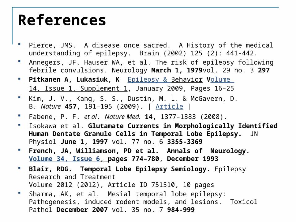
References Pierce, JMS. A disease once sacred. A History of the medical understanding of
epilepsy. Brain (2002) 125 (2): 441-442. Annegers, JF, Hauser WA, et al. The risk of epilepsy following febrile convulsions.
Neurology March 1, 1979vol. 29 no. 3 297 Pitkanen A, Lukasiuk, K Epilepsy & Behavior Volume 14, Issue 1, Supplement 1,
January 2009, Pages 16–25 Kim, J. V., Kang, S. S., Dustin, M. L. & McGavern, D. B. Nature 457, 191–195
(2009). | Article | Fabene, P. F. et al. Nature Med. 14, 1377–1383 (2008). Isokawa et al. Glutamate Currents in Morphologically Identified Human Dentate
Granule Cells in Temporal Lobe Epilepsy. JN Physiol June 1, 1997 vol. 77 no. 6 3355-3369
French, JA, Williamson, PD et al. Annals of Neurology. Volume 34, Issue 6, pages 774–780, December 1993
Blair, RDG. Temporal Lobe Epilepsy Semiology. Epilepsy Research and TreatmentVolume 2012 (2012), Article ID 751510, 10 pages
Sharma, AK, et al. Mesial temporal lobe epilepsy: Pathogenesis, induced rodent models, and lesions. Toxicol Pathol December 2007 vol. 35 no. 7 984-999

Quiz - Seizures and Epilepsy
Thank you for completing this module
Questions? Contact me at:
?

Survey
We would appreciate your feedback on this module. Click on the button below to complete a brief survey. Your responses and comments will be shared with the module’s author, the LSI EdTech team, and LSI curriculum leaders. We will use your feedback to improve future versions of the module.
The survey is both optional and anonymous and should take less than 5 minutes to complete.
Survey