
Epidemiologia das infestações por Oestrus ovis em … · Botucatu e influência da raça ovina no...
141
Campus de Botucatu Instituto de Biociências PG-BGA Epidemiologia das infestações por Oestrus ovis em ovinos criados em Botucatu e influência da raça ovina no parasitismo Bruna Fernanda da Silva Tese apresentada ao Instituto de Biociências, Câmpus de Botucatu, UNESP, para obtenção do título de Doutor no Programa de Pós-Graduação em Biologia Geral e Aplicada, Área de concentração Biologia de Parasitas e Microorganismos. Prof. Titular Dr. Alessandro Francisco Talamini do Amarante Orientador BOTUCATU – SP 2012
Transcript of Epidemiologia das infestações por Oestrus ovis em … · Botucatu e influência da raça ovina no...

Campus de Botucatu
Instituto de Biociências PG-BGA
Epidemiologia das infestações por Oestrus ovis em ovinos criados em
Botucatu e influência da raça ovina no parasitismo
Bruna Fernanda da Silva
Tese apresentada ao Instituto de Biociências, Câmpus de
Botucatu, UNESP, para obtenção do título de Doutor no
Programa de Pós-Graduação em Biologia Geral e
Aplicada, Área de concentração Biologia de Parasitas e
Microorganismos.
Prof. Titular Dr. Alessandro Francisco Talamini do Amarante
Orientador
BOTUCATU – SP
2012
Campus de Botucatu
Instituto de Biociências PG-BGA
UNIVERSIDADE ESTADUAL PAULISTA
“Julio de Mesquita Filho”
INSTITUTO DE BIOCIÊNCIAS DE BOTUCATU
Epidemiologia das infestações por Oestrus ovis em ovinos criados em
Botucatu e influência da raça ovina no parasitismo
Bruna Fernanda da Silva
Bióloga
Prof. Titular Dr. Alessandro Francisco Talamini do Amarante
Orientador
Tese apresentada ao Instituto de Biociências, Campus de
Botucatu, UNESP, para obtenção do título de Doutor no
Programa de Pós-Graduação em Biologia Geral e
Aplicada, Área de concentração Biologia de Parasitas e
Microorganismos.
BOTUCATU – SP
2012

FICHA CATALOGRÁFICA ELABORADA PELA SEÇÃO DE AQUIS. E TRAT. DA INFORMAÇÃO
DIVISÃO TÉCNICA DE BIBLIOTECA E DOCUMENTAÇÃO - CAMPUS DE BOTUCATU - UNESP
BIBLIOTECÁRIA RESPONSÁVEL: ROSEMEIRE APARECIDA VICENTE
Silva, Bruna Fernanda.
Epidemiologia das infestações por Oestrus ovis em ovinos criados em
Botucatu e influência da raça ovina no parasitismo / Bruna Fernanda da Silva. –
Botucatu : [s.n.], 2012
Tese (doutorado) - Universidade Estadual Paulista, Instituto de Biociências
Orientador: Alessandro Francisco Talamini do Amarante
Capes: 21300003
1. Parasitologia. 2. Epidemiologia. 3. Ovino – Doenças – Imunologia.
Palavras-chave: Controle; Epidemiologia; Haemonchus contortus; Oestrus ovis;
Ovinos; Resposta imunológica; Trichostrongylus colubriformis.

“As coisas que são impossíveis aos homens são possíveis a Deus”
Lucas 18:27

Dedico
Aos meus pais, José Horácio e Benedita e a minha irmã Beatriz
Meus exemplos de amor

Agradecimentos
A Deus pelo dom da vida e por todas as maravilhas que tem feito em minha
vida, por iluminar e abençoar meus caminhos e o meu trabalho.
Aos meus pais José Horácio e Benedita e minha irmã Beatriz pelo amor
incondicional, carinho, dedicação, paciência, educação, incentivo... Obrigada por
acreditarem nos meus sonhos e por me darem apoio para realizar cada um deles. Vocês
são a minha fonte de inspiração, meu porto seguro, meu esteio, meu chão. A vocês
dedico todo meu amor.
Ao meu orientador Prof. Dr. Alessandro Amarante por ter me concedido à
oportunidade de estágio em 2005, me abrindo as portas do mundo científico, onde tudo
começou. Obrigada pela confiança, por todo conhecimento transmitido, paciência e
dedicação ao longo de todos estes anos de trabalho. Obrigada pela impagável dedicação
e paciência nas muitas correções de relatórios e artigos e, além disso, por literalmente
colocar as “mãos na massa” no trabalho de campo, auxiliando em todas as coletas e
acompanhando de perto o andamento dos trabalhos. É um privilégio ser orientada por
você... muito obrigada por tudo!
A Fapesp pela bolsa e auxílio financeiro concedidos para a realização deste
trabalho.
A Capes pela bolsa concedida para a realização do estágio de doutorado no
Hopkirk Research Institute – AgResearch, Palmerston North - Nova Zelândia, onde tive
a oportunidade de ampliar os conhecimentos.
Ao Dr. Anton Pernthaner, meu co-orientador durante o estágio de doutorado,
pela oportunidade de estágio, por todo conhecimento transmitido, paciência e amizade.
A minha prima-irmã Carolina por todo amor, carinho, apoio e paciência. E a
toda minha família pelo apoio e incentivo.

A minha mana do coração Cristina P. Cardoso e sua (nossa) família, pela linda e
sincera amizade que nasceu durante esta importante etapa de nossas vidas. Obrigada
pelo privilégio de poder compartilhar tantas coisas boas contigo e nossa família. Amo
vocês!
A todos os animais que doaram suas vidas para a realização deste trabalho e a
todos os ovinocultores que colaboraram com os experimentos.
Ao amigo César Bassetto pela parceria, paciência, amizade e fundamental
colaboração em todas as etapas deste trabalho. Com a tua ajuda, com certeza, eu faria
tudo novamente, inclusive criar bezerros! Aproveito para agradecer toda sua família,
amigos queridos, pelo apoio.
Ao amigo e técnico de laboratório, Valdir Paniguel, por todo auxílio prestado
durante os experimentos.
Aos funcionários da Fazenda Edgardia – Unesp, Nico e Edvaldo, pela amizade,
companheirismo e auxílios prestados.
Ao Sr Serginho e Sr Renato por me ajudarem a cuidar dos animais
experimentais.
Aos amigos do Laboratório de Helmintologia Veterinária: Maria Érika
Picharillo, Jorge K. Xavier, Camila O. Carvalho, César Bassetto, Michelle C. Santos,
Fabiana Almeida e Maurícia „Berne‟. Galerinha especial que “tá na m.... mas é feliz”.
Obrigada pelo companheirismo e auxílio nos experimentos, mas acima de tudo obrigada
pela linda amizade e por todos os momentos que passamos juntos ao longo destes
anos... com vocês até o “trabalho de preso” fica divertido. Obrigada por tudo!
Aos que fizeram parte do início desta jornada, na época do estágio: Raquel
Rocha, Patrizia Bricarello e Kátia Bresciani... o conhecimento transmitido por vocês
foram fundamentais para que eu chegasse até aqui. Muito obrigada!
A todos os estagiários que passaram pelo laboratório durante esses quatro anos,
em especial, Cássio Florencio, Regina Silva, Maurício Wilmsen e Christiano Dreyer.
Obrigada pela amizade e contribuição neste trabalho.

Ao Gustavo P. Machado e Thiago B. Izidoro pela parceria no trabalho
desenvolvido em Itápolis e por todo auxílio prestado.
A todos os médicos veterinários residentes da Clínica de Grandes Animais -
FMVZ, em especial, Juliana, Diego e Giovanni pelos auxílios prestados durante os três
anos de experimento, sempre “socorrendo” meus filhotes.
A Profª Lucia Helena O‟Dwyer pela linda amizade e por ser uma grande
incentivadora do meu trabalho.
Aos professores e funcionários do Departamento de Parasitologia, amigos
queridos: Nilza, Roberto (Bicho), Ângela, Márcia, Profª Mônica, Profª Luciene, Profª
Cristina, Profª Semiramis, Prof° Reinaldo, Prof° Newton e Prof° Paulo.
Aos amigos queridos do Departamento de Parasitologia e agregados: Eriquinha,
Alison, Ericona, Giovana, Larissa, Tatiana (Brusinha), Betina, Denise, Leticinha,
Letícia, Diego, Aline, Luciano, Teresa, Ana Paula (Patologia) e Thiaguinho
(Inspeção)... e aos que já passaram por lá: Karina Santos, Bianca, Marco (Xabi),
Aruaque, Karina Paduan, Adriano, Gustavo e Juliana. Obrigada por tudo... quem tem
amigos tem um tesouro!
A todos os amigos do Hopkirk Research Institute, em especial a Qing Deng,
Leticia Salvador, Rami, Tanu, Sadaf, Joanna, Charlotte e Sheralee. Foi maravilhoso
poder conviver com vocês!
A todos os amigos queridos da Nova Zelândia, em especial a Carol e Rafael,
Camila e Nico, Ricardinho, Dayanne, Svenja e Ryan, Soraya e Sydney e o pequeno
Nicolas... vocês foram a minha família na NZ. Obrigada pela linda amizade, carinho,
companheirismo e pelos inesquecíveis momentos que passamos juntos, vocês são
especiais e estarão para sempre no coração!
Na realidade, foram muitas as pessoas que sonharam junto comigo, que
torceram, apoiaram, incentivaram e hoje podem ver esse sonho se tornar realidade.
Sempre digo que este trabalho não é meu... é nosso!
Agradeço a todos que aqui não foram citados, mas que de alguma forma
contribuíram para que mais este sonho se tornasse realidade... que Deus os abençoe!
Muito obrigada!!!

"Escolhe um trabalho de que gostes, e não terás que trabalhar
nem um dia na tua vida."
Confúcio
"Não conheço ninguém que conseguiu realizar seu sonho, sem sacrificar feriados e
domingos pelo menos uma centena de vezes. O sucesso é construído à noite.
Durante o dia você faz o que todos fazem. Mas, para obter um resultado diferente
da maioria, você tem que ser especial. Se fizer igual a todo mundo, obterá os
mesmos resultados. Não se compare à maioria, pois infelizmente ela não é modelo
de sucesso. Se você quiser atingir uma meta especial, terá que estudar no horário
em que os outros estão tomando cerveja com batata frita. Terá de planejar,
enquanto os outros permanecem à frente da televisão. Terá de trabalhar enquanto os
outros tomam sol à beira da piscina. A realização de um sonho depende de
dedicação. Há muita gente que espera que o sonho se realize por mágica, mas toda
mágica é ilusão e a ilusão não tira ninguém de onde está. Na verdade a ilusão é
combustível dos perdedores, pois: 'Quem quer fazer alguma coisa, encontra um meio.
Quem não quer fazer nada, encontra uma desculpa'."
(Desconheço a autoria)

Sumário
Página
Resumo..............................................................................................................................1
Abstract..............................................................................................................................3
Capítulo 1..........................................................................................................................5
Introdução..............................................................................................................6
Epidemiologia......................................................................................................11
Soroprevalência...................................................................................................14
Ovino x Caprino...................................................................................................15
Resposta imunológica..........................................................................................15
Interação entre a infestação por O. ovis e a infecção por nematódeos
gastrintestinais.................................................................................................................17
Controle...............................................................................................................18
Considerações finais............................................................................................19
Referências bibliográficas....................................................................................20
Capítulo 2........................................................................................................................28
Epidemiology of Oestrus ovis in sheep in the southwest of Brazil…………………….29
Abstract................................................................................................................29
Introduction..........................................................................................................30
Materials and methods.........................................................................................31
Study location…………………………………………………………………..31
Management of tracer lambs……………………………………………………32
Sheep flock management……………………………………………………….33
Statistical analyses……………………………………………………………...33

Results…………………………………………………………………………..33
Clinical signs of oestrosis in the sheep flock…………………………………...33
Prevalence and intensity of infestation in tracer sheep…………………………34
Discussion………………………………………………………………………35
Acknowledgements……………………………………………………………..38
References………………………………………………………………………39
Capítulo 3………………………………………………………………………………46
Prevalence and intensity of Oestrus ovis infestation in sheep in central region of São
Paulo State, Brazil……………………………………………………………………...47
Abstract…………………………………………………………………………47
Introduction……………………………………………………………………..48
Materials and methods………………………………………………………….49
Statistical analyses……………………………………………………………...50
Results…………………………………………………………………………..51
Discussion………………………………………………………………………51
Acknowledgements……………………………………………………………..53
References………………………………………………………………………54
Capítulo 4………………………………………………………………………………60
Parasitism by Oestrus ovis: Influence of sheep breed and nematode infections………61
Abstract………………………………………………………...……………….61
Introduction……………………………………………………………………..62
Material and methods…………………………………………………………...64
Study location…………………………………………………………………..64
Animals and experimental design………………………………………………65
Fecal examination………………………………………………………………66

Pasture infectivity………………………………………………………………66
Hematology……………………………………………………………………..66
Serology – sandwich enzyme immunoassay (EIA) for IgE antibody…………67
Oestrus ovis: clinic signs and larvae count post mortem……………………….67
Worm counts……………………………………………………………………67
Statistical analyses……………………………………………………………...68
Results…………………………………………………………………………..68
Oestrus ovis: clinic signs and larvae count post mortem……………………….68
Fecal examination………………………………………………………………69
Nematode larvae on pasture……………………………………………………70
Hematology……………………………………………………………………..71
Serology – sandwich enzyme immunoassay (EIA) for IgE antibody…………..71
Weight gain……………………………………………………………………..72
Worm counts……………………………………………………………………72
Correlation coefficients…………………………………………………………72
Discussion……………………………………………………………………....73
Acknowledgements……………………………………………………………..77
References………………………………………………………………………79
Capítulo 5………………………………………………………………………………93
Cellular and humoral immune responses in sheep naturally infested with Oestrus ovis
(Diptera: Oestridae) and with nematode infections…………………………………….94
Abstract…………………………………………………………………………95
Introduction……………………………………………………………………..96
Material and methods…………………………………………………………...96
Animals…………………………………………………………………………96

Hematology……………………………………………………………………..97
Histology………………………………………………………………………..97
Mucus…………………………………………………………………………...98
Enzyme-linked immunosorbent assay (ELISA)………………………………..99
Parasites used in antigen production……………………………………………99
Parasite-specific serum IgG…………………………………………………...100
Parasite-specific mucus IgA…………………………………………………..101
Statistical analyses…………………………………………………………….101
Results…………………………………………………………………………102
Discussion……………………………………………………………………..104
Acknowledgement…………………………………………………….............108
References…………………………………………………………………......109

Lista de Figuras
Página
Capítulo 1
Figura 1. Larvas de Oestrus ovis em diferentes fases de desenvolvimento (vista dorsal).
Foto: B. F. Silva, 2009.......................................................................................................7
Figura 2. Vista ventral da larva de primeiro estádio (L1) de Oestrus ovis. Foto: B. F.
Silva, 2009.........................................................................................................................7
Figura 3. Detalhe da região posterior da larva de segundo estádio (L2) de Oestrus ovis
onde estão localizados os espiraculos respiratórios. Foto: B. F. Silva, 2009....................8
Figura 4. Larva de terceiro estádio (L3) de Oestrus ovis em diferentes fases de
desenvolvimento (A, B e C); espiraculos respiratórios localizados na região posterior do
corpo da larva (D). Foto: B. F. Silva, 2009.......................................................................9
Figura 5. Vista dorsal da mosca adulta de Oestrus ovis. Foto: B. F. Silva, 2008..........10
Figura 6. Ovelhas da raça Bergamacia com corrimento nasal, sinal clínico característico
do parasitismo por larvas de Oestrus ovis. Foto: B. F. Silva, 2008.................................11
Capítulo 2
Figure 1. Average of maximum, minimum and mean monthly temperatures (A);
monthly rainfall and relative air humidity (B); and monthly mean intensity of first larval
instar (L1), second larval instar (L2) and third larval instar (L3) of Oestrus ovis
infestation in tracer sheep (C) from April 2008 until March 2011…………………….43

Figure 2. Prevalence of clinical signs of oestrosis (%) in the sheep of the flock and
mean nasal discharge score (ND) in those animals displaying clinical signs. Dyspnea
without nasal mucous: ND = 1; dyspnea with sero-mucous: ND = 2; and dyspnea with
muco-purulent: ND = 3…………………………………………………………………44
Capítulo 4
Figure 1. Mean of fecal egg counts (FEC) of the Strongyle (H. contortus and T.
colubriformis) (A) and Strongyloides papillosus (B) of the Ile de France and Santa Ines
male lambs naturally infested with Oestrus ovis and gastrointestinal nematodes. Bars
are standard error. There was no significant difference between group means (P >
0.05)…………………………………………………………………………………….86
Figure 2. Mean packed cell volume (A), total plasma protein (B) and albumin (C) of the
Ile de France and Santa Ines male lambs naturally infested by Oestrus ovis and
gastrointestinal nematodes. Bars are standard error. There was no significant difference
between group means (P > 0.05)……………………………………………………….87
Figure 3. Means of blood eosinophils (eosinophils/µL) in Ile de France and Santa Ines
male lambs naturally infested by Oestrus ovis and gastrointestinal nematodes. Bars are
standard error. Data on which a significant (P < 0.05) difference was found between the
groups are indicated with an asterisk (*)………………………………………………88
Figure 4. Means of total IgE level in sera in Ile de France and Santa Ines males lambs
naturally infested by Oestrus ovis and gastrointestinal nematodes. Bars are standard
error. Data on which a significant (P < 0.05) difference was found between the groups
are indicated with an asterisk (*)………………………………………………………89

Capítulo 5
Figure 1. Mean number of eosinophils, mast cells and globule leucocytes per mm2 from
digestive tract: abomasum and small intestine; and from upper respiratory tract: septum,
ventral nasal conchae and middle meatus in Ile de France and Santa Ines male lambs
naturally infested by Oestrus ovis and gastrointestinal nematodes. Bars are standard
error. Data on which a significant difference (P < 0.05) was found between the groups
are indicated with an asterisk (*)……………………………………………………...114
Figure 2. Mean of levels of serum IgG against crude extract (CE) (A) and excretory and
secretory products (ESP) (B) of Oestrus ovis second larval instar (L2) in Ile de France
and Santa Ines males lambs naturally infested by O. ovis and gastrointestinal
nematodes. Bars are standard error……………………………………………………115
Figure 3. Mean of levels of serum IgG against third stage larvae (L3) (A) and against
adult (L5) (B) of Haemonchus contortus in Ile de France and Santa Ines males lambs
naturally infested by Oestrus ovis and gastrointestinal nematodes. Bars are standard
error……………………………………………………………………………...……116
Figure 4. Mean of levels of serum IgG against third stage larvae (L3) (A) and against
adult (L5) (B) of Trichostrongylus colubriformis in Ile de France and Santa Ines males
lambs naturally infested by Oestrus ovis and gastrointestinal nematodes. Bars are
standard error…………………………………………………………………………117
Figure 5. Mean level of mucus IgA against crude extract (CE) and excretory and
secretory products (ESP) of Oestrus ovis (Oo) second larval instar (L2) (A); against
third stage larvae (L3) and against adult (L5) of Haemonchus contortus (Hc) (B);
against third stage larvae (L3) and against adult (L5) of Trichostrongylus colubriformis
(Tc) (C) in Ile de France and Santa Ines males lambs naturally infested by O. ovis and

gastrointestinal nematodes. Bars are standard error. There was no significant difference
between means (P > 0.05)……………………………………………………………..118

Lista de Tabelas
Página
Capítulo 1
Tabela 1. Prevalência e intensidade de infestação por larvas de Oestrus ovis em
ovinos...............................................................................................................................13
Capítulo 2
Table 1. Percentage of animals of the sheep flock that received anthelmintic treatment
with levamisole phosphate or moxidectin from April 2008 to March 2011…………...45
Capítulo 3
Table 1. Average of maximum, minimum and mean monthly temperature and rainfall
in the last 10 years in Itápolis city, São Paulo State……………………………………57
Table 2. Numbers of heads infested with Oestrus ovis per month, mean larval burden
per head, number of first, second and third larval stage (L1-L3) and total number of
larvae found per month in sheep from central region of São Paulo State, Brazil……58
Table 3. Prevalence of infestation by Oestrus ovis according to sheep
age………………………………………………………………………………………59
Capítulo 4
Table 1. Mean number of parasites in Ile de France and Santa Ines males lambs
naturally infested by Oestrus ovis and gastrointestinal nematodes…………………….90
Table 2. Correlation coefficients between numbers of H. contortus (Hc), T.
colubriformis (Tc), S. papillosus (Sp) and O. ovis (Oo) in Ile de France and Santa Ines
male lambs……………………………………………………………………………...91
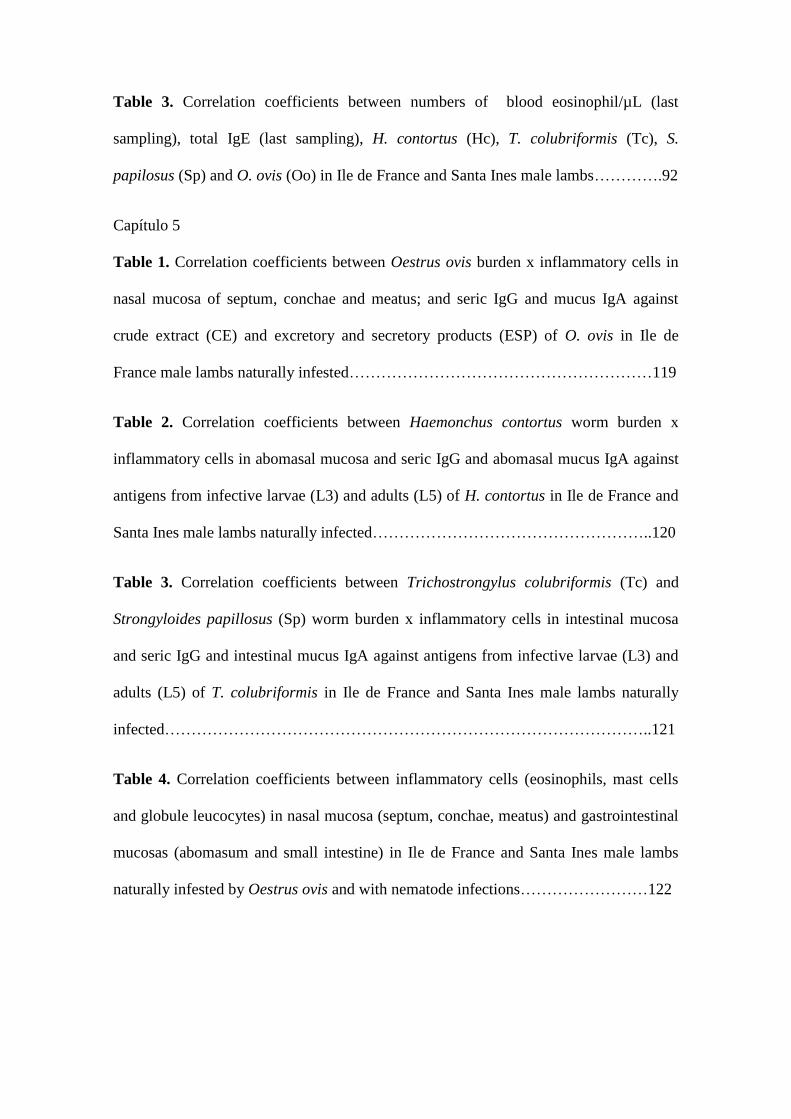
Table 3. Correlation coefficients between numbers of blood eosinophil/µL (last
sampling), total IgE (last sampling), H. contortus (Hc), T. colubriformis (Tc), S.
papilosus (Sp) and O. ovis (Oo) in Ile de France and Santa Ines male lambs………….92
Capítulo 5
Table 1. Correlation coefficients between Oestrus ovis burden x inflammatory cells in
nasal mucosa of septum, conchae and meatus; and seric IgG and mucus IgA against
crude extract (CE) and excretory and secretory products (ESP) of O. ovis in Ile de
France male lambs naturally infested…………………………………………………119
Table 2. Correlation coefficients between Haemonchus contortus worm burden x
inflammatory cells in abomasal mucosa and seric IgG and abomasal mucus IgA against
antigens from infective larvae (L3) and adults (L5) of H. contortus in Ile de France and
Santa Ines male lambs naturally infected……………………………………………..120
Table 3. Correlation coefficients between Trichostrongylus colubriformis (Tc) and
Strongyloides papillosus (Sp) worm burden x inflammatory cells in intestinal mucosa
and seric IgG and intestinal mucus IgA against antigens from infective larvae (L3) and
adults (L5) of T. colubriformis in Ile de France and Santa Ines male lambs naturally
infected………………………………………………………………………………..121
Table 4. Correlation coefficients between inflammatory cells (eosinophils, mast cells
and globule leucocytes) in nasal mucosa (septum, conchae, meatus) and gastrointestinal
mucosas (abomasum and small intestine) in Ile de France and Santa Ines male lambs
naturally infested by Oestrus ovis and with nematode infections……………………122

1
Resumo
A variação sazonal e a intensidade de infestação por larvas de Oestrus ovis em
ovinos criados em Botucatu-SP foi avaliada de abril de 2008 a março de 2011. Dois
cordeiros traçadores foram colocados, mensalmente, junto com um rebanho ovino, onde
permaneceram por 28 dias. Após esse período, os traçadores foram sacrificados e as
larvas de O. ovis recuperadas, identificadas e quantificadas de acordo com o estádio de
desenvolvimento. Dos 72 cordeiros traçadores, 50% estavam infestados por larvas O.
ovis com intensidade média de 16,8 larvas/cabeça com média de 7,8 larvas de primeiro
estádio (L1), 5,3 de segundo (L2) e 3,7 de terceiro (L3). Sinais clínicos de oestrose
foram mensalmente avaliados em todas as ovelhas do rebanho e a prevalência média de
animais com sinais clínicos de oestrose foi de 13,4% (máxima de 31,4% em janeiro de
2009 e mínimo de 1% em setembro de 2010). Além disso, a prevalência do parasitismo
por O. ovis e a intensidade de infestação foram avaliados em cabeças de ovinos obtidas
de um abatedouro localizado em Itápolis – SP. Das 139 cabeças examinadas, 13,7%
estavam parasitadas pelas larvas O. ovis e a intensidade de infestação média mensal
variou de 1 até 10,2 larvas/cabeça com intensidade média geral de 4,5 larvas/cabeça. Do
total de 85 larvas, 21,2% eram L1, 37,6% L2 e 41,2% L3. Os resultados demonstraram
que as condições climáticas do Estado de São Paulo são favoráveis para a atividade da
mosca e desenvolvimento dos estágios larvais praticamente durante todo o ano.
Em outro estudo, foi avaliada, comparativamente, a resistência de cordeiros de
duas raças ovinas, Ile de France (IF) e Santa Inês (SI) contra infestações naturais por O.
ovis, bem como a associação entre a ocorrência deste parasita com as infecções naturais
por nematódeos gastrintestinais. Cordeiros machos SI (n = 12) e IF (n = 12) recém
desmamados, foram mantidos juntos em um piquete de setembro a início de dezembro
de 2009, quanto foram sacrificados. Todos os animais apresentaram infestação pelos

2
diferentes instares larvais de O. ovis, sem diferença entre as raças (P > 0,05). Os
cordeiros da raça SI apresentaram em média 24,8 larvas, sendo que a intensidade de
infestação variou entre 14 e 39 larvas, enquanto que os cordeiros da raça IF
apresentaram em média 23,5 larvas, com infestação máxima e mínima, respectivamente
de 11 e 36 larvas. Os cordeiros SI apresentaram a menor contagem de ovos de
nematódeos por grama de fezes (OPG) e os menores números médios de Haemonchus
contortus, Trichostrongylus colubriformis e Strongyloides papillosus, no entanto, não
houve diferenças significativas entre as raças (P > 0,05). Foi observada relação inversa
entre o número de larvas de O. ovis e nematódeos gastrintestinais em ambas as raças. Os
cordeiros SI apresentaram aumento significativo no número de eosinófilos sanguíneos e
nos níveis totais de IgE sérica e estas variáveis foram negativamente correlacionadas
com as contagens de OPG e carga de H. contortus em ambas as raças. A resposta imune
contra O. ovis e nematódeos gastrintestinais foram muito semelhantes nas duas raças (P
> 0,05) e envolveu o recrutamento de células inflamatórias e produção de
imunoglobulinas parasita-específicas no soro e muco. Os resultados indicaram que a
presença dos anticorpos no soro e muco nasal não foram suficientes para proteger os
animais contra a infestação pelas larvas de O. ovis, mas pareceram promover atraso no
desenvolvimento larval, fato observado especialmente nos cordeiros SI. Em conclusão,
não houve diferença no parasitismo por larvas de O. ovis entre as raças avaliadas, e os
animais parasitados pelas larvas de O. ovis tendem a apresentar menor carga parasitária
de nematódeos gastrintestinais.

3
Abstract
The seasonal factors which influence Oestrus ovis infestation in sheep in
Botucatu-SP were determined from April 2008 until March 2011. Two tracer lambs
were exposed monthly to natural infestation by O. ovis larvae for 28 consecutive days,
by grazing with a sheep flock. Tracer animals were then euthanized and the larvae of O.
ovis recovered from nasal and sinus cavities. Of the 72 tracer lambs, 50% were infested
with O. ovis larvae and the mean intensity of infestation per head infested was 16.8
larvae with an average of 7.8 first instar (L1), 5.3 second instar (L2) and 3.7 third instar
(L3). Clinic signs of oestrosis were evaluated in all sheep of the flock monthly and the
average prevalence of animals with clinical signs of oestrosis was 13.4% (maximum of
31.4% in January 2009 and minimum of 1% in September 2010). Additionally, the O.
ovis prevalence and infestation intensity were evaluated in heads from slaughtered
sheep from Itápolis-SP. Of the 139 head examined 13.7% were parasitized by O. ovis
larvae with monthly mean of intensity of infestation ranging from 1 until 10.2
larvae/infested head with general mean intensity of 4.5 larvae/infested head. Of the total
of 85 larvae, 21.2% were L1, 37.6% L2 and 41.2% L3. The results suggest that the
climatic conditions in São Paulo State are favorable to fly activity and larval
development during the whole year.
In other study, were evaluated comparatively, the resistance in lambs of two
sheep breeds, Ile de France (IF) and Santa Ines (SI) against O. ovis infestation, well as
the association between the occurrence of this parasite with natural infections by
gastrointestinal nematodes (GIN). SI (n=12) and IF (n=12) young male lambs weaned at
two months of age were kept together in a paddock from September to early December
2009, when were sacrificed. All animals were infested by different larval instars of O.
ovis without any statistical difference between breeds (P > 0.05). The SI lambs had an

4
average of 24.8 larvae, and the intensity of infection ranged between 14 and 39 larvae,
while the IF lambs showed an average of 23.5 larvae with the minimum and maximum
from 11 to 36 larvae, respectively. SI lambs presented the lowest nematode fecal egg
counts (FEC) and the lowest mean numbers of Haemonchus contortus, Trichostrongylus
colubriformis and Strongyloides papillosus, however, there was no significant
differences between group means (P > 0.05). Inverse relationship between numbers of
O. ovis larvae and gastrointestinal nematodes was observed in both breeds. SI sheep
showed a significant increase in blood eosinophils and total IgE serum levels and these
variables were negatively correlated with nematode FEC. The immune response against
O. ovis and GIN were very similar in both breeds (P > 0.05) and involved the
recruitment of inflammatory cells and the parasitic specific immunoglobulins
production in serum and mucus. The results indicated that the presence of antibodies in
serum or nasal mucus were not enough to protect them against O. ovis infestation, but
can promote a delay in larval development, fact observed especially in SI lambs. In
conclusion there was no breed difference regarding O. ovis infestation and in each
breed, animals with more nasal bot fly larvae tended to display smaller worm burden.

5
Capítulo 1
6
Introdução
Oestrus ovis L. (Díptera: Oestridae) é um parasita cosmopolita causador de
míiase cavitária e suas larvas são parasitas obrigatórios da cavidade nasal e seios
paranasais de ovinos e caprinos (Zumpt, 1965).
A mosca de O. ovis é vivípara e deposita larvas de primeiro estádio (L1)
diretamente no nariz dos ovinos e caprinos. Estas colonizam rapidamente as cavidades
nasais, septo, turbinas e etmóide, e em seguida, mudam para larva de segundo estádio
(L2) e migram para os seios frontais, onde irá completar seu desenvolvimento em larva
de terceiro estádio (L3). As L3 maduras serão expelidas para o ambiente para o período
de pupação que acontece no solo. A pupa dará origem a mosca, e após o acasalamento,
as fêmeas grávidas atacam os ovinos para depositar as larvas, reiniciando o ciclo
(Zumpt, 1965).
O desenvolvimento das larvas na cavidade nasal do hospedeiro bem como a
atividade da mosca no meio ambiente é muito influenciado pelas condições climáticas
do ambiente (Cobbett and Mitchell, 1941). O desenvolvimento larval pode variar entre
25-35 dias podendo se estender até nove meses dependendo da estação do ano e
condições climáticas da região (Hall and Wall, 1995).
As larvas de O. ovis não são hematófagas e se alimentam de proteínas
plasmáticas, de anticorpos que estão passando pela mucosa nasal durante o processo
inflamatório, de mucina, albumina e colágeno da membrana basal (Frugère et al., 2000;
Tabouret et al., 2003a). Embora seus ganchos e espinhos danifiquem as membranas
nasais, a nutrição larval não é apenas mecânica, mas está relacionada principalmente a
um processo bioquímico e grande liberação de oxido de nitrogênio (Angulo-Valadez et
al., 2010).

7
O comprimento da larva e a coloração/tamanho dos espiraculos posteriores, por
onde as larvas respiram, são um dos parâmetros utilizados para classificar as fases de
desenvolvimento em L1, L2 ou L3 (Fig. 1) (Cepeda-Palacios et al., 1999).
Figura 1. Larvas de Oestrus ovis em diferentes fases de desenvolvimento (vista dorsal).
Foto: B. F. Silva, 2009.
A L1 é depositada na cavidade nasal dos ovinos com cerca de 1 mm de
comprimento, possui ganchos orais relativamente pequenos e espiraculos posteriores
não pigmentados (Fig. 2). A muda de L1 para L2 ocorre com cerca de 4 mm e nesta fase
os espiraculos começam a ficar visíveis na cutícula da L1.
Figura 2. Vista ventral da larva de primeiro estádio (L1) de Oestrus ovis. Foto: B. F.
Silva, 2009.

8
A L2 varia entre > 4 mm até 10 mm de comprimento e nesta fase os espiraculos
posteriores são visíveis, inicialmente com coloração variando entre o amarelo-
alaranjado até o marrom escuro (Fig. 3).
Figura 3. Detalhe da região posterior da larva de segundo estádio (L2) de Oestrus ovis
onde estão localizados os espiraculos respiratórios. Foto: B. F. Silva, 2009.
A L3 possui comprimento entre >10 mm até 22 mm e os espiraculos apresentam
coloração marrom escura. Conforme a maturação, a L3 começa apresentar listas dorsais
pretas e mudam de cor, do branco para a cor creme e depois marrom claro até ficar com
o corpo totalmente escuro, e então estará pronta para ser expelida pelo hospedeiro e
pupar no solo (Fig. 4).

9
Figura 4. Larva de terceiro estádio (L3) de Oestrus ovis em diferentes fases de
desenvolvimento (A, B e C); espiraculos respiratórios localizados na região posterior do
corpo da larva (D). Foto: B. F. Silva, 2009.
A mosca adulta tem coloração acinzentada, possui pêlos e manchas escuras pelo
corpo, o que facilita a camuflagem (Fig. 5). Além disso, voa rápido e possuem olhos
grandes, o que facilita a localização de seus hospedeiros bem como, das fêmeas para a
reprodução. A vida dos adultos é curta, pois não possuem peças bucais funcionais, e,
portanto, não se alimentam no ambiente e dispõem apenas das reservas energéticas
acumuladas durante a fase larval que acontece no aparelho nasal do hospedeiro
(Angulo-Valadez et al., 2010). As fêmeas carregam consigo cerca de 500 larvas e são
capazes de infestar vários ovinos / caprinos durante seu curto período de vida (Cobbett
and Mitchell, 1941), além disso, machos e fêmeas emergem do pupario já sexualmente
maduros, prontos para o acasalamento (Angulo-Valadez et al., 2010) adaptação que
auxilia na economia de energia.
10
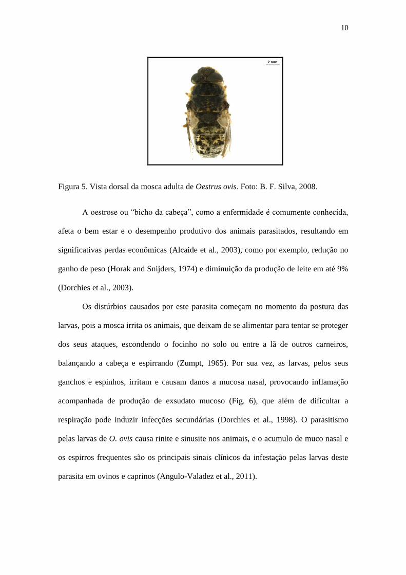
Figura 5. Vista dorsal da mosca adulta de Oestrus ovis. Foto: B. F. Silva, 2008.
A oestrose ou “bicho da cabeça”, como a enfermidade é comumente conhecida,
afeta o bem estar e o desempenho produtivo dos animais parasitados, resultando em
significativas perdas econômicas (Alcaide et al., 2003), como por exemplo, redução no
ganho de peso (Horak and Snijders, 1974) e diminuição da produção de leite em até 9%
(Dorchies et al., 2003).
Os distúrbios causados por este parasita começam no momento da postura das
larvas, pois a mosca irrita os animais, que deixam de se alimentar para tentar se proteger
dos seus ataques, escondendo o focinho no solo ou entre a lã de outros carneiros,
balançando a cabeça e espirrando (Zumpt, 1965). Por sua vez, as larvas, pelos seus
ganchos e espinhos, irritam e causam danos a mucosa nasal, provocando inflamação
acompanhada de produção de exsudato mucoso (Fig. 6), que além de dificultar a
respiração pode induzir infecções secundárias (Dorchies et al., 1998). O parasitismo
pelas larvas de O. ovis causa rinite e sinusite nos animais, e o acumulo de muco nasal e
os espirros frequentes são os principais sinais clínicos da infestação pelas larvas deste
parasita em ovinos e caprinos (Angulo-Valadez et al., 2011).

11
Figura 6. Ovelhas da raça Bergamacia com corrimento nasal, sinal clínico característico
do parasitismo por larvas de Oestrus ovis. Foto: B. F. Silva, 2008.
Epidemiologia
Oestrus ovis é um parasita distribuído mundialmente, mas a atividade da mosca
no ambiente, o desenvolvimento larval no aparelho nasal do hospedeiro, bem como, o
período de pupa que ocorre no solo é muito influenciado pelas condições climáticas do
ambiente.
Os principais fatores climáticos que influenciam a atividade dos oestrideos são a
temperatura, luminosidade e velocidade do vento, mas no caso do O. ovis, a temperatura
é fator determinante para a larviposição (Cepeda-Palacios and Scholl, 2000). Em Baja
California Sur, México, onde a temperatura diurna durante a primavera varia entre 9 –
35 °C foi observado que a atividade da mosca teve início quando a temperatura
ultrapassou os 20 °C, mas a „temperatura ótima‟ para a larviposição variou entre 26 - 28
°C. Porém, O. ovis, por um processo adaptativo, pode se ajustar as características
climáticas da região onde habita e essa „temperatura ótima‟ pode ser diferente

12
dependendo da região estudada (Cepeda-Palacios and Scholl, 2000), mas no geral, os
ataques da mosca acontecem principalmente durante o período mais quente do dia.
Cobbett e Mitchell (1941) foram pioneiros em descrever a influencia do clima na
epidemiologia do O. ovis. Dentre muitas descobertas, os pesquisadores observaram que
durante o inverno o desenvolvimento larval na cavidade nasal dos animais é lento e que
em locais onde o inverno é muito rigoroso, característico de regiões com clima
temperado, a L1 cessa o desenvolvimento e entra em estado de hipobiose, ou seja, não
há desenvolvimento larval neste período até que as condições climáticas voltem a ser
favoráveis para o desenvolvimento. Da mesma forma, em países com clima muito
quente, o desenvolvimento larval também é interrompido durante o período seco
(Dorchies et al., 1998). Essa é uma das estratégias que pode assegurar a perpetuação do
O. ovis em regiões em que as condições climáticas são extremas durante algum período
do ano.
Devido a essa grande influencia do clima no desenvolvimento das larvas de O.
ovis, a prevalência e intensidade de infestação é variável de acordo com o país ou região
estudada, como pode ser observado na tabela abaixo (Tabela 1), bem como a
prevalência dos diferentes instares larvais recuperados da cavidade nasal dos animais
parasitados.
Em locais com clima temperado, como exemplo, na região sul da França
(Dorchies et al., 2000) e região nordeste da Espanha (Gracia et al., 2010), as L1 foram
predominantes durante o ano de estudo, constituindo respectivamente 85,1 e 78,4% da
carga parasitária total. O oposto foi observado na Sicilia, Itália (Caracappa et al., 2000)
e na região sudeste da Espanha (Alcaide et al., 2003) onde todos os diferentes instares
larvais foram simultaneamente recuperados durante o ano de estudo e em proporções
similares. Na Sicilia, por exemplo, a proporção de L1, L2 e L3 foi respectivamente,

13
41,4%, 29,1% e 29,6% o que indica que as condições climáticas da região estudada
foram favoráveis para o desenvolvimento larval. Porém, em ambos os estudos, durante
os meses frios, foi observado um período de desenvolvimento lento onde a proporção de
L1 predominou sob os demais estádios larvais.
Tabela 1. Prevalência e intensidade de infestação por larvas de Oestrus ovis em ovinos.
Local N* Prevalência Intensidade Referência
Canadá 698 50,0% 2,5 (Fallis, 1940)
Estados Unidos 720 91,5% 25,6 (Meleney et al., 1962)
Nova Zelândia 1083 65.8% 3,1 (Kettle, 1973)
África do Sul 542 73,4% 15,2 (Horak, 1977)
Zimbábue 507 21,9% 1,1 (Pandey, 1989)
França (sudeste) 555 65,0% 24,8 (Yilma and Dorchies, 1991)
Itália (Sicilia) 841 55,8% 9,4 (Caracappa et al., 2000)
França (sul) 631 43,4% 10,8 (Dorchies et al., 2000)
Etiópia 248 77,4% 12,7 (Yilma and Genet, 2000)
Itália (Sardenha) 566 91,0% 19,0 (Scala et al., 2001)
Espanha (sudeste) 477 71,1% 18,5 (Alcaide et al., 2003)
Nigéria (norte) 116 62,1% 9,2 (Oniye et al., 2006)
Turquia (Konya) 624 59,0% 23,9 (Uslu and Dik, 2006)
Turquia (Kars) 387 40,3% 4,5 (Arslan et al., 2009)
Irã (Shiraz) 2002 49,7% 6,3 (Shoorijeh et al., 2009)
Espanha (nordeste) 120 84,2% 37,9 (Gracia et al., 2010)
*N = número de cabeças de ovinos examinadas, provenientes de abatedouros.

14
No Brasil, apesar da crescente observação de animais com sinais clínicos de
oestrose, há poucos estudos epidemiológicos sobre esta enfermidade, e estes estão
restritos a região Sul, onde as condições climáticas foram favoráveis em praticamente
todos os meses do ano para a atividade da mosca de O. ovis e desenvolvimento larval na
cavidade nasal dos ovinos (Ribeiro et al., 1990; Ramos et al., 2006).
Em Bagé – RS, das 144 cabeças de ovinos examinadas durante o período de um
ano, 85,4% estavam parasitadas e 1639 larvas foram recuperadas, sendo que destas
68,6% eram L1, 12,3% L2 e 18,9% L3 (Ribeiro et al., 1990). Em Encruzilhada do Sul, a
prevalência foi de 100% em animais abatidos com sinais clínicos de oestrose, com
intensidade média de infestação de 23,8 larvas/animal e a prevalência de L1, L2 e L3 foi
respectivamente de 78,9%, 14,5% e 6,6% (Oliveira et al., 1999). Já em estudo realizado
em Santa Catarina, quando as temperaturas médias foram inferiores a 9,8 ºC, não foram
constatadas larvas de O. ovis nos animais (Ramos et al., 2006).
Soroprevalência
No geral, o diagnóstico de oestrose é baseado em sinais clínicos e na detecção
das larvas post mortem, porém, estudos demonstram que o teste ELISA (enzyme-linked
immunosorbent assay) é sensível para detecção de anticorpos específicos anti O. ovis,
utilizando antígenos totais ou produtos excretórios e secretórios das larvas
(Papadopoulos et al., 2001; Alcaide et al., 2005a; Angulo-Valadez et al., 2008).
Alguns estudos, além de avaliar a prevalência por sorologia, também avaliaram
fatores de risco associados com a oestrose. Em Yucatan – México, 30% dos ovinos
avaliados estavam soropositivos para O. ovis e o tamanho do rebanho (> 25 animais) e a
cor do nariz do ovino (escuro) foram associados como fatores de risco para a ocorrência
da enfermidade (Murguia et al., 2000). Já na região sudeste da Alemanha a prevalência
15
de anticorpos foi de 50% nos ovinos examinados e o tamanho do rebanho (> 50
animais) foi o único fator de risco associado com a oestrose (Bauer et al., 2002).
Na região sudeste da Espanha, das 551 fazendas estudadas, apenas 18 fazendas
estavam livres de animais soropositivos e em 115, todos os animais foram soropositivos
para O. ovis. A prevalência média de animais soropositivos foi de 69,3% e foi
observado que além do tamanho do rebanho (> 250 ovinos) e densidade da população
ovina (> 100 ovinos por km2) foram importantes fatores de risco para a ocorrência desse
parasita na região estudada (Alcaide et al., 2005b).
Ovino x Caprino
Tanto ovinos como caprinos são parasitados por O. ovis, mas a prevalência do
parasitismo é menor nos caprinos do que em ovinos. Em Pézenas, região sul da França,
a prevalência, os sinais clínicos da infestação e a carga parasitária de O. ovis foi menor
em caprinos do que em ovinos (Dorchies et al., 2000). Da mesma forma, na Grécia, a
prevalência de animais com anticorpos específicos anti O. ovis foi menor em caprinos
comparado aos ovinos (Papadopoulos et al., 2001; Papadopoulos et al., 2006).
Há algumas hipóteses para essa diferença no parasitismo por O. ovis entre
caprinos e ovinos. Supõe-se que os caprinos aparentam ser mais sensíveis aos ataques
da mosca e conseguem evitar o contato com a mesma de forma mais eficaz comparado
aos ovinos (Dorchies et al., 1998; Angulo-Valadez et al., 2010). Acredita-se também
que os caprinos co-evoluíram com o O. ovis por um período mais longo do que com os
ovinos e talvez por isso sejam mais bem adaptados ao parasitismo (Angulo-Valadez et
al., 2010).
Resposta imunológica
As infecções parasitárias caracterizam-se por estimular inúmeros mecanismos

16
imunológicos de defesa, sejam eles mediados por anticorpos ou por células e a
eficiência da resposta imunológica depende do parasita em questão e do estágio da
infecção.
A presença das larvas de O. ovis na cavidade nasal dos ovinos induz resposta
imunológica celular, com recrutamento de leucócitos (linfócitos T e B, macrófagos) e
granulócitos (eosinófilos, mastócitos e leucócitos globulares) na mucosa do trato nasal,
e resposta imune humoral local e sistêmica com produção de imunoglobulina G (IgG) e
imunoglobulina A (IgA) anti O. ovis, as quais são encontradas no soro e muco nasal dos
ovinos parasitados (Tabouret et al., 2003b), o que sugere uma resposta imunológica tipo
Th2 (Angulo-Valadez et al., 2011), similar ao que é observado na resposta imune contra
o parasitismo por nematódeos gastrintestinais (Anthony et al., 2007; Rowe et al., 2008).
Além disso, estudos demonstram que os produtos excretórios e secretórios das larvas de
O. ovis causam reação de hipersensibilidade imediata em seus hospedeiros (Dorchies et
al., 1998; Jacquiet et al., 2005).
A resposta inflamatória causada pelas larvas de O. ovis parece estar relacionada
com a regulação da carga parasitária, pois promove redução no crescimento larval, mas
por outro lado, não protege os hospedeiros contra o estabelecimento das larvas (Frugère
et al., 2000; Jacquiet et al., 2005). Além disso, foi observado que os linfócitos de ovinos
previamente infestados não responderam a estimulação com antígenos específicos
(produtos excretórios e secretórios de L2 e L3) após a terceira infestação, ou seja, a
capacidade de resposta dos linfócitos diminui de acordo com o número de exposições,
sugerindo atividade imunossupressora pelo parasita (Jacquiet et al., 2005).
Foi observado que em ovinos imunossuprimidos, que receberam tratamento com
corticóide, o desenvolvimento larval foi mais rápido comparado com o controle que não
recebeu tratamento, onde as L2 foram recuperadas em maior proporção. Comparando o

17
grupo imunossuprimido com o grupo de ovinos que haviam sido previamente
parasitados pelas larvas, o estabelecimento larval foi similar, porém as L2 do grupo
imunossuprimido tiveram peso mais elevado (Jacquiet et al., 2005). Vale ressaltar que
O. ovis só se alimenta durante a fase de vida parasitária, enquanto larva, e, portanto, a
redução do peso da larva madura pode comprometer a viabilidade da mosca (Cepeda-
Palacios et al., 2000).
Portanto, estes estudos demonstram que apesar dos ovinos sofrerem sucessivas
infestações pelas larvas de O. ovis, a aquisição de resistência é muito difícil, oposto do
que é observado no parasitismo por nematódeos gastrintestinais. Mas a resposta
imunológica pode ao menos manter o parasitismo sob controle, regulando a carga
parasitária ou afetando o crescimento das larvas, e, por consequência, a viabilidade das
moscas.
Interação entre a infestação por O. ovis e a infecção por nematódeos gastrintestinais
É muito comum um animal ser parasitado por vários organismos
simultaneamente, a exemplo dos ovinos, que comumente são parasitados por
nematódeos gastrintestinais e por larvas de O. ovis. Vários trabalhos foram realizados a
fim de avaliar a interação entre a infestação por larvas de O. ovis e as infecções com
nematódeos gastrintestinais parasitas de ovinos, como por exemplo a interação entre O.
ovis e o parasita do intestino delgado Trichostrongylus colubriformis (Yacob et al.,
2002; 2004; 2006) ou pelo parasita do abomaso Haemonchus contortus (Dorchies et al.,
1997b; Terefe et al., 2005).
Nestes trabalhos os ovinos foram divididos em quatro grupos de animais:
infestados apenas com larvas de O. ovis; infectados apenas com nematódeo
gastrointestinal (T. colubriformis ou H. contortus); ovinos infectados com ambos
parasitas; e grupo controle que permaneceram livres de infestações/infecções pelos

18
referidos parasitas. Os pesquisadores observaram que a infecção no trato digestivo por
nematódeos não modificou a biologia da população de Oestrus na cavidade nasal. Já a
presença do O. ovis foi relacionada com significativa redução na eliminação de ovos
pelos helmintos, redução no tamanho e fecundidade das fêmeas e na carga parasitária.
Estas mudanças foram associadas com eosinofilia e significativas modificações na
população tecidual de mastócitos, leucócitos e eosinófilos nos tratos respiratório e
digestivo. Com base nesses resultados foi observado que a infecção parasitária em uma
determinada região anatômica provoca “à distância” reações inflamatórias em todo o
sistema de mucosa, pois foram observadas mudanças na população celular tecidual em
região anatômica que não estava parasitada.
Porém, apesar de existir correlação negativa entre o parasitismo por O. ovis e
nematódeos gastrintestinais, a regulação é de natureza transitória e desaparece quando
as larvas de O. ovis são expelidas pelo hospedeiro ou após tratamento com
antiparasitário. O mecanismo envolvido na regulação não é específico e está relacionado
com forte ativação dos eosinófilos sanguíneos que age de forma inespecífica sobre os
vermes (Yacob et al., 2006).
Controle
O controle do parasitismo por O. ovis é feito exclusivamente com o uso de
antiparasitários, tais como os organofosforados, triclorfon e closantel, e as lactonas
macrocíclicas, como a ivermectina (Dorchies et al., 1997a; Lucientes et al., 1998),
moxidectina (Dorchies et al., 1996), doramectina (Oliveira et al., 2000; Dorchies et al.,
2001) e eprinomectina (Hoste et al., 2004; Habela et al., 2006).

19
Considerações finais
Oestrus ovis é um parasita morfológica e biologicamente muito bem adaptado
aos ovinos. Tais adaptações permitem sua sobrevivência sob condições climáticas
adversas e extremas, bem como, a resposta imune do hospedeiro.
Apesar da elevada prevalência e intensidade de infestação observada
mundialmente, a oestrose pode ser considerada uma enfermidade negligenciada no
Brasil. Praticamente não há estudos epidemiológicos sobre este parasita, e apenas com
tal conhecimento é possível predizer quais os fatores climáticos que favorecem a
infestação, bem como, recomendar a melhor época de tratamento.
O Capítulo 2 da Tese intitulado “Epidemiology of Oestrus ovis in sheep in the
southwest of Brazil” e o Capítulo 3 intitulado “Prevalence and intensity of Oestrus ovis
infestation in sheep in central region of São Paulo State, Brazil” tiveram por objetivos
avaliar a variação sazonal e a intensidade de infestação por larvas de O. ovis em ovinos
criados em Botucatu-SP e em Itápolis-SP, respectivamente.
O Capítulo 4 intitulado “Parasitism by Oestrus ovis: Influence of sheep breed
and nematode infections” e o Capítulo 5 intitulado “Cellular and humoral immune
responses in sheep naturally infested with Oestrus ovis (Diptera: Oestridae) and with
nematode infections” tiveram por objetivos avaliar comparativamente, a resistência de
cordeiros de duas raças ovinas, Ile de France e Santa Inês, contra infestações naturais
por O. ovis, bem como a associação entre a ocorrência deste parasita com as infecções
naturais por nematódeos gastrintestinais e a resposta imunológica envolvida na proteção
contra estes parasitas.

20
Referências bibliográficas
Alcaide, M., Reina, D., Frontera, E., Navarrete, I., 2005a. Analysis of larval antigens of
Oestrus ovis for the diagnosis of oestrosis by enzyme-linked immunosorbent
assay. Med. Vet. Entomol. 19, 151-157.
Alcaide, M., Reina, D., Sánchez-López, J., Frontera, E., Navarrete, I., 2005b.
Seroprevalence of Oestrus ovis (Diptera, Oestridae) infestation and associated
risk factors in ovine livestock from Southwestern Spain. J. Med. Entomol. 42,
327-331.
Alcaide, M., Reina, D., Sánchez, J., Frontera, E., Navarrete, I., 2003. Seasonal
variations in the larval burden distribution of Oestrus ovis in sheep in the
southwest of Spain. Vet. Parasitol. 118, 235-241.
Angulo-Valadez, C.E., Ascencio, F., Jacquiet, P., Dorchies, P., Cepeda-Palacios, R.,
2011. Sheep and goat immune responses to nose bot infestation: a review. Med.
Vet. Entomol. 25, 117-125.
Angulo-Valadez, C.E., Scala, A., Grisez, C., Prevot, F., Bergeaud, J.P., Carta, A.,
Cepeda-Palacios, R., Ascencio, F., Terefe, G., Dorchies, P., Jacquiet, P., 2008.
Specific IgG antibody responses in Oestrus ovis L. (Diptera: Oestridae) infected
sheep: Associations with intensity of infection and larval development. Vet.
Parasitol. 155, 257-263.
Angulo-Valadez, C.E., Scholl, P.J., Cepeda-Palacios, R., Jacquiet, P., Dorchies, P.,
2010. Nasal bots... a fascinating world! Vet. Parasitol. 174, 19-25.

21
Anthony, R.M., Rutitzky, L.I., Urban, J.F., Stadecker, M.J., Gause, W.C., 2007.
Protective immune mechanisms in helminth infection. Nat Rev Immunol 7, 975-
987.
Arslan, M., Kara, M., Gicik, Y., 2009. Epidemiology of Oestrus ovis infestations in
sheep in Kars province of north-eastern Turkey. Trop. Anim. Health Prod. 41,
299-305.
Bauer, C., Steng, G., Prevot, F., Dorchies, P., 2002. Seroprevalence of Oestrus ovis
infection in sheep in southwestern Germany. Vet. Parasitol. 110, 137-143.
Caracappa, S., Rilli, S., Zanghi, P., Di Marco, V., Dorchies, P., 2000. Epidemiology of
ovine oestrosis (Oestrus ovis Linné 1761, Diptera: Oestridae) in Sicily. Vet.
Parasitol. 92, 233-237.
Cepeda-Palacios, R., Ávila, A., Ramírez-Orduña, R., Dorchies, P., 1999. Estimation of
the growth patterns of Oestrus ovis L. larvae hosted by goats in Baja California
Sur, Mexico. Vet. Parasitol. 86, 119-126.
Cepeda-Palacios, R., Frugère, S., Dorchies, P., 2000. Expected effects of reducing
Oestrus ovis L. mature larval weight on adult populations. Vet. Parasitol. 90,
239-246.
Cepeda-Palacios, R., Scholl, P.J., 2000. Factors affecting the larvipositional activity of
Oestrus ovis gravid females (Diptera: Oestridae). Vet. Parasitol. 91, 93-105.
Cobbett, N.G., Mitchell, W.C., 1941. Further observations on the life cycle and
incidence of the sheep bot, Oestrus ovis, in New Mexico and Texas. Am. J. Vet.
Res. 2, 358-366.

22
Dorchies, P., Alzieu, J.P., Cadiergues, M.C., 1997a. Comparative curative and
preventive efficacies of ivermectin and closantel on Oestrus ovis (Linné 1758) in
naturally infected sheep. Vet. Parasitol. 72, 179-184.
Dorchies, P., Bergeaud, J., Khanh, N.V., Morand, S., 1997b. Reduced egg counts in
mixed infections with Oestrus ovis and Haemonchus contortus: influence of
eosinophils? Parasitol. Res. 83, 727-730.
Dorchies, P., Bergeaud, J.P., Tabouret, G., Duranton, C., Prevot, F., Jacquiet, P., 2000.
Prevalence and larval burden of Oestrus ovis (Linné 1761) in sheep and goats in
northern Mediterranean region of France. Vet. Parasitol. 88, 269-273.
Dorchies, P., Cardinaud, B., Fournier, R., 1996. Efficacy of moxidectin as a 1%
injectable solution and a 0.1% oral drench against nasal bots, pulmonary and
gastrointestinal nematodes in sheep. Vet. Parasitol. 65, 163-168.
Dorchies, P., Duranton, C., Jacquiet, P., 1998. Pathophysiology of Oestrus ovis
infection in sheep and goats: a review. Vet. Rec. 142, 487.
Dorchies, P., Jacquiet, P., Bergeaud, J.P., Duranton, C., Prévot, F., Alzieu, J.P.,
Gossellin, J., 2001. Efficacy of doramectin injectable against Oestrus ovis and
gastrointestinal nematodes in sheep in the southwestern region of France. Vet.
Parasitol. 96, 147-154.
Dorchies, P., Wahetra, S., Lepetitcolin, E., Prevot, F., Grisez, C., Bergeaud, J.P., Hoste,
H., Jacquiet, P., 2003. The relationship between nasal myiasis and the
prevalence of enzootic nasal tumours and the effects of treatment of Oestrus ovis
and milk production in dairy ewes of Roquefort cheese area. Vet. Parasitol. 113,
169-174.
23
Fallis, A.M., 1940. Studies on Oestrus ovis L. Can. J. Res. 18, 442-446.
Frugère, S., Leon, A.C., Prévot, F., Palacios, R.C., Tabouret, G., Bergeaud, J.P.,
Duranton, C., Dorchies, P., Jacquiet, P., 2000. Immunisation of lambs with
excretory secretory products of Oestrus ovis third instar larvae and subsequent
experimental challenge. Vet. Res. 31, 527-535.
Gracia, M.J., Lucientes, J., Peribáñez, M.A., Castillo, J.A., Calvete, C., Ferrer, L.M.,
2010. Epidemiology of Oestrus ovis infection of sheep in northeast Spain (mid-
Ebro Valley). Trop. Anim. Health Prod. 42, 811-813.
Habela, M., Moreno, A., Gragera-Slikker, A., Gomez, J., Montes, G., Rodriguez, P.,
Alvinerie, M., 2006. Efficacy of eprinomectin pour-on in naturally Oestrus ovis
infested merino sheep in Extremadura, South-West Spain. Parasitol. Res. 99,
275-280.
Hall, M., Wall, R., 1995. Myiasis of Humans and Domestic Animals, In: J.R. Baker,
R.M., Rollinson, D. (Eds.) Advances in Parasitology. Academic Press, pp. 257-
334.
Horak, I.G., 1977. Parasites of domestic and wild animals in South Africa. I. Oestrus
ovis in sheep. Onderstepoort J. Vet. Res. 44, 55-64.
Horak, I.G., Snijders, A.J., 1974. The effect of Oestrus ovis infestation on merino
lambs. Vet. Rec. 94, 12-16.
Hoste, H., Lemercier, P., Jacquiet, P., Dorchies, P., Lespine, A., Alvinerie, M., 2004.
Efficacy of eprinomectin pour-on against gastrointestinal nematodes and the
nasal bot fly (Oestrus ovis) in sheep. Vet. Rec. 154, 782-785.
24
Jacquiet, P., Tran Thi Ngoc, T., Nouvel, X., Prevot, F., Grisez, C., Yacob, H.T.,
Bergeaud, J.-P., Hoste, H., Dorchies, P., Tabouret, G., 2005. Regulation of
Oestrus ovis (Diptera: Oestridae) populations in previously exposed and naïve
sheep. Vet. Immunol. Immunopathol. 105, 95-103.
Kettle, P.R., 1973. A study on the sheep botfly, Oestrus ovis (Diptera: Oestridae) in
New Zealand. N. Z. Entomol. 5, 185-191.
Lucientes, J., Castillo, J.A., Ferrer, L.M., Peribáñez, M.A., Ferrer-Dufol, M., Gracia-
Salinas, M.J., 1998. Efficacy of orally administered ivermectin against larval
stages of Oestrus ovis in sheep. Vet. Parasitol. 75, 255-259.
Meleney, W.P., Cobbett, N.G., Peterson, H.O., 1962. The natural occurrence of Oestrus
ovis in sheep from the Southwestern United States. Am. J. Vet. Res. 23, 1246-
1251.
Murguia, M., Rodriguez, J.C., Torres, F.J., Segura, J.C., 2000. Detection of Oestrus ovis
and associated risk factors in sheep from the central region of Yucatan, Mexico.
Vet. Parasitol. 88, 73-78.
Oliveira, C.M.B., Moro, E., Caproni Jr, L., Gonçalves, L.C.B., Umehara, O., Oliveira,
L.O., 2000. Efficacy of doramectin in the treatment of sheep naturally infested
by Oestrus ovis. Rev. Bras. Parasitol. Vet. 9, 61-64.
Oliveira, C.M.B., Oliveira, L.O., Torres, J.R., 1999. Oestrus ovis larvae distribution in
the head of naturally infested sheep. Arq. Fac. Vet. UFRGS 27, 87-92.

25
Oniye, S., Adebote, D., Ahunanya, C., 2006. Observations on Oestrus ovis L.(Diptera:
Oestridae) myiasis in the Nasal cavities and sinuses of the domestic sheep (Ovis
aries) in Zaria, Northern Nigeria. Int. J. Zool. Res. 2, 178-185.
Pandey, V.S., 1989. Epidemiology of Oestrus ovis infection of sheep in the highveld of
Zimbabwe. Vet. Parasitol. 31, 275-280.
Papadopoulos, E., Prevot, F., Diakou, A., Dorchies, P., 2006. Comparison of infection
rates of Oestrus ovis between sheep and goats kept in mixed flocks. Vet.
Parasitol. 138, 382-385.
Papadopoulos, E., Prevot, F., Jacquiet, P., Duranton, C., Bergeaud, J.P., Kalaitzakis, E.,
Dorchies, P., 2001. Seasonal variation of Oestrus ovis-specific antibodies in
sheep and goats mixed flocks in Greece. Vet. Parasitol. 95, 73-77.
Ramos, C.I., Bellato, V., Souza, A.P., Avila, V.S., Coutinho, G.C., Dalagnol, C.A.,
2006. Epidemiologia de Oestrus ovis (Diptera: Oestridae) em ovinos no Planalto
Catarinense. Ciência Rural 36, 173-178.
Ribeiro, V.L.S., Oliveira, C.M.B., Branco, F.P.J.A., 1990. Prevalência e variações
mensais das larvas de Oestrus ovis (Linneus, 1761) em ovinos no município de
Bagé, RS, Brasil. Arquivo Brasileiro de Medicina Veterinaria e Zootecnia 42,
211-221.
Rowe, A., McMaster, K., Emery, D., Sangster, N., 2008. Haemonchus contortus
infection in sheep: Parasite fecundity correlates with worm size and host
lymphocyte counts. Vet. Parasitol. 153, 285-293.

26
Scala, A., Solinas, G., Citterio, C.V., Kramer, L.H., Genchi, C., 2001. Sheep oestrosis
(Oestrus ovis Linné 1761, Diptera: Oestridae) in Sardinia, Italy. Vet. Parasitol.
102, 133-141.
Shoorijeh, S., Negahban, S., Tamadon, A., Behzadi, M., 2009. Prevalence and intensity
of Oestrus ovis in sheep of Shiraz, southern Iran. Trop. Anim. Health Prod. 41,
1259-1262.
Tabouret, G., Bret-Bennis, L., Dorchies, P., Jacquiet, P., 2003a. Serine protease activity
in excretory–secretory products of Oestrus ovis (Diptera: Oestridae) larvae. Vet.
Parasitol. 114, 305-314.
Tabouret, G., Lacroux, C., Andreoletti, O., Bergeaud, J.P., Hailu-Tolosa, Y., Hoste, H.,
Prevot, F., Grisez, C., Dorchies, P., Jacquiet, P., 2003b. Cellular and humoral
local immune responses in sheep experimentally infected with Oestrus ovis
(Diptera: Oestridae). Vet. Res. 34, 231-241.
Terefe, G., Yacob, H.T., Grisez, C., Prevot, F., Dumas, E., Bergeaud, J.P., Dorchies, P.,
Hoste, H., Jacquiet, P., 2005. Haemonchus contortus egg excretion and female
length reduction in sheep previously infected with Oestrus ovis (Diptera:
Oestridae) larvae. Vet. Parasitol. 128, 271-283.
Uslu, U., Dik, B., 2006. Prevalence and intensity of Oestrus ovis in Akkaraman sheep in
the Konya region of Turkey. Med. Vet. Entomol. 20, 347-349.
Yacob, H.T., Dorchies, P., Jacquiet, P., Bleuart, C., Prevot, F., Grisez, C., Bergeaud,
J.P., Hoste, H., 2004. Concurrent parasitic infections of sheep: depression of
Trichostrongylus colubriformis populations by a subsequent infection with
Oestrus ovis. Vet. Parasitol. 121, 297-306.

27
Yacob, H.T., Duranton-Grisez, C., Prevot, F., Bergeaud, J.P., Bleuart, C., Jacquiet, P.,
Dorchies, P., Hoste, H., 2002. Experimental concurrent infection of sheep with
Oestrus ovis and Trichostrongylus colubriformis: negative interactions between
parasite populations and related changes in the cellular responses of nasal and
digestive mucosae. Vet. Parasitol. 104, 307-317.
Yacob, H.T., Terefe, G., Jacquiet, P., Hoste, H., Grisez, C., Prévot, F., Bergeaud, J.P.,
Dorchies, P., 2006. Experimental concurrent infection of sheep with Oestrus
ovis and Trichostrongylus colubriformis: Effects of antiparasitic treatments on
interactions between parasite populations and blood eosinophilic responses. Vet.
Parasitol. 137, 184-188.
Yilma, J.M., Dorchies, P., 1991. Epidemiology of Oestrus ovis in southwest France.
Vet. Parasitol. 40, 315-323.
Yilma, J.M., Genet, A., 2000. Epidemiology if the sheep nasal bot, Oestrus ovis
(Diptera: Oestridae), in Central Ethiopia. Rev. Med. Vet. 151, 143-150.
Zumpt, P., 1965. Myiasis in man and animals in the Old World. Butterworths, London,
257 p.

28
Capítulo 2

29
Epidemiology of Oestrus ovis in sheep in the southwest of Brazila
B.F. Silva1*
; C.C. Bassetto1; A.F.T. Amarante
1
1UNESP - Univ Estadual Paulista, Departamento de Parasitologia, Instituto de
Biociências, Caixa Postal 510, CEP: 18618-000, Botucatu, SP, Brazil.
*Corresponding author: Bruna Fernanda da Silva, Departamento de Parasitologia,
Instituto de Biociências, Universidade Estadual Paulista (UNESP), Distrito de Rubião
Júnior s/n, Botucatu, São Paulo, CEP: 18618-000 Brazil. Tel.: +55 14 3811 6239; Fax:
+55 14 3815 3744. E-mail address: [email protected]
Abstract
The seasonal factors which influence Oestrus ovis infestation in sheep in São
Paulo State in the southwest of Brazil were determined from April 2008 until March
2011. Two tracer lambs were exposed monthly to natural infestation by O. ovis larvae
for 28 consecutive days, by grazing with a sheep flock. Tracer animals were then
euthanized and the larvae of O. ovis recovered from nasal and sinus cavities. Of the 72
tracer lambs, 50% were infested with O. ovis larvae and the mean intensity of
infestation per head infested was 16.8 larvae with an average of 7.8 L1, 5.3 L2 and 3.7
L3. In addition, clinic signs of oestrosis were evaluated in all sheep of the flock monthly
and the average prevalence of animals with clinical signs of oestrosis was 13.4%
(maximum of 31.4% in January 2009 and minimum of 1% in September 2010). The
results suggest that evolution and development of larval instars practically occurs
a Artigo redigido de acordo com as normas do periódico científico Veterinary Parasitology. Enviado para
publicação no dia 07 de novembro de 2011.

30
throughout the entire year, but O. ovis larval infestation was especially frequent during
spring and summer months.
Key words: Oestrus ovis; epidemiology; sheep; control.
Introduction
Oestrosis is a worldwide myiasis caused by larvae of the fly Oestrus ovis (Linné
1761, Diptera: Oestridae), which are obligatory parasites of nasal and sinus cavities of
sheep and goats. The female fly is viviparous and deposits larvae in or around the
nostrils of its host. These early first instars attach to the mucous membranes in the nasal
cavities, change to second instars and move up to the sinuses where it completes
development in mature third instars, which are expelled for pupation under the soil
(Zumpt, 1965). The length of this parasitic portion of the life cycle is quite variable
from a few weeks to several months depending on the season and climatic conditions
(Hall and Wall, 1995). Clinical respiratory signs such as seromucous or purulent nasal
discharge, frequent sneezing and dyspnea, may severely impair the health of infested
animals. Those pathological affects causes serious economical losses in small ruminant
livestock (Alcaide et al., 2003), i.e., reduction in live weight gain (Horak and Snijders,
1974) and decrease in milk production of almost 9% (Dorchies et al., 2003).
Numerous studies regarding the epidemiology of O. ovis have been carried out
in many countries but results are likely to be influenced by varying geographical and
epidemiological conditions. In southwest France, for example, the infestation was
present in 65% of the heads with mean intensity of 24.8 larvae/infested head consisting
mainly of first larval instar (Yilma and Dorchies, 1991). In Sicily, Italy, the prevalence
of infestation was 55.8% and all different larval stages were simultaneously recovered
in similar proportions (Caracappa et al., 2000). In Brazil there are only few studies

31
about this parasite and these are restricted to States in South region, where favorable
climatic conditions to O. ovis parasitism are observed throughout the year (Ribeiro et
al., 1990; Oliveira et al., 1999; Ramos et al., 2006), excepting periods with temperature
less than 9 °C when no larvae were recovered from tracer sheep (Ramos et al., 2006).
Oestrosis can be considered a neglected disease in Brazil, despite its increasing
occurrence according to farmers and veterinary clinician‟s perception. Therefore, it is
imperative more research about the epidemiology of the disease, since only with such
knowledge it will be possible to predict environmental factors favoring infestation as
well as to recommend the best strategies for oestrosis prophylaxis. This work was
conducted to determine the seasonal variation of O. ovis infestation in sheep in the
southwest of Brazil over a period of three years.
Materials and methods
Study location
The study was carried out in Botucatu, São Paulo state, Brazil, at an altitude of
786 m a.s.l.. Climate data referring to averages of temperatures, relative air humidity
and rainfall were obtained by the Department of Environmental Science, Agronomical
Science College, UNESP, located 8 km from the experimental site (Fig. 1 A and B).
For each month, from April 2008 until March 2011, two tracer lambs were
exposed to natural infestation with O. ovis larvae for 28 consecutive days, while grazing
together always with the same sheep flock. Immediately thereafter the tracer lambs were
euthanized. Heads were removed and cut open along their longitudinal and sagittal axis.
All larvae present in nasal cavity (nasal passage, septum, middle meatus and conchae)
and frontal sinus were collected and counted. Larvae were preserved into 70% alcohol

32
and identified according to their stage of development based on description of the
Zumpt (1965) and Capelle (1966).
Nasal discharge score (clinic signs of oestrosis) were recorded also in animals of
the sheep flock on the first day of each month and in tracer sheep before euthanasia.
According to clinical signs, the scores of nasal discharge (ND) were the
following: no ND = 0; dyspnea without nasal mucous ND = 1; dyspnea with sero-
mucous ND = 2; and dyspnea with muco-purulent ND = 3. Results are presented as
prevalence of sheep with clinical signs of oestrosis (%) and mean of ND score.
Management of tracer lambs
In total, over the three years of observations, 72 Ile de France weathered male
lambs, were purchased from farms located in São Paulo State. Twelve lambs were
purchased every six months, with initial age between two and five months.
Immediately after arrival at the University facilities, animals were housed,
vaccinated against clostridial infections (Sintoxan Polivalente®, Merial, Brazil) and
orally drenched, once daily, for three consecutive days with levamisole phosphate (10
mg/kg, Ripercol®
L 150 F, Fort Dodge) and albendazole (10 mg/kg, Valbazen®
10
Cobalto, Pfizer). One week later, the same protocol was carried with triclorfon (100
mg/kg, Neguvon®, Bayer S.A.) to remove any existing infestation of O. ovis and
infection by nematodes. Tracer lambs that stayed housed for more than one month,
received an additional treatment with triclorfon (100 mg/kg; Neguvon®
- Bayer S.A.)
one week before being taken to the farm to remove any infestation by O. ovis.
While housed, the animals were fed on concentrate (Tech Ovin Unique, Socil®,
with 18% of crude protein) in an amount corresponding to 1% of their mean live weight
and had free access to Tifton hay and tap water. Decoquinate (Deccox®, Alpharma) was
added to the commercial feed to prevent coccidiosis.

33
Sheep flock management
The sheep farm where the tracer lambs were placed belongs to University and
consisted in156 Bergamacia sheep at the beginning of the study and 202 animals at the
end. The sheep were kept in rotational grazing on Panicum maximum cv Tanzania grass
and, every year, from June until early November, due to low amount of forage at this
time, the animals received additional diet with corn silage once daily.
To avoid mortality of animals due to parasitism by gastrointestinal nematodes,
the sheep flock received anthelmintic treatments with levamisole phosphate (10 mg/kg,
Ripercol®
L 150 F, Fort Dodge) or moxidectin (0.2 mg/kg, Cydectin® Fort Dodge).
Details about these treatments are presented in Table 1.
Statistical analyses
Descriptive statistical analyses were performed in agreement with Bush et al.
(1997). The following terms were used:
Prevalence: the number of hosts infested with O. ovis larvae divided by the number of
hosts examined;
Intensity of infestation: the number of O. ovis larvae in a single infested host;
Mean intensity of infestation: the total number of O. ovis larvae found divided by the
number of hosts infected with that parasite.
Results
Clinical signs of oestrosis in the sheep flock
The average prevalence of animals with clinical signs of oestrosis was 13.4%
with maximum of 31.4% in January 2009 and minimum of 1% in September 2010. The
overall ND average score during all years was 1.4 with minimum of 1 and of 1.8 (Fig.
2).
34
Prevalence and intensity of infestation in tracer sheep
O. ovis larvae were recovered from 50% of the tracer sheep during the three
years of experiment. The mean intensity of infestation was 16.8 larvae per infested head
with an average of 7.8 L1, 5.3 L2 and 3.7 L3 (Fig. 1 C).
The highest number of larvae recovered from a single sheep was 66 in
September 2008 and of these, 64 were first stage (L1) and two second stage (L2). The
highest mean larval burden was found at the beginning of spring in September and
October 2008 (40 and 41 larvae/infested head, respectively).
O. ovis larvae were especially present during the spring and summer months
(starting in September until March), except in 2009 when many larvae were also
recovered during autumn (April to June 2009). This high rate of larvae recovery
coincided with mean temperatures between 20 ºC and 25 ºC and the relative air
humidity of around 70% (Fig. 1 A and B). No larvae were recovered in July when the
minimum mean temperatures were < 14 ºC.
Of the total of 606 larvae, 46.4% was L1, while L2 and L3 represented 31.3%
and 22.3% of the total larval burden, respectively. The majority of L1 were located in
the nasal cavity (99.6%). In contrast L3 were found especially in the frontal sinus
(97.8%). L2 were recovered from both sites: 40% from nasal cavity and 60% from
frontal sinus.
The highest intensity of infestation occurred mainly during the first two years of
the experiment. Considering the total number of larvae, 46.9 % was recovered in the
first and 32.5 % in the second year. The lowest intensity of infestation was observed in
third year with 20.6% of the total larvae recovered.
Most of the tracer lambs that were parasitized with O. ovis (30 of 36) did not
show any clinical signs of oestrosis during the experiment. Only six animals showed

35
signs, i.e., dyspnea and sero-mucous discharge with average score of 1.7. The larval
burden of these animals were variable, ranging from 12 to 66 larvae, constituted
especially by L1 and L2.
Discussion
The prevalence of O. ovis in tracer lambs was 50% which is less than that
observed in Bagé, Rio Grande do Sul State, Brazil, where 85.4% of sheep were infested
(Ribeiro et al., 1990). However in Bagé, the animals were exposed to O. ovis infestation
for a longer time period (from one month until one year) while in the present
experiment the sheep were exposed for just 28 days. Moreover, others factors may have
influenced the O. ovis epidemiology, like the climatic conditions of each region, size of
sheep flock and management, which can result in variable prevalence demonstrated by
many studies conducted in slaughterhouses in different countries (Yilma and Dorchies,
1991; Abo-Shehada et al., 2000; Scala et al., 2002; Alcaide et al., 2003; Arslan et al.,
2009; Shoorijeh et al., 2009).
The mean intensity of infestation reported in this study (16.8) was similar than
those previously reported for Sardinia, Italy (Scala et al., 2001) and southwest of Spain
(Alcaide et al., 2003) with mean intensity of 19 and 18.5 larvae by infested head,
respectively, but lower than that found in southwest of France with 24.8 larvae (Yilma
and Dorchies, 1991) and in northeast of Spain with 37.9 (Gracia et al., 2010). It is of
interest to note that a high percentage of first stage larvae were found by those authors,
especially during the colder months. This was not observed in the present study where
the proportion of different larval instars did not show variation throughout the
experimental months.

36
The respective percentages of L1, L2 and L3 were 46.4%, 31.3%, 22.3%. The
presence of different instars indicates that endogenous development and re-infestation
with L1 occurred simultaneously. Otherwise, in southwest region of France, under
temperate weather, L1 predominated with 90.5% of the total burden, while L2 (6.0%)
and L3 (3.5%) displayed a low proportion (Yilma and Dorchies, 1991). A greater
burden of L1 indicates the occurrence of a period of hypobiosis during the year, when
O. ovis cease the development, or at least suggests a decrease in the rate of larval
maturation. This was not observed in the present experiment, where it was observed
synchronous evolution of all larval stages without any evidence of hypobiosis.
Among the climatic factors influencing oestrid fly activity, temperature, light
intensity, and wind are recognized as the most important, but in the case of O. ovis, it
has been reported that temperature is the principal factor determining fly activity
(Cepeda-Palacios and Scholl, 2000). During all years of the study, averages of
maximum temperatures were between 20.8 ºC and 29.8 ºC. It has been demonstrated
that 20 ºC was the minimum temperature for fly activity while optimum temperature
appear to range between 26 and 28 ºC (Cepeda-Palacios and Scholl, 2000). Therefore,
the temperature conditions of the present study were optimal for fly activity during all
months of the experimental years, excepted in two winter months (June and July), when
regardless of mean maximum temperature of around 25 ºC, the mean of minimum were
always less than 14 ºC. In these climatic conditions the fly activity was possibly
reduced, especially in July, which coincided with reduction in number of sheep at the
farm with clinical signs of oestrosis.
Nasal discharges and sneezing were evident in sheep from the farm during all
study, but only six of the 72 tracer animals had clinical symptoms of oestrosis. The
delay between the first infestation and the appearance of clinical signs suggests the

37
possible involvement of a hypersensitivity phenomenon in the modulation of the
infection and these first infestations are possibly not associated with overt oestrosis
clinical signs (Dorchies et al., 1998). Probably, due the short period between the
infestation and euthanasia, most of the tracer sheep did not have time to develop and
display immune response with sneezing and nasal discharge, which are characteristic
clinical signs of parasitism by O. ovis, and the opposite occurred with the sheep from
the farm, continuously exposed to infestation.
Among sheep from farm, the mean prevalence of animals with some clinical
signs of oestrosis was relatively low (13.4%) with average score of 1.4. Studies of O.
ovis distribution analysis demonstrated that the numbers of larvae recovered in the
sheep population followed a negative-binomial distribution (Bart and Minar, 1992;
Abo-Shehada et al., 2000) and similar results were observed with gastrointestinal
nematode infections in sheep: a relatively small part of the flock constituted by
susceptible animals was responsible for the excretion of the majority of gastrointestinal
nematode eggs to environment (Sréter et al., 1994; Stear et al., 1998). Other possible
explanation may be related with the immune response; in this case, oestrosis symptoms
could be due to hypersensitivity reactions displayed only by a minor percentage of the
flock.
A variation in O. ovis larval number was observed between the experimental
years: the two first years concentrated 79.4% of total larval number recovered while just
20.6% of larval number was recovered during the last experimental year. These
differences between the burdens in experimental years were probably a consequence of
administration of broad spectrum anthelmintic to control gastrointestinal nematodes
infection in sheep flock where the tracer lambs were kept. Initially levamisole
phosphate was used and in the second and third year also moxidectin, a macrocycle

38
lactone that acts against O. ovis larvae (Dorchies et al., 1996). These treatments with
moxidectin were necessary due to anthelmintic resistance to other anthelmintics and
probably caused reduction in O. ovis population in the farm.
In conclusion, it is possible to affirm that several annual generations of adult
flies may occur in the region and probably in other areas with similar climatic
conditions. New studies will be necessary to evaluate the impact of oestrosis in sheep
production, as well the best strategies of prophylaxis.
Acknowledgements
We are grateful for the technical assistance provided by Mr. Valdir A. Paniguel,
Mr. Moises dos Santos and Mr. Edvaldo J. Vito. This study was funded by Fundação de
Amparo à Pesquisa do Estado de São Paulo (FAPESP, Grant number 2008/53494-2).
Bruna F. Silva (Grant number 2007/58244-1) and César C. Bassetto (Grant number
2009/03504-4) received financial support by FAPESP and Alessandro F.T. Amarante
from CNPq (Conselho Nacional de Desenvolvimento Científico e Tecnológico, Brazil).
39
References
Abo-Shehada, M.N., Arab, B., Mekbel, R., Williams, D., Torgerson, P.R., 2000. Age
and seasonal variations in the prevalence of Oestrus ovis larvae among sheep in
northern Jordan. Prev. Vet. Med. 47, 205-212.
Alcaide, M., Reina, D., Sánchez, J., Frontera, E., Navarrete, I., 2003. Seasonal
variations in the larval burden distribution of Oestrus ovis in sheep in the
southwest of Spain. Vet. Parasitol. 118, 235-241.
Arslan, M., Kara, M., Gicik, Y., 2009. Epidemiology of Oestrus ovis infestations in
sheep in Kars province of north-eastern Turkey. Trop. Anim. Health Prod. 41,
299-305.
Bart, A.G., Minar, J., 1992. Probability description of regulation on the level of
population and individual in the host-parasite system using Oestrus ovis
(Diptera: Oestridae) as an example. Folia Parasitol. 39, 75-83.
Bush, A.O., Lafferty, K.D., Lotz, J.M., Shostak, A.W., 1997. Parasitology meets
ecology on its own terms: Margolis et al. revisited. J. Parasitol. 83, 575-583.
Capelle, K.J., 1966. The occurrence of Oestrus ovis L. (Diptera: Oestridae) in the
bighorn sheep from Wyoming and Montana. J. Parasitol. 52, 618-621.
Caracappa, S., Rilli, S., Zanghi, P., Di Marco, V., Dorchies, P., 2000. Epidemiology of
ovine oestrosis (Oestrus ovis Linné 1761, Diptera: Oestridae) in Sicily. Vet.
Parasitol. 92, 233-237.
Cepeda-Palacios, R., Scholl, P.J., 2000. Factors affecting the larvipositional activity of
Oestrus ovis gravid females (Diptera: Oestridae). Vet. Parasitol. 91, 93-105.

40
Dorchies, P., Cardinaud, B., Fournier, R., 1996. Efficacy of moxidectin as a 1%
injectable solution and a 0.1% oral drench against nasal bots, pulmonary and
gastrointestinal nematodes in sheep. Vet. Parasitol. 65, 163-168.
Dorchies, P., Duranton, C., Jacquiet, P., 1998. Pathophysiology of Oestrus ovis
infection in sheep and goats: a review. Vet. Rec. 142, 487.
Dorchies, P., Wahetra, S., Lepetitcolin, E., Prevot, F., Grisez, C., Bergeaud, J.P., Hoste,
H., Jacquiet, P., 2003. The relationship between nasal myiasis and the
prevalence of enzootic nasal tumours and the effects of treatment of Oestrus ovis
and milk production in dairy ewes of Roquefort cheese area. Vet. Parasitol. 113,
169-174.
Gracia, M.J., Lucientes, J., Peribáñez, M.A., Castillo, J.A., Calvete, C., Ferrer, L.M.,
2010. Epidemiology of Oestrus ovis infection of sheep in northeast Spain (mid-
Ebro Valley). Trop. Anim. Health Prod. 42, 811-813.
Hall, M., Wall, R., 1995. Myiasis of Humans and Domestic Animals, In: J.R. Baker,
R.M., Rollinson, D. (Eds.) Advances in Parasitology. Academic Press, pp. 257-
334.
Horak, I.G., Snijders, A.J., 1974. The effect of Oestrus ovis infestation on merino
lambs. Vet. Rec. 94, 12-16.
Oliveira, C.M.B., Oliveira, L.O., Torres, J.R., 1999. Oestrus ovis larvae distribution in
the head of naturally infested sheep. Arq. Fac. Vet. UFRGS 27, 87-92.

41
Ramos, C.I., Bellato, V., Souza, A.P., Avila, V.S., Coutinho, G.C., Dalagnol, C.A.,
2006. Epidemiologia de Oestrus ovis (Diptera: Oestridae) em ovinos no Planalto
Catarinense. Cienc. Rural 36, 173-178.
Ribeiro, V.L.S., Oliveira, C.M.B., Branco, F.P.J.A., 1990. Prevalência e variações
mensais das larvas de Oestrus ovis (Linneus, 1761) em ovinos no município de
Bagé, RS, Brasil. Arq. Bras. Med. Vet. Zoot. 42, 211-221.
Scala, A., Paz-Silva, A., Suarez, J., Lopez, C., Diaz, P., Diez-Banos, P., Fernandez, S.,
2002. Chronobiology of Oestrus ovis (Diptera: Oestridae) in Sardinia, Italy:
guidelines to chemoprophylaxis. J. Med. Entomol. 39, 652-657.
Scala, A., Solinas, G., Citterio, C.V., Kramer, L.H., Genchi, C., 2001. Sheep oestrosis
(Oestrus ovis Linné 1761, Diptera: Oestridae) in Sardinia, Italy. Vet. Parasitol.
102, 133-141.
Shoorijeh, S., Negahban, S., Tamadon, A., Behzadi, M., 2009. Prevalence and intensity
of Oestrus ovis in sheep of Shiraz, southern Iran. Trop. Anim. Health Prod. 41,
1259-1262.
Sréter, T., Molnár, V., Kassai, T., 1994. The distribution of nematode egg counts and
larval counts in grazing sheep and their implications for parasite control. Int. J.
Parasitol. 24, 103-108.
Stear, M.J., Bairden, K., Bishop, S.C., Gettinby, G., McKellar, Q.A., Park, M., Strain,
S., Wallace, D.S., 1998. The processes influencing the distribution of parasitic
nematodes among naturally infected lambs. Parasitology 117, 165-171.

42
Yilma, J.M., Dorchies, P., 1991. Epidemiology of Oestrus ovis in southwest France.
Vet. Parasitol. 40, 315-323.
Zumpt, P., 1965. Myiasis in man and animals in the Old World. Butterworths, London,
257 p.
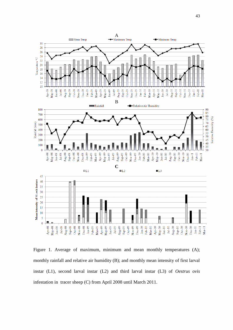
43
A
B
C
Figure 1. Average of maximum, minimum and mean monthly temperatures (A);
monthly rainfall and relative air humidity (B); and monthly mean intensity of first larval
instar (L1), second larval instar (L2) and third larval instar (L3) of Oestrus ovis
infestation in tracer sheep (C) from April 2008 until March 2011.

44
Figure 2. Prevalence of clinical signs of oestrosis (%) in the sheep of the flock and mean
nasal discharge score (ND) in those animals displaying clinical signs. Dyspnea without
nasal mucous: ND = 1; dyspnea with sero-mucous: ND = 2; and dyspnea with muco-
purulent: ND = 3.
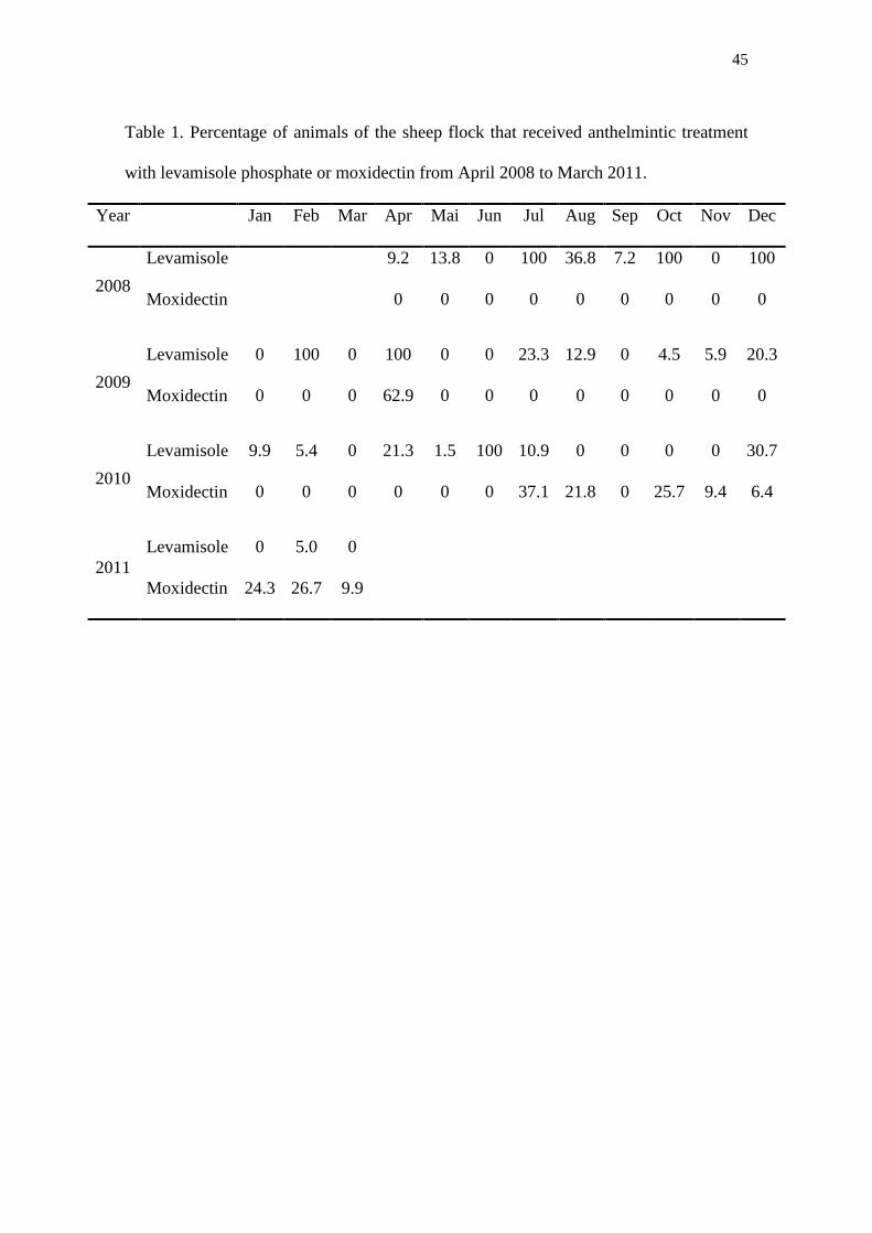
45
Table 1. Percentage of animals of the sheep flock that received anthelmintic treatment
with levamisole phosphate or moxidectin from April 2008 to March 2011.
Year Jan Feb Mar Apr Mai Jun Jul Aug Sep Oct Nov Dec
2008
Levamisole 9.2 13.8 0 100 36.8 7.2 100 0 100
Moxidectin 0 0 0 0 0 0 0 0 0
2009
Levamisole 0 100 0 100 0 0 23.3 12.9 0 4.5 5.9 20.3
Moxidectin 0 0 0 62.9 0 0 0 0 0 0 0 0
2010
Levamisole 9.9 5.4 0 21.3 1.5 100 10.9 0 0 0 0 30.7
Moxidectin 0 0 0 0 0 0 37.1 21.8 0 25.7 9.4 6.4
2011
Levamisole 0 5.0 0
Moxidectin 24.3 26.7 9.9

46
Capítulo 3

47
Prevalence and intensity of Oestrus ovis infestation in sheep in central region of São
Paulo State, Brazilb
Silva, B.F.1*
; Machado, G.P. 2
; Izidoro, T.B. 2
; Amarante, A.F.T. 1
1UNESP - Univ Estadual Paulista, Departamento de Parasitologia, Instituto de
Biociências, Caixa Postal 510, CEP: 18618-000, Botucatu-SP, Brazil.
2UNESP - Univ Estadual Paulista, Departamento de Higiene e Saúde Pública,
Faculdade de Medicina Veterinária e Zootecnia, CEP: 18618-000, Botucatu-SP, Brazil.
*Corresponding author: Bruna Fernanda da Silva, Departamento de Parasitologia,
Instituto de Biociências, Universidade Estadual Paulista (UNESP), Distrito de Rubião
Júnior s/n, Botucatu, São Paulo, CEP: 18618-000 Brazil. Tel.: +55 14 3811 6239; Fax:
+55 14 3815 3744. E-mail address: [email protected]
Abstract
Heads of 139 sheep were examined with the aim of determining the Oestrus ovis
prevalence and infestation intensity in central region of São Paulo State, Brazil. Heads
from slaughtered sheep were examined and the different O. ovis larval instars (L1, L2
and L3) were recovered from the nasal and sinus cavities. O. ovis larvae were detected
in 13.7% of sheep. The monthly mean intensity of infestation ranged from 1 until 10.2
larvae/infested head with general mean intensity of 4.5 larvae/infested head. Of the total
of 85 larvae, 21.2% were L1, 37.6% L2 and 41.2% L3. The present results demonstrate
b Artigo redigido de acordo com as normas do periódico científico Tropical Animal Health and
Production. Enviado para publicação no dia 29 de novembro de 2011.

48
that oestrosis is an important parasitic disease in sheep of the central region of São
Paulo State, which must be considered in programs of prophylaxis of sheep diseases.
Key-words: Oestrus ovis; sheep; prevalence; infestation.
Introduction
Oestrus ovis (Diptera: Oestridae), the sheep nasal bot fly, is a cosmopolitan
parasite whose larvae develop in the head sinuses and nasal passages of sheep and goats
in all sheep-farming areas of the world. The female fly is viviparous and deposit larvae
in or around the nostrils of its host and these early first instars attach to the mucous
membranes in the nasal cavities, change to second instars and move up to the sinuses
where it completes development in mature third instars, which are expelled for pupation
under the soil (Zumpt, 1965). The length of this parasitic portion of the life cycle is
quite variable from a few weeks to several months depending on the season and climatic
conditions (Cobbett and Mitchell, 1941). Clinical respiratory signs such as seromucous
or purulent nasal discharge, frequent sneezing and dyspnea, may severely impair the
health of affected animals (Dorchies et al., 1998).
O. ovis can thrive in different environments and has adapted to the climate
prevailing wherever sheep are kept (Horak, 1977) and the ability to adapt to different
environments allows to the natural persistence of infestation and the difficulties for its
control (Alcaide et al., 2005). Many surveys have demonstrated the high prevalence of
ovine oestrosis in numerous areas all over the world, e.g. Zimbabwe (Pandey, 1989),
France (Yilma and Dorchies, 1991), Italy (Caracappa et al., 2000), Spain (Alcaide et al.,
2003) and Greece (Papadopoulos et al., 2010). Oestrosis is commonly seen in sheep in
Brazil but there are few studies about the epidemiology of this parasite and most of

49
them are restricted to States in south region where favorable climatic conditions are
observed throughout the year.
In Rio Grande do Sul State, the prevalence of oestrosis in sheep was of 85.4% in
Bagé (Ribeiro et al., 1990) and in Encruzilhada do Sul, the mean intensity of infestation
was 23.8 larvae (Oliveira et al., 1999). In Santa Catarina State the O. ovis infestation
intensity were greater during the spring and summer months and no larvae were
recovered from tracer sheep when the temperature was less than 9 °C (Ramos et al.,
2006).
The first study about O. ovis epidemiology in São Paulo State was conducted
recently in southwest region where prevalence was 50% with mean intensity of 16.8
larvae per animal with the highest infestation rate in spring and summer time (Silva et
al., 2011). São Paulo State has different climatic conditions in each region and small
sheep farms could be found in the most part of this State. Therefore detailed research
about the epidemiology of parasitic infestation is required to recommend the best
strategies of oestrosis prophylaxis in small ruminants. This study aimed to determine the
O. ovis prevalence and infestation intensity in slaughtered sheep from central region of
São Paulo State, Brazil.
Materials and methods
A total of 139 sheep heads were examined from July 2009 to February 2010.
The animals were originated from Itápolis, Ibitinga, Borborema, Guarantã and Tapinas,
counties from the central region of São Paulo State. The monthly numbers of sheep
slaughtered in an abattoir in Itápolis city was variable and heads from all animals were
examined. The maximum and minimum numbers of heads examined per month were 26
in July and August 2009 and nine in October 2009, respectively (Table 2).
50
Most of the sheep were ≤ 1 year old (89.2%), Santa Ines (80.6%) breed and its
crosses with Dorper breed. Information on prior antiparasitic treatment and sheep
management were not available. Climate data were obtained by the Center of
Meteorological and Climate Research Applied to Agriculture (CEPAGRI / UNICAMP).
The annual rainfall average for the last 10 years was 1416.5 mm and the annual mean
temperature average for this period was 22.9 °C (Table 1).
Heads of slaughtered sheep were separated from the carcasses, put into an
individual plastic bag to avoid the transfer of larvae from one head to another and to
collect those that came out of nasal cavities. They were transported to Laboratory inside
a cool box with ice. Heads were cut open along their longitudinal and sagittal axis. All
larvae present in nasal cavity (nasal passage, septum, middle meatus and conchae) and
frontal sinus were collected and counted. Larvae were preserved in 70% alcohol and
identified according to their stage of development based on description of the Zumpt
(1965) and Capelle (1966).
Statistical analyses
Descriptive statistical analyses were performed in agreement with Bush et al.
(1997). The following terms were used:
Prevalence: the number of hosts infested with O. ovis larvae divided by the number of
hosts examined;
Intensity of infestation: the number of O. ovis larvae in a single infested host;
Mean intensity of infestation: the total number of O. ovis larvae found divided by the
number of hosts infected with that parasite.
51
Results
Of the 139 heads examined, 19 (13.7 %) were infested with O. ovis larvae with
minimum and maximum prevalence of the 7.7 % in August 2009 and 20.0 % in
February 2010, respectively (Table 2). The monthly mean intensity of infestation ranged
from 1 to 10.2 larvae/infested head with general mean of intensity of 4.5 larvae/infested
head.
In October 2009 no O. ovis larvae was recovered, coinciding with the lowest
number of heads examined. Of the total of 85 larvae, 21.2% was L1, while L2 and L3
represented 37.6% and 41.2%. The sheep heads collected were derived mainly from
young animals (≤ 1 year old) but the prevalence of O. ovis larvae were similar between
the ages groups (Table 3).
Discussion
The prevalence of O. ovis infestation (13.7 %) in this study was lower than those
reported in similar studies carried out in other countries such as France, where 65% of
sheep was infested with O. ovis larvae (Yilma and Dorchies, 1991) or in Sicily with
55.8% of prevalence (Caracappa et al., 2000) and Turkey with 40.6% (Arslan et al.,
2009). Higher prevalence was also observed in other Brazilian studies: 85.4% in
southern Brazil in sheep exposed to natural infestation during one year (Ribeiro et al.,
1990) and 50% of prevalence in tracer sheep in Botucatu, São Paulo State, Brazil (Silva
et al., 2011).
Despite the lower prevalence observed in the present study, O. ovis larvae were
present in all experimental months, except in October, coinciding with the lowest
number of sheep heads examined. Probably if more heads had been monthly examined
we would have more chances to find infested sheep, like is observed in studies done in

52
other countries where the sheep slaughter routine is high and frequent. In Sicily, as
example, 70 heads were examined per month (Caracappa et al., 2000) whereas in the
present study in the maximum were examined 26 sheep heads per month. In São Paulo
State, Brazil, large slaughterhouses are common which are specialized in slaughtering
cattle, poultry and pigs. Slaughter of sheep, in general, is restricted to small abattoirs
and with low slaughter routine, making it difficult to collect a larger number of heads to
examine.
Many factors can influence the O. ovis epidemiology such as sheep management
practices and the particular climatic conditions. The low prevalence of oestrosis in these
animals may have been influenced by previous treatment with drugs, such as
macrocyclic lactones, which are usually used for prophylaxis of infections with
gastrointestinal nematodes and have action against O. ovis larvae instars (Dorchies et
al., 1996; 1997; Lucientes et al., 1998). Influence of anthelmintic treatment on O. ovis
epidemiology was also observed in Botucatu, where a reduction in infestation of tracer
sheep coincided with the treatment of the flock with macrocyclic lactones (Silva et al.,
2011).
Among the climatic factor influencing oestrid fly activity, temperature, light
intensity and wind is considered the most important, but in case of O. ovis, the
temperature is the principal factor affecting the larvipositional activity of gravid females
(Cepeda-Palacios and Scholl, 2000). Although the sheep heads were examined during
the eight months, the climatic conditions of the region of the sheep origin should be
favorable to O. ovis activity during all months of the year, where the mean of
temperature is around 22 ºC and maximum and minimum averages around 16.3 and
29.6 ºC. Previous studies demonstrated that O. ovis fly activity start when the
temperature is around 20 ºC, but the major peak of strikes was seen between 25 and 28

53
ºC (Horak, 1977; Cepeda-Palacios and Scholl, 2000) that coincide with the average of
maximum temperature in this place.
It is interesting to note that mainly L2 and L3 O. ovis larval instars were
recovered in this study. Probably the climatic conditions were suitable to endogenous
larval development and L1 were able to change to L2 in a short period of time. Study
showed that larvae in sheep head can have a rapid development and reach the maturity
in three weeks (Cobbett and Mitchell, 1941). In Santa Catarina State, Brazil, greater L3
numbers also were recovered from tracer sheep (Ramos et al., 2006), but probably
because animals were exposed to infestation for 28 days and then were housed for more
20 days before slaughter, larvae had enough time to complete the development.
In conclusion, despite of the lack of information on prior antiparasitic treatment
and sheep management, which may influence oestrosis prevalence, all different O. ovis
larvae instars were recovered from slaughtered animals. This suggests the existence of
favorable climatic conditions for the fly activity and larval development of O. ovis in
sheep from central region of São Paulo State.
Acknowledgements
We are grateful to slaughterhouse worker for his fundamental cooperation
during the study. Bruna F. Silva received financial support by FAPESP (Fundação de
Amparo à Pesquisa do Estado de São Paulo) and Alessandro F.T. Amarante from CNPq
(Conselho Nacional de Desenvolvimento Científico e Tecnológico, Brazil).
The authors declare that they have no conflict of interested.

54
References
Alcaide, M., Reina, D., Frontera, E., Navarrete, I., 2005. Epidemiology of Oestrus ovis
(Linneo, 1761) infestation in goats in Spain, Veterinary Parasitology, 130, 277-
284
Alcaide, M., Reina, D., Sánchez, J., Frontera, E., Navarrete, I., 2003. Seasonal
variations in the larval burden distribution of Oestrus ovis in sheep in the
southwest of Spain, Veterinary Parasitology, 118, 235-241
Arslan, M., Kara, M., Gicik, Y., 2009. Epidemiology of Oestrus ovis infestations in
sheep in Kars province of north-eastern Turkey, Tropical Animal Health and
Production, 41, 299-305
Bush, A.O., Lafferty, K.D., Lotz, J.M., Shostak, A.W., 1997. Parasitology meets
ecology on its own terms: Margolis et al. revisited, The Journal of Parasitology,
83, 575-583
Capelle, K.J., 1966. The occurrence of Oestrus ovis L. (Diptera: Oestridae) in the
bighorn sheep from Wyoming and Montana, Journal Parasitology, 52, 618-621
Caracappa, S., Rilli, S., Zanghi, P., Di Marco, V., Dorchies, P., 2000. Epidemiology of
ovine oestrosis (Oestrus ovis Linné 1761, Diptera: Oestridae) in Sicily,
Veterinary Parasitology, 92, 233-237
Cepeda-Palacios, R., Scholl, P.J., 2000. Factors affecting the larvipositional activity of
Oestrus ovis gravid females (Diptera: Oestridae), Veterinary Parasitology, 91,
93-105

55
Cobbett, N.G., Mitchell, W.C., 1941. Further observations on the life cycle and
incidence of the sheep bot, Oestrus ovis, in New Mexico and Texas, American
Journal Veterinary Research, 2, 358-366
Dorchies, P., Alzieu, J.P., Cadiergues, M.C., 1997. Comparative curative and preventive
efficacies of ivermectin and closantel on Oestrus ovis (Linné 1758) in naturally
infected sheep, Veterinary Parasitology, 72, 179-184
Dorchies, P., Cardinaud, B., Fournier, R., 1996. Efficacy of moxidectin as a 1%
injectable solution and a 0.1% oral drench against nasal bots, pulmonary and
gastrointestinal nematodes in sheep, Veterinary Parasitology, 65, 163-168
Dorchies, P., Duranton, C., Jacquiet, P., 1998. Pathophysiology of Oestrus ovis
infection in sheep and goats: a review, Veterinary Record, 142, 487
Horak, I.G., 1977. Parasites of domestic and wild animals in South Africa. I. Oestrus
ovis in sheep, Onderstepoort Journal Veterinary Reseach, 44, 55-64
Lucientes, J., Castillo, J.A., Ferrer, L.M., Peribáñez, M.A., Ferrer-Dufol, M., Gracia-
Salinas, M.J., 1998. Efficacy of orally administered ivermectin against larval
stages of Oestrus ovis in sheep, Veterinary Parasitology, 75, 255-259
Oliveira, C.M.B., Oliveira, L.O., Torres, J.R., 1999. Oestrus ovis larvae distribution in
the head of naturally infested sheep, Arquivos da Faculdade de Veterinaria
UFRGS, 27, 87-92
Pandey, V.S., 1989. Epidemiology of Oestrus ovis infection of sheep in the highveld of
Zimbabwe, Veterinary Parasitology, 31, 275-280

56
Papadopoulos, E., Chaligiannis, I., Morgan, E.R., 2010. Epidemiology of Oestrus ovis
L. (Diptera: Oestridae) larvae in sheep and goats in Greece, Small Ruminant
Research, 89, 51-56
Ramos, C.I., Bellato, V., Souza, A.P., Avila, V.S., Coutinho, G.C., Dalagnol, C.A.,
2006. Epidemiologia de Oestrus ovis (Diptera: Oestridae) em ovinos no Planalto
Catarinense, Ciência Rural, 36, 173-178
Ribeiro, V.L.S., Oliveira, C.M.B., Branco, F.P.J.A., 1990. Prevalência e variações
mensais das larvas de Oestrus ovis (Linneus, 1761) em ovinos no município de
Bagé, RS, Brasil, Arquivo Brasileiro de Medicina Veterinaria e Zootecnia 42,
211-221
Silva, B.F., Bassetto, C.C., Amarante, A.F.T., 2011. Epidemiology of Oestrus ovis in
sheep in the southwest of Brazil., Veterinary Parasitology, in press
Yilma, J.M., Dorchies, P., 1991. Epidemiology of Oestrus ovis in southwest France,
Veterinary Parasitology, 40, 315-323
Zumpt, P., 1965. Myiasis in man and animals in the Old World, (Butterworths, London)

57
Table 1. Average of maximum, minimum and mean monthly temperature and rainfall in
the last 10 years in Itápolis city, São Paulo State.
Month
Temperature (ºC)
Rainfall (mm)
Minimum Maximum Mean
January 19.5 30.9 25.2 235.9
February 19.7 31.1 25.4 204.5
March 19.0 30.8 24.9 158.2
April 16.4 29.5 22.9 78.9
May 13.8 27.6 20.7 65.0
June 12.4 26.6 19.5 43.5
July 11.9 26.9 19.4 22.9
August 13.4 29.3 21.4 25.7
September 15.5 30.3 22.9 70.6
October 17.1 30.6 23.9 121.2
November 17.9 30.8 24.3 147.7
December 19.0 30.5 24.7 242.4
Annual 16.3 29.6 22.9 1416.5
Min 11.9 26.6 19.4 22.9
Max 19.7 31.1 25.4 242.4
Source: CEPAGRI / UNICAMP

58
Table 2. Numbers of heads infested with Oestrus ovis per month, mean larval burden
per head, number of first, second and third larval stage (L1-L3) and total number of
larvae found per month in sheep from central region of São Paulo State, Brazil.
Month
Number of
heads
examined
Number of
heads infested
Prevalen
ce (%)
Mean
larval
burden
L1 L2 L3
July 2009 26 5 19.2 10.2 17 19 15
August 2009 26 2 7.7 2.0 0 0 4
September 2009 13 2 15.4 1.0 0 0 2
October 2009 9 0 0 0 0 0 0
November 2009 12 1 8.4 4 0 0 4
December 2009 16 3 18.8 4.7 1 9 4
January 2010 17 2 11.8 1.0 0 0 2
February 2010 20 4 20.0 2.0 0 4 4
Total 139 19 18 32 35
Mean 13.7 4.5
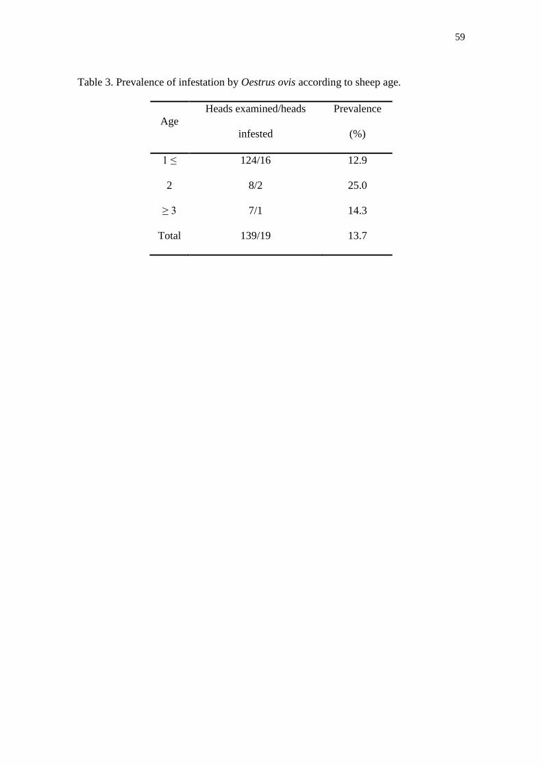
59
Table 3. Prevalence of infestation by Oestrus ovis according to sheep age.
Age
Heads examined/heads
infested
Prevalence
(%)
1 ≤ 124/16 12.9
2 8/2 25.0
≥ 3 7/1 14.3
Total 139/19 13.7

60
Capítulo 4
61
Parasitism by Oestrus ovis: Influence of sheep breed and nematode infectionsc
B.F. Silva1*
; C.C. Bassetto1; R.J. Shaw
2; A.M.O. Canavessi
3; A.F.T. Amarante
1
1UNESP - Universidade Estadual Paulista, Departamento de Parasitologia, Instituto de
Biociências, Caixa Postal 510, CEP 18618-000, Botucatu, SP, Brazil.
2Hopkirk Research Institute, AgResearch, Private Bag 11008, Palmerston North 4442,
New Zealand.
3USP – Universidade de São Paulo, Departamento de Zootecnia, ESALQ, Piracicaba,
CEP 13418-900, Brazil.
*Corresponding author: Bruna Fernanda da Silva, Departamento de Parasitologia,
Instituto de Biociências, Universidade Estadual Paulista (UNESP), Distrito de Rubião
Júnior s/n, Botucatu, São Paulo, CEP: 18618-000 Brazil. Tel.: +55 14 3811 6239; fax:
+55 14 3815 3744. E-mail address: [email protected]
Abstract
Previous studies showed that Santa Ines (SI) hair sheep were more resistant to
gastrointestinal nematode infections (GIN) than Ile de France (IF) sheep. The present
experiment aimed to evaluate if that reported resistance difference against GIN also
occurred against Oestrus ovis infestation and also to evaluate the influence of O. ovis
infestation on the gastrointestinal nematodes (GIN) infections. SI (n=12) and IF (n=12)
young male lambs were weaned at two months of age and moved to a paddock (0.3 ha)
with Brachiaria decumbens grass, where they also received concentrate ration. The
c Artigo redigido de acordo com as normas do periódico científico Veterinary Parasitology.
Aceito para publicação no dia 01 de novembro de 2011 doi:10.1016/j.vetpar.2011.11.007
Disponível: http://www.sciencedirect.com/science/article/pii/S0304401711007229
62
animals were kept together during the experimental period (September to early
December 2009). Fecal and blood samples were taken from all animals every two weeks
and body weight and nasal discharge score (oestrosis clinic signs) were recorded on the
same occasion. In early December 2009, all lambs were sacrificed and O. ovis larvae
and GIN were recovered, counted and identified according to the larval stage. All
animals were infested by different larval instars of O. ovis without any statistical
difference between breeds (P > 0.05). The SI lambs had an average of 24.8 larvae, and
the intensity of infection ranged between 14 and 39 larvae, while the IF lambs showed
an average of 23.5 larvae with the minimum and maximum from 11 to 36 larvae,
respectively. SI lambs presented the lowest nematode fecal egg counts (FEC) and the
lowest mean numbers of Haemonchus contortus, Trichostrongylus colubriformis and
Strongyloides papillosus, however, there was no significant differences between group
means (P > 0.05). Inverse relationship between numbers of O. ovis larvae and
gastrointestinal nematodes was observed in both breeds. SI sheep showed a significant
increase in blood eosinophils and total IgE serum levels and these variables were
negatively correlated with nematode FEC. A negative correlation was observed between
total IgE serum level and H. contortus burden in both breeds. In conclusion, there was
no breed difference regarding O. ovis infestation and in each breed, animals with more
nasal bot fly larvae tended to display smaller worm burden.
Key words: Oestrus ovis, nematodes, genetic resistance, breed-effect, sheep, total IgE
Introduction
Infections caused by gastrointestinal nematodes are the major constraint in small
ruminant production, however concurrent infections of sheep with nasal bot fly Oestrus
ovis and nematodes of the digestive tract are common (Yacob et al., 2002). O. ovis

63
causes cavitary myiasis in sheep and goats with clinical respiratory signs such as
seromucous or purulent nasal discharge, frequent sneezing and dyspnea, which may
severely impair their health. Those pathogenic effects cause serious economical losses
in the small ruminant livestock (Alcaide et al., 2003).
Haemonchus contortus and Trichostrongylus colubriformis are the major
gastrointestinal nematodes infecting sheep in Brazil (Amarante et al., 2004; Ramos et
al., 2004; Louvandini et al., 2006). The increasing development of resistances to
anthelmintics in these nematode populations (Almeida et al., 2010) imposes the need to
find alternative approaches less dependent on anthelmintic treatments for parasitic
control. Such strategies include, for example, pasture decontamination using different
herbivore species (Fernandes et al., 2004; Rocha et al., 2008) and the breeding of
genetically resistant animals (Amarante et al., 2004; Good et al., 2006; Alba-Hurtado et
al., 2010).
Epidemiological observations from field studies indicated that sheep selected for
resistance to gastrointestinal nematodes were more infected with the nasal bot fly O.
ovis than susceptible animals (Yacob et al., 2001). Many studies have shown negative
interactions between parasitism by O. ovis larvae and gastrointestinal nematodes
(Dorchies et al., 1997; Yacob et al., 2002; 2004; 2006; 2008; Terefe et al., 2005). The
presence of O. ovis larvae is correlated with significant reductions in nematode egg
excretion, worm fecundity and worm burdens. It was clear that O. ovis larvae had an
antagonistic effect on the worm populations but in contrast, the presence of nematodes
was not related with any change in the biology and larval burden of O. ovis (Yacob et
al., 2002; Terefe et al., 2005).
Sheep of the Santa Ines breed originated from the northeast of Brazil but has
been widely distributed throughout the southeast region of Brazil in the last few years. It

64
shows greater resistance to H. contortus infection than sheep of European breeds,
common in the same region (Amarante et al., 2004; Rocha et al., 2004; Bricarello et al.,
2005; Amarante et al., 2009). However, there is no information available about the
comparative resistance of Santa Ines and Ile de France sheep breeds to natural
infestation by O. ovis. Therefore, the purpose of this study was to examine the
comparative resistance to a natural infestation by O. ovis larvae in young lambs of two
sheep breeds (Santa Ines and Ile de France) and determine the interaction of this insect
infestation with naturally acquired gastrointestinal nematode infections.
Material and Methods
Study location
The study was carried out in Botucatu, São Paulo state, Brazil, at an altitude of
786 m a.s.l.. Climate data relating to averages in temperatures, relative air humidity and
rainfall were obtained by the Department of Environmental Science, Agronomical
Science College, UNESP, located at 8 km from experimental site. The average monthly
relative humidity was always higher than 76% throughout the experimental period that
occurred from September to December 2009. As usual for the region, rainfall was
abundant (150.5 mm in September, 141.8 mm in October, 289.0 mm in November and
327.1 mm in December) with a total 908.3 mm. Maximum and minimum mean
temperature ranged from 25.5 °C to 15.8 °C in September and from 29.5 °C to 19.8 °C
in November. The time of the year chosen to conduct the experiment was based on an
epidemiological study carried out recently in the same area that showed a high
incidence of oestrosis in sheep from September to December (unpublished data).

65
Animals and experimental design
Twelve young intact male Santa Ines (SI) and Ile de France (IF) lambs were
purchased from farms located in São Paulo state. Four lambs were acquired from each
farm to assure a minimum of genetic variability in each breed. All lambs were born in
June 2009, except four Ile de France lambs, which were born in May. Lambs were
weaned at two months of age and moved in late August to University facilities. The
animals were kept exclusively in pasture during the experimental period in a paddock
(0.3 ha) with Brachiaria decumbens grass, where they had free access to tap water. In
order to prevent massive infection by GIN, the animals were also supplemented, daily,
with commercial feed (Tech Ovin Unique, Socil®, with 18% of crude protein) in an
amount corresponding to 3% of their mean live weight. The amount of the commercial
diet was adjusted every 14 days according to lambs body weight gain. Decoquinate
(Deccox®, Alpharma) was added to the commercial feed to prevent coccidiosis. Early in
the trial, lambs received specific vaccine against clostridial infections (Sintoxan
Polivalente®, Merial, Brazil)
Fecal and blood samples were taken, body weight and nasal discharge score
(oestrosis clinic signs) were recorded from all animals every two weeks.
Fecal examination revealed that animals were infected with gastrointestinal
nematodes at their arrival to University facilities. In order to start the trial with animals
in the same conditions, they were drenched orally, once daily, for three consecutive
days with levamisole phosphate (10 mg/kg, Ripercol® L 150 F, Fort Dodge) and
albendazole (10 mg/kg, Valbazen®
10 Cobalto, Pfizer). The results obtained in the first
sampling were not included in the analysis. Two SI lambs died early in the trial of
unknown causes and the data for these animals were excluded from analyses.
66
At six months of age, in early December 2009, the animals were euthanized for
O. ovis larvae identification and counting and worm burden determination. The fresh
carcass weight was also recorded in an electronic balance (Modelo BCW 15, Welmy®
).
Fecal examination
Fecal samples were collected directly from the rectum of animals for nematode
fecal egg count (FEC) determination. The FEC values were determined using a
modified McMaster technique, in which each nematode egg counted represented 100
eggs per gram (EPG) of feces. Composite fecal cultures for the production of infective
larvae of gastrointestinal nematodes were prepared separately for each breed, and the
larvae obtained were killed, stained with Lugol‟s iodine and identified, according to the
descriptions of Keith (1953).
Pasture infectivity
To determine the number of third stage larvae (L3) per kilogram of dry matter
(L3/kg DM), grass samples were collected manually from paddock, close to the soil,
approximately every 3.5 m. The collector followed a W-track on the paddock (Taylor
1939). Samples were processed in the laboratory, according to Niezen et al. (1998), and
the larvae obtained were killed, stained with Lugol‟s iodine and identified, according to
the descriptions of Keith (1953).
Hematology
Blood samples were collected by jugular vein puncture into vacutainer tubes
with and without anti-coagulant (EDTA). Packed cell volume (PCV) was determined by
micro-haematocrit centrifugation and total plasma protein levels (g/dL) were estimated
using a refractometer (Refractometer SPR-N, Atago). The eosinophil counts in
peripheral blood were made in a Neubauer‟s chamber after staining with Carpentier‟s

67
solution (Dawkins et al., 1989); counts were expressed as the number of cells per µL of
blood. Serum samples were stored at -20 °C until immunoglobulin measurements and
serum albumin was determined using a kit (Protal método colorimétrico® – Laborlab)
and absorbance was read at 625 nm. The albumin/globulin ratio was estimated by the
formula: albumin/globulin = concentration of albumin / (total protein – albumin
concentration).
Serology – sandwich enzyme immunoassay (EIA) for IgE antibody
Total IgE levels in the animal‟s sera were measured using sandwich enzyme
immunoassay (EIA) as previously described (Shaw et al., 1997).
Oestrus ovis: clinic signs and larvae count post mortem
Signs of infestation and their clinical severity were measured by the score of
nasal discharge (ND). The score was the following: no ND = 0; only dyspnea without
secretion = 1; sero-mucous ND = 2; and muco-purulent ND = 3.
After euthanasia, heads were removed and cut open along their longitudinal and
sagittal axis. All larvae present in nasal cavity (nasal passage, septum, middle meatus
and conchae) and frontal sinus were collected and counted. Larvae were preserved into
70% alcohol and identified according to their stage of development based on description
of the Zumpt (1965) and Capelle (1966).
Worm counts
The abomasum was opened along its greater curvature and the contents placed in
a container. A 10% aliquot of the abomasal contents was preserved in 5% formalin. The
mucosal layers of all abomasums were soaked in saline solution at 38 °C for 6 h. All
content of the digested material was collected and preserved in 5% formalin. A similar
procedure was used to process the small intestine, but a 10% aliquot of the material

68
digested was collected. The large intestine was opened and a 10% aliquot of the
contents was collected and preserved. Worm identification and counting procedures
were performed in the preserved material as describe by Ueno and Gonçalves (1998).
Statistical analyses
Significant differences between groups for all variables were assessed by one-
way analysis of variance using Minitab 11.21 statistical software (Minitab Inc., USA).
Group means were considered statistically different when P < 0.05. EPG, worm burden,
Oestrus larval numbers, eosinophils/µL and nasal discharge score data were
transformed using log10 (x+1) prior to analysis. The results in figures and tables are
expressed as arithmetic means (± standard error of the mean). Spearman‟s correlation
coefficient between variables was assessed using the same software. To test if there was
difference between breeds regarding proportions of larval instars of O. ovis, the Chi-
square test for trend was performed using SAS (release 9.2).
Results
Oestrus ovis: clinic signs and larvae count post mortem
The animals, from both breeds, showed no oestrosis clinics signs in the first
month of trial (September), but, from October until the end of experiment, most of the
lambs showed oestrosis clinic signs with average of scores ranging from 0.5 to 1.7. On
18th
November the IF lambs showed average score (1.7) significantly higher (P < 0.05)
than SI lambs (0.8).
When euthanized, all three O. ovis larval instars were observed with prevalence
of 100% in both breeds (Table 1). The mean intensity of infestation was 24.8 larvae /
animal in SI lambs, with the minimum of 14 and maximum of 39 larvae per animal. In
IF lambs the mean intensity of infestation was 23.5 larvae / animal and the number of

69
larvae ranged from 11 to 36 larvae. There was no significant difference between breeds
(P > 0.05). The first stage larvae (L1) were predominant in both breeds (Table 1).
However, there was significant difference (P < 0.05) between breeds regarding the
proportion of larval instars of O. ovis: The total larval burden in SI lambs was
composed by 75.8% L1, 11.7% L2 and 12.5% L3, while IF lambs had 57.5% L1, 18.4%
L2 and 24.1% L3.
Fecal examination
At arrival of the experimental site, the mean of Strongyle and Strongyloides
papillosus FEC were, respectively, of 2275 and 900 EPG in IF lambs and of 3660 and
30 EPG in SI lambs. Haemonchus spp. was the predominant genus in fecal cultures
(93% in IF and 83% in SI) followed by Trichostrongylus spp. (7% in IF and 17% SI).
Fifteen days after the anthelmintic treatment, mean FEC dropped to 60 and 20 EPG
(Strongyle and S. papillosus, respectively) in SI lambs and 158 and 75 EPG (Strongyle
and S. papillosus, respectively) in IF lambs.
During the sampling period, the Strongyle FEC in IF lambs increased gradually
until 4th
November and thereafter there was a slight decrease in the mean FEC. The FEC
in SI lambs also increased during the first collections with the maximum mean FEC of
781.8 (± 194.4) on 21st October, but then the mean Strongyle FEC decreased and was
near zero in the last sampling (80.0 ± 32.7). The S. papillosus FEC also increased
gradually throughout the experiment in both breeds, with IF lambs displaying a higher
mean than SI lambs (725.0 ± 263.7 and 250.0 ± 80.6, respectively) in the last collection.
Despite the IF lambs showing the highest Strongyle and S. papillosus FEC (Fig. 1), no
significant difference was observed between breeds during the experiment (P > 0.05).
The animals were also observed to be infected with Moniezia spp.

70
It was noted that there was variability in susceptibility to GIN parasitism within
each breed. Within SI animals, there were three resistant lambs, whose FEC remained
low from the beginning of the experiment (< 500 EPG), while others showed relatively
high FEC with a maximum of 19,600 EPG in one animal, but over the course of the trial
these lambs developed protective immunity that resulted in a sharp FEC reduction that
were close to zero in all animals at the end of the experiment. Among the IF lambs,
there were resistant lambs; susceptible animals that acquired resistance throughout the
experiment; and four susceptible lambs, which remained with high FEC, with counts up
to 6600 EPG. Despite the high FEC observed in these IF lambs, salvage anthelmintic
treatments were not necessary during the experiment.
Haemonchus spp. was the predominant nematode in fecal cultures ranging
between 67% and 98% in IF and between 86% and 98% in SI, followed by
Trichostrongylus spp. ranging from 2% and 33% in IF and from 2% and 14% in SI
lambs. S. papillosus larvae were also found in fecal cultures in both breeds in all
sampling.
Nematode larvae on pasture
The Haemonchus spp. infective larvae were the major species recovered from
herbage. In the first collection no infective larvae was recovered, but from the 23rd
September significant numbers of Haemonchus spp. infective larvae per kilogram dry
matter (L3/kg DM) were recovered with a peak of 1713.7 L3/kg DM on 4th
November.
Infective larvae of Trichostrongylus spp. were recovered during the experiment but only
in two samplings and in low numbers (41.1 L3/kg DM on 18th
November and 59.5
L3/kg DM on 2nd
December).

71
Hematology
The lambs of both breeds had mean packed cell volume (PCV), total plasma
protein (TPP), albumin and globulin within the normal range for sheep. Initially the
mean of PCV was 33% (± 1.0%) and decreased gradually throughout the experiment
until reaching the lowest mean values of 27.6% (± 0.7%) in IF lambs and 27.7% (±
0.8%) in SI lambs on 4th
November (Fig. 2 A). The TPP in IF lambs ranged between 5.4
and 6.0 g/dL and in SI lambs from 5.1 to 6.0 g/dL throughout the experiment (P > 0.05)
(Fig. 2B).
Albumin values ranged between 3.0 and 3.9 g/dL in IF lambs and between 3.1
and 3.8 g/dL in the SI (P > 0.05) (Fig. 2C). The ratio of albumin/globulin during the
experiment ranged from 2.1 to 2.7 g/dL in IF lambs and between 2.1 to 2.5 g/dL in SI (P
> 0.05).
In all experimental weeks, the SI lambs had significantly higher mean number of
blood eosinophils than IF lambs (P < 0.05), except on 7th
October and 2nd
December,
when there was no significant difference between the breeds. IF lambs had mean (± SE)
values between 189.6 (±49.7) and 814.6 (±181.9) eosinophils/µL while the average of
the SI ranged from 460.0 (±145.6) to 2131.8 (±222.6) eosinophils/µL of blood
throughout the experiment (Fig. 3).
Serology – sandwich enzyme immunoassay (EIA) for IgE antibody
In all experimental weeks, the SI lambs had higher total serum IgE levels
compared with IF lambs (Fig. 4), which were significantly higher on 23rd
September,
21st
October, 4th
November and 2nd
December (P < 0.05).
72
Weight gain
IF lambs showed mean daily weight gain (200 g/day) significantly (P < 0.01)
higher than SI lambs (154 g/day). The animals were sacrificed at 6-7 months of age,
when IF lambs showed a mean of 40.6 kg (± 1.87) and the SI 27.9 kg (± 1.50).
Likewise, the average hot carcass weight was significantly higher in IF lambs (16.6 kg ±
0.94) compared with SI (11.4 ± 0.66 kg) (P < 0.05).
Worm counts
The following species were found in gastrointestinal contents of the animals: H.
contortus, T. colubriformis and S. papillosus (Table 1). The mean worm counts were
always higher in IF lambs than SI, but without significant differences between breeds (P
> 0.05). Moniezia spp. specimens were also recovered from most of the lambs of both
breeds in small numbers.
Correlation coefficients
Although the experimental groups were composed of a limited number of
animals, correlation coefficients were calculated between the number of H. contortus, T.
colubriformis, S. papillosus and O. ovis separately for each breed. Negative correlation
coefficients were observed between O. ovis x H. contortus and O. ovis x T.
colubriformis worm burdens in both breeds, however, significant correlation occurred
only between O. ovis larvae x S. papillosus (r = -0.614; P < 0.05) just in SI lambs
(Table 2).
Significant negative correlations were observed in SI lambs between total IgE x
S. papillosus worm burden (r = -0.648; P < 0.05) and in IF lambs between total IgE x H.
contortus worm burden (r = -0.594; P < 0.05). In SI animals correlation between total
IgE x H. contortus was also negative (r = -0.576, P = 0.08) (Table 3).

73
In some sampling dates, significant negative correlations were observed just in
SI lambs: on 21st October and 4
th November between total IgE x Strongyle‟s FEC (r = -
0.631 and r = -0.699, respectively; P < 0.05); on 18th
November and 2nd
December
between total IgE x FEC of S. papillosus (r = -0.968; r = -0.752, respectively; P < 0.05);
and between blood eosinophils x FEC of Strongyle (r = -0.764 e r = -0.745,
respectively; P < 0.05).
Discussion
Sheep of the SI breed are known to exhibit greater resistance to gastrointestinal
infections than sheep of European breeds (Amarante et al., 2004; 2009; Rocha et al.,
2004; Bricarello et al., 2005). Such difference between breeds was not observed
regarding O. ovis infestation, which were similar in both breeds. However,
epidemiological observations from field studies indicated that sheep selected for
resistance to gastrointestinal nematodes were more infected with the nasal bot fly, O.
ovis than susceptible ones (Yacob et al., 2001).
All different larval instars were recovered from animals of both breeds, but the
proportions of different instars were different between the breeds with higher proportion
of L1 in SI lambs. Possibly, there was delay in larval development in SI lambs caused
by a more intense immune response in comparison with IF. In other studies it was
demonstrated that immune response against O. ovis can impair larval development
(Frugeré et al., 2000; Angulo-Valadez et al., 2007).
In the present study young animals (less than six months old) were evaluated. If
the trial had had a longer duration, perhaps statistical difference between the breeds
could have been evidenced. However, some studies showed that despite sheep of all
ages being affected by oestrosis, the largest infestations were found in sheep more than

74
4-years-old (Abo-Shehada et al., 2000; Uslu and Dik, 2006; Arslan et al., 2009;
Shoorijeh et al., 2009). These results indicate that the ability of animals to become
oestrosis‟ resistant is very limited, in contrast to what occurs with GIN infections.
Concurrent infections of sheep with O. ovis and nematodes of the digestive tract
are common in the field as demonstrated in the present experiment, in which H.
contortus was the predominant nematode species in lambs of both breeds, followed by
T. colubriformis and S. papillosus, in agreement with previous studies in the same
region (Amarante et al., 2004). However, the number of parasites recovered from the
lambs in the present experiment was much lower than it was observed previously by
Amarante et al. (2004), who reported, on average, H. contortus worm burden almost
two times higher in SI sheep and approximately six times higher in IF animals than in
the present trial. Similarly for T. colubrifomis worm burden, almost nine times higher in
SI animals and almost 17 times higher in IF sheep.
The relatively low parasite worm burden observed in animals of the present
experiment may have been due to the influence of two factors. First, parasitism by O.
ovis may have induced nonspecific immune response that indirectly protected against
gastrointestinal nematodes. Several studies have shown that there is negative interaction
between the parasitism by larvae of O. ovis and gastrointestinal nematodes (Dorchies et
al., 1997; Yacob et al., 2002; 2004; 2006; 2008; Terefe et al., 2005). It was observed
that the FEC were significantly higher in lambs infected only with H. contortus
compared to lambs infected with both H. contortus and O. ovis. In this case the
production of blood eosinophils and high inflammatory cellular activity in tissue
stimulated by the presence of O. ovis larvae conferred protection against H. contortus
(Dorchies et al. 1997; Terefe et al., 2005). The same was also observed in experimental
infections with T. colubriformis and O. ovis (Yacob et al., 2002; 2004; 2006). Similarly,

75
negative correlation coefficients were observed between O. ovis and GIN worm burden
in the present trial. In addition, animals infected with O. ovis seem to be more tolerant
to the pathogenic effects of haemoncose because parasitism by O. ovis reduced the
length of H. contortus, and thus minimized the consumption of blood by the parasite
(Terefe et al., 2005). This may be an additional reason why no lamb showed clinical
signs of parasitic gastroenteritis, such as anemia accompanied by bottle jaw or diarrhea.
Secondly, the experimental animals received a high quality diet consisting of
commercial feed with 18% crude protein, the amount of 3% of body weight per day in
addition to grazing high quality braquiaria grass (B. decumbens). The high quality food
may also have favored the immune response of animals against GIN. Bricarello et al.
(2005) observed in SI and IF sheep breeds, artificially infected with H. contortus, that
high level of protein in the diet resulted in increased resistance of the SI compared to IF
sheep. Increased resistance and resilience were also observed in grazing SI lambs
naturally infected with gastrointestinal nematodes that received a diet supplemented
with high protein content (Louvandini et al., 2006). Therefore, the low GIN parasite
burdens displayed by sheep in the present experiment may have been due to the
interaction of several factors, specially the high nutritional standards associated with the
indirect effects of O. ovis infestation.
Eosinophilia and higher level of total IgE in the SI lambs indicate that the
immune responses of these animals against parasites were more pronounced than that of
IF. Significant negative correlation between total IgE and Strongyle and S. papillosus
FEC were observed on two occasions in the SI which was not observed in IF lambs.
Similar results were observed in sheep selected for resistance, artificially infected with
T. colubriformis, which presented higher levels of total IgE in intestinal lymph and
lower FEC when compared to susceptible sheep (Pernthaner et al., 2005). Furthermore,

76
the peripheral eosinophilia and increased serum IgE were associated with the reduced
infection in Gulf Coast Native lambs naturally infected by H. contortus (Shakya et al.,
2009). Moreover, despite IF lambs having serum total IgE levels lower than SI lambs, a
significant negative correlation was observed in IF lambs between total IgE and H.
contortus worm burden. These results demonstrate that the intra-breed differences
observed in the level of infection by GIN may be explained by the efficiency of the
immune response. In Nelore cattle infected naturally by Haemonchus placei and
Cooperia punctata, a resistant group also had significantly higher total serum IgE levels
and higher mean of blood eosinophil counts than a susceptible group (Bricarello et al.,
2007). Therefore, it has been suggested that total IgE levels might be considered useful
as a marker to identify resistant and susceptible cattle and sheep to nematode infections
(Bricarello et al., 2007; Shakya et al., 2009).
The mean number of blood eosinophils was significantly higher in SI than in IF
in almost all samplings and a significant negative correlation was observed between the
number of eosinophils/µL and FEC in SI lambs on two occasions. These results confirm
that eosinophilia can be more pronounced in more resistant animals. In Mexico,
eosinophilia was also associated with resistance to H. contortus infection in native
Criollo lambs that presented more eosinophils and a smaller worm burden than Suffolk
lambs (Alba-Hurtado et al., 2010). The presence of O. ovis larvae in the nasal cavity of
sheep is also related to a strong activation of eosinophils (Jacquiet et al., 2005; Yacob et
al., 2006). The experimental animals showed variation in eosinophil values throughout
the study that could be related to variations in the degree of exposition to infective
stages of parasites that might have occurred in the experimental site.
The results obtained also reinforced the importance of identifying animals in a
flock that are highly susceptible to parasites, which was the case of four IF lambs.
77
These animals are responsible for a large majority of pasture contamination and, once
identified, they should be removed from the flock (Sréter et al., 1994; Amarante et al.,
1998; Stear et al., 1998; Bassetto et al., 2009), a procedure that could minimize the
economic losses due to parasitism by GIN, reducing pasture contamination by infective
larvae and, consequently, reducing the need of anthelmintic prophylactic treatments.
Moreover, the resistance is a heritable characteristic (Sréter et al., 1994; Gruner et al.,
2004).
The IF lambs showed significantly higher weight gain compared to the SI,
averaging 200 g/day versus 154 g/day, respectively. Animals of the same breeds and
age, kept indoors, free of parasitic infections, and receiving a similar diet, presented
body weight gain of 256 g/day (IF lambs) and 223 g/day (SI lambs) (Bricarello et al.,
2005). At least in part, the lower body weight gain of the animals of the present
experiment in comparison with those studied by Bricarello et al. (2005) could be due to
oestrosis and GIN infections. The negative influence of GIN on body weight gain has
been well demonstrated. In ewes, O. ovis induced a decrease of almost 9% in milk
production (Dorchies et al., 2003). However, further studies will be necessary to assess
the influence of oestrosis in sheep productivity in our environmental conditions.
In conclusion, there was no breed difference regarding O. ovis infestation in SI
and IF young sheep and animals with more nasal bot fly larvae tended to display smaller
worm burden.
Acknowledgements
We are grateful for the technical assistance provided by Ms. Camila O. Carvalho
and Mr. Valdir A. Paniguel. This study was funded by Fundação de Amparo à Pesquisa
do Estado de São Paulo (FAPESP, Grant number 2008/53494-2). Bruna F. Silva (Grant

78
number 2007/58244-1) and César C. Bassetto (Grant number 2009/03504-4) received
financial support by FAPESP and Alessandro F. T. Amarante from CNPq (Conselho
Nacional de Desenvolvimento Científico e Tecnológico, Brazil).
79
References
Abo-Shehada, M.N., Araba, B., Mekbela, R., Williams, D., Torgerson, P.R., 2000. Age
and seasonal variations in the prevalence of Oestrus ovis larvae among sheep in
northern Jordan. Prev. Vet. Med. 47, 205-212.
Alba-Hurtado, F., Romero-Escobedo, E., Muñoz-Guzmán, M.A., Torres-Hernández, G.,
Becerril-Pérez, C.M., 2010. Comparison of parasitological and productive traits
of Criollo lambs native to the central Mexican Plateau and Suffolk lambs
experimentally infected with Haemonchus contortus. Vet. Parasitol. 172, 277-
282.
Alcaide, M., Reina, D., Sánchez, J., Frontera, E., Navarrete, I., 2003. Seasonal
variations in the larval burden distribution of Oestrus ovis in sheep in the
southwest of Spain. Vet. Parasitol. 118, 235-241.
Almeida, F.A., Garcia, K.C.O.D., Torgerson, P.R., Amarante, A.F.T., 2010. Multiple
resistance to anthelmintics by Haemonchus contortus and Trichostrongylus
colubriformis in sheep in Brazil. Parasitol. Int. 59, 622-625.
Amarante, A.F.T., Bricarello, P.A., Rocha, R.A., Gennari, S.M., 2004. Resistence of
Santa Ines, Suffolk and Ile de France sheep to naturally acquired gastrointestinal
nematodes infections. Vet. Parasitol. 120, 91-106.
Amarante, A.F.T., Godoy, W.A.C., Barbosa, M.A., 1998. Nematode egg counts, packed
cell volume and body weight as parameters to identify sheep resistant and
susceptible to infections by gastrointestinal nematodes. Ars. Vet. 14, 331-339.
80
Amarante, A.F.T., Susin, I., Rocha, R.A., Silva, M.B., Mendes, C.Q., Pires, A.V., 2009.
Resistance of Santa Ines and crossbred ewes to naturally acquired
gastrointestinal nematode infections. Vet. Parasitol. 165, 273-280.
Angulo-Valadez, C.E., Cepeda-Palacios, R., Jacquiet, P., Dorchies, P., Prévot, F.,
Ascencio-Valle, F., Ramirez-Orduña, J.M., Torres, F., 2007. Effects of
immunization of Pelibuey lambs with Oestrus ovis digestive tract protein
extracts on larval establishment and development. Vet. Parasitol. 143, 140-146.
Arslan, M.O., Kara, M., Gicik, Y., 2009. Epidemiology of Oestrus ovis infestations in
sheep in Kars province of north-eastern Turkey. Trop. Anim. Health Prod. 41,
299-305.
Bassetto, C.C., Silva, B.F., Fernandes, S., Amarante, A.F.T., 2009. Contaminação da
pastagem com larvas infectantes de nematóides gastrintestinais após o pastejo de
ovelhas resistentes ou susceptíveis à verminose. Rev. Bras. Parasitol. Vet. 18,
63-68.
Bricarello, P.A., Amarante, A.F.T., Rocha, R.A., Cabral Filho, S.L., Huntley, J.F.,
Houdijk, J.G.M., Abdalla, A.L., Gennari, S.M., 2005. Influence of dietary
protein supply on resistance to experimental infections with Haemonchus
contortus in Ile de France and Santa Ines lambs. Vet. Parasitol. 134, 99-109.
Bricarello, P.A., Zaros, L.G., Coutinho, L.L., Rocha, R.A., Kooyman, F.N.J., De Vries,
E., Gonçalves, J.R.S., Lima, L.G., Pires, A.V., Amarante, A.F.T., 2007. Field
study on nematode resistance in Nelore-breed cattle. Vet. Parasitol. 148, 272-
278.

81
Capelle, K.J., 1966. The occurrence of Oestrus ovis L. (Diptera: Oestridae) in the
bighorn sheep from Wyoming and Montana. J. Parasitol. 52, 618-621.
Dawkins, H.J.S., Windon, R.G., Eagleston, G.K., 1989. Eosinophil response in sheep
selected for high and low responsiveness to Trichostrongylus colubriformis. Int.
J. Parasitol. 19, 199-205.
Dorchies, P., Bergeaud, J.P., Van Khanh, N., Morand, S., 1997. Reduced egg counts in
mixed infections with Oestrus ovis and Haemonchus contortus: influence of
eosinophils? Parasitol. Res. 83, 727–730.
Dorchies, Ph., Wahetra, S., Lepetitcolin, E., Prevot, F., Grisez, C., Bergeaud, J.P.,
Hoste, H., Jacquiet, Ph., 2003. The relationship between nasal myiasis and the
prevalence of enzootic nasal tumours and the effects of treatment of Oestrus ovis
and milk production in dairy ewes of Roquefort cheese area. Vet. Parasitol. 113,
169-174.
Fernandes, L.H., Seno, M.C.Z., Amarante, A.F.T., Souza, H., Belluzzo, C.E.C., 2004.
Efeito do pastejo rotacionado e alternado com bovinos adultos no controle da
verminose em ovelhas. Arq. Bras. Med. Vet. Zootec. 56, 733–740.
Frugeré, S., Cota Leon, A., Prévot, F., Cepeda Palacios, R., Tabouret, G., Bergeaud,
J.P., Duranton, C., Dorchies, P., Jacquiet, P., 2000. Immunisation of lambs with
excretory secretory products of Oestrus ovis third instar larvae and subsequent
experimental challenge. Vet. Res. 31, 527-535.
Good, B., Hanrahan, J.P., Crowley, B.A., Mulcahy, G., 2006. Texel sheep are more
resistant to natural nematode challenge than Suffolk sheep based on faecal egg
count and nematode burden. Vet. Parasitol. 32, 317-327.

82
Gruner, L., Bouix, J., Brunel, J.C., 2004. High genetic correlation between resistance to
Haemonchus contortus and to Trichostrongylus colubriformis in INRA 401
sheep. Vet. Parasitol. 119, 51-58.
Jacquiet, P., Thi, T.T.N., Nouvel, X., Prevot, F., Grisez, C., Yacob, H.T., Bergeaud,
J.P., Hoste, H., Dorchies, PH., Tabouret, G., 2005. Regulation of Oestrus ovis
(Diptera: Oestridae) populations in previously exposed and naive sheep. Vet.
Immunol. Immunopathol. 105, 95–103.
Keith, R.K., 1953. The differentiation of infective larval of some common nematode
parasites of cattle. Aust. J. Zool. 1, 223-35.
Louvandini, H., Veloso, C.F.M., Paludo, G.R., Dell‟Porto, A., Gennari, S.M.,
McManus, C.M., 2006. Influence of protein supplementation on the resistance
and resilience on young hair sheep naturally infected with gastrointestinal
nematodes during rainy and dry seasons. Vet. Parasitol. 137, 103-111.
Niezen, J.H., Charleston, W.A.G., Hodgson, J., Miller, C.M., Waghorn, T.S.,
Robertson, H.A., 1998. Effect of plants species on the larvae of gastrointestinal
nematodes which parasitise sheep. Int. J. Parasitol., v.28, p.791-803, 1998.
Pernthaner, A., Shaw, R.J., McNeill, M.M., Morrison, L., Hein, W.R., 2005. Total and
nematode-specific IgE responses in intestinal lymph of genetically resistant and
susceptible sheep during infection with Trichostrongylus colubriformis. Vet.
Immunol. Immunopathol. 104, 69-80.
Ramos, C.I., Bellato, V., Souza, A.P., Avila, V.S., Coutinho, G.C., Dalagnol, C,A.,
2004. Epidemiologia das helmintoses gastrintestinais de ovinos no Planalto
Catarinense. Cienc Rural 34, 889–1895.

83
Rocha, R.A., Bresciani, K.D.S., Barros, T.F.M., Fernandes, L.H., Silva, M.B.,
Amarante, A.F.T., 2008. Sheep and cattle grazing alternately: Nematode
parasitism and pasture decontamination. Small Rumin. Res. 75, 135-143.
Rocha, R.A., Amarante, A.F.T., Bricarello, P.A., 2004. Comparison of the susceptibility
of Santa Ines and Ile de France ewes to nematode parasitism around
periparturition and during lactation. Small Rumin. Res. 55, 65–75.
Shakya, K.P., Miller, J.E., Horohov, D.W., 2009. A Th2 type of immune response is
associated with increased resistance to Haemonchus contortus in naturally
infected Gulf Coast Native lambs. Vet. Parasitol. 163, 57-66.
Shaw, R.J., McNeill, M.M., Gatehouse, T.K., Douch, P.G.C., 1997. Quantification of
total sheep IgE concentration using anti-ovine IgE monoclonal antibodies in an
enzyme immunoassay. Vet. Immunol. Immunophatol. 57, 253-265.
Shoorijeh, S. J., Negahban, S., Tamadon, A., Behzadi, M.A., 2009. Prevalence and
intensity of Oestrus ovis in sheep of Shiraz, southern Iran. Trop. Anim. Health
Prod. 41, 1259-1262.
Sréter, T., Molnár, V., Kassai, T., 1994. The distribution of nematode egg counts and
larval counts in grazing sheep and their implications for parasite control. Int. J.
Parasitol. 24, 103-108.
Stear, M.J., Bairden, K., Bishop, S.C., Gettinby, G., McKellar, Q.A., Park, M., Strain,
S., Wallace, D.S., 1998. The processes influencing the distribution of parasitic
nematodes among naturally infected lambs. Parasitology 117, 165-171.
84
Taylor, E.L., 1939. Technique for the estimation of pasture infestation by strongyloid
larvae. Parasitology 31, 473-478.
Terefe, G., Yacob, H.T., Grisez, C., Prevot, F., Dumas, E., Bergeaud, J.P., Dorchies,
Ph., Hoste, H., Jacquiet, P., 2005. Haemonchus contortus egg excretion and
female length reduction in sheep previously infected with Oestrus ovis (Diptera:
Oestridae) larvae. Vet. Parasitol. 128, 271-283.
Ueno, H., Gonçalves, P.C. (4 Ed., Tokyo: Japan International Cooperation Agency),
1998. Manual para diagnóstico das helmintoses de ruminantes. 143 pp.
Uslu, U., Dik, B., 2006. Prevalence and intensity of Oestrus ovis in Akkraman sheep in
the Konya region of Turkey. Med. Vet. Entomol. 20, 347-349.
Yacob, H.T., Basazinew, B.K., Basu, A.K., 2008. Experimental concurrent infection of
Afar breed goats with Oestrus ovis (L1) and Haemonchus contortus (L3):
Interaction between parasite populations, changes in parasitological and basic
haematological parameters. Exp. Parasitol. 120, 180-184.
Yacob, H.T., Dorchies, Ph., Jacquiet, Ph., Bleuart, C., Prevot, F., Grisez, C., Bergeaud,
J.P., Hoste, H., 2004. Concurrent parasitic infections of sheep: depression of
Trichostrongylus colubriformis populations by a subsequent infection with
Oestrus ovis. Vet. Parasitol. 121, 297-306.
Yacob, H.T., Duraton-Grisez, C., Prevot, F., Bergeaud, J.P., Bleuart, C., Jacquiet, Ph.,
Dorchies, Ph., Hoste, H., 2002. Experimental concurrent infection of sheep with
Oestrus ovis and Trichostrongylus colubriformis: and related changes in the
cellular responses of nasal and digestive mucosae. Vet. Parasitol. 104, 307-317.

85
Yacob, H.T., Hoste, H., Jacquiet, Ph., Duraton-Grisez, C., Dorchies, Ph., Gruner, L.,
2001. Mucosal inflammatory responses after experimental infections with
gastrointestinal nematodes in resistant and susceptible sheep naturally infected
with Oestrus ovis. Revue Med. Vet. 152, 717-722.
Yacob, H.T., Terefe, G., Jacquiet, PH., Hoste, H., Grisez, C., Prévot, F., Bergeaud, J.P.,
Dorchies, PH., 2006. Experimental concurrent infection of sheep with Oestrus
ovis and Trichostrongylus colubriformis: Effects of antiparasitic treatments on
interactions between parasite populations and blood eosinophilic response. Vet.
Parasitol. 137, 184-188.
Zumpt, P. (Butterworths, London), 1965. Myiasis in man and animals in the Old World.
257 pp.

86
A
B
Figure 1. Mean of fecal egg counts (FEC) of the Strongyle (H. contortus and T.
colubriformis) (A) and Strongyloides papillosus (B) of the Ile de France and Santa Ines
male lambs naturally infested with Oestrus ovis and gastrointestinal nematodes. Bars
are standard error. There was no significant difference between group means (P > 0.05).

87
A
B
C
Figure 2. Mean packed cell volume (A), total plasma protein (B) and albumin (C) of the
Ile de France and Santa Ines male lambs naturally infested by Oestrus ovis and
gastrointestinal nematodes. Bars are standard error. There was no significant difference
between group means (P > 0.05).
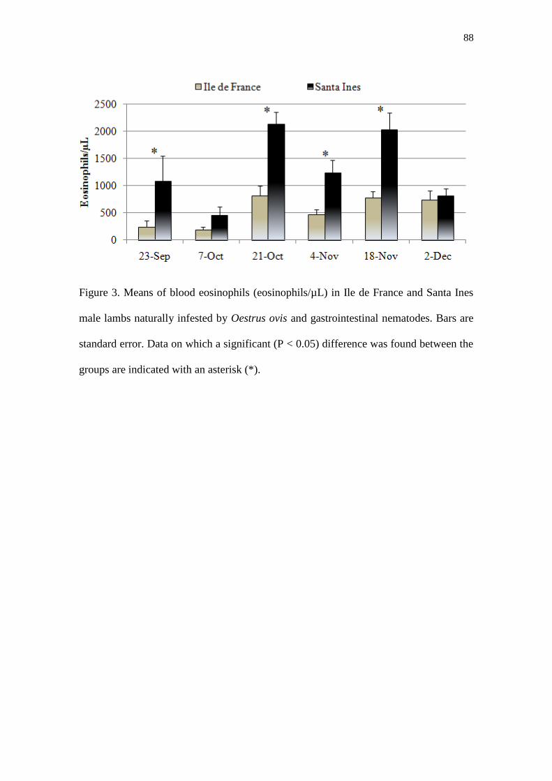
88
Figure 3. Means of blood eosinophils (eosinophils/µL) in Ile de France and Santa Ines
male lambs naturally infested by Oestrus ovis and gastrointestinal nematodes. Bars are
standard error. Data on which a significant (P < 0.05) difference was found between the
groups are indicated with an asterisk (*).

89
Figure 4. Means of total IgE level in sera in Ile de France and Santa Ines males lambs
naturally infested by Oestrus ovis and gastrointestinal nematodes. Bars are standard
error. Data on which a significant (P < 0.05) difference was found between the groups
are indicated with an asterisk (*).

90
Table 1. Mean number of parasites in Ile de France and Santa Ines males lambs
naturally infested by Oestrus ovis and gastrointestinal nematodes.
Species Stage of development Santa Ines Ile de France
H. contortus Early L4 503.0 (0-1640) 626.0 (0-1744)
Late L4 264.0 (0-1020) 115.9 (0-520)
L5 and adults 108.4 (0-734) 900.0 (0-4630)
Total burden 876.0 (0-2704) 1642 (0-5520)
T. colubriformis Total burden 565.0 (150-1350) 774.0 (140-1800)
S. papillosus Total burden 472.0 (0-2140) 967.0 (50-3980)
O. ovis L1 18.8 (7-34) 13.5 (1-29)
L2 2.9 (0-7) 4.3 (0-13)
L3 3.1 (0-10) 5.7 (0-13)
Total burden 24.8 (14-39) 23.5 (11-36)
There was no significant difference between group means (P > 0.05). Minimum and
maximum values are in parenthesis.

91
Table 2. Correlation coefficients between numbers of H. contortus (Hc), T.
colubriformis (Tc), S. papillosus (Sp) and O. ovis (Oo) in Ile de France and Santa Ines
male lambs.
Breed Hc Tc Sp
Ile de France Tc -0.382
Sp 0.070 -0.032
Oo -0.333 -0.323 -0.102
Santa Ines Tc -0.164
Sp 0.552 -0.224
Oo -0.395 -0.249 -0.614*
* P < 0.05.

92
Table 3. Correlation coefficients between numbers of blood eosinophil/µL (last
sampling), total IgE (last sampling), H. contortus (Hc), T. colubriformis (Tc), S.
papilosus (Sp) and O. ovis (Oo) in Ile de France and Santa Ines male lambs.
Breed Eosinophil IgE
Ile de France IgE -0.343
Hc 0.175 -0.594**
Tc 0.326 -0.095
Sp -0.448 -0.154
Oo 0.133 0.375
Santa Ines IgE 0.200
Hc -0.139 -0.576*
Tc 0.164 0.261
Sp 0.006 -0.648 **
Oo -0.219 0.529
* P = 0.08; ** P < 0.05

93
Capítulo 5

94
Cellular and humoral immune responses in sheep naturally infested with Oestrus ovis
(Diptera: Oestridae) and with nematode infectionsd
Silva, B.F.1*
; Bassetto, C.C.1; Amarante, A.F.T.
1
1UNESP - Univ Estadual Paulista, Departamento de Parasitologia, Instituto de
Biociências, Caixa Postal 510, CEP: 18618-000, Botucatu, SP, Brazil.
*Corresponding author: Bruna Fernanda da Silva, Departamento de Parasitologia,
Instituto de Biociências, Universidade Estadual Paulista (UNESP), Distrito de Rubião
Júnior s/n, Botucatu, São Paulo, CEP: 18618-000 Brazil. Tel.: +55 14 3811 6239; Fax:
+55 14 3815 3744. E-mail address: [email protected]
Abstract
This study was carried out to evaluate the humoral and cellular immune response in Ile
de France (IF) and Santa Ines (SI) young sheep naturally infested by Oestrus ovis and
gastrointestinal nematodes (GIN). Mast cells, eosinophils and globule leucocytes were
enumerated in the upper respiratory tract (septum, middle meatus and ventral nasal
conchae) and in the mucosa of abomasum and small intestine. Immunoglobulin G (IgG)
levels in serum samples and immunoglobulin A (IgA) levels in mucus from the nasal,
abomasum and small intestine mucosas were determined against O. ovis, Haemonchus
contortus and Trichostrongylus colubriformis antigens. The immune response against
O. ovis and GIN are very similar in both breeds and involve the recruitment of
inflammatory cells and immunoglobulins production in serum and mucus. Significant
positive correlation was observed in both breeds between the number of O. ovis larvae x
d Artigo redigido de acordo com as normas do periódico científico Veterinary Parasitology.

95
IgG anti-Oestrus crude extract (IF: r = 0.58; SI: r = 0.66; P < 0.05), between O. ovis
larvae x IgG anti-Oestrus excretory and secretory products (IF: r = 0.59; SI: r = 0.63; P
< 0.05). But apparently, the presence of antibodies in serum or nasal mucus as well as
inflammatory cells, were not efficient in the protection against O. ovis infestation.
Regarding GIN, the levels of immunoglobulins and the inflammatory cell numbers in
gastrointestinal mucosa presented significant inverse relationship only with H. contortus
worm burden in Santa Ines animals. Probably this was one of the reasons why these
animals showed the lowest FEC and worm burden compared to IF, since H. contortus
was the more prevalent GIN during the experiment. In conclusion, the immune response
against O. ovis and GIN are very similar and involve the recruitment of inflammatory
cells and production of immunoglobulins against the parasites that can be detected in
serum and mucus. However, O. ovis seems to be very well adapted to sheep and the
immune response against their larvae promotes just the delay in larval development but
not the expulsion of the parasite.
Key words: Immune response; Sheep; Oestrus ovis; Haemonchus contortus;
Trichostrongylus colubriformis.
Introduction
Mixed infections by dipteran larvae and helminthes are quite common in
ruminants, i.e. sheep, are normally parasitized by gastrointestinal nematodes and by
Oestrus ovis larvae. These parasites infections stimulate many immune mechanisms of
defense that can be mediate by antibody or cells, but the efficiency of this immune
response depends of the parasite and the infection stage.
Proinflammatory immune reactions are characteristic of O. ovis infestation and
involve the recruitments of cells (mast cells, eosinophils, macrophages, T and B

96
lymphocytes) and the secretion of immunoglobulins that suggest a type Th2 immune
response (Angulo-Valadez et al., 2011) similar with the protective immune response
against gastrointestinal parasitism by nematodes (Anthony et al., 2007; Rowe et al.,
2008). Many studies about the relationship between O. ovis and helminth co-infections
revealed that there are antagonist interaction between O. ovis larvae and the Strongylide
nematodes Trichostrongylus colubriformis and Haemonchus contortus. The infection of
the digestive tract with nematodes did not modify the biology of Oestrus populations,
but in contrast, infections with O. ovis was related to significant reductions in nematode
egg excretion and worm burdens. These changes were associated with significant
modifications in populations of mast cells, globule leucocytes and eosinophils in the
respiratory and digestive tracts. They also indicate that parasitic infection in one
particular anatomical site induces "at distance" inflammatory reactions of the whole
mucosal system (Dorchies et al., 1997; Yacob et al., 2002; Terefe et al., 2005).
This study was carried out to evaluate the humoral and cellular immune response
in Ile de France and Santa Ines young sheep naturally infested by O. ovis and with
nematode infections. We used samples from previously published study (Silva et al.,
2011b) that demonstrated no breed difference regarding O. ovis infestation, but that
revealed that animals with more nasal bot fly larvae tended to display smaller worm
burden. In the present experiment, we investigated which are the inflammatory cell
population and immunoglobulins involved in the protection against those parasites.
Material and Methods
Animals
The cellular and humoral response was evaluated in the upper respiratory tract
(septum, middle meatus and ventral nasal conchae) and in the digestive tract

97
(abomasum - fundic region and small intestine - 1 m from the pylorus) of the Ile de
France (IF) and Santa Ines (SI) young sheep naturally infested with O. ovis larvae and
infected with gastrointestinal nematodes (GIN). The experimental design has been
describe previously (Silva et al., 2011b). Briefly, 12 IF and 12 SI lambs were purchased
from different farms located in Sao Paulo State. Four lambs were acquired from each
farm to assure a minimum of genetic variability in each breed. All lambs were born in
June 2009, except four IF lambs, which were born in May. Lambs, weaned at two
months of age, were moved in late August to University facilities. The animals were
kept exclusively in pasture during the experimental period in a paddock (0.3 ha) with
Brachiaria decumbens grass, where they had free access to tap water.
At the beginning of the trial, in order to start the study with animals in the same
conditions, all lambs were treated with anthelmintics (levamisole phosphate +
albendazole). Two SI lambs died early in the trial of unknown cause and the data for
these animals were excluded from analyses.
At six months of age, in early December 2009, the animals were euthanized.
Blood serum, tissue and mucus samples were collected for immunological and
histological analysis.
Hematology
Blood samples were collected by jugular vein puncture into vacutainer tubes
without EDTA and serum samples were stored at -20 °C until use for immunoglobulin
G (IgG) measurements.
Histology
Immediately after death, tissue samples were taken from three anatomical
regions in the upper respiratory tract, i.e. septum, middle meatus and ventral nasal
98
conchae and from two sites of the digestive tract, i.e. abomasum (fundic region) and
small intestine (1 m from the pylorus) for counting of mucosal mast cells, eosinophils
and globule leucocytes. All tissue samples were fixed in 10% buffered formaldehyde for
48 h.. Afterwards, the samples were dehydrated with alcohol and embedded in paraffin
wax. Sections, 2 µm thick, were stained with toluidine blue 1% or haematoxylin and
eosin (H&E).
Mast cells were counted in sections stained with toluidine blue and eosinophils
and globule leucocytes in sections stained with H&E. Cells were enumerated under a
10x eye piece containing a calibrated graticule and 100x objective lens viewing an area
of 0.01 mm2. Thirty fields, randomly selected, were observed per animal for each
histological region and the mean numbers of cell/surface were calculated and compared
between the groups. The counts were expressed as number of cells per mm2 of mucosa.
Mucus
Mucus was taken from the nasal cavities, abomasum and small intestine
mucosas to determine the levels of immunoglobulin A (IgA).
While the larvae of O. ovis were collected, mucus from nasal mucosa was
extracted by lightly scraping the mucosal surface with a glass slide and mucus was
stored in a falcon tube at -20 °C until processing. A 5 cm piece of abomasum and small
intestine were sampled for the extraction of mucus and stored at -20 °C until processing.
Tissues were thawed and mucus was scraped off with a glass slide. The scrapings were
collected in a falcon tube on ice. Three milliliters of ice cold PBS supplemented with
protease inhibitors (Complete®, Roche) was added to each sample. The samples were
shaken for 1 h at 4 °C and centrifuged for 30 min at 4 °C and 3000 x g. The supernatant
was collected and centrifuged again for 30 min at 4 °C and 15000 x g (Kanobana et al.,
2002). Protein concentrations were determined using a kit (Protal método
99
colorimétrico® – Laborlab, Brazil) and the samples of abomasum mucus were adjusted
to protein concentration of 0.4 g/dL; small intestine to 0.1 g/dL and nasal mucus to 0.7
g/dL using PBS supplemented with protease inhibitors.
Enzyme-linked immunosorbent assay (ELISA)
IgG levels in serum samples were determined against excretory and secretory
products (ESP) and crude extract (CE) antigens from second instar (L2) O. ovis larvae;
and against third stage larvae (L3) and adults (L5) of H. contortus and T. colubriformis
antigens.
IgA levels in nasal mucus was tested against excretory and secretory products
(ESP) and crude extract (CE) from L2 O. ovis larvae; abomasal mucus was tested
against L3 and L5 of H. contortus and small intestine mucus against L3 and L5 of T.
colubriformis.
Parasites used in antigen production
Second instar of O. ovis (L2) were collected from naturally infested sheep heads
and they were washed several times in phosphate-buffered saline (PBS pH 7.2) with
240.000 UI of penicillin and 100 mg/ml of streptomycin and the viability of larvae were
checked under a stereomicroscope.
The excretory and secretory products were obtained from five L2 maintained in
a culture medium in vitro. The L2 were placed in a tube containing 10 ml RPMI-1640
(Sigma; 8758) with penicillin and streptomycin and were incubated in darkness for 24 h
in a 5% CO2 atmosphere at 37 °C. Supernatants were collected, centrifuged at 2000 x g
for 20 min at 4 °C and stored at -80 °C until use.
To obtain the crude extract, 10 L2 were fragmented/homogenized, using
homogenizer (T10 basic, IKA), in 5 ml of the PBS pH 7.2 supplemented with protease

100
inhibitor (Complete®, Roche). The extract was centrifuged at 15000 x g for 30 min at 4
°C and supernatants were collected and centrifuged newly. Protein concentrations of O.
ovis antigens were determined using a kit (Bicinchoninic Acid Protein Assay Kit –
Sigma) and absorbance was read at 562 nm. The antigen extracts were stored in aliquots
at -80 °C until further use.
The production of antigens of infective third stage larvae (L3) and adults (L5) of
H. contortus and T. colubriformis were previously described by Amarante et al. (2009)
and Cardia et al. (2011), respectively.
Parasite-specific serum IgG
Polystyrene micro-titer plates (Nunc, USA) were coated with 100 µl of the
different antigens (5µg/ml) diluted in carbonate-bicarbonate buffer (pH 9.6); plates were
incubated overnight at 4 °C. All subsequent incubations were carried out for 1 h at 37
°C using, in each well, with a total of 100 µl of reagents. Between each step, plates were
washed three times with ultra pure water (EASYpure II UV, Barnstead, USA)
containing 0.05% Tween 20 (ProPure® - Amresco).
After coating, blocking was carried out with 0.1 % Gelatin (Amresco, USA) and
0.05 % Tween 20 (ProPure®
- Amresco) in PBS 7.2 (PBS-GT). Serum samples were
diluted in PBS-GT (1:500) and applied in duplicate. Plates were then incubated with
rabbit-anti sheep IgG peroxidase conjugated diluted at 1:10000 (A130-101P, Bethyl
Laboratories, Inc. USA). Finally, OPD substrate solution (1,2-phenylenediamine
dihydrochloride, Dako, Denmark) was added to each well and the enzymatic reaction
was allowed to proceed at room temperature, in the dark for 15 min and stopped with
sulphuric acid solution at 5% and plates were immediately read using an automated
ELISA reader (Biotrak II, Amersham-Biosciences, UK) at 492 nm.

101
The positive standard serum for O. ovis was obtained from a sheep evaluated by
titration of the all serum samples tested from this experiment and as negative control,
serum samples was obtained from young animals kept indoors that had no contact with
adult bot flies. The standard positive serum for H. contortus and T. colubriformis were
obtained from a sheep repeatedly infected with these nematodes. Results were expressed
as the percentage of the optical density value (OD) of the positive standard serum and
employing the following formula: % OD = [(OD mean of the tested serum – OD mean
of blank) / (OD mean of the positive standard serum – OD mean of blank)] x 100
(Kanobana et al., 2001).
Parasite-specific mucus IgA
The ELISA reactions for parasite-specific mucus IgA were as previously
described for serum analysis with 1:10 mucus dilution to abomasum and nasal mucus
and with 1:2 mucus dilution to small intestine. Rabbit-anti sheep IgA peroxidase
conjugated was diluted at 1:10000 (A130-108P, Bethyl Laboratories, Inc. USA).
Finally, OPD substrate solution (1,2-phenylenediamine dihydrochloride, Dako,
Denmark) was added to each well and the enzymatic reaction was allowed to proceed at
room temperature, in the dark for 15 min and stopped with sulphuric acid solution at 5%
and plates were immediately read using an automated ELISA reader (Biotrak II,
Amersham-Biosciences, UK) at 492 nm. The results were expressed as the percentage
of OD of sample minus OD of blank (Kanobana et al., 2001).
Statistical analyses
Significant differences between groups for all variables were assessed by
oneway analysis of variance using statistical software, Minitab 11.21 (Minitab Inc.,
USA). Group means were compared using Tukey test, significant level at 5%. The data

102
relative to cell counts, IgG and IgA was transformed to log10(x+1). Statistical
significance was taken as P < 0.05. Spearman‟s correlation coefficient between
variables was assessed using the same software. To test if there was effect of the time in
serum IgG levels, the repeated measures analyses was performed using SAS (release
9.2). The figures and tables in the results are expressed as arithmetic means (± standard
error of the mean).
Results
The data on FEC, nematode and O. ovis burdens of IF and SI lambs has been
presented in detail by (Silva et al., 2011b). In brief, the IF lambs showed the highest
Strongyle and S. papillosus FEC and worm burden, but no statistical significant
difference was observed between breeds during the experiment. FEC were relatively
low in most animals, but were high in a small number of sheep, especially IF.
All three O. ovis larval instars were observed with prevalence of 100% in both
breeds. The mean intensity of infestation in IF and SI lambs was 23.5 and 24.8
larvae/animal, respectively. There was no difference between breeds.
No significant differences between groups were found in the number of
inflammatory cells counted in nasal and digestive mucosa, except in globules
leucocytes/mm2 average in abomasum that was significantly higher in IF than in SI
lambs (P < 0.05) (Fig. 1).
The serum IgG anti-Oestrus crude extract was higher than the mean values of
IgG anti-Oestrus excretory and secretory products in both breeds. In the first month of
experiment (September 2009) the IgG anti-Oestrus levels were close to zero (Fig. 2),
but started to increase on 7th
October 2009 at the same time that the animals of both
breeds started to display clinical signs of oestrosis. The serum IgG anti-Oestrus levels

103
increased gradually throughout the experiment until reaching the highest mean value in
the last collection day (2nd
December 2009), being similar in both breeds (P > 0.05).
At the beginning of experiment, all lambs were parasitized by GIN, therefore,
they already displayed relatively high levels of serum IgG anti-L5 and anti-L3 of H.
contortus (Fig. 3) and T. colubriformis (Fig. 4). There was no difference between the
breeds to antigens tested (P > 0.05) and there was low variation in serum IgG levels
against GIN throughout of experiment, excepting the level of IgG anti-L5 of T.
colubriformis and anti-L3 of H. contortus that significantly increased until the end of
experiment in both breeds (P < 0.05).
The IgA in nasal mucus anti-Oestrus CE was higher than the mean values of IgA
anti-Oestrus ESP (Fig. 5A). The IF lambs showed higher levels of IgA than SI lambs,
but no significant difference was observed between the breeds (P > 0.05).
The IgA in gastrointestinal mucus anti-L5 and anti-L3 of H. contortus (Fig. 5B)
and anti-L5 of T. colubriformis (Fig. 5C) were higher in SI lambs than in IF, but no
significant difference was observed between breeds (P > 0.05).
Correlation coefficients between parasitological and immunological variables
are presented in Tables 1, 2 and 3. Although the experimental groups were composed of
a limited number of animals, significant (P < 0.05) positive correlation was observed in
both breeds between the number of O. ovis larvae x IgG anti-Oestrus crude extract in IF
(r = 0.58) and SI (r = 0.66), between O. ovis larvae x IgG anti-Oestrus excretory and
secretory in IF (r = 0.59) and SI (r = 0.63).
IF lambs showed significant positive correlation between the number of O. ovis
larvae x globule leucocytes from nasal meatus (r = 0.71; P < 0.05).
Regarding to GIN burden and immune response, significant correlations were
observed just in SI lambs: abomasum mast cells x H. contortus burden (r = -0.73; P <

104
0.05); IgG anti-L3 Hc x H. contortus burden (r = -0.72; P < 0.05); IgA anti-L5 Hc x H.
contortus burden (r = -0.61; P = 0.07); and mast cells from small intestine x T.
colubriformis burden (r = 0.60; P = 0.07).
No significant correlation coefficients was observed between inflammatory cells
from nasal tract x GIN tract (Table 4), excepting between globule leucocytes from nasal
conchae x small intestine in IF lambs (r = 0.63; P < 0.05).
Discussion
The parasitism with GIN and O. ovis causes an increase in inflammatory cell
numbers in mucosas of the upper respiratory and gastrointestinal tracts and also the
production of anti-parasite specific immunoglobulins (Yacob et al., 2002; Bricarello et
al., 2005; Terefe et al., 2005; Cardia et al., 2011), changes that were also observed in the
present study. Such immune response was similar in animals of both breeds and resulted
in no breed difference regarding O. ovis infestation or GIN worm burdens. However, SI
lambs showed a higher proportion of L1of O. ovis compared to IF, indicating a more
efficient immune response of the former breed (Silva et al., 2011b). The immune
response is involved in the regulation of O. ovis populations (Jacquiet et al., 2005), and
may have an inhibitory effect on O. ovis larval growth, delaying development (Frugère
et al., 2000; Angulo-Valadez et al., 2007b).
At the beginning of this experiment the serum IgG levels anti-O. ovis was close
to zero in SI and IF lambs, probably because the lambs never have had previous contact
with this parasite, but after the first experimental month, most of lambs started to show
clinical signs of oestrosis, i.e., dyspnea and nasal discharge, coinciding with the
significantly gradual rise of serum IgG levels. In O. ovis infestation, humoral systemic
response of IgG usually reaches seroconversion 2-4 weeks post-first infection and the

105
highest levels are observed during the development of L2 and L3 larvae (Alcaide et al.,
2005; Angulo-Valadez et al., 2011).
In an epidemiological study with tracer lambs exposed to O. ovis infestation
during 28 days, several animals presented considerable parasitic burden, but no clinical
signs of oestrosis (Silva et al., 2011a). This indicates that the major symptoms of
infestation, nasal discharge and frequent sneezing, are immunologically mediated, i.e.,
depends of the acquisition of immune response against the parasite. These symptoms
are more intense in some animals indicating hypersensitivity. In animals with this
clinical manifestations, larvae, especially L1s in the nasal cavities, are at high risk of
becoming trapped in dense mucus, asphyxed and expulsed from the host (Angulo-
Valadez et al., 2011).
A large percentage of the animals do not display intense symptoms of
infestation. Probably, there is a strategy of immune tolerance on the part of the host in
order to avoid self-damage or a strategy on the part of the parasite to avoid
hyperimmunostimulation that may lead to a “self-curing” phenomenon, similar to that
observed in gastrointestinal helminth parasites. Studies suggest that O. ovis uses
immunosuppressive strategies such as the depletion of lymphocytes and the degradation
of immunoglobulins to evade defensive attacks from the host (Tabouret et al., 2003;
Jacquiet et al., 2005) and L1 have an important role in the regulation of inflammatory
reactions (Duranton et al., 1999). It is well known that larvae stimulate the mucus
production, which is utilized in their nutrition. Salivary gland products of O. ovis
contain thermostable proteases, which appear to be important in larval nutrition and
host–parasite interaction (Angulo-Valadez et al., 2007a).
In the present study, animals with the highest levels of IgG and IgA anti-O. ovis
had the highest numbers of O. ovis larvae, while inflammatory cell numbers did not

106
present any consistent association with O. ovis larval burden. Apparently, the presence
of antibodies in serum or nasal mucus as well as inflammatory cells, were not efficient
in the protection against O. ovis infestation. However, it is likely that mucus IgA
associate with humoral and cellular immune response promote the regulation of O. ovis
burden in host (Jacquiet et al., 2005) and also can have influence in larval weight and
consequently in the viability of adults flies (Cepeda-Palacios et al., 2000), i.e., although
the immune response is not enough to limit the parasite establishment, this can at least
maintain the O. ovis population under control in the host.
Regarding GIN, the levels of immunoglobulins and the inflammatory cell
numbers in gastrointestinal mucosa presented significant inverse relationship only with
H. contortus worm burden in Santa Ines animals. Probably this was one of the reasons
why these animals showed the lowest FEC and worm burden compared to IF, since H.
contortus was the more prevalent GIN during the experiment. In addition, the SI sheep
showed higher blood eosinophils and higher level of total serum IgE which indicated
that the immune responses of these animals against GIN parasites were more
pronounced than that of IF and these variables were also negatively correlated with
nematode FEC (Silva et al., 2011b). In other studies, inflammatory cells and IgA-
parasite-specific in abomasum were also inversely associated with H. contortus worm
burden and FEC indicating that they may impair parasite development or fecundity
(Strain and Stear, 2001; Amarante et al., 2005; Bricarello et al., 2005).
The lambs arrived in the experimental site already parasitized by GIN, and
consequently, serum IgG anti-GIN was observed since the beginning of the study. The
serum IgG levels anti-L3 of H. contortus and anti-L5 of T. colubriformis had a
significant rise until the end of experiment in lambs of both breeds as a consequence to
continue exposure to nematode larvae on pasture.

107
It was also observed that animals with more nasal bot fly larvae tended to
display smaller worm burden (Silva et al., 2011b). In other trials, it was demonstrated
that nematode egg production and worm burden were significantly depressed in mixed
infections with O. ovis, as well, the clinical signs of GIN infections (Dorchies et al.,
1997; Terefe et al., 2005; Yacob et al., 2006). O. ovis infestation stimulates the immune
response which possibly had a negative influence in the GIN parasitism. This influence
was probably through the enhanced recruitment of activated inflammatory cells
(eosinophils, mast cells and globule leucocytes) and/or their products towards the gut
mucosa that finally created an unfavourable environment to the nematodes, thereby
reducing worm length and fecundity (Terefe et al., 2005), fact that could explain the low
FEC and worm burden in animals of both breeds in this study. Eosinophils are
considered very important in the response against helminth infections and are frequently
associated with the expression of resistance to parasites (Dawkins et al., 1989; Stear et
al., 2002; Balic et al., 2006; Shakya et al., 2011) as well as immunoglobulin E (Sayers
et al., 2008; MacKinnon et al., 2010).
Although the IF seems to have had the immune response less effective than SI
lambs, the former breed showed greater productivity compared to SI lambs, with higher
body weight gain, fact that could be attributed to the high quality of the diet offered to
animals during the study (Silva et al., 2011b). Immunity to parasitic infection was
considered as a function which competes for nutrient resources against the requirement
of the host to maintain other body functions (Coop and Kyriazakis, 1999). The resistant
breeds can present inferior productivity when compared with other breeds selected for
higher weight gain and meat quality, for example, SI sheep prioritize survival to the
detriment of productive development, while IF animals, which have higher productive
capabilities, prioritize growth (Amarante et al., 2009).

108
In conclusion, the immune response against O. ovis and GIN are very similar
and involve the recruitment of inflammatory cells and production of immunoglobulins
against the parasites that can be detected in serum and mucus. However, the immune
response seems to be more efficient against GIN in comparison with bot fly. O. ovis
seems to be very well adapted to sheep and the immune response against their larvae
promotes just the delay in larval development but not the expulsion of the parasite.
More studies are necessary to prove if the immune response of SI sheep can became
more efficient and able to limit O. ovis larval establishment when animals are exposed
to infestation for longer periods of time.
Acknowledgments
We are grateful for the technical assistance provided by Ms. Camila O. Carvalho
and Mr. Valdir A. Paniguel. This study was funded by Fundação de Amparo à Pesquisa
do Estado de São Paulo (FAPESP, Grant number 2008/53494-2). Bruna F. Silva (Grant
number 2007/58244-1) and César C. Bassetto (Grant number 2009/03504-4) received
financial support from FAPESP and Alessandro F. T. Amarante from CNPq (Conselho
Nacional de Desenvolvimento Científico e Tecnológico, Brazil).

109
References
Alcaide, M., Reina, D., Frontera, E., Navarrete, I., 2005. Analysis of larval antigens of
Oestrus ovis for the diagnosis of oestrosis by enzyme-linked immunosorbent
assay. Med. Vet. Entomol. 19, 151-157.
Amarante, A.F.T., Bricarello, P.A., Huntley, J.F., Mazzolin, L.P., Gomes, J.C., 2005.
Relationship of abomasal histology and parasite-specific immunoglobulin A
with the resistance to Haemonchus contortus infection in three breeds of sheep.
Vet. Parasitol. 128, 99-107.
Amarante, A.F.T., Susin, I., Rocha, R.A., Silva, M.B., Mendes, C.Q., Pires, A.V., 2009.
Resistance of Santa Ines and crossbred ewes to naturally acquired
gastrointestinal nematode infections. Vet. Parasitol. 165, 273-280.
Angulo-Valadez, C.E., Ascencio, F., Jacquiet, P., Dorchies, P., Cepeda-Palacios, R.,
2011. Sheep and goat immune responses to nose bot infestation: a review. Med.
Vet. Entomol. 25, 117-125.
Angulo-Valadez, C.E., Cepeda-Palacios, R., Ascencio, F., Jacquiet, P., Dorchies, P.,
Romero, M.J., Khelifa, R.M., 2007a. Proteolytic activity in salivary gland
products of sheep bot fly (Oestrus ovis) larvae. Vet. Parasitol. 149, 117-125.
Angulo-Valadez, C.E., Cepeda-Palacios, R., Jacquiet, P., Dorchies, P., Prévot, F.,
Ascencio-Valle, F., Ramirez-Orduña, J.M., Torres, F., 2007b. Effects of
immunization of Pelibuey lambs with Oestrus ovis digestive tract protein
extracts on larval establishment and development. Vet. Parasitol. 143, 140-146.

110
Anthony, R.M., Rutitzky, L.I., Urban, J.F., Stadecker, M.J., Gause, W.C., 2007.
Protective immune mechanisms in helminth infection. Nat Rev Immunol 7, 975-
987.
Balic, A., Cunningham, C.P., Meeusen, E.N.T., 2006. Eosinophil interactions with
Haemonchus contortus larvae in the ovine gastrointestinal tract. Parasite
Immunol. 28, 107-115.
Bricarello, P.A., Amarante, A.F.T., Rocha, R.A., Cabral Filho, S.L., Huntley, J.F.,
Houdijk, J.G.M., Abdalla, A.L., Gennari, S.M., 2005. Influence of dietary
protein supply on resistance to experimental infections with Haemonchus
contortus in Ile de France and Santa Ines lambs. Vet. Parasitol. 134, 99-109.
Cardia, D.F.F., Rocha-Oliveira, R.A., Tsunemi, M.H., Amarante, A.F.T., 2011. Immune
response and performance of growing Santa Ines lambs to artificial
Trichostrongylus colubriformis infections. Vet. Parasitol. 182, 248-258.
Cepeda-Palacios, R., Frugère, S., Dorchies, P., 2000. Expected effects of reducing
Oestrus ovis L. mature larval weight on adult populations. Vet. Parasitol. 90,
239-246.
Coop, R.L., Kyriazakis, I., 1999. Nutrition-parasite interaction. Vet. Parasitol. 84, 187-
204.
Dawkins, H.J.S., Windon, R.G., Eagleson, G.K., 1989. Eosinophil responses in sheep
selected for high and low responsiveness to Trichostrongylus colubriformis. Int.
J. Parasitol. 19, 199-205.

111
Dorchies, P., Bergeaud, J., Khanh, N.V., Morand, S., 1997. Reduced egg counts in
mixed infections with Oestrus ovis and Haemonchus contortus: influence of
eosinophils? Parasitol. Res. 83, 727-730.
Duranton, C., Dorchies, P., Grand, S., Lesure, C., Oswald, I.P., 1999. Changing
reactivity of caprine and ovine mononuclear phagocytes throughout part of the
life cycle of Oestrus ovis: assessment through spontaneous and inductible NO
production. Vet. Res. 30, 371-376.
Frugère, S., Leon, A.C., Prévot, F., Palacios, R.C., Tabouret, G., Bergeaud, J.P.,
Duranton, C., Dorchies, P., Jacquiet, P., 2000. Immunisation of lambs with
excretory secretory products of Oestrus ovis third instar larvae and subsequent
experimental challenge. Vet. Res. 31, 527-535.
Jacquiet, P., Tran Thi Ngoc, T., Nouvel, X., Prevot, F., Grisez, C., Yacob, H.T.,
Bergeaud, J.-P., Hoste, H., Dorchies, P., Tabouret, G., 2005. Regulation of
Oestrus ovis (Diptera: Oestridae) populations in previously exposed and naïve
sheep. Vet. Immunol. Immunopathol. 105, 95-103.
Kanobana, K., Ploeger, H.W., Vervelde, L., 2002. Immune expulsion of the
trichostrongylid Cooperia oncophora is associated with increased eosinophilia
and mucosal IgA. Int. J. Parasitol. 32, 1389-1398.
Kanobana, K., Vervelde, L., Van Der Veer, M., Eysker, M., Ploeger, H., 2001.
Characterization of host responder types after a single Cooperia oncophora
infection: kinetics of the systemic immune response. Parasite Immunol. 23, 641-
653.

112
MacKinnon, K.M., Zajac, A.M., Kooyman, F.N.J., Notter, D.R., 2010. Differences in
immune parameters are associated with resistance to Haemonchus contortus in
Caribbean hair sheep. Parasite Immunol. 32, 484-493.
Rowe, A., McMaster, K., Emery, D., Sangster, N., 2008. Haemonchus contortus
infection in sheep: Parasite fecundity correlates with worm size and host
lymphocyte counts. Vet. Parasitol. 153, 285-293.
Sayers, G., Good, B., Hanrahan, J.P., O'Donovan, J., Mulcahy, G., Sweeney, T., 2008.
Breed differences in mucosal and systemic antibody response to nematode
infection in sheep: an important role for IgE? Parasitology 135, 71-80.
Shakya, K.P., Miller, J.E., Lomax, L.G., Burnett, D.D., 2011. Evaluation of immune
response to artificial infections of Haemonchus contortus in Gulf Coast Native
compared with Suffolk lambs. Vet. Parasitol. 181, 239-247.
Silva, B.F., Bassetto, C.C., Amarante, A.F.T., 2011a. Epidemiology of Oestrus ovis in
sheep in the southwest of Brazil. Vet. Parasitol., in press.
Silva, B.F., Bassetto, C.C., Shaw, R.J., Canavessi, A.M.O., Amarante, A.F.T., 2011b.
Parasitism by Oestrus ovis: Influence of sheep breed and nematode infections.
Vet. Parasitol. doi:10.1016/j.vetpar.2011.11.007
Stear, M.J., Henderson, N.G., Kerr, A., McKellar, Q.A., Mitchell, S., Seeley, C.,
Bishop, S.C., 2002. Eosinophilia as a marker of resistance to Teladorsagia
circumcincta in Scottish Blackface lambs. Parasitology 124, 553-560.
Strain, S., Stear, M., 2001. The influence of protein supplementation on the immune
response to Haemonchus contortus. Parasite Immunol. 23, 527-531.

113
Tabouret, G., Bret-Bennis, L., Dorchies, P., Jacquiet, P., 2003. Serine protease activity
in excretory–secretory products of Oestrus ovis (Diptera: Oestridae) larvae. Vet.
Parasitol. 114, 305-314.
Terefe, G., Yacob, H.T., Grisez, C., Prevot, F., Dumas, E., Bergeaud, J.P., Dorchies, P.,
Hoste, H., Jacquiet, P., 2005. Haemonchus contortus egg excretion and female
length reduction in sheep previously infected with Oestrus ovis (Diptera:
Oestridae) larvae. Vet. Parasitol. 128, 271-283.
Yacob, H.T., Duranton-Grisez, C., Prevot, F., Bergeaud, J.P., Bleuart, C., Jacquiet, P.,
Dorchies, P., Hoste, H., 2002. Experimental concurrent infection of sheep with
Oestrus ovis and Trichostrongylus colubriformis: negative interactions between
parasite populations and related changes in the cellular responses of nasal and
digestive mucosae. Vet. Parasitol. 104, 307-317.
Yacob, H.T., Terefe, G., Jacquiet, P., Hoste, H., Grisez, C., Prévot, F., Bergeaud, J.P.,
Dorchies, P., 2006. Experimental concurrent infection of sheep with Oestrus
ovis and Trichostrongylus colubriformis: Effects of antiparasitic treatments on
interactions between parasite populations and blood eosinophilic responses. Vet.
Parasitol. 137, 184-188.

114
A
B
C
Figure 1. Mean number of eosinophils, mast cells and globule leucocytes per mm2 from
digestive tract: abomasum and small intestine; and from upper respiratory tract: septum,
ventral nasal conchae and middle meatus in Ile de France and Santa Ines male lambs
naturally infested by Oestrus ovis and gastrointestinal nematodes. Bars are standard
error. Data on which a significant difference (P < 0.05) was found between the groups
are indicated with an asterisk (*).

115
A
B
Figure 2. Mean of levels of serum IgG against crude extract (CE) (A) and excretory and
secretory products (ESP) (B) of Oestrus ovis second larval instar (L2) in Ile de France
and Santa Ines males lambs naturally infested by O. ovis and gastrointestinal
nematodes. Bars are standard error.

116
A
B
Figure 3. Mean of levels of serum IgG against third stage larvae (L3) (A) and against
adult (L5) (B) of Haemonchus contortus in Ile de France and Santa Ines males lambs
naturally infested by Oestrus ovis and gastrointestinal nematodes. Bars are standard
error.

117
A
B
Figure 4. Mean of levels of serum IgG against third stage larvae (L3) (A) and against
adult (L5) (B) of Trichostrongylus colubriformis in Ile de France and Santa Ines males
lambs naturally infested by Oestrus ovis and gastrointestinal nematodes. Bars are
standard error.

118
A
B
C
Figure 5. Mean level of mucus IgA against crude extract (CE) and excretory and
secretory products (ESP) of Oestrus ovis (Oo) second larval instar (L2) (A); against
third stage larvae (L3) and against adult (L5) of Haemonchus contortus (Hc) (B);
against third stage larvae (L3) and against adult (L5) of Trichostrongylus colubriformis
(Tc) (C) in Ile de France and Santa Ines males lambs naturally infested by O. ovis and
gastrointestinal nematodes. Bars are standard error. There was no significant difference
between means (P > 0.05).

119
Table 1. Correlation coefficients between Oestrus ovis burden x inflammatory cells in
nasal mucosa of septum, conchae and meatus; and seric IgG and mucus IgA against
crude extract (CE) and excretory and secretory products (ESP) of O. ovis in Ile de
France male lambs naturally infested.
Oestrus ovis burden
Ile de France Santa Ines
IgG anti-CE 0.58* 0.66*
IgG anti-ESP 0.59* 0.63*
IgA anti-CE 0.11 0.54
IgA anti-ESP 0.24 0.35
Eosinophils
Septum
0.17
0.03
Conchae -0.18 -0.18
Meatus -0.13 0.31
Mast Cells
Septum
0.45
-0.29
Conchae 0.06 0.14
Meatus 0.29 -0.01
Globule leucocytes
Septum
-0.26
0.17
Conchae -0.16 -0.32
Meatus 0.71* -0.33
* P < 0.05

120
Table 2. Correlation coefficients between Haemonchus contortus worm burden x
inflammatory cells in abomasal mucosa and seric IgG and abomasal mucus IgA against
antigens from infective larvae (L3) and adults (L5) of H. contortus in Ile de France and
Santa Ines male lambs naturally infected.
H. contortus worm burden
Ile de France Santa Ines
Eosinophils -0.13 -0.24
Mast Cells -0.38 -0.73*
Globule Leucocytes -0.41 -0.46
IgG anti-L3 0.10 -0.72*
IgG anti-L5 0.14 -0.56
IgA anti-L3 -0.09 -0.36
IgA anti-L5 0.01 -0.61**
* P < 0.01 ** P = 0.07

121
Table 3. Correlation coefficients between Trichostrongylus colubriformis (Tc) and
Strongyloides papillosus (Sp) worm burden x inflammatory cells in intestinal mucosa
and seric IgG and intestinal mucus IgA against antigens from infective larvae (L3) and
adults (L5) of T. colubriformis in Ile de France and Santa Ines male lambs naturally
infected.
Ile de France Santa Ines
Tc Sp Tc Sp
Eosinophils -0.02 -0.30 0.44 -0.56
Mast Cells -0.02 -0.07 0.60* 0.17
Globules Leucocytes -0.02 -0.09 0.18 -0.20
IgG anti-L3 -0.09 0.40
IgG anti-L5 -0.20 0.27
IgA anti-L3 0.04 0.12
IgA anti-L5 0.00 0.12
* P = 0.07
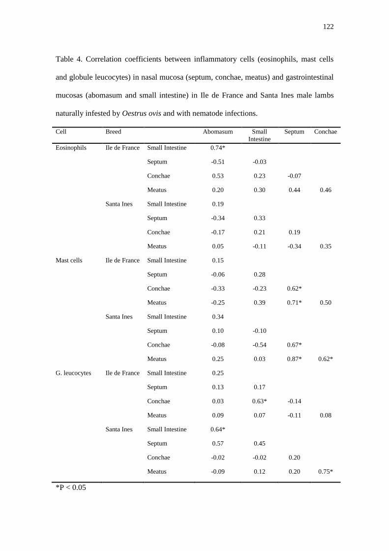
122
Table 4. Correlation coefficients between inflammatory cells (eosinophils, mast cells
and globule leucocytes) in nasal mucosa (septum, conchae, meatus) and gastrointestinal
mucosas (abomasum and small intestine) in Ile de France and Santa Ines male lambs
naturally infested by Oestrus ovis and with nematode infections.
Cell Breed Abomasum Small
Intestine
Septum Conchae
Eosinophils Ile de France Small Intestine 0.74*
Septum -0.51 -0.03
Conchae 0.53 0.23 -0.07
Meatus 0.20 0.30 0.44 0.46
Santa Ines Small Intestine 0.19
Septum -0.34 0.33
Conchae -0.17 0.21 0.19
Meatus 0.05 -0.11 -0.34 0.35
Mast cells Ile de France Small Intestine 0.15
Septum -0.06 0.28
Conchae -0.33 -0.23 0.62*
Meatus -0.25 0.39 0.71* 0.50
Santa Ines Small Intestine 0.34
Septum 0.10 -0.10
Conchae -0.08 -0.54 0.67*
Meatus 0.25 0.03 0.87* 0.62*
G. leucocytes Ile de France Small Intestine 0.25
Septum 0.13 0.17
Conchae 0.03 0.63* -0.14
Meatus 0.09 0.07 -0.11 0.08
Santa Ines Small Intestine 0.64*
Septum 0.57 0.45
Conchae -0.02 -0.02 0.20
Meatus -0.09 0.12 0.20 0.75*
*P < 0.05