Epidemiologia, clinica e diagnosi delle polmoniti · Epidemiologia, clinica e diagnosi delle...
53
Epidemiologia, clinica e diagnosi delle polmoniti Maddalena Peghin MD, PhD Clinica di Malattie Infettive Presidio ospedaliero di Udine Azienda Sanitaria Universitaria Integrata di Udine BUON USO DEGLI ANTIBIOTICI NELL’ERA DELLE RESISTENZE Come far si che il miracolo continui 15 giugno 2019
Transcript of Epidemiologia, clinica e diagnosi delle polmoniti · Epidemiologia, clinica e diagnosi delle...
Epidemiologia, clinica e diagnosi delle polmonitiMaddalena Peghin MD, PhD
Clinica di Malattie InfettivePresidio ospedaliero di Udine Azienda Sanitaria Universitaria Integrata di Udine
BUON USO DEGLI ANTIBIOTICI NELL’ERA DELLE RESISTENZE
Come far si che il miracolo continui
15 giugno 2019
Mr. C clinical case
• Mr. C is a 70 year old male who presents to the medical clinic where you work theday 15 January
• He is accompanied by his daughter
• Mr. C’s daughter states that Mr. C has a bad cold with a cough and that he hasbeen confused. He fell as he was getting dressed this morning
• Previous contact with a grandson with a cold

Mr. C Past Medical History
• Coronary artery disease
• Recovering alcoholic
• Class I obesity (BMI 34 kg/m2)
• Depressive syndrome
• Vascular ulcer lower limb, wound care at hospital twice a week
• No recent immunisations

Mr. C’s physical assessment
• T 37.8. BP 138/70, HR 90 RR 34. pulse ox 90%
• Using accessory muscles to breath. Crackles in the left lower lobe
• He reports pain on inspiration
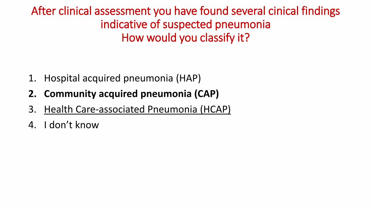
After clinical assessment you have found several cinical findings indicative of suspected pneumonia
How would you classify it?
1. Hospital acquired pneumonia (HAP)
2. Community acquired pneumonia (CAP)
3. Health Care-associated Pneumonia (HCAP)
4. I don’t know

After clinical assessment you have found several cinical findings indicative of suspected pneumonia
How would you classify it?
1. Hospital acquired pneumonia (HAP)
2. Community acquired pneumonia (CAP)
3. Health Care-associated Pneumonia (HCAP)
4. I don’t know
After clinical assessment you have found several cinical findings indicative of suspected pneumonia
How would you classify it?
1. Hospital acquired pneumonia (HAP)
2. Community acquired pneumonia (CAP)
3. Health Care-associated Pneumonia (HCAP)
4. I don’t know
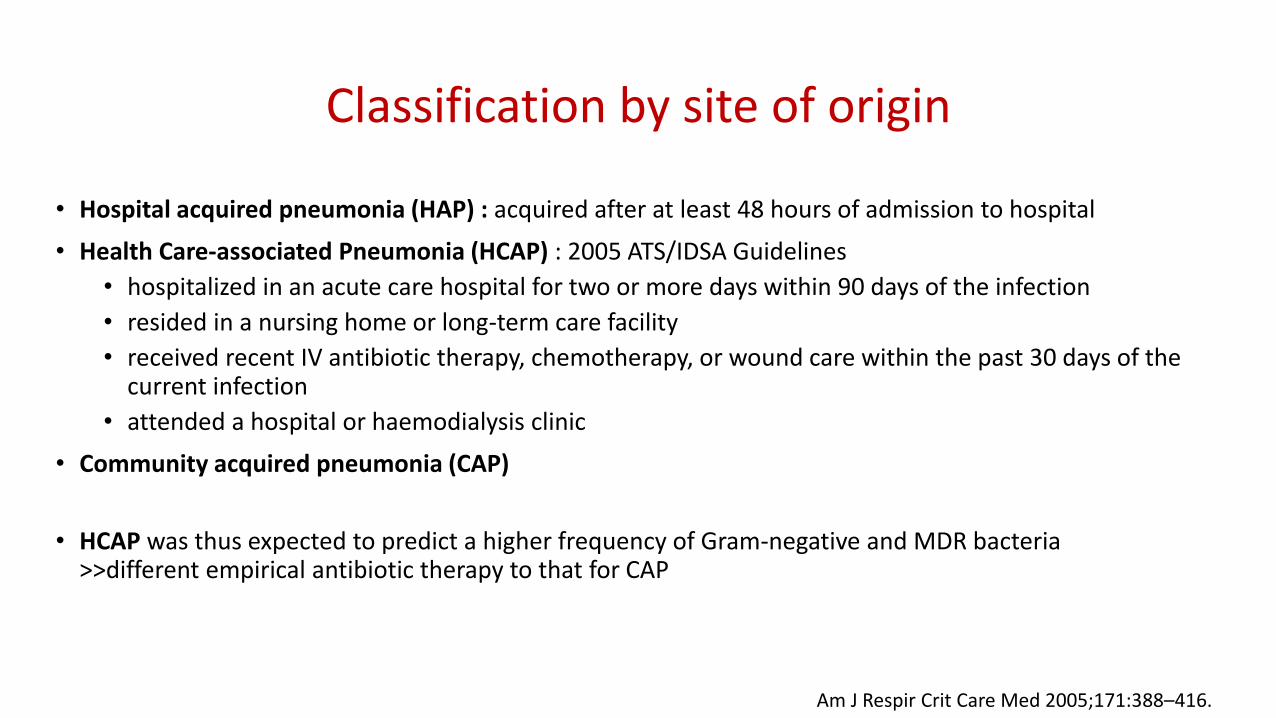
Classification by site of origin
• Hospital acquired pneumonia (HAP) : acquired after at least 48 hours of admission to hospital
• Health Care-associated Pneumonia (HCAP) : 2005 ATS/IDSA Guidelines
• hospitalized in an acute care hospital for two or more days within 90 days of the infection
• resided in a nursing home or long-term care facility
• received recent IV antibiotic therapy, chemotherapy, or wound care within the past 30 days of the current infection
• attended a hospital or haemodialysis clinic
• Community acquired pneumonia (CAP)
• HCAP was thus expected to predict a higher frequency of Gram-negative and MDR bacteria>>different empirical antibiotic therapy to that for CAP
Am J Respir Crit Care Med 2005;171:388–416.

However…HCAP : new avenue or just a cul-de-sac?
• Similar frequency and spectrum of causative pathogens in HCAP and CAP
• HCAP for the identification of pneumonia patients with a worse prognosis >>worse outcome related to patient factors
• HCAP should not be used to direct empirical antibiotic therapy:• opposite effect to that intended
• promote the development of bacterial antibiotic resistance
• 2016 ATS/IDSA Guidelines on HAP/VAP: removal of the concept of HCAP
• Recommendations regarding coverage for MDR pathogens among CAP based on validated risk factors for MDR pathogens
Woodhead et al. Thorax 2013;68:985–986.
Khalil et al. Clinical Infectious Diseases 2016;63(5):e61–111

Overview on Community-acquired pneumonia (CAP)
CAP is a leading cause of hospitalization and death worldwide
• The annual estimated CAP burden in USA : 5 million cases
• 80% of these cases are treated as outpatients (1% mortality)
• 20–25% treated in the hospital setting (23% mortality)
Gramegna et al. BMC Infectious Diseases (2018) 18:677
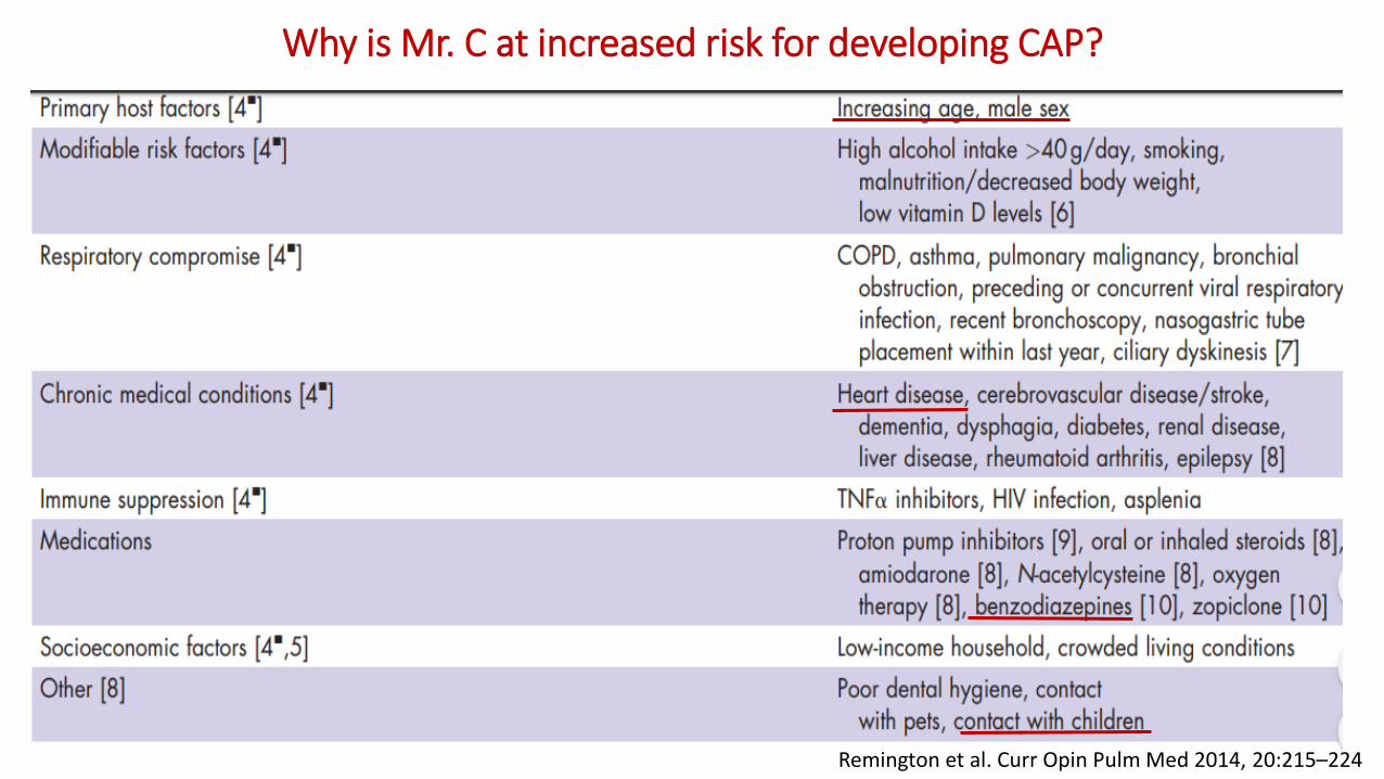
Why is Mr. C at increased risk for developing CAP?
Remington et al. Curr Opin Pulm Med 2014, 20:215–224

To Admit or Not?Pneumonia Severity & Deciding Site of Care
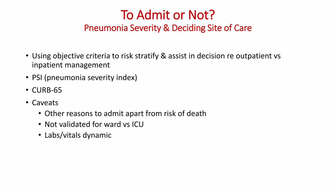
To Admit or Not?Pneumonia Severity & Deciding Site of Care
• Using objective criteria to risk stratify & assist in decision re outpatient vs inpatient management
• PSI (pneumonia severity index)
• CURB-65
• Caveats
• Other reasons to admit apart from risk of death
• Not validated for ward vs ICU
• Labs/vitals dynamic
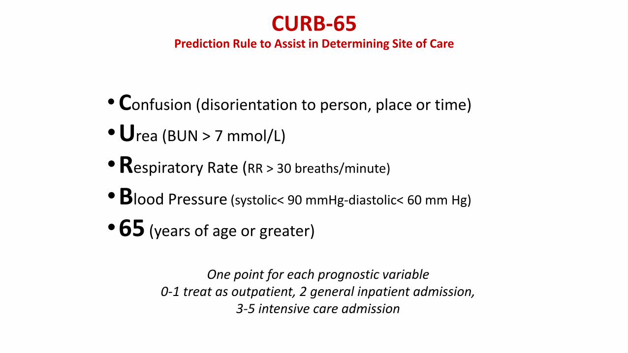
CURB-65Prediction Rule to Assist in Determining Site of Care
•Confusion (disorientation to person, place or time)
•Urea (BUN > 7 mmol/L)
•Respiratory Rate (RR > 30 breaths/minute)
•Blood Pressure (systolic< 90 mmHg-diastolic< 60 mm Hg)
•65 (years of age or greater)
One point for each prognostic variable 0-1 treat as outpatient, 2 general inpatient admission,
3-5 intensive care admission
(Uptodate 2012)

E.R.
Due to the baseline risk factors and clinical and physical assessment the patient is referred to the emergency room

Laboratory and radiographic findings
• RADIOGRAPHIC FINDINGS • LABORATORY FINDINGS
• WBC count: 13,500 cells/µL (with 82% polymorphonuclear cells and 11% band forms, lymphocytes 7%), platelets; 180,000 cells/mL
• CRP 223 mg/L
• AST 18 IU/L; ALT 23 IU/L
• Sodium 135 mEq/L; Potassium 4.2 mEq/L Chloride 99 mmol/L Calcium 8 mg/dL
• BUN 19 mg/dL Creatinine 1.9 mg/dL
• Glucose 116 mg/dL
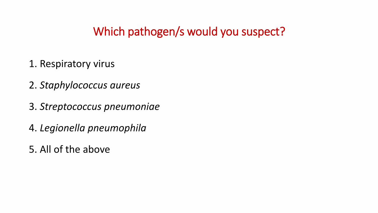
Which pathogen/s would you suspect?
1. Respiratory virus
2. Staphylococcus aureus
3. Streptococcus pneumoniae
4. Legionella pneumophila
5. All of the above
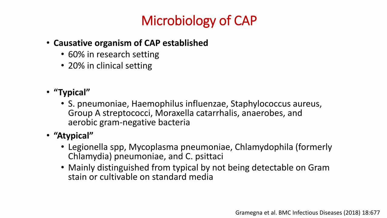
Microbiology of CAP
• Causative organism of CAP established • 60% in research setting• 20% in clinical setting
• “Typical”• S. pneumoniae, Haemophilus influenzae, Staphylococcus aureus,
Group A streptococci, Moraxella catarrhalis, anaerobes, and aerobic gram-negative bacteria
• “Atypical” • Legionella spp, Mycoplasma pneumoniae, Chlamydophila (formerly
Chlamydia) pneumoniae, and C. psittaci• Mainly distinguished from typical by not being detectable on Gram
stain or cultivable on standard media
Gramegna et al. BMC Infectious Diseases (2018) 18:677
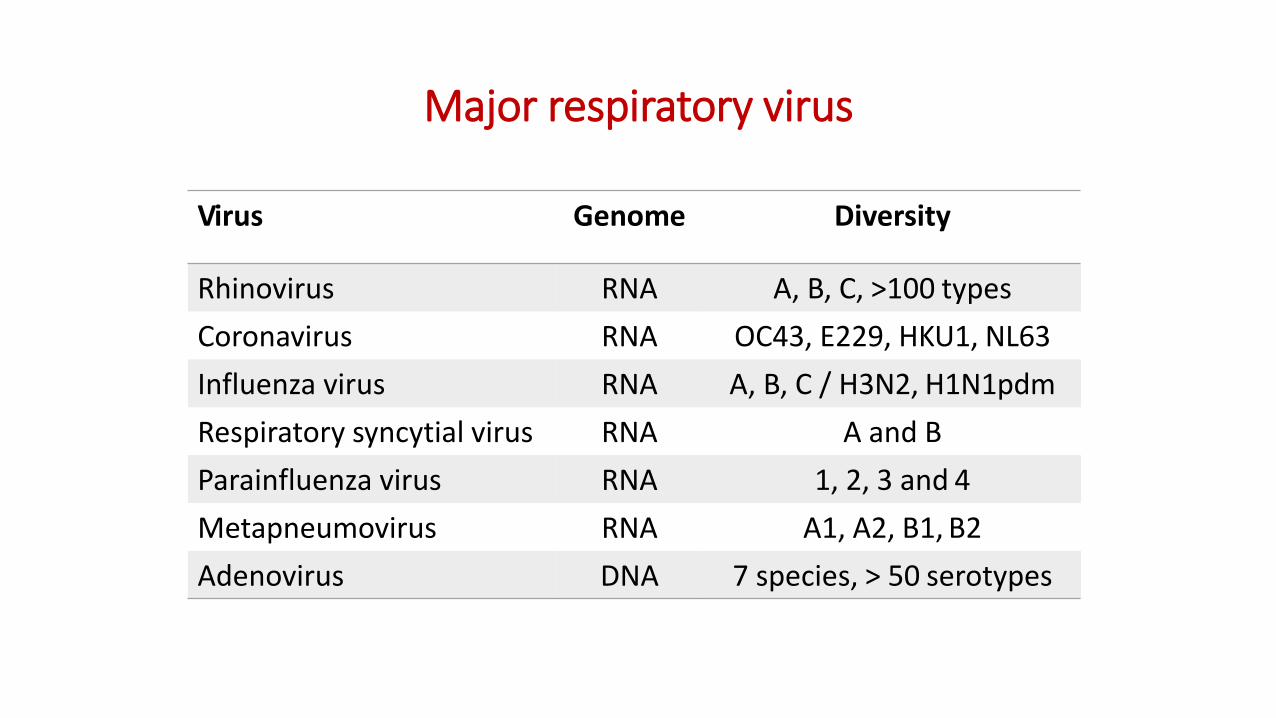
Major respiratory virus
Virus Genome Diversity
Rhinovirus RNA A, B, C, >100 types
Coronavirus RNA OC43, E229, HKU1, NL63
Influenza virus RNA A, B, C / H3N2, H1N1pdm
Respiratory syncytial virus RNA A and B
Parainfluenza virus RNA 1, 2, 3 and 4
Metapneumovirus RNA A1, A2, B1, B2
Adenovirus DNA 7 species, > 50 serotypes

Diagnostic tests for CAP
• Culture of respiratory tract specimens (sputum, tracheoaspirate, BAL): • very low sensitivity for atypical bacteria and RV
• Blood cultures• yield 5-15% ; stronger indication for severe CAP
• Direct fluorescent antibody (DFA) assay for atypical bacteria and RV• suboptimal sensitivity
• Serologic assays: antibodies to for atypical bacteria and RV • fourfold rise in either IgM or IgG titer is needed to confirm active disease
repeat 4-6 weeks later
• S.pneumoniae and L. pneumophila urine antigen• S.pneumoniae urine antigen: Sensitivity 60-75%, specificity : 95-99%
• L. pneumophila urine antigen: Legionella serogroup 1-specific (80%) >>>>>not useful for other serogroups
• Molecular testing
Arnold et al. Semin Respir Crit Care Med 2016;37:819–828
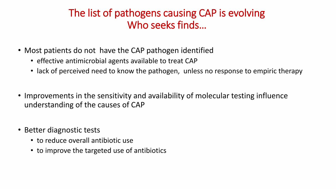
The list of pathogens causing CAP is evolvingWho seeks finds…
• Most patients do not have the CAP pathogen identified• effective antimicrobial agents available to treat CAP
• lack of perceived need to know the pathogen, unless no response to empiric therapy
• Improvements in the sensitivity and availability of molecular testing influenceunderstanding of the causes of CAP
• Better diagnostic tests • to reduce overall antibiotic use
• to improve the targeted use of antibiotics
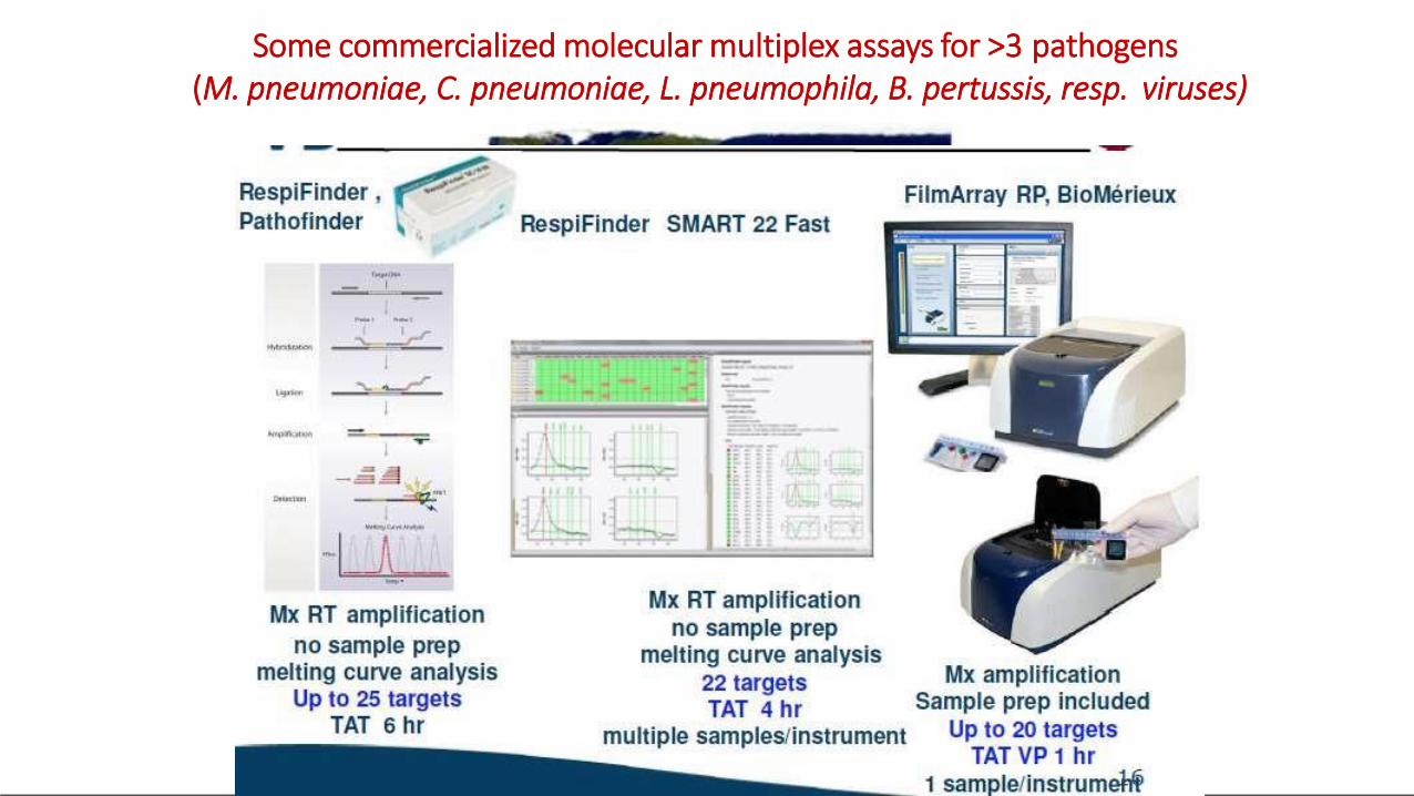
Some commercialized molecular multiplex assays for >3 pathogens(M. pneumoniae, C. pneumoniae, L. pneumophila, B. pertussis, resp. viruses)
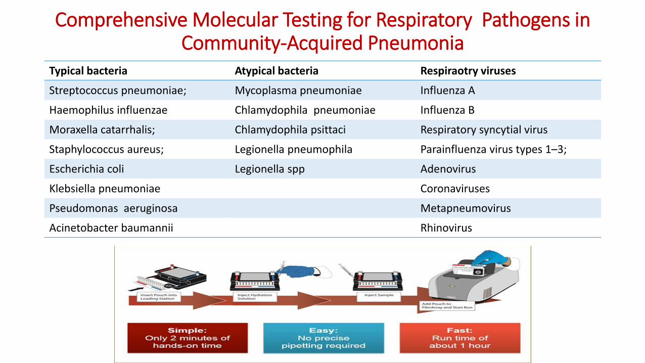
Comprehensive Molecular Testing for Respiratory Pathogens in Community-Acquired Pneumonia
Typical bacteria Atypical bacteria Respiraotry viruses
Streptococcus pneumoniae; Mycoplasma pneumoniae Influenza A
Haemophilus influenzae Chlamydophila pneumoniae Influenza B
Moraxella catarrhalis; Chlamydophila psittaci Respiratory syncytial virus
Staphylococcus aureus; Legionella pneumophila Parainfluenza virus types 1–3;
Escherichia coli Legionella spp Adenovirus
Klebsiella pneumoniae Coronaviruses
Pseudomonas aeruginosa Metapneumovirus
Acinetobacter baumannii Rhinovirus
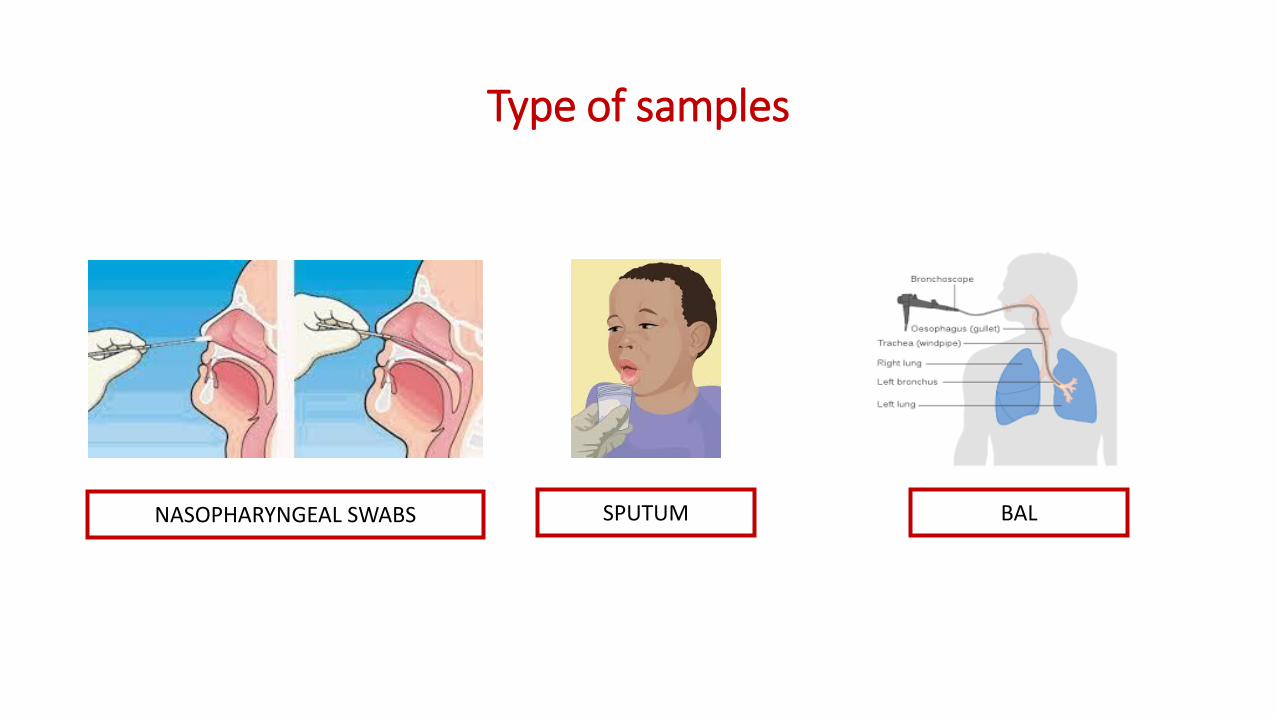
Type of samples
NASOPHARYNGEAL SWABS SPUTUM BAL

Epidemiology-atypical pathogens
• increased trend of atypical pathogens over the last 15 years, with prevalence ranging from 6 to 40% in both Europe and USA
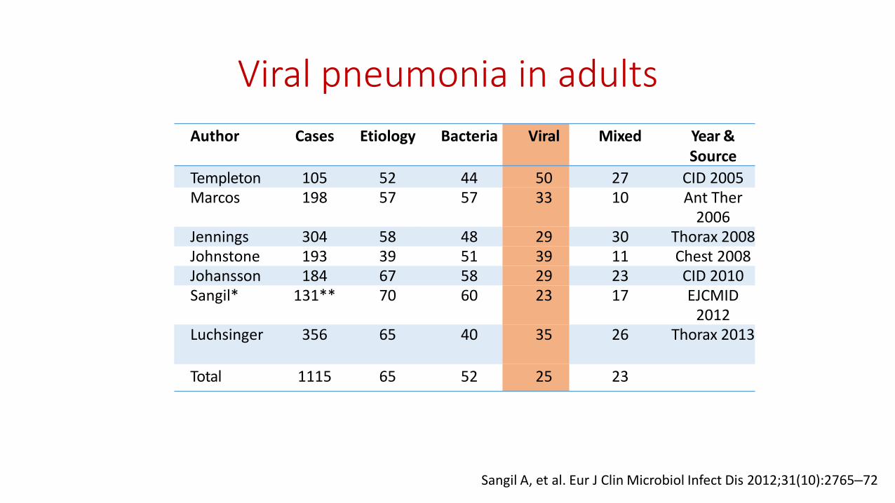
Viral pneumonia in adults
Author Cases Etiology Bacteria Viral Mixed Year &Source
Templeton 105 52 44 50 27 CID 2005Marcos 198 57 57 33 10 Ant Ther
2006Jennings 304 58 48 29 30 Thorax 2008Johnstone 193 39 51 39 11 Chest 2008Johansson 184 67 58 29 23 CID 2010Sangil* 131** 70 60 23 17 EJCMID
2012Luchsinger 356 65 40 35 26 Thorax 2013
Total 1115 65 52 25 23
Sangil A, et al. Eur J Clin Microbiol Infect Dis 2012;31(10):2765–72
Alimi Y et al. J Clin Virol. 2017; 95:26-35
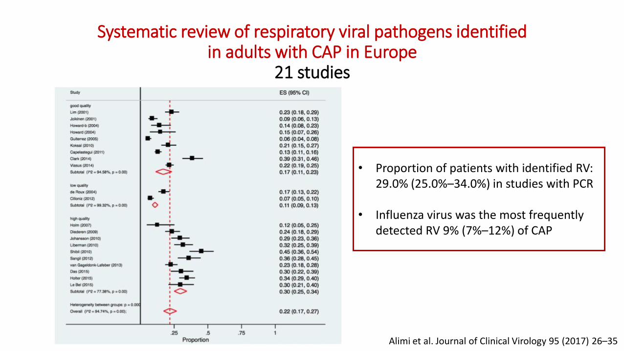
Systematic review of respiratory viral pathogens identified in adults with CAP in Europe
21 studies
• Proportion of patients with identified RV: 29.0% (25.0%–34.0%) in studies with PCR
• Influenza virus was the most frequently detected RV 9% (7%–12%) of CAP
Alimi et al. Journal of Clinical Virology 95 (2017) 26–35
What treatment would you start?
1. Beta-lactam plus macrolide/levofloxacin
2. Beta-lactam plus levofloxacin plus oseltamivir
3. Beta-lactam plus macrolide plus oseltamivir
4. Beta-lactam alone
What treatment would you start?
1. Beta-lactam plus macrolide/levofloxacin
2. Beta-lactam plus levofloxacin plus oseltamivir
3. Beta-lactam plus macrolide plus oseltamivir
4. Beta-lactam alone
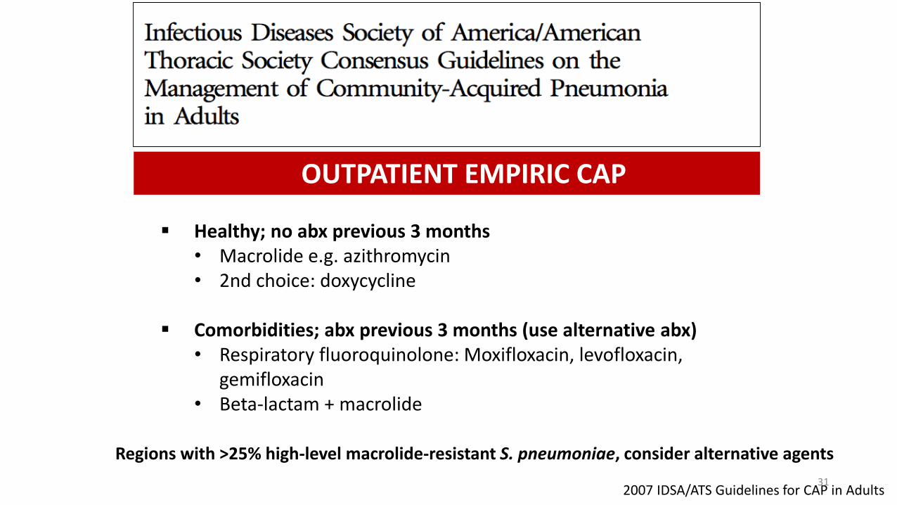
▪ Healthy; no abx previous 3 months• Macrolide e.g. azithromycin• 2nd choice: doxycycline
▪ Comorbidities; abx previous 3 months (use alternative abx)• Respiratory fluoroquinolone: Moxifloxacin, levofloxacin,
gemifloxacin• Beta-lactam + macrolide
OUTPATIENT EMPIRIC CAP
2007 IDSA/ATS Guidelines for CAP in Adults
Regions with >25% high-level macrolide-resistant S. pneumoniae, consider alternative agents
31

▪ Inpatients Ward• Respiratory fluoroquinolone• ß-lactam + macrolide
▪ Inpatients ICU• ß-lactam (cefotaxime/ceftriaxone or ampicillin/sulbactam) + macrolide• Respiratory fluoroquinolone for PCN-allergic pts
▪ Special pathogens• Pseudomonas anti-pseudomonal ß-lactam + cipro/levofloxacin or
aminoglycoside and azitromycin• MRSA add vancomycin or linezolid
2007 IDSA/ATS Guidelines for CAP in Adults
INPATIENT EMPIRIC CAP
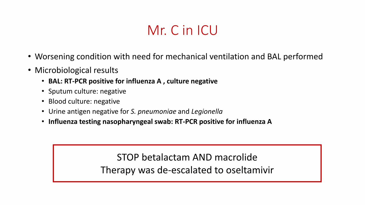
Mr. C in ICU
• Worsening condition with need for mechanical ventilation and BAL performed
• Microbiological results• BAL: RT-PCR positive for influenza A , culture negative
• Sputum culture: negative
• Blood culture: negative
• Urine antigen negative for S. pneumoniae and Legionella
• Influenza testing nasopharyngeal swab: RT-PCR positive for influenza A
STOP betalactam AND macrolide Therapy was de-escalated to oseltamivir
35
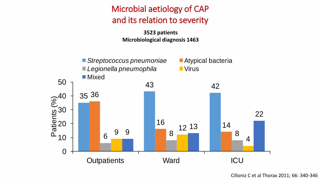
43 4236
16 14
6 8 8912
49
13
22
0
10
20
30
40
50
Outpatients Ward ICU
Pa
tie
nts
(%)
Streptococcus pneumoniae Atypical bacteria
Legionella pneumophila Virus
Mixed
Microbial aetiology of CAP and its relation to severity
Cilloniz C et al Thorax 2011; 66: 340-346
3523 patientsMicrobiological diagnosis 1463
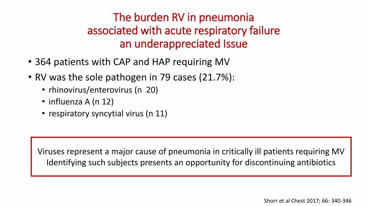
The burden RV in pneumoniaassociated with acute respiratory failure
an underappreciated Issue
• 364 patients with CAP and HAP requiring MV
• RV was the sole pathogen in 79 cases (21.7%):• rhinovirus/enterovirus (n 20)
• influenza A (n 12)
• respiratory syncytial virus (n 11)
Shorr et al Chest 2017; 66: 340-346
Viruses represent a major cause of pneumonia in critically ill patients requiring MV Identifying such subjects presents an opportunity for discontinuing antibiotics

After discharge
• The patient is sent home with recommendation of seeing the family doctor and planning antipneumococcal vaccination and influenza vaccination
• However the patient complains that in previous years he got vaccinated but he developed fever after vaccination and he still got the flu ...
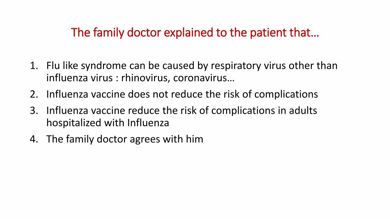
The family doctor explained to the patient that…
1. Flu like syndrome can be caused by respiratory virus other than influenza virus : rhinovirus, coronavirus…
2. Influenza vaccine does not reduce the risk of complications
3. Influenza vaccine reduce the risk of complications in adultshospitalized with Influenza
4. The family doctor agrees with him
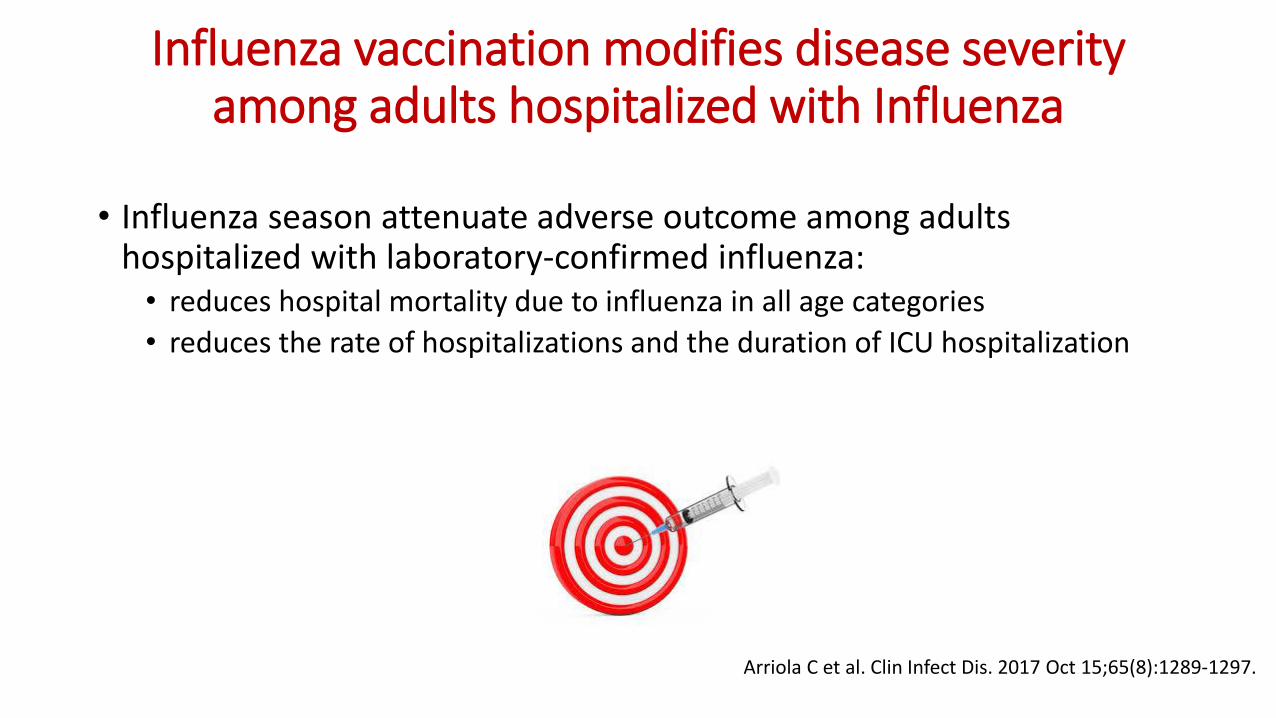
Influenza vaccination modifies disease severity among adults hospitalized with Influenza
• Influenza season attenuate adverse outcome among adults hospitalized with laboratory-confirmed influenza:• reduces hospital mortality due to influenza in all age categories
• reduces the rate of hospitalizations and the duration of ICU hospitalization
Arriola C et al. Clin Infect Dis. 2017 Oct 15;65(8):1289-1297.
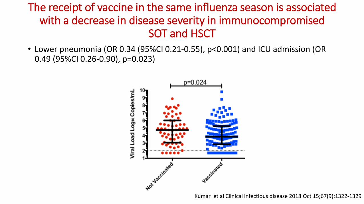
The receipt of vaccine in the same influenza season is associated with a decrease in disease severity in immunocompromised
SOT and HSCT• Lower pneumonia (OR 0.34 (95%CI 0.21-0.55), p<0.001) and ICU admission (OR
0.49 (95%CI 0.26-0.90), p=0.023)
Kumar et al Clinical infectious disease 2018 Oct 15;67(9):1322-1329

Mr. BW clinical case
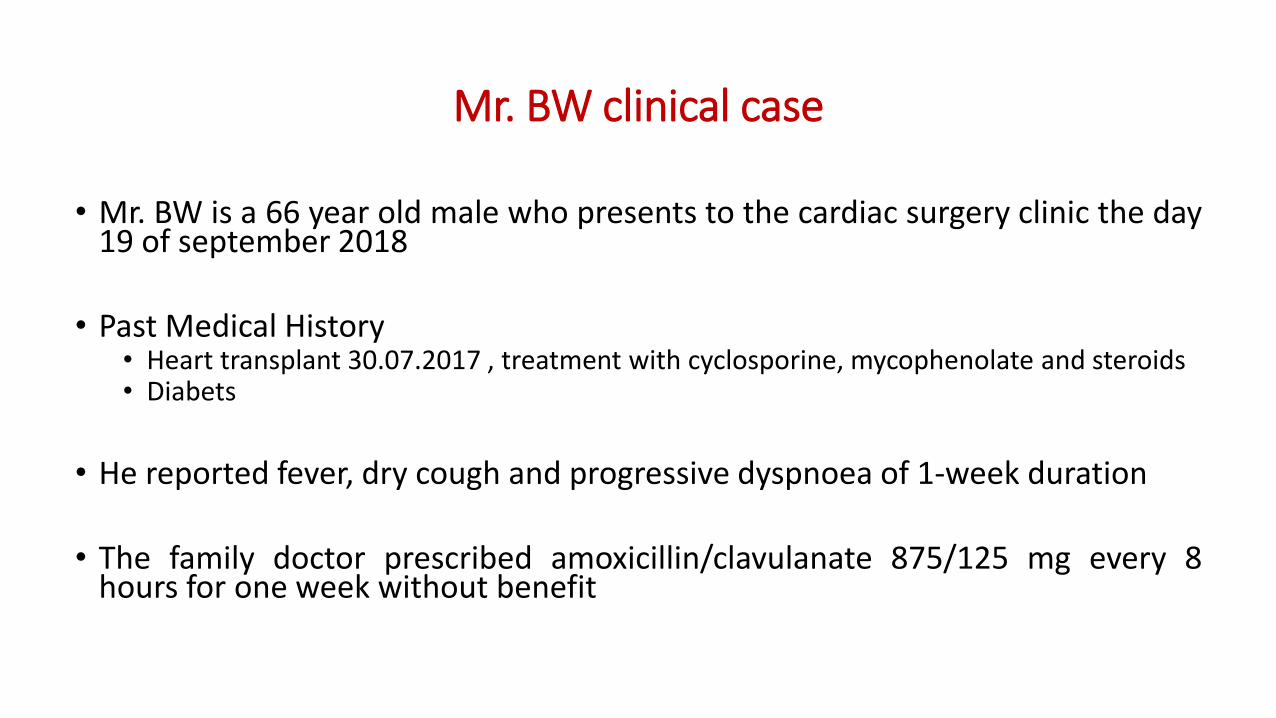
Mr. BW clinical case
• Mr. BW is a 66 year old male who presents to the cardiac surgery clinic the day19 of september 2018
• Past Medical History• Heart transplant 30.07.2017 , treatment with cyclosporine, mycophenolate and steroids• Diabets
• He reported fever, dry cough and progressive dyspnoea of 1-week duration
• The family doctor prescribed amoxicillin/clavulanate 875/125 mg every 8hours for one week without benefit

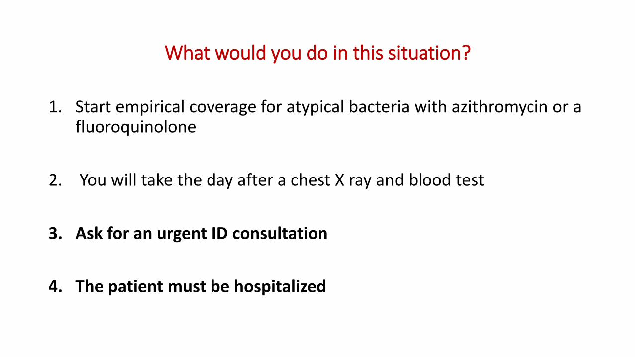
What would you do in this situation?
1. Start empirical coverage for atypical bacteria with azithromycin or a fluoroquinolone
2. You will take the day after a chest X ray and blood test
3. Ask for an urgent ID consultation
4. The patient must be hospitalized
What would you do in this situation?
1. Start empirical coverage for atypical bacteria with azithromycin or a fluoroquinolone
2. You will take the day after a chest X ray and blood test
3. Ask for an urgent ID consultation
4. The patient must be hospitalized
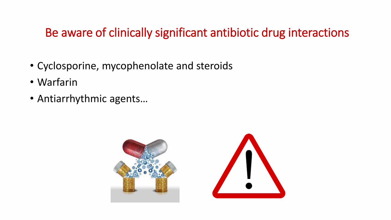
Be aware of clinically significant antibiotic drug interactions
• Cyclosporine, mycophenolate and steroids
• Warfarin
• Antiarrhythmic agents…
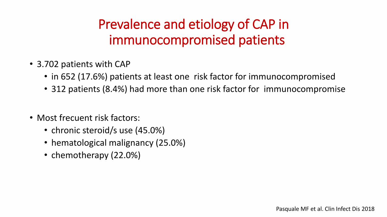
Prevalence and etiology of CAP inimmunocompromised patients
• 3.702 patients with CAP
• in 652 (17.6%) patients at least one risk factor for immunocompromised
• 312 patients (8.4%) had more than one risk factor for immunocompromise
• Most frecuent risk factors:
• chronic steroid/s use (45.0%)
• hematological malignancy (25.0%)
• chemotherapy (22.0%)
Di Pasquale MF et al. Clin Infect Dis 2018
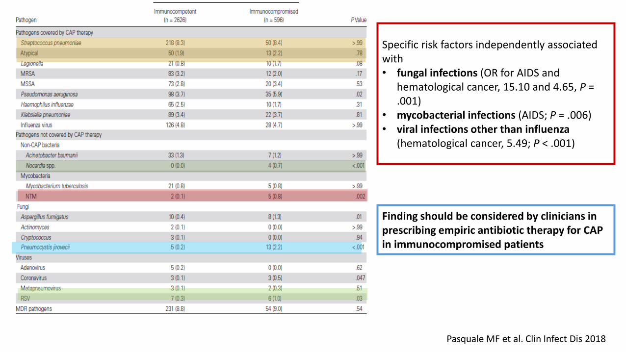
Di Pasquale MF et al. Clin Infect Dis 2018
Specific risk factors independently associated with• fungal infections (OR for AIDS and
hematological cancer, 15.10 and 4.65, P = .001)
• mycobacterial infections (AIDS; P = .006)• viral infections other than influenza
(hematological cancer, 5.49; P < .001)
Finding should be considered by clinicians in prescribing empiric antibiotic therapy for CAP in immunocompromised patients
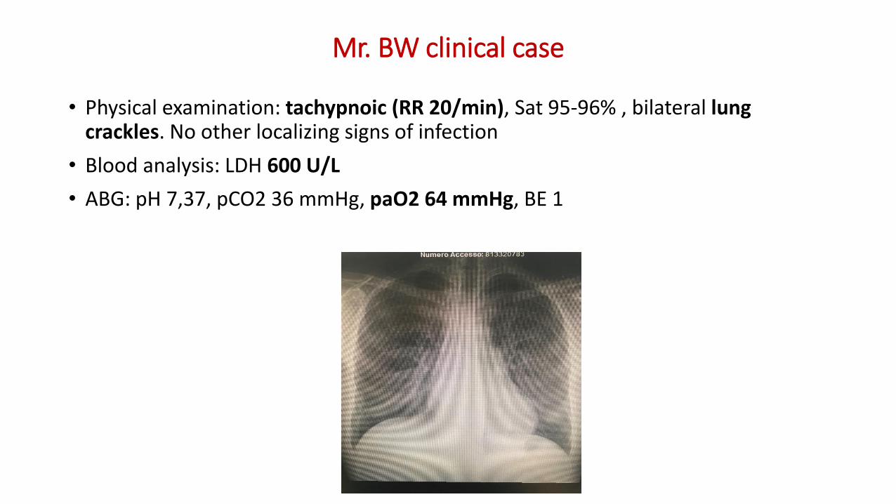
Mr. BW clinical case
• Physical examination: tachypnoic (RR 20/min), Sat 95-96% , bilateral lungcrackles. No other localizing signs of infection
• Blood analysis: LDH 600 U/L
• ABG: pH 7,37, pCO2 36 mmHg, paO2 64 mmHg, BE 1
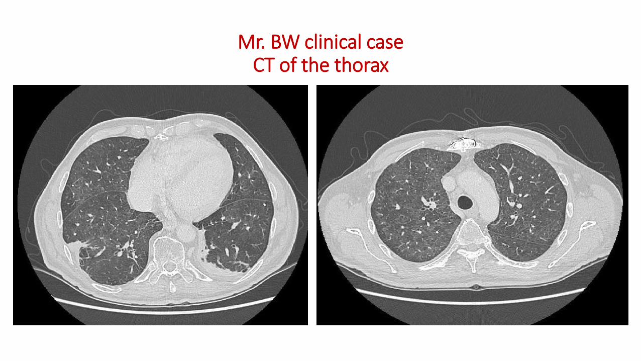
Mr. BW clinical case CT of the thorax
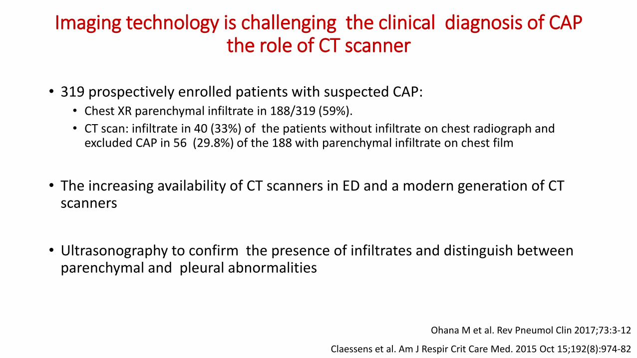
Imaging technology is challenging the clinical diagnosis of CAPthe role of CT scanner
• 319 prospectively enrolled patients with suspected CAP:• Chest XR parenchymal infiltrate in 188/319 (59%).
• CT scan: infiltrate in 40 (33%) of the patients without infiltrate on chest radiograph and excluded CAP in 56 (29.8%) of the 188 with parenchymal infiltrate on chest film
• The increasing availability of CT scanners in ED and a modern generation of CT scanners
• Ultrasonography to confirm the presence of infiltrates and distinguish betweenparenchymal and pleural abnormalities )
Claessens et al. Am J Respir Crit Care Med. 2015 Oct 15;192(8):974-82
Ohana M et al. Rev Pneumol Clin 2017;73:3-12
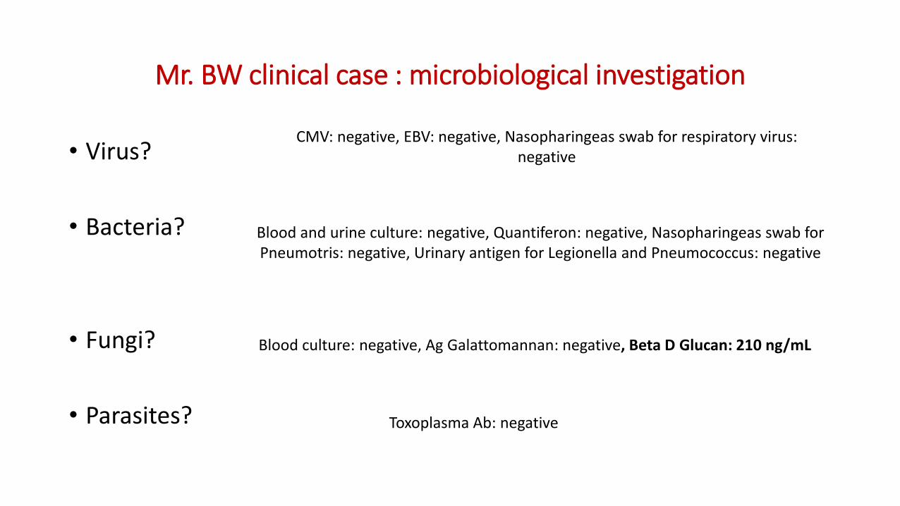
Mr. BW clinical case : microbiological investigation
• Virus?
• Bacteria?
• Fungi?
• Parasites?
CMV: negative, EBV: negative, Nasopharingeas swab for respiratory virus: negative
Blood and urine culture: negative, Quantiferon: negative, Nasopharingeas swab for Pneumotris: negative, Urinary antigen for Legionella and Pneumococcus: negative
Blood culture: negative, Ag Galattomannan: negative, Beta D Glucan: 210 ng/mL
Toxoplasma Ab: negative
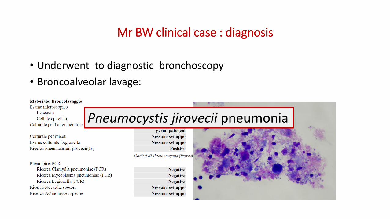
Mr BW clinical case : diagnosis
• Underwent to diagnostic bronchoscopy
• Broncoalveolar lavage:
Pneumocystis jirovecii pneumonia
Take home messages
• Removal of the concept of HCAP
• Changing epidemiology of CAP: new diagnostic methods and their impact in management
• Emerging role of respiratory viruses in CAP
• The role of prevention and influenza vaccination
• Empiric antibiotic therapy for CAP should not be prescribed in immunocompromised patients

GRAZIE