Epidemic Hand, Foot and Mouth Disease Caused by Human ... · Disease Caused by Human Enterovirus...
8
RESEARCH 78 Emerging Infectious Diseases • Vol. 9, No. 1, January 2003 Epidemic Hand, Foot and Mouth Disease Caused by Human Enterovirus 71, Singapore Kwai Peng Chan,* Kee Tai Goh,† Chia Yin Chong,‡ Eng Swee Teo,§ Gilbert Lau,§ and Ai Ee Ling* Singapore experienced a large epidemic of hand, foot and mouth disease (HFMD) in 2000. After reviewing HFMD notifi- cations from doctors and child-care centers, we found that the incidence of HFMD rose in September and declined at the end of October. During this period, 3,790 cases were reported. We performed enteroviral cultures on 311 and 157 specimens from 175 HFMD patients and 107 non-HFMD patients, respectively; human enterovirus 71 (HEV71) was the most frequently isolated virus from both groups. Most of the HFMD patients were < 4 years of age. Three HFMD and two non-HFMD patients died. Specimens from two HFMD and both non-HFMD patients were culture positive for HEV71; a third HFMD patient was possibly associated with the virus. Autopsies performed on all three HFMD and one of the non- HFMD case-patients showed encephalitis, interstitial pneu- monitis, and myocarditis. A preparedness plan for severe HFMD outbreaks provided for the prompt, coordinated actions needed to control the epidemic. and, foot and mouth disease (HFMD) is typically a benign and common self-limiting childhood disease, characterized by rapidly ulcerating vesicles in the mouth and lesions, usually vesicular, on the hands and feet (1). Lesions also frequently occur on the buttocks, but other parts of the body are usually not affected (2). HFMD is caused by a few serotypes of enteroviruses, most frequently coxsackie virus A16 (CAV16) and human enterovirus 71 (HEV71). Other viruses associated with the syndrome are coxsackie virus A (CAV) 4, 5, 9, and 10 and coxsackie virus B (CBV) 2 and 5 (1). The first recognized HFMD outbreak in Singapore occurred in 1970; the etiologic agent was unknown (3). Two other outbreaks were reported in 1972 and 1981 and involved 104 and 742 persons, respectively; in both outbreaks, CAV16 was implicated as the cause (4,5). After epidemics of HFMD in Sarawak, East Malaysia, and the Malaysian Peninsula in 1997 (6–8) and Taiwan in 1998 (9,10), which were associated with complications of encepha- litis, myocarditis, and death, a system of surveillance for the disease, based on notifications from child-care centers, was implemented in Singapore in April 1998. Reporting the dis- ease was made legally mandatory on October 1, 2000. Concur- rent with the intensified surveillance, an interministry and interhospital HFMD Task Force, composed of representatives from the Ministries of Health, Environment, Education, and Community Development and Sports, as well as virologists and pediatricians, was created in 1998 to formulate a prepared- ness response plan to monitor and manage severe HFMD out- breaks in Singapore. At the end of 2000, Singapore experienced its largest known outbreak of HFMD. After media reports in September of HFMD-related deaths in Singaporean children, many patient samples were sent for virologic investigation to the Virology Laboratory of the Department of Pathology, Sin- gapore General Hospital. Because the Virology Laboratory receives all requests for virus culture or enterovirus typing from the entire country, it was the repository of information on virtually all laboratory investigations during the HFMD epi- demic. We describe the epidemiologic, virologic, and pathologic features of this epidemic. Methods In this study, we used a case definition for HFMD of fever, accompanied by oral ulcers and a rash, maculopapular or vesicular, on the hands and feet, with or without buttock involvement. We reviewed records of HFMD notifications to the Ministry of the Environment for the incidence and trend of the disease. All children with suspected HFMD reported by preschool centers were examined, and the cases were certified by family physicians. Cases reported by parents or school principals and teachers were excluded unless a medical certifi- cate from a physician verified them. At the same time, Minis- try of the Environment nurses conducted active case detection in both preschools and primary schools. All case-patients were identified by a unique national registration identification num- ber, and duplicate reports were eliminated by the computer. Data obtained from samples received by the Virology Lab- oratory at Singapore General Hospital for enterovirus isolation during the epidemic were also analyzed. In addition to stool samples, samples included swabs of vesicles, mouth, throat, rectum, and ulcers, and samples from the brain, heart, lung, tonsil, lymph node, spleen, and intestine of those with fatal disease. The samples were added into HeLa, HEp-2, human embryonic lung fibroblasts, and human rhabdomyosarcoma *Singapore General Hospital, Singapore, Republic of Singapore; †National Environment Agency, Singapore, Republic of Singapore, ‡Kandang Kerbau Women’s and Children’s Hospital, Singapore, Republic of Singapore; and §Centre for Forensic Medicine, Health Sci- ences Authority, Singapore, Republic of Singapore H
Transcript of Epidemic Hand, Foot and Mouth Disease Caused by Human ... · Disease Caused by Human Enterovirus...

RESEARCH
78 Emerging Infectious Diseases • Vol. 9, No. 1, January 2003
Epidemic Hand, Foot and Mouth Disease Caused by Human Enterovirus 71, Singapore
Kwai Peng Chan,* Kee Tai Goh,† Chia Yin Chong,‡ Eng Swee Teo,§ Gilbert Lau,§ and Ai Ee Ling*
Singapore experienced a large epidemic of hand, foot andmouth disease (HFMD) in 2000. After reviewing HFMD notifi-cations from doctors and child-care centers, we found that theincidence of HFMD rose in September and declined at theend of October. During this period, 3,790 cases werereported. We performed enteroviral cultures on 311 and 157specimens from 175 HFMD patients and 107 non-HFMDpatients, respectively; human enterovirus 71 (HEV71) was themost frequently isolated virus from both groups. Most of theHFMD patients were <4 years of age. Three HFMD and twonon-HFMD patients died. Specimens from two HFMD andboth non-HFMD patients were culture positive for HEV71; athird HFMD patient was possibly associated with the virus.Autopsies performed on all three HFMD and one of the non-HFMD case-patients showed encephalitis, interstitial pneu-monitis, and myocarditis. A preparedness plan for severeHFMD outbreaks provided for the prompt, coordinated actionsneeded to control the epidemic.
and, foot and mouth disease (HFMD) is typically abenign and common self-limiting childhood disease,
characterized by rapidly ulcerating vesicles in the mouth andlesions, usually vesicular, on the hands and feet (1). Lesionsalso frequently occur on the buttocks, but other parts of thebody are usually not affected (2). HFMD is caused by a fewserotypes of enteroviruses, most frequently coxsackie virusA16 (CAV16) and human enterovirus 71 (HEV71). Otherviruses associated with the syndrome are coxsackie virus A(CAV) 4, 5, 9, and 10 and coxsackie virus B (CBV) 2 and 5(1). The first recognized HFMD outbreak in Singaporeoccurred in 1970; the etiologic agent was unknown (3). Twoother outbreaks were reported in 1972 and 1981 and involved104 and 742 persons, respectively; in both outbreaks, CAV16was implicated as the cause (4,5).
After epidemics of HFMD in Sarawak, East Malaysia, andthe Malaysian Peninsula in 1997 (6–8) and Taiwan in 1998(9,10), which were associated with complications of encepha-litis, myocarditis, and death, a system of surveillance for thedisease, based on notifications from child-care centers, was
implemented in Singapore in April 1998. Reporting the dis-ease was made legally mandatory on October 1, 2000. Concur-rent with the intensified surveillance, an interministry andinterhospital HFMD Task Force, composed of representativesfrom the Ministries of Health, Environment, Education, andCommunity Development and Sports, as well as virologistsand pediatricians, was created in 1998 to formulate a prepared-ness response plan to monitor and manage severe HFMD out-breaks in Singapore.
At the end of 2000, Singapore experienced its largestknown outbreak of HFMD. After media reports in Septemberof HFMD-related deaths in Singaporean children, manypatient samples were sent for virologic investigation to theVirology Laboratory of the Department of Pathology, Sin-gapore General Hospital. Because the Virology Laboratoryreceives all requests for virus culture or enterovirus typingfrom the entire country, it was the repository of information onvirtually all laboratory investigations during the HFMD epi-demic.
We describe the epidemiologic, virologic, and pathologicfeatures of this epidemic.
MethodsIn this study, we used a case definition for HFMD of fever,
accompanied by oral ulcers and a rash, maculopapular orvesicular, on the hands and feet, with or without buttockinvolvement. We reviewed records of HFMD notifications tothe Ministry of the Environment for the incidence and trend ofthe disease. All children with suspected HFMD reported bypreschool centers were examined, and the cases were certifiedby family physicians. Cases reported by parents or schoolprincipals and teachers were excluded unless a medical certifi-cate from a physician verified them. At the same time, Minis-try of the Environment nurses conducted active case detectionin both preschools and primary schools. All case-patients wereidentified by a unique national registration identification num-ber, and duplicate reports were eliminated by the computer.
Data obtained from samples received by the Virology Lab-oratory at Singapore General Hospital for enterovirus isolationduring the epidemic were also analyzed. In addition to stoolsamples, samples included swabs of vesicles, mouth, throat,rectum, and ulcers, and samples from the brain, heart, lung,tonsil, lymph node, spleen, and intestine of those with fataldisease. The samples were added into HeLa, HEp-2, humanembryonic lung fibroblasts, and human rhabdomyosarcoma
*Singapore General Hospital, Singapore, Republic of Singapore;†National Environment Agency, Singapore, Republic of Singapore,‡Kandang Kerbau Women’s and Children’s Hospital, Singapore,Republic of Singapore; and §Centre for Forensic Medicine, Health Sci-ences Authority, Singapore, Republic of Singapore
H

Emerging Infectious Diseases • Vol. 9, No. 1, January 2003 79
RESEARCH
cells. The cultures were incubated at 36°C and examined dailyfor cytopathic effects for 21 to 28 days.
Enteroviruses cultured from the samples were typed bymicro-neutralization tests (11) by using Lim Benyesh-MelnickA-H equine antiserum pools (World Health Organization, Stat-ens Serum Institut, Copenhagen, Denmark), equine antiserumpools (Rijksinstituut voor Volksgesondheid en Milieuhygiene,Bilthoven, the Netherlands), rabbit 385JS HEV71-specificpolyclonal antiserum (Victorian Infectious Diseases ReferenceLaboratory, Melbourne, Australia), and rabbit or monkey anti-sera specific for CAV serotypes (National Institutes of Health,Bethesda, MD). Nonenteroviruses that produced cytopathiceffects characteristic of Cytomegalovirus (CMV) or herpessimplex virus were identified by immunofluorescence assay asdescribed (12), by using mouse monoclonal antibodies toCMV (Bartels CMV DFA kit, Trinity Biotech plc, Wicklow,Ireland) and herpes simplex virus (MicroTrak HSV1/HSV2culture identification/typing test, Trinity Biotech plc). Whenthe presence of rhinovirus was suggested from the cytopathiceffects, the virus was identified by the acid lability test (13).Autopsies were performed on four patients who died, and tis-sue samples were subjected to virus cultures.
Results
CasesThe number of notifications of HFMD cases to the Minis-
try of the Environment increased in early September 2000(Figure 1). The incidence peaked at 308 cases per day onOctober 10 and decreased to 10 cases per day by October 28.Hospital and general practice physicians and preschool-centeroperators reported a total of 3,790 cases during these 2months.
During the epidemic, 311 samples from 175 clinicallydiagnosed HFMD case-patients were submitted for virus cul-ture. A total of 138 (78.8%) of these patients were <4 years ofage, with 12 (6.9%) >10 years of age, the oldest being 71 yearsold (Table 1). The male-to-female ratio was 1.7:1.
At least one virus was isolated from 147 (47.3%) of thesamples collected from 104 (59.4%) HFMD patients, includ-ing 2 of 3 who died. Almost all (91.5%) of these patients were<5 years of age with the peak incidence at 1 year (Table 1). A21-year-old woman was the only patient >10 years of age toyield a virus, identified as HEV71, from vesicles on her handsand feet.
HEV71 was the most commonly isolated virus, detected in76 (73.1%) of 104 case-patients (Table 2). Three of thesepatients had a second virus isolated concurrently: echovirus(EV) 25, Rhinovirus, and CMV. Other enteroviruses were iso-lated in 24 (23.1%) of samples from case-patients. Five casesof CAV16 were identified, as well as four cases each of CAV6,CAV24, and EV18; three cases of CAV10; and one case eachof CAV4, CBV3, CBV4, and CBV5. Four patients (3.8%)tested positive for nonenteroviruses; CMV was isolated fromtheir mouth and from throat swabs.
The two patients with fatal HFMD, from whom HEV71was isolated, were siblings, a 14-month-old girl and her 2-year-old brother. The girl was admitted to the hospital withfever, rashes on the hands and feet, and oral ulcers of 3 days’duration. Progressive hemodynamic instability, oliguria, meta-bolic acidosis, and hyperkalemia developed; despite intensivecare and resuscitative efforts, she died on day 2 after admis-sion. At autopsy, her lungs showed acute pulmonary edema,acute intraalveolar hemorrhage and diffuse alveolar damageassociated with interstitial lymphocytic infiltrates, extensivehyaline membrane formation, patchy atelectasis, and focalpneumocyte desquamation and hypertrophy (Figure 2). Sam-ples from her brain tissue showed lymphocytic leptomeningitiswith widespread perivascular cuffing by lymphocytes andplasma cells within the cortex and white matter (Figure 3). Thepons, in particular, showed evidence of encephalitis, associ-ated with localized perivascular hemorrhage, focal neuronalnecrosis, and microglial reaction (Figure 4). Features of myo-carditis were observed; the myocardium showed occasional
Figure 1. Number of cases of hand, foot and mouth disease reported tothe Singapore Ministry of Environment as surveillance for the disease,July 2000–March 2001. Each epidemiologic week begins on Sunday.Mandatory reporting of the disease began on October 1, 2000.
Table 1. Age distribution of clinical and virus-positive hand, foot and mouth disease patients
Age (yrs) No. clinical cases (%) No. virus-positive cases (%)
<1 16 (9.1) 11 (10.6)
1 44 (25.2) 32 (30.8)
2 41 (23.4) 24 (23.1)
3 21 (12.0) 14 (13.5)
4 16 (9.1) 8 (7.7)
5 8 (4.6) 6 (5.8)
6 6 (3.4) 4 (3.9)
7 4 (2.3) 1 (0.9)
8 3 (1.7) 2 (1.9)
9 1 (0.6) 0 (0.0)
10 3 (1.7) 1 (0.9)
>10 12 (6.9) 1 (0.9)
Total 175 (100.0) 104 (100.0)

RESEARCH
80 Emerging Infectious Diseases • Vol. 9, No. 1, January 2003
interstitial infiltrates of lymphocytes and plasma cells associ-ated with focal myonecrosis (Figure 5). HEV71 was isolatedfrom samples taken from the brain, tonsils, intestines, stools,and throat and from swabs of the mouth and rectum. Viral cul-tures of the lung, heart, and spleen were negative.
This patient had two older brothers. The 2-year-old brothershowed an almost identical clinical course. After 3 days offever, rash on the hands and feet, and oral ulcers, he too deteri-orated under intensive care and died about 2 hours after his sis-ter, within 24 hours of hospitalization. Autopsy findings weresimilar, showing evidence of acute interstitial pneumonitis,pulmonary edema, encephalitis (including focal neuronalnecrosis of the pons), and myocarditis. HEV71 was isolatedfrom postmortem specimens of the tonsils and intestines. Viralcultures of the brain, heart, lungs, spleen, and lymph nodesshowed negative results.
The 5-year-old brother of these patients was also admittedto the hospital at the same time with rashes and mouth ulcers,which in his case did not progress to severe disease. However,because of the rapid deaths of his siblings, intravenous immu-noglobulin was administered prophylactically 2 days after hos-pital admission. After two doses, vomiting and a headachedeveloped. A computed tomographic scan of his head showednormal results, and after due assessment, the headache andvomiting were attributed to the intravenous immunoglobulin,which was then stopped. The symptoms ceased and the boyrecovered well. HEV71 was cultured from his throat swab andstool.
Another 2-year-old boy with 5 days of fever, cough, andrash on the hands and feet was diagnosed with HFMD and
died on the same day he was admitted to the hospital. Postmor-tem examination showed pulmonary edema, interstitial pneu-monitis, leptomeningeal infiltrates of lymphocytes and plasmacells, and occasional foci of perivascular lymphoid cuffingwithin the cerebral cortex. Although the heart containedpatchy epicardial lymphoid infiltrates, no evidence of myone-crosis was found. Virus was not isolated from the brain, heart,lungs, intestine, and tracheal swab specimen.
One other complicated HFMD case was seen. Asepticmeningitis manifested by headache and terminal neck stiffnessdeveloped in an 8-year-old girl with characteristic symptomsof HFMD. HEV71 was isolated from her oral ulcers, but hercerebrospinal fluid (CSF) was negative for viruses and bacte-ria. After she was treated for her symptoms, the patient was
Table 2. Viruses isolated from HFMD cases during the epidemica
VirusNo. HFMD patients (%)
No. non-HFMD patients (%)
HEV71 HEV71 only HEV71 + EV25 HEV71 + Rhinovirus HEV71 + CMV
76 (73.1)73111
5 (29.4)5000
CAV4 1 (1.0) 0
CAV6 4 (3.8) 0
CAV10 3 (2.9) 1 (5.9)
CAV16 5 (4.8) 0
CAV24 4 (3.8) 3 (17.6)
CBV3 1 (1.0) 1 (5.9)
CBV4 1 (1.0) 2 (11.8)
CBV5 1 (1.0) 3 (17.6)
EV18 4 (3.8) 0
CMV 4 (3.8) 0
Herpes simplex virus 1 0 2 (11.8)
Total 104 (100.0) 17 (100.0)aHFMD, hand, foot and mouth disease; HEV, human enterovirus; EV, echovirus; CMV, Cytomegalovirus; CAV, coxsackie virus A; CBV, coxsackie virus B.
Figure 2. Interstitial pneumonitis in the 14-month-old girl who died ofhuman enterovirus 71 disease. Photomicrograph shows alveolar wallcongestion, intra-alveolar hemorrhage, and interstitial lymphocytic infil-trate. (Hematoxylin and eosin stain, original magnification x 200).
Figure 3. Perivascular cuffing in the brain. (Hematoxylin and eosinstain, original magnification x 200).
Emerging Infectious Diseases • Vol. 9, No. 1, January 2003 81
RESEARCH
discharged from the hospital after 4 days. No patient in thisepidemic showed acute flaccid paralysis.
Over the same period, 107 other patients who did not haveHFMD were investigated for enteroviral infection. HEV71was isolated from 5 of 17 culture-positive patients (Table 2).Of the five HEV71 patients, three had a nonspecific febrile ill-ness without rash. The other two patients died. One was a 1-year-old boy with a 3-day history of fever, vomiting, diarrhea,and increasing restlessness; he died the day after being admit-ted to the hospital. Tests for bacteria, Dengue virus, Rotavirus,and Plasmodium spp. were negative; however, HEV71 wasisolated from his throat and rectal swabs. Cardiac enzyme lev-els were raised, and a clinical diagnosis of myocarditis wasmade. No autopsy was performed. The other patient who diedwas a 19-year-old man who had a fever and headache for 3days, associated with slurred speech and an episode of gener-alized tonic-clonic seizure. He died about 16 hours after hospi-tal admission in spite of maximum resuscitative efforts.Postmortem investigation found meningoencephalitis involv-ing the cerebral cortex and pons. The latter also showed focalliquefactive necrosis. His lungs showed marked intraalveolarhemorrhage and edema, and enlarged pneumocytes withintense nuclear smudging consistent with acute interstitialpneumonitis. The myocardium did not show notable inflam-mation or necrosis. The culture of brain tissue showed HEV71,but cultures of his heart, lungs, and intestines were negativefor viruses.
Non-HEV71 enteroviruses were cultured from patientswith hemorrhagic conjunctivitis (CAV24), aseptic meningitis(CBV4, CBV5), neonatal pyrexia (CBV3, CBV4), gastroen-teritis (CAV24), sudden infant death (CAV24), and pharyngitis(CAV10) (Table 2). Two patients in whom herpangina wasdiagnosed had herpes simplex virus type 1 cultured from theiroral swabs.
SpecimensThe majority of specimens received from HFMD case-
patients included those from stool, vesicles, and mouth andthroat swabs (Table 3), for which the HEV71 culture-positivity
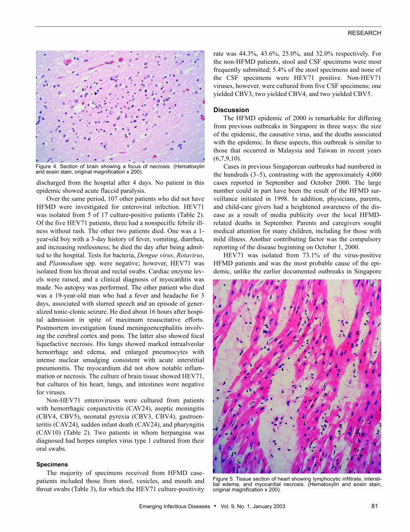
rate was 44.3%, 43.6%, 25.0%, and 32.0% respectively. Forthe non-HFMD patients, stool and CSF specimens were mostfrequently submitted; 5.4% of the stool specimens and none ofthe CSF specimens were HEV71 positive. Non-HEV71viruses, however, were cultured from five CSF specimens; oneyielded CBV3, two yielded CBV4, and two yielded CBV5.
DiscussionThe HFMD epidemic of 2000 is remarkable for differing
from previous outbreaks in Singapore in three ways: the sizeof the epidemic, the causative virus, and the deaths associatedwith the epidemic. In these aspects, this outbreak is similar tothose that occurred in Malaysia and Taiwan in recent years(6,7,9,10).
Cases in previous Singaporean outbreaks had numbered inthe hundreds (3–5), contrasting with the approximately 4,000cases reported in September and October 2000. The largenumber could in part have been the result of the HFMD sur-veillance initiated in 1998. In addition, physicians, parents,and child-care givers had a heightened awareness of the dis-ease as a result of media publicity over the local HFMD-related deaths in September. Parents and caregivers soughtmedical attention for many children, including for those withmild illness. Another contributing factor was the compulsoryreporting of the disease beginning on October 1, 2000.
HEV71 was isolated from 73.1% of the virus-positiveHFMD patients and was the most probable cause of the epi-demic, unlike the earlier documented outbreaks in Singapore
Figure 4. Section of brain showing a focus of necrosis. (Hematoxylinand eosin stain, original magnification x 200).
Figure 5. Tissue section of heart showing lymphocytic infiltrate, intersti-tial edema, and myocardial necrosis. (Hematoxylin and eosin stain,original magnification x 200).

RESEARCH
82 Emerging Infectious Diseases • Vol. 9, No. 1, January 2003
(4,5), which were attributed to CAV16. Other viruses culturedin smaller numbers included CAV16, CAV4, CAV10, andCBV5, known etiologic agents of HFMD, as well as CAV6,CAV24, CBV3, CBV4, and EV18, cocirculating enterovirusesthat may have caused at least some cases of HFMD. CAV6was isolated from the vesicles of two patients and CAV24from the vesicles of one patient. Some of these non-HEV71enteroviruses could have played an indirect role in the HEV71epidemic. Indeed, the possibility of HEV71 interacting withother enteroviruses in a previous HEV71 epidemic has beenraised (14).
Among the patients with suspected enteroviral infectionbut without the classic symptoms of HFMD, the most fre-quently isolated virus was still HEV71. These cases repre-sented the extremes of the clinical spectrum of HEV71,including nonspecific febrile illness in three patients and deathfrom myocarditis and encephalitis in two patients. The otherclinical presentations of the non-HFMD patients includedaseptic meningitis, herpangina, and Guillain-Barré syndrome,conditions that could also be caused by HEV71. However, the
viruses isolated from these patients were CBV4, CBV5, andherpes simplex virus 1. Of the total of 81 patients with cultureevidence of HEV71 infection, most (93.8%) showed illnessconsistent with HFMD.
Until this epidemic occurred, no deaths had been associ-ated with HFMD in Singapore, although HEV71-relateddeaths from encephalitis (15–17), pulmonary edema, and hem-orrhage (8,18) have occurred elsewhere since the virus wasfirst isolated in 1969 (15). In this epidemic in Singapore, thecase-fatality rate among all reported HFMD case-patients was0.08%, which is similar to the rate of 0.06% experienced in the1998 Taiwanese outbreak (9).
Four deaths (two HFMD and two non-HFMD cases) wereassociated with HEV71. All occurred rapidly despite intensivecare, within a day of the patient’s hospital admission, and afteran average of 3.4 days of illness. The circumstances of thesedeaths were reminiscent of recent HEV71 deaths in the region(10,18–20). Of these four case-patients, three were autopsied,including a pair of siblings with HFMD and a patient withnon-HFMD encephalitis. Their postmortem findings were sim-
Table 3. Virus yield by specimen typea
Specimen type
No. specimens
HFMD patients Non-HFMD patients
Culture + HEV 71 + No. tested Culture + HEV 71 + No. tested
Stool 58 39b 88 11 4 74
Rectal swab 6 5 8 1 1 3
Vesicle 31 27 62 0 0 3
Oral swab 23 15 60 2 0 6
Throat swab 19 16c 50 2 1 9
Nasal aspirate 3 3d 6 1 1 1
Saliva 1 1d 3 0 0 0
Tonsil 2 2 2 0 0 0
Intestine and contents 2 2 5 1 0 6
Brain 1 1 3 1 1 7
Cerebrospinal fluid 0 0 2 5 0 32
Ulcer 1 1 2 0 0 0
Conjunctiva 0 0 1 1 0 1
Blood 0 0 2 0 0 0
Heart 0 0 4 0 0 12
Tracheal swab 0 0 4 0 0 1
Lymph node 0 0 1 0 0 0
Spleen 0 0 2 0 0 0
Lung 0 0 4 0 0 1
Nasal swab 0 0 2 0 0 0
BAL 0 0 0 0 0 1
Total 147 112 311 25 8 157aHFMD, hand, foot and mouth disease; HEV, human enterovirus; +, positive; EV, echovirus; CMV, Cytomegalovirus; BAL, broncho-alveolar lavage. bDual isolation of HEV71 and EV25 from one specimen.cDual isolation of HEV71 and Rhinovirus from one specimen.dDual isolation of HEV71 and CMV from one specimen. Both double-virus positive specimens were received from the same patient.

Emerging Infectious Diseases • Vol. 9, No. 1, January 2003 83
RESEARCH
ilar, with HEV71 isolated from the brains of two case-patientsand from the tonsils and intestines of the third. WhetherHEV71 caused the death of the patient with myocarditis whowas not autopsied is less clear since HEV71 was isolated fromnonsterile sites (the throat and rectum), although the illnessand epidemiology suggest the possibility.
During the epidemic, a fifth death occurred involving aboy with HFMD on whom an autopsy was conducted. Novirus was cultured from him, possibly because of the advancedpostmortem degradation of his tissues. However, HEV71 waslikely also to have been the cause of death on the basis of thesimilarity of his clinical and postmortem findings to those ofthe siblings who died, as well as the epidemiologic links toage, time, and place.
Like other fatal HEV71 cases reported elsewhere(7,16,20,21), the primary pathologic changes found at autopsyof four case-patients in this study were in the brain, includingthe brainstem, which showed extensive inflammatory cellinfiltrate and focal necrosis. In addition, pneumonitis wasfound in all the case-patients and myocarditis in two. In theMalaysian and Taiwanese outbreaks (7,18,20), however, nosignificant inflammation was found in the lungs of patientswith fatal cases. Notably, the myocardium of 10 Malaysianpatients was described as normal (7), whereas autopsy reportsof 2 patients from the Taiwanese outbreak described mildmyocarditis in 1 (20) and no myocarditis in the other (18).
HEV71 was cultured from the brain specimens of two ofour autopsied case-patients, but viral cultures of the lung andheart were negative. Similarly, no HEV71 was isolated from34 CSF samples studied, notwithstanding the diagnosis ofaseptic meningitis. Besides encephalitis and death, other com-plications (such as aseptic meningitis and acute flaccid paraly-sis) have also been reported in other HFMD outbreaks (7,9).However, other than the three fatal cases and one case of asep-tic meningitis, all HFMD cases in the Singapore epidemicwere uncomplicated, despite the large number of patients.
Most HFMD patients were very young children (<4 yearsof age) with the peak incidence at 1 year, a finding consistentwith other HFMD outbreaks (7,9,22,23). Male patients out-numbered female patients by 1.7 to 1. This predominance hasbeen observed in other enteroviral infections in which themale-to-female ratio ranges from 1.5:1 to 2.5:1 (24). The rea-son for this finding is not clear but may suggest a susceptibilityat the host genetic level. That two siblings died of HEV71 dis-ease, which has a low case-fatality rate, further strengthensthis suspicion. Further studies are warranted on the possiblerole of host genetic factors in the pathogenesis of HEV71 dis-ease.
Since 1997, HEV71 outbreaks have occurred in Sarawak(6,7), the Malaysian Peninsula (8), Taiwan (9), Singapore (25),and Australia (26). To account for this wave of HEV71 out-breaks in the region, we suspected the presence of a suscepti-ble population as a plausible explanation; however, HEV71had appeared previously in Singapore in 1984 (27). The virusdisappeared for a time, resurfacing initially in small numbers
of patients in 1997–1999 (25); the number of infections thenjumped in 2000. Because the same group of children with thehighest incidence of infection in 2000 would have had beenexposed to HEV71 since 1997, why a large outbreak did notoccur earlier is unclear. Likewise, the large HEV71 outbreakin Taiwan (9) also took place when most of the population hadapparent immunity. We suspect that changes in viral factors,including virulence and tropism, are possible factors in theseoccurrences.
The genetic sequences of the complete VP1 gene ofHEV71 isolates from the four patients who died and three ofthe patients who did not, obtained during the Singapore out-break in 2000, were compared in a recent study involving 66HEV71 strains isolated between 1999 and 2001 from Malay-sia, Singapore, and Western Australia (28). That study showedthat the Singapore 2000 strains, like those isolated in 2000 inSarawak, Malaysia, belong to genogroup B4, whereas the Sin-gapore 1998 and Western Australia 1999 strains (from nonfa-tal case-patients) and Malaysian 1997 strains (including fatalcase-patients) belong to the closely related B3 genogroup. Thestrains from the Taiwanese outbreak of 1998 were found to bein the more distantly related C2 genogroup. The viruses thatcaused fatalities in outbreaks in Malaysia (1997), Taiwan(1998), and Singapore (2000) were thus not genetically simi-lar, at least in the VP1 region. These viruses did not belong tothe same genogroup, which would have explained the similarcharacteristics of the outbreaks. Furthermore, although thesame study suggests that a substitution of alanine with valineat position 170 of the VP1 region of genogroup C2 (lineage 1)strains may be associated with increased neurovirulence, nosimilar virulence-related mutation in the same genomic regionwas found for genogroups B3 and B4, to which the Malaysianand Singaporean fatal strains belong. No evidence exists fromthe deduced VP1 amino acid sequences of these two geno-groups to link specific amino acid residues with the severity ofillness or death. These observations indicate that the geneticdeterminants for virulence are still unclear.
Coinfection with a second virus has been suggested as yetanother possible pathogenetic factor (6,9), and this theory issupported by the concomitant isolation of a subgenus B aden-ovirus with an enterovirus from three persons who died duringa HFMD outbreak in Sarawak (6). Among the Singaporeanpatients with HEV71 infection, three had a second virus iso-lated concurrently. However, the presence of dual viruses didnot result in severe disease, although a child with HEV71 andCAV16 coinfection died in Singapore in 1997 (29).
We considered whether any unusual medications, treat-ments, or dietary exposures contributed to the deaths. No evi-dence from the fatalities in Singapore suggests this possibility.Conversely, we reviewed whether any particular therapeuticmodality improved clinical outcome, but found that this ideacannot be argued conclusively because the HFMD patient withaseptic meningitis recovered well with symptomatic treatment.The child who survived his two younger siblings had no com-plications from HFMD and was not cared for differently from

RESEARCH
84 Emerging Infectious Diseases • Vol. 9, No. 1, January 2003
his siblings before hospitalization. He was given prophylacticintravenous immunoglobulin solely because his siblings died.Whether this treatment, which was not administered to thepatients who died, helped prevent severe disease in him isuncertain.
Considering that transmission of enteroviruses is mainlyfecal-oral and through the respiratory route (to some extent)(22), we note that spread of the viruses is prevalent in child-care centers. To break the chain of transmission during the epi-demic, the HFMD Task Force coordinated a swift, nationwideclosure of preschool centers on October 1, 2000, reopeningthem on October 16, 2000, only when the HFMD reportsrecorded a declining trend, and no additional severe cases anddeaths associated with the disease were reported. Other mea-sures included repeated public health education through themass media on observance of good personal hygiene, cleaningand disinfection of premises and articles both at home and atpreschool centers, and keeping children away from crowds.These interventions may have played a role in bringing theepidemic under control by the end of October, although theoutbreak may have also run its natural course by that time.
In September and October 2000, HEV71 caused the largestHFMD epidemic recorded to date in Singapore, an epidemicthat involved mainly young children <4 years of age. Fivedeaths occurred, and HEV71 was isolated from four case-patients. Autopsies of four case-patients showed encephalitis,interstitial pneumonitis, and myocarditis. Virulence determi-nants of HEV71 and the precipitating factors for the epidemicitself unfortunately remain unknown. Based on our experi-ences during this epidemic, we found that an HFMD epidemicpreparedness plan was useful in providing the framework forprompt actions to monitor the situation, identify the causativeagent, interrupt virus transmission, and communicate with andsolicit the cooperation of the media, parents, physicians, andpreschool center personnel.
AcknowledgmentsWe thank Seng Eng Hong and staff of the Virology Laboratory
who provided their usual excellent technical support. We also thankthe team at the Quarantine and Epidemiology Department who pains-takingly recorded and followed up the hand, foot and mouth diseasenotifications. We are indebted to Margery Kennett for the generousgifts of human enterovirus 71 and coxsackie virus A antisera.
Dr. Chan is a consultant virologist at the Department of Pathol-ogy, Singapore General Hospital. She is a diagnostician and collabo-rates with the World Health Organization on poliomyelitis, measles,and influenza studies.
References 1. Melnick JL. Enteroviruses: polioviruses, coxsackieviruses, echoviruses,
and newer enteroviruses. In: Fields BN, Knipe DM, Howley PM, Chan-lock RM, Melnick JL, Monath TP, et al., editors. Field’s virology. 3rd ed.Philadelphia: Lippincott-Raven Publishers; 1996. p. 655–712.
2. Tunnessen WW Jr. Erythema infectiosum, roseola, and enteroviral exan-thems. In: Gorbach SL, Bartlett JG, Blacklow NR, editors. Infectious dis-eases. Philadelphia: W. B. Saunders Company; 1992. p. 1120–5.
3. Chan MCK, Wong HB. Hand-foot and mouth disease. J Singapore Paedi-atr Soc 1973;15:31–4.
4. Tay CH, Gaw CYN, Low T, Ong C, Chia KW, Yeo H, et al. Outbreak ofhand, foot and mouth disease in Singapore. Singapore Med J1974;15:174–83.
5. Goh KT, Doraisingham S, Tan JL, Lim GN, Chew SE. An outbreak ofhand, foot and mouth disease in Singapore. Bull World Health Organ1982;60:965–9.
6. Cardosa MJ, Krishnan S, Tio PH, Perera D, Wong SC. Isolation of subge-nus B adenovirus during a fatal outbreak of enterovirus 71–associatedhand, foot, and mouth disease in Sibu, Sarawak. Lancet 1999;354:987–91.
7. Chan LG, Parashar UD, Lye MS, Ong FGL, Zaki SR, Alexander JP, et al.Deaths of children during an outbreak of hand, foot and mouth disease inSarawak, Malaysia: clinical and pathological characteristics of the dis-ease. Clin Infect Dis 2000;31:678–83.
8. Lum LC, Wong KT, Lam SK, Chua KB, Goh AY. Neurogenic pulmonaryoedema and enterovirus 71 encephalomyelitis. Lancet 1998;352:1391.
9. Ho M, Chen ER, Hsu KH, Twu SJ, Chen KT, Tsai SF, et al. An epidemicof enterovirus 71 infection in Taiwan. N Engl J Med 1999;341:929–35.
10. Liu CC, Tseng HW, Wang SM, Wang JR, Su IJ. An outbreak of enterovi-rus 71 infection in Taiwan, 1998: epidemiologic and clinical manifesta-tions. J Clin Virol 2000;17:23–30.
11. Grandien M, Fosgren M, Ehrnst A. Enteroviruses and reoviruses. In:Schmidt NJ, Emmons RW, editors. Diagnostic procedures for viral, rick-ettsial and chlamydial infections. 6th ed. Washington: American PublicHealth Association; 1989. p. 513–69.
12. Ng W, Rajadurai VS, Pradeepkumar VK, Tan KW, Chan KP. Parainflu-enza type 3 viral outbreak in a neonatal nursery. Ann Acad Med Sin-gapore 1999;28:471–5.
13. Grist NR, Ross CA, Bell EJ, editors. Diagnostic methods in clinical virol-ogy. Oxford: Blackwell Scientific Publications; 1974.
14. Dolin R. Enterovirus 71—emerging infections and emerging questions[editorial]. N Engl J Med 1999;341:984–5.
15. Schmidt NJ, Lennette EH, Ho HH. An apparently new enterovirus iso-lated from patients with disease of the central nervous system. J InfectDis 1974;129:304–9.
16. Shindarov LM, Chumakov MP, Voroshilova MK, Bojinv S, VasilenkoSM, Iordanov I, et al. Epidemiological, clinical and pathomorphologicalcharacteristics of epidemic poliomyelitis-like disease caused by enterovi-rus 71. J Hyg Epidemiol Microbiol Immunol 1979;23:284–95.
17. Landry ML, Fonseca SNS, Cohen S, Bogue CW. Fatal enterovirus type71 infection: rapid detection and diagnostic pitfalls. Pediatr Infect Dis J1995;14:1095–100.
18. Chang LY, Huang YC, Lin TY. Fulminant neurogenic pulmonary oedemawith hand, foot and mouth disease. Lancet 1998;352:367–8.
19. Lum LCS, Wong KT, Lam SK, Chua KB, Goh AY, Lim WL, et al. Fatalenterovirus 71 encephalomyelitis. J Pediatr 1998;133:795–8.
20. Yan J-J, Wang J-R, Liu C-C, Yang H-B, Su I-J. An outbreak of enterovi-rus 71 infection in Taiwan 1998: A comprehensive pathological, virologi-cal, and molecular study on a case of fulminant encephalitis. J Clin Virol2000;17:13–22.
21. Komatsu H, Shimizu Y, Takeuchi Y, Ishiko H, Takada H. Outbreak ofsevere neurologic involvement associated with Enterovirus 71 infection.Pediatr Neurol 1999;20:17–23.
22. Bendig JW, Fleming DM. Epidemiological, virological, and clinical fea-tures of an epidemic of hand, foot and mouth disease in England andWales. Commun Dis Rep CDR Rev 1996;6:R81–6.
23. Gilbert GL, Dickson KE, Waters M-J, Kennett ML, Land SA, SneddonM. Outbreak of enterovirus 71 infection in Victoria, Australia, with a highincidence of neurologic involvement. Pediatr Infect Dis J 1988;7:484–8.
Emerging Infectious Diseases • Vol. 9, No. 1, January 2003 85
RESEARCH
24. Morens DM, Pallansch MA. Epidemiology. In: Rotbart HA, editor.Human enterovirus infections. Washington: American Society for Micro-biology; 1995. p. 3–23.
25. Singh S, Chow VTK, Chan KP, Ling AE, Poh CL. RT-PCR, nucleotide,amino-acid and phylogenetic analyses of enterovirus type 71 in Asia. JVirol Methods 2000;88:193–204.
26. McMinn PC, Stratov I, Nagarajan L, Davis S. Neurological manifesta-tions of enterovirus 71 infection in children during a hand, foot andmouth disease outbreak in Western Australia. Clin Infect Dis2001;32:236–42.
27. Doraisingham S. Isolation of enterovirus type 71 in Singapore. VirusInformation Exchange Newsletter for South-East Asia and the WesternPacific 1985;2:39.
28. McMinn P, Lindsay K, Perera D, Chan HM, Chan KP, Cardosa MJ. Aphylogenetic analysis of enterovirus 71 strains isolated during linked epi-demics in Malaysia, Singapore and Western Australia. J Virol2001;75:7732–8.
29. Committee on Epidemic Diseases. Hand, foot and mouth disease andenterovirus infection in Singapore. Epidemiological News Bulletin1998;24:37.
Address for correspondence: Dr. Kwai Peng Chan, Virology Laboratory,Department of Pathology, Singapore General Hospital, Outram Road, Sin-gapore 169608; fax: (65) 6323 4972; e-mail: [email protected]
Search past issues of EID at www.cdc.gov/eid