
Environmental scanning electron microscopy · Environmental scanning electron microscopy a...
39
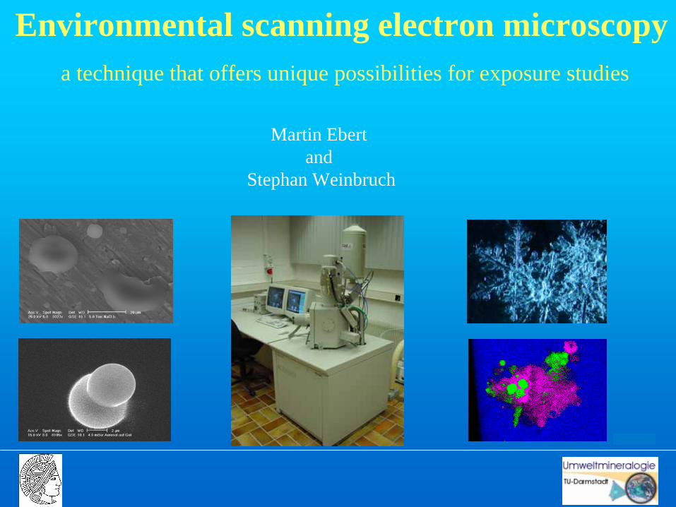
Environmental scanning electron microscopy a technique that offers unique possibilities for exposure studies Martin Ebert and Stephan Weinbruch
Transcript of Environmental scanning electron microscopy · Environmental scanning electron microscopy a...
Environmental scanning electron microscopya technique that offers unique possibilities for exposure studies
Martin Ebert and
Stephan Weinbruch
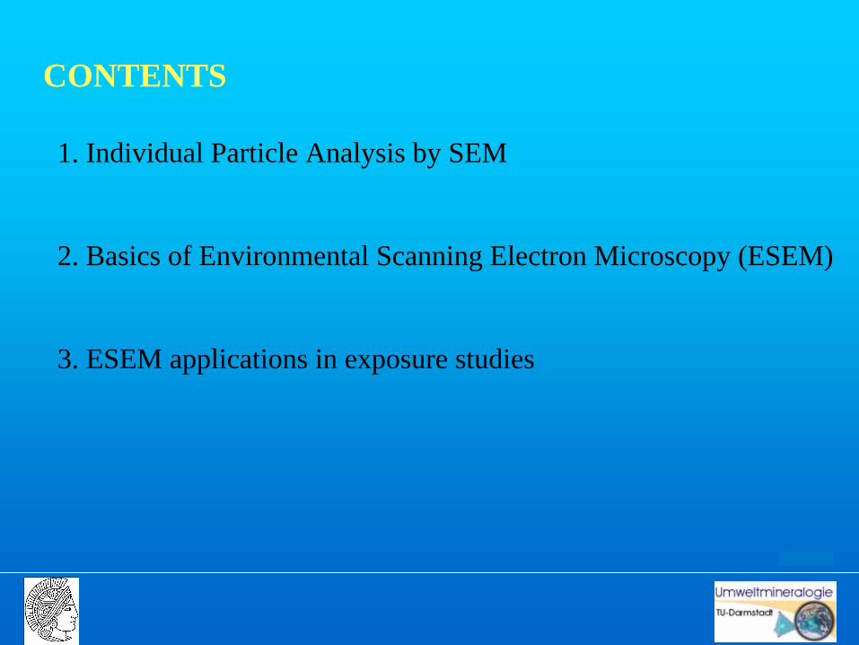
CONTENTS
1. Individual Particle Analysis by SEM
2. Basics of Environmental Scanning Electron Microscopy (ESEM)
3. ESEM applications in exposure studies
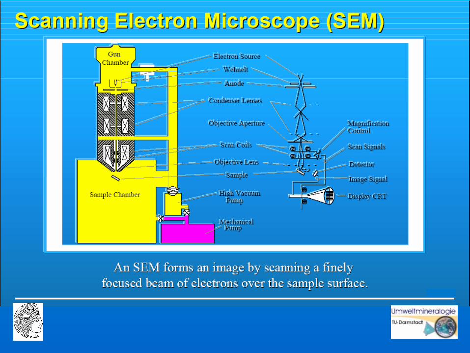
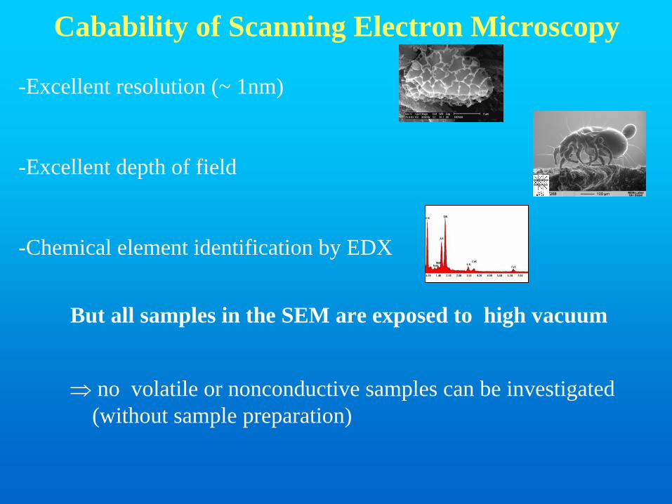
Cabability of Scanning Electron Microscopy
-Excellent resolution (~ 1nm)
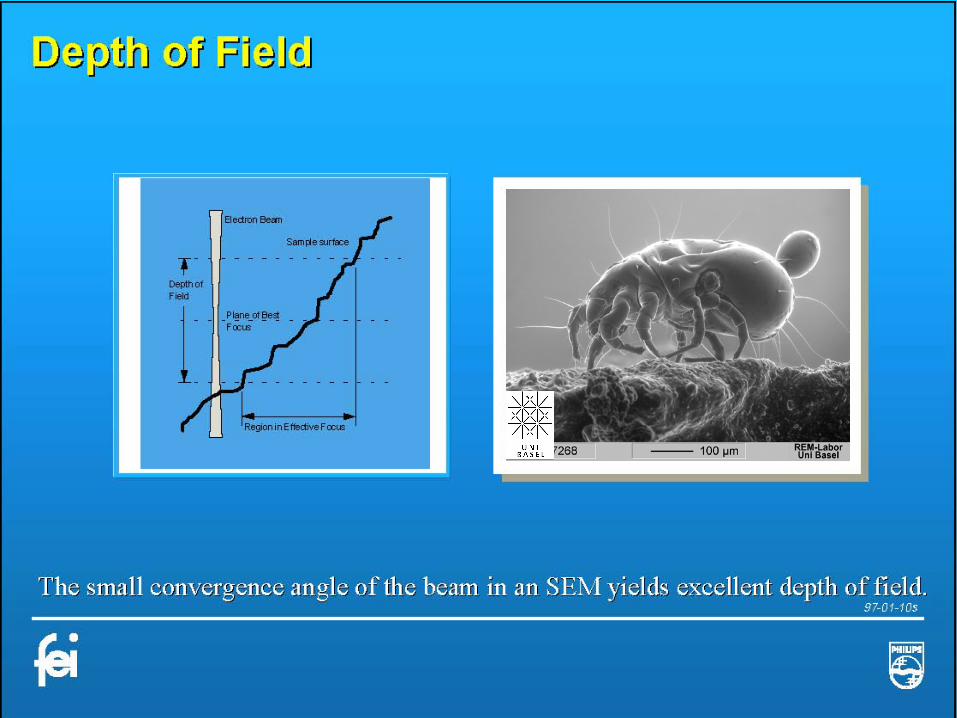
-Excellent depth of field
-Chemical element identification by EDX
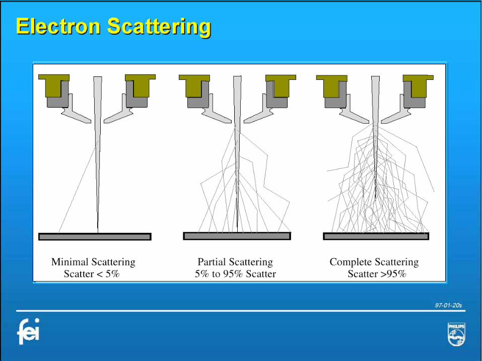
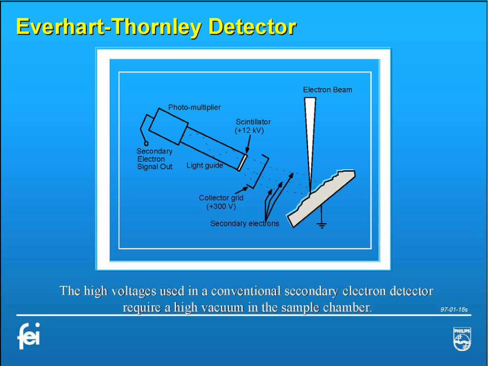
But all samples in the SEM are exposed to high vacuum
⇒ no volatile or nonconductive samples can be investigated(without sample preparation)

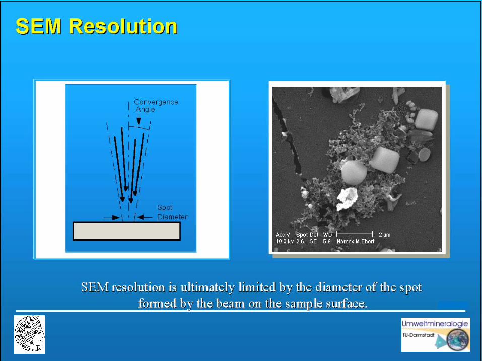
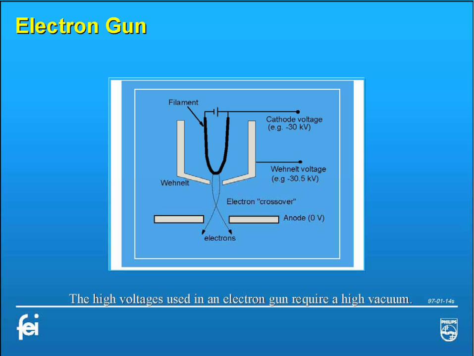
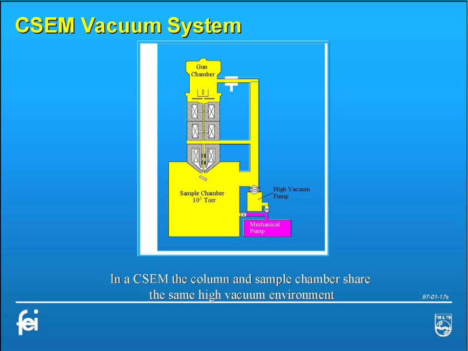
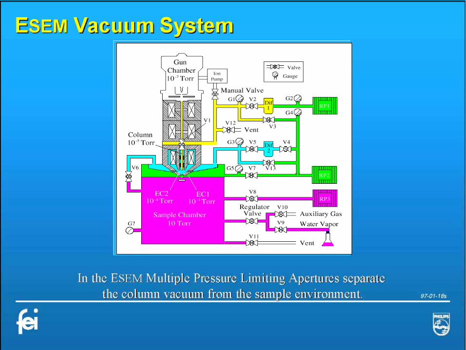
Why do we need high vacuum in a SEM ?
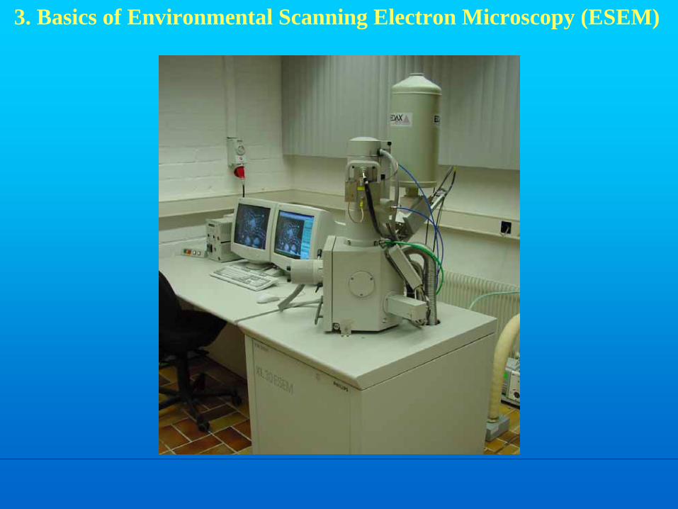
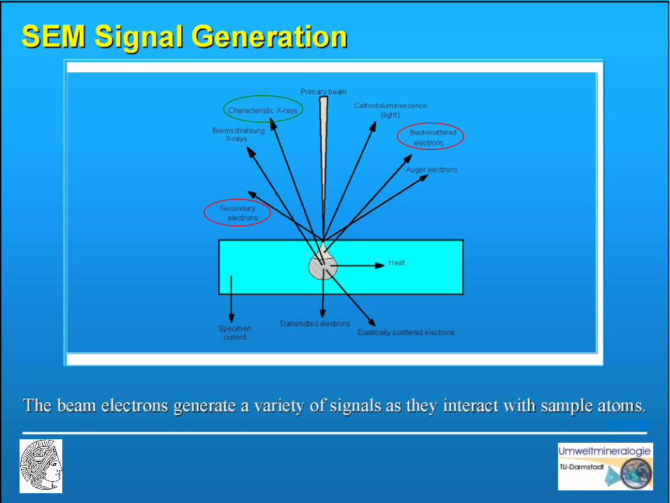
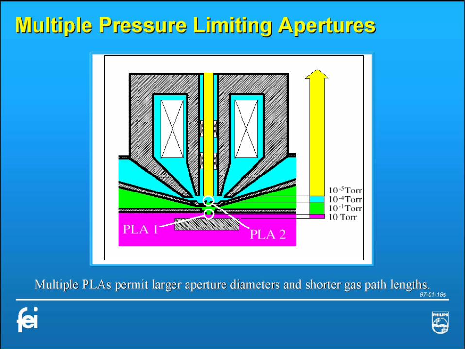
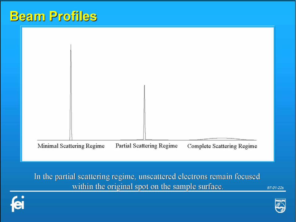
3. Basics of Environmental Scanning Electron Microscopy (ESEM)
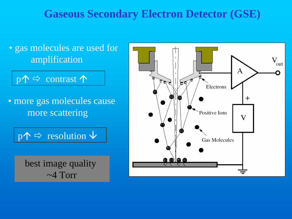
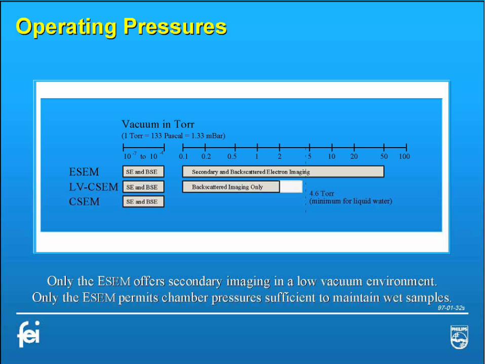
Gaseous Secondary Electron Detector (GSE)
• gas molecules are used foramplification
p contrast
best image quality~4 Torr
• more gas molecules cause more scattering
p resolution
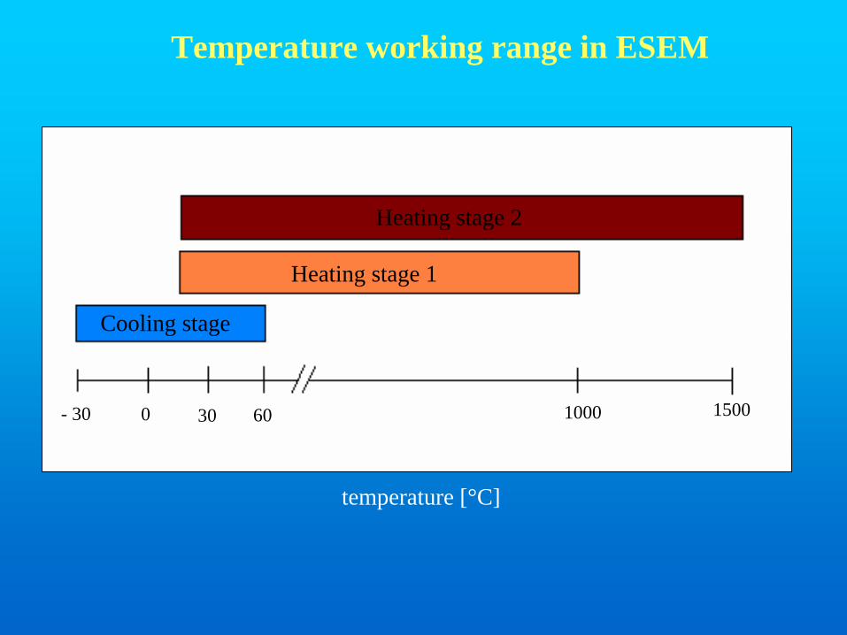
- 30 0 30 60 1000 1500
temperature [°C]
Cooling stage
Heating stage 1
Heating stage 2
Temperature working range in ESEM
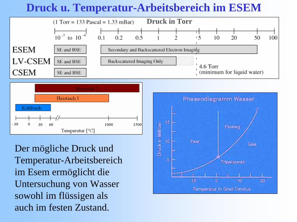
Druck u. Temperatur-Arbeitsbereich im ESEM
Der mögliche Druck und Temperatur-Arbeitsbereich im Esem ermöglicht die Untersuchung von Wasser sowohl im flüssigen als auch im festen Zustand.
Druck in Torr
4. ESEM applications in exposure studies
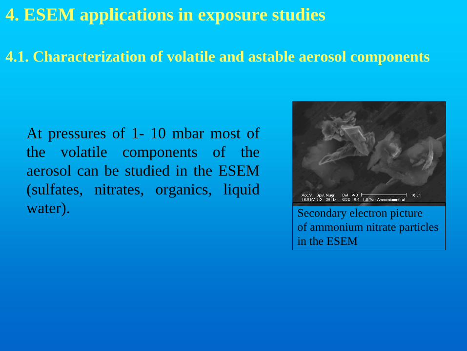
4.1. Characterization of volatile and astable aerosol components
At pressures of 1- 10 mbar most of the volatile components of theaerosol can be studied in the ESEM (sulfates, nitrates, organics, liquid water). Secondary electron picture
of ammonium nitrate particlesin the ESEM
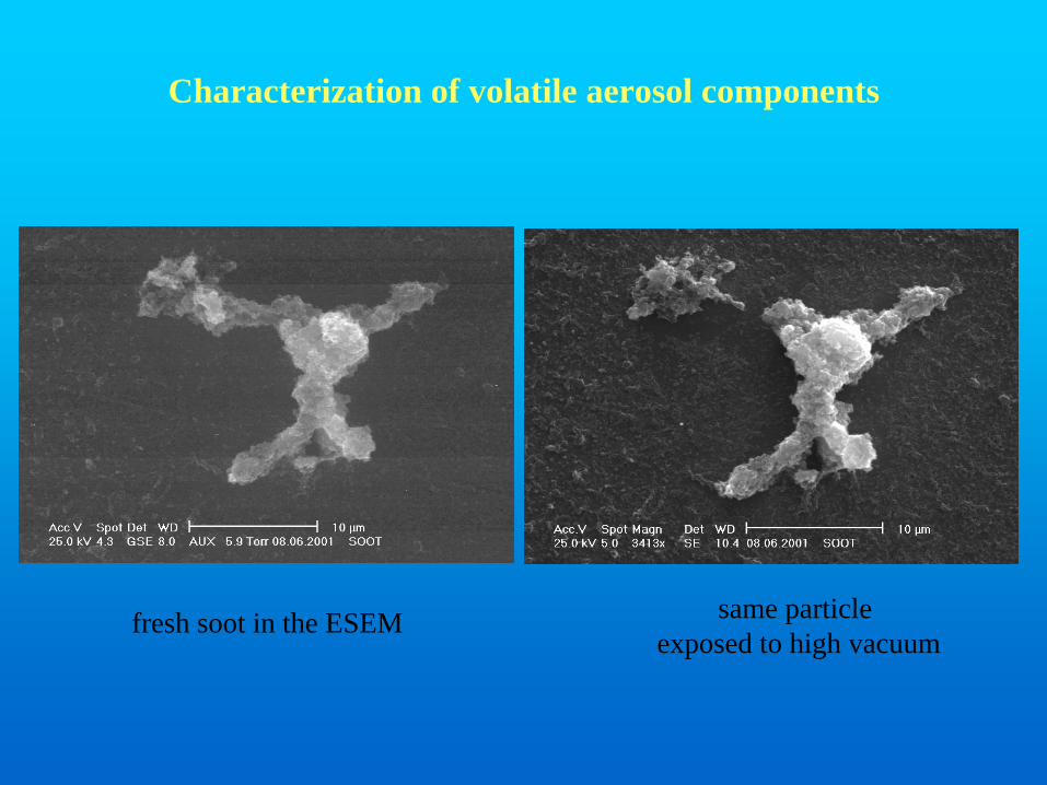
fresh soot in the ESEM same particleexposed to high vacuum
Characterization of volatile aerosol components
a b
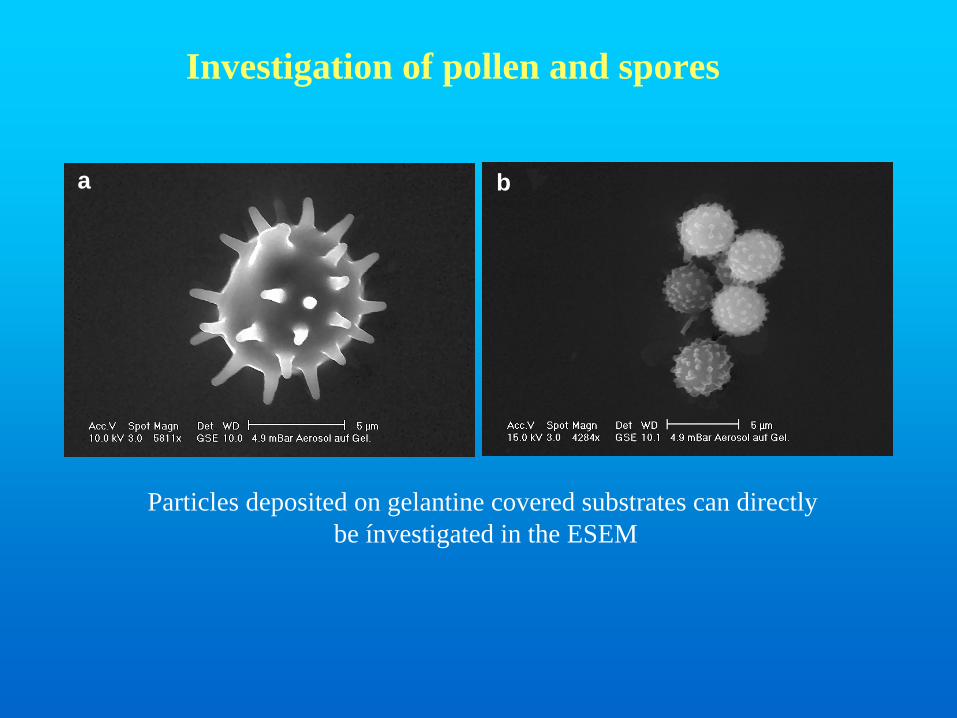
Particles deposited on gelantine covered substrates can directlybe ínvestigated in the ESEM
Investigation of pollen and spores
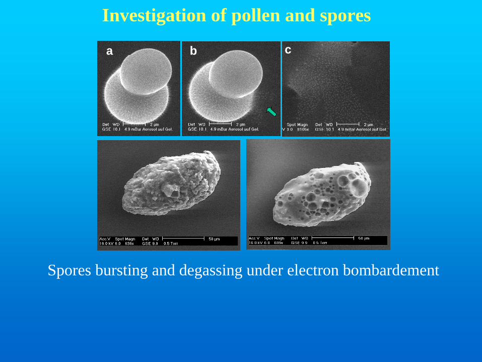
Investigation of pollen and spores
a b c
Spores bursting and degassing under electron bombardement
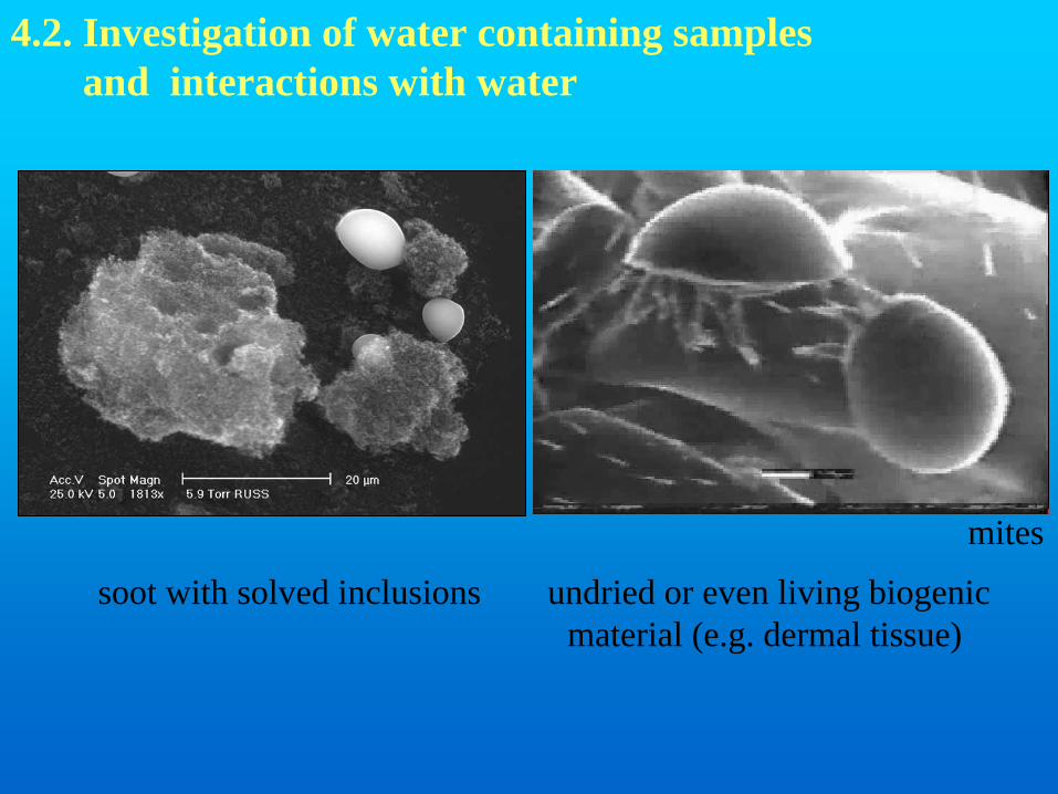
4.2. Investigation of water containing samplesand interactions with water
soot with solved inclusions undried or even living biogenicmaterial (e.g. dermal tissue)
mites
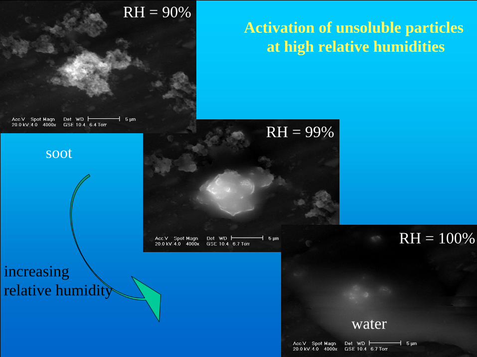
increasingrelative humidity
Activation of unsoluble particlesat high relative humidities
RH = 90%
RH = 99%
RH = 100%
soot
water
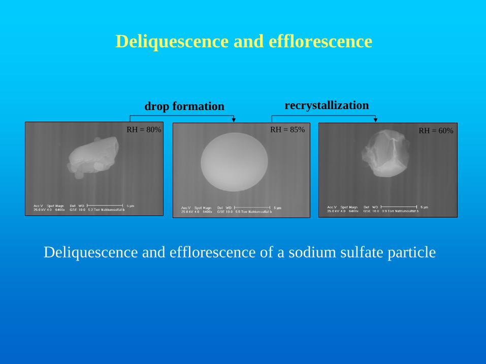
drop formation recrystallization
RH = 80% RH = 85% RH = 60%
Deliquescence and efflorescence
Deliquescence and efflorescence of a sodium sulfate particle
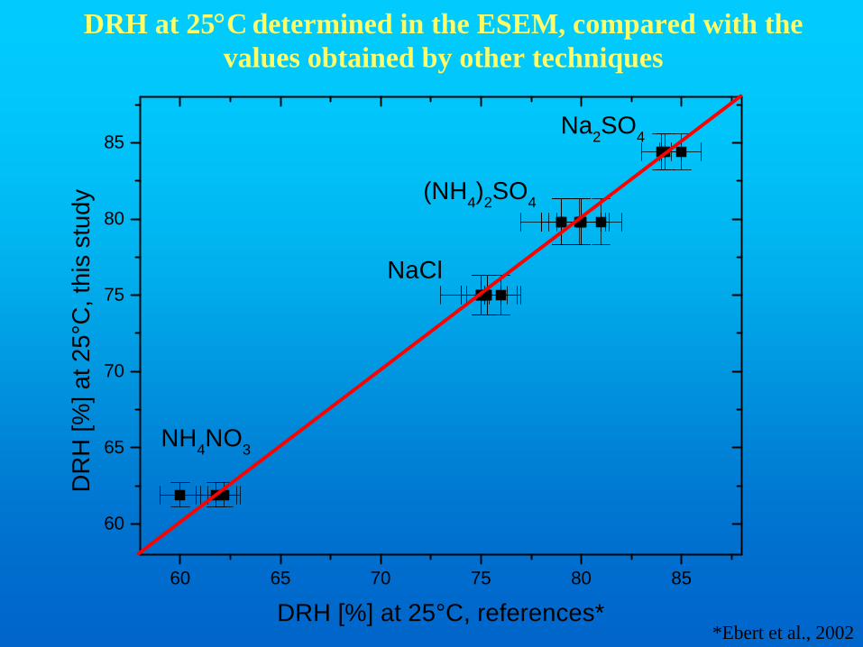
60 65 70 75 80 85
60
65
70
75
80
85Na2SO4
(NH4)2SO4
NaCl
NH4NO3
DR
H [%
] at 2
5°C
, thi
s st
udy
DRH [%] at 25°C, references*
DRH at 25°C determined in the ESEM, compared with the values obtained by other techniques
*Ebert et al., 2002
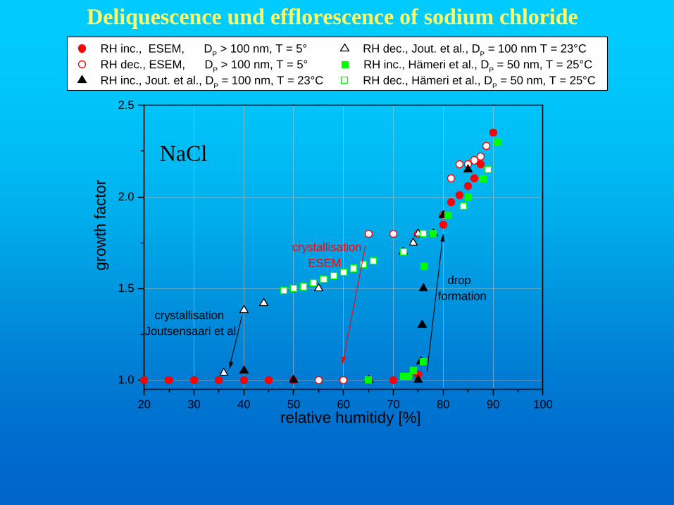
NaCl
Deliquescence und efflorescence of sodium chloride
20 30 40 50 60 70 80 90 100
1.0
1.5
2.0
2.5
crystallisationJoutsensaari et al.
crystallisation ESEM
dropformation
RH inc., ESEM, DP > 100 nm, T = 5° RH dec., Jout. et al., DP = 100 nm T = 23°C RH dec., ESEM, DP > 100 nm, T = 5° RH inc., Hämeri et al., DP = 50 nm, T = 25°C RH inc., Jout. et al., DP = 100 nm, T = 23°C RH dec., Hämeri et al., DP = 50 nm, T = 25°C
grow
th fa
ctor
relative humitidy [%]
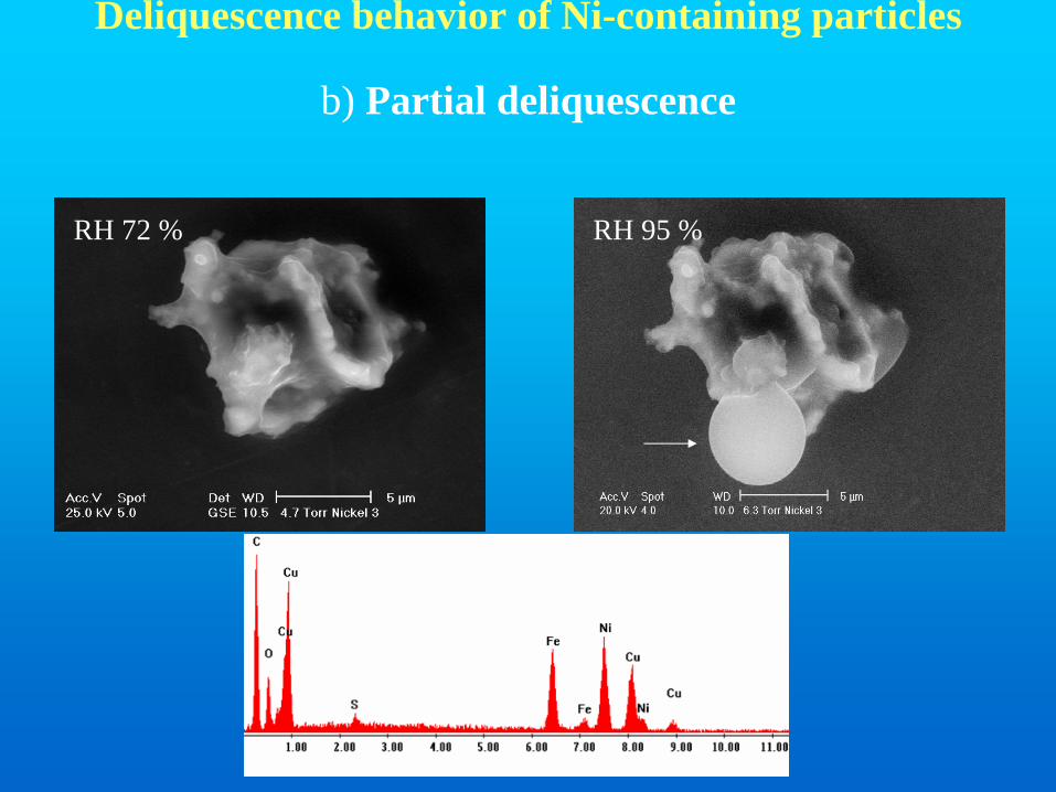
RH 72 % RH 95 %
Deliquescence behavior of Ni-containing particles
b) Partial deliquescence
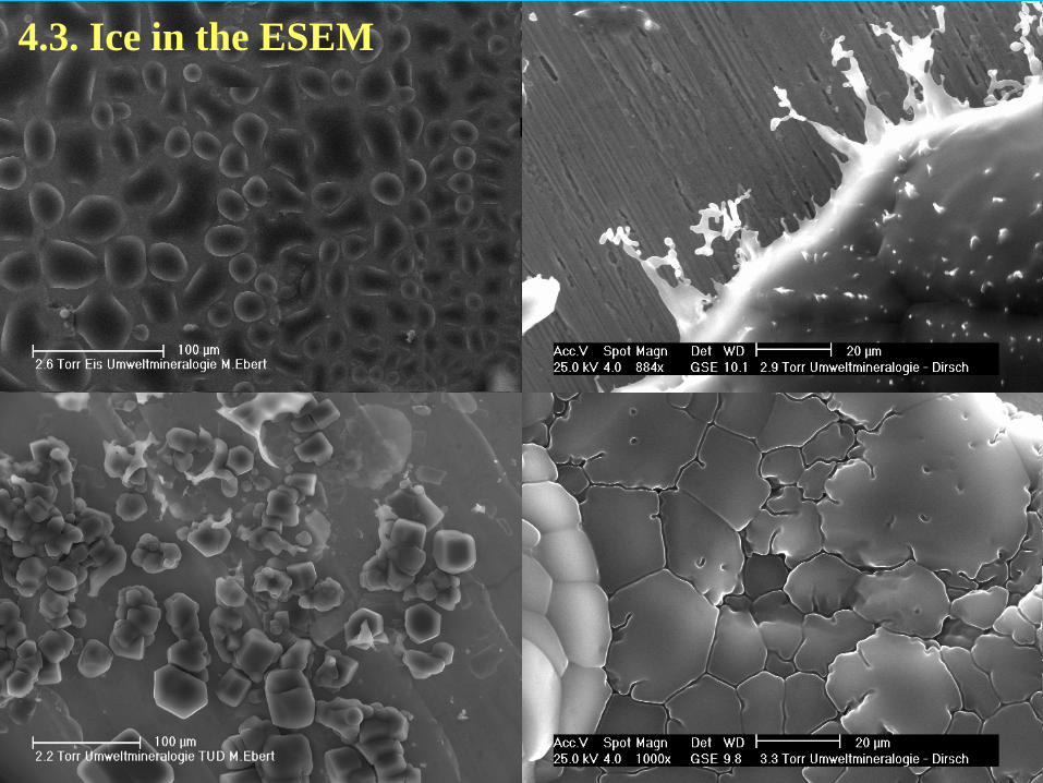
4.3. Ice in the ESEM
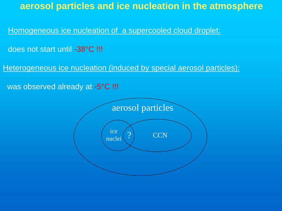
aerosol particles and ice nucleation in the atmosphere
Heterogeneous ice nucleation (induced by special aerosol particles):
was observed already at -5°C !!!
Homogeneous ice nucleation of a supercooled cloud droplet:
does not start until -38°C !!!
aerosol particles
? CCNicenuclei
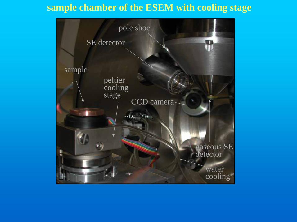
sample chamber of the ESEM with cooling stage
samplepeltiercoolingstage
watercooling
SE detector
gaseous SE detector
pole shoe
CCD camera
mica ice
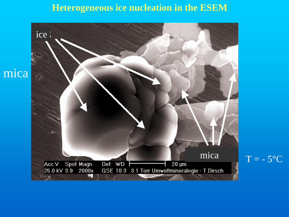
T = - 5°C
Heterogeneous ice nucleation in the ESEM
ice
mica
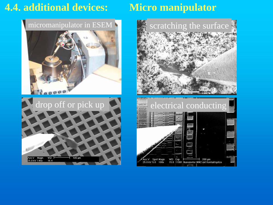
4.4. additional devices: Micro manipulator
scratching the surface
electrical conducting
micromanipulator in ESEM
drop off or pick up

micro injector (drop off or pick up solution)
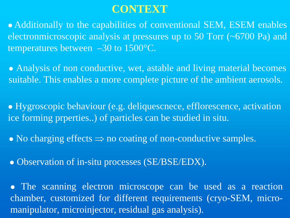
CONTEXTAdditionally to the capabilities of conventional SEM, ESEM enables
electronmicroscopic analysis at pressures up to 50 Torr (~6700 Pa) and temperatures between –30 to 1500°C.
No charging effects ⇒ no coating of non-conductive samples.
Analysis of non conductive, wet, astable and living material becomessuitable. This enables a more complete picture of the ambient aerosols.
Observation of in-situ processes (SE/BSE/EDX).
The scanning electron microscope can be used as a reactionchamber, customized for different requirements (cryo-SEM, micro-manipulator, microinjector, residual gas analysis).
Hygroscopic behaviour (e.g. deliquescnece, efflorescence, activationice forming prperties..) of particles can be studied in situ.


![Ultrafast transmission electron microscopy using a laser ...transmission electron microscopy [4], scanning electron microscopy [5], x-ray diffraction [6], scanning tunneling and atomic](https://static.fdocuments.net/doc/165x107/607eb1335ce8082131294459/ultrafast-transmission-electron-microscopy-using-a-laser-transmission-electron.jpg)
