Environmental Microbiology Athens September 2010...
52
http:// http:// www.bio.warwick.ac.uk www.bio.warwick.ac.uk /wellington /wellington Environmental Microbiology Environmental Microbiology Athens September 2010 MSc Athens September 2010 MSc Microbial detection methods Microbial detection methods E M H Wellington, Department of Biological E M H Wellington, Department of Biological Sciences, Sciences, University of Warwick, Coventry UK University of Warwick, Coventry UK
-
Upload
nguyenkhanh -
Category
Documents
-
view
215 -
download
1
Transcript of Environmental Microbiology Athens September 2010...

http://http://www.bio.warwick.ac.ukwww.bio.warwick.ac.uk/wellington/wellington
Environmental MicrobiologyEnvironmental Microbiology
Athens September 2010 MScAthens September 2010 MSc
Microbial detection methodsMicrobial detection methods
E M H Wellington, Department of Biological E M H Wellington, Department of Biological Sciences, Sciences,
University of Warwick, Coventry UKUniversity of Warwick, Coventry UK

Objectives for enumeration and isolation of bacteriaObjectives for enumeration and isolation of bacteria
Detection of pathogen reservoirs Detection of pathogen reservoirs
Determination pollution impactsDetermination pollution impacts
Exploiting bacterial diversityExploiting bacterial diversity
Investigate aspects of biogeochemistryInvestigate aspects of biogeochemistry
Tracking Tracking biocontrolbiocontrol inoculants / inoculants / GMOsGMOs

Approaches to isolating bacteria from the environmentApproaches to isolating bacteria from the environment
* Pretreatment of the sample, enrichments, soil fractionation
* Selective media, baiting, liquid culture, chemostats
* Immunomagnetic capture, lectins, extraction of cells
* Inhibition of dominant groups/fast growing bacteria by use of selective inhibitors such as specific phage, antibiotics, protozoa
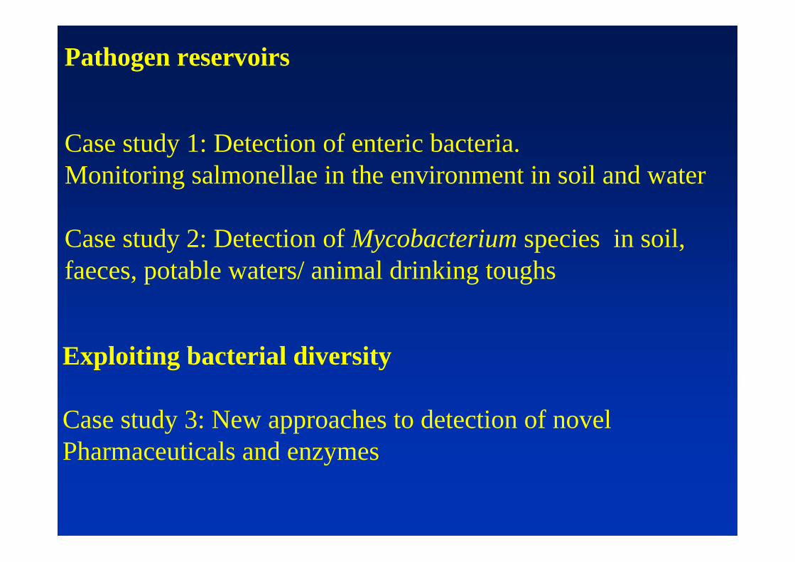
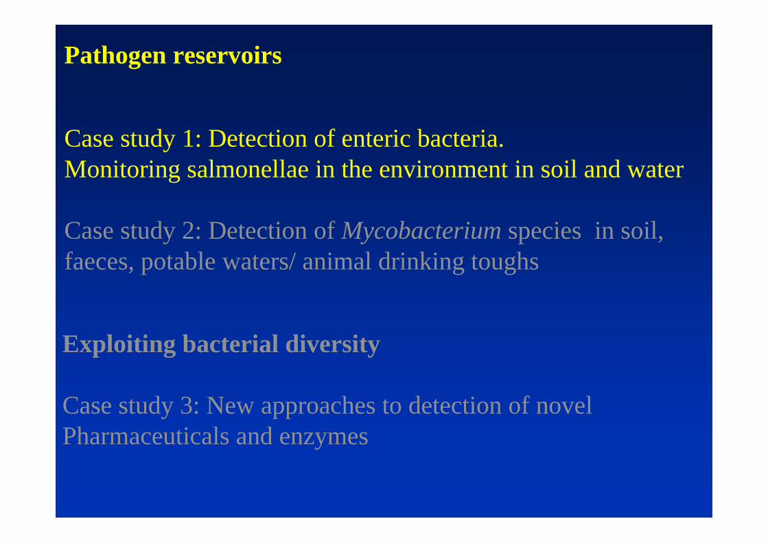
Pathogen reservoirs
Case study 1: Detection of enteric bacteria. Monitoring salmonellae in the environment in soil and water
Case study 2: Detection of Mycobacterium species in soil, faeces, potable waters/ animal drinking toughs
Exploiting bacterial diversity
Case study 3: New approaches to detection of novel Pharmaceuticals and enzymes

Extraction and cultivationExtraction and cultivation : : sensitivity sensitivity and selectivityand selectivity
Part 1
Pathogen reservoirs
Case study 1: Detection of enteric bacteria. Monitoring salmonellae in the environment in soil and water
Case study 2: Detection of Mycobacterium species in soil, faeces, potable waters/ animal drinking toughs
Exploiting bacterial diversity
Case study 3: New approaches to detection of novel Pharmaceuticals and enzymes

Application of sewage sludge to land: what is the fate of enteric bacteria in soil?
Sewage treatment and disposal


Fate of exotic bacteria in the environment
Problems to be addressed
lDo exotic bacteria enter a non-culturable state; if so what is physiological state of cells? Extent survival of culturable populationWhich environmental factors induce this state, and what is the mechanism of induction?
lHow can cells be selectively detected and resuscitated?
lCan existing clinical detection methods be appliedSelective isolation media, typing methods, specific antibodies

Growth 0.5h 6h 100h
Changes in Vibrio during multiple nutrient starvation
After Östling et al., 1992

Bacterium
Aeromonas Salmonicida
Agrobacterium tumefaciens
Campylobacter jejuni
Enterobacter aerogenes
Klebsiella pneumoniae
Salmonella enteritidis
Shigella dysenteriae
Vibrio cholerae
V. vulnificus
Reference
Morgan et al. (1993)
Byrd et al. (1991)
Medema et al. (1992)
Byrd et al. (1991)
Byrd et al. (1991)
Roszak et al. (1984)
Islam et al. (1993)
Wolf and Oliver (1992)
Wolf and Oliver (1992)
Examples of the nonculturable state detected in bacteria
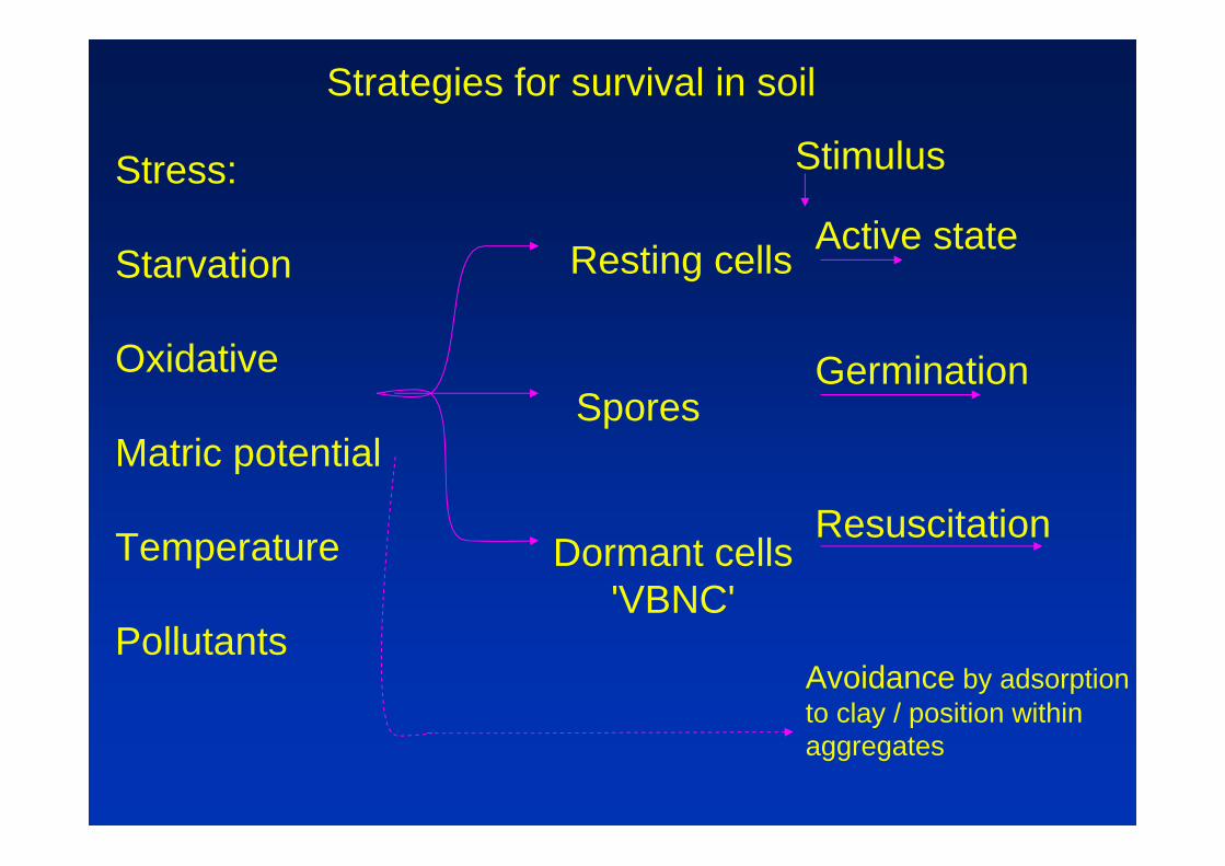
Strategies for survival in soil
Stress:
Starvation
Oxidative
Matric potential
Temperature
Pollutants
Resting cells
Spores
Dormant cells'VBNC'
Active state
Germination
Resuscitation
Avoidance by adsorptionto clay / position withinaggregates
Stimulus

Detection of the nonculturable state in soil
Bacterium
Salmonella typhimuriumVibrio harveyiE. coli
Indigenous
Alcaligenes eutrophusEnterobacter clloacaePseudomonas fluorescensPseudomonas aeruginosa
Pseudomonas fluorescensPseudomonas syringaePseudomonas fluorescensFlavobacterium
Method
Direct countluminescence+ yeast extract
CTC stainingmicrocoloniesDirect viable counts (DVC)
Microcolony epifluorescence (AO)DNA / resuscitation
CTC staining
ImmunofluarescenceDVC, CTC staining
Reference
Turpin et al. 1993Duncan et al. 1994
Winding et al. 1994Pedersen & Jacobsen 1993Binnerup et al. 1993Leung et al. 1995Oliver et al. 1995Van Overvbeaket al. 1995Heijnen et al 1995
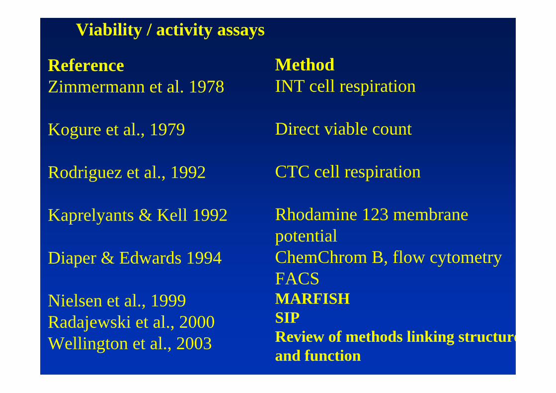
Viability / activity assays
ReferenceZimmermann et al. 1978
Kogure et al., 1979
Rodriguez et al., 1992
Kaprelyants & Kell 1992
Diaper & Edwards 1994
Nielsen et al., 1999Radajewski et al., 2000Wellington et al., 2003
MethodINT cell respiration
Direct viable count
CTC cell respiration
Rhodamine 123 membranepotentialChemChrom B, flow cytometryFACSMARFISHSIPReview of methods linking structureand function

100
10
1
0.1
0.0120 40 60 80 100 120 140
Times (s)
Survival (%)
Starved 5dViable
DAPI
Log phaseViable
DAPI
Resistance of Vibrio cells to sonication
after Weichart and Kjellberg, 1996

MetMethods must fit objectives:Monitoring only: +/- detection, can be enrichment in situDetection and enumeration: specific, quantitative molecular or
indirect cultivationQuantification and typing: tracking source, establish infectivity-
limited to recovery of cells
Spread of plasmids carrying transposons mediated streptothricin resistance among various bacterial populations (Witte, 1996)
Origin 1982 1983 1984 1985 1986 1987
GI flora of nat- fed pigs - + + + + +GI flora of pig farm personnel - - + + + +GI flora of family of personnel - - + + + +GI flora of healthy adults - - - + + +GU tract infections - - - + + +Shigella sonnei - - - - - +
Meat productsHuman GI tract
Faeces
Slurry Water
Animal feed
Food
Infection
Possible pathways of dissemination of antibiotic-resistant bacteria between animalsand humans
e.g

Genus Selective medium for viable counts
Pseudomonas Pseudomonas CFC agarKings A agar
Salmonella XLD (xylose deoxycholate) agarSalmonella Shigella AgarBrilliant Green agar
Micromonospora Colloidal chitin agarStarch-Casein agar
Streptomyces RASS agar
E.coli McConkey agarEMB agarTryptone Bile agar
Methods for selective isolation of bacteria

Application of sewage sludge to land: what is the fate of enteric bacteria in soil?
l Do salmonellae survive and what affects death rate?
l Can enteric pathogens grow in soil?
l Is there evidence for intracellular growth of pathogenic bacteria within protozoans?
l What are the best strategies for detection and monitoring?

. . . .. .. .. ... ...
The monitoring methods
Directcount(FITC)
ViableCount
Lux phage DNA(PCR)

0 10 20 30 40 50 60 703
4
5
6
7
9
10
0
0.5
1
1.5
2
2.5
3
3.5
Time (days)
Log cfu/g orlog cells/g soil
Abs.492nm
8
Uncultured populationSalmonella typhimurium
produced in nonsterile soil
Plate count on XLD agar
Fluorescent antibody direct count with live inoculum
UV-killed inoculum
♦ ELISA
15% moisture, 22°C
``

Concentration of cells from soil & DNA extraction
lSample dispersion and cell concentration by differential centrifugation (optional: immuno-magnetic capture (IMC) of cells)
lDNA extraction and purification by either:1) physical disruption of cells, partition & ion-exchange chromatography, or 2) heat-lysis of magnetically captured cells
lMultiplex PCR of triple targets on chromosomeQuantitative PCR (QPCR); MPN or densitometry ofproduct

EXTRACTION OF CELLS FROM SOIL
Breaking forces binding cells to aggregates and clay particles:
Ion exchange-resin based extraction :
Soil + Chelex 100 + PEG 6000 + sodium deoxycholateshake
Differential centrifugation & filtering
Concentrated biomass, separated from soil

Concentration of cells from soil for DNA and other extractions
10 g soil + 10 g Chelex 100 (BioRad) + 10 ml 2.5% polyethylene glycol 6000shake gently for 2 hrs
centrifuge at 177 x g 30 sec
re-centrifuge liquid phase at 3500 x g 15 minthree alternative analyses
DNA extraction viable count immunomagneticresuspend pellet in 1 ml plate dilutions of capture20 mg.ml-1 lysozyme extract on agar resuspend pellet in37ºC for 30 min phosphate buffered saline
(pH 7.2)SDS lysis or physicallydisrupt cells rotate 30min
add magnetic beads coatedprecipitate salt with potassium with antibody (Dynal)acetate
capture bead-cell complex byethanol precipitation of DNA holding magnet against tube
PCR wash and process as required(PCR, selective plating etc.)

Multiplex PCR of putative Salmonella isolates from field samples
Lanes 1-8, putative Salmonella isolates. Lane 9, S. typhimuriumLT2
phoPHinH-li
1 2 3 4 5 6 7 8 9

DNA extracted from soil & PCR of Salmonella -specific targets
Nonsterile soil inoculated with S. typhimurium.
Top lanes =soil DNA (inoculum cfu/g): 1, 10 ; 2, 10 ;
3, 10 ; 4, 10 ; 5, 10 ; 6, negative soil control
Bottom lanes =PCR of H-li from soil DNA.
Lanes 1 - 6 as top lanes; 7, positive control;
8, negative control
7 6
5 4 3
H-li
1 2 3 4 5 6
1 2 3 4 5 6 7 8
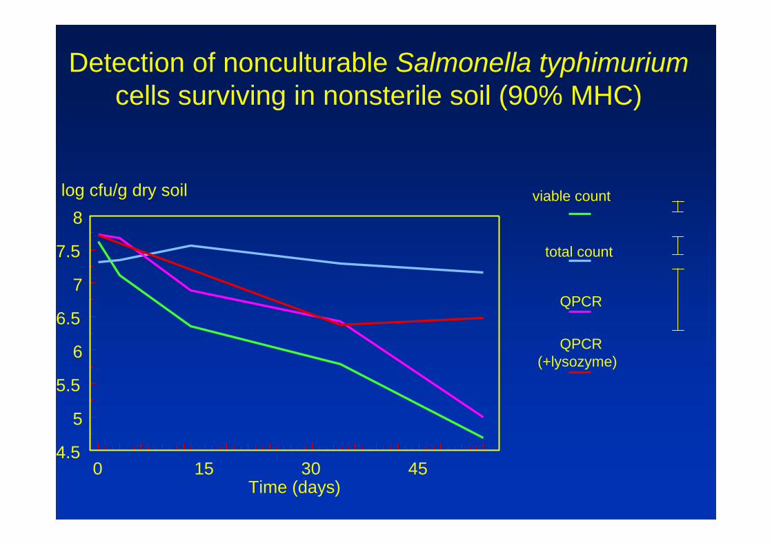
0 15 30 454.5
5
5.5
6
6.5
7
7.5
8
Time (days)
log cfu/g dry soil viable count
total count
QPCR
QPCR(+lysozyme)
Detection of nonculturable Salmonella typhimuriumcells surviving in nonsterile soil (90% MHC)

Detection of Salmonella typhimurium cells in soil: proportion of active cells
CTC - active count FITC - total count

Factors affecting the survival of salmonella in soil
Factor Comments
Moisturecontent
Bacteria susceptible to desiccation. Survival is longer in wet soils and during rainy periods
Organic matter
Bacterial survival increased when more organic matteris present
pH UK soils range pH 4 to 8. Highest survival pH 7; shorter survival in acid soils (pH 3-5)
Temperature Longer survival at moderate temperatures (>0ºC)
Physical state Physical state of cell when exposed to environment
Sunlight Lower survival a t soil surface by UV action/ photoinactivation /desiccation

Application of sewage sludge to land: what is the fate of enteric bacteria in soil?
l Do salmonellae survive beyond ‘no grazing period’ ? Methods for detection
l Can enteric pathogens grow in soil?
l Is there evidence for intracellular growth of pathogenic bacteria within protozoans?

Soil food web: Soil food web: bacteria consumed by amoebaebacteria consumed by amoebae
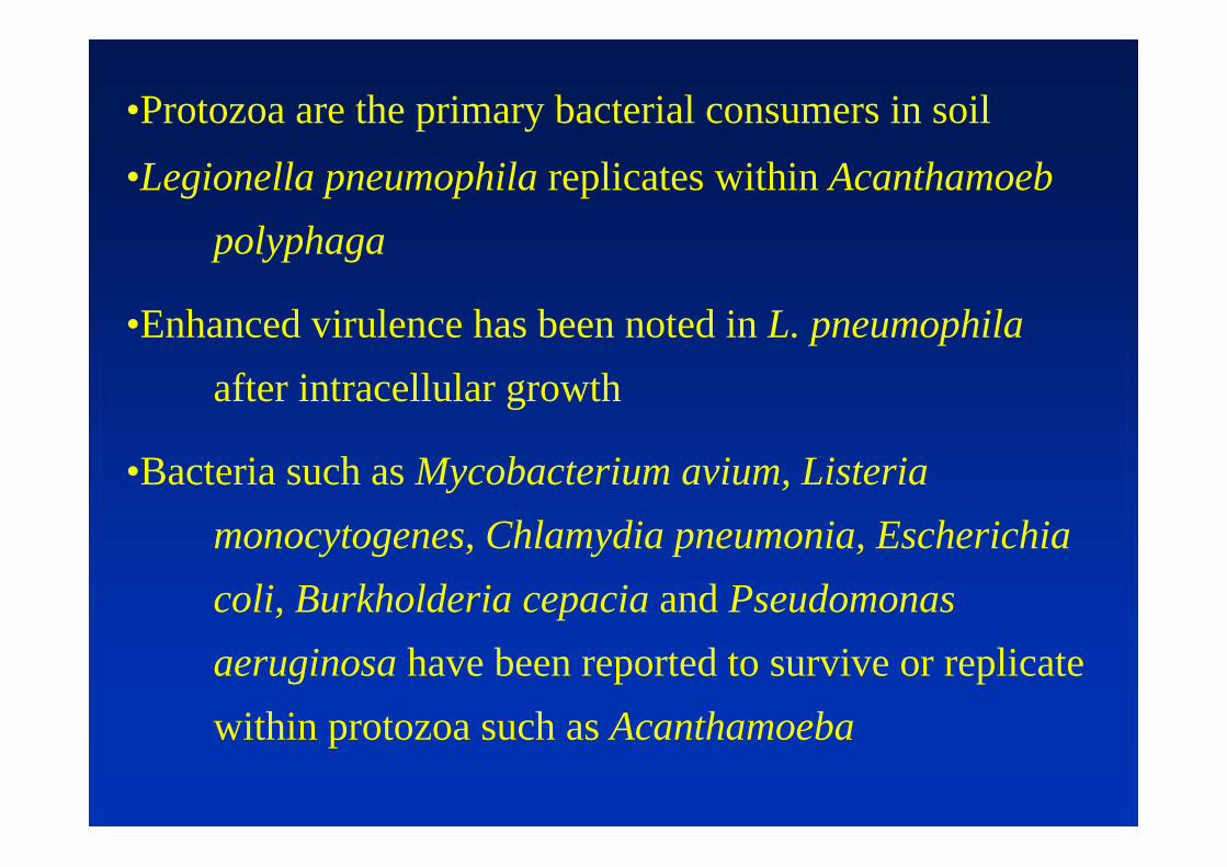
•Protozoa are the primary bacterial consumers in soil
•Legionella pneumophila replicates within Acanthamoebpolyphaga
•Enhanced virulence has been noted in L. pneumophilaafter intracellular growth
•Bacteria such as Mycobacterium avium, Listeriamonocytogenes, Chlamydia pneumonia, Escherichia coli, Burkholderia cepacia and Pseudomonas aeruginosa have been reported to survive or replicate within protozoa such as Acanthamoeba

•Investigate S. typhimurium phagocytosed by A. polyphaga doesintracellular survival or replication occur?
•Assess the dynamics of the system, using standardised image sets and time lapse image sequences to determine amoebal grazing rates, duration of digestive cycle, locomotory rate, locomotory behaviour, bacterial growth rates, bacterial survival

Monitoring the interaction
•fis:gfp reporter, fis (factor-for-inversion stimulation protein) is a DNA architectural protein which is involved in specialised DNA recombination events and regulation of gene expression. Fis protein and mRNA levels rapidly increase during early logarithmic growthphase in response to a nutritional upshift but become virtually undetectable during late logarithmic and stationary phases.
•Plasmid pPDT105 was derived from pPDT100 by insertion of egfpdownstream of the fis fragment.
Investigate S. typhimurium phagocytosed by A. polyphaga does
intracellular survival or replication occur?

a) phase contrast image of A. polyphaga with contractile vacuole infected by S. typhimurium.
b) fluorescence image of (a), fluorescence intensity indicates expression of fis.
ba b
Amoebae may facilitate growth of certain bacteria- cocultures non-nutrient agar biofilm , drop inoculate amoebae centre of lawn

c) 3D profile of pixel intensity of (b)
d) thresholded image of (b) with relative pixel intensities below 30% subtracted, showing relative up-regulation of fis indicating entry into logarithmic growth
c d

Growth in the contractile vacuole

0
2
4
6
8
10
12
14
0 10 20 30 40 50 60
time (seconds)
cv diameter "salmonallae"cv diameter empty
Relative diameter of contractile vacuole in an infected and uninfected amoeba
μm

A. polyphaga and S. typhimurium, phase contrast (a) and fluorescence images (b). Up-regulation of the fis promoter in some bacterial cells contained within phagocytic food vacuoles suggests that intravacuolar growth was occurring and replication was possible

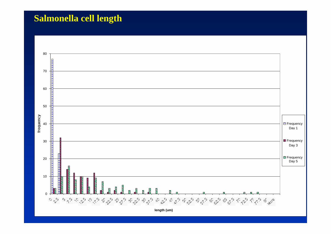
Filamentous growth of S. typhimurium
0
10
20
30
40
50
60
70
80
length (um)
Frequency
Frequency
Frequency
Day 1
Day 3
Day 5
Salmonella cell length
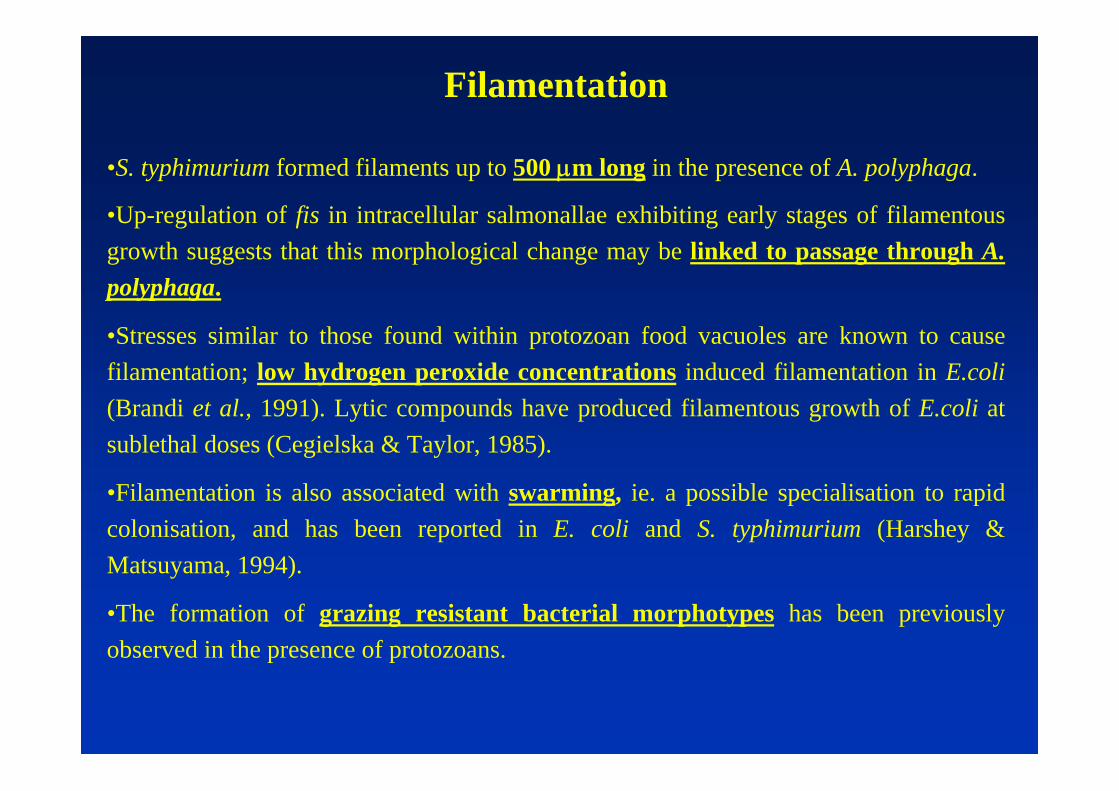
Filamentation
•S. typhimurium formed filaments up to 500 μm long in the presence of A. polyphaga.
•Up-regulation of fis in intracellular salmonallae exhibiting early stages of filamentous growth suggests that this morphological change may be linked to passage through A. polyphaga.
•Stresses similar to those found within protozoan food vacuoles are known to cause filamentation; low hydrogen peroxide concentrations induced filamentation in E.coli(Brandi et al., 1991). Lytic compounds have produced filamentous growth of E.coli at sublethal doses (Cegielska & Taylor, 1985).
•Filamentation is also associated with swarming, ie. a possible specialisation to rapid colonisation, and has been reported in E. coli and S. typhimurium (Harshey & Matsuyama, 1994).
•The formation of grazing resistant bacterial morphotypes has been previously observed in the presence of protozoans.

Amoebae-bacteria population level interactions
1. Matrices of 100 fields of view, 2 x phase contrast with 4 second interval + 1 fluorescence image. 10 x objective, non-nutrient agar, no coverslip.
2. Time-lapse image sequences, 1 frame per minute (approx. 600x)

Spatial and temporal analysis

Spatial and temporal analysis

TRACKING

Saprophytic growth of passaged cells

Deposition of faecal pellets and colony growth

Each colony was formed from an egested food vacuole or “faecal pellet” which were deposited at regular intervals by an amoeba. This illustrates bacterial translocation, survival of digestion and subsequent growth on nutrients contained in the faecal pellet and/or secreted by the amoebae
Dissemination of viable salmonellae

Bacterial colony growth on NNA along an amoebal trail, subsequently colonised by amoebae.
Saprophytic growth of passaged cells


Summary of interactions with amoebae
•A proportion of salmonellae can survive digestion, and are egested in a pellet and disseminated
•Rapid bacterial growth occurs on pellets
•Salmonellae can infect contractile vacuoles (osmoregulatoryorganelles)
•Bacterial filamentation can occur during interactions with amoebae, possible a phenotypic change induced by ingestion and passage