Environment Responsive Metal–Organic Frameworks as Drug ...
8
Yan et al. Nanoscale Res Lett (2021) 16:140 https://doi.org/10.1186/s11671-021-03597-w NANO REVIEW Environment Responsive Metal–Organic Frameworks as Drug Delivery System for Tumor Therapy Chao Yan 1 , Yue Jin 1 and Chuanxiang Zhao 2* Abstract Nanoparticles as drug delivery systems can alter the drugs’ hydrophilicity to affect drug uptake and efflux in tissues. They prevent drugs from non-specifically binding with bio-macromolecules and enhance drug accumulation at the lesion sites, improving therapy effects and reducing unnecessary side effects. Metal–organic frameworks (MOFs), the typical nanoparticles, a class of crystalline porous materials via self-assembled organic linkers and metal ions, exhibit excellent biodegradability, pore shape and sizes, and finely tunable chemical composition. MOFs have a rigid molecu- lar structure, and tunable pore size can improve the encapsulation drug’s stability under harsh conditions. Besides, the surface of MOFs can be modified with small-molecule ligands and biomolecule, and binding with the biomarkers which is overexpressed on the surface of cancer cells. MOFs formulations for therapeutic have been developed to effectively respond to the unique tumor microenvironment (TEM), such as high H 2 O 2 levels, hypoxia, and high con- centration glutathione (GSH). Thus, MOFs as a drug delivery system should avoid drugs leaking during blood circula- tion and releasing at the lesion sites via a controlling manner. In this article, we will summary environment responsive MOFs as drug delivery systems for tumor therapy under different stimuli. Keywords: Nanoparticles, Metal–organic frameworks, Unique tumor microenvironment © The Author(s) 2021. Open Access This article is licensed under a Creative Commons Attribution 4.0 International License, which permits use, sharing, adaptation, distribution and reproduction in any medium or format, as long as you give appropriate credit to the original author(s) and the source, provide a link to the Creative Commons licence, and indicate if changes were made. The images or other third party material in this article are included in the article’s Creative Commons licence, unless indicated otherwise in a credit line to the material. If material is not included in the article’s Creative Commons licence and your intended use is not permitted by statutory regulation or exceeds the permitted use, you will need to obtain permission directly from the copyright holder. To view a copy of this licence, visit http://creativecommons.org/licenses/by/4.0/. Introduction Tumor is a multifactorial disease with high mortality and recurrence rates that threaten human health [1]. In clinics, chemotherapeutic drugs and surgery applied for tumor therapy have achieved tumor inhibition but often with serious side effects, which promoted us to develop superior therapeutic methods [2, 3]. Over the past dec- ades, nanocarriers have been developed for tumor imag- ing, theranostics and therapy [4]. In all kinds of nanocarriers, metal–organic frame- works (MOFs) have attracted increasing attention, as they can be stimulated by different environment [5, 6]. MOFs, as a class of high crystalline inorganic–organic porous materials, consist of metal ions or clusters linked by organic bridging ligands and have attracted tremen- dous attention in recent years in different fields [7]. Ear- lier than the 1990s, MOFs has been widely applied in gas storage, separation catalysis, energy conversion, lumines- cence and chemical sensing, and biomedical field, due to their finely tunable chemical composition, pore shape and size, morphology, large surface area and excellent biodegradability [8, 9]. MOFs have organic active sites and accessible, open- ing porous architectures, chemical stability, and sufficient thermal effects [10]. us various functional groups can integrate into MOFs via three strategies: encapsulation, grafting, and infiltration, which can improve their bio- compatibility, solubility and interactivity with a target molecules [11]. In particular, the encapsulation approach through coprecipitation and biomimetic mineraliza- tion method is the rapid and convenient approach using Open Access *Correspondence: [email protected] 2 School of Medical Technology, Jiangsu College of Nursing, Huai’an City, Jiangsu Province, China Full list of author information is available at the end of the article
Transcript of Environment Responsive Metal–Organic Frameworks as Drug ...

Yan et al. Nanoscale Res Lett (2021) 16:140 https://doi.org/10.1186/s11671-021-03597-w
NANO REVIEW
Environment Responsive Metal–Organic Frameworks as Drug Delivery System for Tumor TherapyChao Yan1, Yue Jin1 and Chuanxiang Zhao2*
Abstract
Nanoparticles as drug delivery systems can alter the drugs’ hydrophilicity to affect drug uptake and efflux in tissues. They prevent drugs from non-specifically binding with bio-macromolecules and enhance drug accumulation at the lesion sites, improving therapy effects and reducing unnecessary side effects. Metal–organic frameworks (MOFs), the typical nanoparticles, a class of crystalline porous materials via self-assembled organic linkers and metal ions, exhibit excellent biodegradability, pore shape and sizes, and finely tunable chemical composition. MOFs have a rigid molecu-lar structure, and tunable pore size can improve the encapsulation drug’s stability under harsh conditions. Besides, the surface of MOFs can be modified with small-molecule ligands and biomolecule, and binding with the biomarkers which is overexpressed on the surface of cancer cells. MOFs formulations for therapeutic have been developed to effectively respond to the unique tumor microenvironment (TEM), such as high H2O2 levels, hypoxia, and high con-centration glutathione (GSH). Thus, MOFs as a drug delivery system should avoid drugs leaking during blood circula-tion and releasing at the lesion sites via a controlling manner. In this article, we will summary environment responsive MOFs as drug delivery systems for tumor therapy under different stimuli.
Keywords: Nanoparticles, Metal–organic frameworks, Unique tumor microenvironment
© The Author(s) 2021. Open Access This article is licensed under a Creative Commons Attribution 4.0 International License, which permits use, sharing, adaptation, distribution and reproduction in any medium or format, as long as you give appropriate credit to the original author(s) and the source, provide a link to the Creative Commons licence, and indicate if changes were made. The images or other third party material in this article are included in the article’s Creative Commons licence, unless indicated otherwise in a credit line to the material. If material is not included in the article’s Creative Commons licence and your intended use is not permitted by statutory regulation or exceeds the permitted use, you will need to obtain permission directly from the copyright holder. To view a copy of this licence, visit http:// creat iveco mmons. org/ licen ses/ by/4. 0/.
IntroductionTumor is a multifactorial disease with high mortality and recurrence rates that threaten human health [1]. In clinics, chemotherapeutic drugs and surgery applied for tumor therapy have achieved tumor inhibition but often with serious side effects, which promoted us to develop superior therapeutic methods [2, 3]. Over the past dec-ades, nanocarriers have been developed for tumor imag-ing, theranostics and therapy [4].
In all kinds of nanocarriers, metal–organic frame-works (MOFs) have attracted increasing attention, as they can be stimulated by different environment [5, 6]. MOFs, as a class of high crystalline inorganic–organic
porous materials, consist of metal ions or clusters linked by organic bridging ligands and have attracted tremen-dous attention in recent years in different fields [7]. Ear-lier than the 1990s, MOFs has been widely applied in gas storage, separation catalysis, energy conversion, lumines-cence and chemical sensing, and biomedical field, due to their finely tunable chemical composition, pore shape and size, morphology, large surface area and excellent biodegradability [8, 9].
MOFs have organic active sites and accessible, open-ing porous architectures, chemical stability, and sufficient thermal effects [10]. Thus various functional groups can integrate into MOFs via three strategies: encapsulation, grafting, and infiltration, which can improve their bio-compatibility, solubility and interactivity with a target molecules [11]. In particular, the encapsulation approach through coprecipitation and biomimetic mineraliza-tion method is the rapid and convenient approach using
Open Access
*Correspondence: [email protected] School of Medical Technology, Jiangsu College of Nursing, Huai’an City, Jiangsu Province, ChinaFull list of author information is available at the end of the article
Page 2 of 8Yan et al. Nanoscale Res Lett (2021) 16:140
the organic ligands and metal ions to achieve one-step embedding of drugs into MOFs [12, 13]. Inspired from these excellent merits, various methods have been made to identify its feasibility and effectiveness of utilize. How-ever, MOFs can easily grow at different substrates to form multifunctional complexes [14].Thus, some therapeutic agents can directly incorporate into MOFs via synthesis progress, which can circumvent crystal growth problems when applying pre-functionalized ligands [15, 16]. Such a strategy provides a high atomic economy and leads to extremely satisfactory drug payloads [14].
Although MOFs as drugs delivery system for tumor therapy has unparalleled advantages, their application has been restricted by many intractable drawbacks. For example, MOFs are a complicated synthetic progress, eliminated by the body’s immune system, and has a short half-life in the blood [17–19]. In this article, we will summarize some basic environment stimuli-responsive MOFs to enhance tumor therapy and review the current state of the tumor theranostics.
pH/ATP ResponsiveZeolitic imidazolate frameworks (ZIFs), as the specific subclass of MOFs, have tunable pore size, ultra-large sur-face area, and facile synthesis progress. ZIFs are synthe-sized via biomimetic mineralization and coprecipitation used as the ideal drug carrier for tumor theranostics [20]. Moreover, ZIFs nanoparticles can achieve endosome escape, ascribed to the protonation of the imidazole-2-carboxaldehyde (2-ICA) in the acidic endosome that drives the "proton sponge" effect [21].
Gene therapy has attracted great attention both in basic and clinical research for tumor therapy in the past decades [22]. However, naked nucleic acids are easily
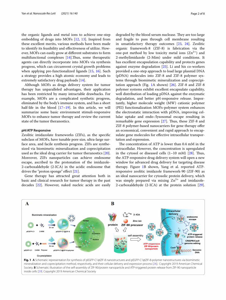
degraded by the blood serum nuclease. They are too large and fragile to pass through cell membrane resulting in unsatisfactory therapy outcomes [23, 24]. Zeolitic organic framework-8 (ZIF-8) is fabrication via the one-pot method by low toxicity metal ions (Zn2+) and 2-methylimidazole (2-Mim) under mild conditions. It has excellent encapsulation capability and protects genes against enzyme degradation [25]. Li and his co-workers provided a one-step approach to load large plasmid DNA (pDNA) molecules into ZIF-8 and ZIF-8 polymer sys-tems through biomimetic mineralization and coprecipi-tation approach (Fig. 1A shown) [26]. ZIF-8 and ZIF-8 polymer systems exhibit excellent encapsulate capability, well distribution of loading pDNA against the enzymatic degradation, and better pH-responsive release. Impor-tantly, higher molecule weight (MW) cationic polymer (PEI) functionalization MOFs-polymer system enhances the electrostatic interaction with pDNA, improving cel-lular uptake and endo-/lysosomal escape resulting in remarkable gene expression [27]. Thus, these ZIF-8 and ZIF-8 polymer-based nanocarriers for gene therapy offer an economical, convenient and rapid approach to encap-sulate gene molecules for effective intracellular transpor-tation and expression.
The concentration of ATP is lower than 0.4 mM in the extracellular. However, the concentration is upregulated in the cytosol or diseased cells (1–10 mM) [28]. Thus, the ATP-responsive drug delivery system will open a new window for advanced drug delivery for targeting disease therapy. Figure 1B shown, Yang et al. reported ATP-responsive zeolitic imidazole framework-90 (ZIF-90) as an ideal nanocarrier for cytosolic protein delivery, which was simply prepared via mixing Zn2+ and imidazole-2-carboxaldehyde (2-ICA) at the protein solution [29].
Fig. 1 A Schematic representation for synthesis of pEGFP-C1@ZIF-8 nanostructures and pEGFP-C1@ZIF-8-polymer nanostructures via biomimetic mineralization and coprecipitation method, respectively, and their cellular delivery and expression process [26]. Copyright 2019 American Chemical Society. B Schematic illustration of the self-assembly of ZIF-90/protein nanoparticle and ATP-triggered protein release from ZIF-90 nanoparticle inside cells [29]. Copyright 2019 American Chemical Society

Page 3 of 8Yan et al. Nanoscale Res Lett (2021) 16:140
At the tumor sites, as-prepared ZIF-90/protein MOFs will gradually degrade to release preload protein due to the competitive coordination between the Zn2+ and ATP that disassembles ZIF-90 and the releasing protein can effectively inhibit cancer cells growth. Thus, we can speculate that ZIP-90 MOFs can encapsulate molecular weighted protein regardless of molecular weight and pro-tein size. This includes superoxide dismutase and bovine serum albumin with minimal effects on protein function for tumor therapy.
Due to the abnormal TME, this ATP-responsive pro-tein delivery system illustrated in this section not only expands the chemistry of MOFs in biomedical applica-tions, but also opens up a new window for protein deliv-ery and genome editing techique for targeting disease therapy.
Light ResponsiveAs a "green" approach, photothermal therapy has minimal toxicity to surrounding tissues, widely applied in tumor therapy [30, 31]. High temperatures can induce severe irreversible damage to tissues when the temperature sustains over 44 °C. It is enough to cause cell membrane damage, mitochondrial dysfunction, and disruption RNA synthesis to induce cell death [32]. Unlike normal tissues that can dissipate heat and keep the temperature con-stant by blood circulation via neuromodulation, locking of autonomous regulatory function made tumor tissues a heat reservoir. This provides a huge advantage for subse-quence photothermal therapy [33].
Based on these merits mentioned above and poor heat-dissipating ability, photo-based therapy may be suit-able for tumor therapy. Photodynamic therapy (PDT) is the typical approach of photothermal therapy, which is constituted by three basic elements (near-infrared light
irradiation, plenty of oxygen, and photosensitizers) [34]. Near-infrared light irradiation (NIR light) as external stimulus exhibits high spatial and temporal control of local heating with minimal adverse side effects [35, 36]. PSs utilized surrounding oxygen to generate poisonous reactive oxygen species (ROS) to destroy cancer cells under laser irradiation [37, 38]. As shown in Fig. 2A, Park et al. designed Zr(IV)-based porphyrinic metal–organic framework (Zr-MOF) that can generate ROS under NIR light [39]. Up injection into the body, Zr-MOF can accumulate at the tumor tissues via the enhanced per-meability and retention (EPR) effects. However, the tar-geting ability was not satisfactory, which could increase unnecessary side effects [40]. Thus, Zr-MOF was further modified with folic acid, improving Zr-MOF targeting ability during blood circulation time and enhancing PDT efficacy.
With the assistance of contrast agents, this can pro-vide precise therapy navigation and determine the suit-able therapeutic time [41. As shown in Fig. 2B, Zhang and his co-workers developed Mn-porphyrin MOFs via self-assembling of Mn-tetrakis (4- carboxyphenyl) porphyrin and Zr4+ ions, which endow Mn-porphyrin MOFs with the magnetic resonance imaging (MRI) and photothermal conversion capacity without increasing tedious synthesis progress [42]. These novel MOFs can further conjugate with the type heat unstable NO donor s-nitrosothiol (SNO) [43]. Therefore, this MOFs platform can achieve the photothermal and MRI-guided NO syn-ergistic treatment. MOFs-SNO can efficiently accumu-late at the tumor areas through intravenous injection, and realize high photothermal conversion ability for PTT and control NO release for NO synergistic therapy with less photo-damage. Thus, theranostic agents integrated into the MOFs are a feasible approach for enhancing the
Fig. 2 A Illustration of PCN-224 structure. 6-connected Zr6 cluster (Zr6O4(OH)4(H2O)6(OH)6(COO)6), tetratopic linker (tetrakis (4-carboxyphenyl)porphyrin (H2TCPP)), and 3D nanoporous framework of PCN-224. (b) A cubic unit of PCN-224 and schematic illustration of spherical PCN-224 nanoparticles on the basis of construction of cubic units, yielding different sizes [39]. Copyright 2018 American Chemical Society. B Scheme for the synthesis of the NMOF–SNO nanocomposite and the NIR light-triggered NO release and PTT [42]. Copyright 2018 American Chemical Society

Page 4 of 8Yan et al. Nanoscale Res Lett (2021) 16:140
diagnosis and provide precise therapy navigation and determine the suitable therapeutic time.
Due to free porphyrin has optical properties, when porphyrin integrated into the MOFs, the obtained por-phyrin MOFs has fluorescence imaging and PDT, which will opens new opportunities for next-generation tumor theranostics.
H2O2 ResponsiveHigh levels of H2O2, hypoxia, low pH value, and high concentration glutathione (GSH) are common feature in the tumor microenvironment (TME) [44–46]. Therefore, ameliorating or changing unique TME can inhibit tumor growth and enhance therapeutic effects [47, 48]. Many literatures have reported that MnO2 has nanoenzyme activity can decompose into Mn2+ and release amount O2 under the circumstances of H2O2, which can increase oxygen concentration inside the solid tumors and genera-tion abound reactive oxygen species (ROS) under laser irradiation [49, 50]. ROS, as the intracellular chemical substrate, can modulate cell signal and play an impor-tant role in the cell cycle [51]. Important, cancer cells are more sensitive to high levels of ROS and susceptible to apoptosis [52]. As Fig. 3 shows, Sun et al. constructed bovine serum albumin-MnO2/chlorin e6@ZIF-8 (BSA-MnO2/Ce6@ZIF-8) nanosystem exhibits pH/H2O2 con-trollability for O2 production capacity, which offered a safe and efficient PDT therapy administration progress [53]. Photosensitizer chlorin e6 (Ce6) loading into the ZIF-8 can resolve the low dissolubility problem in the aqueous environment and generate ROS to induce can-cer cells apoptotic and necrotic under 650 nm laser irra-diation. Bovine serum albumin (BSA)-MnO2 decorated into the surface of Ce6@ZIF-8, the obtained BSA-MnO2/Ce6@ZIF-8 has excellent dispersibility, low toxicity, sufficient oxygen generation ability, and minimal side effects in vitro/in vivo. This well-prepared BSA-MnO2/Ce6@ZIF-8 nanosystem possesses a pH/H2O2-sensitive capacity and follows the MRI-guided PDT, which holds enormous potential for more accurate diagnosis and improvements to the antitumor effects.
GSH ResponsivePDT has achieved a distinct advantage in tumor ther-apy; a high concentration of glutathione (GSH) in can-cer cells (2–10 mM) not only resists PDT, radiotherapy, and chemotherapy, but also serves as an antioxidant to scavenge cellular ROS and severely compromises the PDT application [54, 55]. More specifically, it has been reported that excessive ROS can cause inflammation to tumor tissues and serious phototoxicity to normal tissues [56, 57]. Thus, it is urgent to develop an intelligent MOFs system, which can simultaneously achieve PSs-mediated
ROS generation and reduce the negative effects of intra-cellular GSH on the cytotoxicity of ROS at the tumor areas.
In order to meet these requirements, Wan et al. pro-vided a GSH-unlocked Mn (III)-sealed MOFs nanosys-tem to undergo a reductive disintegration by high-level GSH in tumor sites. This can control GSH depletion and ROS generation exhibited comprehensive tumor inhibi-tion by improving the therapeutic effects of PDT (Fig. 4A shown) [58]. However, the major challenge of MOFs in medical applications are their unfavorable biocompat-ibility and short blood half-life. Thus many strategies to optimize MOFs in vivo application have attracted sig-nificant attention [59]. Inspiring from circulating blood cells, biomimetic cloaking with the plasma membrane is a powerful approach to coordinate the fate of inorganic nanomaterials in vivo [60–62]. As shown in Fig. 4B, Min and his co-colleagues illustrated multifunctional biomi-metic MOFs nanoparticles with 4T1 breast cancer cell membrane camouflage for synergic anticancer therapy of PDT and antiangiogenesis [55]. Such design can keep the surface proteins inherited from the donor cells and endow 4T1 cells decorated MnO2 coated porphyrinic Zr-MOF loaded vascular endothelial growth factor recep-tor 2 MOFs (aMMTm) additional biological function to escape macrophage recognition and target tumor tissue via homotypic affinity in vivo. More importantly, MnO2 decorated into the surface of MOFs to neutralize high
Fig. 3 Schematic illustration for the Formation of a BSA-MnO2/Ce6@ZIF-8 Nanoplatform and Schematic Illustration Showing the TME Responsiveness and Generation of ROS Irradiation upon 650 nm NIR Laser for MRI-Guided Photodynamic Cancer Treatment [53]. Copyright 2019 American Chemical Society

Page 5 of 8Yan et al. Nanoscale Res Lett (2021) 16:140
intratumoral levels of GSH and H2O2 to ameliorate the unique tumor microenvironment, which can boost the PDT outcomes. When the MnO2 shell was gradually degraded, the released Mn2+ can act as an MRI contrast agent and apatinib neutralized the PDT-induced revascu-larization and prevented tumor progress. We believe that this multifunctional drug delivery system has enormous potential capacity in mechanism-based customization of antitumor therapy.
The as-fabricated biomimetic nanosystem for dual imaging-guided synergistic tumor therapy was a simple theranostic system, which would pave a new avenue for tumor diagnosis and therapy.
Hydrogen Sulfide (H2S) ResponsiveEndogenous hydrogen sulfide (H2S), as the third gas-otransmitter, is generated from the enzyme system of cystathionine β-synthase via the catalysis process [63, 64]. Cu-based MOFs have a strong binding ability of Cu2+ with S2−, and their inherent activity of Cu2+ possessed higher catalytic activity in acid [65]. In recent years, Cu-MOFs have been exploited to detect the toxic H2S gas in the serum or solution [66]. Thus, H2S can be recognized as a specific "target signal" for ovarian and colon tumor diagnosis and therapy [67]. As shown in Fig. 5, Li and his co-workers provided endogenous H2S-activated Cu-MOF is in the "OFF" state and no obvious adsorption at the NIR region. However, when Cu-MOFs entered into the colon tumor tissues where H2S was overexpressed, Cu-MOFs can change into the “ON” state by reacting
with high levels of H2S concentration to generate pho-toactive copper sulfide with stronger NIR absorption, which promoted photothermal therapy (PTT) [68]. Cu-MOFs has the mimicking-peroxidase activity and reacted with overexpressed H2O2 to produce toxical hydroxyl radical for hemodynamic therapy after endo-cytosed by the cancer cells [69]. Thus, H2S-triggering ‘turn-on’ strategy exhibits excellent antitumor outcomes and avoid unnecessary side effects in tumor therapy. This H2S-triggered nanocarrier can significant inhibit colon cancer cells grown in vivo, and this biomarker triggered therapeutic agents show enormous potential for tumor diagnosis and therapeutic.
PerspectivesMOFs as drug delivery systems for tumor therapy, show unparalleled advantages due to their intrinsic features, including structural tenability, high porosity, multifunc-tionality, and biocompatibility. Although MOFs have achieved impressive progress in the biomedical field, sev-eral key problems need to be addressed before MOFs can be permitted to clinical translation stages. These include complexed synthesis, early clearance by body immune system, system toxicity, unsatisfactory pharmacokinet-ics and biodistribution, off-target accumulation, and untimely drug release ability.
In order to solve these multileveled problems, biomi-metic cloaking with the plasma membrane is a power-ful strategy to tune the fate of MOFs in vivo. All kinds of cell membranes have been widely applied to camouflage
Fig. 4 Schematic illustration of an endocytosis Mn(III)-sealed MOF nanosystem for MRI and OI-guided PDT by controlled ROS generation and GSH depletion after being unlocked by overexpressed GSH in tumor cells [58]. Copyright 2019 American Chemical Society. B Schematic illustration of aMMTm preparation and proposed combination therapy of PDT and antiangiogenesis[55]. Copyright 2019 WILEY–VCH Verlag GmbH & Co. KGaA, Weinheim

Page 6 of 8Yan et al. Nanoscale Res Lett (2021) 16:140
MOFs. This biomimetic approach can make up MOFs with the biointerface of cell membranes, which can keep the surface proteins inherited from the donor cell, reduce their elimination from the body immune system to prolong their half-life in the blood, and enhance MOFs accumulated at the tumor tissues via permeability and retention effects. Based on these merits, cell membrane and MOFs combined biomimetic platforms to maximize the therapeutic agents to tumor tissues and effectively achieve tumor therapy.
Especially, the distorted cancer blood vessels and can-cer cells’ rapid proliferation would cause low oxygen concentration and acidification in the tumor microenvi-ronment (TME). Hypoxia, low pH, and high GSH con-centration are the common features in the TEM, which promote cancer metastasis and angiogenesis and lead to therapeutic resistance and compromise therapy out-comes. Developing environment responsive and intel-ligent MOFs triggering by tumor microenvironment is a feasible approach for the substantial elevation in precise diagnosis, and reduction in unnecessary side effects in tumor therapy.
ConclusionIn this article, we summarized various kinds of MOFs based on their unique mechanisms and structures. Com-plex design, high operating costs, and lengthy preparation steps, are obstacles MOFs encounter in real application to the clinical field. Ultimately, targeting delivery, low to none toxicity, and outstanding therapeutic effects are the critical factors for successful translating MOFs to clinical application.
AcknowledgementsNot applicable.
Authors’ contributionsCY made substantial contributions to the conception, paper collecting, and analyzing of the work, YJ drafting the work, CZ final approval of the version to be published. All authors read and approved the final manuscript.
FundingNone.
Availability of data and materialsNot applicable.
Declarations
Competing interestsThe authors declare that they have no competing interests.
Author details1 The Affiliated Huai’an Hospital of Xuzhou Medical University and The Second People’s Hospital of Huai’an, No. 62, Huaihai Road (S.), Huai’an 223002, China. 2 School of Medical Technology, Jiangsu College of Nursing, Huai’an City, Jiangsu Province, China.
Received: 1 July 2021 Accepted: 27 August 2021
References 1. Liu Y, Zhen W, Jin L, Zhang S, Sun G, Zhang T et al (2018) All-in-one
theranostic nanoagent with enhanced reactive oxygen species genera-tion and modulating tumor microenvironment ability for effective tumor eradication. ACS Nano 12:4886–4893
2. Chung H, Barron PM, Novotny RW, Son H-T, Chunhua Hu, Choe W (2009) Structural variation in porphyrin pillared homologous series: influence of distinct coordination centers for pillars on framework topology. Cryst Growth Des 9:7
3. Datta S, Rajnish KN, Samuel MS, Pugazlendhi A, Selvarajan E (2020) Metagenomic applications in microbial diversity, bioremediation, pollu-tion monitoring, enzyme and drug discovery. A review. Environ Chem Lett 18:1229–1241
Fig. 5 Schematic Illustration of the H2S-Triggered Transformation of Non-photoactive Cu-MOFs Nanoenzyme into an Near-NIR-Activatable Photothermal Agent by in Situ Sulfifidation Reaction and Its Further Synergic Photothermal and Chemodynamic Therapy for Colon Cancer [68]. Copyright 2020 American Chemical Society

Page 7 of 8Yan et al. Nanoscale Res Lett (2021) 16:140
4. Wool RP (2008) Self-healing materials: a review. Soft Matter 4:400–418 5. Samuel MS, Savunthari KV, Ethiraj S (2021) Synthesis of a copper (II)
metal-organic framework for photocatalytic degradation of rhodamine B dye in water. Environ Sci Pollut Res Int 28:40835–40843
6. Samuel MS, Selvarajan E, Chidambaram R, Patel H, Brindhadevi K (2021) Clean approach for chromium removal in aqueous environments and role of nanomaterials in bioremediation: Present research and future perspective. Chemosphere 284:131368
7. Samuela MS, Bhattacharyaa J, Parthibanb C, Viswanathanc G, Pradeep Singh ND (2018) Ultrasound-assisted synthesis of metal organic frame-work for the photocatalytic reduction of 4-nitrophenol under direct sunlight. Ultrasonics-Sonochemistry 49:215–221
8. Deng J, Wang K, Wang M, Ping Yu, Mao L (2017) Mitochondria targeted nanoscale zeolitic imidazole framework-90 (ZIF-90) for ATP imaging in live cells. J Am Chem Soc 139:5877–5882
9. Ruoyu Xu, Wang Y, Duan X, Kuangda Lu, Micheroni D, Aiguo Hu, Lin W (2016) Nanoscale metal-organic frameworks for ratiometric oxygen sens-ing in live cells. J Am Chem Soc 138:2158–2161
10. Samuel MS, Kirankumar VS, Selvarajan E (2021) Fabrication of a metal-organic framework composite for removal of Aflatoxin B1 from water. J Environ Chem Eng 9:104966
11. Li J-R, Yu J, Lu W, Sun L-B, Sculley J, Balbuena PB, Zhou H-C (2013) Porous materials with pre-designed single-molecule traps for CO2 selective adsorption. Nat Commun 4:1538–1546
12. Doonan C, Ricco R, Liang K, Bradshaw D, Falcaro P (2017) Metal−organic frameworks at the biointerface: synthetic strategies and applications. Acc Chem Res 50:1423–1432
13. Liang K, Carbonell C, Styles MJ, Ricco R, Cui J, Richardson JJ et al (2015) Biomimetic replication of microscopic metal-organic framework patterns using printed protein patterns. Adv Mater 27:7293–7298
14. Shieh F-K, Wang S-C, Yen C-I, Chang-Cheng Wu, Dutta S, Chou L-Y et al (2015) Imparting functionality to biocatalysts via embedding enzymes into nanoporous materials by a de novo approach: size-selective shelter-ing of catalase in metal−organic framework microcrystals. J Am Chem Soc 137:4276–4279
15. XizhenLian Yu, Fang EJ, Wang Qi, Li J, Banerjee S et al (2017) Enzyme–MOF (metal–organic framework) composites. Chem Soc Rev 46:3386–3401
16. Katsoulidis AP, Park KS, Antypov D, Mart-Gastaldo C, Miller GJ, War-ren JE et al (2014) Guest-adaptable and water-stable peptide-based porous materials by imidazolate side chain control. Angew Chem Int Ed 53:193–198
17 Mantion A, Massüger L, Rabu P, Palivan C, McCusker LB, Taubert A (2008) Metal-peptide frameworks (MPFs): “Bioinspired” metal organic frame-works. J Am Chem Soc 130:2517–2526
18. Zhuang J, Gong H, Zhou J, Zhang Q, Gao W, Fang RH et al (2020) Targeted gene silencing in vivo by platelet membrane–coated metal-organic framework nanoparticles. Sci Adv 6:6108–6019
19. Wang S, Mao J, Liu H, Huang S, Cai J, Gui W et al (2020) pH-Sensitive nan-otheranostics for dual-modality imaging guided nanoenzyme catalysis therapy and phototherapy. J Mater Chem B 8:4859–4869
20 Zhu P, Chen Y, Shi J (2018) Nanoenzyme-augmented cancer sonody-namic therapy by catalytic tumor oxygenation. ACS Nano 12:3780–3795
21 Kaneti YV, Dutta S, Hossain MSA, Shiddiky MJA, Tung K-L, Shieh F-K et al (2017) Strategies for improving the functionality of zeolitic imidazolate frameworks: tailoring nanoarchitectures for functional applications. Adv Mater 29:1700213–1700244
22. Nel AE, Mädler L, Velegol D, Xia T, Hoek EMV, Somasundaran P et al (2009) understanding biophysicochemical interactions at the nano–biointer-face. Nat Mater 8:543–557
23. Shi B, Zheng M, Tao W, Chung R, Jin D, Ghaffari D et al (2017) Challenges in DNA delivery and recent advances in multifunctional polymeric DNA delivery systems. Biomacromol 18:2231–2246
24. Keles E, Song Y, Dan Du, Dong W-J, Lin Y (2016) Recent progress in nano-materials for gene delivery applications. Biomater Sci 4:1291–1309
25. Zheng H, Zhang Y, Liu L, Wan W, Guo P, Nystrom AM et al (2016) One-pot synthesis of metal−organic frameworks with encapsulated target mol-ecules and their applications for controlled drug delivery. J Am Chem Soc 138:962–968
26. Li Y, Zhang K, Liu P, Chen Mo, Zhong Y, Ye Q et al (2019) Encapsulation of plasmid DNA by nanoscale metal-organic frameworks for efficient gene transportation and expression. Adv Mater 31:1901570–1901579
27 Zakeri A, Kouhbanani MAJ, Beheshtkhoo N, Beigi V, Mousavi SM, Hashemi SAR et al (2018) Polyethylenimine-based nanocarriers in co-delivery of drug and gene: a developing horizon. Nano Rev Exp 9:1488497–1488511
28. Mo R, Jiang T, DiSanto R, Tai W, Zhen Gu (2014) ATP-triggered anticancer drug delivery. Nat Commun 5:3364–3374
29. Yang X, Tang Q, Jiang Y, Zhang M, Wang M, Mao L (2019) Nanoscale ATP-responsive zeolitic imidazole framework-90 as a general platform for cytosolic protein delivery and genome editing. J Am Chem Soc 141:3782–3786
30. Kuangda Lu, He C, Lin W (2014) Nanoscale metal−organic framework for highly effective photodynamic therapy of resistant head and neck cancer. J Am Chem Soc 136:16712–16715
31. Jingjing Liu Yu, Yang WZ, Yi X, Dong Z, Xiaona Xu et al (2016) Nanoscale metalorganic frameworks for combined photodynamic & radiation therapy in cancer treatment. Biomaterials 97:1–9
32 Yarmolenko PS, Moon EJ, Landon C, Manzoor A, Hochman DW, Viglianti BL, Dewhirst MW (2011) Thresholds for thermal damage to normal tis-sues: an update. Int J Hyperthermia 27:320–343
33. Nikfarjam M, Muralidharan V, Christophi C (2005) Mechanisms of focal heat destruction of liver tumors. J Surg Res 127:208–223
34. Wan X, Zhang G, Liu S (2011) pH-disintegrable polyelectrolyte multilayer-coated mesoporous silica nanoparticles exhibiting triggered co-release of cisplatin and model drug molecules. Macromol Rapid Commun 32(14):1082–1089
35. Chen Q, Ligeng Xu, Liang C, Wang C, Peng R, Liu Z (2016) Photothermal therapy with immune-adjuvant nanoparticles together with check-point blockade for effective cancer immunotherapy. Nat Commun 7:13193–13206
36 Lin H, Gao S, Dai C, Chen Y, Shi J (2017) Correction to “A two-dimen-sional biodegradable niobium carbide (MXene) for photothermal Tumor ERADICATION in NIR-I and NIR-II biowindows.” J Am Chem Soc 139:16235–16247
37. Schulze A, Harris AL (2012) How cancer metabolism is tuned for prolifera-tion and vulnerable to disruption. Nature 491:364–373
38. Liu Y, Liu Y, Wenbo Bu, Cheng C, Zuo C, Xiao Q et al (2015) Hypoxia induced by upconversion-based photodynamic therapy: towards highly effective synergistic bioreductive therapy in tumors. Angew Chem Int Ed 54:8105–8109
39. Park J, Jiang Q, Feng D, Mao L, Zhou H-C (2016) Size-controlled synthesis of porphyrinic metal−organic framework and functionalization for targeted photodynamic therapy. J Am Chem Soc 138:3518–3525
40. Ming-Xue Wu, Yang Y-W (2017) Metal-organic framework (MOF)-based drug/cargo delivery and cancer therapy. Adv Mater 29:1606134–1606154
41. Wang Z, Deng X, Ding J, Zhou W, Zheng X, Tang G (2018) Mechanisms of drug release in pH-sensitive micelles for tumour targeted drug delivery system: A review. Int J Pharm 535:253–260
42. Zhang H, Tian X-T, Shang Y, Li Y-H, Yin X-B (2018) Theranostic Mn-porphy-rin metal−organic frameworks for magnetic resonance imaging-guided nitric oxide and photothermal synergistic therapy. ACS Appl Mater Interfaces 10:28390–28398
43. Riccio DA, Nugent JL, Schoenfisch MH (2011) Stöber synthesis of nitric oxide-releasing S-nitrosothiolmodified silica particles. Chem Mater 23:1727–1735
44. Teng Z, Wang C, Tang Y, Li W, Bao L, Zhang X et al (2018) Deformable hollow periodic mesoporous organosilica nanocapsules for significantly improved cellular uptake. J Am Chem Soc 140:1385–1393
45 Gao S, Lin H, Zhang H, Yao H, Chen Y, Shi J (2019) Nanocatalytic tumor therapy by biomimetic dual inorganic nanozyme-catalyzed cascade reac-tion. Adv Sci 6:1801733–1801745
46. Wang L, Huo M, Chen Y, Shi J (2018) Tumor microenvironment-enabled nanotherapy. Adv Healthcare Mater 7:1701156–1701179
47 Bai J, Jia X, Zhen W, Cheng W, Jiang X (2018) A facile ion-doping strategy to regulate tumor microenvironments for enhanced multimodal tumor theranostics. J Am Chem Soc 140:106–109
48 Cao Z, Zhang L, Liang K, Cheong S, Boyer C, Gooding JJ et al (2018) Bio-degradable 2D Fe–Al hydroxide for nanocatalytic tumor-dynamic therapy with tumor specificity. Adv Sci 5:1801155–1801165
49. He Z, Xiao Y, Zhang J-R, Zhang P, Zhu J-J (2018) In situ formation of large pore silica–MnO2 nanocomposites with H+ /H2O2 sensitivity for O2-elevated photodynamic therapy and potential MR imaging. Chem Commun 54:2962–2965

Page 8 of 8Yan et al. Nanoscale Res Lett (2021) 16:140
50. Yang G, Ligeng Xu, Chao Yu, Jun Xu, Sun X, Yifan Wu et al (2017) Hollow MnO2 as a tumor-microenvironmentresponsive biodegradable nano-platform for combination therapy favoring antitumor immune responses. Nat Commun 8:902–915
51. Zhou Z, Song J, Nie L, Chen X (2016) Reactive oxygen species generating systems meeting challenges of photodynamic cancer therapy. Chem Soc Rev 45:6597–6626
52. Cheng D-B, Zhang X-H, Gao Y-J, Ji L, Hou D, Wang Z, Wanhai Xu et al (2019) Endogenous reactive oxygen species-triggered morphology transformation for enhanced cooperative interaction with mitochondria. J Am Chem Soc 141:7235–7239
53. Sun Q, Bi H, Wang Z, Li C, Wang C, Jiating Xu et al (2019) O2-generating metal−organic framework-based hydrophobic photosensitizer delivery system for enhanced photodynamic therapy. ACS Appl Mater Interfaces 11:36347–36358
54. Diehn M, Cho RW, Lobo NA, Kalisky T, Dorie MJ, Kulp AN et al (2006) Asso-ciation of reactive oxygen species levels and radioresistance in cancer stem cells. Nat Rev Cancer 6:535–545
55. Min H, Wang J, Qi Y, Zhang Y, Han X, Ying Xu et al (2019) Biomimetic metal-organic framework nanoparticles for cooperative combination of antiangiogenesis and photodynamic therapy for enhanced efficacy. Adv Mater 31:1808200–1808211
56. Castano AP, Mroz P, Hamblin MR (2006) Photodynamic therapy and anti-tumour immunity. Nat Rev Cancer 6:535–545
57. Zeng J-Y, Zou M-Z, Zhang M, Wang X-S, Zeng X, Cong H et al (2018) π-Extended benzoporphyrin-based metal−organic framework for inhibi-tion of tumor metastasis. ACS Nano 12:4630–4640
58. Wan S-S, Cheng Q, Zeng X, Zhang X-Z (2019) A Mn(III)-sealed metal−organic framework nanosystem for redox-unlocked tumor theranostics. ACS Nano 13:6561–6571
59. Wuttke S, Zimpel A, Bein T, Braig S, Stoiber K, Vollmar A et al (2017) Validating metal-organic framework nanoparticles for their nanosafety in diverse biomedical applications. Adv Healthcare Mater 6:1600818–1600829
60. Sun H, Jinghan Su, Meng Q, Yin Qi, Chen L, Wangwen Gu et al (2016) Cancer-cell-biomimetic nanoparticles for targeted therapy of homotypic tumors. Adv Mater 28:9581–9588
61. Ding H, Lv Y, Ni D, Wang J, Tian Z, Wei W et al (2015) Erythrocyte mem-brane-coated NIR-triggered biomimetic nanovectors with programmed delivery for photodynamic therapy of cancer. Nanoscale 7:9806–9815
62. Zhang W, Zi-Li Yu, Min Wu, Ren J-G, Xia H-F, Sa G-L et al (2017) Magnetic and folate functionalization enables rapid isolation and enhanced tumor-targeting of cell-derived microvesicles. ACS Nano 11:277–290
63. Szabo C, Coletta C, Chao C, Módis K, Szczesny B, Papapetropoulos A et al (2013) Tumor-derived hydrogen sulfide, produced by cystathionine-β-synthase, stimulates bioenergetics, cell proliferation, and angiogenesis in colon cancer. Proc Natl Acad Sci USA 110:12474–12479
64. Dong H, Zhou Qi, Zhang L, Tian Y (2019) Rational design of specific recognition molecules for simultaneously monitoring of endogenous polysulfide and hydrogen sulfide in the mouse brain. Angew Chem Int Ed Engl 58:13948–13953
65. Ming Xu, Yuan S, Chen X-Y, Chang Y-J, Day G, Zhi-Yuan Gu et al (2017) Two-dimensional metal-organic framework nanosheets as an enzyme inhibitor: modulation of the α-chymotrypsin activity. J Am Chem Soc 139:8312–8319
66. Zhang X, Quan Hu, Xia T, Jun Zhang Yu, Yang YC et al (2016) Turn-on and ratiometric luminescent sensing of hydrogen sulfide based on metal-organic frameworks. ACS Appl Mater Interfaces 8:32259–32265
67 Chen W, Ni D, Rosenkrans ZT, Cao T, Cai W (2019) Smart H2S-triggered/therapeutic system (SHTS)-based nanomedicine. Adv Sci 6:1901724–1901751
68. Li Y, Zhou J, Wang L, Xie Z (2020) Endogenous hydrogen sulfide-triggered MOF-based nanoenzyme for synergic cancer therapy. ACS Appl Mater Interfaces 12:30213–30220
69. Xiao J, Zhu Y, Huddleston S, Li P, Xiao B, Farha OK et al (2018) Copper metal-organic framework nanoparticles stabilized with folic acid improve wound healing in diabetes. ACS Nano 12:1023–1032
Publisher’s NoteSpringer Nature remains neutral with regard to jurisdictional claims in pub-lished maps and institutional affiliations.