Enhancement of Human Prolactin Synthesis by Sodium Butyrate ...
11
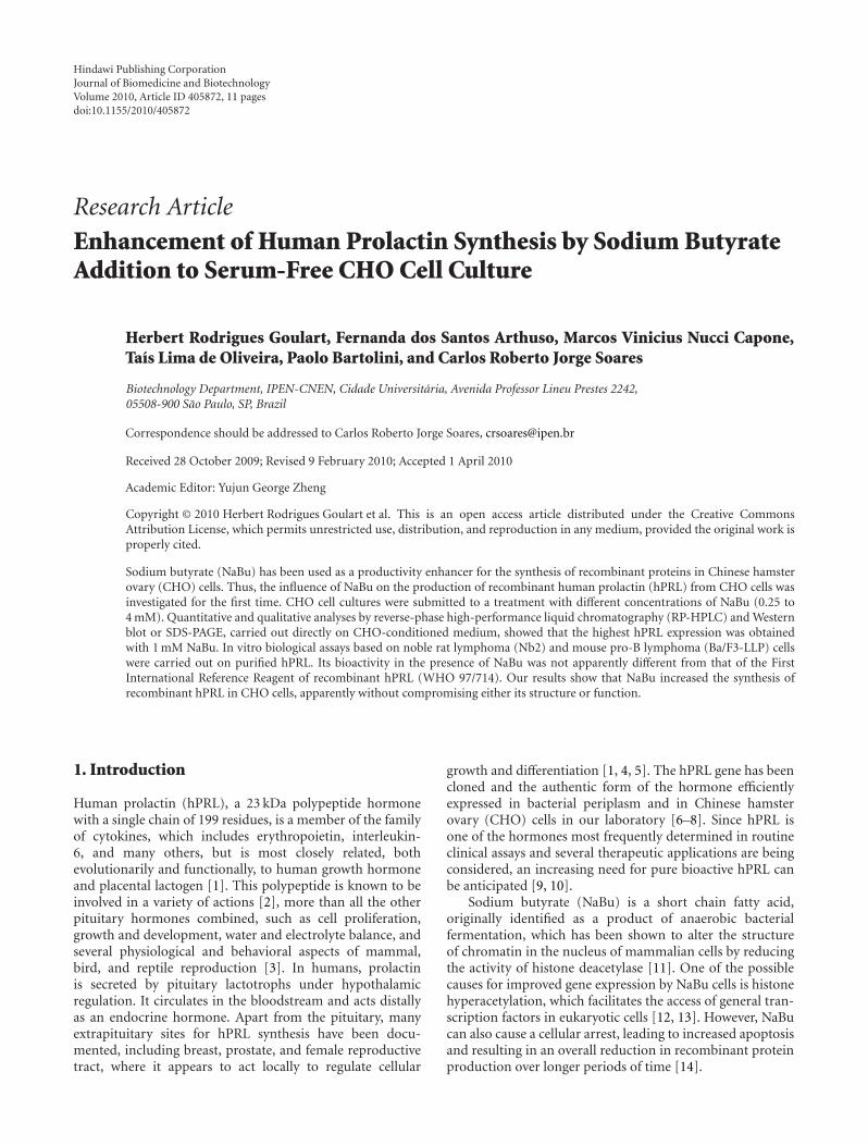
Hindawi Publishing Corporation Journal of Biomedicine and Biotechnology Volume 2010, Article ID 405872, 11 pages doi:10.1155/2010/405872 Research Article Enhancement of Human Prolactin Synthesis by Sodium Butyrate Addition to Serum-Free CHO Cell Culture Herbert Rodrigues Goulart, Fernanda dos Santos Arthuso, Marcos Vinicius Nucci Capone, Ta´ ıs Lima de Oliveira, Paolo Bartolini, and Carlos Roberto Jorge Soares Biotechnology Department, IPEN-CNEN, Cidade Universit´ aria, Avenida Professor Lineu Prestes 2242, 05508-900 S˜ ao Paulo, SP, Brazil Correspondence should be addressed to Carlos Roberto Jorge Soares, [email protected] Received 28 October 2009; Revised 9 February 2010; Accepted 1 April 2010 Academic Editor: Yujun George Zheng Copyright © 2010 Herbert Rodrigues Goulart et al. This is an open access article distributed under the Creative Commons Attribution License, which permits unrestricted use, distribution, and reproduction in any medium, provided the original work is properly cited. Sodium butyrate (NaBu) has been used as a productivity enhancer for the synthesis of recombinant proteins in Chinese hamster ovary (CHO) cells. Thus, the influence of NaBu on the production of recombinant human prolactin (hPRL) from CHO cells was investigated for the first time. CHO cell cultures were submitted to a treatment with different concentrations of NaBu (0.25 to 4 mM). Quantitative and qualitative analyses by reverse-phase high-performance liquid chromatography (RP-HPLC) and Western blot or SDS-PAGE, carried out directly on CHO-conditioned medium, showed that the highest hPRL expression was obtained with 1 mM NaBu. In vitro biological assays based on noble rat lymphoma (Nb2) and mouse pro-B lymphoma (Ba/F3-LLP) cells were carried out on purified hPRL. Its bioactivity in the presence of NaBu was not apparently different from that of the First International Reference Reagent of recombinant hPRL (WHO 97/714). Our results show that NaBu increased the synthesis of recombinant hPRL in CHO cells, apparently without compromising either its structure or function. 1. Introduction Human prolactin (hPRL), a 23 kDa polypeptide hormone with a single chain of 199 residues, is a member of the family of cytokines, which includes erythropoietin, interleukin- 6, and many others, but is most closely related, both evolutionarily and functionally, to human growth hormone and placental lactogen [1]. This polypeptide is known to be involved in a variety of actions [2], more than all the other pituitary hormones combined, such as cell proliferation, growth and development, water and electrolyte balance, and several physiological and behavioral aspects of mammal, bird, and reptile reproduction [3]. In humans, prolactin is secreted by pituitary lactotrophs under hypothalamic regulation. It circulates in the bloodstream and acts distally as an endocrine hormone. Apart from the pituitary, many extrapituitary sites for hPRL synthesis have been docu- mented, including breast, prostate, and female reproductive tract, where it appears to act locally to regulate cellular growth and differentiation [1, 4, 5]. The hPRL gene has been cloned and the authentic form of the hormone efficiently expressed in bacterial periplasm and in Chinese hamster ovary (CHO) cells in our laboratory [6–8]. Since hPRL is one of the hormones most frequently determined in routine clinical assays and several therapeutic applications are being considered, an increasing need for pure bioactive hPRL can be anticipated [9, 10]. Sodium butyrate (NaBu) is a short chain fatty acid, originally identified as a product of anaerobic bacterial fermentation, which has been shown to alter the structure of chromatin in the nucleus of mammalian cells by reducing the activity of histone deacetylase [11]. One of the possible causes for improved gene expression by NaBu cells is histone hyperacetylation, which facilitates the access of general tran- scription factors in eukaryotic cells [12, 13]. However, NaBu can also cause a cellular arrest, leading to increased apoptosis and resulting in an overall reduction in recombinant protein production over longer periods of time [14].
-
Upload
vuongquynh -
Category
Documents
-
view
215 -
download
0
Transcript of Enhancement of Human Prolactin Synthesis by Sodium Butyrate ...
Hindawi Publishing CorporationJournal of Biomedicine and BiotechnologyVolume 2010, Article ID 405872, 11 pagesdoi:10.1155/2010/405872
Research Article
Enhancement of Human Prolactin Synthesis by Sodium ButyrateAddition to Serum-Free CHO Cell Culture
Herbert Rodrigues Goulart, Fernanda dos Santos Arthuso, Marcos Vinicius Nucci Capone,Taıs Lima de Oliveira, Paolo Bartolini, and Carlos Roberto Jorge Soares
Biotechnology Department, IPEN-CNEN, Cidade Universitaria, Avenida Professor Lineu Prestes 2242,05508-900 Sao Paulo, SP, Brazil
Correspondence should be addressed to Carlos Roberto Jorge Soares, [email protected]
Received 28 October 2009; Revised 9 February 2010; Accepted 1 April 2010
Academic Editor: Yujun George Zheng
Copyright © 2010 Herbert Rodrigues Goulart et al. This is an open access article distributed under the Creative CommonsAttribution License, which permits unrestricted use, distribution, and reproduction in any medium, provided the original work isproperly cited.
Sodium butyrate (NaBu) has been used as a productivity enhancer for the synthesis of recombinant proteins in Chinese hamsterovary (CHO) cells. Thus, the influence of NaBu on the production of recombinant human prolactin (hPRL) from CHO cells wasinvestigated for the first time. CHO cell cultures were submitted to a treatment with different concentrations of NaBu (0.25 to4 mM). Quantitative and qualitative analyses by reverse-phase high-performance liquid chromatography (RP-HPLC) and Westernblot or SDS-PAGE, carried out directly on CHO-conditioned medium, showed that the highest hPRL expression was obtainedwith 1 mM NaBu. In vitro biological assays based on noble rat lymphoma (Nb2) and mouse pro-B lymphoma (Ba/F3-LLP) cellswere carried out on purified hPRL. Its bioactivity in the presence of NaBu was not apparently different from that of the FirstInternational Reference Reagent of recombinant hPRL (WHO 97/714). Our results show that NaBu increased the synthesis ofrecombinant hPRL in CHO cells, apparently without compromising either its structure or function.
1. Introduction
Human prolactin (hPRL), a 23 kDa polypeptide hormonewith a single chain of 199 residues, is a member of the familyof cytokines, which includes erythropoietin, interleukin-6, and many others, but is most closely related, bothevolutionarily and functionally, to human growth hormoneand placental lactogen [1]. This polypeptide is known to beinvolved in a variety of actions [2], more than all the otherpituitary hormones combined, such as cell proliferation,growth and development, water and electrolyte balance, andseveral physiological and behavioral aspects of mammal,bird, and reptile reproduction [3]. In humans, prolactinis secreted by pituitary lactotrophs under hypothalamicregulation. It circulates in the bloodstream and acts distallyas an endocrine hormone. Apart from the pituitary, manyextrapituitary sites for hPRL synthesis have been docu-mented, including breast, prostate, and female reproductivetract, where it appears to act locally to regulate cellular
growth and differentiation [1, 4, 5]. The hPRL gene has beencloned and the authentic form of the hormone efficientlyexpressed in bacterial periplasm and in Chinese hamsterovary (CHO) cells in our laboratory [6–8]. Since hPRL isone of the hormones most frequently determined in routineclinical assays and several therapeutic applications are beingconsidered, an increasing need for pure bioactive hPRL canbe anticipated [9, 10].
Sodium butyrate (NaBu) is a short chain fatty acid,originally identified as a product of anaerobic bacterialfermentation, which has been shown to alter the structureof chromatin in the nucleus of mammalian cells by reducingthe activity of histone deacetylase [11]. One of the possiblecauses for improved gene expression by NaBu cells is histonehyperacetylation, which facilitates the access of general tran-scription factors in eukaryotic cells [12, 13]. However, NaBucan also cause a cellular arrest, leading to increased apoptosisand resulting in an overall reduction in recombinant proteinproduction over longer periods of time [14].

2 Journal of Biomedicine and Biotechnology
NaBu
0
2
4
6
8×10⁵
Cel
lden
sity
(cel
ls/m
L)
0 1 2 3 4 5 6 7 8 9 10Culture time (days)
0 mM0.25 mM0.5 mM
1 mM2 mM4 mM
(a)
0
20
40
60
80
100
Via
bilit
y(%
)
0 1 2 3 4
NaBu
5 6 7 8 9 10Culture time (days)
0 mM0.25 mM0.5 mM
1 mM2 mM4 mM
(b)
Figure 1: NaBu influence on cellular growth and viability of hPRL-secreting CHO cells. (a) Cellular growth and (b) cellular viability in a 10-day hPRL production period under different NaBu concentrations. The cell viability was determined by the trypan blue exclusion procedure,following trypsinization. Values are the mean of two independent determinations.
NaBu treatment has been shown, via immunoassaydetermination, to increase the expression levels of foreignproteins such as human thrombopoietin, interferon-β-1a,and chimeric IgG3 antibodies in CHO cell cultures [15–17]by factors of 2-, 2.5-, and 3.6-fold, respectively, relative tonontreated cells. This led us to investigate, for the first time,via physicochemical techniques, whether addition of NaBuwould also increase hPRL production in these cell cultures. Inorder to characterize the product obtained, we took advan-tage of a specific RP-HPLC methodology previously devel-oped in our laboratory [18], which permits qualitative andquantitative analysis of hPRL directly in CHO-conditionedmedium. High-Performance Size-Exclusion Chromatogra-phy (HPSEC) methodology was used for both analytical andpreparative purposes [19] and also allowed us to evaluatedhPRL activity directly in two bioassays, after its purificationfrom CHO-conditioned medium [9].
2. Materials and Methods
2.1. Cell Culture. The clone expressing hPRL, derived fromCHO dhfr− cells (DUKX-B11) that had been transfectedwith the vector pEDdc-hPRL, was obtained in our laboratory[8]. Cells were thawed into T-flasks (75 cm2, from CorningCostar Corporation, Cambridge, MA) containing α-minimalessential medium (α-MEM, Gibco-Invitrogen Corporation,Grand Island, NY, USA) with L-glutamine and withoutribonucleosides and deoxyribonucleosides, supplementedwith 10% dialysed fetal bovine serum (dFBS), 40 μg/mL gen-tamycin, 4 g/L glucose, and 100 nM methotrexate. Cultureswere maintained in a humidified incubator at 37◦C in thepresence of 5% CO2.
NaBu (mM)
M 0 0.25 0.5 1 2 4
Figure 2: Electrophoretic patterns of DNA isolated from NaBu-treated and untreated CHO cells. Isolated chromosomal DNA(2 μg) was electrophoresed on 5% polyacrylamide gel and intactand fragmented DNA bands were revealed by silver staining.Arrowheads designate DNA ladder formation. M: molecular weightmarker, 100-bp DNA ladder. DNAs were isolated from day 8cultures exposed to different concentrations of Nabu.
2.2. NaBu Treatment. 100 mM NaBu was prepared inphosphate-buffered saline (PBS) (0.007 M Na2HPO4, 0.01 MNaH2PO4, pH 7.4 and 0.15 M NaCl), sterilized by passing
Journal of Biomedicine and Biotechnology 3
hPRL (IR)
hPRL (1 mM NaBu)
hPRL (0 mM NaBu)
0
25
50
75
100
mA
bs
0 5 10 15 20 25 30 35 40(min)
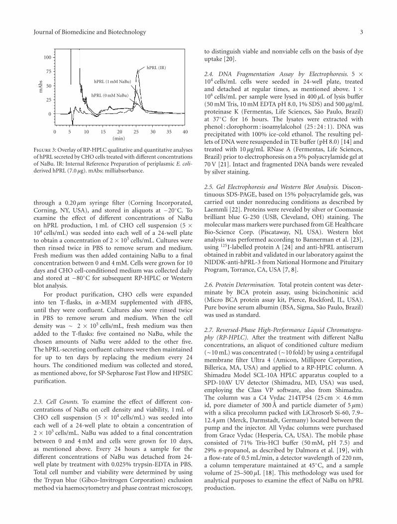
Figure 3: Overlay of RP-HPLC qualitative and quantitative analysesof hPRL secreted by CHO cells treated with different concentrationsof NaBu. IR: Internal Reference Preparation of periplasmic E. coli-derived hPRL (7.0 μg). mAbs: milliabsorbance.
through a 0.20 μm syringe filter (Corning Incorporated,Corning, NY, USA), and stored in aliquots at −20◦C. Toexamine the effect of different concentrations of NaBuon hPRL production, 1 mL of CHO cell suspension (5 ×10⁴ cells/mL) was seeded into each well of a 24-well plateto obtain a concentration of 2 × 10⁵ cells/mL. Cultures werethen rinsed twice in PBS to remove serum and medium.Fresh medium was then added containing NaBu to a finalconcentration between 0 and 4 mM. Cells were grown for 10days and CHO cell-conditioned medium was collected dailyand stored at −80◦C for subsequent RP-HPLC or Westernblot analysis.
For product purification, CHO cells were expandedinto ten T-flasks, in α-MEM supplemented with dFBS,until they were confluent. Cultures also were rinsed twicein PBS to remove serum and medium. When the celldensity was ∼ 2 × 10⁵ cells/mL, fresh medium was thenadded to the T-flasks: five contained no NaBu, while thechosen amounts of NaBu were added to the other five.The hPRL-secreting confluent cultures were then maintainedfor up to ten days by replacing the medium every 24hours. The conditioned medium was collected and stored,as mentioned above, for SP-Sepharose Fast Flow and HPSECpurification.
2.3. Cell Counts. To examine the effect of different con-centrations of NaBu on cell density and viability, 1 mL ofCHO cell suspension (5 × 10⁴ cells/mL) was seeded intoeach well of a 24-well plate to obtain a concentration of2 × 105 cells/mL. NaBu was added to a final concentrationbetween 0 and 4 mM and cells were grown for 10 days,as mentioned above. Every 24 hours a sample for thedifferent concentrations of NaBu was detached from 24-well plate by treatment with 0.025% trypsin-EDTA in PBS.Total cell number and viability were determined by usingthe Trypan blue (Gibco-Invitrogen Corporation) exclusionmethod via haemocytometry and phase contrast microscopy,
to distinguish viable and nonviable cells on the basis of dyeuptake [20].
2.4. DNA Fragmentation Assay by Electrophoresis. 5 ×10⁴ cells/mL cells were seeded in 24-well plate, treatedand detached at regular times, as mentioned above. 1 ×10⁶ cells/mL per sample were lysed in 400 μL of lysis buffer(50 mM Tris, 10 mM EDTA pH 8.0, 1% SDS) and 500 μg/mLproteinase K (Fermentas, Life Sciences, Sao Paulo, Brazil)at 37◦C for 16 hours. The lysates were extracted withphenol : clorophorm : isoamylalcohol (25 : 24 : 1). DNA wasprecipitated with 100% ice-cold ethanol. The resulting pel-lets of DNA were resuspended in TE buffer (pH 8.0) [14] andtreated with 10 μg/mL RNase A (Fermentas, Life Sciences,Brazil) prior to electrophoresis on a 5% polyacrylamide gel at70 V [21]. Intact and fragmented DNA bands were revealedby silver staining.
2.5. Gel Electrophoresis and Western Blot Analysis. Discon-tinuous SDS-PAGE, based on 15% polyacrylamide gels, wascarried out under nonreducing conditions as described byLaemmli [22]. Proteins were revealed by silver or Coomassiebrilliant blue G-250 (USB, Cleveland, OH) staining. Themolecular mass markers were purchased from GE HealthcareBio-Science Corp. (Piscataway, NJ, USA). Western blotanalysis was performed according to Bannerman et al. [23],using 125I-labelled protein A [24] and anti-hPRL antiserumobtained in rabbit and validated in our laboratory against theNIDDK-anti-hPRL-3 from National Hormone and PituitaryProgram, Torrance, CA, USA [7, 8].
2.6. Protein Determination. Total protein content was deter-minate by BCA protein assay, using bicinchoninic acid(Micro BCA protein assay kit, Pierce, Rockford, IL, USA).Pure bovine serum albumin (BSA, Sigma, Sao Paulo, Brazil)was used as standard.
2.7. Reversed-Phase High-Performance Liquid Chromatogra-phy (RP-HPLC). After the treatment with different NaBuconcentrations, an aliquot of conditioned culture medium(∼10 mL) was concentrated (∼10 fold) by using a centrifugalmembrane filter Ultra 4 (Amicon, Millipore Corporation,Billerica, MA, USA) and applied to a RP-HPLC column. AShimadzu Model SCL-10A HPLC apparatus coupled to aSPD-10AV UV detector (Shimadzu, MD, USA) was used,employing the Class VP software, also from Shimadzu.The column was a C4 Vydac 214TP54 (25 cm × 4.6 mmid, pore diameter of 300 A and particle diameter of 5 μm)with a silica precolumn packed with LiChrosorb Si-60, 7.9–12.4 μm (Merck, Darmstadt, Germany) located between thepump and the injector. All Vydac columns were purchasedfrom Grace Vydac (Hesperia, CA, USA). The mobile phaseconsisted of 71% Tris-HCl buffer (50 mM, pH 7.5) and29% n-propanol, as described by Dalmora et al. [19], witha flow-rate of 0.5 mL/min, a detector wavelength of 220 nm,a column temperature maintained at 45◦C, and a samplevolume of 25–500 μL [18]. This methodology was used foranalytical purposes to examine the effect of NaBu on hPRLproduction.
4 Journal of Biomedicine and Biotechnology
NaBu (mM)
IR 0 0.25 0.5 1 2 4
2325
(kDa)
(a)
NaBu (mM)
IR 0 0.25 0.5 1 2 4
23
25
(kDa)
(b)
NaBu (mM)
IR 0 0.25 0.5 1 2 4
2325
(kDa)
(c)
NaBu (mM)
IR 0 0.25 0.5 1 2 4
23
25
(kDa)
(d)
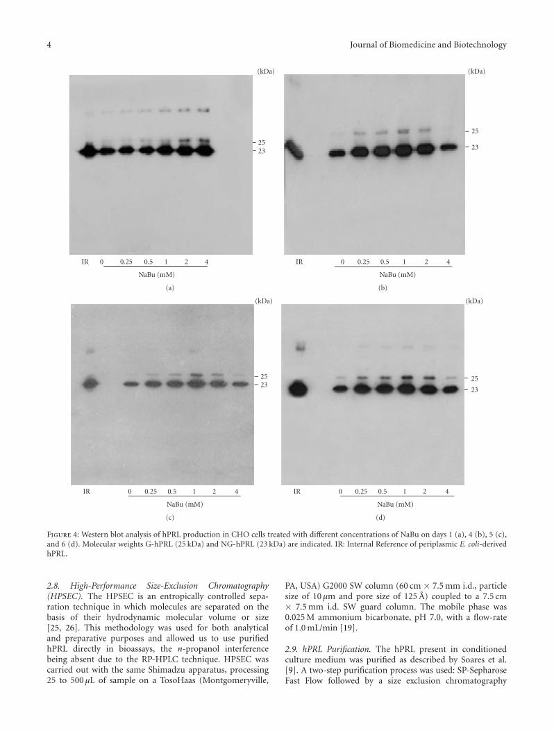
Figure 4: Western blot analysis of hPRL production in CHO cells treated with different concentrations of NaBu on days 1 (a), 4 (b), 5 (c),and 6 (d). Molecular weights G-hPRL (25 kDa) and NG-hPRL (23 kDa) are indicated. IR: Internal Reference of periplasmic E. coli-derivedhPRL.
2.8. High-Performance Size-Exclusion Chromatography(HPSEC). The HPSEC is an entropically controlled sepa-ration technique in which molecules are separated on thebasis of their hydrodynamic molecular volume or size[25, 26]. This methodology was used for both analyticaland preparative purposes and allowed us to use purifiedhPRL directly in bioassays, the n-propanol interferencebeing absent due to the RP-HPLC technique. HPSEC wascarried out with the same Shimadzu apparatus, processing25 to 500 μL of sample on a TosoHaas (Montgomeryville,
PA, USA) G2000 SW column (60 cm × 7.5 mm i.d., particlesize of 10 μm and pore size of 125 A) coupled to a 7.5 cm× 7.5 mm i.d. SW guard column. The mobile phase was0.025 M ammonium bicarbonate, pH 7.0, with a flow-rateof 1.0 mL/min [19].
2.9. hPRL Purification. The hPRL present in conditionedculture medium was purified as described by Soares et al.[9]. A two-step purification process was used: SP-SepharoseFast Flow followed by a size exclusion chromatography
Journal of Biomedicine and Biotechnology 5
NaBu (mM)
IR
NG
-hP
RL
0 0.25 0.5 1 2 4
23
(kDa)
(a)
NaBu (mM)
IR
NG
-hP
RL
Mr0 0.25 0.5 1 2 4
14
20
304566
97
(kDa)
(b)
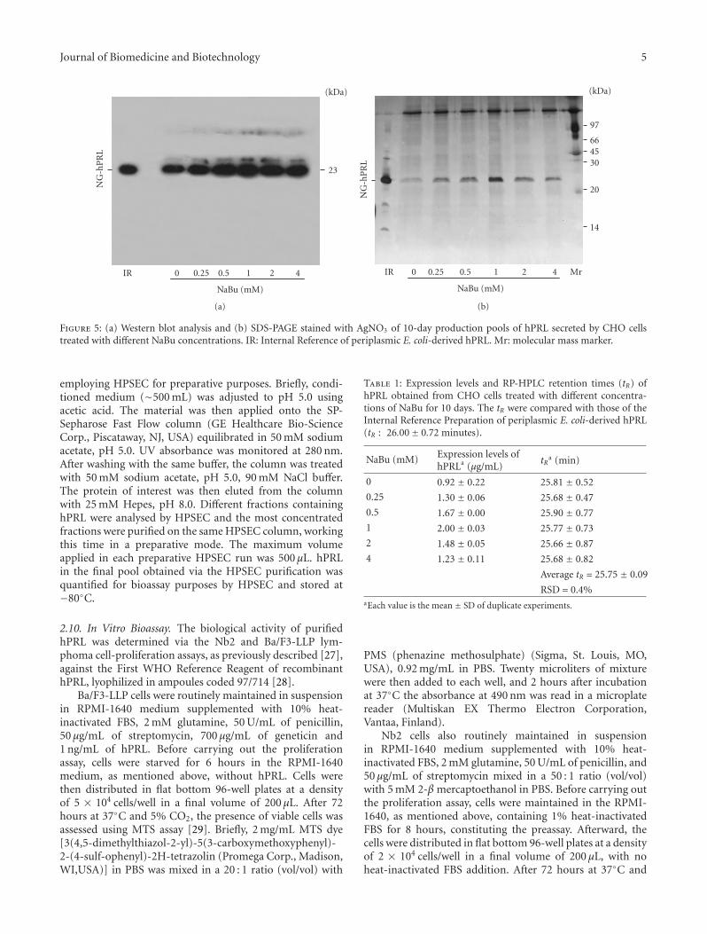
Figure 5: (a) Western blot analysis and (b) SDS-PAGE stained with AgNO3 of 10-day production pools of hPRL secreted by CHO cellstreated with different NaBu concentrations. IR: Internal Reference of periplasmic E. coli-derived hPRL. Mr: molecular mass marker.
employing HPSEC for preparative purposes. Briefly, condi-tioned medium (∼500 mL) was adjusted to pH 5.0 usingacetic acid. The material was then applied onto the SP-Sepharose Fast Flow column (GE Healthcare Bio-ScienceCorp., Piscataway, NJ, USA) equilibrated in 50 mM sodiumacetate, pH 5.0. UV absorbance was monitored at 280 nm.After washing with the same buffer, the column was treatedwith 50 mM sodium acetate, pH 5.0, 90 mM NaCl buffer.The protein of interest was then eluted from the columnwith 25 mM Hepes, pH 8.0. Different fractions containinghPRL were analysed by HPSEC and the most concentratedfractions were purified on the same HPSEC column, workingthis time in a preparative mode. The maximum volumeapplied in each preparative HPSEC run was 500 μL. hPRLin the final pool obtained via the HPSEC purification wasquantified for bioassay purposes by HPSEC and stored at−80◦C.
2.10. In Vitro Bioassay. The biological activity of purifiedhPRL was determined via the Nb2 and Ba/F3-LLP lym-phoma cell-proliferation assays, as previously described [27],against the First WHO Reference Reagent of recombinanthPRL, lyophilized in ampoules coded 97/714 [28].
Ba/F3-LLP cells were routinely maintained in suspensionin RPMI-1640 medium supplemented with 10% heat-inactivated FBS, 2 mM glutamine, 50 U/mL of penicillin,50 μg/mL of streptomycin, 700 μg/mL of geneticin and1 ng/mL of hPRL. Before carrying out the proliferationassay, cells were starved for 6 hours in the RPMI-1640medium, as mentioned above, without hPRL. Cells werethen distributed in flat bottom 96-well plates at a densityof 5 × 10⁴ cells/well in a final volume of 200 μL. After 72hours at 37◦C and 5% CO2, the presence of viable cells wasassessed using MTS assay [29]. Briefly, 2 mg/mL MTS dye[3(4,5-dimethylthiazol-2-yl)-5(3-carboxymethoxyphenyl)-2-(4-sulf-ophenyl)-2H-tetrazolin (Promega Corp., Madison,WI,USA)] in PBS was mixed in a 20 : 1 ratio (vol/vol) with
Table 1: Expression levels and RP-HPLC retention times (tR) ofhPRL obtained from CHO cells treated with different concentra-tions of NaBu for 10 days. The tR were compared with those of theInternal Reference Preparation of periplasmic E. coli-derived hPRL(tR : 26.00± 0.72 minutes).
NaBu (mM) Expression levels ofhPRLa (μg/mL)
tRa (min)
0 0.92 ± 0.22 25.81 ± 0.52
0.25 1.30 ± 0.06 25.68 ± 0.47
0.5 1.67 ± 0.00 25.90 ± 0.77
1 2.00 ± 0.03 25.77 ± 0.73
2 1.48 ± 0.05 25.66 ± 0.87
4 1.23 ± 0.11 25.68 ± 0.82
Average tR = 25.75 ± 0.09
RSD = 0.4%aEach value is the mean ± SD of duplicate experiments.
PMS (phenazine methosulphate) (Sigma, St. Louis, MO,USA), 0.92 mg/mL in PBS. Twenty microliters of mixturewere then added to each well, and 2 hours after incubationat 37◦C the absorbance at 490 nm was read in a microplatereader (Multiskan EX Thermo Electron Corporation,Vantaa, Finland).
Nb2 cells also routinely maintained in suspensionin RPMI-1640 medium supplemented with 10% heat-inactivated FBS, 2 mM glutamine, 50 U/mL of penicillin, and50 μg/mL of streptomycin mixed in a 50 : 1 ratio (vol/vol)with 5 mM 2-β mercaptoethanol in PBS. Before carrying outthe proliferation assay, cells were maintained in the RPMI-1640, as mentioned above, containing 1% heat-inactivatedFBS for 8 hours, constituting the preassay. Afterward, thecells were distributed in flat bottom 96-well plates at a densityof 2 × 10⁴ cells/well in a final volume of 200 μL, with noheat-inactivated FBS addition. After 72 hours at 37◦C and
6 Journal of Biomedicine and Biotechnology
hPRL
Elution
6
17
30 G-hPRLNG-hPRL22
4260
(kDa) 1 2 3 4 5 6 7 8
#27 28 29Sample application
50 mM NaAc pH 5
50 mM NaAc pH 5+
90 mM NaCl 25 mM HEPES pH 8
SP-Sepharose Fast Flow SDS-PAGE
(a)
hPRL
Elution
6
17
30 G-hPRLNG-hPRL22
4260
(kDa)1 2 3 4 5 6 7 8
#27 28 29Sample application
50 mM NaAc pH 5
50 mM NaAc pH 5+
90 mM NaCl 25 mM HEPES pH 8
(b)
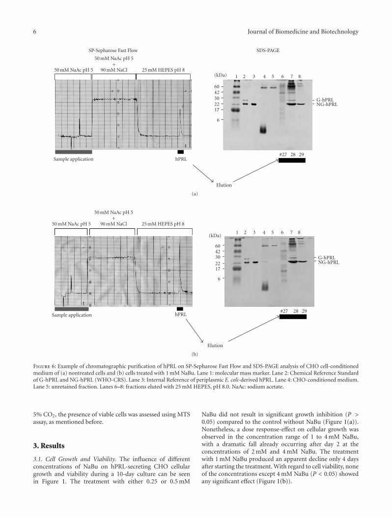
Figure 6: Example of chromatographic purification of hPRL on SP-Sepharose Fast Flow and SDS-PAGE analysis of CHO cell-conditionedmedium of (a) nontreated cells and (b) cells treated with 1 mM NaBu. Lane 1: molecular mass marker. Lane 2: Chemical Reference Standardof G-hPRL and NG-hPRL (WHO-CRS). Lane 3: Internal Reference of periplasmic E. coli-derived hPRL. Lane 4: CHO-conditioned medium.Lane 5: unretained fraction. Lanes 6–8: fractions eluted with 25 mM HEPES, pH 8.0. NaAc: sodium acetate.
5% CO2, the presence of viable cells was assessed using MTSassay, as mentioned before.
3. Results
3.1. Cell Growth and Viability. The influence of differentconcentrations of NaBu on hPRL-secreting CHO cellulargrowth and viability during a 10-day culture can be seenin Figure 1. The treatment with either 0.25 or 0.5 mM
NaBu did not result in significant growth inhibition (P >0.05) compared to the control without NaBu (Figure 1(a)).Nonetheless, a dose response-effect on cellular growth wasobserved in the concentration range of 1 to 4 mM NaBu,with a dramatic fall already occurring after day 2 at theconcentrations of 2 mM and 4 mM NaBu. The treatmentwith 1 mM NaBu produced an apparent decline only 4 daysafter starting the treatment. With regard to cell viability, noneof the concentrations except 4 mM NaBu (P < 0.05) showedany significant effect (Figure 1(b)).
Journal of Biomedicine and Biotechnology 7
19.79 hPRL (#28)
19.84 hPRL (#29)19.68 hPRL (IR)
#27
0 mM NaBu
0
50
100
150
200
250
300
350
mA
bs
0 5 10 15 20 25 30 35
(min)
(a)
19.69 hPRL (#28)
19.76 hPRL (#29)
19.68 hPRL (IR)#27
0
50
100
150
200
250
300
1 mM NaBu350
mA
bs
0 5 10 15 20 25 30 35
(min)
(b)
19.58 hPRL (1 mM NaBu)
19.72 hPRL (0 mM NaBu)
0
5
10
15
20
25
30
mA
bs
0 5 10 15 20 25 30 35
(min)
(c)
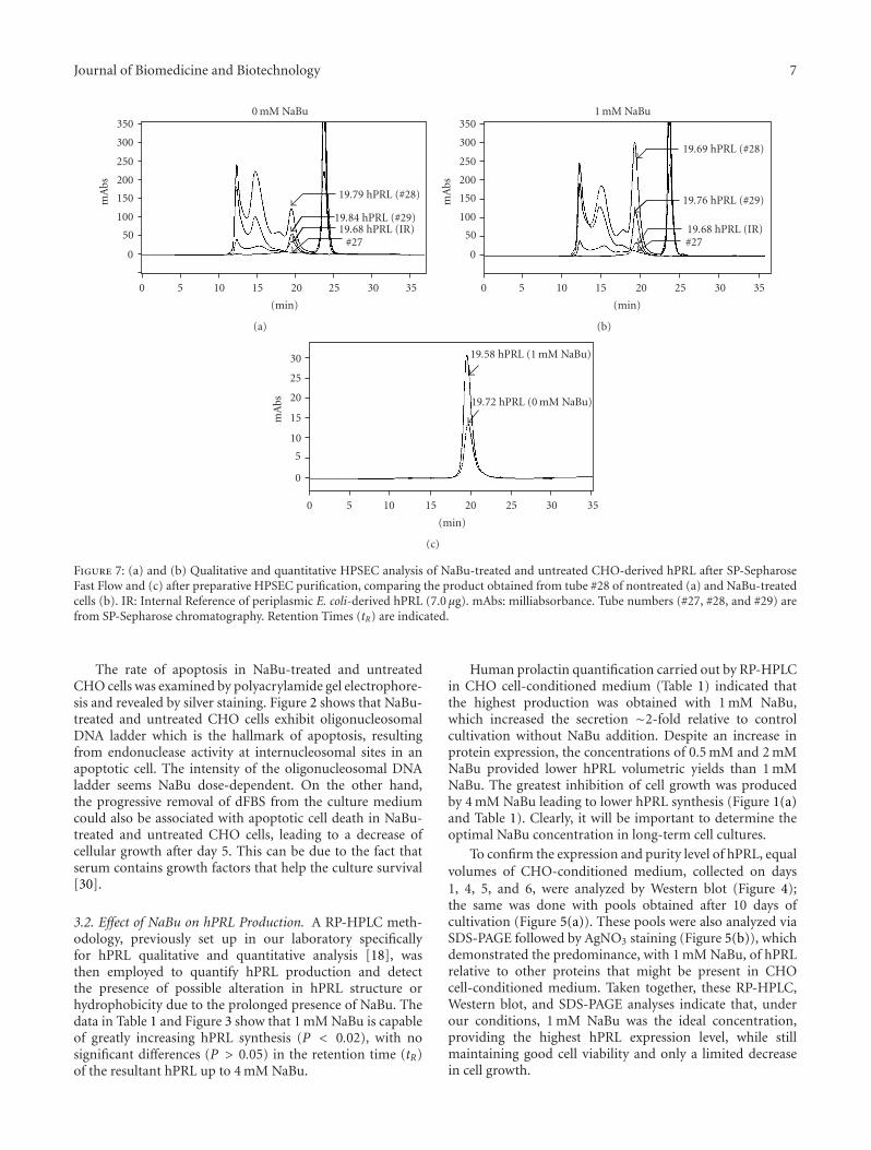
Figure 7: (a) and (b) Qualitative and quantitative HPSEC analysis of NaBu-treated and untreated CHO-derived hPRL after SP-SepharoseFast Flow and (c) after preparative HPSEC purification, comparing the product obtained from tube #28 of nontreated (a) and NaBu-treatedcells (b). IR: Internal Reference of periplasmic E. coli-derived hPRL (7.0 μg). mAbs: milliabsorbance. Tube numbers (#27, #28, and #29) arefrom SP-Sepharose chromatography. Retention Times (tR) are indicated.
The rate of apoptosis in NaBu-treated and untreatedCHO cells was examined by polyacrylamide gel electrophore-sis and revealed by silver staining. Figure 2 shows that NaBu-treated and untreated CHO cells exhibit oligonucleosomalDNA ladder which is the hallmark of apoptosis, resultingfrom endonuclease activity at internucleosomal sites in anapoptotic cell. The intensity of the oligonucleosomal DNAladder seems NaBu dose-dependent. On the other hand,the progressive removal of dFBS from the culture mediumcould also be associated with apoptotic cell death in NaBu-treated and untreated CHO cells, leading to a decrease ofcellular growth after day 5. This can be due to the fact thatserum contains growth factors that help the culture survival[30].
3.2. Effect of NaBu on hPRL Production. A RP-HPLC meth-odology, previously set up in our laboratory specificallyfor hPRL qualitative and quantitative analysis [18], wasthen employed to quantify hPRL production and detectthe presence of possible alteration in hPRL structure orhydrophobicity due to the prolonged presence of NaBu. Thedata in Table 1 and Figure 3 show that 1 mM NaBu is capableof greatly increasing hPRL synthesis (P < 0.02), with nosignificant differences (P > 0.05) in the retention time (tR)of the resultant hPRL up to 4 mM NaBu.
Human prolactin quantification carried out by RP-HPLCin CHO cell-conditioned medium (Table 1) indicated thatthe highest production was obtained with 1 mM NaBu,which increased the secretion ∼2-fold relative to controlcultivation without NaBu addition. Despite an increase inprotein expression, the concentrations of 0.5 mM and 2 mMNaBu provided lower hPRL volumetric yields than 1 mMNaBu. The greatest inhibition of cell growth was producedby 4 mM NaBu leading to lower hPRL synthesis (Figure 1(a)and Table 1). Clearly, it will be important to determine theoptimal NaBu concentration in long-term cell cultures.
To confirm the expression and purity level of hPRL, equalvolumes of CHO-conditioned medium, collected on days1, 4, 5, and 6, were analyzed by Western blot (Figure 4);the same was done with pools obtained after 10 days ofcultivation (Figure 5(a)). These pools were also analyzed viaSDS-PAGE followed by AgNO3 staining (Figure 5(b)), whichdemonstrated the predominance, with 1 mM NaBu, of hPRLrelative to other proteins that might be present in CHOcell-conditioned medium. Taken together, these RP-HPLC,Western blot, and SDS-PAGE analyses indicate that, underour conditions, 1 mM NaBu was the ideal concentration,providing the highest hPRL expression level, while stillmaintaining good cell viability and only a limited decreasein cell growth.
8 Journal of Biomedicine and Biotechnology
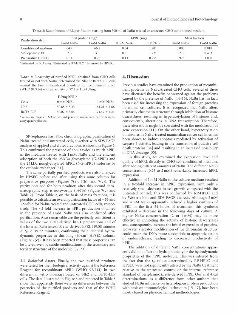
Table 2: Recombinant hPRL purification starting from 500 mL of NaBu-treated or untreated CHO-conditioned medium.
Purification stepTotal protein (mg)a hPRL (mg) Mass fraction
0 mM NaBu 1 mM NaBu 0 mM NaBu 1 mM NaBu 0 mM NaBu 1 mM NaBu
Conditioned medium 64.7 66.2 0.54 1.20b 0.008 0.018
SP-Sepharose FF 2.3 3.0 0.51 1.22c 0.219 0.403
Preparative HPSEC 0.14 0.25 0.13 0.25c 0.978 1.000aEstimated by BCA assay. bEstimated by RP-HPLC. cEstimated by HPSEC.
Table 3: Bioactivity of purified hPRL obtained from CHO cellstreated or not with NaBu, determined via Nb2 or Ba/F3-LLP cellsagainst the First International Standard for recombinant hPRL(WHO 97/714) with an activity of 57.2 ± 11.4 IU/mg.
IU/mg hPRLa
Cells 0 mM NaBu 1 mM NaBu
Nb2 50.88 ± 5.33 61.21 ± 4.66
Ba/F3-LLP 50.07 ± 5.64 71.47 ± 4.33aValues are means ± SD of two independent assays, each run with intra-assay quadruplicates.
SP-Sepharose Fast Flow chromatographic purification ofNaBu-treated and untreated cells, together with SDS-PAGEanalysis of applied and eluted fractions, is shown in Figure 6.This confirmed the presence of about twice as much hPRLin the medium treated with 1 mM NaBu and the efficientadsorption of both the 25 kDa glycosylated (G-hPRL) andthe 23 kDa nonglycosylated hPRL (NG-hPRL) isoforms bythe cationic exchanger [31].
The same partially purified products were also analyzedby HPSEC before and after using this same column forpreparative purposes (Figures 7(a), 7(b), and 7(c)). Thepurity obtained for both products after this second chro-matographic step is noteworthy (>97%) (Figure 7(c) andTable 2). From Table 2, on the basis of mass fractions, it ispossible to calculate an overall purification factor of∼55 and122-fold for NaBu-treated and untreated CHO cells, respec-tively. The ∼2-fold increase in hPRL production obtainedin the presence of 1mM NaBu was also confirmed afterpurification. Also remarkable are the perfectly coincident tRvalues of the two CHO-derived hPRL preparations and ofthe Internal Reference of E. coli-derived hPRL (19.58 minutes< tR < 19.72 minutes), confirming their identical hydro-dynamic properties in this long (60 cm) HPSEC column(Figure 7(c)). It has been reported that these properties canbe altered even by subtle modifications in the secondary andtertiary structure of the molecule [32, 33].
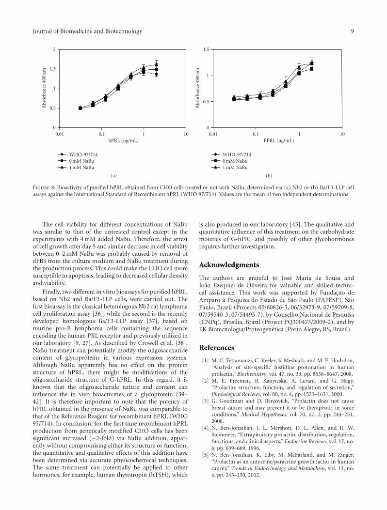
3.3. Biological Assays. Finally, the two purified productswere tested for their biological activity against the ReferenceReagent for recombinant hPRL (WHO 97/714) in twodifferent in vitro bioassays based on Nb2 and Ba/F3-LLPcells. The data illustrated in Figure 8 and reported in Table 3show that apparently there were no differences between thepotencies of the purified products and that of the WHOReference Reagent.
4. Discussion
Previous studies have examined the production of recombi-nant proteins by NaBu-treated CHO cells. Several of thesehave discussed the benefits or warned against the problemscaused by the presence of NaBu [14–16]. NaBu has, in fact,been used for increasing the expression of foreign proteinsin animal cell cultures. It is recognized that NaBu altersprimarily chromatin structure through inhibition of histonedeacetylases, resulting in hyperacetylation of histones and,consequently, alterations in DNA transcription. Therefore,these alterations might be correlated with the modulation ofgene expression [11]. On the other hand, hyperacetylationof histones in NaBu-treated mammalian cancer cell lines hasbeen shown to induce apoptosis mediated by activation ofcaspase-3 activity, leading to the translation of putative celldeath proteins [34] and resulting in an increased possibilityof DNA cleavage [35].
In this study, we examined the expression level andquality of hPRL directly in CHO cell-conditioned medium,after adding different amounts of NaBu. The different NaBuconcentrations (0.25 to 2 mM) remarkably increased hPRLexpression.
Addition of 1 mM NaBu to the culture medium resultedin a twofold increase in hPRL expression, with only arelatively small decrease in cell growth compared with theuntreated control; this was confirmed by RP-HPLC andby Western blot and SDS-PAGE analysis. Although 2 mMand 4 mM NaBu apparently induced a higher synthesis ofhPRL in the first 24 hours of treatment, this synthesisexhibited a decrease in the following days of culture. Ahigher NaBu concentration (2 or 4 mM) may be moreeffective in inhibiting the activity of histone deacetylasesand, consequently, increase the initial expression of proteins.However, a greater modification of the chromatin structurecould make the DNA more susceptible to apoptotic actionof endonucleases, leading to decreased productivity ofhPRL.
The addition of different NaBu concentrations appar-ently did not affect the hydrophobicity or the hydrodynamicproprieties of the hPRL molecule. This was inferred fromthe fact that the tR values determined by RP-HPLC andHPSEC were not significantly altered by the NaBu treatmentrelative to the untreated control or the internal referencestandard of periplasmic E. coli-derived hPRL. Our analyticaldeterminations, as a difference from other authors thatstudied NaBu influence on heterologous protein productionwith basis on immunological techniques [15–17], have beenmostly based on physicochemical methodologies.
Journal of Biomedicine and Biotechnology 9
0
0.5
1
1.5
2A
bsor
ban
ce49
0n
m
0.01 0.1 1 10hPRL (ng/mL)
WHO-97/7140 mM NaBu1 mM NaBu
(a)
0
0.5
1
1.5
Abs
orba
nce
490
nm
0.01 0.1 1 10hPRL (ng/mL)
WHO-97/7140 mM NaBu1 mM NaBu
(b)
Figure 8: Bioactivity of purified hPRL obtained from CHO cells treated or not with NaBu, determined via (a) Nb2 or (b) Ba/F3-LLP cellassays against the International Standard of Recombinant hPRL (WHO 97/714). Values are the mean of two independent determinations.
The cell viability for different concentrations of NaBuwas similar to that of the untreated control except in theexperiments with 4 mM added NaBu. Therefore, the arrestof cell growth after day 5 and similar decrease in cell viabilitybetween 0–2 mM NaBu was probably caused by removal ofdFBS from the culture medium and NaBu treatment duringthe production process. This could make the CHO cell moresusceptible to apoptosis, leading to decreased cellular densityand viability.
Finally, two different in vitro bioassays for purified hPRL,based on Nb2 and Ba/F3-LLP cells, were carried out. Thefirst bioassay is the classical heterologous Nb2 rat lymphomacell proliferation assay [36], while the second is the recentlydeveloped homologous Ba/F3-LLP assay [37], based onmurine pro-B lymphoma cells containing the sequenceencoding the human PRL receptor and previously utilized inour laboratory [9, 27]. As described by Crowell et al. [38],NaBu treatment can potentially modify the oligosaccharidecontent of glycoproteins in various expression systems.Although NaBu apparently has no effect on the proteinstructure of hPRL, there might be modifications of theoligosaccharide structure of G-hPRL. In this regard, it isknown that the oligosaccharide nature and content caninfluence the in vivo bioactivities of a glycoprotein [39–42]. It is therefore important to note that the potency ofhPRL obtained in the presence of NaBu was comparable tothat of the Reference Reagent for recombinant hPRL (WHO97/714). In conclusion, for the first time recombinant hPRLproduction from genetically modified CHO cells has beensignificant increased (∼2-fold) via NaBu addition, appar-ently without compromising either its structure or function;the quantitative and qualitative effects of this addition havebeen determined via accurate physicochemical techniques.The same treatment can potentially be applied to otherhormones, for example, human thyrotropin (hTSH), which
is also produced in our laboratory [43]. The qualitative andquantitative influence of this treatment on the carbohydratemoieties of G-hPRL and possibly of other glycohormonesrequires further investigation.
Acknowledgments
The authors are grateful to Jose Maria de Sousa andJoao Ezequiel de Oliveira for valuable and skilled techni-cal assistance. This work was supported by Fundacao deAmparo a Pesquisa do Estado de Sao Paulo (FAPESP), SaoPaulo, Brazil (Projects 05/60826-3, 06/52973-9, 07/59709-8,07/59540-3, 07/54493-7), by Conselho Nacional de Pesquisa(CNPq), Brasılia, Brazil (Project PQ300473/2009-2), and byFK Biotecnologia/Proteogenetica (Porto Alegre, RS, Brazil).
References
[1] M. C. Tettamanzi, C. Keeler, S. Meshack, and M. E. Hodsdon,“Analysis of site-specific histidine protonation in humanprolactin,” Biochemistry, vol. 47, no. 33, pp. 8638–8647, 2008.
[2] M. E. Freeman, B. Kanyicska, A. Lerant, and G. Nagy,“Prolactin: structure, function, and regulation of secretion,”Physiological Reviews, vol. 80, no. 4, pp. 1523–1631, 2000.
[3] G. Goodman and D. Bercovich, “Prolactin does not causebreast cancer and may prevent it or be therapeutic in someconditions,” Medical Hypotheses, vol. 70, no. 2, pp. 244–251,2008.
[4] N. Ben-Jonathan, J. L. Mershon, D. L. Allen, and R. W.Steinmetz, “Extrapituitary prolactin: distribution, regulation,functions, and clinical aspects,” Endocrine Reviews, vol. 17, no.6, pp. 639–669, 1996.
[5] N. Ben-Jonathan, K. Liby, M. McFarland, and M. Zinger,“Prolactin as an autocrine/paracrine growth factor in humancancer,” Trends in Endocrinology and Metabolism, vol. 13, no.6, pp. 245–250, 2002.
10 Journal of Biomedicine and Biotechnology
[6] L. Morganti, M. Huyer, P. W. Gout, and P. Bartolini, “Pro-duction and characterization of biologically active Ala-Ser-(His)6-lle-Glu-Gly-Arg-human prolactin (tag-hPRL) secretedin the periplasmic space of Escherichia coli,” Biotechnology andApplied Biochemistry, vol. 23, no. 1, pp. 67–75, 1996.
[7] L. Morganti, C. R. J. Scares, R. Affonso, P. W. Gout, andP. Bartolini, “Synthesis and characterization of recombinant,authentic human prolactin secreted into the periplasmic spaceof Escherichia coli,” Biotechnology and Applied Biochemistry,vol. 27, part 1, pp. 63–70, 1998.
[8] C. R. J. Soares, L. Morganti, B. Miloux, J. H. Lupker, P. Ferrara,and P. Bartolini, “High-level synthesis of human prolactinin Chinese-hamster ovary cells,” Biotechnology and AppliedBiochemistry, vol. 32, no. 2, pp. 127–135, 2000.
[9] C. R. J. Soares, A. Glezer, K. Okazaki, et al., “Physico-chemicaland biological characterizations of two human prolactinanalogs exhibiting controversial bioactivity, synthesized inChinese hamster ovary (CHO) cells,” Protein Expression andPurification, vol. 48, no. 2, pp. 182–194, 2006.
[10] C. R. J. Soares, E. K. M. Ueda, T. L. Oliveira, F. I. C. Gomide, S.R. Heller, and P. Bartolini, “Distinct human prolactin (hPRL)and growth hormone (hGH) behavior under bacteriophagelambda P promoter control: temperature plays a major rolein protein yields,” Journal of Biotechnology, vol. 133, no. 1, pp.27–35, 2008.
[11] J. R. Davie, “Inhibition of histone deacetylase activity bybutyrate,” Journal of Nutrition, vol. 133, no. 7, supplement, pp.2485S–2493S, 2003.
[12] M.-P. Leibovitch, S.-A. Leibovitch, J. Harel, and J. Kruh,“Effect of sodium butyrate on messenger RNA populations inmyogenic cells in culture,” Differentiation, vol. 22, no. 2, pp.106–112, 1982.
[13] D. Y. Lee, J. J. Hayes, D. Pruss, and A. P. Wolffe, “A positiverole for histone acetylation in transcription factor access tonucleosomal DNA,” Cell, vol. 72, no. 1, pp. 73–84, 1993.
[14] N. S. Kim and G. M. Lee, “Overexpression of bcl-2 inhibitssodium butyrate-induced apoptosis in Chinese hamster ovarycells resulting in enhanced humanized antibody production,”Biotechnology and Bioengineering, vol. 71, no. 3, pp. 184–193,2000.
[15] Y. Mimura, J. Lund, S. Church, et al., “Butyrate increasesproduction of human chimeric IgG in CHO-K1 cells whilstmaintaining function and glycoform profile,” Journal ofImmunological Methods, vol. 247, no. 1-2, pp. 205–216, 2001.
[16] K. O. Han, K. S. Moon, J. Yang, et al., “Effect of N-Acetylcystein on butyrate-treated Chinese hamster ovary cellsto improve the production of recombinant human interferon-beta-1a,” Biotechnology Progress, vol. 21, no. 4, pp. 1154–1164,2005.
[17] H. S. Yun and M. L. Gyun, “Enhanced human throm-bopoietin production by sodium butyrate addition to serum-free suspension culture of Bcl-2-overexpressing CHO cells,”Biotechnology Progress, vol. 21, no. 1, pp. 50–57, 2005.
[18] C. R. J. Soares, I. M. C. Camargo, L. Morganti, et al.,“Reversed-phase high-performance liquid chromatographymethod for the determination of prolactin in bacterial extractsand in its purified form,” Journal of Chromatography A, vol.955, no. 2, pp. 229–236, 2002.
[19] S. Dalmora, J. E. Oliveira, R. Affonso, E. Gimbo, M. T. C.P. Ribela, and P. Bartolini, “Analysis of recombinant humangrowth hormone directly in osmotic shock fluids,” Journal ofChromatography A, vol. 782, no. 2, pp. 199–210, 1997.
[20] J. R. Tennant, “Evaluation of the trypan blue technique fordetermination of cell viability,” Transplantation, vol. 2, pp.685–694, 1964.
[21] B. J. Bassam, G. Caetano-Anolles, and P. M. Gresshoff, “Fastand sensitive silver staining of DNA in polyacrylamide gels,”Analytical Biochemistry, vol. 196, no. 1, pp. 80–83, 1991.
[22] U. K. Laemmli, “Cleavage of structural proteins during theassembly of the head of bacteriophage T4,” Nature, vol. 227,no. 5259, pp. 680–685, 1970.
[23] D. D. Bannerman, J. C. Tupper, R. D. Erwert, R. K. Winn,and J. M. Harlan, “Divergence of bacterial lipopolysaccharidepro-apoptotic signaling downstream of IRAK-1,” Journal ofBiological Chemistry, vol. 277, no. 10, pp. 8048–8053, 2002.
[24] M. T. C. P. Ribela, A. C. Bianco, and P. Bartolini, “Theuse of recombinant human thyrotropin produced by Chinesehamster ovary cells for the preparation of immunoassayreagents,” Journal of Clinical Endocrinology and Metabolism,vol. 81, no. 1, pp. 249–256, 1996.
[25] M. T. C. P. Ribela, P. W. Gout, J. E. Oliveira, and P. Bartolini,“HPLC analysis of human pituitary hormones for pharmaceu-tical applications,” Current Pharmaceutical Analysis, vol. 2, no.2, pp. 103–126, 2006.
[26] G. W. White, T. Katona, and J. P. Zodda, “The use of high-performance size exclusion chromatography (HPSEC) as amolecular weight screening technique for polygalacturonicacid for use in pharmaceutical applications,” Journal ofPharmaceutical and Biomedical Analysis, vol. 20, no. 6, pp.905–912, 1999.
[27] A. Glezer, C. R. J. Soares, J. G. Vieira, et al., “Human macro-prolactin displays low biological activity via its homologousreceptor in a new sensitive bioassay,” Journal of ClinicalEndocrinology and Metabolism, vol. 91, no. 3, pp. 1048–1055,2006.
[28] B. Rafferty, P. Rigsby, R. E. Gaines-Das, et al., “Draft report ofan international collaborative study of proposed WHO refer-ence reagents for rDNA-derived prolactin and its glycosylatedand non-glycosylated componentes,” WHO Technical ReportSeries, vol. 52, no. 924, 2001.
[29] T.-J. Chen, C. B. Kuo, K. F. Tsai, J. O.-W. Liu, D.-Y. Chen,and A. M. Walker, “Development of recombinant humanprolactin receptor antagonists by molecular mimicry of thephosphorylated hormone,” Endocrinology, vol. 139, no. 2, pp.609–616, 1998.
[30] Y.-S. Lee, H. Nakajima, Y.-C. Chang, et al., “Alleviation ofapoptosis by serum in Chinese hamster ovary cells ectopicallyexpressing human Fas antigen,” Molecules and Cells, vol. 8, no.3, pp. 272–279, 1998.
[31] S. R. Heller, H. Rodrigues Goulart, F. S. Arthuso, T. L. Oliveira,P. Bartolini, and C. R. J. Soares, “Synthesis, purification andcharacterization of recombinant glycosylated human prolactin(G-hPRL) secreted by cycloheximide-treated CHO cells,”Journal of Biotechnology, vol. 145, no. 4, pp. 334–340, 2010.
[32] M. T. C. P. Ribella and P. Bartolini, “Stokes radius deter-mination of radioiodinated polypeptide hormones by gelfiltration,” Analytical Biochemistry, vol. 174, no. 2, pp. 693–697, 1988.
[33] C. M. Carvalho, J. E. Oliveira, B. E. Almeida, et al., “Efficientisolation of the subunits of recombinant and pituitary glyco-protein hormones,” Journal of Chromatography A, vol. 1216,no. 9, pp. 1431–1438, 2009.
[34] V. Medina, B. Edmonds, G. P. Young, R. James, S. Appleton,and P. D. Zalewski, “Induction of caspase-3 protease activityand apoptosis by butyrate and trichostatin a (inhibitorsof histone deacetylase): dependence on protein synthesis
Journal of Biomedicine and Biotechnology 11
and synergy with a mitochondrial/cytochrome c-dependentpathway,” Cancer Research, vol. 57, no. 17, pp. 3697–3707,1997.
[35] P. J. Smith, “n-Butyrate alters chromatin accessibility to DNArepair enzymes,” Carcinogenesis, vol. 7, no. 3, pp. 423–429,1986.
[36] T. Tanaka, R. P. C. Shiu, and P. W. Gout, “A new sensitiveand specific bioassay for lactogenic hormones: measurementof prolactin and growth hormone in human serum,” Journal ofClinical Endocrinology & Metabolism, vol. 51, no. 5, pp. 1058–1063, 1980.
[37] S. Bernichtein, S. Jeay, R. Vaudry, P. A. Kelly, and V. Goffin,“New homologous bioassays for human lactogens show thatagonism or antagonism of various analogs is a function ofassay sensitivity,” Endocrine, vol. 20, no. 1-2, pp. 177–190,2003.
[38] C. K. Crowell, Q. Qin, G. E. Grampp, R. A. Radcliffe, G. N.Rogers, and R. I. Scheinman, “Sodium butyrate alters erythro-poietin glycosylation via multiple mechanisms,” Biotechnologyand Bioengineering, vol. 99, no. 1, pp. 201–213, 2008.
[39] R. Damiani, J. E. Oliveira, K. Vorauer-Uhl, et al., “Sta-ble expression of a human-like sialylated recombinant thy-rotropin in a Chinese hamster ovary cell line expressingalpha2,6-sialyltransferase,” Protein Expression and Purification,vol. 67, no. 1, pp. 7–14, 2009.
[40] C. F. Goochee, M. J. Gramer, D. C. Andersen, J. B. Bahr,and J. R. Rasmussen, “The oligosaccharides of glycoproteins:bioprocess factors affecting oligosaccharide structure and theireffect on glycoprotein properties,” Biotechnology, vol. 9, no. 12,pp. 1347–1355, 1991.
[41] F. Mendonca, J. E. Oliveira, P. Bartolini, and M. T. C. P.Ribela, “Two-step chromatographic purification of recombi-nant human thyrotrophin and its immunological, biological,physico-chemical and mass spectral characterization,” Journalof Chromatography A, vol. 1062, no. 1, pp. 103–112, 2005.
[42] M. W. Szkudlinski, V. Fremont, C. Ronin, and B. D.Weintraub, “Thyroid-stimulating hormone and thyroid-stimulating hormone receptor structure-function relation-ships,” Physiological Reviews, vol. 82, no. 2, pp. 473–502, 2002.
[43] C. N. Peroni, C. R. J. Soares, E. Gimbo, L. Morganti, M. T. C.P. Ribela, and P. Bartolini, “High-level expression of humanthyroid-stimulating hormone in Chinese hamster ovary cellsby co-transfection of dicistronic expression vectors followedby a dual-marker amplification strategy,” Biotechnology andApplied Biochemistry, vol. 35, part 1, pp. 19–26, 2002.