Unfractionated Heparin and New Heparin Analogues from Ascidians ...
Upload
truongkietCategory
view
219download
1
MINI-REVIEW
Engineering of routes to heparin and related polysaccharides
Ujjwal Bhaskar & Eric Sterner & Anne Marie Hickey &
Akihiro Onishi & Fuming Zhang & Jonathan S. Dordick &
Robert J. Linhardt
Received: 15 August 2011 /Revised: 23 September 2011 /Accepted: 8 October 2011 /Published online: 3 November 2011# Springer-Verlag 2011
Abstract Anticoagulant heparin has been shown to possessimportant biological functions that vary according to itsfine structure. Variability within heparin’s structure occursowing to its biosynthesis and animal tissue-based recoveryand adds another dimension to its complex polymericstructure. The structural variations in chain length andsulfation patterns mediate its interaction with manyheparin-binding proteins, thereby eliciting complex biolog-ical responses. The advent of novel chemical and enzymatic
approaches for polysaccharide synthesis coupled with highthroughput combinatorial approaches for drug discoveryhave facilitated an increased effort to understand heparin’sstructure–activity relationships. An improved understandingwould offer potential for new therapeutic developmentthrough the engineering of polysaccharides. Such a bioen-gineering approach requires the amalgamation of severaldifferent disciplines, including carbohydrate synthesis,applied enzymology, metabolic engineering, and processbiochemistry.
Keywords Bioengineered heparin . Applied enzymology .
Biosynthesis . Chemical synthesis . Chemoenzymaticsynthesis .Metabolic engineering
Introduction
Heparin (Fig. 1a) was discovered in 1916 and entered earlyclinical trials as the first biopolymeric drug during 1930sbefore the establishment of the US Food and DrugAdministration (FDA) (Linhardt and Gunay 1999). It isstill in widespread clinical use as an intravenous anticoag-ulant with more than 100,000 kilograms produced annuallyworldwide (Liu et al. 2009). Nearly a century after itsdiscovery, heparin is still derived from animal sources suchas porcine intestine and bovine lung. Heparin has acomplex and diverse fine structure and is a polydispersemixture of varying polysaccharide chain lengths. Theheparin polysaccharide is comprised primarily of a trisul-fated disaccharide repeating unit, 2-O-sulfo-!-L-iduronicacid 1–4 linked to 6-O-sulfo-N-sulfo-!-D-glucosamine(Pervin et al. 1995). Heparin polydispersity, defined as theratio of weight average molecular weight to numberaverage molecular weight, varies from 1.1 to 1.6 (Liu et
U. Bhaskar : E. Sterner : F. Zhang : J. S. Dordick :R. J. Linhardt (*)Department of Chemical and Biological Engineering,Rensselaer Polytechnic Institute,Troy, NY, USAe-mail: [email protected]
A. M. Hickey :A. Onishi : F. Zhang : J. S. Dordick :R. J. LinhardtCenter for Biotechnology and Interdisciplinary Studies,Rensselaer Polytechnic Institute,Troy, NY, USA
J. S. Dordick :R. J. LinhardtDepartment of Biology, Rensselaer Polytechnic Institute,Troy, NY, USA
J. S. Dordick :R. J. LinhardtDepartment of Biomedical Engineering,Rensselaer Polytechnic Institute,Troy, NY, USA
J. S. DordickDepartment of Materials Science & Engineering,Rensselaer Polytechnic Institute,Troy, NY, USA
R. J. LinhardtDepartment of Chemistry and Chemical Biology,Rensselaer Polytechnic Institute,Troy, NY, USA
Appl Microbiol Biotechnol (2012) 93:1–16DOI 10.1007/s00253-011-3641-4

al. 2009; Gray et al. 2008). This high polydispersitysuggests the presence of a high mole percentage of smallerlength chains within certain pharmaceutical heparin prepa-rations. This is consistent with a molecular weight rangethat varies between 5,000 and 40,000 Da for pharmaceu-tical heparin (Linhardt and Gunay 1999; Gray et al. 2008).In addition to the trisulfated disaccharide, heparin alsocontains disaccharides with lesser degrees of sulfation, leadingto its structural heterogeneity. Chains composed mainly ofundersulfated (unsulfated or monosulfated) disaccharide are
typically classified as heparan sulfate (HS). Together withheparin, HS comprises one family of glycosaminoglycans(GAGs) and shares the same biosynthetic pathway.
In addition to their anticoagulant activity, heparin andHS are known to possess a wide variety of other biologicalfunctions. This family of GAGs plays critical roles inembryonic development, inflammatory response, viral/bacterial infections, and cell differentiation. These biologicalactivities are primarily associated with charge interactionsbetween heparin/HS and heparin-binding proteins, thereby,
A
B
C
Fig. 1 Structure of heparin, LMWH, and ULMWH fondaparinux. aA representative sequence of heparin comprising a major trisulfateddisaccharide sequence, an AT-binding site, and a minor disulfateddisaccharide sequence. b LMWH structures generated through peroxide
oxidation, deaminative degradation, and chemical or enzymatic "-elimination. c ULMWH, a commercial synthetic pentasaccharideAT-binding domain stabilized as the methyl glycoside called“fondaparinux”
2 Appl Microbiol Biotechnol (2012) 93:1–16

making control of sulfation level and distribution, throughcontrolled biosynthesis, of critical importance. The advent oflow molecular weight heparin (LMWH) (Fig. 1b), derivedfrom heparin through its controlled depolymerization, alongwith identification of serine protease inhibitor, antithrombinIII (AT), and heparin-binding coagulation factors, likethrombin and factor Xa, have led to an improved understand-ing of the blood coagulation cascade. Development ofLMWH was initially aimed at the control of heparin’shemorrhagic effect by exploiting heparin chain size-dependent differences in binding to antithrombin and factorXa (Johnson et al. 1976). The ensuing research led to theestablishment of LMWHs as new generation of anticoagulantdrugs. Anticoagulant activity associated with heparin and HSis primarily attributed to this highly specific interaction withan AT-binding pentasaccharide sequence containing a central3,6 di-O-sulfo, 2-N-sulfo glucosamine residue (Van Boeckelet al. 1995).When this pentasaccharide sequence is bound toAT, AT undergoes a conformational change thereby acceler-ating its inhibition of factor Xa in the blood coagulationcascade. AT and thrombin both need to bind to the sameheparin chain in a ternary bridging complex to inhibitthrombin and prevent conversion of fibrinogen into aninsoluble fibrin clot. This pentasaccharide sequence was firstidentified through AT-based affinity chromatography follow-ing the digestion of heparin. This was followed by efforts tosynthesize an ultra LMWH (ULMWH) containing thisunique AT pentasaccharide sequence in the 1980s, resultingin the first pentasaccharide analogue, with the aldehydegroup at the reducing-end permanently protected as themethyl glycoside (Fig. 1c). The synthesis and purification ofsuch derivatives became simpler, resulting in their industrial-scale production (Petitou et al. 1987). In 2001, a stablemethyl glycoside derivative of AT-binding pentasaccharide“fondaparinux” passed through clinical development as thefirst ULMWH drug leading to Arixtra® and its registration asa new antithrombotic drug in the USA and Europe (Petitouand van Boeckel 2004). Recently, a generic version offondaparinux has been approved by the US FDA. With anestimated 110 metric tons/year of heparin produced world-wide, its annual market is estimated to be around $3–4billion. This production volume surpasses other such anti-coagulants with sales for Arixtra nearing 500 million dollarsannually in the USA and Europe.
An international health crisis, associated with contami-nation of several heparin batches, began early 2008,reportedly resulting in nearly 100 deaths alone in theUSA. The patients displayed minor symptoms like rash,fainting, racing heart, and other more severe symptoms,including hypotension leading to death. Since there was nohistory of such crisis related to heparin use, it led tosuspicions about possible heparin contamination. Thesecontaminated batches, however, had cleared existing quality
control and quality assurance tests designed to detectproteins, lipids, DNA, dioxins, and other contaminants.These adverse side effects resulted in the withdrawal of anumber of heparin batches from US markets in March 2008followed by an investigation for the presence of contami-nants in these batches. Oversulfated chondroitin sulfate(OSCS), an 18-kDa semisynthetic polysaccharide havinga high charge density of !5 (sulfo+carboxyl groups)/disaccharide compared to !3.7/disaccharide for heparin),was discovered to be the contaminating agent (Guerriniet al. 2008). This contaminant was also found to be carriedthrough the production process of certain LMWH products(Zhang et al. 2008a). The rapid and acute response elicitedby this OSCS was associated with an anaphylactoidresponse generated due to activation of kinin–kallikreinpathway in human plasma, leading to formation ofvasoactive mediator bradykinin, which caused vasodilationand a sometimes lethal drop in blood pressure (Kishimotoet al. 2008; Li et al. 2009).
Significant efforts have been directed towards elucidat-ing the biological and functional properties of heparin andits related poly-/oligosaccharides to provide for a betterunderstanding of this complex biopolymer. Interdisciplinarystudies involving several branches of science have provideda better understanding of heparin’s complex structure andhave resulted in novel synthetic approaches and structure–activity relationship (SAR) studies (Fig. 2). The healthcrisis in 2008 brought to light flaws within animal tissuebased heparin production, which begins at slaughterhouses.This mini-review examines the various aspects of polysac-charide development including research currently underwayto develop a biologically and structurally similar, bioen-gineered heparin that can provide a clinically safe optionreplacing the currently used animal-derived heparin.
Current production process for heparinand heparin-like polysaccharides
Commercial-scale heparin production from animal tissueshas moved from dog liver to beef lung and finally toporcine intestine. While being similar, these GAGs derivedfrom animal sources have variation in fine structure thatrelates to difference in functional activities, such as AT- andthrombin-binding affinities (Liu et al. 2009). Additionalvariation into an already complex structure is added by thevariability inherent in raising domesticated animals, includ-ing their diet and breed. For example, approximately30,000–50,000 U ("300 mg per animal) of USP heparincan be derived from American pigs, while this number isslightly higher for Chinese pigs (Liu et al. 2009).
A general outline for heparin processing is presented inFig. 3. Initial processing is carried out at slaughterhouse
Appl Microbiol Biotechnol (2012) 93:1–16 3

without current good manufacturing practices (cGMP)facilities providing an opportunity for contamination.Salting of freed intestines is first carried out using sodiummetabisulfite or sodium chloride to preserve the tissuesfrom degradation and dewatering (Liu et al. 2009; Mozenand Evans 1962; Vidic 1981). Tissues are next solubilizedusing proteases, which leads to a decrease in protein content,followed by heat deactivation for protease removal (Panasyuk2011). The heparin capture step is either a precipitation stepusing a hydrophobic quaternary ammonium salt or resin-based chromatographic step using a strong anion exchangeresin. After resolubilizing heparin in saline, it can then beprecipitated using ethanol or methanol. All these steps areoften performed at or near slaughterhouses generating rawheparin, which can be consolidated and shipped forpurification at cGMP facilities. Quaternary ammonium saltscan often selectively precipitate heparin from tissue extractswithout the need for a separate protein removal step. Thisinvolves isolation of the insoluble complex followed byresolubilization of the same complex with highly concen-
trated sodium chloride solution (Mozen and Evans 1962) orwith higher alcohols. The heparin salt of Hyamine 1622flocculates easily at low pH, thereby allowing filtration, andthe precipitate can be resolubilized in butanol followed bymetathesis, leading to aqueous phase separation (Nominéand Barthelemy 1961). This can then be separated by saltingout or through precipitation by miscible solvents like ethanol(Nominé and Barthelemy 1961), leading to purified heparinin high yields and in high potency. The brine formed due tosalting of extracted tissues contains heparin with lowimpurity levels, thereby requiring milder treatments (Vidic1981). In case of ion-exchange, the negative charge onheparin (!3.7/disaccharide unit) is used for adsorption ontomacroreticular anion exchange resin with elution carried outunder basic or high salt conditions depending upon the resin.Affinity purification techniques based on affinity for AT hasbeen developed (Rosenberg 1981, 1985); however, thescalability of these approaches poses major problems.
At cGMP facilities, raw heparin is resolubilized andfiltered at low pH for the removal of protein followed by
Fig. 2 SAR characterization of heparin
4 Appl Microbiol Biotechnol (2012) 93:1–16

bleaching step. A microreticular cation exchange resin canbe employed for the removal of unwanted cations and theuse of ethanol precipitation for nucleotide removal.Remaining salt present within the product is removedthrough membrane filtration following, which recoveredheparin can be dried and analyzed.
LMWHs have a characteristic average MWof <8,000 Da(compared to 12,000–20,000 for heparin) with most of thechains weight having MW below 8,000 (Linhardt andGunay 1999; Gray et al. 2008). LMWHs generally possessmore than 70 U/mg of anti-factor Xa activity with ratio ofanti-factor Xa to antifactor IIa activity #1.5 (Linhardt andGunay 1999). LMWHs can be directly recovered fromanimal-derived heparin by size exclusion chromatography,but such a process is unsuitable for large-scale production;instead, either chemical or enzymatic depolymerization ofanimal-derived heparin is used to prepare LMWHs (Fig. 3).
The instability of heparin towards reactive oxygenspecies is utilized in hydrogen peroxide-based degradationmethod employed for production of ardeparin and parna-parin (Fig. 3). This proceeds by selective oxidation ofsusceptible nonsulfated uronic residues (Linhardt andGunay 1999). In one such method, heparin is converted toits heparinic acid form by maintaining the pH around 3–5followed by heating in the presence of an oxidizing agentresulting in fractions with molecular weights between 4,000
and 12,000 Da (Smith et al. 1984). Compared to othermethods of depolymerization, oxidative treatment based onOH radical does not result in any residual structuralmodification in the LMWH chains (Gray et al. 2008)(Fig. 1b). Oxidative depolymerization using deaminativecleavage with nitrous acid or isoamyl nitrite generatesanhydromannose residue at the reducing end of LMWHchains, leading to anhydromannitol residue (Fig. 1b). Thepresence of N-nitroso compounds obtained as contaminantswith LMWHs derived from this degradation has beenremoved through UV irradiation (Branellec et al. 1997). Ina separate strategy, the degrading enzyme heparinase (Liuet al. 2008; Linhardt 1996) can be employed for degrada-tion via a "-elimination cleavage mechanism. Enzymaticaction can be mimicked through chemical steps in whichthe carboxyl group of uronic acid is first esterified, andbase treatment leads to selective "-elimination cleavage(Linhardt 1996; Gray et al. 2008).The products generatedusing enzymatic/chemical "-elimination tends to have acharacteristic unsaturated uronic acid residue at the non-reducing end (Mardiguian 1984).
The introduction of Arixtra® (Fig. 1c) as a newanticoagulant drug in 2001 paved the way for industrialproduction of this AT-binding pentasaccharide. Multi-stepchemical synthesis of Arixtra® leads to high productioncosts, making it more than 1,000-fold costlier than heparin.
Fig. 3 Process flow diagram for production of heparin and LMWHs
Appl Microbiol Biotechnol (2012) 93:1–16 5

Expiration of patent protecting Arixtra® has led to develop-ment of a generic version at lower costs. Levulinate protected2-glucuronic acid-anhydro sugar coupling methodology withdeprotection and sequential sulfonation reactions leads togeneration of fondaparinux (Nadji et al. 2011). This strategyoffers reduced reaction time and high coupling yields of "-isomer with increased selectivity making it an efficient andscalable process for industrial-scale production.
Structural and biological activity of heparin
The biological activity of heparin is closely related to itsstructure and depends upon the interaction of heparin withheparin-binding proteins (Fig. 2). Heparin-binding proteinsgenerally have patches of basic amino acid residues on theirsurface that interact with the negative sulfo and carboxylgroups present (!3.7/disaccharide unit) throughout theheparin chain length (Liu et al. 2009).
Disaccharide compositional and oligosaccharide map-ping analysis are often used to study heparin and LMWHstructure. In these methods, disaccharide and/or oligosac-charide fragments of heparin are prepared through enzy-matic or chemical digestion and subsequently analyzed toprovide detailed structural information (Ly et al. 2010;Yang et al. 2011; Turnbull 1993). Fourier transform ioncyclotron resonance mass spectroscopy (MS), a highresolution technique, can be used to directly determine thechain length and composition of GAGs (Chi et al. 2008;Laremore et al. 2010). Quadupole mass filter leads toremoval of ions, thereby leading to an improved signal tonoise ratio in Fourier transform MS (Laremore et al. 2010).Disaccharides derived from heparin have been analyzedusing liquid chromatography under reverse phased ion-pairing mode coupled to electrospray ionization massspectrometry with ion trap mass analyzer (Yang et al.2011). Capillary electrophoresis (CE) coupled with ultra-violet or laser-induced fluorescence detection has beenroutinely used earlier for detection of heparin/heparansulfate disaccharides (Pervin et al. 1993; Mao et al. 2002).Multi-dimensional NMR has been employed for assignmentof 13C–15N labeled bioengineered heparin (Zhang et al.2008b). These structural evaluation tools were criticaltowards determination of contaminant in adulterated hepa-rin batches. 1D NMR and CE showed distinct variationbetween control lots and contaminated batches with differ-ences in acetyl region (2.0–2.2 ppm) along with a leadingpeak before heparin in CE (Liu et al. 2009). NMR wasemployed for determination of oversulfated chondroitinsulfate as the contaminant present in heparin batches(Guerrini et al. 2008).
Bioassays are routinely used for activity studies ofheparin (Linhardt et al. 1992). Surface plasmon resonance
can also be used to determine kinetics and thermodynamicsof heparin interaction with heparin-binding proteins afford-ing binding kinetics and thermodynamics (Muñoz et al.2005; Beaudet et al. 2011). Pharmacokinetic and pharma-codynamic studies have been performed on heparindelivered through oral, subcutaneous, and intravenousroutes (Mousa et al. 2007).
Biosyntheis of heparin/HS
Heparin and HS share the same eukaryotic biosyntheticpathway (Fig. 4), beginning in the endoplasmic reticulumwith most modifications occurring within the Golgi(Fig. 5). Different sulfation patterns in heparin and HSprovide for diverse biological activities with their SAR(Fig. 2) controlled through the action of genome-encodedbiosynthetic enzymes. Heparin is biosynthesized as aproteoglycan, a number of GAG chains attached to theserine residues of serglycin core protein (Sawesi et al.2010), while HS chains are attached to a number ofdifferent core proteins (Kramer and Yost 2003). Theheparin and HS GAGs are attached to their core proteinthrough a common tetrasaccharide linker (–GlcA"1–3Gal"1–3Gal"1–4Xyl"1–), the synthesis of which takesplace in the endoplasmic reticulum (Robinson et al. 1978;Carlsson et al. 2008) (Fig. 4). A repeating GlcNAc–GlcAdisaccharide unit is then attached to this tetrasaccharidelinker with the sequential addition, to the non-reducingend, of uridine diphosphate (UDP) sugars through theaction of EXT (Exotosin genes) glycosyltranferases (Eskoand Selleck 2002). This is followed by N-deacetylation andN-sulfonation through the action of a bifunctional N-deacetylase/N-sulfotranferase (NDST) (four isoforms)(Aikawa et al. 2001). This serves as a critical step,providing substrate specificity for the appropriate actionof the other modification enzymes (Kusche et al. 1991a).This N-sulfo group containing polysaccharide is thenmodified by C5-epimerase, converting GlcA flanked byGlcNS or GlcN to IdoA (Jacobsson et al. 1984; Gorsi andStringer 2007).The action of 2-O-sulfotranferase thenmodifies the C2 position of IdoA prior to 6 and 3-O-sulfonation. The 6-O-sulfotranferase modifies the sulfateddomains by placing a 6-O-sulfo group on GlcNS orGlcNAc near GlcNS/IdoA (Habuchi et al. 2003). Thereare three different isoforms of the 6-OST each havingsimilar substrate specificity, but the 6-OST-1 seems toprefer the absence of 2-O-sulfo group (Habuchi et al. 2003).The 3-O-sulfotranferases add a 3-O-sulfo group to GlcNSor GlcNS6S (Gorsi and Stringer 2007). There are sixdifferent 3-OST isoforms, which mostly act on GlcNS nextto GlcA/IdoA2S. The 3-OST-1 requires 6-O-sulfo GlcNS.These O-sulfo transferases and C5-epimerase are responsible
6 Appl Microbiol Biotechnol (2012) 93:1–16

Fig. 4 Biosynthetic pathway for heparin and HS. Synthesis of tetrasaccharide linker attached to serine residue of core protein takes place in theendoplasmic reticulum followed by enzymatic elongation and modification of polymeric chains occurring within the Golgi
Fig. 5 Biosynthesis of HSand heparin in eukaryotic cells.Arrows in red indicate next stepwithin the biosynthetic pathway.C5-Epi (C5-epimerase), NDST(N-deacetylase/N-sulfotranferase),2,3,6-OST (2,3,6-O-sulfotransferases) are the enzymesinvolved in heparin/HSbiosynthetic pathway
Appl Microbiol Biotechnol (2012) 93:1–16 7
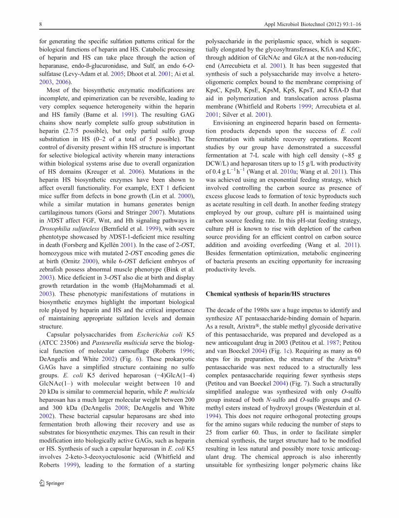
for generating the specific sulfation patterns critical for thebiological functions of heparin and HS. Catabolic processingof heparin and HS can take place through the action ofheparanase, endo-ß-glucuronidase, and Sulf, an endo 6-O-sulfatase (Levy-Adam et al. 2005; Dhoot et al. 2001; Ai et al.2003, 2006).
Most of the biosynthetic enzymatic modifications areincomplete, and epimerization can be reversible, leading tovery complex sequence heterogeneity within the heparinand HS family (Bame et al. 1991). The resulting GAGchains show nearly complete sulfo group substitution inheparin (2.7/5 possible), but only partial sulfo groupsubstitution in HS (0–2 of a total of 5 possible). Thecontrol of diversity present within HS structure is importantfor selective biological activity wherein many interactionswithin biological systems arise due to overall organizationof HS domains (Kreuger et al. 2006). Mutations in theheparin HS biosynthetic enzymes have been shown toaffect overall functionality. For example, EXT 1 deficientmice suffer from defects in bone growth (Lin et al. 2000),while a similar mutation in humans generates benigncartilaginous tumors (Gorsi and Stringer 2007). Mutationsin NDST affect FGF, Wnt, and Hh signaling pathways inDrosophilia sulfateless (Bernfield et al. 1999), with severephentotype showcased by NDST-1-deficient mice resultingin death (Forsberg and Kjellén 2001). In the case of 2-OST,homozygous mice with mutated 2-OST encoding genes dieat birth (Ornitz 2000), while 6-OST deficient embryos ofzebrafish possess abnormal muscle phenotype (Bink et al.2003). Mice deficient in 3-OST also die at birth and displaygrowth retardation in the womb (HajMohammadi et al.2003). These phenotypic manifestations of mutations inbiosynthetic enzymes highlight the important biologicalrole played by heparin and HS and the critical importanceof maintaining appropriate sulfation levels and domainstructure.
Capsular polysaccharides from Escherichia coli K5(ATCC 23506) and Pasteurella multicida serve the biolog-ical function of molecular camouflage (Roberts 1996;DeAngelis and White 2002) (Fig. 6). These prokaryoticGAGs have a simplified structure containing no sulfogroups. E. coli K5 derived heparosan (!4)GlcA(1–4)GlcNAc(1–) with molecular weight between 10 and20 kDa is similar to commercial heparin, while P. multicidaheparosan has a much larger molecular weight between 200and 300 kDa (DeAngelis 2008; DeAngelis and White2002). These bacterial capsular heparosans are shed intofermentation broth allowing their recovery and use assubstrates for biosynthetic enzymes. This can result in theirmodification into biologically active GAGs, such as heparinor HS. Synthesis of such a capsular heparosan in E. coli K5involves 2-keto-3-deoxyoctulosonic acid (Whitfield andRoberts 1999), leading to the formation of a starting
polysaccharide in the periplasmic space, which is sequen-tially elongated by the glycosyltransferases, KfiA and KfiC,through addition of GlcNAc and GlcA at the non-reducingend (Arrecubieta et al. 2001). It has been suggested thatsynthesis of such a polysaccharide may involve a hetero-oligomeric complex bound to the membrane comprising ofKpsC, KpsD, KpsE, KpsM, KpS, KpsT, and KfiA-D thataid in polymerization and translocation across plasmamembrane (Whitfield and Roberts 1999; Arrecubieta et al.2001; Silver et al. 2001).
Envisioning an engineered heparin based on fermenta-tion products depends upon the success of E. colifermentation with suitable recovery operations. Recentstudies by our group have demonstrated a successfulfermentation at 7-L scale with high cell density ("85 gDCW/L) and heparosan titers up to 15 g/L with productivityof 0.4 g L!1h!1 (Wang et al. 2010a; Wang et al. 2011). Thiswas achieved using an exponential feeding strategy, whichinvolved controlling the carbon source as presence ofexcess glucose leads to formation of toxic byproducts suchas acetate resulting in cell death. In another feeding strategyemployed by our group, culture pH is maintained usingcarbon source feeding rate. In this pH-stat feeding strategy,culture pH is known to rise with depletion of the carbonsource providing for an efficient control on carbon sourceaddition and avoiding overfeeding (Wang et al. 2011).Besides fermentation optimization, metabolic engineeringof bacteria presents an exciting opportunity for increasingproductivity levels.
Chemical synthesis of heparin/HS structures
The decade of the 1980s saw a huge impetus to identify andsynthesize AT pentasaccharide-binding domain of heparin.As a result, Arixtra®, the stable methyl glycoside derivativeof this pentasaccharide, was prepared and developed as anew anticoagulant drug in 2003 (Petitou et al. 1987; Petitouand van Boeckel 2004) (Fig. 1c). Requiring as many as 60steps for its preparation, the structure of the Arixtra®pentasaccharide was next reduced to a structurally lesscomplex pentasaccharide requiring fewer synthesis steps(Petitou and van Boeckel 2004) (Fig. 7). Such a structurallysimplified analogue was synthesized with only O-sulfogroup instead of both N-sulfo and O-sulfo groups and O-methyl esters instead of hydroxyl groups (Westerduin et al.1994). This does not require orthogonal protecting groupsfor the amino sugars while reducing the number of steps to25 from earlier 60. Thus, in order to facilitate simplerchemical synthesis, the target structure had to be modifiedresulting in less natural and possibly more toxic anticoag-ulant drug. The chemical approach is also inherentlyunsuitable for synthesizing longer polymeric chains like
8 Appl Microbiol Biotechnol (2012) 93:1–16

heparin because of the large number of modest yield stepsand side-product formation. Furthermore, separation ofproducts at each step provides for a challenge in scalingup these syntheses when competing against low-costheparin and LMWHs. The flexibility of chemical synthesisfor delivering unnatural oligosaccharides, however, providedan insight into the specific interactions of oligosaccharides,natural and unnatural, and helped to elucidate the specificityof heparin–protein interactions (Lee et al. 2004).
Synthesis of heparin oligosaccharides (Lee et al. 2004)are typically carried out using a stepwise convergentsynthesis as shown in the retrosynthetic scheme in Fig. 7.One pot synthesis and selective activation has recently beenapplied for oligosaccharide synthesis to reduce the numberof synthetic steps (Polat and Wong 2007). This one potsynthesis employs rapid oligosaccharide assembly bysequential addition of thioglycoside-building blocks inreducing order of their reactivity (Polat and Wong 2007;Zhang et al. 1999). AT-binding pentasaccharide has beensynthesized using one pot synthesis with starting mono-saccharides followed by global deprotection and sulfona-
tion (Zhang et al. 1999). In another such effort forsynthesizing AT-binding pentasaccharide, sequential glyco-sylation starting with monomer building blocks was used.This involved 1-thio uronic acid building blocks, which areactivated by the use of potent sulfonium activators resultingin highly stereoselective glycosylations leading towards afully protected pentasaccharide (Codée et al. 2005).Numerous oligosaccharide containing structural variationsare required for efficient SAR studies to completelyunderstand heparin specificity with respect to a given targetprotein (Arungundram et al. 2009). This has been achievedthrough a preactivation-based one-pot combinatorial syn-thesis of heparin hexasaccharides (Wang et al. 2010b). Inthis study, thioglycosyl building blocks activated bythiophilic promoters were used with matching of donorand acceptor pairs, thereby allowing formation of stereo-specific disaccharide building blocks. This resulted in apool of 12 hexasaccharides from initial six disaccharidesderived from two common intermediate disaccharides(Arungundram et al. 2009). Analysis of oligosaccharidesfor binding with FGF-2 showed the importance of N-sulfo
Fig. 6 Chemoenzymaticsynthesis of heparin andneoheparin starting from E.coli capsular polysaccharideheparosan. a This pathway forthe chemoenzymatic synthesisof bioengineered heparin closelyresembles the heparin/HSbiosynthetic pathway. Thechemical de-N-acetylation/N-sulfonation reaction mightbe replaced by an enzymaticreaction relying on NDST. bThis pathway for the synthesisof neoheparin requires selectivedesulfonation and resulfonationsteps and affords some 3-O-sulfoglucuronic acid andiduronic acid residues notfound in animal-derived heparin
Appl Microbiol Biotechnol (2012) 93:1–16 9

groups and GlcNS–IdoA2S–GlcNS (Wang et al. 2010b).Click chemistry has also been applied to development ofunnatural disaccharide and tetrasaccharide analogue ofheparosan (Bera and Linhardt 2011). Protected glucosamineand glucuronic acid building blocks with azide and alkynefunctional groups allow for the rapid and efficient iterative1,3-dipolar cycloaddition, leading to formation of "(1–4)linkage as heparosan analogues. Microarrays for screeningprotein binding to synthetic heparin oligosaccharides orpolysaccharides attached to a chip through an amine linker,compatible with protection group chemistry, have led tohigh throughput screening (de Paz et al. 2006; Park et al.2008). Development of such combinatorial oligosaccharidelibraries based on chemical synthesis provides for apowerful tool in SAR studies (Fig. 2) for heparin and HSthrough the use of such high throughput microarrays.
Chemoenzymatic synthesis of heparin, HS, and heparinoligosaccharides
Commercial-scale production of clinically safe heparinrequires a robust process that retains structural andfunctional properties of USP heparin derived from animalsources. The capsular polysaccharide derived from E. coliK5 strain, heparosan, consisting of repeating [!4) "-D-glucuronic acid (GlcA) (1–4) N-acetyl-!-D-glucosamine(GlcNAc)(1!]n units, has been characterized as a precursor
to heparin (Wang et al. 2010a; DeAngelis and White 2002).Heparosan biosynthesis has also been demonstrated in P.multicida (DeAngelis and White 2002). This capsularpolysaccharide might be modified into heparin/HS usingchemical or enzymatic steps involved in the biosyntheticpathway (Fig. 6).
N-Deacetylated and N-sulfonated heparosan is requiredfor recognition by C-5 epimerase and O-sulfotransferases(Chen et al. 2007; Bame et al. 1991). While the NDSTenzyme is involved in the biosynthetic pathway (Aikawaet al. 2001); hydrazine/hydrazine sulfate or NaOH can beused to chemically N-deacteylate heparosan followed bychemical N-sulfonation using trimethylamine-sulfur trioxidecomplex resulting in a chemically derived N-deacetylated/N-sulfonated intermediate called N-sulfo, N-acetyl heparosan(Wang et al. 2010a)
Structural constraints for formation of pentasacchar-ide sequence include a GlcNAc terminated GlcNAc–GlcA–GlcNS–IdoA–GlcNS substrate recognition of 3-O-sulfotransferase (Kusche et al. 1991b) for AT binding(Hricovíni et al. 2001). Lindahl and coworkers havedemonstrated gram-scale production of heparin-like polysac-charide, “neoheparin,” incorporating antithrombin bindingand anticoagulant activity starting from N-deacetylated/N-sulfated heparosan (Lindahl et al. 2004) (Fig. 6). In thisapproach, C5-epimerization of N-sulfoheparosan is carriedout enzymatically using C5-epimerase derived from insectcells with higher conversion ("60%) of GlcA–IdoA in the
Fig. 7 Convergent chemical synthesis of fondaparinux, which resembles the pentasaccharide AT-binding domain of heparin, stabilized as themethyl glycoside
10 Appl Microbiol Biotechnol (2012) 93:1–16

presence of divalent cations (Naggi et al. 2001). This is thenfollowed by per-O-sulfonation of OH groups present in N-sulfoheparosan. Graded solvolytic desulfonation leads toremoval of more of the unnatural 3-O-sulfo groups thannatural 2-O-sulfo groups in IdoA residues. The 3-O sulfogroups of GlcNAc residues were resistant to this desulfona-tion procedure and were retained compared to 6-O sulfogroups of GlcNAc that were lost. The presence of significantnumber of 3-O-sulfated GlcNAc residues adjacent tounsulfated GlcA affords a pentasaccharide structurallysimilar to that in the natural AT-binding site. Selective O-desulfonation and re-N,6-O-sulfonation of this per-O-sulfonated product led to the generation of a low molecularweight product with anticoagulant activity. Unfortunately, theneoheparin thus generated had a high proportion of 3-O-sulfated glucuronic acid sequences, which are absent inanimal-derived heparin, along with a significant amount ofthe unnatural 3-O-sulfo IdoA, which impede anti-factor Xaactivity (Rej et al. 1991). Oversulfated chondroitin sulfate,which resulted in the 2008 contamination crisis, alsopossesses unnatural 3-O-sulfo glucuronic acid residues, andthe presence of such unnatural sulfated patterns withinneoheparin leads to concerns regarding its clinical safety.
The problems encountered using the chemoenzymaticapproach to synthesize neoheparin show the challenges incontrolling sulfation patterns. Enzymatic approaches lackO-sulfo groups in unnatural sites like C3 of IdoA and GlcA.Enzymes involved in heparin and HS biosynthesis havebeen recently cloned and over-expressed in E. coli. Byscaling-up enzyme production, immobilizing theseenzymes, and preparing inexpensive 3!-phosphoadenosine5!-phosphosulfate (PAPS) cofactor, these sulfotransferasesmay be suitable for large-scale production (Chen et al.2007; Chen et al. 2005). A small-scale enzymatic approachinvolving enzymes from heparin and HS biosyntheticpathways has been demonstrated (Zhang et al. 2008b)(Fig. 6). In this study, isotopically labeled heparosan wasderived from E. coli K5, while recombinant human C5-epimerase, hamster 2-OST, hamster 6-OST-1, mouse 6-OST-3, and mouse 3-OST-1 were expressed and purifiedfrom E. coli. This approach is preceded by chemical N-deacetylation/N-sulfonation of heparosan using sodiumhydroxide and trimethylamine–sulfur trioxide complex,giving N-sulfoheparosan with reduced chain length due toalkaline hydrolysis. Treatment of N-sulfoheparosan with 2-OST and C5-epimerase yields an undersulfated heparin thatcan further be 6-O-sulfonated by the action of 6-OSTleading to heparin. The use of 3-OST-1, responsible formodification of very few residues, was the final stepresulting in anticoagulant heparin. Heparin and anticoagu-lant heparin thus generated resembled pharmaceuticalheparin with trisulfated IdoA2S(1,4)GlcNS6S, making up86–89% of the structure. Greater degree of control over
sulfation in this approach along with milligram-scaleproduction makes it a rational approach towards preparinga bioengineered heparin comparable to the animal-derivedproduct.
An important aspect of this chemoenzymatic approach iscoupling of PAPS regeneration system in enzymaticmodifications (Burkart et al. 2000). This involves the useof p-nitrophenyl sulfate as the sulfo donor in the presenceof arylsulfotransferase, which can easily be expressed in E.coli, with a catalytic amount of PAPS. This and otherinnovative technologies (Zhou et al. 2011) reduced theimpact of PAPS cost in the chemoenzymatic process,thereby improving scalability.
In addition to the potential for industrial production, achemoenzymatic approach provides for combinatorial syn-thesis of oligosaccharides and polysaccharides with novelheparin-like structures. Such combinatorial libraries areessential in elucidating the substrate specificity of variousheparin/HS biosynthetic pathway enzymes as well asproviding novel biologically active structures. In one suchapproach, chemically desulfonated N-sulfo heparin wasused as a starting material for modification using recombi-nantly produced immobilized HS sulfotransferases coupledwith PAPS regeneration system (Chen et al. 2005). Thisresulted in heparin/HS-like structures with varying sulfationpattern generated in a block fashion. These enzymaticallymodified products exhibited biological activities associatedwith heparin-like FGF2 binding, anticoagulant activity, andbinding to herpes simplex virus glycoprotein D (gD). Thismethod can be similarly used to generate an oligosaccha-ride library starting from disaccharide with unnatural UDP-monosaccharide as donor (Liu et al. 2010). The generatedoligosaccharides possess N-sulfo groups specifically placedalong the chain and provide for critical control in enzymaticsynthesis of HS oligosaccharides and polysaccharides. Thiscombinatorial approach is further strengthened by alteredsubstrate specificities of engineered sulfotransferases, lead-ing to an increased diversity within such oligosaccharide/polysaccharide library.
The combinatorial approach described above can beutilized for development of HS polysaccharides havinghighly defined functional activity. Using N-sulfo-heparosan,the generation of differentially sulfated HS structures hasshown that IdoA residue is not important for binding to AT-III as has been suggested in the literature (Chen et al. 2007).This has significant impact on reducing the complexity ofengineered heparin structures as epimerization of startingGlcA is reversible, thereby resulting in significant structuralheterogeneity. While IdoA has been deemed important inproviding conformational flexibility, thereby leading tofavorable binding to AT, it has been demonstrated that thisrequirement for flexibility is size dependent. This recombi-nant heparin version termed “Recomparin” lacks IdoA and
Appl Microbiol Biotechnol (2012) 93:1–16 11

a 2-O-sulfo group but shows anti-Xa activity at levelssimilar to heparin.
A recent work has shown that ultralow molecular weightheparins (ULMWHs), resembling porcine and bovineheparin’s pentasaccharide-binding domain, have been syn-thesized, starting from disaccharide acceptor followed bychain elongation using UDP-sugars and glycosyltrans-ferases. Modification of this backbone chain by heparin/HS biosynthetic pathway enzymes results in constructsresembling the pentasaccharide-binding domain providingfor a general chemoenzymatic method aimed at designingoligosaccharides with defined structures similar to Arixtra®.This scalable process provides for an easier method fordevelopment of oligosaccharides compared to cumbersomemultistep chemical synthesis (Xu et al. 2011).
In summary, these chemoenzymatic methods involvingrecombinant heparin/HS biosynthetic pathway enzymesprovide for scalable processes for bioengineered heparin/HS structures along with combinatorial tools for discoveryof new therapeutics having simplified structures along withdefined biological activity.
Metabolic engineering for heparin production
As a result of being produced as a serglycin proteoglycan ineukaryotic mast cells Golgi (Linhardt and Gunay 1999), thegenetic engineering of bacteria for heparin synthesis istechnologically infeasible. Such an approach would requireengineering of biosynthetic pathways and enzymes alongwith incorporation of activities associated with Golgi withinthe periplasmic space in bacteria that are known to producestructurally simple GAGs like heparosan, hyaluronan, andchondroitin (DeAngelis and White 2002; Wang et al.2010a). Owing to these problems, the biosynthesis ofheparin within eukaryotic systems like yeast, insect cells,and Chinese hamster ovary (CHO) cells appear to be moreachievable. Both yeast and CHO cells are widely used inthe biotechnological field with protocols in place for theirgrowth and purification of therapeutics derived fromthem while getting rid of the contaminating proteins(Yang et al. 2011).
Yeast strains have been shown to be capable ofgenerating essential glycosylation patterns in mammals.Engineered yeast strains have been shown to be capable ofproducing antibodies (e.g., anti CD antibodies) with uniqueglycosylation, thereby resulting in enhancement of biolog-ical activity (Li et al. 2006). They have been shown to becapable of generating complex hybrid mammalian glyco-proteins from mannose yeast glycoproteins (Hamilton andGerngross 2007). However, yeast does not produce HS, andbiosynthesis of heparin in yeast cells would be extremelychallenging, entailing the expression of high levels of core
protein along with controlled expression of enzymes fromthe entire biosynthetic pathway (Laremore et al. 2009).However, as a simpler recombinant protein expressionsystem, efforts have been underway for expression ofsulfotransferases (N-sulfotransferase, 3-OST, 2-OST, and6-OST) and the preparation of PAPS in yeast (Zhou et al.2011). In addition to their ease of purification, thesesecreted enzymes have also been shown to possess higherspecific activity and thermostability and do not containendotoxins. Yeast expression might serve as a criticaloptimization feature for scaling-up the chemoenzymaticproduction of bioengineered heparin. Glycoproteins withglycan structures similar to humans have been expressed ininsect cell systems (Wolff et al. 2001). Insect cells are alsocapable of the biosynthesis of HS (Staatz et al. 2001;Bernfield et al. 1999) and thus might be metabolicallyengineered to produce heparin. One issue using insect cellsis the difficulty associated with controlling baculorvirusexpressing system for commercial scale production (Yinet al. 2007). Further studies of insect cells might, however,be warranted.
CHO cells are mammalian cells known to produce HS;therefore, they may be capable of producing heparin as it isproduced through the same biosynthetic pathway (Bameet al. 1991; Zhang et al. 2006). CHO cells are deficient inseveral GAG modification enzymes and also lack granulesrequired for the storage of the heparin proteoglycans,serglycin. They express two out of four N-sulfotransferases,one out of three 6-O-sulfotranferases, and none of the 3-O-sulfotranferases (Zhang et al. 2006). Mutant CHO cellshaving deficient N-sulfotransferase have been shown topossess more sparse NS distribution when compared to thewild type (Bame et al. 1991). As N-sulfonation provides foran in increase in the substrate specificity of the O-sulfotransferases, genetic engineering of CHO cells forheparin production seems promising. Mutant CHO cellshave been shown to gain AT binding (Zhang et al. 2001),heparin cofactor II binding, and herpes simplex virus entry(O’Donnell et al. 2006). However, the production levels ofHS in CHO cells are very low in comparison to the highlevels of heparin found in (Zhang et al. 2006) mammalianmast cells. The generation of immortalized mastocytomacell lines, a rare form of mast cell cancer, might offeranother target for metabolic engineering. However, CHOcells currently provide the best alternative route for generationof heparin in eukaryotic cell lines.
The recent thrust towards development of an engineeredversion of heparin has focused on heparosan derived fromsimpler prokaryotic bacterial cells. Metabolic engineeringof bacterial cells provides for an alternative approachtowards increasing productivity levels of heparosan, which,when coupled with optimized fermentation strategies, canpotentially lead to an economically viable engineered
12 Appl Microbiol Biotechnol (2012) 93:1–16

heparin. It has been suggested that heparosan shares itsbiosynthetic pathway partially with cell wall biosynthesis.Towards this goal, UDP glucose dehydrogenase, thought tobe the rate-limiting step in mammalian GAG synthesis, wasoverexpressed in E. coli, resulting in decreased heparosanproduction with unaltered chain length (Roman et al. 2003),possibly due to reduction in UDP-GlcNAc, which becomesrate limiting besides interfering with polymerization (Romanet al. 2003; Wang et al. 2011). Modulating cell surfaceinteractions with capsules through mutation of waaR geneinvolved in lipopolysaccharide outer-core biosynthesis hasbeen shown to increase heparosan production (Taylor et al.2006). In another metabolic engineering approach, controlledover-expression of glycosyltransferases, KfiA and KfiC, candirect the biosynthesis of excess heparosan, drawing sugarsaway from cell wall biosynthesis (Wang et al. 2011).
Conclusion and future work
Understanding complex heparin structures that showwidespread involvement in diverse biological functionshas generated interest in the preparation and synthesis ofheparin and related poly-/oligosaccharides. Significantcontrol over heparin’s functions might be achieved bymodifying its fine structure, thereby leading to efficaciousnew drugs that can be targeted towards specific biologicalfunctions. Chemical synthesis will continue to drive thescreening of novel and unnatural GAG sequences. Enzyme-assisted synthesis should provide greater control withregards to modification of heparin structure leading to anenhanced understanding of heparin in SAR (Fig. 2). SARstudies are currently focused on reducing the detectionlimits and the use of high throughput microarray andmicrofluidic methods (Barbulovic-Nad et al. 2008; Martinet al. 2009), which can help in deciphering these complexstructures obtained from diverse sources. Evolution of abioengineered version of heparin derived from non-animalsources having well-defined structural and functionalproperties with good control over its properties could leadto a clinically safer and potent version of heparin. Thismight also reduce side effects and health concerns aroundthe oldest biopolymeric drug and open up new opportuni-ties for the development of novel carbohydrate basedtherapeutics.
References
Ai X, Do AT, Lozynska O, Kusche-Gullberg M, Lindahl U, EmersonCP Jr (2003) QSulf1 remodels the 6-O sulfation states of cellsurface heparan sulfate proteoglycans to promote Wnt signaling.J Cell Biol 162:341–351
Ai X, Do AT, Kusche-Gullberg M, Lindahl U, Lu K, Emerson CP(2006) Substrate specificity and domain functions of extracellularheparan sulfate 6-O-endosulfatases, QSulf1 and QSulf2. J BiolChem 281:4969–4976
Aikawa J, Grobe K, Tsujimoto M, Esko JD (2001) Multiple isozymesof heparan sulfate/heparin GlcNAcN-deacetylase/GlcN N-sulfotransferase. Structure and activity of the fourth member,NDST4. J Biol Chem 276:5876–5888
Arrecubieta C, Hammarton TC, Barrett B, Chareonsudjai S, HodsonN, Rainey D, Roberts IS (2001) The transport of group 2 capsularpolysaccharides across the periplasmic space in Escherichia coli.Roles for the KpsE and KpsD proteins. J Biol Chem 276:4245–4250
Arungundram S, Al-Mafraji K, Asong J, Leach FE III, Amster IJ,Venot A, Turnbull JE, Boons G-J (2009) Modular synthesis ofheparan sulfate oligosaccharides for structure!activity relation-ship studies. J Am Chem Soc 131:17394–17405
Bame KJ, Lidholt K, Lindahl U, Esko JD (1991) Biosynthesis ofheparan sulfate. Coordination of polymer-modification reactionsin a Chinese hamster ovary cell mutant defective in N-sulfotransferase. J Biol Chem 266:10287–10293
Barbulovic-Nad I, Yang H, Park PS, Wheeler AR (2008) Digitalmicrofluidics for cell-based assays. Lab Chip 8:519–526
Beaudet JM, Weyers A, Solakyildirim K, Yang B, Takieddin M,Mousa S, Zhang F, Linhardt RJ (2011) Impact of autoclavesterilization on the activity and structure of formulated heparin. JPharm Sci 100:3396–3404
Bera S, Linhardt RJ (2011) Design and synthesis of unnaturalheparosan and chondroitin building blocks. J Org Chem76:3181–3193
Bernfield M, Götte M, Park PW, Reizes O, Fitzgerald ML, LincecumJ, Zako M (1999) Functions of cell surface heparan sulfateproteoglycans. Annu Rev Biochem 68:729–777
Bink RJ, Habuchi H, Lele Z, Dolk E, Joore J, Rauch GJ, Geisler R,Wilson SW, den Hertog J, Kimata K, Zivkovic D (2003) Heparansulfate 6-O-sulfotransferase is essential for muscle developmentin zebrafish. J Biol Chem 278:31118–31127
Branellec J, Espejo J, Picart P (1997) Purified heparin fractions,method for obtaining them and pharmaceutical compositionscontaining them. US Patent 5,599,801
Burkart MD, Izumi M, Chapman E, Lin CH, Wong CH (2000)Regeneration of PAPS for the enzymatic synthesis of sulfatedoligosaccharides. J Org Chem 65:5565–5574
Carlsson P, Presto J, Spillmann D, Lindahl U, Kjellén L (2008)Heparin/heparan sulfate biosynthesis. Processive formation of N-sulfated domains. J Biol Chem 283:20008–20014
Chen J, Avci FY, Muñoz EM, McDowell LM, Chen M, Pedersen LC,Zhang L, Linhardt RJ, Liu J (2005) Enzymatic redesigning ofbiologically active heparan sulfate. J Biol Chem 280:42817–42825
Chen J, Jones CL, Liu J (2007) Using an enzymatic combinatorialapproach to identify anticoagulant heparan sulfate structures.Chem Biol 14:986–993
Chi L, Wolff JJ, Laremore TN, Restaino OF, Xie J, Schiraldi C, ToidaT, Amster IJ, Linhardt RJ (2008) Structural analysis of bikuninglycosaminoglycan. J Am Chem Soc 130:2617–2625
Codée JDC, Stubba B, Schiattarella M, Overkleeft HS, van BoeckelCAA, van Boom JH, van der Marel GA (2005) A modularstrategy toward the synthesis of heparin-like oligosaccharidesusing monomeric building blocks in a sequential glycosylationstrategy. J Am Chem Soc 127:3767–3773
de Paz JL, Noti C, Seeberger PH (2006) Microarrays of syntheticheparin oligosaccharides. J Am Chem Soc 128:2766–2767
DeAngelis PL (2008) Heparosan-based biomaterials and coatings andmethods of production and use thereof. US patent application2008/0226690 A1
Appl Microbiol Biotechnol (2012) 93:1–16 13

DeAngelis PL, White CL (2002) Identification and molecular cloningof a heparosan synthase from Pasteurella multocida Type D. JBiol Chem 277:7209–7213
Dhoot GK, Gustafsson MK, Ai X, Sun W, Standiford DM, EmersonCP Jr (2001) Regulation of Wnt signaling and embryo patterningby an extracellular sulfatase. Science 293:1663–1666
Esko JD, Selleck SB (2002) Order out of chaos: assembly of ligandbinding sites in heparan sulfate. Annu Rev Biochem 71:435–471
Forsberg E, Kjellén L (2001) Heparan sulfate: lessons from knockoutmice. J Clin Invest 108:175–180
Gorsi B, Stringer SE (2007) Tinkering with heparan sulfate sulfationto steer development. Trends Cell Biol 17:173–177
Gray E, Mulloy B, Barrowcliffe TW (2008) Heparin and low-molecular-weight heparin. Thromb Haemost 99:807–818
Guerrini M, Beccati D, Shriver Z, Naggi A, Viswanathan K, Bisio A,Capila I, Lansing JC, Guglieri S, Fraser B, Al-Hakim A, GunayNS, Zhang Z, Robinson L, Buhse L, Nasr M, Woodcock J,Langer R, Venkataraman G, Linhardt RJ, Casu B, Torri G,Sasisekharan R (2008) Oversulfated chondroitin sulfate is acontaminant in heparin associated with adverse clinical events.Nat Biotechnol 26:669–675
Habuchi H, Miyake G, Nogami K, Kuroiwa A, Matsuda Y, Kusche-Gullberg M, Habuchi O, Tanaka M, Kimata K (2003) Biosyn-thesis of heparan sulphate with diverse structures and functions:two alternatively spliced forms of human heparan sulphate 6-O-sulphotransferase-2 having different expression patterns andproperties. Biochem J 371:131–142
HajMohammadi S, Enjyoji K, Princivalle M, Christi P, Lech M,Beeler D, Rayburn H, Schwartz JJ, Barzegar S, de Agostini AI,Post MJ, Rosenberg RD, Shworak NW (2003) Normal levels ofanticoagulant heparan sulfate are not essential for normalhemostasis. J Clin Invest 111:989–999
Hamilton SR, Gerngross TU (2007) Glycosylation engineering inyeast: the advent of fully humanized yeast. Curr Opin Biotechnol18:387–392
Hricovíni M, Guerrini M, Bisio A, Torri G, Petitou M, Casu B (2001)Conformation of heparin pentasaccharide bound to antithrombinIII. Biochem J 359:265–272
Jacobsson I, Lindahl U, Jensen J, Rodén L, Prihar H, Feingold DS(1984) Biosynthesis of heparin. Substrate specificity of heparosanN-sulfate D-glucuronosyl 5-epimerase. J Biol Chem 259:1056–1063
Johnson EA, Kirkwood TB, Stirling Y, Perez-Requejo JL, Ingram GI,Bangham DR, Brozovi! M (1976) Four heparin preparations:anti-Xa potentiating effect of heparin after subcutaneousinjection. Thromb Haemost 35:586–591
Kishimoto TK, Viswanathan K, Ganguly T, Elankumaran S, Smith S,Pelzer K, Lansing JC, Sriranganathan N, Zhao G, Galcheva-Gargova Z, Al-Hakim A, Bailey GS, Fraser B, Roy S, Rogers-Cotrone T, Buhse L, Whary M, Fox J, Nasr M, Dal Pan GJ,Shriver Z, Langer RS, Venkataraman G, Austen KF, Woodcock J,Sasisekharan R (2008) Contaminated heparin associated withadverse clinical events and activation of the contact system. NEngl J Med 358:2457–2467
Kramer KL, Yost HJ (2003) Heparan sulfate core proteins in cell-cellsignaling. Annu Rev Genet 37:461–484
Kreuger J, Spillmann D, Li JP, Lindahl U (2006) Interactions betweenheparan sulfate and proteins: the concept of specificity. J CellBiol 174:323–327
Kusche M, Hannesson HH, Lindahl U (1991a) Biosynthesis ofheparin. Use of Escherichia coli K5 capsular polysaccharide asa model substrate in enzymic polymer-modification reactions.Biochem J 275:151–158
Kusche M, Oscarsson LG, Reynertson R, Rodén L, Lindahl U (1991b)Biosynthesis of heparin. Enzymatic sulfation of pentasaccharides.J Biol Chem 266:7400–7409
Laremore TN, Zhang F, Dordick JS, Liu J, Linhardt RJ (2009) Recentprogress and applications in glycosaminoglycan and heparinresearch. Curr Opin Chem Biol 13:633–640
Laremore TN, Leach FE III, Amster IJ, Linhardt RJ (2010) Electro-spray ionization Fourier transform mass spectrometric analysis ofintact bikunin glycosaminoglycan from normal human plasma.Int J Mass spectrom 305:109–115
Lee JC, Lu XA, Kulkarni SS, Wen YS, Hung SC (2004) Synthesis ofheparin oligosaccharides. J Am Chem Soc 126:476–477
Levy-Adam F, Abboud-Jarrous G, Guerrini M, Beccati D, VlodavskyI, Ilan N (2005) Identification and characterization of heparin/heparan sulfate binding domains of the endoglycosidaseheparanase. J Biol Chem 280:20457–20466
Li H, Sethuraman N, Stadheim TA, Zha D, Prinz B, Ballew N,Bobrowicz P, Choi BK, Cook WJ, Cukan M, Houston-Cummings NR, Davidson R, Gong B, Hamilton SR, HoopesJP, Jiang Y, Kim N, Mansfield R, Nett JH, Rios S,Strawbridge R, Wildt S, Gerngross TU (2006) Optimizationof humanized IgGs in glycoengineered Pichia pastoris. NatBiotechnol 24:210–215
Li B, Suwan J, Martin JG, Zhang F, Zhang Z, Hoppensteadt D, ClarkM, Fareed J, Linhardt RJ (2009) Oversulfated chondroitin sulfateinteraction with heparin-binding proteins: new insights intoadverse reactions from contaminated heparins. Biochem Pharmacol78:292–300
Lin X, Wei G, Shi Z, Dryer L, Esko JD, Wells DE, Matzuk MM(2000) Disruption of gastrulation and heparan sulfate biosynthe-sis in EXT1-deficient mice. Dev Biol 224:299–311
Lindahl U, Li JP, Kusche-Gullberg M, Salmivirta M, Alaranta S,Veromaa T, Emeis J, Roberts I, Taylor C, Oreste P, Zoppetti G,Naggi A, Torri G, Casu B (2004) Generation of “Neoheparin”from E. coli K5 capsular polysaccharide. J Med Chem 48:349–352
Linhardt RJ (1996) Analysis of glycosaminoglycans with polysaccha-ride lyases. Curr Protoc Mol Biol 17.13B.1–17.13B.16
Linhardt RJ, Gunay NS (1999) Production and chemical processing oflow molecular weight heparins. Semin Thromb Hemost 25:5–16
Linhardt RJ, Ampofo SA, Fareed J, Hoppensteadt D, Mulliken JB,Folkman J (1992) Isolation and characterization of humanheparin. Biochemistry 31:12441–12445
Liu D, Pojasek K, Shriver Z, Holley K, El-Shabrawi Y, VenkataramanG, Sasisekharan R (2008) Methods for preparing low molecularweight heparin with modified heparinase III. US Patent7,390,633 B2
Liu H, Zhang Z, Linhardt RJ (2009) Lessons learned from thecontamination of heparin. Nat Prod Rep 26:313–321
Liu R, Xu Y, Chen M, Weïwer M, Zhou X, Bridges AS, DeAngelisPL, Zhang Q, Linhardt RJ, Liu J (2010) Chemoenzymatic designof heparan sulfate oligosaccharides. J Biol Chem 285:34240–34249
Ly M, Laremore TN, Linhardt RJ (2010) Proteoglycomics: recentprogress and future challenges. OMICS 14:389–399
Mao W, Thanawiroon C, Linhardt RJ (2002) Capillary electrophoresisfor the analysis of glycosaminoglycans and glycosaminoglycanderived oligosaccharides. Biomed Chromatogr 16:77–94
Mardiguian JS (1984) Heparin esters and processes for theirpreparation. US Patent 4,440,926
Martin JG, Gupta M, Xu Y, Akella S, Liu J, Dordick JS, Linhardt RJ(2009) Toward an artificial Golgi: redesigning the biologicalactivities of heparan sulfate on a digital microfluidic chip. J AmChem Soc 131:11041–11048
Mousa SA, Zhang F, Aljada A, Chaturvedi S, Takieddin M, Zhang H,Chi L, Castelli MC, Friedman K, Goldberg MM, Linhardt RJ(2007) Pharmacokinetics and pharmacodynamics of oral heparinsolid dosage form in healthy human subjects. J Clin Pharmacol47:1508–1520
14 Appl Microbiol Biotechnol (2012) 93:1–16

Mozen MM, Evans TD (1962) Process for purifying heparin. USPatent 3,058,884
Muñoz EM, Yu H, Hallock J, Edens RE, Linhardt RJ (2005) Poly(ethylene glycol)-based biosensor chip to study hepari proteininteractions. Anal Biochem 343:176–178
Nadji S, Smoot JT, Vanartsdalen JA (2011) Process for preparaingfondaparinux sodium and intermediates useful in the synthesisthereof. US Patent Application US 2011/0105418 A1
Naggi A, Torri G, Casu B, Oreste P, Zoppetti G, Li JP, Lindahl U(2001) Toward a biotechnological heparin through combinedchemical and enzymatic modification of the Escherichia coli K5polysaccharide. Semin Thromb Hemost 27:437–443
Nominé G, Barthelemy P (1961) Process of purifying heparin USPatent 2,989,438
O’Donnell CD, Tiwari V, Oh MJ, Shukla D (2006) A role for heparansulfate 3-O-sulfotransferase isoform 2 in herpes simplex virustype 1 entry and spread. Virology 346:452–459
Ornitz DM (2000) FGFs, heparan sulfate and FGFRs: complexinteractions essential for development. Bioessays 22:108–112
Panasyuk AF (2011) Method for producing sulphated glycosamino-glycans from biological tissues. US Patent 7,943,764 B2
Park T-J, Lee M-Y, Dordick JS, Linhardt RJ (2008) Signalamplification of target protein on heparin glycan microarray.Anal Biochem 383:116–121
Pervin A, Gu K, Linhardt R (1993) Capillary electrophoresis tomeasure sulfoesterase activity on chondroitin sulfate and heparinderived disaccharides. Appl Theor Electrophor 3:297–303
Pervin A, Gallo C, Jandik KA, Han XJ, Linhardt RJ (1995)Preparation and structural characterization of large heparin-derived oligosaccharides. Glycobiology 5:83–95
Petitou M, van Boeckel CAA (2004) A synthetic antithrombin IIIbinding pentasaccharide is now a drug! What comes next?Angew Chem Int Ed Engl 43:3118–3133
Petitou M, Duchaussoy P, Lederman I, Choay J, Jacquinet JC, Sinay P,Torri G (1987) Synthesis of heparin fragments: a methyl [alpha]-pentaoside with high affinity for antithrombin III. Carbohydr Res167:67–75
Polat T, Wong CH (2007) Anomeric reactivity-based one-potsynthesis of heparin-like oligosaccharides. J Am Chem Soc129:12795–12800
Rej RN, Ludwig-Baxter KG, Perlin AS (1991) Sulfation of somechemically-modified heparins. Formation of a 3-sulfate analog ofheparin. Carbohydr Res 210:299–310
Roberts IS (1996) The biochemistry and genetics of capsularpolysaccharide production in bacteria. Annu Rev Microbiol50:285–315
Robinson H, Horner A, Höök M, Ogren S, Lindahl U (1978) Aproteoglycan form of heparin and its degradation to single-chainmolecules. J Biol Chem 253:6687–6693
Roman E, Roberts I, Lidholt K, Kusche-Gullberg M (2003) Over-expression of UDP-glucose dehydrogenase in Escherichia coliresults in decreased biosynthesis of K5 polysaccharide. BiochemJ 374:767–772
Rosenberg RD (1981) Heparin preparation. US Patent 4,301,153Rosenberg RD (1985) Affinity fractionation of heparin on immobi-
lized Concanavalin A. US Patent 4,539,398Sawesi O, Spillmann D, Lundén A, Wernersson S, Åbrink M (2010)
Serglycin-independent release of active mast cell proteases inresponse to Toxoplasma gondii infection. J Biol Chem285:38005–38013
Silver RP, Prior K, Nsahlai C, Wright LF (2001) ABC transporters andthe export of capsular polysaccharides from Gram-negativebacteria. Res Microbiol 152:357–364
Smith MR, Amaya E, Fussi F (1984) Process for manufacturing lowmolecular weight heparins by depolymerization of normalheparin. Eur Pat Appl EP0101141
Staatz WD, Toyoda H, Kinoshita-Toyoda A, Chhor K, Selleck SB(2001) Analysis of proteoglycans and glycosaminoglycans fromDrosophila. Methods Mol Biol 171:41–52
Taylor CM, Goldrick M, Lord L, Roberts IS (2006) Mutations inthe waaR gene of Escherichia coli which disrupt lipopoly-saccharide outer core biosynthesis affect cell surface retentionof group 2 capsular polysaccharides. J Bacteriol 188:1165–1168
Turnbull JE (1993) Oligosaccharide mapping and sequenceanalysis of glycosaminoglycans. Methods Mol Biol 19:253–267
van Boeckel CAA, Grootenhuis PDJ, Meuleman D, Westerduin P(1995) Glycosaminoglycans: synthetic fragments and theirinteraction with serine protease inhibitors. Pure Appl Chem67:1663–1672
Vidic HJ (1981) Process for the preparation of heparin. US Patent4,283,530
Wang Z, Ly M, Zhang F, Zhong W, Suen A, Hickey AM, Dordick JS,Linhardt RJ (2010a) E. coli K5 fermentation and the preparationof heparosan, a bioengineered heparin precursor. BiotechnolBioeng 107:964–973
Wang Z, Xu Y, Yang B, Tiruchinapally G, Sun B, Liu R, Dulaney S,Liu J, Huang X (2010b) Preactivation based, one pot combina-torial synthesis of heparin like hexasaccharides for the analysis ofheparin–protein interactions. Chemistry 16:8365–8375
Wang Z, Dordick JS, Linhardt RJ (2011) E. coli K5 heparosanfermentation and improvement by genetic engineering. BioengBugs 2:63–67
Westerduin P, van Boeckel CAA, Basten JEM, Broekhoven MA,Lucas H, Rood A, van der Heijden H, van Amsterdam RGM, vanDinther TG, Meuleman DG, Visser A, Vogel GMT, Damm JBL,Overklift GT (1994) Feasible synthesis and biological propertiesof six ‘non-glycosamino’ glycan analogues of the antithrombinIII binding heparin pentasaccharide. Bioorg Med Chem 2:1267–1280
Whitfield C, Roberts IS (1999) Structure, assembly and regulation ofexpression of capsules in Escherichia coli. Mol Microbiol31:1307–1319
Wolff MW, Zhang F, Roberg JJ, Caldwell EEO, Kaul PR, Serrahn JN,Murhammer DW, Linhardt RJ, Weiler JM (2001) Expression ofC1 esterase inhibitor by the baculovirus expression vectorsystem: preparation, purification, and characterization. ProteinExpr Purif 22:414–421
Xu Y, Masuko S, Takieddin M, Xu H, Liu R, Jing J, Mousa S,Linhardt RJ, Liu J, (2011) Chemoenzymatic synthesis ofstructurally homogeneous ultra-low molecular weight heparins.Science (in press)
Yang B, Weyers A, Baik JY, Sterner E, Sharfstein S, Mousa SA,Zhang F, Dordick JS, Linhardt RJ (2011) Ultra-performance ion-pairing liquid chromatography with on-line electrospray ion trapmass spectrometry for heparin disaccharide analysis. AnalBiochem 415:59–66
Yin J, Li G, Ren X, Herrler G (2007) Select what you need: acomparative evaluation of the advantages and limitations offrequently used expression systems for foreign genes. J Bio-technol 127:335–347
Zhang Z, Ollmann IR, Ye X-S, Wischnat R, Baasov T, Wong C-H(1999) Programmable one-pot oligosaccharide synthesis. J AmChem Soc 121:734–753
Zhang L, Beeler DL, Lawrence R, Lech M, Liu J, Davis JC, Shriver Z,Sasisekharan R, Rosenberg RD (2001) 6-O-sulfotransferase-1represents a critical enzyme in the anticoagulant heparan sulfatebiosynthetic pathway. J Biol Chem 276:42311–42321
Zhang L, Lawrence R, Frazier BA, Esko JD (2006) CHOglycosylation mutants: proteoglycans. Methods Enzymol 416:205–221
Appl Microbiol Biotechnol (2012) 93:1–16 15

Zhang Z, Weïwer M, Li B, Kemp MM, Daman TH, Linhardt RJ (2008a)Oversulfated chondroitin sulfate: impact of a heparin impurity,associated with adverse clinical events, on low-molecular-weightheparin preparation. J Med Chem 51:5498–5501
Zhang Z, McCallum SA, Xie J, Nieto L, Corzana F, Jiménez-BarberoJ, Chen M, Liu J, Linhardt RJ (2008b) Solution structures of
chemoenzymatically synthesized heparin and its precursors. JAm Chem Soc 130:12998–13007
Zhou X, Chandarajoti K, Pham TQ, Liu R, Liu J (2011) Expression ofheparan sulfate sulfotransferases in Kluyveromyces lactis andpreparation of 3-phosphoadenosine-5-phosphosulfate. Glycobiology21:771–780
16 Appl Microbiol Biotechnol (2012) 93:1–16