Engineering of Lipid Metabolism in the Diatom...
106
Engineering of Lipid Metabolism in the Diatom Phaeodactylum tricornutum Kenneth GOOSSENS Master’s dissertation submitted to obtain the degree of Master of Biochemistry and Biotechnology Major Structural Biotechnology and Biochemistry Academic year 2011-2012 Promoters: Prof. dr. Alain Goossens and Prof. dr. Wim Vyverman Scientific supervisor: Michiel Matthijs Department Plant Biotechnology and Bioinformatics VIB - Department Plant Systems Biology
Transcript of Engineering of Lipid Metabolism in the Diatom...

Engineering of Lipid Metabolism in the Diatom
Phaeodactylum tricornutum
Kenneth GOOSSENS
Master’s dissertation submitted to obtain the degree of
Master of Biochemistry and Biotechnology
Major Structural Biotechnology and Biochemistry
Academic year 2011-2012
Promoters: Prof. dr. Alain Goossens and
Prof. dr. Wim Vyverman
Scientific supervisor: Michiel Matthijs
Department Plant Biotechnology and Bioinformatics
VIB - Department Plant Systems Biology


I
Acknowledgements
I would like to thank those people that made it possible for me to finish my master thesis. Most thank goes to all the people in the lab that helped me gain some insight in what it means to be a scientist. Special gratitude goes to the following people: Prof. Dr. Alain Goossens, Michele and the other members of the metabol group for their advice about a number of protocols; Michiel and Dr. Guino Baart for their editing skills; Wilson for processing our numerous sequencing requests; Sophie for her help with the practical work; my promoter Prof. Dr. Alain Goossens, copromotor Prof. Dr. Wim Vyverman and the members of the diatom group (Sophie, Michele, Michiel and Dr. Guino Baart) for providing me with an interesting master thesis subject and sufficient background information.
Special thanks goes to my supervisor Michiel Matthijs. I would like to thank him for his support, his patience and his scholarship to help me finishing this master project to a successful end.

II

III
Table of contents
Acknowledgements .................................................................................................................................. I
Table of contents .................................................................................................................................... III
List of abbreviations ................................................................................................................................ V
Nederlandse samenvatting ................................................................................................................... VII
English summary .................................................................................................................................... IX
Part 1: Introduction ................................................................................................................................. 1
1.1. General characteristics of diatoms .......................................................................................... 1
1.2. Potential applications of diatoms ............................................................................................ 2
1.3. Lipid metabolism ..................................................................................................................... 4
1.3.1. Typical mammalian lipid features (Voet, 2008) .............................................................. 4
1.3.2. Typical plant lipid features .............................................................................................. 7
1.3.3. Typical diatom lipid features ........................................................................................... 7
1.4. The model organism: Phaeodactylum tricornutum .............................................................. 10
1.5. Metabolic engineering of the lipid metabolism .................................................................... 10
1.5.1. The metabolic engineering strategy .............................................................................. 10
1.5.2. Previously conducted work ........................................................................................... 11
1.5.3. Gene targets .................................................................................................................. 12
Part 2: Aim of the research project ....................................................................................................... 15
Part 3: Results ........................................................................................................................................ 17
3.1. Vector construction ............................................................................................................... 17
3.1.1. Overexpression vectors ................................................................................................. 17
3.1.2. RNAi vector .................................................................................................................... 18
3.2. Metabolic engineering of the target genes ........................................................................... 20
3.2.1. Strategy 1: creating a dominant negative kinase dead AMPK ...................................... 20
3.2.2. Strategy 2: overexpressing and knocking down TOR interactors .................................. 40
3.2.3. Strategy 3: overexpressing and knocking down geranyl. .............................................. 44
Part 4: Discussion .................................................................................................................................. 45
4.1. Construction of the RNAi vector............................................................................................ 45
4.2. Analysis of the dominant negative AMPK transformants ..................................................... 46
4.3. Other target genes and future prospects .............................................................................. 47
Part 5: Materials and methods .............................................................................................................. 49
5.1. Cloning of target genes ......................................................................................................... 49
5.2. Vector construction ............................................................................................................... 50

IV
5.3. Generation of a kinase dead AMPK ....................................................................................... 52
5.4. Generation of transgenic Phaeodactylum tricornutum ........................................................ 53
5.5. Monitoring the expression of the transgenes using QPCR ................................................... 53
5.6. Phenotypic analysis .................................................................................................................... 54
5.6.1. (Fluorescence) microscopy of the diatom cells ............................................................. 54
5.6.2. Growth analysis using OD measurement ...................................................................... 54
5.6.3. Analysis of the cell cycle using flow cytometry ............................................................. 54
5.6.4. Quantitative sugar analysis: phenol-sulphate method ................................................. 55
References ............................................................................................................................................. 57
Attachments .......................................................................................................................................... 63
Protocols ............................................................................................................................................ 63
Composition of media ....................................................................................................................... 80
Samenvatting (discussie) ................................................................................................................... 82
Primers .............................................................................................................................................. 86
Raw data ............................................................................................................................................ 88

V
List of abbreviations
3PGA, 3-phosphoglyceric acid; ACCase, acetyl-CoA carboxylase; ACP, acyl carrier protein; AMPK, AMP activated protein kinase; ARA, arachidonic acid; αγ-SBS, α and γ subunit binding sequence; BLAST, basic local alignment search tool; β-SID, β subunit interacting domain; CAT, chloramphenicol resistance; CBS, cystathionine β synthase; cDNA, copy DNA; CrAT, carnitine acyltransferase; Ct, cycle threshold; DAGAT, diacylglycerol acyltransferase; DAPI, 4,6-diamidino-2-phenylindole; DHA, docosahexaenoic acid; DHAP, dihydroxyacetone phosphate; DIC, differential interference contrast; EPA, eicosapentaenoic acid; E(N)R, Enoyl-ACP reductase; ESAW, enriched seawater, artificial water; EST, expressed sequence tag; FAS, fatty acid synthase; FAT, fatty acyl-ACP thioesterase; FSC, forward light scatter; FU, fluorescence units; FW, forward; G3PDH, glycerol-3-phosphate dehydrogenase; GBD,
glycogen binding domain; gDNA, genomic DNA; GFP, green fluorescence protein; GPAT, glycerol-3-phosphate-acyltransferase; GSK, glycogen synthase kinase; GW, gateway; HD, β-hydroxyacyl-ACP dehydrase; JGI, joint genome institute; KAR, β-ketoacyl-ACP reductase; KAS, β-ketoacyl-ACP synthase; LPAAT, lysophosphatidic acid acyltransferase; LPAT, lysophosphatidylcholine acyltransferase; MAPK, mitogen activated protein kinase; MAT, malonyl/acetyl-CoA-ACP-transacylase; ME, metabolic engineering; MORG, MAPK organizer; mTOR, mammalian target of rapamycin; NADPH, nicotinamide adenine dinucleotide phosphate; OD, optical density; PCR, polymerase chain reaction; PUFAs, polyunsaturated fatty acids; QPCR, quantitative polymerase chain reaction RACE, rapid amplifying cDNA ends; RE, restriction enzyme; Rheb, Ras homolog enriched in brain; Sir, silent information
regulator; RPS, ribosomal proteins ;RV, reverse; TAGs, triacylglycerols; TBP, TATA-box binding protein; TE, thioesterase, TSAP, thermo sensitive shrimp alkaline phosphatise; tubB, tubuline subunit b; YFP, yellow fluorescence protein

VI

VII
Nederlandse samenvatting
Diatomeeën zijn eencellige algen die abundant voorkomen in mariene en andere aquatische niches. Deze organismen bezitten enkele opmerkelijke eigenschappen die momenteel intensief worden bestudeerd. Eén van deze eigenschappen is de aanwezigheid van lipiden met een nutritionele en economische waarde, waaronder enkele polyonverzadigde lipiden (PUFAs). Dit draagt bij tot het gebruik van diatomeeën als potentiële bron van lipiden voor de productie van biobrandstof en voor de extractie van polyonverzadigde lipiden voor nutritionele doeleinden. Helaas kunnen diatomeeën op dit moment niet concurreren met alternatieven, zoals planten-, en visolie, door hun beperkte productie capaciteit. De moleculaire kennis beschikbaar voor de manipulatie en engineering van diatomeeën is bovendien beperkt. Het doel van dit onderzoek was tweevoudig. In de
eerste plaats probeerden we de moleculaire hulpmiddelen uit te breiden door de constructie van een RNAi vector die de expressie van bepaalde target genen inhibeert. Een vector werd gecreëerd die werkzaam leek op en moleculair niveau, maar waarvoor functionaliteit nog niet kon worden aangetoond op het transcriptieniveau. Ten tweede probeerde dit onderzoek bij te dragen tot een verhoogde synthese van lipiden in diatomeeën door gebruik te maken van metabolische engineering technieken. Voor deze strategie werden verschillende targetgenen uitgeprobeerd. Deze genen werden geplaatst in een andere regulatorische context en getransformeerd in diatomeeën. Er werden transformante celculturen gecreëerd met een gewijzigd genotype voor een aantal TOR (target of rapamycin) interactoren en voor AMPK (AMP activated protein kinase). TOR interactoren
werden ofwel gebruikt in een overexpressie strategie, ofwel in een knockdown strategie. AMPK werd geïnhibeerd door de creatie van een dominant negatieve, kinase disfunctionele vorm. Transformante AMPK lijnen werden geanalyseerd op fenotypisch niveau. Een aantal celculturen vertoonden wijzigingen in groei en celcyclus progressie, maar geen sluitend bewijs kon verkregen worden voor deze vaststellingen. Studie van de getransformeerde culturen op het lipiden niveau en optimalisatie van de RNAi vector zijn nog steeds gaande.

VIII

IX
English summary
Diatoms are unicellular algae which are abundant in marine and other aquatic environments. These organisms possess interesting characteristics which are currently under intense study. One of these characteristics is the presence of lipids of nutritional and economic importance, such as polyunsaturated fatty acids (PUFAs). This makes diatoms a potential source for the extraction of lipids for the production of biofuels and polyunsaturated lipids for nutrition. However, diatoms can still not economically compete with the alternatives such as plant and fish oil, due to limited productivity. The molecular tools available for manipulation and engineering of diatoms are quite limited. The goal of this research was twofold. First, we tried to expand the molecular toolkit of diatoms by constructing an RNAi vector to knock down expression of certain target genes. Creation of
this vector led to a construct which seemed workable at a molecular level, but for which functionality could not yet be proved on the transcript level. Second, this research tried to contribute to an improved lipid production in diatoms by making use of metabolic engineering strategies. For this strategy different target genes were assessed. These target genes were put under a different regulatory context and were transformed in diatom cells. Transformed cell cultures were created with an altered genotype for a number of TOR (target of rapamycin) interactors and for AMPK (AMP activated protein kinase). The TOR interactors were either targeted for overexpression or knocked down, AMPK was inhibited by the creation of a dominant negative kinase dead form. Transformant AMPK lines were analysed for changes on the phenotype level. A number of cell cultures showed changes in
growth and cell cycle progression, but no definite evidence for these changes was obtained. Study of the transformant cell cultures on the lipid level and further improvement of the RNAi vector are still ongoing.

X

Part 1: Introduction
1
Part 1: Introduction
1.1. General characteristics of diatoms
Diatoms are photoautotrophic unicellular algae (figure 1). They are an important factor for the ecological balance, because their fixation of carbon dioxide is responsible for about
20-25% of the global primary production and they contribute to about 40% of the organic matter production in the ocean. Current numbers estimate that there are about 100,000 species and 250 genera (Lebeau & Robert, 2003). The major storage molecules of diatoms are triacylglycerols (TAGs) and polysaccharides. The predominant storage glucan is chrysolaminaran. It is used as a respiratory substrate and it is an important source for the synthesis of precursors of amino acids and other components (Granum & Myklestad, 2001). Other noteworthy characteristics are the presence of a silica cell wall (a frustule) and the
presence of four cell membranes around the chloroplast, this is most likely a remnant of a secondary endosymbiosis event. This four layered membrane contributes to the evolutionary development theory for diatoms. Presumably, ancestors of red and green algae
arose through an endosymbiosis of a photosynthetic prokaryote and a eukaryotic cell. Next, diatoms developed through a second endosymbiosis of a red algae ancestor and a heterotrophic eukaryote. Some studies even suggest the acquisition of a plastidial organelle through endosymbiosis with either a green or a red algae ancestor, meaning diatoms can’t be considered as a monophyletic group (Moustafa et al, 2009). This evolutionary trajectory was supported by genome analysis: homologous genes of the animal and plant kingdom were found, but also hundreds of genes that resemble those of prokaryotes. These prokaryotic genes probably got inserted in the diatom genome by horizontal gene transfer (Saade & Bowler, 2009). This evolutionary descent, leading to the presence of genes from different origins, has promoted the existence of a flexible metabolism for these organisms. This history could be at the basis for the rapid speciation and explain why diatoms are one of
the most successful aquatic organisms.
Figure 1: Phaeodactylum tricornutum, a pennate
marine diatom. The model organism used in the
experiments conducted in this project.

Part 1: Introduction
2
1.2. Potential applications of diatoms
Diatoms form certain metabolites which make them interesting for industrial use. Two examples of this are the synthesis of TAGs to produce biodiesel (Chisti, 2007), and the synthesis of PUFAs (= polyunsaturated fatty acids), which possess certain characteristics beneficial for human health (Hoffmann et al, 2008). In both examples it are lipids which make diatoms interesting for industrial and economical purposes.
Looking at the lipid content and fatty acid composition of the diatoms, some interesting observations have been made. The most common lipids are neutral lipids, such as TAGs which function as storage lipids (Berge, 1995; Hu et al, 2008; Yongmanitchai & Ward, 1991). Berge et al observed in S. costatum that the predominant lipid species were the polar lipids,
while the amount of storage or neutral lipids were quite low compared to the total amount (Berge, 1995). A possible explanation for this observation is that membranes are mostly build from glyco- and phospholipids. But it has been frequently reported that, when diatoms are put under stress conditions (e.g. photo-oxidative stress or nutrient starvation), the concentration of storage lipids rises. This phenomenon can partially be explained by a shift in lipid metabolism from the synthesis of membrane to storage lipids (Hu et al, 2008; Guschina
& Harwood, 2006; Yongmanitchai & Ward, 1991). The building blocks for the lipids are fatty acids. These fatty acids consist of an acyl chain and a carboxylic end group. Differences can be found in the number of carbon atoms in the acyl chain, the saturation grade, and the place(s) of the double bound. Three forms of saturation grade are possible, either completely saturated (no double bounding), mono-unsaturated (one double bond) or
polyunsaturated (two or more double bonds). This last form of fatty acids are called PUFAs (Hu et al, 2008). Examples of PUFAs are arachidonic acid (ARA, C20:46), eicosapentaenoic acid (EPA, C20:53) and docosahexaenoic acid (DHA, C22:63). Which fatty acids occur most in the lipids of diatoms differs from species to species. In most algae saturated and mono-unsaturated fatty acids are predominant, but some species are inconsistent with this observation. Phaeodactylum tricornutum for example synthesises large amounts of long-chain PUFAs, mostly EPA (Lebeau & Robert, 2003). Concluding, the amount and composition of lipids and fatty acids depends on the species studied and the culture conditions under which the organism was grown.
Since humans are not capable to synthesize enough of certain groups of fatty acids, a
number of them need to be taken up in the diet. One group of these dietary fatty acids are the long-chain PUFAs (ARA, EPA and DHA). They have anti-inflammatory characteristics and seem to play an important role in fetal development and in the function of the central nervous system. The most important source of these PUFAs is through the consumption of fish and fish oil, but fish stocks are declining and alternative producers need to be found. A number of diatoms are considered to be good candidates for PUFA production (Hoffmann et al, 2008; Certik & Shimizu, 1999). Microalgae have also been frequently mentioned as a possible alternative, next to terrestrial plants, for the production of biodiesel by processing TAGs. Transestrification of TAGs with methanol produces glycerol and methyl esters. These methyl esters are further processed to biodiesel.

Part 1: Introduction
3
Using diatoms or other algae has a number of benefits (Chisti, 2007; Hu et al, 2008; Radakovits et al, 2010).
- The biomass production is higher compared to terrestrial plants. - They store more TAGs and use sunlight more efficiently. - Growth is possible on marginal land, so competition with resources for agricultural
food is limited. - They consume carbon dioxide.
But a number of disadvantages exist as well. Most of them are technical barriers (Chisti, 2007; Hu et al, 2008; Radakovits et al, 2010).
- Harvesting the microalgal cells and extracting the lipids from the algal biomass remain expensive and they are complex procedures. There simply is not enough knowhow to make these processes cheap and easy.
- The TAG composition is far from ideal for the production of biodiesel. PUFAs for example are susceptible to oxidation leading to storage problems of the biodiesel.
Of course, the biotechnological uses for diatoms mentioned here, are only a small fraction of what can possibly be done with these organisms. Several other relevant products are also being studied. Examples are, the use of the biomass rich in lipids and proteins, as feedstock. For example, the diatom Skeletonema costatum is used as a feedstock in aquacultures to feed larvae of shellfish (Berge, 1995). Other examples are the use of the
silylated cell wall in biofilters, using the amino acids in cosmetics, usage of algae in depollution and many others. Lebeau et al have written a nice review about these biotechnical applications but further discussion about these options is beyond the scope of this project (Lebeau & Robert, 2003).
Before diatoms can be used as producers of PUFAs and TAGs for biodiesel, a number of existing problems need to be overcome to truly make them an economically valid alternative. That is why getting a better understanding of the metabolism is vital to the further use of microalgae. With this better understanding, one can hopefully manipulate the lipid composition and quantity, without having to change the cultivation conditions to create stressful environment for the diatoms.

Part 1: Introduction
4
1.3. Lipid metabolism
The (fatty acid) biosynthesis pathways in diatoms and other algae have been poorly studied, most information has been inferred from model organisms. The set of reactions necessary to produce the completely saturted backbone palmitate are conserved throughout the eukaryotic kingdom. The enzymatic steps to produce palmitate or stearate from acetyl-CoA, are the same for plants, mammals and algae. The most important differences are compartmentalisation and the further processing of palmitate to other fatty acid forms, meaning different saturation and elongation processes. The general production of fatty acids and TAGs will be explained by the mammalian model. The most important differences and some peculiarities of plants and diatoms will be given afterwards.
1.3.1. Typical mammalian lipid features (Voet, 2008)
Fatty acid synthesis starts with acetyl-CoA, produced in the mitochondria through oxidation of fatty acids or through oxidative decarboxylation of pyruvate. Fatty acid synthesis occurs in the cytosol, meaning acetyl-CoA first has to be transported through the mitochondrial envelope . First acetyl-CoA is converted to malonyl-CoA by acetyl-CoA decarboxylase (ACCase). This is the committing step of the pathway. The following enzymatic steps are all catalysed by the same multifunctional enzyme, the fatty acid synthase (FAS). An overview of the complete pathway can be seen in figure 2.
- Malonyl/acetyl-CoA-ACP-transacylase (MAT): catalyses the transfer of acetyl and
malonyl to an acyl carrier protein (ACP). The ACP functions as an anchor for the condensation reaction.
- β-ketoacyl-ACP synthase (KAS): catalyses the transfer of the acetyl to the growing acyl chain. In the first cycle an acetoacetyl-ACP is produced by transfer of the acetyl (from acetyl-ACP) to malonyl-ACP.
- β-ketoacyl-ACP reductase (KAR): reduces the added keton group. - β-hydroxyacyl-ACP dehydrase (HD): dehydrates its substrate. - Enoyl-ACP reductase (ER): reduces its substrate and an acyl chain is formed. - This cycle is repeated six times until palmitoyl-ACP is produced. - At the end, the ACP is removed by palmitoyl thioesterase (TE) and palmitate is
formed.
A second pathway for the formation of fatty acids exists within the mitochondria. This
mitochondrial elongation starts from two acetyl-CoA molecules and can be considered as the reverse fatty acid oxidation (the breakdown of fatty acids). Initially the two acetyl-CoAs are condensed by a thiolase. This is followed by a process of reduction, dehydration and again a reduction, analogous as the cytosolar pathway. The main difference is the use of acyl chains elongated on its CoA-derivative instead of the ACP form.
After the formation of palmitate, fatty acids can be further processed by elongation and desaturation. Elongation by elongases occurs in the mitochondria and endoplasmatic reticulum. Desaturation is carried out by terminal desaturases. The mammalian genome only consists of four different desaturases: Δ9-, Δ6-, Δ5- and Δ4-fatty acyl-CoA desaturases.
TAGs are formed by an estrification of fatty acyl-CoA esters with glycerol. Two major pathways exist: one is the Kennedy or glycerol phosphate pathway, the other is the

Part 1: Introduction
5
monoacylglycerol pathway (figure 3). Both pathways occur in the endoplasmatic reticulum. The Kennedy pathway starts with dihydroxyacetone phosphate (DHAP), an intermediate from the glycolysis pathway which is converted to glycerol-3-phoshate. The first acyl chain is put on by glycerol-3-phosphate-acyltransferase (GPAT). That way lysophosphatidic acid is formed, which is the substrate for a second acyltransferase, lysophosphatidic acid acyltransferase (LPAAT). This reaction leads to the formation of phosphatidic acid, which can be used to form two types of lipids, phospholipids or TAGs (by a diacylglycerol intermediate). When converted to TAGs, the phosphatidic acid is dephosphorylated by a phosphatase to form diacylglycerol. The monoacylglycerol pathway is a salvage pathway in which 2-monoacylglycerols are used. These monoacylglycerols are products of TAG hydrolysis or they are acquired through the diet. They are converted to diacylglycerol by again an
acyltransferase (MGAT). Diacylglycerol, synthesized by either pathways are in the end converted to TAGs. This enzymatic step is conducted by an acylCoA:diacylglycerol acyltransferase (DGAT) enzyme, and is considered to be the committed step in the biosynthesis of TAGs (Yen et al, 2008).
Figure 2: Overview of the fatty acid synthesis reactions. Acetyl-CoA is used as the starting
molecule for the production of different types of fatty acids. One acetyl-CoA unit is used in each
cycle for the elongation of the fatty acid with two extra carbon atoms. Acetyl-CoA carboxylase
synthesises malonyl-CoA from acetyl-CoA, which will be used as an anchor for the initial
elongation step. β-ketoacyl-ACP synthase (KAS) catalyses the transfer of the acetyl to the
growing acyl chain. β-ketoacyl-ACP reductase (KAR) reduces the ketoacyl-ACP. β-hydroxyacyl-
ACP dehydrase (HD) dehydrates hydroxyacyl-ACP. Enoyl-ACP reductase (ER) reduces enoyl-ACP
thereby forming an acyl chain. This cycle is repeated for a number of times until a acyl chain of
suitable length is formed. Picture taken from the AOCS lipid library.

Part 1: Introduction
6
Figure 3: (A) overview of the phospholipid and TAG synthesis. The glycerol phosphate pathway: glycerol-3-
phosphate-acyltransferase (GPAT) catalyses the first transfer of an acyl chain on glycerol-3-phosphate.
Lysophosphatidic acid is the substrate for the second acyltransferase (AGPAT). Phosphatidic acid, which is
converted to a diacylglycerol intermediate, can be used to form two types of lipids, phospholipids or TAGs. The
monoacylglycerol (salvage) pathway: monoacylglycerols are converted to diacylglycerol by an acyltransferase
(MGAT). Diacylglycerol, synthesized by either pathways are converted to TAGs. This enzymatic step is
conducted by an acylCoA:diacylglycerol acyltransferase (DGAT) enzyme. (B) The reaction catalysed by DGAT.
The final acyl chain is transferred to diacylglycerol (Yen et al, 2008).

Part 1: Introduction
7
1.3.2. Typical plant lipid features
The number of enzymes involved in fatty acid and lipid synthesis is higher for plants then for mammalians. Reasons for this are the existence of multiple gene families and the presence of both ‘prokaryotic’ and ‘eukaryotic’ pathways. The genes analogous to prokaryotic counterforms are most probably acquired through the endosymbiosis of a prokaryotic cell which gave rise to plastids. Some of the most important differences are:
1. Fatty acid synthesis doesn’t occur in the cytosol, but in the plastids. Therefore acetyl-CoA accumulated in the cytosol (by for example glycolysis) needs to be transported to the plastids. But the TAG synthesis and most of the elongation steps occur in the endoplasmic reticulum (Rawsthorne, 2002; Hu et al, 2008; Christie., 2011).
2. Two forms of the ACCase are found in plants. Type 1 is analogous to the ACCase found in yeast and mammals. It is the cytosolic ACCase which is presumed to have a function in fatty acid elongation by providing malonyl-CoA. Type 2 is analogous to the prokaryote form. It functions in the plastids where it carries out the rate limiting step from the fatty acid synthesis (Rawsthorne, 2002; Christie., 2011).
3. Also a number of possible types exist for the FAS multiprotein complex. The
functional FAS enzyme in fatty acid synthesis is the FAS II form. Differences lie in the KAS enzyme unit. KAS is encoded by a gene family (KAS I – III) which encode different KAS enzymes with different substrate specificity. KAS I catalyses the initial condensation reaction. KAS II takes over for acyl chains going from 2-14 Cs and KAS III has palmitoyl-ACP as a substrate (Christie., 2011).
4. The formed fatty acids are then used to produce lipids such as TAGs. Two lipid synthesis pathways are possible. Either the fatty acids are retained in the plastids where they are then used in a prokaryotic form of the lipid synthesis, or they are transported out of the plastids to go into a eukaryotic synthesis pathway (Christie., 2011).
5. The number of elongases and desaturases encoded in the plant genome is more impressive when comparing with the human genome, as they need to be self-sufficient in their lipid acquirement (Christie., 2011).
1.3.3. Typical diatom lipid features
The algae metabolism is poorly studied compared to that of mammals or higher plants. But based on sequence homology, it is reasoned that the lipid biosynthetic pathways of algae are quite homologous to those of higher plants, also consisting of both a ‘eukaryotic’ and a ‘prokaryotic’ pathway (Hu et al, 2008).
One of the most interesting types of lipids produced by diatoms (and other algae) are the long chain PUFAs. As mentioned before these lipids have beneficial effects on human health. That is why the synthesis of lipids containing these fatty acids is currently under intensive study (Hoffmann et al, 2008; Guschina & Harwood, 2006; Certik & Shimizu, 1999; Domergue et al, 2003). Unlike diatoms, higher plants are not capable of making long chain PUFAs with
acyl chains of more than 18 carbon atoms. The capacity of these organisms to synthesise these fatty acids is due to the presence of multiple genes which encode desaturases and elongases (figure 4). These enzymes have different substrate specificities and different

Part 1: Introduction
8
positions of action on the acyl chain. Not only the diversity of these enzymes, but also the existence of multiple synthesis pathways, makes the process of PUFA synthesis very complex and difficult to understand (Certik & Shimizu, 1999). But elaborating on the synthesis of these PUFAs is beyond the scope of this project, as the aim is the increase of lipid production not lipid quality. The second most important difference between plants and diatoms is the compartment in which the lipid synthesis takes place. Because diatoms are unicellular organisms, the complete process of fatty acid and lipid synthesis takes place within one cell, producing and storing of energy occurs at the same place. While in plants this synthesis takes place in cells from specialised organs and tissues, such as seeds, leading to a separated production and storage of energy (Hu et al, 2008). A schematic of the complete fatty acid and TAG synthesis pathways can be seen in figure 5.
Figure 4: overview of the downstream processing of fatty acid. Three processes can contribute to
the diversity of fatty acids in diatoms: elongation, desaturation, and other modification reactions.
These enzymatic steps are the most diverse among different organisms. Picture taken from the AOCS
lipid library.

Part 1: Introduction
9
Figure 5: overview of the most important metabolic pathways involved in the synthesis of TAGs in diatoms.
Synthesis of the fatty acids takes place in the plastid, synthesis of the TAGs happens in the cytosol and is
catalyzed by enzymes associated with the endoplasmatic reticulum (ER) membrane. The main source for
carbon synthesis is acquired through fixation of carbon dioxide via the calvin cycle. The calvin cycle yields 3-
phosphoglyceric acid (3PGA), which is converted to pyruvate through glycolysis. Pyruvate is decarboxylated
which forms acetyl-CoA, the building block for fatty acids. The fatty acid synthesis cycle leads to the
formation of fatty acids which are build in TAGs by the glycerol phosphate pathway. PDH, pyruvaat
dehydrogenase; ACCase, acetyl-CoA carboxylase; MAT, malonyl-CoA:ACP acyltransferase; KAS, β-ketoacyl-
ACP synthase; KAR, β-ketoacyl-ACP reductase; HD, hydroxyacyl-ACP dehydrase; ENR, Enoyl-ACP reductase;
FAT, fatty acyl-ACP thioesterase; G3PDH, glycerol-3-phosphate dehydrogenase; GPAT, glycerol-3-phosphate-
acyltransferase; LPAAT, lysophosphatidic acid acyltransferase; LPAT, lysophosphatidylcholine acyltransferase;
DAGAT, diacylglycerol acyltransferase. Enzymes are shown in red (Radakovits et al, 2010).

Part 1: Introduction
10
1.4. The model organism: Phaeodactylum tricornutum
Most of the studies performed on diatoms have been limited to two species, Thalassiosira pseudonana en Phaeodactylum tricornutum. The species that will be used in this project is P. tricornutum, a pennate marine diatom (figure 1). Some of the reasons are listed below:
- The genome sequence is known, which makes it easier to use this species for genomic and transcriptomic studies (Saade & Bowler, 2009).
- The amount of TAGs is very high, which makes them interesting for the production and extraction of lipids.
- It can be grown easily on a laboratory scale:
o It can, but it doesn’t need to form a frustule. Meaning it can be grown in medium without silicon.
o Only mitotic cell devision occurs in the cultures used in research. This makes cultivation easier than with most diatoms, because the lack of sexual reproduction also means there is no size reduction over successive generation as in other diatoms.
o Growth requirements are easy as it does not require vitamins or complex media.
Another major advantage of using P. tricornutum is the availability of molecular tools to analyse gene functions, such as the use of biolistic transformation to create transformants
(Saade & Bowler, 2009). This transformation technique makes use of high pressure bombardment ‘to shoot’ DNA in the cells and allows for simultaneous integration of two different plasmids (BioRad, 2011). Falciatore et al showed that integration of foreign DNA in the genome of P. tricornutum unfortunately happens by illegitimate recombination at several independent sites, and that the occurrence of homologous recombination events is low to non-existing (Falciatore et al, 1999).
1.5. Metabolic engineering of the lipid metabolism
1.5.1. The metabolic engineering strategy
Once a better understanding about the metabolism of diatoms is gained, the search for target genes for metabolic engineering (ME) can begin. A good definition for ME has been stated by Stephanopoulos: “metabolic engineering is the directed improvement of product formation or cellular properties through the modification of specific biochemical reaction or introduction of new ones with the use of genetic methods (Stephanopoulos & Gill, 2001).” In this case, the goal of these ME techniques is to improve the flux of the lipid synthesis pathway.

Part 1: Introduction
11
To start with this approach, one needs to begin with choosing good target genes. Making a good choice is an important step, because it can help avoid repeating the whole procedure due to targeting of a gene with low importance on the total pathway. In other words if an enzyme is chosen which has limited influence on the flux of a pathway, changing its expression level will not affect the flux in a significant way. Important assets into making the right choice, is having a good understanding of the metabolism (which is discussed above), and getting a good overview of which genes might be important for regulation of that metabolism. For example, one can predict that targeting a transcription factor might prove a better strategy then targeting a single enzyme, since transcription factors can influence the activation or repression of multiple enzymes. To find good target genes two general strategies can be applied. The first strategy is based on knowledge that has already been
obtained from other organisms. Using a combination of literature and bioinformatics tools, such as a homology based search, to identify genes which might have an (important) role in the pathway under study. Second, one can set up a transcriptome analysis. Genes which are upregulated when the wanted phenotype is present and downregulated when that phenotype is absent, might also be considered good candidate genes. Of course, a combination of these approaches might be the best strategy yet.
1.5.2. Previously conducted work
These aspects, to get a good ME experiment going, have been and are being conducted on P. tricornutum in the framework of the PhD thesis of Drs. Fabris and Drs. Matthijs. Their work is contributing to the overall Sunlight project, which aims to make diatoms an
economically attractive production vessel (Eetvelde, 2011). In the research of Drs. Fabris a metabolic model from the lipid synthesis pathway was created. This was done using genome annotation. The predicted genes were blasted with the genome of well-known organisms and based on the homology output, the functions of these genes were predicted. That way part of the metabolic framework could be filled in. This informatics data was then complemented with biochemical data from literature, transcriptome and metabolome profiles to get the best prediction of the metabolic model. This work was not only conducted on the lipid metabolism, but on the whole genome of P. tricornutum. Using this orthology-based method, a genome database named DiatomCyc was constructed (Fabris et al, 2012). This database could then contribute to a better understanding of the cellular metabolism,
and thereby be used to develop a suitable ME strategy. The research of Drs. Matthijs focuses on the ME of P. tricornutum. To start with, a transcriptome analysis using AFLP (amplified
fragment length polymorphism) was conducted. This expression analysis was performed on diatoms grown under two different conditions. One culture was grown under normal and the other under stress conditions. Based on the expression patterns, genes could then be identified whose expression was altered between the two conditions. When grown under stress conditions (e.g. photo-oxidative stress or nutrient starvation), the concentration of storage lipids in a diatom cell rises (Hu et al, 2008; Guschina & Harwood, 2006; Yongmanitchai & Ward, 1991). This observation was used to state that a number of differentially expressed genes needed to play a role in the lipid synthesis metabolism. A number of these differentially expressed genes will then be targeted for ME studies. Further
limiting these candidates could be done by filtering out those with most likelihood to truly contribute to lipid metabolism based on the metabolic model that was created (Matthijs, 2010). This thesis project will contribute to the work on ME of P. tricornutum.

Part 1: Introduction
12
1.5.3. Gene targets
The genes that can be targeted can have a number of functions in the production of your desired compound, either directly or indirectly:
1. Genes encoding enzymes in the anabolic pathway. By overexpressing these genes an increased flux can be obtained.
2. Genes encoding enzymes in the catabolic pathway. By knocking these genes down or out, breakdown of the desired compound can be decreased.
3. Genes encoding proteins with regulatory functions, such as transcription factors or kinases.
These target genes are then put in a different regulatory context. For example, instead of putting the gene behind an inducible promoter active under stress conditions, a constitutive alternative can be used which is also active in healthy situations. Another inducible promoter can be used as well, but then a promoter is chosen which can be easily controlled, for example by changing the conditions of the medium or adding a certain agent to the medium. This new regulatory context will then lead to a change in the genotype of a transformed cell. In the best case this altered genotype will lead to a significant change of the phenotype.
Previous studies on the ME of the fatty acid and TAG synthesis mention that the overexpression of enzymes of these pathways might not have a significant effect. For example in the review of Radakovits et al, it is mentioned that even the overexpression of
what would be a rate-limiting enzyme, acetyl-CoA caboxylase (ACCase), led to disappointing effects. Of course exceptions do exist, because in the same review they mention the successful increase of fatty acid synthesis by 40%, due to the overexpression of glycerol-3-phosphate dehydrogenase. This enzyme catalyses the synthesis of glycerol-3-phosphate, which is a necessary compound for the production of TAGs (Radakovits et al, 2010). But because there is a trend towards small changes when targeting enzymes in ME, this project will mainly focus on the use of regulatory proteins, such as transcription factors and kinases. Two of these regulatory proteins are AMPK (AMP activated protein kinase) and the homologue of mTOR (mammalian target of rapamycin). Both genes have homologues in diatom cells, which is proven by blasting these genes against a P. tricornutum database. Targeting these genes in ME might be a good approach to reach an altered metabolic state.
AMPK and TOR are both important regulators in the balance between anabolic and catabolic pathways. They sense the available nutrients, either directly or indirectly, and adjust the flux through the pathways according to these nutrients (Lindsley & Rutter, 2004). The AMPK complex is a heterotrimeric complex consisting of subunits α, β, and γ. The γ subunit is built from four tandem repeats called CBS (cystathionine β synthase) motifs which play a role in sensing the ratio of AMP on ATP by binding to AMP. The β subunit is capable of binding glycogen and interacts with the other subunits. Both the β and γ subunit are therefore important links between energy availability and the operability of AMPK. The α subunit contains the catalytic site (kinase domain) for phosphorylation of its substrates and a COOH-terminal domain for interaction with the β subunit (Stapleton et al, 1996; Polekhina et
al, 2003; Steinberg & Kemp, 2009). A schematic overview of the mammalian AMPK can be seen in figure 6. Every subunit has several isoforms encoded in the mammalian genome. Two paralogs exist for the α subunit (α1 and α2), two for the β subunit (β1 and β2), and

Part 1: Introduction
13
three for the γ subunit (γ1, γ2 and γ3) (Stapleton et al, 1996). In yeast only one isoform exists for the α and γ subunit, but three paralogs can be found for the β subunit. In plants the diversity is even greater, where βγ fusions occur (Steinberg & Kemp, 2009).
Figure 6: (human) AMPK subunit features. α1: contains two major domains, the catalytic kinase domain and a
β subunit interacting domain (β-SID). β1: also contains two major domains, a glycogen binding domain (GBD)
and an α and γ subunit binding sequence (αγ-SBS). The amino acid residues pictured on the GBD domain are
responsible for interaction with glycogen. γ1: is built from four tandem repeats called CBS (cystathionine β
synthase) motifs. The amino acid residues pictured on γ1 are responsible for interaction with AMP (Steinberg &
Kemp, 2009).
AMPK is mostly active when the energy requirement of cells is high. When ATP is consumed, it will be the function of AMPK to restore the original ATP concentrations. It will achieve this state by promoting catabolic pathways (e.g. the lipid oxidation pathway, the
glycolytic pathway) and inhibiting anabolic pathways. It will mainly do this by phosphorylating a number of target proteins. These target proteins will alter their activity due to this new phosphorylation state. One of those targets is ACCase. By phosphorylation of ACCase, its activity will be reduced and the flux towards fatty acid synthesis will drop (Schimmack et al, 2006). That is why reducing AMPK activity by making a dominant negative form might be an interesting approach to increase the flux towards lipid synthesis.
A second possible metabolic regulator that can be targeted is TOR. It is known to play an important role in regulating cell growth and lipid biosynthesis. The activation of TOR will promote the activation of anabolic processes, such as the fatty acid synthesis (Laplante & Sabatini, 2009). So, in contrary to AMPK, activation of TOR might be highly wanted when
trying to increase the flux in lipid synthesis. Noteworthy, due to their opposite functions an increased AMPK activation can lead to an inhibition of TOR, thereby inhibiting its anabolic enhancing activity (Hay & Sonenberg, 2004). Since both AMPK and TOR are kinases with

Part 1: Introduction
14
multiple interaction partners, one of the goals of this project will be the characterisation of these interaction partners. Identification of these proteins will be studied using a combination of literature and bioinformatics tools. Some of these interactors might be good candidates for targeting in ME experiments.
To conclude, one can try out many ME strategies to improve or alter the lipid synthesis in diatoms. Picking the target which will give the best result might prove difficult, but using strategies described above (such as literature search and transcriptome analysis), the list of possible candidates can be shrunken to a number of promising genes. This project will focus on a small sets of carefully chosen targets, such as AMPK, to achieve an alteration in the lipid profile of P. tricornutum.
Part 2: Aim of the research project
15
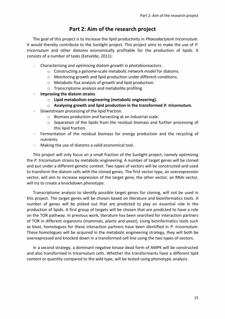
Part 2: Aim of the research project
The goal of this project is to increase the lipid productivity in Phaeodactylum tricornutum. It would thereby contribute to the Sunlight project. This project aims to make the use of P. tricornutum and other diatoms economically profitable for the production of lipids. It consists of a number of tasks (Eetvelde, 2011):
- Characterising and optimising diatom growth in photobioreactors. o Constructing a genome-scale metabolic network model for diatoms. o Monitoring growth and lipid production under different conditions. o Metabolic flux analysis of growth and lipid production. o Transcriptome analysis and metabolite profiling.
- Improving the diatom strains o Lipid metabolism engineering (metabolic engineering). o Analysing growth and lipid production in the transformed P. tricornutum.
- Downstream processing of the lipid fraction. o Biomass production and harvesting at an industrial scale. o Separation of the lipids from the residual biomass and further processing of
this lipid fraction. - Fermentation of the residual biomass for energy production and the recycling of
nutrients. - Making the use of diatoms a valid economical tool.
This project will only focus on a small fraction of the Sunlight project, namely optimizing the P. tricornutum strains by metabolic engineering. A number of target genes will be cloned and put under a different genetic context. Two types of vectors will be constructed and used to transform the diatom cells with the cloned genes. The first vector type, an overexpression vector, will aim to increase expression of the target gene, the other vector, an RNAi vector, will try to create a knockdown phenotype.
Transcriptome analysis to identify possible target genes for cloning, will not be used in this project. The target genes will be chosen based on literature and bioinformatics tools. A number of genes will be picked out that are predicted to play an essential role in the production of lipids. A first group of targets will be chosen that are predicted to have a role
on the TOR pathway. In previous work, literature has been searched for interaction partners of TOR in different organisms (mammals, plants and yeast). Using bioinformatics tools such as blast, homologues for these interaction partners have been identified in P. tricornutum. These homologues will be acquired in the metabolic engineering strategy, they will both be overexpressed and knocked down in a transformed cell line using the two types of vectors.
In a second strategy, a dominant negative kinase dead form of AMPK will be constructed and also transformed in tricornutum cells. Whether the transformants have a different lipid content or quantity compared to the wild type, will be tested using phenotypic analysis.

Part 2: Aim of the research project
16

Part 3: Results
17
Part 3: Results
3.1. Vector construction
The molecular tools for P. tricornutum are still limited compared to the plant or animal field. In order to improve the situation two novel types of vectors were created, an RNAi vector and an overexpression vector which has the resistance marker transcriptionally fused to the GW site. Two types of destination vectors were used for transformation, an overexpression vector to increase expression of the target gene, and an RNAi vector to knock down the expression. Vector constructs were made using a combination of restriction enzyme techniques and gateway cloning. The advantage of working with this gateway technology is that the same destination vector can be used multiple times for each donor
vector containing the target gene of interest.
The vector that was used mostly for the transformation work, was the pPha-T1 vector. This construct was specifically made for transformation of P. tricornutum. It contains a selection marker, named sh ble, making it able to grow on medium containing zeocin, and a multiple cloning site flanked by a suitable promoter (Zaslavskaia et al, 2000). The pPha-T1 vector has the advantage of already containing the selection marker for transformation, that way the sh ble resistence gene is present on the same vector as the gene of interest.
3.1.1. Overexpression vectors
To overexpress the target gene two types of promoters and two types of vector
backbones were used. The histon H4 promoter is a constitutive promoter, the FCP (fucoxanthin binding protein) is only active during illumination (figure 7). These two types of promoters will lead to transcription activation of the downstream gene.
Figure 7: Overexpression vector with FCP promoter (a) and histon H4 promoter (b). (a) FcpB promoter =
fucoxanthin binding protein promoter, induces transcription under light conditions. FcpA terminator =
terminator sequence, which stops the transcription process. HA epitope tag = hemagglutinin tag, used to check
if the gene of interest is expressed. AttR gateway cassette = sequence which will be recombined out with LR
clonase, and replaced by a gene of interest. It contains two coding sequences, the ccdB gene and the CAT
marker gene. (b) H4 promoter = constitutive promoter.

Part 3: Results
18
Two types of overexpression vectors are used, pDEST-H4-Cterm, which doesn’t contain a resistance marker, and pPha-T1. To select for P. tricornutum cells containing the desired pDEST-H4-Cterm vector, the cells will be cotransformed with a selection vector, PAF6, containing the selection marker sh ble under control of the FCP promotor. By using biolistic transformation, both vectors (the destination vector and the selection vector) will be inserted in the genome of the transformed cells (Falciatore et al, 1999). Growing the cells on medium containing zeocin allows to select for stably transformed cells.
A new overexpression vector was developed that uses the pPha-T1 backbone as a blueprint (figure 8). It consists of a GW sequence which is transcriptionally fused the selection marker by using a F2A linker. That way, when the sh ble gene is transcribed and translated, the chance that the same happens for the gene of interest will be very high. At
the protein level the F2A linker will break without the help of any other enzyme, thereby leading to the formation of two separated proteins (De Felipe et al, 2003). This strategy lowers the chance of picking up transformed diatom cells which do not have an active target gene.
3.1.2. RNAi vector
Making knockout lines is currently infeasible due to the low frequency of homologous recombination and the suspected absence of the sexual reproduction cycle of P. tricornutum. Therefore, heterozygous mutants are the only lines which have currently been made. To overcome this problem, two options are available. The first option is making a dominant negative mutant, which can have the same effect on the phenotype as a homozygous knockout, but this strategy is only possible in some cases. These dominant negative forms can be expressed using the available overexpression vector. The second
option is making use of the RNAi machinery of P. tricornutum. Therefore a second type of vector, an RNAi vector, was constructed. To make use of the RNAi machinery the transcribed RNA needs to make a hairpin construct by the presence of inverted repeats, therefore a
Figure 8: pPha-T1 overexpression vector. This vector contains a GW site which is transcriptionally fused to
the sh ble selection marker using a F2A peptide linker. FCP promoter = fucoxanthin binding protein
promoter. CDS2 = coding sequence two, which is an ampicilline resistance marker

Part 3: Results
19
destination vector was made containing two gateway sites. These gateway sites are positioned in a different orientation, thereby forming an inverted repeat, and are connected via an intron, forming the loop in the hairpin construct. These types of constructs have proven to work best in plants (Wesley et al, 2001), and it is hoped that this hairpin RNA will have the same effect in diatoms.
A first attempt was made to construct the RNAi vector. This vector was constructed by using unaltered GW sites and a small intron sequence, unfortunately it could not be build. At the final step of the construction no E. coli colonies grew on the selective medium, meaning that the final RNAi vector was either not constructed or destroyed. Destruction of the final construct was probably caused by unforeseen recombination events between the inverted repeats (Achaz et al, 2003). Since the first attempt was a failure, a new strategy was devised
(figure 9). It was assumed that the reason for the previous failure was the presence of long inverted repeats, namely the two gateway sites, and only a short intron sequence in between. This construct is assumed to be very sensitive to recombination events, thereby preventing stable propagation of the plasmid in E. coli. To overcome this problem two adaptations were made:
- The inverted gateway sequences were shortened by removal of the chloramphenicol resistance gene.
- The intron sequence was enlarged, by using the CAT marker which was removed from the gateway site. That way, when grown on medium containing chloramphenicol, there will be selection for vectors containing the intron.
Figure 9: RNAi vector. The two gateway sites are put in a different orientation to form inverted repeats and
no longer contain the CAT marker gene. This marker gene is now added to the intron sequence. FcpB
promoter (P 2) = fucoxanthin binding protein promoter, induces transcription under light conditions. FcpA
terminator 1/2 = terminator sequence, which stops the transcription process. attR gateway cassette =
sequence which will be recombined out with LR clonase, and replaced by a gene of interest. Intron + CAT =
sequence which will form a loop in the hairpin construct of the RNA product that will be transcribed. CDS2 =
coding sequence two, which is an ampicilline resistance marker. Zeocin resistance marker = sh ble gene

Part 3: Results
20
Construction of the second vector was successful. Restriction enzyme analysis of the vector revealed logical patterns. Now, the RNAi vector just had to be tested on its functionality by using it in a transformation experiment. The vector was used to target a number of genes that encode proteins interacting with TOR. The results of these transformation are discussed in section 3.2.2.
3.2. Metabolic engineering of the target genes
Different target genes were chosen based on promising characteristics of each (group of) gene(s). Not every target gene was completely analyzed from cloning to transformation and phenotypical analysis. An overview of the completed work can be seen in table 1. The genes that are mentioned in this table are only the ones which were successfully cloned.
Vector construct Target gene Cloning Transformation QPCR analysis Phenotypic analysis
Overexpression: pDEST H4 Cterm
AMPK K45A
AMPK K45R
Rheb
Sty1
Overexpression: pPHAT1: GW -
F2A - SHBLE
GSK3
MORG1
Sir1
Sir2
Sir4
Sir5
RNAi: pPHAT1 LST8
geranyl
Rheb
Sty1
CrAT
MORG1
Sir1
Sir4
Sir5
Tabel 1: completed work. Phases in which the analysis of the different transgenes has ended during this
project.
3.2.1. Strategy 1: creating a dominant negative kinase dead AMPK
AMPK plays a central role in energy homeostasis. When the energy consumption is high, it will be the role of the AMPK complex to restore the steady-state ATP levels by activating catabolism and inhibiting anabolism (Lindsley & Rutter, 2004; Schimmack et al, 2006). Therefore, creating a dominant negative AMPK might be a good approach to enhance lipid production. It is reasoned, when AMPK is inactive, it will no longer be able to inhibit lipid anabolism. To create a dysfunctional AMPK complex it should suffice to target one of its subunits, preferably the subunit which confers the enzymatic activity, meaning the α subunit
(Stapleton et al, 1996). AMPKα is an evolutionary very conserved domain. Alignment of the AMPKα orthologs from different organisms shows the occurrence of a conserved NH2-terminal kinase domain (figure 10A). Therefore, one can assume that this complex will also

Part 3: Results
21
be functional in diatoms and a good candidate for ME. Figure 10B shows that the AMPKα protein of P. tricornutum and other algae (such as Thalassiosira pseudonana, Fragilariopsis cylindrus, Ectocarpus siliculosus and Chlamydomonas reinhardtii) clusters more closely to their ortholog in plants such as the soy plant, as compared to the human and fungal relatives.
AMPKα was inhibited by making a kinase dead form. This mutant was created by
introducing a point mutation using a site-directed mutagenesis protocol (Zheng et al, 2004). The same strategy was applied by Mu et al. They disabled the phosphorylation function by creating a point mutation in the amino acid sequence. By mutating the lysine, at position 45 (of the human sequence), into an arginine (K45R), they could show that AMPKα could no longer efficiently phosphorylate its targets by applying a SAMS peptide assay (Mu et al, 2001). This strategy was adapted and supplemented with a point mutation of the same lysine into an alanine (K45A). This second mutant was added to ensure that most activity of the active center was disabled. The presence of an arginine means that a positive charge remains in the active center. This positive charge might still be able to represent some residual activity of the mutant AMPK. To transform P. tricornutum, the pDEST-H4-Cterm overexpression vector was used. That way the activity of the endogenous AMPKα will be
negligible compared to the transgene. The wild type form of AMPKα will still be present, but this will not lead to functional AMPK complexes since AMPK is active as a heterotrimer. The
Figure 10: (A) Alignment of AMPKα: aligning several AMPKα orthologs shows the presence of a conserved
kinase domain which confers the catalytic activity of the protein. The lysine at position 45 (K45) of the human
sequence is essential for the catalytic activity and has therefore been targeted in P. tricornutum for mutation.
(B) Guide tree of AMPKα: sequences of different organisms were aligned using ClustalW to compare their
relatedness. The sequences that were used, were the ones that gave the best hit when blasting the AMPKα
sequence from P. tricornutum (PHATRDRAFT 8773). The guide tree shows that AMPKα from diatoms closely
resembles those from plants, compared to their human and fungal orthologs. Both figures were created using
ClustalW (EMBL-EBI, 2012).

Part 3: Results
22
mutant form that is introduced in the cells is dominant negative. The remaining functional wild type complexes will form a minority as the mutant form will be transcribed at a considerably higher level compared to the wild type. The phenotype of this mutant should resemble a knockout of AMPKα.
In a first attempt, the overexpression vector with the FCP promoter was used. No transformed cells for both constructs (K45R and K45A) could be found. Either no transformants were created, or all transformants died. In a second attempt the histon H4 promoter was used, which is weaker compared to the FCP promoter and has the additional advantage of being transcribed in darkness. This attempt gave rise to viable mutant cells.
Checking for the presence of the transgene using PCR
To confirm integration and expression of the target gene in the genome a PCR was run on the cDNA using primers that target the transgene. In a first attempt, three different FW primers were used that target the AMPKα gene (FW 12, 24 and 44). The RV primer anneals to the regulatory region downstream of the gene, namely to the FCP terminator. The original expression vector that was used to transform the diatom cells was used as a positive control. The gel output can be seen in figure 11. A second PCR was conducted using the same FW primers, but the RV primer now annealed with the 3’ region of the endogenous AMPKα (figure 12). This PCR was conducted as a control.
Figure 11: PCR gel after AMPKα transgene amplification. Three different FW primers, all three located on the
AMPK coding sequence, were used (12, 24 and 44).The same RV primer, located on the FCP terminator, was
used in all three constructs. Sequence of loading: ladder (L), A1, A3, A5, A6, A7, Paf6 (P), R2, R3, R4 and a
positive control (expression vector - +). The band sizes of the ladder are displayed in the right-bottom figure.
The band that corresponds with the size of the desired amplicon are indicated with an asterix (*).

Part 3: Results
23
Unfortunately, the gel electrophoresis output showed multiple PCR products, probably due to aspecific amplification. Sometimes a light band could be seen with the size that was expected to correspond to the desired PCR product, but also the PAF6 control, which should not contain a transgenic AMPKα, showed this signal. Therefore a second strategy was adapted to amplify the transgene. The same RV primer was used that targeted the FCP terminator, but the FW primer now annealed with the H4 promotor instead of the AMPKα gene (figure 13).
No logical output was obtained, aspecific amplification appeared again. A number of different other primer combinations were used (data not shown), but each time the PCR
Figure 12: PCR gel after AMPKα endogene amplification. Three different FW primers, all three located on the
AMPK coding sequence, were used (12, 24 and 44).The same RV primer, located on the 3’ region of the
endogenous AMPKα, was used in all three constructs. Sequence of loading: ladder (L), A1, A3, A5, A6, A7, Paf6
(P), R2, R3, R4 and a positive control (expression vector +). The band sizes of the ladder are displayed in the
right-bottom figure.
Figure 13: PCR gel after AMPKα transgene amplification (2nd
attempt). The FW primers was located on the H4
promotor and the RV primer annealed to the FCP terminator region. Sequence of loading: ladder (L), A1, A3,
A5, A6, A7, Paf6 (P), R2, R3, R4, a positive control (expression vector - +) and a negative control (-).
Expected size of the PCR product

Part 3: Results
24
yielded either multiple bands or the absence of a signal. Of course, this lack of evidence that transformants were created will make it difficult to prove that the dominant negative AMPKα contributes to any phenotype that is observed.
QPCR analysis
To check whether the transformation method had truly been successful, in other words to confirm overexpression of the AMPKα transgene, a QPCR analysis was conducted. QPCR primers (table 4 in the addendum) were constructed against the gene of interest and a SYBR green based method was applied to measure the gene expression. SYBR green will bind preferentially to dsDNA and will produce a detectable fluorescence signal after a number of cycles have passed during the QPCR run. The number of cycles that are needed to surpass
this threshold value is represented by the Ct (cycle threshold) value, and this value is correlated with the original mRNA concentration of the gene of interest. If a gene is highly expressed, it will have a low Ct value and vice versa. The expression of the target genes of the transformants was compared with a control group, which was solely transformed with the selection marker vector PAF6.
To cope with differential expression results as a consequence of different concentrations of starting material, the QPCR analysis will simultaneously be performed on two housekeeping genes. These genes are considered to have a constant expression level and can therefore be used to even out differences that are not caused by differential expression. One of the causes of these differences can be the harvesting of a different amount of cells,
or using a different RNA quantity when synthesizing the cDNA. The genes that were used for this correction are TubB, H4, TBP and RPS which encode for tubuline subunit b, histon H4, the TATA-box binding protein and a ribosomal protein respectively. The output of the QPCR can be seen in figure 15.
To analyse the expression of AMPK six different sets of QPCR primers were used (numbered with 24, 43, 44, 45, 46 and 47). This lowers the chance for a false conclusion to be made when using a primer pair that doesn’t function properly. A false signal during the QPCR run might occur because of primer mismatching, or incoherent signals might be found due to unknown presence of different isoforms of the AMPK gene. The region which is amplified by the primer pairs is visualised in figure 14.
Figure 14: AMPKα primer amplicon positions. The regions on the AMPKα gene that are amplified by the
different primer combinations (24, 43, 44, 45, 46 and 47 FW and RV) are annealed to the full length gene.

Part 3: Results
25
Figure 15: QPCR expression analysis of AMPKα (analysis 1). The expression of AMPKα was tested using three
primer combinations (24, 43 and 44). Two housekeeping genes, histon H4 and TubB were coanalysed as control
genes. Expression values are expressed relative to the highest value, which is equal to one. Error bars indicate
the standard deviation on the measurement.
No overexpression mutant was found and the different primer pairs contradict each
other, yet a different phenotype could be observed between the transformants and the Paf6
control lines. The transformants appeared to grow slower. Two cell lines (AMPK K45A 1 and
K45R 3) even stopped growing after they were diluted and they could not be further used for
phenotypic analysis. A second QPCR analysis was run to check whether this output could be
confirmed or whether it was due to technical errors. This experiment yielded analogous
results (figure 16).

Part 3: Results
26
Figure 16: QPCR expression analysis of AMPKα (analysis 2). The expression of AMPKα was retested using five
primer combinations (43, 44, 45, 46 and 47). Two housekeeping genes, histon H4 and TBP were coanalysed as
control genes. Expression values are expressed relative to the highest value, which is equal to one. Error bars
indicate the standard deviation on the measurement.

Part 3: Results
27
Phenotypic analysis using epifluorescence microscopy
To check for phenotypic characteristics of the transformants, without conducting a biochemical assay, cell cultures were stained with nile red (9-diethylamino-5H-benzo-α-phenoxazine-5-one) and visualised with a fluorescence microscope. Nile red interacts with neutral lipids, it excites at 485 nm and emits at 525 nm (green light), but only in an apolar environment (Greenspan et al, 1985). In P. tricornutum this means that lipid droplets can be visualized using a fluorescent epifluorescent microscope.
The transformant cell lines AMPK K45A and K45R (further named A3, 4, 5, 6, 7 and R2, 4) were compared with a Paf6 control line. The samples were analysed using two kinds of light. Differential interference contrast (DIC) light (or visible light) was used to check for
differences in shape and size, and the fluorescent light emitted by nile red was used to get a first impression of the lipid content. The microscopic pictures can be seen in figure 17.
Some abnormal phenotypes were observed for a number of cell lines:
- A3: appeared quite normal. - A4: mostly normal cells, but also a couple of small (and fat) once. Being fat means
having more or larger lipid droplets than Paf6 lines. - A5: the number of small and fat diatoms was even higher than for A4. - A6: appeared to look normal (sometimes a short and fat cell occurred). - A7: did not agree with the other cell lines. Instead of being small and fat, the cells
were stretched, had abnormal shapes and contained about the same amount of lipid droplets as the control line.
- R2 and R4: had the most severe phenotypes of being small and fat.

Part 3: Results
28
Figure 17A
Small cells
Fat cell
Normal looking cells from Paf6 line

Part 3: Results
29
Figure 17: microscopic pictures of the AMPK transformant lines. Figure A shows a control Paf6 line and
transformant lines A3 to A5. Figure B shows the remaining transformant lines A6, A7, R2 and R4. Each line was
visualised using DIC light (left column) and using fluorescent light from nile red (right column). Some abnormal
phenotypes are mentioned on the figures. Pictures were taken with a 10x40x magnification using an
epifluorescent microscope.
Figure 17B
Stretched cells
Very abnormal
shape
Small cells
Fat cells

Part 3: Results
30
Growth analysis using OD measurements, flow cytometry and sugar quantification
All transformant cell lines (A3, 4, 5, 6, 7 and R2, 4) and three control Paf6 lines (named Paf6 3, 4 and 5) were grown in constant light condition for a period of two days. At the beginning of the growth, cultures were diluted to the same OD (optical density) value. Each culture was subjected to three conditions, they were grown under normal conditions (control), in the dark (dark) or without a nitrogen source (noN). A sample was taken at three different time points, after 0, 24 and 48 hours. Unfortunately four dark samples (Paf6 4, A3, A6 and A7) were destroyed during the experiment before the last sample could be taken.
Measuring cell density using OD measurements
To assess the growth of the different transformant lines, OD was measured at 405 nm (figure 18).
Figure 18: OD measurement of the transformant cell lines and the PAF6 control lines at 405 nm. OD was
measured at three time points (0 h, 24 h and 48 h) and under three conditions (control = normal conditions,
noN = without nitrogen source, dark = without light).

Part 3: Results
31
The OD value increases linearly with a growing diatom population. This linear increase is present for normal conditions and growth without nitrogen, but absent at dark conditions. However, growing in medium without nitrogen leads to lower cell densities compared to optimal control conditions. The (near) absence of growth in the dark can be explained by a loss of energy. ATP will no longer be formed by means of photosynthesis and the cell will no longer invest energy in mitosis.
Two transformant lines appeared to differ slightly from the control lines. In normal conditions growth of A3 and A4 appeared to flat out, which contributes to the observation of the slower growth compared to the PAF6 lines. Unfortunately this observation was not made for the cultures growing in medium without nitrogen. Cell line A3 slightly flats out, but A4 grows linearly. The data from this experiment is not enough to come to a conclusion about
the growth, therefore this data was supplemented with flow cytometry experiments.
Analyzing cell cycle state using flow cytometry of DAPI stained cell cultures
To analyse the cell cycle state of the cell cultures, cell were stained using DAPI, which bind to dsDNA and emits fluorescent light at 461 nm. The intensity of this fluorescent light correlates to the DNA concentration and can therefore be used to estimate to which state of the cell cycle a diatom has progressed. These DAPI stained cell cultures were analysed with a flow cytometer, cells were counted and their fluorescence intensity was measured. This gives an output in the form of a flow histogram, which shows how many cells were counted with a certain DAPI intensity. These histograms show one large peak, which corresponds to
cells that are not duplicating their genome (diploid cells), and a second smaller peak, which corresponds to cells that have undergone genome duplication but haven’t divided yet (cells in the G2/M phase). This second peak is mostly absent in the histograms in figures 19, 20 and 21, probably because the cells were not synchronized. The region in between corresponds to cell that are in the process of duplicating their genome. To get a first impression, only the samples that were taken after 24h were analysed using this technique. The profile of the histograms of the transformant cell lines were compared to the control lines. This comparison is displayed in figure 19 A, B and C.
Two observation were made when analysing the histograms:
- First, culture A7 seemed to have the most pronounced deviation from the control
culture. It seemed to contain a lot of cells with high DAPI intensity. It was therefore
not in line with the other transformants. To make sure this phenotype was correct,
samples at time point 0 and 48h were also analysed (figure 20).
- Second, there seemed to be a slight trend in the control conditions for a number of
transformants (mostly in A3, A4 and A5) to have a larger number of cells with a lower
DAPI intensity, meaning these cultures possess less dividing cells. The transformant
lines grown in normal conditions seemed quite similar to their equivalents grown
without nitrogen and were therefore compared as well (figure 21).

Part 3: Results
32
Figure 19A: The
control samples
Diploid cells

Part 3: Results
33
Figure 19B: The
dark samples

Part 3: Results
34
Diploid cells
tetraploid cells
Figure 19C: The
noN samples
Figure 19: flow histogram
comparison of transformant
lines and Paf6 lines at time
point 24h. The red graph
indicates the Paf6 4 output
and the black graph indicates
the output of the
transformants. In the last
histogram of each condition
Paf6 4 is compared to the
remaining Paf6 lines (3 and
5) to illustrate the natural
fluctuation of the control
lines. (A) Samples from the
cultures grown in normal
conditions (C24). (B) Samples
from the cultures grown in
dark conditions (D24). (C)
Samples from the cultures
grown in medium without
nitrogen source (N24). The x-
axis displays the number of
counts, the y-axis displays
the DAPI intensity.
Histograms were
smoothened using FCS
express software.

Part 3: Results
35
Figure 20: flow histogram comparison of transformant line A7 and Paf6 line 3 at time point 0, 24 and 48h.
The red graph indicates the Paf6 3 output and the black graph indicates the output of A7. Column 1: samples
from the cultures grown in normal conditions (C0, C24 and C48). Column 2: samples from the cultures grown in
dark conditions (D0 and D24). Column 3: Samples from the cultures grown in medium without nitrogen source
(N0, N24 and N48). The x-axis displays the number of counts, the y-axis displays the DAPI intensity. Histograms
were smoothened using FCS express software.
Unfortunately, the observation that A7 contains more cells with more DNA is not always visible, although the trend for the black graph (from A7) to be on the right side of the control
line remains.

Part 3: Results
36
Figure 21: flow histogram
comparison of transformant
lines grown in normal vs.
noN conditions at time
point 24h. The blue graph
indicates the Paf6 4 output
grown in noN (N24), the
black graph of the
transformants grown in
control conditions (C24) and
the red graph of the
transformants grown in noN
(N24). In the last histogram
Paf6 4 is compared to Paf6 3
to illustrate the natural
difference of the cell lines
grown in normal vs. noN
conditions. The x-axis
displays the number of
counts, the y-axis displays
the DAPI intensity.
Histograms were
smoothened using FCS
express software.

Part 3: Results
37
Again no consequent effects could be observed, but transformants A3, A4 and A5 still
seemed to have a similar cell cycle profile when their cultures were grown in normal
conditions versus noN.
Measuring sugar content of the different cell cultures
The last phenotypic analysis that was conducted was a sugar quantification assay. Since AMPK plays a role in regulating catabolism, an effect of the transgene was hoped to be seen on the chrysolaminaran concentration. Chrysolaminaran, a β-1,3-glucan, is the most important storage glycan in P. tricornutum and contains mainly glucose units (Granum & Myklestad, 2001). The sugars were extracted from the cells (using soft acidic conditions),
they were hydrolysed (using harsh acidic conditions) and quantified using a colorimetric assay. Monosaccharides reacted with phenol, thereby forming an aromatic complex which has an absorbance at 493 nm. The OD measured at this wavelength corresponds to the initial chrysolaminaran concentration. The sugar concentration is calculated using a calibration curve. This curve indicates the OD value for glucose samples with known concentrations. The raw data of this analysis can be found in the addendum. Each sample was taken in triplo (technical replicates).
Figure 22 shows the absolute sugar concentration for each condition over two days. The main focus was trying to see a difference between the transformant lines and Paf6, unfortunately no clear effects of the transgene seem visible. The samples grown in control conditions even have unexpected values. Normally, the sugar concentration should rise over
the complete two days, but at time point 48h only Paf6 4 shows a normal concentration. The low values of the other cultures was probably due to experimental failure. The evolution of the samples over 48h in the other conditions appears normal. Only Paf6 5 grown in the dark has an unexpected value, its sugar concentration doesn’t drop after one day. Again, this problem is probably due to technical errors.
Figure 23 shows the sugar concentration that was corrected for growth of the cells. These relative values filter out a rise or decline in sugar concentration due to a rise or decline in cell density. The same conclusion can be drawn from these graphs as the once for absolute concentrations.

Part 3: Results
38
Figure 22: Absolute sugar quanticication. For all three conditions (control, dark and noN) samples were taken
at time point 0, 24 and 48h. There sugar concentration was calculated using a calibration curve. No clear effect
of the transgene can be seen, however some technical errors were made. The y-axis shows the sugar
concentration (mg/ml), and the x-axis shows which cell line was measured. Error bars indicate the standard
deviation on the measurement.
Abnormal values
(probably caused
by experimental
error).

Part 3: Results
39
Figure 23: Relative sugar quanticication. For all three conditions (control, dark and noN) samples were taken at
time point 0, 24 and 48h. There sugar concentration was calculated using a calibration curve and corrected for
cell density. No clear effect of the transgene can be seen, however some technical errors were made. The y-
axis shows the relative value of the sugar content (has no dimension), and the x-axis shows which cell line was
measured. Error bars indicate the standard deviation on the measurement.
Abnormal values
(probably caused
by experimental
error).

Part 3: Results
40
3.2.2. Strategy 2: overexpressing and knocking down TOR interactors
Proteins which interact with TOR were found using a combination of literature search and bioinformatics tools. Based on the knowledge of well-known organisms, such as Saccharomyces cerevisiae and mammals, some interactors of TOR were already known. To check whether these proteins are also encoded in the P. tricornutum the coding sequence of these interactors was blasted against the tricornutum genome. Those genes that gave a hit, were analysed whether they were good candidates to apply a ME strategy. For example, if a protein family consisting of paralogs was found, these genes were removed from the candidate list. These genes are considered bad target genes due to their likeliness of being redundant. To transform P. tricornutum both an overexpression vector and an RNAi vector were used. That way the target gene will be, in one cell line, overexpressed, and in the other
down regulated. The genes were first cloned, either from the cDNA using PCR, or from a cDNA library using RACE (rapid amplification of cDNA ends). The primers that were used are mentioned in table 3 of the addendum. The cDNA that was used was obtained from wild type cells that were grown in normal conditions (without stress). The cloned genes were put in a destination vector and this construct was transformed in tricornutum cells.
Rheb (Ras homolog enriched in brain)
In mammalian cells this small GTPase was found to be active upstream of TOR and to be in an activated state when bound to GTP. In the research of Hay et al, they found that this protein is a positive regulator of TOR in mammalian cells. Meaning, when it is overexpressed
it will lead to the activation of TOR (Hay & Sonenberg, 2004). The sequence that was picked up was consistent with the annotation model (PHATRDRAFT 9927).
Raptor
In mammalian cells this protein interacts directly with TOR forming a nutrient sensitive complex. In the research of Hay et al, they again found that this protein is a positive regulator of TOR in mammalian cells, although exceptions were found (Hay & Sonenberg, 2004). In P. tricornutum two homologs were found, RPTOR 1 and RPTOR2. Cloning of these genes from cDNA proved unsuccessful, probably due to bad annotation (PHATRDRAFT 45807 and 18549). The RACE PCR strategy revealed the possible expression of RPTOR 1 but
sequencing of the fragment failed. Also picking up a fragment of the genes for use in the RNAi strategy failed.
LST8
LST8 will associate to the nutrient sensitive TOR-Raptor complex and thereby stabilize this complex. It was found that both in mammalian cells and in S. cerevisiae LST8 is a positive regulator of TOR. Cloning of the complete gene from cDNA proved unsuccessful (PHATRDRAFT 36142), but using the cDNA library revealed a possible LST8 transcript. Unfortunately sequencing of the PCR product was not successful. Picking up a fragment for cloning in the RNAi vector was a success.

Part 3: Results
41
STY1
It was found in Schizosaccharomyces pombe that the homolog of Sty1 ,namely Spc1, is activated due to stress factors such as high temperature and oxidative stress. Spc1 itself promotes the activation of a number of stress survival genes (Degols et al, 1996). Cloning of the RNAi fragment and the complete gene was a success and the sequenced gene was consistent with the annotation model (PHATRDRAFT 21410).
Carnitine acyltransferase (CrAT)
CrAT catalyses the transfer of an acyl chain from carnitine to coenzyme A, thereby transporting these acyl chains through the mitochondrial membrane. This gene is therefore
essential for appropriate oxidation of long-chain fatty acids (Jogl et al, 2004). Again, cloning of the complete gene didn’t work (PHATRDRAFT 22418), colony PCR conducted on the E. coli cells after the BP reaction revealed variable band sizes, meaning a non-uniform gene product had been picked up. Fortunately, for this target the RNAi strategy is more suitable and picking up a fragment for this purpose was a success.
MAPK organizer (MORG) 1
This gene encodes for a protein which is a member of the WD-40 protein family. It associates with a number of other components of the mitogen-activated protein kinase (MAPK)/ extracellular signal-regulated kinase (ERK) pathway (Vomastek et al, 2004). Its specific role in lipid metabolism remains unclear, yet it is found to interact with the TOR
pathway. Both the RNAi fragment and the complete gene were picked up. The annotation model (PHATRDRAFT 18217) predicted that the gene had two introns, but the second predicted intron was not spliced out of the gene from the transcript that was picked up from the cDNA.
Silent information regulator (Sir)
It was observed that mice carrying two null alleles for Sir are hypermetabolic and have elevated levels of lipid oxidation (Boily et al, 2008). That is why this gene was ideal for overexpression. When annotating this gene on the P. tricornutum genome, five paralogs were found (numbered 1 to 5). The gene that was annotated to be the functional Sir enzyme
was Sir 4, but all five paralogs were targeted for engineering.
- Sir 1 (PHATRDRAFT 8827): both the RNAi fragment and the complete gene were cloned. The annotation model didn’t start with a methionine, therefore the gene was enlarged upstream to the first methionine after the first encountered stop codon. The model also predicted the presence of an intron, but this wasn’t supported by the sequenced gene.
- Sir 2 (PHATRDRAFT 16859): only the complete gene was successfully cloned. The annotation model didn’t start with a methionine nor did it end with a stop codon, therefore the gene was enlarged upstream to the first methionine after the first encountered stop codon, and downstream to the first encountered stop.
- Sir 3 (PHATRDRAFT 12305): cloning of both constructs failed.

Part 3: Results
42
- Sir 4 (PHATRDRAFT 45850): both the RNAi fragment and the complete gene were cloned. The sequence of the annotation model was consistent with the sequenced gene.
- Sir 5 (PHATRDRAFT 21543): both the RNAi fragment and the complete gene were cloned. The annotation model didn’t end with a stop codon, therefore the gene was enlarged downstream to the first encountered stop. The model also predicted the presence of an intron, but this wasn’t supported by the sequenced gene.
Glycogen synthase kinase (GSK) 3
It is found that GSK3 is capable of inhibiting glycogen, lipid and protein synthesis in mammals, where it is regulated by insulin (Saltiel & Kahn, 2001). Knocking down this protein
in tricornutum cells might therefore prove a worthwhile strategy, but only the complete fragment was successfully cloned. A large fragment which was predicted to be part of the exon sequence of the gene, appeared missing on the sequenced cloning target (PHATRDRAFT 29223). Either an intron was overlooked by the model, or something had gone wrong with the cloning process. Either way, the cloned gene was further used for transformation.
QPCR analysis
For each cloned gene either an RNAi line or an overexpression line (or both) were made. To check whether the transformation method had truly been successful a QPCR analysis was
conducted. The output of the relative expression values can be seen in figure 24. Unfortunately, not every transformation yielded multiple transformants. The lines that were assessed are:
- Geranyl (see section 3.2.3.): five RNAi lines. - Rheb: one overexpression line and four RNAi lines. - STY1: four overexpression lines and one RNAi line.
The output on figure 24 shows that RNAi lines and overexpression lines do not give the expression values that were expected. RNAi lines do not show lower expression than the Paf6 lines, and overexpression lines do not show higher expression. However, it should not be considered abnormal that the transcript levels of the RNAi lines do not drop below the
levels of the control. It has been mentioned in previous studies that creation of RNAi transformants in P. tricornutum can have an effect on the protein level without having an effect on the transcript level by means of translational inhibition (Lavaud et al, 2012). This mechanism has already been proven to occur in plants (Brodersen et al, 2008).
Two YFP RNAi lines were created as well, but since a gene which encodes for YFP is not present in P. tricornutum, one expects to find no expression of the gene when conducting a QPCR experiment. Therefore QPCR primers which target the YFP gene were included in the analysis as a negative control. However, expression of YFP does show up in the QPCR analysis, which should not be possible. Ct values were reasonably low enough to conclude that YFP was expressed, therefore it was concluded that this QPCR output could not be
reliable.

Part 3: Results
43
Figure 24: QPCR expression analysis of TOR interactors (RNAi and overexpression lines). The expression of
geranyl, Rheb and STY1 was tested using QPCR primers for each gene. Two housekeeping genes, histon H4 and
RPS were coanalysed as control genes. The expression of YFP was also analysed as a negative control.
Expression values are expressed relative to the highest value, which is equal to one. Error bars indicate the
standard deviation on the measurement.

Part 3: Results
44
3.2.3. Strategy 3: overexpressing and knocking down geranyl.
Next to targeting the lipid metabolism, one can also try to improve the energy production of a cell. This increased energy status of a cell, can be used for the production of extra storage molecules. In line with this vision the geranyl gene, which encodes a geranylgeranyl (diphosphate) reductase, was targeted for ME. Geranylgeranyl reductase plays a role in the production of terpenoids such as carotenoids and chlorophyll, it catalyses the reduction of geranylgeranyl pyrophosphate to phytyl phosphate by using NADPH as a cofactor (KEGG, 2012). By lowering the production levels of chlorophyll, thereby reducing the size of the chlorophyll antenna, the transformed P. tricornutum will absorb less light. This phenotype has a dual benefit. The cells that are closest to the light source will be less damaged by photo oxidation, and more light, meaning more energy, will be available for the diatom cells which
are growing in the shadowed regions of an aquaculture. In theory this should lead to an increase in cell mass (Ort & Melis, 2011). The annotation model (PHATRDRAFT 50650) seemed correct, but cloning of the complete sequence was unsuccessful, only an RNAi fragment could be picked up. The QPCR results of the transformation with an RNAi vector can be seen in figure 24 above.

Part 4: Discussion
45
Part 4: Discussion
There is an increasing interest to use diatoms as a production vessel for the synthesis of certain valuable metabolites. Diatoms produce a number of interesting PUFAs among which a number possess a nutritional use. Besides synthesis of speciality products diatom cells can potentially be harvested for the production of biofuels. Unfortunately, knowledge of these organisms on a molecular level and knowhow of growing cultures on an industrial scale is limited. Therefore, diatoms can not economically compete with fossil fuels. The goal of this research was to contribute to an improved lipid synthesis in diatoms, as part of the covering Sunlight project which aims to make diatoms economically valuable. This study set of to create an RNAi vector to expand the molecular toolkit available for engineering diatom cells. This knockdown vector together with overexpression strategies were applied in metabolic
engineering of the diatom cell to alter their lipid content. The main results of this study focus on the construction of this RNAi vector and on the analysis of diatom cultures which were transformed with a dominant negative AMPKα.
4.1. Construction of the RNAi vector
A vector was constructed which had the purpose of knocking down the expression of a target gene. Constructs with this purpose were already available but they didn’t work as desired, knockdown of certain target genes was insufficient. A new construct was developed which consisted of two gateway (GW) cassettes with a different direction, separated by an intron which contained a chloramphenicol resistance (CAT) marker. In a first attempt the
CAT marker was not removed from the GW cassettes, and the intron was much shorter due to the absence of a resistance gene. This strategy failed to be constructed, which was probably due to the susceptibility of the long inverted repeat to recombination events in E. coli (Achaz et al, 2003). The strategy was adapted such that it formed a hairpin-loop with a smaller hairpin and a bigger loop. GW sites were shortened by removal of the CAT marker and the intron was enlarged by addition of that same CAT marker. This adapted construct led to the creation of a stable RNAi vector (figure 9).
Exchange of the GW site by a fragment of a target gene should lead to the expression and formation of a hairpin-loop with a hairpin region resembling the target gene. This hairpin-loop should be recognized by the RNAi machinery of the diatom cells, leading to a
knockdown of the target gene. This hypothesis was tested by targeting a number of genes which encode proteins that interact with TOR (an important metabolic regulator). Expression of these genes was analysed using a QPCR (figure 24). Results of this experiment could not provide conclusive evidence of the desired functionality of the RNAi vector, genes were not knocked down. However, RNAi silencing can have an effect on the protein level without affecting the transcript levels by translational inhibition (Brodersen et al, 2008; Lavaud et al, 2012), but whether this occurred is not clear. The negative control that was added to the experiment (an RNAi vector targeting YFP which is not encoded in the P. tricornutum genome), gave a significant signal what should not be possible. Meaning, we could not prove the functionality of the RNAi vector, but due to technical errors in the QPCR experiment, we could not prove the dysfunctionality as well. Reasons for the failed set-up of the experiment
could not yet be found. Research to whether the RNAi vector is suitable for metabolic engineering is still ongoing.

Part 4: Discussion
46
4.2. Analysis of the dominant negative AMPK transformants
A dominant negative AMPKα was created. This AMPKα is no longer capable of phosphorylating its interaction partner due to a mutation (K45A or K45R) in the catalytic site of the kinase domain. By overexpressing this dominant negative form, it complexes with the AMPK β and γ residues from the wild type AMPKα, thereby leading to a severely reduced number of functional AMPK complexes (Mu et al, 2001). In other words, this dominant negative overexpression genotype is meant to closely resemble an AMPKα knockout. By reducing the activity of AMPK, we also tried to reduce the activity of catabolic processes and enhance anabolic processes, thereby increasing the amount of lipids in a diatom cell. Transformants were created, but proving the insertion of the transgene in the genome and its expression appeared problematic. Picking up the transgene by PCR on the cDNA of
transformed diatom cells led to aspecific amplification, even with different combinations of primers (figure 11 and 13). Conducting a QPCR on the cDNA of the different cell cultures led to a similar problem. Different primer combinations gave different expression outputs and none of these outputs proved the overexpression of AMPKα (figure 15 and 16). It was concluded that studying AMPK expression proved difficult with the used techniques. In future transformation events, one might think of fusion AMPK to a tag in order to prove its transcription. Primers that target this tag can then be used to study expression, or one can work on the protein level using antibodies against this tag, to prove the presence of the dominant negative AMPK.
However, although (over)expression of the transgene could not be proven, a significant
phenotype could be seen when growing the transformant cell lines. Most cultures showed delayed growth compared to the control Paf6 line, and two cultures even stopped growing after dilution. Therefore, the cultures were analysed using (fluorescence) microscopy. This experiment showed that a number of cell lines had altered shapes and sizes, mainly reduced cell size, plus they appeared to contain larger lipid droplets than Paf6 (figure 17). Cell line K45A 7 seemed inconsistent with the others, instead of being smaller, the cells were enlarged and they did not show a change in lipid content. To prove a significant phenotypical change in the transformants, growth and sugar content were analysed for a period of two days. Growth analysis did not show any abnormalities, there was only a trend visible that the growth of a number of transformant cell lines flatted out quicker than for Paf6 (figure 18).
The same can be said about the flow cytometry experiments, again no obvious difference between control and transformants was visible. Some cell lines (K45A 3 to 5) showed a trend
to have less dividing cells, but cell line K45A 7 showed the opposite, it seemed to possess a lot of dividing cells instead (figure 19 to 21). But since only one cell culture showed this phenotype it could not be proven whether this observation was a consequence of the transgene expression or due to an random insertion of the expression or selection vector in an essential region of the genome. Sugar analysis of the cultures showed no significant change in the chrysolaminaran concentration. All seven cultures are now in the process of having their lipid content analysed by quantitative and qualitative assays. We hope to see a significant and consequent change to rule out the possibility that the observed phenotype was caused by something other than the expression of the dominant negative AMPK.

Part 4: Discussion
47
To conclude, we can’t prove whether the phenotypes we encountered are due to the presence of the AMPK transgene, due to the deletion of an essential gene as a consequence of the insertion in the genome or due to (in the worst case) environmental conditions or badly conducted experiments. To prove the functionality of the dominant negative AMPK, we need to find a good assay to prove the expression of the transgene, plus we need to produce more transformant cell lines to get statistical valid data. Meanwhile, new transformants have already been created (data not yet available), and these cell cultures show the same phenotype as the cultures that are discussed, microscopy also revealed the presence of smaller cells with larger lipid droplets.
4.3. Other target genes and future prospects
In a second strategy, genes were targeted that encode proteins that interact with TOR, which plays an important role in regulating anabolic pathways (Laplante & Sabatini, 2009). These genes were chosen based on literature searches and bioinformatics tools such as blasting of the genes against the P. tricornutum genome. The genes were either cloned in an overexpression or in the RNAi vector. The expression of the target genes was analysed in the same QPCR experiment as the one that was used to test the functionality of the RNAi vector.
The same conclusion can be drawn as mentioned above, due to a failed set-up of the experiment no theories could be ruled out and further studies were demanded. These transformant cultures were not yet ready for phenotypic analysis and study of significant changes at that level are still ongoing. One of the main problems of phenotypical analysis of the transformants is the lack of a high throughput method to screen for quantitative changes
in the lipid content.
Since results of this project are inconclusive, further research has to be conducted. If the problems with the QPCR set-up are solved, we can adapt (if necessary) the RNAi vector accordingly. Plus, in order to take into account the possibility of translational inhibition, one should also study the effect of the transformation on the protein level. If a proper RNAi vector is available, metabolic engineering of diatom cells would become much easier. We would no longer have to rely on mere overexpression. Knocking down genes with an RNAi strategy might prove valuable to create diatom cultures that synthesize more of the desired fatty acids (such as EPA, DHA and ARA) and other metabolites. If for example, the AMPKα kinase dead transformant leads to a dead end, one can try to knock it down instead.
A number of alternative ME strategies can be chosen to further optimize the creation of fat diatoms. One of these strategies can be the engineering of multiple target genes instead of one. Using this strategy already proved useful in a number of other success stories conducted in plants and prokaryotes. Combining a number of genotypical changes might give a synergistic effect when trying to achieve an improved flux in the anabolic pathways. If we would combine for example overexpression of genes that positively regulate the fatty acid synthesis, together with inhibition of genes which positively regulate the fatty acid β-oxidation, the increase in the flux of fatty acid synthesis might yield satisfactory results. When adjusting this strategy to P. tricornutum, one will probably encounter a number of problems, such as the lack of knowledge about the sexual reproduction to combine
transformants, and the difficulty to select for transformed cells that possess every transgene. However, if this strategy would lead to creation of optimised cell lines, looking into this possibility might prove worthwhile.

Part 4: Discussion
48
To conclude, using diatoms as little cell factories still isn’t economically feasible, but if research about these organisms keeps uncovering new knowledge about their molecular built-up, and if metabolic engineering succeeds in creating diatoms that produce more of the desired products, in the future these organisms might be on the same level as their alternatives. Some of the speciality products synthesized by diatoms have the potential to contribute to human health, nutrition and prosperity, therefore it is important to go deeper into the fundamental research concerning these organisms. The Sunlight project will continue to contribute to this vision.
(The Dutch summary can be found in the addendum.)

Part 5: Materials and methods
49
Part 5: Materials and methods
5.1. Cloning of target genes
Annotation of the target genes was retrieved using the Phaeodactylum tricornutum genome database of the DOE joint genome institute (JGI, 2012). This annotation model was checked using the basic local alignment search tool (BLAST) from NCBI (NCBI, 2012) and the expressed sequence tag (EST) database from DiatomCyc (DiatomCyc, 2012). If homologous genes showed significantly different annotations from the model in the JGI database, or if the EST did not support this model, the annotation of the genes was manually adjusted. Having the most probable coding sequence of the target genes at hand, primers were created to clone these genes (table 3 of the addendum). General guidelines for the
construction of these primers (as mentioned in the addendum) were used. Genes were picked up with a polymerase chain reaction (PCR) using the Pfu polymerase, which has proofreading activity. If the primers were not successful at cloning the genes, a RACE (rapid amplifying cDNA ends) PCR strategy was used. This technique uses a cDNA plasmid library to check if the targeted genes are expressed. Two GoTaq PCRs were used to guarantee the presence of the gene. The fist PCR used the cDNA plasmid library as template, which gave
multiple bands. The band(s) which was/were more likely to be corresponding to the gene of interest were purified and used as a template in the second PCR. For this second PCR a different reverse primer was used (targeting a sequence upstream of the previous reverse primer). If a signal was found (meaning the gene was most likely expressed but wrongly annotated), these genes were sequenced and reannotated.
The primers used to pick up the genes were flanked by attB sites. After amplifying the target genes using Pfu PCR, they were cloned in a donor vector (pDON221 – figure 25) using the gateway technology from Invitrogen (Invitrogen, 2011). A gateway cassette (figure 26) flanked by attP sites is recombined out by BP clonase and the gene of interest is inserted in the vector. That way an entry vector is created which contains the gene of interest flanked by attL sites. To select for the entry vector containing the target gene, and not for the donor vector containing the gateway cassette, two types of selection are used. When the gateway cassette is replaced by the target gene, the vector loses its lethal ccdB gene. If the original pDONR221 is transformed into non-resistant E. coli cells, the cell dies. Cells containing the desired entry vector are selected on medium containing kanamycin. The entry vector can
also be used to check for the correct target sequence. By sequencing the vector, using the M13 primerpairs, the sequence can be checked for mutations, which might have occurred during the cloning process.
The entry vector can then be used to get the gene of interest in the desired destination vector by using a LR clonase. This clonase catalyses the recombination event by which the attR flanked gateway in the destination vector is exchanged for the gene of interest flanked by the attL sites.

Part 5: Materials and methods
50
5.2. Vector construction
All vectors were maintained and amplified using Escherichia coli. The vectors were isolated using the GeneJET Plasmid Miniprep Kit from Fermentas Life Sciences (Fermentas, 2012). Three types of destination vectors were used to create transgenic Phaeodactylum
tricornutum, two types of overexpression vectors and one RNAi vector. The first type of overexpression vector was already available for use and was based on the pDEST vector from Invitrogen. The RNAi vector and the second overexpression vector had to be constructed. The destination RNAi vector was created using the pPha-T1 vector as a backbone. A number of restriction enzyme digests (RE digests) was needed to construct the necessary backbone. The CAT marker (chloramphenicol resistance gene) was removed from the gateway (GW) cassette (present in the pGEM vector) using the BamHI RE. The first GW was added to the opened pPha-T1 vector using EcoRV (figure 27A). EcoRV cleaves DNA leaving behind blunt ends, therefore a control digest needed to be performed to select for a
pPha-T1 vector which contained the GW site in the correct orientation. The PstI RE was used
for this control digest, since it cuts the GW sequence in an asymmetrical manner. The second GW was added to the construct simultaneously with the intron containing the CAT marker, using a three-point-ligation. The pPha-T1 backbone was cut open using SacI and XbaI RE. The
Figure 25: pDONR221. Donor vector
containing a gateway cassette flanking
attP sites. M13 forward and reverse
primers are used to sequence the
vector. Kan(R) = kanamycin resistance
gene. AttP1 and attP2 = the sites
flanking the gateway site, which are
recognized by the BP clonase, which
catalyses the recombination event with
a sequence flanked by attB sites. CcdB =
lethal gene whose protein product
targets the DNA gyrase. Cm(R) =
chloramphenicol resistance gene.
Figure 26: annotated gateway cassette. The gateway site is used in donor and destination vectors and will be
recombined out of the vector by a clonase protein. In a donor vector, the gateway site is flanked by attP sites
and will be recombined out by a BP clonase (thereby forming a entry clone). In a destination vector, the
gateway is flanked by attR sites and will be recombined out by a LR clonase (thereby forming a expression
clone). The gateway site contains two coding sequences: a chloramphenicol resistance gene (the CAT marker)
and the lethal ccdB gene. Therefore, to select for transformants which contain a vector with a gateway
cassette, use of ccdB resistant E. coli cells is obligated.
Annotated gateway

Part 5: Materials and methods
51
second GW was cut out of the pGEM vector with XhoI and XbaI, and the intron was cut with SacI and XhoI (figure 27B). The resulting hairpin-loop structure formed by the GW sites and the intron can be seen in figure 27 part C. In order to avoid self-closure of the pPha-T1 vector, this backbone was simultaneously dephosphorylated whilst performing the RE digest. The enzyme used for this procedure was the thermo sensitive shrimp alkaline phosphatase (TSAP, Promega). To construct an intron containing the CAT marker, a site-directed mutagenesis strategy was equipped (Zheng et al, 2004). The H4 intron was cloned from gDNA using Pfu mediated PCR, with primers flanked by RE recognition sites (SacI and XhoI sites). After the intron had been picked up, it was ligated in a pJET vector. This vector was then used in a mutagenesis strategy. Primers targeting the intron sequence were flanked by BamHI RE recognition sites. That way the intron sequence got an extra BamHI site in its
sequence. This site was used to add the CAT marker which was cut out of the GW using BamHI (figure 28). In order to get rid of the pJET vector with the original intron sequence, this vector was digested using DpnI, which only cleaves the methylated wild type DNA. After the three-point-ligation two control digests were performed to test for the right vector map of the RNAi vector, one with NotI and the other with EcoRI.
Figure 27: (A) Step 1: addition of the first GW site to the pPha-T1 backbone. The GW was cut out of the pGEM backbone using EcoRV
and was ligated into the pPha-T1 vector which was also cut open with EcoRV. (B) Step 2: construction of the RNAi vector using a three-
point ligation. The backbone is cut open with SacI and XbaI, the second GW is cleaved from pGEM using XbaI and XhoI, and the intron
containing the CAT marker is cut with SacI and XhoI. (C) The hairpin-loop structure. The two GW sites flanked by the (att)R sites (1 and
2) form the hairpin, and the intron containing CAT will form the loop.

Part 5: Materials and methods
52
The second overexpression vector was also constructed using a pPha-T1 backbone. This work was conducted by Michiel Matthijs. The selection marker sh ble was transcriptionally fused to the GW site using a F2A linker. The selection marker gene which was already present on the pPha-T1 backbone was removed using the XhoI RE.
5.3. Generation of a kinase dead AMPK
To create a kinase dead AMPK a site-directed mutagenesis strategy was equipped (Zheng et al, 2004). This is the same mutagenesis strategy that was used to create the mutated
intron for the construction of the RNAi vector. Primers were constructed which contained the desired point mutation. The lysine at position 43 (position 45 of the human sequence)
Figure 28: Construction of the intron containing the chloramphenicol resistance marker (CAT). The intron is
first cloned from gDNA using primers that target the exons surrounding the intron. The FW primer is flanked by
a SacI RE recognition site, the RV primer by a XhoI recognition site. The intron is ligated in a pJET vector and
was subjected to a mutagenesis strategy to insert a BamHI RE recognition site. The mutated intron was cut
open using BamHI and the CAT marker was ligated into the mutated intron. The intron that was needed for the
final three-point ligation to construct the RNAi vector, was obtained by using SacI and XhoI RE.

Part 5: Materials and methods
53
was either mutated to an arginine (K45R) or into an alanine (K45A). The oligonucleotides that were used for K45A are FW, GCGGTGGCAATCCTGAACAAGAACAAAATCAAGCAGCTG and RV, GATTGCCACCGCTACCTTGTGTCCGGTTACGGCATGTG , and for K45R FW, GCGGTGAGAATC CTGAACAAGAACAAAATCAAGCAGCTG and RV, GATTCTCACCGCTACCTTGTGTCCGGTTACGGCA TGTG. The adapted codon is underlined.
5.4. Generation of transgenic Phaeodactylum tricornutum
Cells were transformed using the Biolistic PDS-1000/He Particle Delivery System from Biorad (BioRad, 2011). The expression vector was constructed using the GW technology (Invitrogen, 2011). An LR reaction was used to get the target gene in a suitable destination vector. The resulting expression vector was coated onto a number of tungsten particles.
These particles were shot into the cells by applying high pressure in a vacuum chamber. To select for stably transformed cells cotransformation with a selection marker vector (PAF6) was performed. In case the pPha-T1 vector was used (as with the RNAi vector), this cotransformation was not needed as the selection marker sh ble is already present on the pPha-T1 backbone. After transformation the cells were grown on F2 plates for 24 hours. The cells were then transferred to plates containing the antibiotic zeocin. These plates were then
incubated at 21°C in constant white light. Resistant colonies became visible after 2-3 weeks. These colonies were then restreaked on F2 plates containing zeocin, and after sufficient growth, they were transferred to ESAW medium.
5.5. Monitoring the expression of the transgenes using QPCR
To analyze the gene expression, RNA is isolated using a combination of Trizol and the RNA extraction kit from Qiagen (Qiagen, 2012). First the diatom cells are harvested, washed and freeze dried using liquid nitrogen. Trizol is added to lyse the freeze dried cells, and a phase separation is obtained by addition of chloroform. RNA is further purified using the RNeasy kit, with a DNase step to remove residual gDNA. RNA quality is tested using the NanoDrop spectrophotometer ND-1000 from Thermo Scientific (ThermoScientific, 2012), which tests for impurities. Gel electrophoresis is used to check for stability. RNA is stored at -70°C and 1 µg of the RNA is used to make cDNA using the iSCRIPT cDNA kit from BioRad (BioRad, 2012). This cDNA is further used to analyse expression.
First the cDNA is used to test for the incorporation of the target gene in the genome. A GoTaq PCR is run using primers which should only amplify the inserted transgene. A gel
electrophoresis experiment is conducted to check whether the amplified DNA fragment has the correct length. In order to check for the expression of the transgenes, a quantitative polymerase chain reaction (QPCR) procedure was applied. Samples were handled using the JANUS automated workstation of PerkinElmer (PerkinElmer, 2012). This robotic system was used to prepare the PCR mixture for the analysis. This mixture contains: SYBR green , cDNA, QPCR primers specific for the gene of interest, and the necessary reagents to perform a PCR, such as dNTPs and a polymerase enzyme. After preparation, a second automated system was applied for the QPCR analysis, namely the LightCycler 480 Real-Time PCR system from Roche (Roche, 2012). To measure the DNA concentration after each cycle, SYBR Green was
used, which binds preferentially to dsDNA. The LightCycler system will measure the fluorescence intensity after each cycle of the QPCR. The threshold for detecting the light signal is correlated with the original mRNA concentration.

Part 5: Materials and methods
54
To measure the expression of the gene of interest, QPCR primers were developed (table 4 of the addendum). These primers were chosen such that they should not be able to amplify the RNAi fragment in case of the knock down lines. Otherwise, one would measure the expression of the RNAi transgene instead of the mRNA target. Second, these primers were preferably constructed such that they cross an exon-exon boundary, thereby preventing amplification of possible gDNA contaminations during the QPCR run.
5.6. Phenotypic analysis
Before phenotypic analysis was conducted, different cell lines were grown in comparable circumstances. Cultures were grown in ESAW medium, in continuous light chambers at 21°C. For analysis of the growth, the sugar content and the cell cycle, cultures were first diluted to
the same OD. During two days samples were taken from each culture after 0, 24 and 48h. Each culture was grown in three circumstances, one was grown in normal condition, another without light and the last without a nitrogen source.
5.6.1. (Fluorescence) microscopy of the diatom cells
To check for differences at the cell level between the transformant lines and the Paf6 control lines, cells were analysed using the Axio Imager epifluorescent microscope from Zeiss (Zeiss, 2012). This microscope allowed to study the cultures simultaneously at visible and fluorescent light. The diatoms were checked for atypical shapes and sizes and for changes in the lipid content. To study lipid content changes, cells were stained using nile red, a
fluorescent dye which binds preferentially to neutral lipids. The dye was added to the cell cultures to achieve a volume concentration between 5 and 10%. After staining, nile red was visualized using the GFP filters (excitation 488 nm- emission maximum 509 nm) of the Axio Imager, which only allows transmission of green light. Lipid droplets became visible as green dots, by analysing the number and size of these dots a first impression of the lipid content was achieved. The DIC light was used to study cell sizes and shapes.
5.6.2. Growth analysis using OD measurement
To assess the growth speed of the transformant lines, the OD was measured during two days. Samples (of 250 µl) were taken and the OD was measured at a wavelength of 405 nm using a spectrophotometer.
5.6.3. Analysis of the cell cycle using flow cytometry
Samples of the cell cultures were fixed (the cell cycle was blocked) using 70% ethanol. The cells (2 ml) were stained using 1 µl of DAPI (1 µg/ml), a fluorescent dye which excites at 358 nm and binds preferentially to dsDNA, thereby emitting fluorescent light at 461 nm (DAPI, 2012). The amount of DAPI binding correlates to the concentration of DNA, and thereby gives information about the cell cycle state of the cell. The higher the intensity of the fluorescent light, the more DNA is present. DAPI intensity of each cell was measured using flow cytometry. Samples were first cleaned from debry by sending them over a filter, next they were sucked up by the flow cytometer at a speed such that one cell at a time
passes through the laser light (about 100 cells per second). By passing through the laser, two types of light are generated, the fluorescent light from DAPI which correlates to the DNA concentration, and the forward light scatter (FSC) which correlates with the cell size and

Part 5: Materials and methods
55
shape. Cells are counted until a total number of 10.000 have passed through the laser. The device that was used for this experiment was the CyFlow flow cytometer from Partec (Partec, 2012). After measuring the DAPI intensity of each cell, the output can be visualised using a scatter plot and a flow histogram. The scatter plot indicates for each data point (each cell) the value for the DAPI intensity (in fluorescence units FU) and the value for FSC. That way remaining cell debry, which has abnormal FSC values, can be filtered out of the analysis by using a gate. With the remaining data points, flow histograms are constructed which indicate the amount of cells that were measured with a specific DAPI intensity. Using these graphical outputs, one can analyse the fraction of cells that were in the process of duplicating there genome. The histograms were processed with the FCS Express Cytometry programme from De Novo Software (DeNovoSoftware, 2012).
5.6.4. Quantitative sugar analysis: phenol-sulphate method
This analytical technique for sugar quantification was first described by granum et al (Granum & Myklestad, 2002). This protocol was optimised to determine the amount of chrysolaminaran in a diatom cell. Cells are first treated in mild acidic conditions (0,05 M H2SO4) at 60°C to extract the chrysolaminaran from the cell. By working at 60°C the cell
membrane of the diatoms gets disrupted such that the storage glycans diffuse out of the cell. By working in mild acidic conditions, extraction of the cell wall glycans is avoided. After sugar extraction, samples are freeze-dried and sublimated in a speedvac to remove water from the extract. After water removal the sugars get hydrolysed to monosaccharides (mostly glucose) by using concentrated H2SO4. These monosaccharides will react with phenol (5%) to
form an aromatic complex which has a typical absorbance at 493 nm. This absorbance is measured using a spectrophotometer and correlates with the initial chrysolaminaran concentration in the cells. To determine the concentration a calibration curve is used, which determines the linear relationship between the absorbance and the monosaccharide concentration. For this calibration glucose samples were used with known concentrations (6,25 – 3,125 – 1,5625 – 0,7813 – 0,3906 – 0,1953 – 0,0977 mg/ml).

Part 5: Materials and methods
56

References
57
References
Achaz G, Coissac E, Netter P & Rocha EPC (2003) Associations between inverted repeats and the structural evolution of bacterial genomes. Genetics 164: 1279-89 Available at: http://www.pubmedcentral.nih.gov/articlerender.fcgi?artid=1462642&tool=pmcentrez&rendertype=abstract
Berge J (1995) Reassessment of lipid composition of the diatom, Skeletonema costatum. Phytochemistry 39: 1017-1021 Available at: http://linkinghub.elsevier.com/retrieve/pii/003194229400156N
BioRad (2011) Biolistic transformation. Available at: http://www3.bio-rad.com/B2B/vanity/gexp/product.do?Failed_Reason=Invalid+timestamp,+engine+has+been+restarted&ccatoid=-35448&pcatoid=-35440&com.broadvision.session.new=Yes&com.broadvision.session.new=Yes&root=/Product+Family/GX/Home&country=US&BV_SessionID=@@@@0023634116.1320502167@@@@&BV_EngineID=ccccadffdhkjkdlcfngcfkmdhkkdfll.0 [Accessed November 5, 2011]
BioRad (2012) iSCRIP cDNA Synthesis Kit. Available at: http://www3.bio-rad.com/cmc_upload/Literature/53875/4106228A.pdf
Boily G, Seifert EL, Bevilacqua L, He XH, Sabourin G, Estey C, Moffat C, Crawford S, Saliba S, Jardine K, Xuan J, Evans M, Harper M-E & McBurney MW (2008) SirT1 Regulates Energy Metabolism and Response to Caloric Restriction in Mice. PLoS ONE 3: 12 Available at: http://www.ncbi.nlm.nih.gov/pubmed/18335035
Brodersen P, Sakvarelidze-Achard L, Bruun-Rasmussen M, Dunoyer P, Yamamoto YY, Sieburth L & Voinnet O (2008) Widespread translational inhibition by plant miRNAs and siRNAs. Science 320: 1185-1190 Available at: http://www.ncbi.nlm.nih.gov/pubmed/18483398
Certik M & Shimizu S (1999) Biosynthesis and regulation of microbial polyunsaturated fatty acid production. Journal of Bioscience and Bioengineering 87: 1-14 Available at: http://www.sciencedirect.com/science/article/B6VSD-3WWD5W4-1/2/5bec382a9d89aa75af8a68214331a245
Chisti Y (2007) Biodiesel from microalgae. Biotechnology Advances 25: 294-306 Available at: http://www.ncbi.nlm.nih.gov/pubmed/17350212
Christie. W (2011) Plant fatty acid synthesis - The AOCS Lipid Library. AOCS Available at: http://lipidlibrary.aocs.org/index.html [Accessed October 23, 2011]
DAPI (2012) Available at: http://probes.invitrogen.com/media/pis/mp01306.pdf
DeNovoSoftware (2012) FCS Express Flow Cytometry. Available at: http://www.denovosoftware.com/
Degols G, Shiozaki K & Russell P (1996) Activation and regulation of the Spc1 stress-activated protein kinase in Schizosaccharomyces pombe. Molecular and Cellular Biology 16: 2870-2877 Available at: http://www.pubmedcentral.nih.gov/articlerender.fcgi?artid=231280&tool=pmcentrez&rendertype=abstract
DiatomCyc (2012) Available at: http://www.diatomcyc.org/
Domergue F, Spiekermann P, Lerchl J, Beckmann C, Kilian O, Kroth PG, Boland W, Zähringer U & Heinz E (2003) New insight into Phaeodactylum tricornutum fatty acid metabolism. Cloning and functional

References
58
characterization of plastidial and microsomal delta12-fatty acid desaturases. Plant Physiology 131: 1648-1660 Available at: http://www.plantphysiol.org/cgi/content/abstract/131/4/1648
EMBL-EBI (2012) ClustalW. Available at: http://www.ebi.ac.uk/Tools/msa/clustalw2/
Eetvelde GV (2011) Sunlight (revised program). : 4
Fabris M, Matthijs M, Rombauts S, Vyverman W, Goossens A & Baart G (2012) The metabolic blueprint of Phaeodactylum tricornutum reveals a eukaryotic Entner-Doudoroff glycolytic pathway. The Plant Journal
Falciatore a, Casotti R, Leblanc C, Abrescia C & Bowler C (1999) Transformation of Nonselectable Reporter Genes in Marine Diatoms. Marine biotechnology (New York, N.Y.) 1: 239-251 Available at: http://www.ncbi.nlm.nih.gov/pubmed/10383998
De Felipe P, Hughes LE, Ryan MD & Brown JD (2003) Co-translational, intraribosomal cleavage of polypeptides by the foot-and-mouth disease virus 2A peptide. The Journal of Biological Chemistry 278: 11441-11448 Available at: http://www.ncbi.nlm.nih.gov/pubmed/12522142
Fermentas (2012) GeneJet Plasmid MiniPrep Kit. Available at: http://www.fermentas.com/templates/files/tiny_mce/coa_pdf/coa_k0502.pdf
Granum E & Myklestad SM (2001) Mobilization of beta-1,3-glucan and biosynthesis of amino acids induced by NH4+ addition to TO N-limited cells of the marine diatom skeletonema costatum (Bacillariophyceae). Journal of Phycology 37: 772-782 Available at: http://doi.wiley.com/10.1046/j.1529-8817.2001.01005.x
Granum E & Myklestad SM (2002) A simple combined method for determination of β-1 , 3-glucan and cell wall polysaccharides in diatoms. Science And Technology: 155-161
Greenspan P, Mayer EP & Fowler SD (1985) Nile red: a selective fluorescent stain for intracellular lipid droplets. The Journal of cell biology 100: 965-73 Available at: http://www.pubmedcentral.nih.gov/articlerender.fcgi?artid=2113505&tool=pmcentrez&rendertype=abstract
Guschina IA & Harwood JL (2006) Lipids and lipid metabolism in eukaryotic algae. Progress in Lipid Research 45: 26 Available at: http://www.sciencedirect.com/science?_ob=ArticleURL&_udi=B6TBP-4J6NGJ5-1&_user=1578745&_coverDate=03/31/2006&_alid=1560070980&_rdoc=6&_fmt=high&_orig=search&_origin=search&_zone=rslt_list_item&_cdi=5148&_sort=r&_st=13&_docanchor=&view=c&_ct=95343&_acct=C000053850&_version=1&_urlVersion=0&_userid=1578745&md5=c6cfc1fbb9ba2a25d7a091b1bdde335f&searchtype=a
Hay N & Sonenberg N (2004) Upstream and downstream of mTOR. Genes & Development 18: 1926-1945 Available at: http://www.ncbi.nlm.nih.gov/pubmed/15314020
Hoffmann M, Wagner M, Abbadi A, Fulda M & Feussner I (2008) Metabolic engineering of omega3-very long chain polyunsaturated fatty acid production by an exclusively acyl-CoA-dependent pathway. The Journal of biological chemistry 283: 22352-62 Available at: http://www.ncbi.nlm.nih.gov/pubmed/18556660 [Accessed August 15, 2011]
Hu Q, Sommerfeld M, Jarvis E, Ghirardi M, Posewitz M, Seibert M & Darzins A (2008) Microalgal triacylglycerols as feedstocks for biofuel production: perspectives and advances. The Plant journal : for cell and molecular biology 54: 621-39 Available at: http://www.ncbi.nlm.nih.gov/pubmed/18476868 [Accessed June 20, 2011]
Invitrogen (2011) GATEWAY cloning. Available at: http://www.invitrogen.com/site/us/en/home/Products-and-Services/Applications/Cloning/Gateway-Cloning.html [Accessed November 5, 2011]

References
59
JGI (2012) Phaeodactylum tricornutum genome database. Available at: http://genome.jgi-psf.org/Phatr2/Phatr2.home.html
Jogl G, Hsiao Y-S & Tong L (2004) Structure and function of carnitine acyltransferases. Annals Of The New York Academy Of Sciences 1033: 17-29 Available at: http://www.ncbi.nlm.nih.gov/pubmed/15591000
KEGG (2012) geranylgeranyl reductase reaction. Available at: http://www.genome.jp/dbget-bin/www_bget?rn:R02063
Laplante M & Sabatini DM (2009) An emerging role of mTOR in lipid biosynthesis. Current Biology 19: R1046-R1052 Available at: http://www.ncbi.nlm.nih.gov/pubmed/19948145
Lavaud J, Materna AC, Sturm S, Vugrinec S & Kroth PG (2012) Silencing of the Violaxanthin De-Epoxidase Gene in the Diatom Phaeodactylum tricornutum Reduces Diatoxanthin Synthesis and Non-Photochemical Quenching. PLoS ONE 7: e36806 Available at: http://dx.plos.org/10.1371/journal.pone.0036806
Lebeau T & Robert J-M (2003) Diatom cultivation and biotechnologically relevant products. Part II: current and putative products. Applied microbiology and biotechnology 60: 624-32 Available at: http://www.ncbi.nlm.nih.gov/pubmed/12664140 [Accessed July 9, 2011]
Lindsley JE & Rutter J (2004) Nutrient sensing and metabolic decisions. Comparative biochemistry and physiology Part B Biochemistry molecular biology 139: 543-559 Available at: http://www.ncbi.nlm.nih.gov/pubmed/15581787
Matthijs M (2010) IWT aanvraag - Metabolic engineering van vetzuurbiosynthese in Phaeodactylum tricornutum. : 19
Moustafa A, Beszteri B, Maier UG, Bowler C, Valentin K & Bhattacharya D (2009) Genomic footprints of a cryptic plastid endosymbiosis in diatoms. Science (New York, N.Y.) 324: 1724-6 Available at: http://www.ncbi.nlm.nih.gov/pubmed/19556510 [Accessed July 22, 2011]
Mu J, Brozinick JT, Valladares O, Bucan M & Birnbaum MJ (2001) A role for AMP-activated protein kinase in contraction- and hypoxia-regulated glucose transport in skeletal muscle. Molecular Cell 7: 1085-1094 Available at: http://www.ncbi.nlm.nih.gov/pubmed/11389854
NCBI (2012) basic local alignment search tool. Available at: http://blast.ncbi.nlm.nih.gov/Blast.cgi
Ort DR & Melis A (2011) Optimizing antenna size to maximize photosynthetic efficiency. Plant Physiology 155: 79-85 Available at: http://www.pubmedcentral.nih.gov/articlerender.fcgi?artid=3014228&tool=pmcentrez&rendertype=abstract
Partec (2012) CyFlow flow cytometer. Available at: http://www.partec.com/instrumentation/flow-cytometry.html
PerkinElmer (2012) JANUS automated workstation. Available at: http://www.perkinelmer.com/Catalog/Category/ID/JANUS
Polekhina G, Gupta A, Michell BJ, Van Denderen B, Murthy S, Feil SC, Jennings IG, Campbell DJ, Witters LA, Parker MW, Kemp BE & Stapleton D (2003) AMPK beta subunit targets metabolic stress sensing to glycogen. Current Biology 13: 867-871
Qiagen (2012) RNeasy mini Kit. Available at: ftp://ftp.arabidopsis.org/Protocols/RNeasy.pdf

References
60
Radakovits R, Jinkerson RE, Darzins A & Posewitz MC (2010) Genetic engineering of algae for enhanced biofuel production. Eukaryotic cell 9: 486-501 Available at: http://www.pubmedcentral.nih.gov/articlerender.fcgi?artid=2863401&tool=pmcentrez&rendertype=abstract [Accessed June 13, 2011]
Rawsthorne S (2002) Carbon flux and fatty acid synthesis in plants. Progress in lipid research 41: 182-96 Available at: http://www.ncbi.nlm.nih.gov/pubmed/11755683
Roche (2012) LightCycler 480 Real-Time PCR System. Available at: https://www.roche-applied-science.com/sis/rtpcr/htc/index.jsp
Saade A & Bowler C (2009) Molecular Tools for Discovering the Secrets of Diatoms. BioScience 59: 757-765 Available at: http://www.jstor.org/stable/27735985
Saltiel AR & Kahn CR (2001) Insulin signalling and the regulation of glucose and lipid metabolism. Nature 414: 799-806 Available at: http://www.ncbi.nlm.nih.gov/pubmed/11742412
Schimmack G, Defronzo R a & Musi N (2006) AMP-activated protein kinase: Role in metabolism and therapeutic implications. Diabetes, obesity & metabolism 8: 591-602 Available at: http://www.ncbi.nlm.nih.gov/pubmed/17026483 [Accessed August 8, 2011]
Stapleton D, Mitchelhill KI, Gao G, Widmer J, Michell BJ, Teh T, House CM, Fernandez CS, Cox T, Witters LA & Kemp BE (1996) Mammalian AMP-activated protein kinase subfamily. The Journal of Biological Chemistry 271: 611-614 Available at: http://www.ncbi.nlm.nih.gov/pubmed/8557660
Steinberg GR & Kemp BE (2009) AMPK in Health and Disease. Physiological Reviews 89: 1025-1078 Available at: http://www.ncbi.nlm.nih.gov/pubmed/19584320
Stephanopoulos G & Gill RT (2001) After a decade of progress, an expanded role for metabolic engineering. Advances in biochemical engineeringbiotechnology 73: 1-8 Available at: http://www.ncbi.nlm.nih.gov/pubmed/11816808
ThermoScientific (2012) NanoDrop Spectrophotometer ND-1000. Available at: http://www.nanodrop.com/library/nd-1000-v3.7-users-manual-8.5x11.pdf
Voet (2008) Principles of Biochemistry 3th ed.
Vomastek T, Schaeffer H-J, Tarcsafalvi A, Smolkin ME, Bissonette EA & Weber MJ (2004) Modular construction of a signaling scaffold: MORG1 interacts with components of the ERK cascade and links ERK signaling to specific agonists. Proceedings of the National Academy of Sciences of the United States of America 101: 6981-6986 Available at: http://www.pubmedcentral.nih.gov/articlerender.fcgi?artid=406452&tool=pmcentrez&rendertype=abstract
Wesley SV, Helliwell CA, Smith NA, Wang MB, Rouse DT, Liu Q, Gooding PS, Singh SP, Abbott D, Stoutjesdijk PA, Robinson SP, Gleave AP, Green AG & Waterhouse PM (2001) Construct design for efficient, effective and high-throughput gene silencing in plants. The Plant Journal 27: 581-590 Available at: http://www.ncbi.nlm.nih.gov/pubmed/11576441
Yen C-LE, Stone SJ, Koliwad S, Harris C & Farese RV (2008) Thematic review series: glycerolipids. DGAT enzymes and triacylglycerol biosynthesis. Journal of lipid research 49: 2283-301 Available at: http://www.ncbi.nlm.nih.gov/pubmed/18757836 [Accessed July 20, 2011]
Yongmanitchai W & Ward OP (1991) Growth of and omega-3 fatty acid production by Phaeodactylum tricornutum under different culture conditions. Applied and environmental microbiology 57: 419-25

References
61
Available at: http://www.pubmedcentral.nih.gov/articlerender.fcgi?artid=182726&tool=pmcentrez&rendertype=abstract
Zaslavskaia LA, Lippmeier JC, Kroth PG, Grossman AR & Apt KE (2000) Transformation of the diatom Phaeodactylum tricornutum (Bacillariophyceae) with a variety of selectable marker and reporter genes. Journal of Phycology 36: 379-386 Available at: http://doi.wiley.com/10.1046/j.1529-8817.2000.99164.x
Zeiss (2012) Axio Imager. Available at: http://www.zeiss.com/c12567be0045acf1/Contents-Frame/f70396ab9c6e8f75c1256f230058ce5b
Zheng L, Baumann U & Reymond J-L (2004) An efficient one-step site-directed and site-saturation mutagenesis protocol. Nucleic Acids Research 32: e115 Available at: http://www.pubmedcentral.nih.gov/articlerender.fcgi?artid=514394&tool=pmcentrez&rendertype=abstract

References
62

Attachments
63
Attachments
Protocols
Gene cloning:
General guidelines for primer construction
Avoid runs over 3 nucleotide (GGGG) Long stretches of G, in particular, give problems. 18-30bp in length. (5' tails do not significantly affect annealing) Primer pairs should differ in length by less than 3bp.
3’ end should be G or C (stronger bond) Primer melting temp (Tm) should be 50-60°C with low FIR difference (<5°C, <2°C
better) GC content between 40% and 60%. Avoid palindromes and inverted repeat sequences. Avoid complementarity between members of a primer pair. Check for dimer binding and hairpins (avoid structures with ΔG < -5kcal/mol). Long primers (those approximately >50 bp or those needed for sensitive applications)
should be purified. Verify that your primers are designed and ordered in the correct orientation (oligos
are always specified 5' to 3', left to right).
If you plan to cut your PCR product near the ends of the linear DNA fragment, note that some enzymes do not cut efficiently at the ends of linear DNA. So include extra bases to increase the efficiency of cutting. Many enzymes work with 4 bases supposedly but XhoI was found to require more than 4 bases (8 bases was used successfully). Thus, to be on the safe side, use 8 bases whenever possible.
Rule of thumb: if a PCR fails, try it again. The second time around, work a bit harder by varying the annealing temperature or something else. If it fails again, redesign your primers.
Polymerase chain reaction (PCR):
1. GoTaq
Reaction mixture: Template 2 µl FW primer (5 µM) 1.6 µl RV primer (5 uM) 1.6 µl 5 X buffer 4 µl MgCl2 (10mM) 1.6 µl Go Taq 0.1 µl dNTPs (10mM) 0.8 µl H2O 13.34 µl

Attachments
64
PCR temperature profile: 95°C 1’
95°C 30” 45°C 30” 5x 72°C 1’/kB
95°C 30” 55°C 30” 25x 72°C 1’/kB
72°C 4’
4°C hold
2. Taqara Primestar
Reaction mixture: Template 2 µl FW primer 3 µl RV primer 3 µl 5 X buffer 4 µl Taqara 0.5 µl dNTPs 4 µl H2O 27.5 µl
PCR temperature profile: 98°C 10” 55°C 10” 30x 72°C 1’/kB
4°C hold
3. Phusion PCR
Reaction mixture: Template 1 µl FW primer 1 µl RV primer 1 µl Buffer 10 µl
Phusion 0.2 µl dNTPs 1 µl H2O 35.8 µl
PCR temperature profile: 98°C 30”
98°C 30” 55°C 30” 35x 72°C 30”/kB
72°C 3’
4°C hold

Attachments
65
4. Pfu
Reaction mixture: Template 2 µl FW primer 3 µl RV primer 3 µl Buffer 5 µl Pfu 0.5 µl dNTPs 1 µl H2O 35.5 µl
PCR temperature profile: 95°C 1’
95°C 30” 55°C 30” 30x 74°C 2’/kB
74°C 5’
4°C hold
RACE (rapid amplifying cDNA ends) PCR using a cDNA library
- Perform a GoTaq PCR on the cDNA library using a FW primer which targets the plasmid sequence and a RV primer which targets your gene of interest. Use the cDNA library (0.5µl of a 1/100 dilution) as a template.
Reaction mixture: Template cDNA library 0.5 µl FW primer (5 µM) 1.6 µl RV primer (5 uM) 1.6 µl 5 X buffer 4 µl MgCl2 (10mM) 1.6 µl Go Taq 0.1 µl dNTPs (10mM) 0.8 µl
H2O 14.84 µl
PCR temperature profile: 95°C 1’
95°C 30” 45°C 30” 5x 72°C 1’/kB
95°C 30” 55°C 30” 25x 72°C 1’/kB
72°C 4’
4°C hold

Attachments
66
- Run an agarose gel (100 mV, 30 min) and cut out the band which has the appropriate size. Purify the PCR product using the GeneJet purification kit.
- Perform a second GoTaq PCR using the same FW primer. This time use the purified PCR product as a template and use a RV primer which is situated upstream on the gene of interest compared to the previous RV primer. If this second PCR gives a signal it means your gene of interest is present in the cDNA library.
- Send the PCR product for sequencing - Now, you can perform a PCR on the full length gene with the new annotation based
on the sequencing data.
Gateway cloning
http://www.invitrogen.com/site/us/en/home/Products-and-Services/Applications/Cloning/Gateway-Cloning/GatewayC-Misc/Protocols.html#bp
BP Reaction set-up
1. Add the following components to a 1.5 ml tube at room temperature and mix: - attB-PCR product (=10 ng/µl; final amount ~15-150 ng) 1-7 µl - Donor vector (150 ng/µl) 1 µl = pDON221
2. Thaw on ice the BP Clonase™ II enzyme mix for about 2 minutes. Vortex the BP Clonase™ II enzyme mix briefly twice (2 seconds each time).
3. To each sample (Step 1, above), add 2 µl of BP Clonase™ II enzyme mix to the reaction and mix well by vortexing briefly twice. Microcentrifuge briefly.
4. Return BP Clonase™ II enzyme mix to -20°C or -80°C storage. 5. Incubate reactions at 25°C for 2-3 hour. 6. Add 1 µl of the proteinase K solution to each sample to terminate the reaction.
Vortex briefly. Incubate samples at 37°C for 10 minutes.
LR Reaction set-up
1. Add the following components to a 1.5 ml tube at room temperature and mix: Entry clone (50-150 ng) 1-7 µl
Destination vector (150 ng/µl) 1 µl TE buffer, pH 8.0 to 8 µl
2. Thaw on ice the LR Clonase ™ II enzyme mix for about 2 minutes. Vortex the LR Clonase ™ II enzyme mix briefly twice (2 seconds each time).
3. To each sample (Step 1, above), add 2 µl of LR Clonase ™II enzyme mix to the reaction and mix well by vortexing briefly twice. Microcentrifuge briefly.
4. Return LR Clonase ™ II enzyme mix to -20°C or -80°C storage. 5. Incubate reactions at 25°C overnight. 6. Add 1 µl of the proteinase K solution to each sample to terminate the reaction.
Vortex briefly. Incubate samples at 37°C for 10 minutes.

Attachments
67
Transformation of Escherichia coli using heat shock 1) Take competent E.coli cells from -80°C freezer. Use DH5α cells in most cases. 2) Turn on water bath to 42°C. 3) Put competent cells in a 1.5 ml tube (Eppendorf or similar). For transforming a DNA construct, use 50 ul of competent cells. 4) Keep tubes on ice for 30 min to thaw competent cells. 5) Add 5 µl of BP or ligation product to E.coli cells. Incubate on ice for 30 min. 6) Heat shock: put tube(s) with DNA and E.coli into water bath at 42°C for 40-60 seconds. 7) Put tubes back on ice for 2 minutes to reduce damage to the E.coli cells. 8) Incubate tubes for 1 hour at 37°C.
9) Spread about 100 µl of the resulting culture on LB plates (with appropriate antibiotic added – usually Ampicillin or Kanamycin.) Grow overnight. 10) Pick colonies about 12-16 hours later.
Colony PCR 1) Pick colonies from LB plates. Transfer the colony to a PCR tube containing 100 µl selective medium. 2) Incubate for 2 hours at 37°C in a shaker. 3) Pipette 10 µl in a new PCR tube and put the other 90 µl in a fridge. 4) Denature 10 min at 95°C.
5) Add the PCR mixture (40 µl). 6) Perform PCR and run a gel PCR programme: Reaction mixture: FW primer (5 µM) 4 µl
RV primer (5 uM) 4 µl 5 X buffer 10 µl MgCl2 (10mM) 4 µl Go Taq 0.25 µl dNTPs (10mM) 1 µl
H2O 16.75 µl
PCR temperature profile: 95°C 1’
95°C 30” 45°C 30” 30x 72°C 1’/kB
72°C 2’
4°C hold

Attachments
68
Site directed mutagenesis using DpnI Background
DNA is normally methylated by the dam methylase in DH5a (check the NEB catalog for dam+ if you use a different strain). DpnI is a four cutter restriction enzyme that cuts only methylated DNA. Since DNA generated by a PCR is unmethylated we can exploit this to degrade the original template plasmid and keep only the mutated plasmid.
Primer Design
First primers need to be designed that contain the mutation. Follow the following guidelines to design a forward and a reverse primer:
- Place the mutation (1-3 bases) in the middle of the primer - Flank by at least 10 basepairs - A melting temperature of about 68°C
The PCR
Pfu polymerase is preferable over Taq in this reaction in order to avoid extension of the primer dimers but any polymerase without terminal A addition is usable.
Reaction set up:
- 1 ul of plasmid (30ng/ul) - 5 ul of 10X Pfu buffer
- 2 ul of Primer mutant FW (10 uM) - 2 ul of Primer mutant RV (10 uM) - 1 ul of dNTPs (10mM each) - 1 ul of Pfu - 37 ul of H2O
The extension temperature is reduced to 68C to avoid primer displacement by the polymerase. Set the extension time so that the entire plasmid can be synthesized by the polymerase.
PCR program
- 94°C 5' - 25 cycles
o 94°C 45” o 52°C 45” o 68°C 2min/Kb
- 72°C 5' - 10°C for ever
Add to the PCR:
- 5ul of Buffer B - 1 ul of DpnI (Promega)
Incubate 4 hours to overnight at 37°C and subsequently heatshock 3-5ul of the mix. The product is a nicked circle so there is no need to ligate.

Attachments
69
Plasmid extraction: miniprep (GenJet Plasmid MiniPrep Kit from Fermentas) This protocol is designed for purification of up to 20 μg of high-copy plasmid DNA from 1–5 ml overnight cultures of E. coli in LB (Luria-Bertani) medium. Note: All protocol steps should be carried out at room temperature.
Procedure:
1. Resuspend pelleted bacterial cells in 250 µl Buffer P1 (kept at 4°C) and transfer to a
microcentrifuge tube. Ensure that RNase A has been added to Buffer P1. No cell clumps should be visible after resuspension of the pellet.
2. Add 250 μl lysis buffer and gently invert the tube 4–6 times to mix. Mix gently by
inverting the tube. Do not vortex, as this will result in shearing of genomic DNA. If necessary, continue inverting the tube until the solution becomes viscous and slightly clear. Do not allow the lysis reaction to proceed for more than 5 min.
3. Add 350 μl neutralization buffer and invert the tube immediately but gently 4–6 times. To avoid localized precipitation, mix the solution gently but thoroughly, immediately after addition of buffer. The solution should become cloudy.
4. Centrifuge for 10 min at 13,000 rpm (~17,900 x g) in a table-top microcentrifuge. A compact white pellet will form.
5. Apply the supernatants from step 4 to the QIAprep spin column by decanting or pipetting.
6. Centrifuge for 30–60 s. Discard the flow-through.
7. Wash the QIAprep spin column by adding 0.5 ml washing buffer and centrifuging for 30–60 s. Discard the flow-through.
8. Repeat washing step.
9. Discard the flow-through, and centrifuge for an additional 1 min to remove residual wash buffer.
10. Place the QIAprep column in a clean 1.5 ml microcentrifuge tube. To elute DNA, add 50 μl elution buffer (10 mM Tris·Cl, pH 8.5) or water to the center of each QIAprep spin column, let stand for 2 min, and centrifuge for 2 min.
Extraction of PCR product/plasmid from gel: GeneJet Purification Kit All purification steps should be carried out at room temperature. All centrifugations should be carried out in a table-top microcentrifuge at >12000 x g (10 000-14 000 rpm, depending on the rotor type). Step Procedure 1) Add a 1:1 volume of Binding Buffer to completed PCR mixture (e.g. for every 100 µl of reaction mixture, add 100 µl of Binding Buffer). Mix thoroughly. Check the colour of the solution. A yellow colour indicates an optimal pH for DNA binding. If the colour of the solution is orange or violet, add 10 µl of 3 M sodium acetate, pH 5.2 solution and mix. The
colour of the mix will become yellow.

Attachments
70
2) for DNA≤500 bp Optional: if the DNA fragment is ≤500 bp, add a 1:2 volume of 100% isopropanol (e.g., 100 µl of isopropanol should be added to 100 µl of PCR mixture combined with 100 µl of Binding Buffer). Mix thoroughly. Note. If PCR mixture contains primer-dimers, purification without isopropanol is recommended. However, the yield of the target DNA fragment will be lower. 3) Transfer up to 800 µl of the solution from step 1 (or optional step 2) to the GeneJET™ purification column. Centrifuge for 30-60 s. Discard the flow-through. Note. If the total volume exceeds 800 µl, the solution can be added to the column in stages. After the addition of 800 µl of solution, centrifuge the column for 30-60 s and discard
flowthrough. Repeat until the entire solution has been added to the column membrane. 4) Add 700 µl of Wash Buffer (diluted with the ethanol) to the GeneJET™ purification column. Centrifuge for 30-60 s. Discard the flow-through and place the purification column back into the collection tube. 5) Centrifuge the empty GeneJET™ purification column for an additional 1 min to completely remove any residual wash buffer. Note. This step is essential as the presence of residual ethanol in the DNA sample may inhibit subsequent reactions.
6) Transfer the GeneJET™ purification column to a clean 1.5 ml microcentrifuge tube. Add 50 µl of Elution Buffer to the center of the GeneJET™ purification column membrane and centrifuge for 1 min. Note. For low DNA amounts the elution volumes can be reduced to increase DNA concentration. An elution volume between 20-50 ùl does not significantly reduce the DNA yield. However, elution volumes less than 10 µl are not recommended. If DNA fragment is >10 kb, prewarm Elution Buffer to 65°C before applying to column. If the elution volume is 10 µl and DNA amount is ≥5 µg, incubate column for 1 min at room temperature before centrifugation. 7) Discard the GeneJET™ purification column and store the purified DNA at -20°C.
Concentrating DNA
- Add 11µl NaOAc (3M) per 100 µl of plasmid - Add 75µl isopropanol per 100 µl of plasmid and mix - Centrifuge 30 min at 14000 rpm (4°C) - Wash with 300µl 70% EtOH and vortex - Centrifuge 15 min at 14000 rpm (4°C) and remove supernatans - Shortspin and remove supernatans - Air dry until EtOH has evaporated (about 20 min) - Resolve DNA in water (20-50 µl)

Attachments
71
DNA restriction digest Add to a 1.5 ml eppendorf tube:
- Buffer 2.5 µl The type of buffer is chosen according to the restriction enzymes used http://www.promega.com/guides/re_guide/RESearch/default.aspx?search=buffer
- Plasmid (1µg) or PCR product 3 µl - Restriction enzyme 1 µl of each enzyme - H20 x µl - Total volume 25 µl
Incubate at 37°C for 1 hour.
DNA restriction digest with dephosphorylation Add to a 1.5 ml eppendorf tube:
- Buffer 2.5 µl - Plasmid (1µg) or PCR product 3 µl - Restriction enzyme 1 µl of each enzyme - BSA (bovine serum albumin) 0.25 µl - TSAP (thermosensitive shrimp alkaline phosphatise) 1 µl - H20 x µl - Total volume 25 µl
Incubate at 37°C for 1 hour.
Ligation protocol
1. Add appropriate amount of H2O to 1.5 mL tube (total volume 20 µl)
2. Add 4 μL ligation buffer to the tube.
3. Add appropriate amount of insert to the tube.
4. Add appropriate amount of vector to the tube (10 ng).
5. Add 0.5 μL ligase.
6. Incubate at 25°C for 1 hour.
7. Denature the ligase at 65°C for 10min
8. Store at -20°C
Calculating Insert Amount

Attachments
72
The insert to vector molar ratio can have a significant effect on the outcome of a ligation and subsequent transformation step. Molar ratios can vary from a 1:1 insert to vector molar ratio to 10:1. It may be necessary to try several ratios in parallel for best results.
RNA extraction Harvest
From a 106 cells/mL culture, take 100 ml (108 cells) in 2*50ml falcon tube Centrifuge 15’ at 3,000 rpm at 4°C (HB6 or SH3000) Wash pellet in 2ml PBS 1X and put in a 2 ml eppendorf tube Centrifuge 3’ at 10,000 rpm 4°C
Discard supernatant and put tube with pellet in liquid nitrogen before putting your samples in -80°C. Alternative:
Filter 30-50 ml cells (OD405 0.2-1) on a 2cm whatman 598/2 filter Freeze in liquid nitrogen in 2 ml eppendorf
RNA extraction:
1. Add 1.5ml of trireagent (Trizol) to resuspend the frozen pellet or filter 2. Leave 5’ at room temperature after homogenization 3. Centrifuge for 5’ @ 12.000 g 4 C
4. Transfer supernatans to new 2 ml phase separation eppendorf (prespin separation eppendorf 5’ @ 14.000 g)
5. Add 300µL of chloroform 6. Mix for 15” (do not use vortex) 7. Incubate 15’ at room temperature 8. Centrifuge 15’ at 12,000 g 4°C 9. Take the upper phase without touching the white interphase (genomic DNA) and put
in a new tube (approximately 600ul) Or if DNA clearance is needed: see appendix A (usage of Rneasy kit)
10. Add 750 µL of isopropanol 11. Mix well by inverting tubes
12. Incubate 10’ at room temperature 13. Centrifuge 10’ at 12,000 g at 4°C 14. Discard supernatant, short spin, take supernatant off again 15. Add 1.5 ml of 75% Ethanol (cold) 16. Vortex 3” 17. Centrifuge 5’ at 7,500 g at 4°C 18. Carefully remove all the supernatans 19. Repeat wash step with 1,5 mL of 75% ethanol 20. Discard supernatant, pipet all ethanol away after short spin and air dry pellet 21. Resuspend in 30-50 µL DEPC treated water 22. Place 10’ at 55°C
23. Spin down condensation 24. Nanodrop

Attachments
73
Appendix A
Add 6ooul 70% ethanol, mix well Transfer immediately to the pink RNA easy column and spin 30’’ at 10,000 rpm Wash with 500 ul RPE and spin 30’’ at 10,000 rpm Perform DNAeasy treatment (p70 of Rneasy mini handbook and afterwards continu from step 8 p55)
Biolistic transformation of Phaeodactylum tricornutum
Plate preparation
50% SW plate Per liter:
10 gr plant agar 500ml seawater 500 ml miliq Add F/2 nutrients after autoclavation (sterile!) For selective plate add phleomycin (20 mg/ml; filter sterilized) to a final volume of
100 µg/ml Preparation of Cells
1. Collect 100ml of cell suspension containing approximately 1 to 2 million cells
2. Centrifuge @ 3000 rpm for 15 min 3. Resuspend gently in the remaining medium 4. Bring cells on the middle of a 50% SW plate 5. Let dry in en grow for 1 day in continous light
Prepartion of tungsten particles
1 mg per transformation needed for 3 transformations:
1. Weigh of 3 mg of tungsten 2. Add 500 µl of 100% EtOH and vortex well for 2 min
3. Centrifuge 1’ @ 8000g 4. Remove supernatans, repeat 3 times
5. Wash with 500 µl of sterile water 6. Centrifuge 1’ @ 8000g and remove supernatans, repeat 2 times 7. Resuspend in 50 µl, keep on ice (final volume of 16,6 µl per transformation) 8. Vortex particles for 5’ 9. Add (this is ok for 3 transformation events, if other change according to the number
of events) a. 8 µg of destination vector (around 2 µg/µl) b. Add 8 µg of paf6 (around 2 µg/µl), mix a and b in advance (better for co-
transformation) and then add it to the particles
c. Add 50 µl of CaCl (2.5M) d. Add 20 µl of fresh spermidine (0.1M)
10. Vortex 3’ @ 2200, do not vortex any more after this!

Attachments
74
11. Let rest for 1’ at RT 12. 5”short spin 13. Wash with 250 µl of 100% EtOH 14. Repeat 15. Resuspend with 15 µl EtOH per mg of Tungsten
You can prepare spermidine from a 1M solution kept in the freezer, otherwise it degrades easily.
Biolistic transformation
1. Screw the copper valve open
2. Open pressure vessle, turn copper valve until a pressure of 1750 psi is reached 3. Disinfect stopping screens, macro-carriers in 100 % EtOH, let dry
Empty shot
4. Wet a rupture disk in isopropanol, place a stopping screen in the holder 5. Turn on vacuum pump, power supply, biolistic machine
6. Assemble all parts of the biolistic, turn the rupture disk holder with the special tool until the rod touches the border.
7. Close chamber, bring vacuum to 25 8. Increase pressure until rupture, check that disk breaks at correct pressure
Transformation
9. Bring the tungsten on the membrane after pipetting softly up and down, place in holder ring beware of DNA orientation. Let dry shortly
10. Place the assembly in the first slot and the plate at 6cm (third slot, one empty space in between)
11. Transform as before, DO NOT FORGET THE STOPPING SCREEN! 12. Let grow for two days in continous light on the original plate. 13. After growth use 1ml of F/2 to wash cells of the plate and transfer cells to selective
medium (big plate) 14. Dots should appear after 3 weeks, ‘colonies’ after 4 to 5 weeks
15. Expected frequency is 20 transformants per co transformation (3 repeats)
Colony picking
1. Take up the colonies with a sterile toothpick or tip 2. Redissolve in 1 ml of ESAW half salt strength + 100ug/ml phleomycin in a 24 well
plate 3. Incubate for 1 week in continous light, while shaking 4. Add 1 ml of ESAW half salt strength to each well, let grow for 1 day

Attachments
75
DNA extraction
Lysis buffer (for 100 ml)
1% P40 (1 ml) 0.14 M NaCl (0.82gr) 5 mM KCl (0.37gr) 10 mM Tris
Set pH at 7.5
1. Take 1ml of ul of culture in a 1.5 ml eppendorf 2. Spin down 5 min at 7000 rpm
3. Remove supernatans (you can use the vacuum pump) 4. Add 50 ul of lysis buffer, vortex 10 seconds 5. Keep 2 minutes on ice 6. Spin down 5 minutes at maximum centrifugation speed 7. Remove supernatans and redissolve pellet in 20 ul of MQ H2O (appears green)
Run the PCR in 50ul with 10ul as template. Check for the presence of the SH-Ble Gene or the introduced gene with specific primers. Always check if the DNA prep worked by including a genomic sequence in the PCR (e.g. The nitrate reductase promoter)
iSCRIPT cDNA synthesis http://www3.bio-rad.com/cmc_upload/Literature/53875/4106228A.pdf Storage and Stability Store the iScript™ cDNA Synthesis Kit at -20 °C in a constant temperature freezer. When stored under these conditions the kit components are stable for a minimum of one year after ship date. Nuclease-free water can be stored at room temperature. Reaction Set Up: Component Volume per reaction
- 5x iScript Reaction Mix 4μl
- iScript Reverse Transcriptase 1μl - Nuclease-free water xμl - RNA template (100fg to 1μg Total RNA)* xμl - Total Volume 20 μl
Reaction Protocol Incubate complete reaction mix: 5 minutes at 25°C 30 minutes at 42°C 5 minutes at 85°C Hold at 4°C (optional)

Attachments
76
QPCR analysis
Preparation:
Primers X samples = 128 max (Janus robot)
Dilute ss cdna 1/8 (1ug RNA starting material), transfer appropriate volume to new tube
add FW and RV primer together in one eppendorf and dilute 1/20 (20 ul each, 360 ul water)
QPCR primer design:
- pair towards 3' end (often more specific, some cDNAs don't contain)
- pair separated by an exon-exon boundary (reduces genomic background) - amplified region must be no bigger than 200 bp; usually 60-150 bp - GC content: 50-60% - min length: 18, max length 24 (best: 20 nt) - melting temperature: min 60, max 63, best 60 - max Tm difference: 10 (shouldn't be more than 1 in final pair) - max 3' self complementary: 1 - max poly-x: 3
At the JANUS automated workstation:
- Fill empty eppendorfs as much as there are cDNA samples
- Make sure the tips are filled - Username: roche - password: LCyc480 - Initialize, press refill tipboxes. - Execute test - Delete unused entries - Calculate volumes -> Reset tip boxes - Name plate, no spaces in the name allowed - Start
When the robot is finished, seal the plate with foil, shortspin, fill the tip boxes of the robot
and put the blocks back in the fridge
QPCR run using the LightCycler 480 Real-Time PCR System:
- Insert the plate in the robotic system - Start a new experiment - Import the workfile of the JANUS workstation - Start the run
After QPCR run:
Calculate Ct values (threshold values).
Right click on graph to export to XML, no spaces in the name allowed.

Attachments
77
Calculate Tm values and check if each well contained only one product.
Analysis
Import both the worklist from the lightcycler as the XML file into the program.
Check values for gene efficiency and reproducibility between wells.
Chloroform/methanol extraction – total lipid content
Reagents
50-70 mg lyophilized algae Methanol Chloroform 15 ml screw capped tubes Sonication bath Funnel and whatman nr 1 filter paper
Method
50-70 mg sample weighed accurate in the 15 ml screw capped tube Add 4 ml of methanol
Add 2 ml of chloroform and 0,4 ml water, vortex 30 sec. Sonicate in the sonication bath for 15 min – add some ice to prevent the tubes to
become too hot! Add 2 ml of chloroform and 2 ml of water, vortex 30 sec Centrifuge the tubes at 2000 rpm for 10 min Remove the upper layer with a Pasteur pipette, this layer is waste Transfer the lower layer to a clean tube with a Pasteur pipette; leave the solids in the
tube. Reextract the solids using 4 ml of 1/1 (v/v) chloroform/methanol Filter both solvent phases through a 2,5 cm layer sodiumsulphate to remove water.
Use whatman filter paper nr 1 and funnel in a flask useable for rotavap.
Protocol Carbonization
Timing
Approximately 2.5 hours included bligh and dyer extraction. Maximum 16 samples
Required products:
Chloroform Methanol 15 ml pyrex tubes
Concentrated H2SO4

Attachments
78
Protocol
1. Perform standard bligh and dyer extraction 2. Take off 200ul of chloroform phase and evaporate in a speedvac 3. Add 1 ml of H2SO4 Vortex well 4. Do not forget a blank and tripalmitin standard 5. Place in oven at 200 C for 15 minutes 6. Cool by placing in room temperature water bath for 5 min 7. Place on ice , slowly add 1 ml of water VERY EXOTHERMIC REACTION keep tube end
pointed away from you 8. Vortex well 9. Measure 150ul triplicates of each sample in 96 well plate at 375nm
Chrysolaminaran analysis
- Spin down two ml of cells in a 2 ml eppendorf. - Add 1 ml 0.05 M H2SO4 vortex briefly and incubate 1 hour at 60 C. - Spin down for 10 minutes at 14.000 rpm, transfer the extract to a new tube by
decanting or pipetting. - Evaporate the extract in a speedvac do the same for the dilution series of glucose and
a empty tube. - Add 150 µl concentrated sulfuric acid.
- Add immediately 30 µl 5% phenol (in MQ-H2O, freshly made). - Vortex briefly. - Incubate for 1 h at 90°C while slightly shaking (500 rpm). - Spin down for 3 min at 14000 rpm. - Dilute accordingly (or not) depending on the sample. - Fill three wells of a microplate with each time 150 µl diluted sample. - Measure absorbance at 493nm and take a spectrum from 450nm till 550nm. - Coloration stays stable for a limited amount of time, do not store samples longer
than 3 hours. - Substract the blank calculate the amount of sugar using the standard curve.
Flow cytometry
Sample preparation
1. Centrifuge 2ml of culture containing approximately 1 million cells per ml at 3000 rpm during 10 minutes.
2. Remove supernatans add 2 ml of cold 70% ethanol. (This sample can be stored indefinitely at 4 degrees.)
3. Transfer 1 ml of sample to a 1,5 ml eppendorf. 4. Spin down 10 min at 3000 rpm 4°C.
5. Wash with 1 ml of PBS. 6. Vortex 3”. 7. Spin down. Repeat wash step.

Attachments
79
8. Redissolve in 1 ml of PBS, add 1 µl of 1 µg/ml DAPI. 9. Let stain for 15 min in an eppendorf covered in aluminium foil on ice. (DAPI staining
is time dependent, stain maximum 3-5 sample simultanousely for best results.) 10. Send 750 µl over a clean green sieve, transfer into flow cytometer. 11. Add 750 µl of ddH2O.
Starting flow cytometer
12. Open the cover of the flow cytometer and check if the correct filter is in place (blue = DAPI, green = GFP).
13. Check if the waste bottle has been emptied and the water bottle is full with dH2O. 14. Turn on the PC, log in
15. After the PC has started turn on the flow cytometer and the Partec FloMax program. 16. Click the “load instrument settings” button -> Load and open the file
C:\Flomax\Data04403\settings\Diatom_linear_laser_2011.
Cleaning and running
17. Set run speed to 0.5. 18. Fill a glass vial with 2-3cm of green cleaning fluid. 19. Remove the water sample, run the cleaning fluid until the flow cytometer is clean.
(No more peaks or dot appearing, press clear repeatedly to check.) 20. Press stop to end cleaning.
21. Run a new dH2O vial to check if the instrument is clean. (Make sure that after every sample the machine cycles through reposition-> end otherwise the next sample could be lost.)
22. Load first wild type sample. 23. Adjust speed until +/- 100 counts per second (max speed 2 to prevent clogging). 24. Adjust the gain of the instrument so the first peak lies at 50. 25. Press clear a few times to obtain the best possible cytogram. 26. Go to analysis -> Gating region -> Polygon. 27. In the scatter panel (bottom right) select the region corresponding to the diatoms,
excluding as much debris as possible. 28. Click setup, set gating region to R1 and set the number of counts (minimum 10 000).
29. Stop and save. 30. When finished clean the machine with green solution. 31. Check for debris using blank run. 32. Place the electrode in a vial filled with 2-3cm water to protect. 33. Clean waste bottles, refill dH2O bottle.

Attachments
80
Composition of media
ESAW This is an enriched artificial seawater medium designed for coastal and open ocean phytoplankton. The recipe is from Berges et al. (2001) and has been modified from the earlier version (Harrison et al. 1980). Modifications include adding borate only in the salt solution (not in trace metals), an inorganic phosphate in place of glycerophosphate, and the silicate stock solution is made at half strength without acidification to facilitate dissolution. Three trace elements have been added: Na2MoO4.2H2O, Na2SeO3 and NiCl2. 6H2O. The iron is now added, solely as chloride (to remove ammonium), from a separate stock with equimolar EDTA.
The anyhydrous and hydrated salts must be dissolved separately. Dissolve specified quantity of the anhydrous salts in 600 mL of dH2O and dissolve the hydrated salts in 300 mL dH2O. Combine salt solutions I and II, and then add 1 mL of the nitrate and phosphate solutions, 2 mL of the silicate solution, 1 mL of the Iron-EDTA solution, 1 mL of the Trace Metals Solution and 1 mL of the vitamin stock solution (table 2). Bring the final volume to 1L with dH2O. Filter sterilization is recommended, e.g., a 147 mm Millipore GS filter (pore size 0.22 µm) with a Gelman A/E prefilter. Autoclaving the final medium often causes precipitants to form. If autoclaving is necessary, autoclave the two salt solutions separately, and when they are completely cooled, aseptically combine them. The 1 or 2 mL nutrient additions should be
added using a 0.2 µm pore size sterile filter and a syringe. The medium should be bubbled with filtered air for 12 hours before use. The final pH is 8.2.
Salt composition: Anhydrous salts (g):
- NaCL: 22,194 - Na2SO4: 3,55 - KCl: 0,599 - NaHCO3: 0,174 - KBr: 0,0863
- H3BO3: 0,023
- NaF: 0,0028 Hydrous salts (g):
- MgCl2 6H2O: 9,592 - CaCl2 2H2O: 1,344 - SrCl2 6H2O: 0,0218
F2 medium (for 1L)
- 500 ml seawater - 500 ml distilled water.
- 1 ml NaNO3 (75 g/L) - 1 ml NaH2PO4.H2O (50 g/L) - 1 ml F2 trace (composition see table 2)

Attachments
81
Tabel 2: composition of the trace metal and vitamin solution

Attachments
82
Samenvatting (discussie)
Er bestaat een toenemende interesse om diatomeeën te gebruiken als productie eenheid voor de synthese van kostbare metabolieten. Diatomeeën produceren een aantal interessante PUFA’s (sommigen met koolstofketens die meer dan 18 koolstoffen bevatten), waarvan enkele medisch waardevol zijn. Naast de synthese van gespecialiseerde producten kunnen de cellen van diatomeeën ook gebruikt worden voor de productie van biobrandstoffen. Helaas is de moleculaire kennis van deze organismen beperkt en is er onvoldoende knowhow om culturen te groeien op een industriële schaal. Daardoor kunnen diatomeeën voorlopig niet concurreren met hun alternatieven. Het doel van dit onderzoek was om bij te dragen tot een verbeterde lipidensynthese in een diatomee cel. Het levert daartoe een bijdrage aan het overkoepelende Sunlight project dat probeert om diatomeeën
economisch waardevol te maken. Deze studie startte met de creatie van een RNAi vector om de moleculaire toolkit om diatomeeën te engineeren uit te breiden. Deze knockdown vector werd samen met overexpressie strategieën toegepast bij de metabolische engineering van diatomee cellen om hun lipideninhoud te wijzigen. De voornaamste resultaten van deze studie focussen op de constructie van deze RNAi vector en op de analyse van culturen die getransformeerd werden met een dominant negatieve vorm van AMPKα.
Constructie van de RNAi vector
Er werd een vector geconstrueerd die als doel had de expressie van targetgenen te inhiberen. Zulke constructen waren reeds beschikbaar maar deze werkten niet zoals
gewenst. Er werd een nieuw construct ontwikkeld die bestaat uit twee GW sites met een verschillende oriëntatie, gescheiden door een intron met een chloramfenicol resistentie (CAT) merker. In een eerste poging werd de CAT merker nog niet verwijderd uit de GW site, en het intron was toen veel korter door de afwezigheid van een resistentiemerker. Deze blauwdruk kon niet worden gecreëerd, waarschijnlijk door de vatbaarheid van de lange geïnverteerde repeats aan recombinatie events in E. coli. De blauwdruk werd aangepast zodat er een hairpin-loop gevormd werd met een kleinere hairpin en een grotere loop. De GW sites werden verkort door de verwijdering van de CAT merker en het intron werd vergroot door de toevoeging van diezelfde merker. Dit aangepaste construct leidde tot de creatie van een stabiele RNAi vector.
Uitwisseling van de GW site door een fragment van de targetgenen hoorde te leiden tot de expressie en vorming van een hairpin-loop met een hairpin regio die complementair is aan het targetgen in kwestie. Dit construct wordt dan normaal gezien herkend door de RNAi machinerie van diatomeeën, wat leidt tot knockdown van het targetgen. Deze hypothese werd getest door een aantal genen te targetten die coderen voor eiwitten die interageren met TOR (een belangrijke metabolsiche regulator). Expressie van deze genen werd geanalyseerd met een QPCR experiment. Resultaten van dit experiment konden geen sluitend bewijs leveren voor de gewenste functionaliteit van de RNAi vector, maar door een slechte set-up van het QPCR experiment konden we de disfunctionaliteit ook niet bewijzen. Redenen voor de gefaalde set-up konden nog niet gevonden worden. Onderzoek om te bepalen of de RNAi vector geschikt is voor metabolische engineering is nog steeds gaande.

Attachments
83
Analyse van de dominant negatieve AMPK transformanten
Er werd een dominant negatieve AMPKα gecreëerd. Deze AMPKα is niet langer in staat om zijn interactiepartners te fosforyleren als gevolg van een mutatie (K45A of K45R) in de katalytische site van het kinase domein. Door overexpressie van deze dominant negatieve vorm steelt het de AMPK β en γ residu’s van het wild type AMPKα. Dit leidt tot een sterk gereduceerd aantal functionele AMPK complexen. Met andere woorden, deze dominant negatieve vorm heeft als doel de AMPK activiteit te reduceren. Door deze verminderde activiteit werd bovendien getracht de activiteit van de catabolische processen te reduceren, waardoor in principe de hoeveelheid lipiden in een diatomee cel zou stijgen. Transformanten werden gecreëerd, maar expressie en insertie in het genoom van het transgen aantonen bleek problematisch. Het transgen oppikken door middel van PCR op het cDNA van
getransformeerde diatomeeën leidde tot aspecifieke amplificatie, zelfs met verschillende primercombinaties. Het uitvoeren van een QPCR op het cDNA van de verschillende celculturen leidde tot een gelijkaardig probleem. Verschillende primercombinaties gaven een verschillende expressie output en geen enkele van deze outputs bewees de overexpressie van het AMPKα gen. Er werd geconcludeerd dat AMPK expressie bestuderen met deze technieken te moeilijk is. Bij toekomstige transformatie experimenten zou men eventueel kunnen werken met een AMPKα gefuseerd aan een tag om de expressie van het transgen aan te tonen. Primers gericht tegen deze tag kunnen gebruikt worden om de expressie te bestuderen, of men kan werken met antilichamen gericht tegen deze tag om de aanwezigheid van de dominant negatieve AMPK te bewijzen.
Alhoewel (over)expressie van het transgen niet kon aangetoond worden, vertoonden de transformante cellijnen wel een significant fenotype. De meeste culturen vertoonden een vertraagde groei in vergelijking met de Paf6 controlelijn, en twee culturen stopten zelfs met groeien na verdunning. Daarom werden de culturen geanalyseerd met (fluorescentie) microscopie. Dit experiment toonde aan dat een aantal celculturen gewijzigde vormen en groottes had, vooral gereduceerde celgrootte. Bovendien bleken ze grotere lipidendruppels te bevatten ten opzichte van Paf6. Cellijn K45A 7 leek inconsistent met de andere culturen, in plaats van kleiner waren de cellen uitgerokken en leken ze geen gewijzigde hoeveelheid lipidendruppels te hebben. Om een significante wijziging aan te tonen in het fenotype van de transformanten werden groei en suikerhoeveelheid geanalyseerd over een periode van twee
dagen. Groei analyse vertoonde geen abnormaliteiten, er was enkel een trend zichtbaar dat de groei van een aantal transformante cellijnen sneller afvlakte dan die van Paf6. Hetzelfde
kan gezegd worden van de flow cytometry experimenten, opnieuw kon geen duidelijk verschil waargenomen worden tussen de controles en de transformanten. Sommige cellijnen (K45A 3 tot 5) vertoonden een trend om minder delende cellen te bezitten, maar cellijn K45A 7 vertoonde het tegengestelde, het bezat meer delende cellen. Maar omdat dit fenotype slechts zichtbaar was in één cultuur, kon niet worden besloten of dit kenmerk een gevolg was van transgen expressie of door een toevallige insertie van de expressie of selectie vector in een essentiële regio van het genoom. Suikeranalyse van de culturen kon geen significant wijziging in de chrysolaminarine concentratie opsporen. Alle culturen worden nu geanalyseerd op het lipidenniveau door middel van kwalitatieve en kwantitatieve assays. We
hopen een significante en consequente wijziging te zien om de mogelijkheid uit te sluiten dat het geobserveerde fenotype een resultaat was van iets anders dan de expressie van een dominant negatieve AMPK.

Attachments
84
Om te besluiten, we kunnen niet bewijzen of het geobserveerde fenotype een gevolg was van de aanwezigheid van het AMPK transgen, van de deletie van een essentieel gen als gevolg van de insertie in het genoom of (in het ergste geval) van verschillende omgevingscondities. Om de functionaliteit van de dominant negatieve AMPK aan te tonen, hebben we nood aan een goede assay om de expressie van het transgen aan te tonen en moeten we meer transformante cellijnen creëren om statistisch waardevolle data te verkrijgen.
Andere targetgenen en toekomstige perspectieven
In een tweede strategie werden genen getarget die coderen voor eiwitten die
interageren met TOR, dat een belangrijke rol speelt in de regulatie van anabolische
pathways. Deze genen werden gekozen op basis van literatuurstudie en bioinformatica tools
zoals het blasten van de genen tegen het genoom van P. tricornutum. De genen werden
ofwel in een overexpressie ofwel in een RNAi vector gekloneerd. De expressie van de
targetgenen werd geanalyseerd in hetzelfde QPCR experiment als datgene dat gebruikt werd
om de functionaliteit van de RNAi vector aan te tonen. Dezelfde conclusie kan dus getrokken
worden als hierboven, door de gefaalde set-up van het experiment konden geen theorieën
uitgesloten worden en verdere studies zijn dus vereist. Deze getransformeerde culturen
waren nog niet klaar voor fenotypische analyse en onderzoek naar significante wijzigingen
op dat niveau zijn nog steeds gaande. Een van de voornaamste problemen bij fenotypische
analyse is het gebrek aan een high-throughput methode om te screenen naar kwalitatieve
veranderingen van de lipidenhoeveelheid. Lipiden analyse vereist een hoog aantal cellen om
te oogsten waardoor het weken kan duren, nadat een transformante cellijn gecreëerd is, om
te constateren of een wijziging is opgetreden.
Omdat de resultaten van dit onderzoek niet verhelderend zijn, moet verder onderzoek
plaatsvinden. Als we ooit ontdekken wat het probleem was met de QPCR set-up om de
functionaliteit van de RNAi vector te bewijzen, kunnen we indien nodig de blauwdruk van de
vector wijzigen. Als een geschikte RNAi vector beschikbaar is, dan kan metabolische
engineering van diatomeeën veel gemakkelijker worden. We zouden niet langer moeten
terugvallen op de overexpressie van dominant negatieve transgenen (wat niet eens mogelijk
is voor alle targets) om een gen efficiënt te inhiberen. Genen inhiberen met een RNAi
strategie kan waardevol blijken om celculturen te creëren die meer van de gewenste
vetzuren (of andere metabolieten) maken. Als de kinase dode vorm van AMPKα in een
doodlopend straatje zou eindigen, kan altijd nog geprobeerd worden om de RNAi strategie
toe te passen.
Een aantal alternatieve ME strategieën kan uitgevoerd worden om de creatie van vette
diatomeeën verder te optimaliseren. Een van die strategieën is bijvoorbeeld het engineeren
van meerder targetgenen in plaats van één. Deze strategie bleek al bruikbaar in een aantal
andere succesverhalen uitgevoerd in planten en prokaryoten. Het combineren van een
aantal genotypische wijzigingen kan eventueel een synergistisch effect geven wanneer
geprobeerd wordt om een verhoogde flux te verkrijgen in de anabolische pathways. Als

Attachments
85
bijvoorbeeld overexpressie van genen, die vetzuursynthese positief reguleren,
gecombineerd wordt met inhibitie van genen, die vetzuurafbraak positief reguleren, kan
misschien een veelbelovende resultaat verkregen worden in de vetzuurhoeveelheid.
Wanneer deze strategie zou toegepast worden op diatomeeën, dan zal men waarschijnlijk
stoten op een aantal problemen, zoals het gebrek aan kennis over de seksuele reproductie
om transformanten te combineren, en de moeilijkheid om te selecteren naar
getransformeerde cellen die alle transgenen bevatten. Desondanks, als deze strategie zou
leiden tot de creatie van vette diatomeeën loont het misschien de moeite om deze aanpak
een kans te geven.
Diatomeeën gebruiken als kleine celfabriekjes is nog steeds niet economisch interessant,
maar als onderzoek aangaande deze organismen steeds nieuwe ontdekkingen blootlegt, en
als metabolische engineering er in slaagt om diatomeeën te creëren die meer van de
gewenste metabolieten maken, dan zouden deze organismen ooit op hetzelfde niveau
kunnen staan als hun alternatieven. Het Sunlight project zal in de toekomst blijven bijdragen
tot deze visie.

Attachments
86
Primers
Primers for gene cloning:
FW primers were preceded by an attB1 site: GGGGACAAGTTTGTACAAAAAAGCAGGCTCC or GGGGACAAGTTTGTACAAAAAAGCAGGCTTA.
RV primers were preceded by an attB2 site: GGGGACCACTTTGTACAAGAAAGCTGGGTC.
Gene (PHATRDRAFT number) FW primer RV primer
AMPKa2 (8773) ATGGATCAGGCTCCCGTACAAATAG TCMAGGAGCCGGACCTGGTTGC
geranyl (50650) ATGAAGTTCACCCTTGCT TCMAATGCGCTTTTGGCTCTCCA
LST8 (36142) ATGAGTGTCGTTTTCGCA TCMTACACTACTGTCGTTGAGAGCGACG
Rheb (9927) ATGAATAAAGGGAAGGAAC TCMGAAAATGGCACAACCGGT
RPTOR2 (45807) ATGGAGCCTGAGCCTTTGCCAG TCMATTCTCCAAATGGAGTTTTTCGATC
RPTOR1 (18549) ATGGATCCTCCAGATGTT TCMAATGAATCTTTTCTTGG
STY1 (21410) ATGGAGGACACGGTGACC TCMCGGGTCTGCATTTCG
RNAi_geranyl (50650) TCCAGTCTCCAAGTCTGCCT ACAATTTCGGCACCGTAGTC
RNAi_LST8 (36142) GTTTCACAAAGATGGGCGTT AGCAGTAAGTGCCAGGAGGA
RNAi_Rheb (9927) CCATCAGATTCCGCAAAGTT AATACATCGGCCACGTTCTC
RNAi_RPTOR2 (45807) CCAACCCACAATGGAGAGAT TTCGATCGTTGAAAACACCA
RNAi_RPTOR1 (18549) CTCTGAAGGCGAATGGCTAC CTCACTGTGTGATTGGTGGG
RNAi_STY1 (21410) GTGGTAATCTCCGCCAAAGA GACCGCGTAAAACCTGGTAA
Sir2a_1 (8827) ATGCCGAGGGCCAAAGCTGA TCMAGCCACTGTCACCTCACGTACAT
Sir2a_2 (16859) ATGTCTAGTGAAAGCAGTACCGGGGG TCMAGATTTTTTGCTTTTTGTTTTTG
Sir2a_3 (12305) ATGGCGGACGACGGTATTCCA TCMGGACTTTTCGATGTGTCCGTTTTCTTC
Sir2a_4 (45850) ATGAAGATCGCCCCTCCGGT TCMCGAGGCTCCATAGTTTGCATTCCTA
Sir2a_5 (21543) ATGATGAAGGCTCGGAGCCGG TCMTAACGAGTCGGTGCTTACGTGTCTTT
GSK3 (29223) ATGTCCACATCGTTCTCGGCG TCMTCGAAGGGTAGCTTCCTGCTT
CrAT (22418) ATGTCCGACCAAACTTCCTCTCG TCMAAGCTTACTAGCGTACCCGTGGG
AICARFT (23429) ATGGCACACATCCTCTCCGCC TCMATGATGGAACAAACGAACACCG
MORG1 (18217) ATGATAGCATCCAATAGAGACGTGGG TCMCTCTTCCCATCTCATGAATGAAGAT
RNAi_Sir2a_1 (8827) AAGCCCACAGCCTTTTTACA GGCACTCACTTTCCGCTTAC
RNAi_Sir2a_2 (16859) TACCGAAGGAATCCACAACC AGGGTGCAAGTTCGCTAAGA
RNAi_Sir2a_4 (45850) GTGCGACGACCTTTCATTTT CCATGAACCGGAACAATCTT
RNAi_Sir2a_5 (21543) ACAATGACGATATGCGACCA GCTCTTACATGGCGAAAAGC
RNAi_GSK3 (29223) CTGGGGACACCAACTAAGGA AGTCGACTCCTGTCGCAGTT
RNAi_CrAT (22418) GTTTGGCCACATTCTCGTTT TCGGGATAGGAAAACAATCG
RNAi_MORG1 (18217) CGATGTCGTAACCAAACGTG TGGGAGTATGACTGCGACTG
Tabel 3: FW and RV primers for gene cloning.

Attachments
87
QPCR primers:
Gene FW primer RV primer
TubB TAACGCGACTCTTTCCATCCA TTGGCAATCAATTGGTTCAGG
H4 AGGTCCTTCGCGACAATATC ACGGAATCACGAATGACGTT
TBP ACCGGAGTCAAGAGCACACAC CGGAATGCGCGTATACCAGT
RPS CGAAGTCAACCAGGAAACCAA GTGCAAGAGACCGGACATACC
AMPK 12 ATGCGAACCCCGAATCG
AMPK 24 AGAGCTGATGGAGAAGCAGGAGC GAAGACGAGTGTGCTTGTGATCGAG
AMPK 43 GGATCTCTTCCGTTCGATGATG CTCTGGAGGGTGCCTCAAATAAG
AMPK 44 AGGATTTGCGATGCGCGTAC CGACTACCGCCGGTTCCATA
AMPK 45 CTGTACGCCGGACCAGAAGT TCAACTTCCAACATGCGCGG
AMPK 46 GAAGCAGGAGCGAATCGTCG GTACGCGCATCGCAAATCCT
AMPK 47 TGGATCAGGCTCCCGTACAAATAGG CGAATAATGTGTGGGTGCGTGCA
geranyl CGGACCTACGTACACCGTCTTGGA GGGGTTGTTTCCCTGCACTCTCTT
Rheb CCCCGTGTTCTGGTTGGAAGTATG CGCCGGATTCCTTCAAAAGACC
STY1 GGGCTCCCGAGATCATGCTTG GGGGAGTTTGCCAAGCTTTCTAGC
Sir2a_1 GTCCGGAAGAATTGTTCGATTGGG CGCCTCTTGCTCCAAGCCGT
Sir2a_2 CGAACTTGCACCCTGACAAACTGG CAATGGGAGCATTTCGGTGCC
Sir2a_3 TTTCGCAGTGCGGACGGTCTA CTAATGTCGCTTTCCAACGTCGG
Sir2a_4 ACAAGACAAGCAGGCCCCGAA CCAAATCTGGTAGATCGTGCTCCG
Sir2a_5 GGACGTTTCGTTCTGGTCACCG CGGCATGAAAGCTATTGCGGTC
GSK3 GATCGACGGATTACACCACCGC TCTCCTCCTTAGTTGGTGTCCCCAG
CrAT TGCCCCACGCAATATATCTGCAG CCGGACAACCTGACCCGATACC
AICARFT AACTCCAGCAGGCCACCGGAT TCGGAGGTATGCCAAGGCTACGG
MORG1 GGGCGTATTAACGCGGTTGCTATC CCGCCATGATGACGGCATTT
Tabel 4: FW and RV QPCR primers for expression analysis.

Attachments
88
Raw data
Unprocessed flow cytometry histograms

Attachments
89

Attachments
90

Attachments
91
Absorbance measurements of the quantitative sugar assay
Absorbance measurements (493 nm)
oh Blanc A3 A4 A5 A6 A7 R2 R4 Paf3 Paf4 Paf5
Control 0,208 0,359 0,367 0,276 0,436 0,32 0,333 0,298 0,371 0,303 0,41
0,121 0,449 0,327 0,314 0,509 0,268 0,197 0,389 0,352 0,31 0,37
0,181 0,393 0,312 0,424 0,313 0,265 0,331 0,407 0,302 0,426
average 0,17 0,400333 0,347 0,300667 0,456333 0,300333 0,265 0,339333 0,376667 0,305 0,402
SD 0,044531 0,045446 0,028284 0,021385 0,046004 0,028219 0,068 0,046069 0,027934 0,004359 0,028844
noN 0,208 0,542 0,427 0,395 0,562 0,43 0,569 0,522 0,44 0,347 0,553
0,121 0,513 0,43 0,397 0,534 0,495 0,477 0,784 0,665 0,431 0,501
0,181 0,518 0,42 0,408 0,588 0,368 0,438 0,678 0,464 0,421 0,53
average 0,17 0,524333 0,425667 0,4 0,561333 0,431 0,494667 0,661333 0,523 0,399667 0,528
SD 0,044531 0,015503 0,005132 0,007 0,027006 0,063506 0,067263 0,131793 0,12356 0,045884 0,026058
Dark 0,208 0,422 0,388 0,294 0,466 0,362 0,512 0,429 0,363 0,332 0,342
0,121 0,425 0,498 0,259 0,421 0,389 0,51 0,445 0,365 0,463 0,274
0,181 0,472 0,407 0,279 0,33 0,361 0,364 0,48 0,365 0,339 0,368
average 0,17 0,439667 0,431 0,277333 0,405667 0,370667 0,462 0,451333 0,364333 0,378 0,328
SD 0,044531 0,028042 0,058796 0,017559 0,069284 0,015885 0,084876 0,026083 0,001155 0,073695 0,048539
24h Blanc A3 A4 A5 A6 A7 R2 R4 Paf3 Paf4 Paf5
Control 0,108 0,515 0,878 0,61 0,797 0,751 1,006 0,769 0,872 0,986 0,636
0,111 0,688 0,851 0,563 0,934 0,661 1,175 0,642 1,078 1,299 0,591
0,118 0,807 1,006 0,652 0,886 0,575 1,158 0,696 0,971 1,035 0,713
average 0,112333 0,67 0,911667 0,608333 0,872333 0,662333 1,113 0,702333 0,973667 1,106667 0,646667
SD 0,005132 0,14683 0,082803 0,044523 0,069515 0,088008 0,093054 0,063736 0,103026 0,168358 0,061695
noN 0,134 2,098 1,666 1,458 2,16 1,856 1,928 2,448 2,202 1,744 2,26
0,11 1,756 1,896 1,474 2,484 2,044 1,992 2,062 2,246 1,76 2,45
0,146 1,842 1,748 1,426 2,048 1,498 2,034 1,908 2,016 1,802 2,51
average 0,13 1,898667 1,77 1,452667 2,230667 1,799333 1,984667 2,139333 2,154667 1,768667 2,406667
SD 0,01833 0,177903 0,116568 0,02444 0,226427 0,277376 0,053379 0,278182 0,122087 0,029956 0,130512
Dark 0,134 0,162 0,193 0,146 0,256 0,228 0,219 0,238 0,307 0,189 0,239
0,11 0,187 0,174 0,167 0,218 0,222 0,268 0,237 0,21 0,193 0,273
0,146 0,155 0,212 0,158 0,198 0,241 0,213 0,163 0,205 0,215 0,334
average 0,13 0,168 0,193 0,157 0,224 0,230333 0,233333 0,212667 0,240667 0,199 0,282
SD 0,01833 0,016823 0,019 0,010536 0,029462 0,009713 0,030172 0,043016 0,057501 0,014 0,048135

Attachments
92
48h Blanc A3 A4 A5 A6 A7 R2 R4 Paf3 Paf4 Paf5
Control 0,108 0,254 0,346 0,411 0,365 0,359 0,293 0,247 0,301 2,263 0,512
0,111 0,251 0,302 0,405 0,504 0,506 0,447 0,268 0,492 2,631 0,314
0,118 0,269 0,354 0,394 0,542 0,368 0,439 0,589 0,533 2,711 0,538
average 0,112333 0,258 0,334 0,403333 0,470333 0,411 0,393 0,368 0,442 2,535 0,454667
SD 0,005132 0,009644 0,028 0,008622 0,093179 0,082395 0,086695 0,191679 0,123818 0,238931 0,122513
noN 0,134 1,948 2,248 2,07 2,69 2,564 2,572 2,664 2,492 2,21 3,248
0,11 2,808 2,162 1,902 2,902 3,158 2,58 2,566 3,154 2,06 3,702
0,146 2,712 2,198 2,01 2,85 2,816 3,278 2,786 2,7 2,138 2,79
average 0,13 2,489333 2,202667 1,994 2,814 2,846 2,81 2,672 2,782 2,136 3,246667
SD 0,01833 0,471259 0,04319 0,085135 0,11049 0,298134 0,40532 0,110218 0,338532 0,07502 0,456001
Dark 0,108 0,195 0,19 0,231 0,192 0,234 0,143
0,111 0,213 0,172 0,212 0,194 0,195 0,154
0,118 0,217 0,177 0,216 0,18 0,178 0,21
average 0,112333 0,208333 0,179667 0,219667 0,188667 0,202333 0,169
SD 0,005132 0,011719 0,009292 0,010017 0,007572 0,028711 0,03593
Calibration curve
Glucose conc (mg/mL) 6,25 3,125 1,5625 0,78125 0,390625 0,195313 0,097656 0
OD 493 nm (exp 1) 1,864 1,098 0,557 0,339 0,27 0,216 0,186 0,17
OD 493 nm (exp 2) 1,798 0,793 0,444 0,283 0,242 0,215 0,163 0,112333
OD 493 nm (exp 3) 2,305 1,553 0,684 0,428 0,257 0,262 0,171 0,112333
y = 0,2769x + 0,1583 R² = 0,9965
y = 0,2588x + 0,105 R² = 0,9861
y = 0,3617x + 0,1608 R² = 0,9795
0
0,5
1
1,5
2
2,5
3
0 2 4 6 8
OD
(4
93
nm
)
Glucose concentration (mg/ml)
Calibration curve
OD (exp 1)
OD (exp 2)
OD (exp 3)