Engineering Baculo- and Lentiviral Vectors for Enhanced and
110
Engineering Baculo- and Lentiviral Vectors for Enhanced and Targeted Gene Delivery Doctoral dissertation To be presented by permission of the Faculty of Medicine of the University of Kuopio for public examination in Auditorium, Tietoteknia building, University of Kuopio, on Friday 23 rd May 2008, at 12 noon Department of Biotechnology and Molecular Medicine A.I. Virtanen Institute for Molecular Sciences University of Kuopio MINNA KAIKKONEN JOKA KUOPIO 2008 KUOPION YLIOPISTON JULKAISUJA G. - A.I. VIRTANEN -INSTITUUTTI 63 KUOPIO UNIVERSITY PUBLICATIONS G. A.I. VIRTANEN INSTITUTE FOR MOLECULAR SCIENCES 63
Transcript of Engineering Baculo- and Lentiviral Vectors for Enhanced and

Engineering Baculo- and Lentiviral Vectors for Enhanced and
Targeted Gene Delivery
Doctoral dissertation
To be presented by permission of the Faculty of Medicine of the University of Kuopio
for public examination in Auditorium, Tietoteknia building, University of Kuopio,
on Friday 23rd May 2008, at 12 noon
Department of Biotechnology and Molecular MedicineA.I. Virtanen Institute for Molecular Sciences
University of Kuopio
MINNA KAIKKONEN
JOKAKUOPIO 2008
KUOPION YLIOPISTON JULKAISUJA G. - A.I. VIRTANEN -INSTITUUTTI 63KUOPIO UNIVERSITY PUBLICATIONS G.
A.I. VIRTANEN INSTITUTE FOR MOLECULAR SCIENCES 63

Distributor : Kuopio University Library P.O. Box 1627 FI-70211 KUOPIO FINLAND Tel. +358 17 163 430 Fax +358 17 163 410 http://www.uku.fi/kirjasto/julkaisutoiminta/julkmyyn.html
Series Editors: Research Director Olli Gröhn, Ph.D. Department of Neurobiology A.I . Virtanen Institute for Molecular Sciences
Research Director Michael Courtney, Ph.D. Department of Neurobiology A.I . Virtanen Institute for Molecular Sciences
Author’s address: Department of Biotechnology and Molecular Medicine A.I . Virtanen Institute for Molecular Sciences University of Kuopio P.O. Box 1627 FI-70211 KUOPIO FINLAND E-mail : [email protected]
Supervisors: Professor Seppo Ylä-Herttuala, M.D., Ph.D. Department of Biotechnology and Molecular Medicine A.I . Virtanen Institute for Molecular Sciences
Professor Kari Airenne, Ph.D. Department of Biotechnology and Molecular Medicine A.I . Virtanen Institute for Molecular Sciences
Reviewers: Professor Seppo Vainio, Ph.D. Department of Medical Biochemistry and Molecular Biology Biocenter Oulu, University of Oulu
Docent David Mottershead, Ph.D. Department of Bacteriology and Immunology Haartman Institute, University of Helsinki
Opponent: Docent Varpu Marjomäki, Ph.D. Department of Biological and Environmental Sciences/ Nanoscience Center University of Jyväskylä
ISBN 978-951-27-1122-2ISBN 978-951-27-1104-8 (PDF)ISSN 1458-7335
KopijyväKuopio 2008Finland

Kaikkonen, Minna. Engineering baculo- and lentiviral vectors for enhanced and targeted gene delivery. Kuopio University Publications G. - A.I. Virtanen Institute for Molecular Sciences 63. 2008. 109 p. ISBN 978-951-27-1122-2 ISBN 978-951-27-1104-8 (PDF) ISSN 1458-7335 ABSTRACT One of the major goals of gene therapy is the development of vectors able to precisely deliver a gene of interest to specific cells or organs in vivo. In this study we aimed at introducing more efficient and targetable baculo- and lentiviral vectors to the field of gene therapy. In addition, we studied the effects of baculovirus nuclear entry and viral gene transcription in human cells. In the first article we show that a 21- amino acid EctoDomain in conjunction with transmembrane and cytoplasmic tail domains of VSV-G (VSV-GED), deprived of its tropism-mediating epitope, augments baculovirus-mediated gene-delivery to vertebrate cells in vitro and in vivo. We suggest that VSV-GED enhances baculovirus transduction by potentiating the membrane fusion activity of baculovirus envelope protein gp64. However, VSV-GED does not provide cell specificity and this is why other targeting strategies were sought. An ideal targeting strategy would use a general system eliminating the need to engineer new vectors for each new ligand. The use of (strept)avidin and its extraordinary tight interaction with biotin (Kd ~ 10-13-10-15M) could offer an effective approach. In the second article we developed a targeting strategy based on metabolical biotinylation of baculovirus vectors. This was achieved by displaying a small biotin acceptor peptide, BAP, fused either to different sites in the baculovirus glycoprotein gp64 or to VSV-GED. Transduction efficiencies of different contructs showed significant differences highlighting the importance in choosing the peptide insertion site. Only vectors displaying BAP inserted at amino acid position 283 of the gp64 protein showed improved transduction when targeted to cancer cell lines with biotinylated ligands or antibodies. These vectors could also be concentrated by streptavidin conjugated paramagnetic particles to reach titers up to 1010 pfu/ml. For applications requiring long-term transgene expression development of targeted lentiviral vectors is of great importance. In the third article we constructed lentiviral vectors displaying avidin and streptavidin fused to VSV-GED, codisplayed with gp64. We present data on targeting of these lentivirus vectors to transferrin, epidermal growth factor and CD46 receptors overexpressed on tumor cells in vitro. Further, we demonstrate the capability of avidin-display in non-invasive imaging in vivo. The insect baculoviruses have the ability to transduce mammalian cell lines without replication. However, the baculovirus transduction can lead to the expression of some baculoviral immediate early genes in mammalian cells. In the last article we further studied the transcription and expression of viral immediate-early genes in human cells and examined the interactions between viral components and subnuclear structures. In conclusion, this work presents a simple means to enhance baculoviral gene transfer by VSV-GED pseudotyping and gives the proof of principle of the utility of avidin-biotin display as a versatile tool for targeting baculo- and lentivirus transduction. This conjugate-based strategy is readily adaptable for different targets in order to increase the gene delivery for ex vivo and in vivo applications. Finally, we elucidated the intranuclear events followed by baculovirus transduction in human cells. Together these results provide new insights into the future design of safer and more specific gene therapy vectors. National Library of Medicine Classification: QU 195, QU 350, QU 470, QU 475, QU 55.7, QW 162, QW 168.5.H6, QZ 52, WN 185, WN 203 Medical Subject Headings: Avidin; Baculoviridae; Biotin; Biotinylation; Cell Nucleus Structures; Cell Line; Cells, Cultured; Choroid Plexus; Gene Expression; Gene Targeting; Gene Therapy; Gene Transfer Techniques; Genetic Vectors; HIV-1; Lentivirus; Membrane Glycoproteins; Magnetic Resonance Imaging; Muscle, Skeletal; Rabbit; Rats; Receptor; Brain; Tomography, Emission-Computed, Single- Photon; Transcription, Genetic; Vertebrates; Viral Envelope Proteins; Viral Fusion Proteins


Acknowledgements This study was carried out at the Department of Biotechnology and Molecular Medicine, A.I. Virtanen Institute, University of Kuopio, during the years 2003-2008. Here I wish to acknowledge the numerous people who have contributed to this study. First, I would like to express my deep gratitude to both of my supervisors Seppo Ylä-Herttuala and Kari Airenne for making this all possible. I am forever thankful to Seppo for the guidance, endless optimism and confidence in my capabilities. Seppo has the unparalleled ability to encourage people so that everybody coming out of his office has a smile on their face and I admire him for that. Kari, I am deeply indebted to him for guiding and supporting the dissertation with his vast scientific expertise and endless enthousiasm for science. It is with this enthousiasm that he infected me. Or was it baculovirus? I must record a special note of gratitude to the official reviewers of this thesis, Professor Seppo Vainio and Docent David Mottershead for their quick revision and invaluable comments. I am also very grateful to Docent Varpu Marjomäki, who kindly accepted the invitation to serve as an opponent of in the public examination of the dissertation. I am extremely grateful to my co-authors Jani Räty, Hanna Lesch and Johanna Laakkonen for the fruitful collaboration and friendship. Your expertise and collaboration has been essential in this dissertation. I wish to thank Jani for introducing me to the world of science making during my MSc studies. During my thesis work, I also had the priviledge to supervise the MSc thesis of Jenni Viholainen. I want to acknowledge her excellent competence and thank for her contribution to the thesis. I also wish to acknowledge the other people who have contributed to this work. I am grateful to Jere Pikkarainen, Tommi Heikura, Thomas Wirth, Haritha Samaranayake and Ann-Marie Määttä for their important contributions in the animal experiments. I owe my thanks to Ale Närvänen for the optimization of ELISAs used in these studies and for SPECT/CT work done together with Tuulia Huhtala. For the MRI expertise my thanks goes to Olli Gröhn, Teemu Laitinen and Pasi Tuunanen. I am grateful to Miia Taavitsainen and Taina Vuorio for their contribution in cloning and tissue staining. A special thanks to Olli Laitinen, Anssi Mähonen, Teemu Ihalainen, Einari Niskanen and Maija Vihinen-Ranta for interesting discussions about avidins and baculoviruses. I give special thanks Tarja Taskinen, Riikka Eisto, Joonas Malinen, Anneli Miettinen, Siiri Väistö, Anne Martikainen, Juha Rutanen, Seija Sahrio, Riina Kylätie and Mervi Nieminen for excellent technical assistance. I am also grateful to Marja Poikolainen, Helena Pernu, Johanna Konttinen and Jenni Tuovinen for their secretarial help and Ville Harjulampi and Eero Paananen for assistance with computer problems. I am also thankful for the researchers at the “University side” for all the help I have received during these years. My deep appreciation goes to all my former and present research colleagues at Ark Therapeutics for making it easy to come to work every day. One could not ask for a better atmosphere to work in. A special thanks to Tytteli, Diana, Miia, Tiina and Pyry for their moral support along this leg of journey. Thank you all for your help, support, friendship and humour in and out of office.

This dissertation would not have been possible without the emotional and social support from my friends and family. I wish to thank deeply all my friends who have encouraged and delighted me during these years. Hanna-Riikka, Laura and Torssonen, for your frienship which has lasted from the upper secondary school and endured the months without news. I thank also Janne, Kuju, Jenni, Iiwo, Mari, Liisa, Tiihonen, Kirsi, Tuomas and other Määttä Bros’ families for memorable times. Special thanks to Aila for her support. My warmest thanks goes to my family, my parents, siblings, grandparents and other relatives. I especially want to thank Mum for her lifelong love, support and friendship. Finally, I wish to express my deepest graditude to Antti for his love and support and for reminding me that work is not everything. I am grateful to have you in my life. Kuopio, May 2008 Minna Kaikkonen This study was supported by Ark Therapeutics Oy, European Union (LHSB-CT-2006-037541) and grants from the Academy of Finland, Sigrid Juselius Foundation, Finnish Cultural Foundation of North Savo, Finnish Foundation for Cardiovascular Disease, Aarne and Aili Turunen Foundation and Finnish Concordia Fund. Big thanks to Olympus Finland, Nuppulinnan Laboratoriopalvelu, Biofellows, Oligomer, Tamro MedLab, VWR International and Immunodiagnostics for sponsoring the events of the dissertation day.

ABBREVIATIONS AcMNPV Autographa californica multiple nuclear polyhedrovirus Ad adenovirus ATCC American type culture Collection ATP adenosine triphosphate AVD avidin BAP biotin acceptor peptide BEVS baculovirus expression vector system BirA bacterial biotin ligase BIV bovine immunodeficiency virus BV budded virus CAR coxsackie virus and adenovirus receptor CB cajal body CEA carsinoembryonic antigen CMV cytomegalovirus CNS central nervous system CP choroid plexux CRAd conditionally replicating adenovirus CTD cytoplasmic tail domain CT computed tomography DC dendritic cell DNA deoxyribonucleic acid EGF epidermal growth factor EGFP enhanced green fluorescent protein EGFR epidermal growth factor receptor EIAV equine infectious anemia virus ER endoplasmic reticulum Fab antigen-binding fragment of immunoglobulin Fc crystallizable fragment of immunoglobulin FIV feline immunodeficiency virus FGFR1 fibroblast growth factor receptor OV occluded virus
GALV gibbon ape leukemia virus GFP green fluorescent protein GP64 baculovirus major envelope protein GV granulovirus HBV hepatitis B virus hD2R human dopamine receptor HGFR hepatocyte growh factor receptor hHF human heavy chain ferritin HIV human immunodeficiency Virus hNIS human sodium iodine symporter HS heparan sulfate HSC hematopoietic stem cells HSPG heparin sulfate proteoglycan HSV herpes simplex virus HS heparan sulfate HSC hematopoietic stem cells HSPG heparin sulfate proteoglycan HSV herpes simplex virus ie immediate early gene IFN interferon IgG immunoglobulin G IL interleukin IP infectious virus particle ITR inverted terminal repeats JSRV Jaagsiekte sheep retrovirus LamR laminin receptor lef late expression factor gene LCMV lymphocytic choriomeningitis virus LTR long terminal repeat MHC major histocompatibility complex MLV murine leukemia virus MOI multiplicity of infection MRI magnetic resonance imaging NB nuclear body NPV nucleopolyhedrovirus ODV occlusion derived virus ORF open reading frame PBS phosphate buffered saline

PCR polymerase chain reaction PDGF platelet derived growth factor PEG polyethylene glycol PEI polyethylenimine PET positron emission tomography PFU plaque forming unit p.i. post-infection PIB polyhedral inclusion body PIC pre-integration complex PML promyelotic PMP paramagnetic particle PS phosphatidylserine PSTCD P. shermanii transcarboxylase domain p.t. post-transduction R1 relaxation rate (1/T1) R2 relaxation rate (1/T2) RD114 feline endogenous virus RGD arginine-glycine-aspartic acid RNA ribonucleic acid RRE Rev-responsive element RRV Ross River virus SA streptavidin scFv single chain variable fragment SFV Semliki Forest virus SIN self-inactivating lentivirus SIV simian immunodeficiency virus SMC smooth muscle cells SPECT single photon emission tomography T1 time 1, longitudinal relaxation time T2 time 2, transverse relaxation time TfR transferrin receptor TM transmembrane TNF tumor necrosis factor TP total virus particle VCAM vascular cell adhesion molecule
VSV-G vesicular stomatitis virus G protein VSV-GED VSV-G EctoDomain VV vaccinia virus WT wild-type X-SCID X-linked severe combined Immunodeficiency ZZ-domain IgG binding domain of protein A

LIST OF ORIGINAL PUBLICATIONS I Kaikkonen MU*, Räty JK*, Airenne KJ, Wirth T, Heikura T, Ylä-Herttuala S Truncated vesicular stomatitis virus G protein improves baculovirus transduction efficiency in vitro and in vivo. Gene Therapy 2006 Feb;13(4):304-12. II Kaikkonen MU, Viholainen J, Närvänen A, Ylä-Herttuala S, Airenne KJ Targeting and purification of metabolically biotinylated baculoviruses Human Gene Therapy 2008 Jun;19(6): In Press III Kaikkonen MU*, Lesch HP*, Pikkarainen J, Räty JK, Vuorio T, Huhtala T, Taavitsainen M, Laitinen T, Tuunanen P, Gröhn O, Närvänen A, Airenne KJ, Ylä-Herttuala S Avidin-displaying lentiviruses as versatile tools for targeting and dual-imaging of gene delivery Submitted for publication IV Laakkonen JP, Kaikkonen MU, Ronkainen PH, Ihalainen TO, Niskanen EA, Häkkinen M, Salminen M, Kulomaa MS, Ylä-Herttuala S, Airenne KJ, Vihinen-Ranta M. Baculovirus-mediated immediate-early gene expression and nuclear reorganization in human cells. Cellular Microbiology 2008 10(3): 667–681 *equal contribution


1. INTRODUCTION....................................................................................................................13
2. REVIEW OF THE LITERATURE........................................................................................14
2.1 Gene therapy.......................................................................................................................14
2.2 Gene delivery vectors .........................................................................................................14
2.2.1 Baculoviruses ................................................................................................................15 2.2.1.1 Virion structure .................................................................................................................................. 16 2.2.1.2 Major envelope glycoprotein gp64 .................................................................................................... 17 2.2.1.3 Baculovirus life cycle ........................................................................................................................ 17 2.2.1.3 Baculovirus entry and gene delivery.................................................................................................. 18
2.2.2 Retro- and lentiviruses ..................................................................................................20 2.2.2.1 Lentivirus and -vector structure and genome..................................................................................... 20 2.2.2.2 Lentivirus life cycle ........................................................................................................................... 22 2.2.2.3 Lentivirus as a gene therapy vector.................................................................................................... 23
2.2.3 Adenoviruses .................................................................................................................24
2.2.4 Adeno-associated virus (AAV) ......................................................................................25
2.2.5 Other viruses .................................................................................................................26
2.2.6 Nonviral vectors ............................................................................................................27
2.3 Targeted gene delivery.......................................................................................................28
2.3.1 Targeting of membrane-enveloped viruses ...................................................................29 2.3.1.1 Vesicular stomatitis virus G protein................................................................................................... 33
2.3.2 Targeted capsid viruses.................................................................................................35
2.3.3 Targeting non-viral vectors...........................................................................................39
2.3.4 Avidin and streptavidin- biotin technology ...................................................................40 2.3.4.1 Avidin and streptavidin...................................................................................................................... 40 2.3.4.2 Modified avidins ................................................................................................................................ 41 2.3.4.3 Biotin and biotinylation ..................................................................................................................... 43 2.3.4.4 (Strept)avidin-biotin technology in gene therapy .............................................................................. 44
2.4 Detection of viral particles and gene expression .............................................................46
2.4.1 Optical imaging.............................................................................................................47
2.4.2 PET/SPECT...................................................................................................................48
2.4.3 MRI................................................................................................................................49
3. AIMS .........................................................................................................................................51

4. MATERIALS AND METHODS.............................................................................................52
4.1 Methods ...............................................................................................................................52
4.2 Plasmids and DNA oligomers............................................................................................53
4.3 Vectors.................................................................................................................................55
4.4 Antibodies and ligands.......................................................................................................56
4.5 Cell lines ..............................................................................................................................57
4.6 In vivo experiments ............................................................................................................57
5. RESULTS AND DISCUSSION...............................................................................................60
5.1 VSV-GED-displaying baculovirus for improved gene delivery (I)................................60
5.1.1 Generation and characterization of VSV-GED pseudotyped virus...............................60
5.1.2 Improved transduction efficiency and serum stability in vitro......................................61
5.1.3 Improved transduction efficiency in vivo ......................................................................63
5.1.4 Mechanism of action of VSV-GED................................................................................64
5.2 Targeting of metabolically biotinylated baculoviruses (II) ............................................67
5.2.1 Biotin display on the surface of baculoviruses..............................................................67
5.2.2 Vector retargeting by biotinylated ligands and antibodies in vitro ..............................69
5.2.3 Magnetic targeting ........................................................................................................69
5.3 Purification of metabolically biotinylated baculoviruses (II).........................................70
5.4 (Strept)avidin-displaying lentiviruses for vector targeting (III)....................................72
5.4.1 (Strept)avidin-VSV-GED incorporation on lentivirus surface......................................72
5.4.2 Vector retargeting to tumor cells in vitro......................................................................73
5.5 Imaging of streptavidin-displaying lentivirus (III) .........................................................74
5.5.1 SPECT/CT imaging of virus biodistribution .................................................................74
5.5.2 MRI imaging of viral gene delivery...............................................................................75
5.6 Characterization of baculovirus transduction in mammalian cells (IV) ......................76
5.6.1 Baculovirus-mediated immediate early gene expression ..............................................76
5.6.2 Baculovirus induced nuclear reorganization................................................................78
6. SUMMARY AND CONCLUSIONS.......................................................................................80
7. REFERENCES.........................................................................................................................81

13
INTRODUCTION
Gene therapy is a process by which nucleic acids are delivered into the cells with the goal of treating or curing a disease. Gene therapy was initially developed to treat monogenic diseases by replacing the missing or defective gene with the functional one. However, over the last decade, more emphasis has been put on the possibilities of treating a broader spectrum of disease conditions, such as cardiovascular diseases and cancer. Major limiting factor in gene therapy continues to be the poor performance of vectors and their inability to precisely deliver a gene of interest to specific cells or organs in vivo. Viral vectors are known to be the most efficient tools for gene transfer. Because different diseases require either transient or persistent expression of the therapeutic gene, a single vector system is unlikely to be sufficient for all gene therapy purposes. Due to this, the development of a more general targeting method, applicable to different vector types, would be of great value for future evolution of gene therapy. In this work we developed novel viral vectors for enhanced and targeted gene delivery. We studied the utility of avidin and biotin display for vector targeting, purification and imaging of viral biodistribution and transgene expression. Baculoviral and lentiviral vectors were chosen as technology platforms to improve their potential use for therapeutic purposes.

14
2. REVIEW OF THE LITERATURE
2.1 Gene therapy Gene therapy is a process by which DNA encoding specific proteins is delivered into the cells to treat or cure a disease. In comparison to classical medicines, gene therapy has the potential to mediate the highest possible level of therapeutic specificity. Over the last two decades gene therapy has moved from preclinical to clinical studies ranging from single gene disorders to more complex diseases such as cancer and cardiovascular disorders (Figure 1). Every year around 100 clinical trials are approved worldwide. Figure 1. The indications addressed by gene therapy clinical trials.
2.2 Gene delivery vectors
In practice, we face the problem in realizing the concept of gene therapy: the gene delivery into target cells is very ineffective and presents a formidable challenge. Vectors that have been developed to overcome these obstacles include nonviral and viral vectors. Viral vectors have been reported as the most efficient tools for gene transfer in vitro and in vivo. Most of the clinical trials have focused on the use of vectors based on mammalian viruses, such as retroviruses, adenoviruses, adeno-associated viruses, vaccinia viruses and herpes simplex viruses (Figure 2). Their advantage is the natural adaptation to mammalian hosts. On the other hand, practical use of viral vectors is often limited by the emergence of replication competent viruses, cytotoxicity and immune responses, which presents a minimal problem for nonviral vectors. It is thus evident that the currently used classes of vectors have their own characteristics, advantages, drawbacks and applications. The next chapters will introduce some of the current vectors with a special focus on baculovirus and lentivirus.

15
Figure 2. Vectors used in gene therapy clinical trials. 2.2.1 Baculoviruses The virus family Baculoviridae has been known for hundreds of years. They comprise a diverse group of over 600 viruses, which infect only arthropod hosts. Studies since 1920’s have acknowledged baculoviruses as effective natural insecticides against forestry and agriculture pests (Black et al., 1997). The research into the biology of these viruses and ways of improving them as a pest control method has lead to extensive studies of baculovirus genetics, ecology (Miller, 1997) and biosafety (Burges et al., 1980; Kost and Condreay, 2002). Since the late-1980’s the baculovirus expression vector system (BEVS) became a popular choice for the production of numerous recombinant proteins in insect culture and larvae (Kost et al., 2005). This technology has also led to the development of baculovirus surface display for the proper presentation of antigens, construction of eukaryotic libraries and for the enhancement of baculovirus-mediated transduction (Makela and Oker-Blom, 2006; Oker-Blom et al., 2003). As with other eukaryotic expression systems, baculovirus expression of heterologous genes permits folding, post-translational modification and oligomerization in manners that are often similar to those that occur in mammalian cells (Kost et al., 2005). Moreover, the flexibility of the capsid system allows insertion of very large genes into the AcMNPV genome and the expression of heterologous proteins under the control of strong viral p10 or polyhedrin promoter enables high production levels (Fraser, 1986). In the early 1980 it was discovered that baculoviruses can enter into non-host cells, including many mammalian cells, without infectious reproduction. A few years later it was discovered that baculoviruses containing mammalian expression cassettes can transduce mammalian cells (Carbonell et al., 1985). During the late 1990s several studies confirmed the initial findings and the list of suitable target cells has continued to expand (Hu, 2006). Since then baculoviruses have gained popularity as potential vectors for both in vitro and in vivo gene therapy.
16
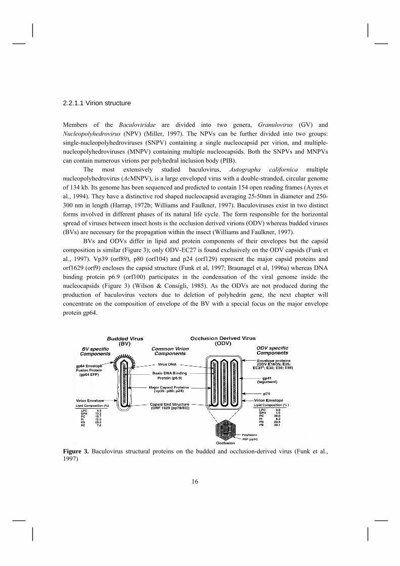
2.2.1.1 Virion structure Members of the Baculoviridae are divided into two genera, Granulovirus (GV) and Nucleopolyhedrovirus (NPV) (Miller, 1997). The NPVs can be further divided into two groups: single-nucleopolyhedroviruses (SNPV) containing a single nucleocapsid per virion, and multiple-nucleopolyhedroviruses (MNPV) containing multiple nucleocapsids. Both the SNPVs and MNPVs can contain numerous virions per polyhedral inclusion body (PIB). The most extensively studied baculovirus, Autographa californica multiple nucleopolyhedrovirus (AcMNPV), is a large enveloped virus with a double-stranded, circular genome of 134 kb. Its genome has been sequenced and predicted to contain 154 open reading frames (Ayres et al., 1994). They have a distinctive rod shaped nucleocapsid averaging 25-50nm in diameter and 250-300 nm in length (Harrap, 1972b; Williams and Faulkner, 1997). Baculoviruses exist in two distinct forms involved in different phases of its natural life cycle. The form responsible for the horizontal spread of viruses between insect hosts is the occlusion derived virions (ODV) whereas budded viruses (BVs) are necessary for the propagation within the insect (Williams and Faulkner, 1997). BVs and ODVs differ in lipid and protein components of their envelopes but the capsid composition is similar (Figure 3); only ODV-EC27 is found exclusively on the ODV capsids (Funk et al., 1997). Vp39 (orf89), p80 (orf104) and p24 (orf129) represent the major capsid proteins and orf1629 (orf9) encloses the capsid structure (Funk et al, 1997; Braunagel et al, 1996a) whereas DNA binding protein p6.9 (orf100) participates in the condensation of the viral genome inside the nucleocapsids (Figure 3) (Wilson & Consigli, 1985). As the ODVs are not produced during the production of baculovirus vectors due to deletion of polyhedrin gene, the next chapter will concentrate on the composition of envelope of the BV with a special focus on the major envelope protein gp64.
Figure 3. Baculovirus structural proteins on the budded and occlusion-derived virus (Funk et al., 1997)

17
2.2.1.2 Major envelope glycoprotein gp64 Budded virions contain one nucleocapsid surrounded by an envelope with gp64 major envelope protein found associated at one pole of the virus particles as peplomer structures (Figure 3). One virion is estimated to contain ~1000 gp64 peplomers (Wickham et al., 1990). The Gp64 exists as a disulfide-linked trimer with a molecular mass of 175 kDa (Oomens et al., 1995). The gp64 protein contains an N-terminal signal peptide and a C-terminal anchor domain. Gp64 accumulates at the plasma membrane during the early and late phases of infection, 8 and 24 hours p.i. (Blissard & Rohrmann, 1989; Monsma et al 1996; Monsma & Blissard, 1995; Volkman & Knudson, 1986). Nucleocapsids become surrounded by gp64-containing plasma membrane during budding from the cell surface in the late phase of infection. Furthermore, gp64 is required for efficient viral budding (Oomens and Blissard, 1999) and cell-to-cell transmission (Monsma et al., 1996). Gp64 mediates also virus binding to cell surface (Duisit et al., 1999; Ghosh et al., 2002; Hefferon et al., 1999; Hofmann et al., 1995) and low-pH-dependent membrane fusion (Blissard and Wenz, 1992). Successful membrane fusion requires the assembly of stable gp64 trimers into multiprotein aggregates in cell-cell contact regions (Markovic et al., 1998). 2.2.1.3 Baculovirus life cycle The baculovirus infection is initiated by ODVs in the gut epithelium (Figure 4). Occluded virions in large PIBs are protected from the environmental factors by a crystalline polyhedrin matrix (Braunagel and Summers, 1994; Harrap, 1972a), but in the alkaline midgut of insect larva the matrix is solubilized and the occluded viruses are released (Harrap et al, 1974). Occluded viruses enter the midgut epithelial cells via direct membrane fusion (Granados, 1978; Summers, 1971). Transcription of viral genes begins immediately after the virus DNA is transported to the nucleus. Baculovirus infection can be divided into three phases, early (0-6 h post-infection), late (6-24 h p.i.) and very late phase (18-24 to 72 h p.i.) (Williams and Faulkner, 1997). During the early phase of infection genes involved in the regulation of the replication cascade and those involved in preventing host responses are expressed. Early genes of the baculovirus are transcribed by the host RNA polymerase (Friesen, 1997). The late phase viral gene expression includes the replication of the viral DNA, the shutdown of host cell transcription and translation and the production of the budded form of the virus (Williams and Faulkner, 1997). The switch from early to late gene expression involves the appearance of a novel virus-induced RNA polymerase activity (Yang et al., 1991). In the very late phase the virus becomes occluded in the protein polyhedrin and the polyhedral envelope (calyx) is produced. Polyhedral inclusion bodies are released by cell lysis and the spreading of infection by adsorptive endocytosis leads eventually to the death of larva and the release of PIBs into the environment (Granados and Lawler, 1981). The cycle begins again when new insect ingests infected food.

18
Figure 4. Baculovirus life cycle consisting of the primary infection (on right) and the secondary infection (on left) (Airenne et al., 2008). 2.2.1.3 Baculovirus entry and gene delivery Budded viruses attach to and enter insect cells by absorptive endocytosis (Blissard and Wenz, 1992; Volkman and Goldsmith, 1985; Wang et al., 1997) followed by internalization into clathrin-coated vesicles. Recent observations in vertebrate cells also suggest involvement of macropinocytosis and caveolae (Long et al., 2006; Matilainen et al., 2005) The sheer number of mammalian cell lines that can be transduced by baculovirus vectors suggests that uptake of baculovirus by mammalian cells is a general phenomenon. The nature of the cell surface molecule that interacts with baculovirus is unclear but the involvement of receptors (Hofmann et al., 1995), electrostatic interactions (Duisit et al., 1999) and phospholipids (Tani et al., 2001) has been proposed. One possible explanation for these contradictory results is that mechanisms of virus-cell interactions are different between cell types. Following endosomal escape, nucleocapsids traverse the cytoplasm potentially with the help of actin filaments and enter the nucleus (van Loo et al., 2001) where the viral genome is released in response to the phosphorylation of basic core protein p6.9 (Funk and Consigli, 1993; Wilson and Consigli, 1985).
19
Baculoviruses are gaining popularity as potential vectors for gene transfer technology (Table 1). They are easily manipulated and produced in high titers (1010-1012 pfu/ml). The inherent inability of baculoviruses to replicate in mammalian cells and low cytotoxicity and lack of pre-existing immunity makes them good candidates for gene therapy in vivo (Hu, 2006). The transient nature of baculovirus-mediated gene delivery makes it an attractive candidate for the treatment of cancer (Song and Boyce, 2001; Wang et al., 2006) and cardiovascular diseases (Airenne et al., 2000; Grassi et al., 2006). A number of studies have also implicated the potential use of baculoviruses for bone (Chuang et al., 2007) and cartilage tissue engineering (Sung et al., 2007) and for gene delivery into nervous system (Lehtolainen et al., 2002b; Sarkis et al., 2000; Tani et al., 2003; Wang et al., 2007). Even though considerable progress has been made in elucidating the biology of baculovirus vectors, some limitations regarding the efficacy and specificity of these vectors have slowed their widespread applications. The major hurdle for baculovirus-mediated transduction lies in the stage of nuclear entrance since the viral DNA is unable to enter the nucleus of many vertebrate cells (Kukkonen et al., 2003; Volkman and Goldsmith, 1983). This might be due to the inability of the virus to escape from endosomes (Barsoum et al., 1997) or blockage of the transport or entry into the nucleus (Kukkonen et al., 2003; van Loo et al., 2001). It has been suggested that microtubules may constitute a barrier to nucleocapsid transport towards the nucleus in the cytoplasm (Salminen et al., 2005). Attempts to enhance baculovirus-mediated gene delivery have mainly focused on the virion surface modifications (Makela and Oker-Blom, 2006), promoter choices (Spenger et al., 2004; Wang et al., 2006), insertion of transgene expression enhancing elements (Mahonen et al., 2007; Venkaiah et al., 2004) and optimization of the transduction protocol in vitro (Condreay et al., 1999; Hsu et al., 2004; Shen et al., 2007). Despite these advances, in vivo gene delivery is still unsatisfactory. One obstacle is the inactivation of baculovirus by serum complement (Hofmann and Strauss, 1998). Different strategies have been pursued to overcome the problem of complement: to inactivate the complement system for the period of infection, to generate complement-resistant vectors (Huser et al., 2001) and to deliver viruses into immunopriviledged areas (Haeseleer et al., 2001; Lehtolainen et al., 2002b; Sarkis et al., 2000) or to sites where the exposure to the complement can be avoided (Airenne et al., 2000; Sandig et al., 1996). Baculovirus transduction leads to transient expression peaking at 3-5 days (Airenne et al., 2000; Lehtolainen et al., 2002b) and can last up to 200 days in the absence of complement (Pieroni et al., 2001). The gradual disappearance of the transgene expression is attributed to the degradation of baculoviral DNA (Ho et al., 2004). The transgene expression has been substantially prolonged by using baculovirus hybrid vectors, taking advantage of AAV ITRs necessary for replication and integration (Palombo et al., 1998; Wang and Wang, 2005; Zeng et al., 2007), or viruses capable of episomal replication (Shan et al., 2006). Even though baculoviruses are non-pathogenic to humans, recent evidence suggests that baculovirus transduction can induce the expression of some baculoviral immediate early genes in mammalian cells, namely ie-0, ie-1, pe-38, gp64 and p35 (Fujita et al., 2006; Kitajima et al., 2006). All these genes belong to the essential (p143, ie-1, lef-1, lef-2 and lef-3) or to the stimulatory (dnapol, p35, ie-2, lef-7, and pe38) genes involved in viral replication in the host cells (Kool et al., 1994; Lu

20
and Miller, 1995). This has shown to alter the expression profiles of mammalian genes although the physiology of the cells is not altered (Fujita et al., 2006; Kenoutis et al., 2006). Furthermore, administration of baculovirus induces expression of interferons and cytokines such as TNF-α, IL- 1α, IL-1β and IL-6 (Abe et al., 2003; Abe et al., 2005; Gronowski et al., 1999). These safety issues have to be taken into consideration when designing new vectors and therapies but also open new avenues for baculovirus-based vaccination and cancer immunotherapy (Kitajima and Takaku, 2008). 2.2.2 Retro- and lentiviruses Retroviridae is a large family of enveloped RNA viruses found in all vertebrates. The most peculiar feature of retroviruses is their ability to integrate the viral genome into the host chromosomal DNA, which can lead to lifelong expression. Retroviruses are currently classified into seven genera based on nucleotide sequence relationship: alpharetroviruses, betaretrovirus, gammaretroviruses, deltaretroviruses, episilonretroviruses, spumaviruses and lentiviruses (Goff, 2001). Gammaretroviruses, based on the murine leukaemia virus (MLV), are among the first viral delivery systems developed for gene therapy applications in 1990. Over the past decade, however, lentiviruses have gained a lot of attention due to their ability to transduce non-dividing cells. The advantages and disadvantages of these vectors are listed in Table 1. 2.2.2.1 Lentivirus structure and genome Figure 5. Structure of lentivirus with major viral proteins presented.
Envelope protein(s)(SU, TM)
Matrix (p17)
Capsid (p24)
Reverse transcriptase(p66, p51)
Genomic RNA innucleocapsid (p6, p7)
Lipid bilayer
Integrase (p32)
Protease (p10)

21
Lentivirus virions are roughly spherical particles with a diameter of 100-150 nm. Lentivirus genome is diploid and contains two plus-stranded RNA copies of its genome. Like other members of the retroviral family, the HIV genome contains the gag, pol and env genes (Wang et al., 2000). The env encodes for complex envelope protein, which consists of an outer protruding surface protein (SU) and a stem transmembrane protein (TM) (Figure 5). The gag gene products produce the protein core of viral particles consisting of p17 (matrix), p24 (capsid), p7 and p6 (nucleocapsid). In addition to nucleocapsid, the major elements contained within the viral core are two single strands of 9 kb RNA genome and three enzyme proteins, p66/p51 (reverse transcriptase), p11 (protease) and p32 (integrase), encoded by the pol gene (Figure 5). In addition, several other nonstructural proteins which serve regulatory functions including tat, rev, nef, vif, vpu and vpr are encoded by the HIV genome (Wang et al., 2000). Tat induces the transcriptional activation of the promoter situated at the long terminal repeat (LTR) whereas Rev plays a role in the nuclear export of viral mRNAs. The other accessory proteins Nef, Vif, Vpu and Vpr contribute to the replication and persistence of infection in vivo (Seelamgari et al., 2004). There are also a number of cis-acting elements required at different stages of the virus life cycle including the LTRs, packaging and dimerization signal (Ψ), Rev-responsive element (RRE), and the central polypurine tract (cPPT) (Wang et al., 2000). Lentivirus vectors. The general strategy in designing lentiviral vectors for gene therapy is based on the deletion and alteration of the native viral sequences, in order to prevent the generation of replication competent retroviruses. The state-of-the-art 3rd generation lentivirus vector system consists of four plasmids (Figure 6) (Delenda, 2004). The minimal transgene expression cassette contains the LTRs, packaging signal, a heterologous promoter and the transgene. Three additional plasmids provide the factors required for virus production and packaging (gag, pol, rev, env). The envelope proteins are typically replaced by a heterologous viral glycoprotein, most commonly vesicular stomatitis virus G-protein (VSV-G) (Naldini et al., 1996b), to modify the host range of the vector. An important safety feature is also the deletion of the promoter-enhancer region form the 3’ LTR preventing transcription from this region and subsequent viral replication (self-inactivating vector; SIN) (Miyoshi et al., 1998).
Figure 6. The four-plasmid transfection system for lentivirus production. The vector plasmid (1), the packaging plasmid (2), rev (3), and an envelope plasmid (4) are needed for HIV vector production. The packaging signal (Ψ), the rev-binding element (RRE) are indicated. (Sinn et al., 2005b)

22
2.2.2.2 Lentivirus life cycle Lentivirus infection is initiated by binding of the virion surface protein (SU) to the cognate receptor (Figure 7). The SU protein is attached to the virus by a non-covalent binding to the transmembrane protein (TM) which anchors the complex in the lentiviral envelope. SU receptor binding triggers conformational changes in the TM protein leading to the fusion between the viral membrane and the host cell membrane (Freed and Martin, 2007). For HIV-1, however, the binding of SU (gp120) to CD4 receptor is followed by the exposure of a chemokine receptor (CXCR4 or CCR5) binding site on SU protein and only subsequent binding to this co-receptor is able to trigger TM(gp41)-mediated fusion (Nisole and Saib, 2004) . Figure 7. The lentivirus replication cycle (http://www.retrovirus.info). The fusion of viral and cellular membranes delivers the viral core into the cytoplasm, where it undergoes a partial and progressive disassembly leading to the generation of pre-integration complexes (PICs). Subsequently the viral RNA is reverse transcribed into double-stranded cDNA in a process mediated by viral reverse transcriptase enzyme (Figure 7). The PIC associated cDNA enters the nucleus with the help of integrase, matrix protein and Vpr (Sherman and Greene, 2002). In the nucleus, the integrase protein catalyzes the integration of the viral cDNA into the host genome (Freed and Martin, 2007). Transcriptional regulation of HIV-1 gene expression is controlled by several host cell transcription factors and the viral Tat protein (Rohr et al., 2003). In the early phase of viral gene expression the newly transcribed mRNA is spliced by the cellular splicing machinery into multiply spliced transcripts, which mainly produces Tat, Rev and Nef proteins. When Rev has accumulated to a critical level the mRNA production shifts from multiply spliced to the singly spliced and unspliced
23
transcripts (e.g. gag, vif, env), characteristic of the late phase of gene expression. The Rev binding to RRE leads to the nuclear export of the late-phase transcripts (Freed and Martin, 2007). Following the production of viral structural proteins, the virus particle is assembled at the plasma membrane (Figure 7) (Bukrinskaya, 2004). In this process the Gag and Gag-Pol polyproteins interact with each other by protein-protein interaction, most probably via the capsid protein domain (Gelderblom, 1991). The viral genome is packaged in a process in which the packaging signal is recognized by the nucleocapsid protein domain of the Gag protein (Zhang et al., 1998). The mature HIV particles bud from the host membrane ready to infect another cell and to begin the replication process all over again. 2.2.2.3 Lentivirus as a gene therapy vector Lentiviruses have gained much attention as a gene delivery tool over the past decade due to their ability to transduce non-dividing cells, giving rise to first clinical trials in 2001. Lentivirus development has mainly focused on human immunodeficiency virus type 1 (HIV-1) and improvements of the vector have enabled efficient in vivo and ex vivo gene delivery to many tissues. However, also non-human pathogens, such as feline (FIV), simian (SIV) and bovine (BIV) immunodeficiency virus and equine anemia infectious virus (EIAV) are currently being investigated for gene therapy due to their safety advantages (Romano, 2005). Lentiviral vectors are rapidly becoming the vectors of choice for hematopoietic stem cell (HSC) gene therapy due to capacity to transduce also quiescent cells, in which state most of HSCs are (Naldini et al., 1996b). Lentiviral vectors can also deliver genes to HSCs with a superior efficiency to MLV vectors without affecting the repopulating capacity of these cells (Kay et al., 2001). Consequently the first clinical studies with HIV-based lentiviral vectors were concentrated on delivering anti-HIV-, antisense- or RNAi- genes to the HSCs of HIV infected patients (http://www.wiley.co.uk/genmed/clinical/). Lentiviral gene transfer to HSC has been also proposed to provide a potential cure for many inherited diseases such as sickle cell disease (Pawliuk et al., 2001) and chronic granulomatous disease (Roesler et al., 2002) and β-thalassaemia (Imren et al., 2002; May et al., 2002; Vacek et al., 2003) where progression towards the clinic can be seen (Bank et al., 2005). Another promising target area for lentivirus-mediated gene delivery is the brain. The VSV-G pseudotyped vectors based on HIV (Kordower et al., 1999; Naldini et al., 1996a), FIV (Alisky et al., 2000) and EIAV (Mitrophanous et al., 1999) vectors were shown to efficiently transduce neurons in various areas of the brain while leading to long-lived transgene expression. Since then, several studies have demonstrated convincing therapeutic efficacy of lentivirus-mediated gene delivery in animal models of lysosomal enzyme deficiency disorders, Huntington’s disease, Alzheimer’s disease and Parkinson’s disease (Wong et al., 2006). Like MLV, HIV integrates randomly into the host genome. This poses a risk of insertional mutagenesis as was demonstrated by the appearance of several cases of leukaemia in the gene therapy trials for X-SCID (Gaspar et al., 2004; Hacein-Bey-Abina et al., 2003; Wilson, 2008). This malignant

24
transformation is likely related to gammaretroviruses’ inherent disposition to integration near the 5’ends of transcription units leading to proto-oncogene activation (Wu et al., 2003). In contrast, lentiviruses strongly favor integration within active transcription units which might be a safer alternative (Mitchell et al., 2004; Schroder et al., 2002). In addition, the careful design of 3rd generation SIN vectors might further reduce the risk of insertional gene-inactivation or proto-oncogene deregulation in the case of lentiviruses (Miyoshi et al., 1998; Thornhill et al., 2008). Other strategies to overcome this problem have led to the development of non-integrating (Philpott and Thrasher, 2007) or site-specifically integrating vectors (Bushman, 1994; Tan et al., 2006). 2.2.3 Adenoviruses Adenoviruses (Ad) belong to the family of Adenoviridae which to date includes 51 immunologically distinct human adenovirus serotypes (A-F) that can cause human infections ranging from respiratory disease, and conjunctivitis, to gastroenteritis. Replication defective viruses based on subgroup C adenovirus type 5 (Ad5) and type 2 (Ad2) are the most widely used for gene transfer in many applications (Shenk, 2001). Adenoviruses are double-stranded DNA viruses with a genome of 36 kb (Chroboczek et al., 1992). The virions are nonenveloped and icosahedral in shape with a diameter of 70-90 nm. The viral capsid containins four principal protein components: the hexon, fiber, penton base and protein IX (Figure 8). Ads enter the host cells by receptor-mediated endocytosis. Initial interaction with the host cells is mediated by the fiber protein and the coxsackie virus and adenovirus receptor (CAR) (Bergelson et al., 1997) and subsequent internalization results from the activation of αv integrin by penton base (Wickham et al., 1993). Figure 8. Structure of adenovirus as a simplified cross-section of the capsid showing the capsid proteins and adenovirus genome (Noureddini and Curiel, 2005).

25
Adenoviruses have passed retroviruses as the most commonly applied viral vectors with over 340 clinical trials finished or ongoing (Figure 2). The major advantages of adenovirus vectors is the the large DNA insertion capacity, the easy generation and purification of high titer stock (1010-1012 pfu/ml) (Table 1). They have a broad host range and can efficiently transfer genes into both dividing and non-dividing cells. Adenoviral genomes do not integrate into the host genome, making them safe vectors for transient gene expression. However, both natural immunity against adenovirus (Chen et al., 2000) and acute inflammatory (Knowles et al., 1995; Yei et al., 1994) and immunological responses (Zoltick et al., 2001) have limited the current clinical applications to few areas such as localized cancer and cardiovascular gene therapy. The concomitant knowledge about adenovirus biology has led to the development of conditionally replicative adenoviruses (CRAds) which exhibit tumor specific amplification resulting in lysis of the cancer cells (Heise and Kirn, 2000). On the other side of the scope, the aspiration to enlargen the field of adenovirus application has led to development of methods to minimize the viral gene content (gutless Ads), to decrease the immunogenicity of the vectors and to retarget the vector tropism (Campos and Barry, 2007; Ghosh et al., 2006). 2.2.4 Adeno-associated viruses (AAVs) AAV is one of the smallest viruses with a non-enveloped icosahedral capsid of approximately 20-25 nm in diameter belonging to the parvoviridae family (Xie et al., 2002). To date, at least 11 serologically distinct AAVs have been identified from humans or primates (Mori et al., 2004). The most extensively studied AAV is AAV type 2 (AAV-2), it also being the most common in active clinical trials (Coura and Nardi, 2007). The most peculiar feature of AAV is its dependence on helper viruses (e.g. adenovirus or herpes virus) for productive infection (Muzyczka and Berns, 2001). It infects humans and some other primate species but the virus has not been linked to any human diseases. Despite the nonpathogenic nature of virions, most humans are seropositive to AAV which may limit the gene delivery efficiency in vivo. The pros and cons of AAV vectors are listed in more detail in Table 1. The AAV has a linear 5 kB single-stranded genome of either plus or minus polarity. The genome harbors two open reading frames (ORFs): one encoding for Rep proteins involved in regulation of replication and transcription and the other for virus capsid proteins VP1, VP2 and VP3 which form the virion in ratio (1:1:10) (Muzyczka and Berns, 2001). AAV-2 capsid proteins mediate the virion attachment to heparin sulfate proteoglycan (HSPG), fibroblast growth factor receptor-1 (FGFR1), integrin αvβ5 and hepatocyte growh factor receptor (HGFR) on host cell membrane and subsequent endocytosis through clathrin-coated pits (Kashiwakura et al., 2005; Qing et al., 1999; Summerford and Samulski, 1998). The ends of the genome form short inverted terminal repeats (ITRs), which serve as origins of viral replication. The ITRs are the only cis element required for replication and packaging of the virus and therefore all the other elements, provided in trans, have been deleted from the recombinant AAV-2 vectors (rAAV-2).

26
The wild type AAV-2 is able to integrate into the genome of the host with the help of Rep proteins with a site preference on human chromosome 19 (Samulski et al., 1991). However, even in the absence of rep-genes on AAV-2 vectors the rAAV genome has been shown to persist in episomal (Afione et al., 1996; Yang et al., 1999) or integrated (Nakai et al., 2001) form. The mechanism lying beneath integration has been thought to involve the host cell DNA break repair machinery, which inserts rAAV genome into existing chromosomal breaks (Nakai et al., 2003). For gene therapy applications, this feature of rAAV is a double-edged sword: rAAVs can maintain high levels of transgene expression but at the same time insertional mutagenesis becomes an issue. However, since rAAVs don’t create but instead insert into existing chromosomal breaks, they could be considered safer than retroviruses. rAAV has shown great potential for the gene delivery to muscle, brain, liver and eye. The current clinical trials are mainly concentrated for the treatment of monogenic diseases, especially cystic fibrosis, hemophilia B, retinal degeneration, and cancer (http://www.wiley.co.uk/genmed/ clinical/). The results so far have shown that rAAVs are safe and efficient tools for gene delivery but the therapeutic benefit to human patients is still limited by the inadequate organ-specific transgene expression (Coura and Nardi, 2007). 2.2.5 Other viruses Over 200 clinical trials have been conducted with less conventional viral vectors including poxviruses, herpes simplex virus, Semliki forest virus, Sendai virus, Simian virus, measles virus, poliovirus, flavivirus and Venezuelan equine encephalitis Newcastle disease virus. The former two represent the vast majority of the studies and will be discussed in more detail. Poxviruses are enveloped viruses which can infect as a family both vertebrate and invertebrate animals (Moss, 2001). Vaccinia virus (VV) is the prototypical recombinant poxvirus. Vaccinia virus has been used clinically as a vaccine for smallpox since the late 18th century, and has thus a well known biology and extensive clinical experience (Niemialtowski et al., 1996). Recombinant VVs, as non-replicating viral vectors, have been demonstrated to have great potential as vaccines due to their safety, low cytotoxicity, high level of protein expression and ability to generate potent antibody and T-cell responses. A number of clinical trials using recombinant VV as vaccines have shown promising results for treating HIV and cancer (Moroziewicz and Kaufman, 2005). On the other hand, replicating VVa are promising candidates for oncolytic virotherapy (Thorne et al., 2005). Herpes simplex virus (HSV) is a human infecting pathogen with a double stranded genome of 152 kB (Whitley, 2001). Among the herpes viruses, HSV-1 is an attractive vector for gene transfer to the nervous system because the natural infection leads to lifelong persistence of viral genomes in neurons in which the latent phase and lytic phase alternates. Two types of vectors have been developed for gene therapy applications: replication defective vectors, whose cytotoxicity has been abolished by deleting lytic gene products, and amplicon vectors, which are plasmids packaged into HSV particles with the aid of a helper virus (Whitley, 2001). Logically, these vectors have been primarily used for neuronal gene delivery for the treatment of neuropathies (Parkinson's disease, pain,

27
stroke) and lysosomal storage disorders (Berto et al., 2005). The majority of the clinical studies with HSV-1 are, however, concentrated on cancer therapy. For these applications, the HSV-1 vectors high infectivity and inherent cytotoxicity is harnessed to conditionally drive viral replication in tumor cells leading eventually to cell lysis (Shen and Nemunaitis, 2006). Similarly, other viruses have been studied as candidates for the oncolytic viral treatment of tumors, including Newcastle disease virus, reovirus, measles virus, Semliki forest virus, sindbisvirus, vesicular stomatitis virus, influenza virus and poliovirus (Kelly and Russell, 2007). 2.2.6 Nonviral vectors Nonviral vectors represent an attractive alternative to viral vectors due to the ease of large-scale production, large insertion capacity, stability, flexibility and lack of immune response. Nonviral gene delivery can be divided into two broad categories: naked DNA delivery by a physical method and delivery by a complex of DNA with a cationic carrier. The latter group can be further divided into lipoplexes (cationic lipid/DNA complex) and polyplexes (cationic polymer/DNA complex) and more recently to the lipid-polymer hybrid systems (Gao et al., 2007). The physical approaches consist direct delivery of DNA to the cytoplasm of target cells by microinjection, gene gun, electroporation, sonoporation or laser irradiation (Mehier-Humbert and Guy, 2005). For systemic administration, however, the plasmid DNA needs to be protected from the nucleases and mononuclear phagocyte system (Kawabata et al., 1995; Mahato et al., 1995). Therefore, plasmid DNA is often shielded from the degradation by cationic compounds. Cationic lipoplexes (Felgner et al., 1987) and polyplexes (Wu and Wu, 1987) were introduced already in 1987 and are today the most studied strategy for nonviral gene delivery. These compounds condense and decrease the negative charge of DNA and thus facilitate its interaction with the cell membrane. Following binding, endocytosis or endocytosis-like mechanisms are proposed to be responsible for the entry of lipoplexes and polyplexes (Elouahabi and Ruysschaert, 2005). One of the major bottlenecks for effective transfection has been the subsequent release of DNA-complexes from the endosomes. This has been circumvented by the use fusogenic “helper” lipids such as dioleoylphosphatidylethanolamine (DOPE) (Farhood et al., 1995) or polymers with intrinsic endosomolytic activity, the most popular being polyethylenimine (PEI) (Boussif et al., 1995). Both vectors have shown excellent efficiency in cell culture but the in vivo gene delivery is still unsatisfactory. In addition, the in vivo administration can sometimes lead to aggregation, toxicity and acute immune responses (Gao et al., 2007). Various strategies have evolved to overcome these problems, the most promising being the shielding of the cationic compounds with polyethyleneglycol (PEG) (Ambegia et al., 2005; Kichler, 2004; Song et al., 2002). Despite some drawbacks, efficient in vivo gene delivery has been achieved to the lungs, brain, kidney and tumors and some of the vectors have undergone clinical trials for the treatment of cancer and cystic fibrosis (Nishikawa and Hashida, 2002)(www.wiley.co.uk/genmed/clinical/).

28
In the future, the combination of the best features of viral and non-viral vector systems by creating chemically modified viral vectors or synthetic virus-like systems could provide significant therapeutic benefits over the traditional vector systems (Boeckle and Wagner, 2006). Table 1. Properties of the most common gene delivery vectors (Gao et al., 2007; Kootstra and Verma, 2003; Moroziewicz and Kaufman, 2005; Waehler et al., 2007). Pros Cons
Baculovirus • High titers (1010-1012 pfu/ml) • Large insertion capacity > 100 kB • Non-human pathogen, safety
• Inactivation by complement • Immunogenic • Large size
Retro-and lentiviruses
• Stable gene expression • Insert capacity 8-9 kB • No pre-existing immunity • Moderate titers 106-1010 TU/ml
• Risk of insertional mutagenesis • Risk of replication competent
virus formation • Inactivation by complement
Adenovirus • High titers (1010-1012 pfu/ml) • Insert capacity 7-8 kB, for gutless vectors
36 kB • Broad tropism • High short-term gene expression • Oncolytic potential
• Pre-existing immunity: neutralizing antibodies
• Acute inflammatory and immunological responses
• Complicated vector genome
Adeno-associated virus (AAV)
• Stable gene expression possible • Nonpathogenic • Highly stable virions • Small size (22 nm) • No need for viral genes in vectors
• Small insert capacity, 4.6 kB • Slow onset of gene expression • Risk of insertional mutagenesis • Production requires helper viruses • Large-scale production difficult
Vaccinia • Well established safety profile • Oncolytic potential
• Immunogenicity
Herpes simplex virus (HSV)
• Long-term expression in neuronal cells, neurotropism
• High titers, 108-1011 pfu/ml • Transgene capacity 30 kB, for amplicons
152 kB • Oncolytic potential
• Host immune responses, inflammation and toxicity
• Complicated vector genome
Nonviral vectors
• Low degree of toxicity, non-infectious • Easy and simple production • High efficiency in vitro • No insert size limit
• Low transfection effic. in vivo • Only transient expression • For some vectors acute immunity,
toxicity, aggregation in vivo
2.3 Targeted gene delivery Key issues for future development of gene therapy include improved gene delivery and targeting. In theory, targeted therapeutic gene delivery can be achieved by targeting entry of the vector (transductional targeting) or by targeting the gene expression (transcriptional targeting) to certain cell types or tissues. Transcriptional targeting has been shown to be highly feasible in the context of most viral vectors (Miller and Whelan, 1997). It provides a safety net by limiting the transgene expression

29
to specific target cells using tissue specific promoters. However, transcriptional targeting does not obviate the need for transductional targeting which is essential for allowing the administered therapeutic dose to be reduced, thereby lessening toxic side effects and costs for the treatment. Thereby the focus of this chapter will be on targeted transduction. 2.3.1 Targeting of membrane-enveloped viruses Targeted gene delivery is currently the most attractive concept to achieve specificity and, in principle, this strategy is applicable for all current vectors (Waehler et al., 2007). The outer surface of virus, through its interaction with cellular receptors, plays a major role in determining the tropism of the virus. There are several strategies for modifying the binding characteristics of membrane-enveloped virus vectors and most of the studies have been conducted using retro- and lentiviruses because they are highly permissive for incorporation of heterologous attachment proteins. In theory, all of these targeting approaches can be extrapolated to other enveloped viruses on condition that the virus budding, fusion activity and infectivity is not compromised. The focus of this chapter will therefore be on these vectors with an extension to baculovirus. Pseudotyping. The simplest form of transductional targeting consists of changing the virus surface protein itself for the envelope or capsid protein of another virus which is not of the same genus. This approach is called pseudotyping. One of the most commonly used pseudotyping tools is Vesicular stomatitis virus G protein, VSV-G (chapter 2.3.1.1). It is routinely used to broaden the target range and enhance the transduction efficiency of retroviruses (Emi et al., 1991) and HIV-1-, HIV-2-, SIV-, FIV-, EIAV- and BIV- based lentiviruses (Cronin et al., 2005; Naldini et al., 1996b; Reiser et al., 1996). Significant advantage of VSV-G pseudotyping is its ability to confer high vector particle stability allowing virus concentration by ultracentrifugation (Burns et al., 1993). There are also several reports of VSV-G pseudotyped baculoviruses which show improved transduction efficiency (Barsoum et al., 1997; Tani et al., 2001; Tani et al., 2003). Unfortunately, VSV-G is cytotoxic to producer cell lines (Burns et al., 1993; Ory et al., 1996; Schauber et al., 2004) and there have also been reports where the VSV-G included in the viral envelope increased the toxicity of the vector (Facciabene et al., 2004; Park et al., 2000; Watson et al., 2002). Together these features can limit the clinical use of VSV-G and alternative glycoproteins have been extensively studied. Some of the most prominent lentivirus pseudotypes and their target cells/organs are presented in Table 2. These include glycoproteins from the families rhabdoviridae, arenaviridae, togaviridae, filoviridae, paramyxoviridae, orthomyxoviridae, and hepadnaviridae (Cronin et al., 2005). In addition, lentiviral vectors pseudotyped with baculovirus envelope glycoprotein gp64 have been produced. Gp64-displaying HIV-1 vectors were produced at similar titers to VSV-G with no associated cytotoxicity and concetration by ultracentrifugation was well tolerated (Kumar et al., 2003). These vectors transduced efficiently various cell types, with a tropism restriction against hematopoietic cell types (Schauber et al., 2004). More recently, two reports have demonstrated the utility Gp64 pseudotyped FIV-vectors for hepatocyte and nasal epithelia targeting (Kang et al., 2005; Sinn et al., 2005; Sinn et al., 2007).

30
Table 2. Cell and organ preferences of lentivirus pseudotypes. Modified from (Cronin et al., 2005).
Glycoproteins (Genus)
Target cell/organ
Remarks References
VSV-G (Rhabdoviridae)
Liver CNS
Retina
Toxicity issues Targets primary neurons
Photoreceptors and retinal pigm. epithelium
(Park, 2003) (Blomer et al., 1997)
(Auricchio et al., 2001; Miyoshi et al., 1997)
Rabies (Rhabdoviridae)
CNS
Cancer
Retro- and anterograde axonal transport Neuroblastoma
(Mazarakis et al., 2001; Wong et al., 2004)
(Steffens et al., 2004) Mokola
(Rhabdoviridae) CNS
Retina Muscle Cancer
Neurons
Retinal pigm. epithelium Cardiomyocytes Neuroblastoma
(Desmaris et al., 2001; Watson et al., 2002)
(Auricchio et al., 2001) (MacKenzie et al., 2002)
(Steffens et al., 2004) LCMV
(Arenaviridae) Liver CNS
Pancreas Cancer
Non-toxic Neural progenitor cells
Astrocytes Islet cells
Malignant glioma
(Park, 2003) (Stein et al., 2005)
(Miletic et al., 2004) (Kobinger et al., 2004)
(Miletic et al., 2004; Steffens et al., 2004)
RRV (Togaviridae) Liver CNS
Nonhepatocytes, nontoxic
Neuroglial cells Complement resistance
(Kang et al., 2002) (Kang et al., 2002) (Strang et al., 2005)
Ebola (Filoviridae) Lung airway epithelia Muscle
Apical surface preference
Cardiomyocytes
(Kobinger et al., 2001)
(MacKenzie et al., 2002)
Marburg (Filoviridae)
Lung airway epithelia
Apical surface preference
(Sinn et al., 2003)
JSRV (Betaretrovirus)
Lung Alveolar type II cells (Sinn et al., 2005a)
MLV (Gammaretrovirus)
Cancer Neuroblastoma (Steffens et al., 2004)
GALV (Gammaretrovirus)
Hematopoietic system Cancer
Increased serum stability
Fusogenic glycoprotein
(Sandrin et al., 2002)
(Diaz et al., 2000) RD114
(Gammaretrovirus) Hematopoietic
system Less toxic and more
efficient than VSV-G (Sandrin et al., 2002)
Sendai (Paramyxoviridae)
Lung airway epithelia
Apical and basolateral surfaces
(Kobayashi et al., 2003)
Influenza A (Orthomyxoviridae)
Airway epithelia
Apical surface preference
(Sinn et al., 2005)
HBV (Hepadnaviridae)
Liver Primary hepatocytes (Chai et al., 2007)
Baculovirus (Baculoviridae)
Liver Airway epithelia
Non-toxic Apical surface
preference
(Kang et al., 2005) (Sinn et al., 2005)

31
Despite enhancing the transduction efficiency, pseudotyping often provides a wide host range and lacks sufficient target cell specificity. Therefore other strategies have been sought, based the modification of envelope glycoproteins genetically or by using bispecific adaptor-molecules. Genetic and adaptor-based targeting of retro- and lentiviruses. Retargeting based on genetic modification of the glycoproteins was first tested with retroviral vectors (Russell et al., 1993). Efforts to target retroviral vectors have concentrated largely on engineering the natural retroviral envelope proteins such as the ecotropic murine leukemia virus MLV protein. Several strategies have been taken to produce targeted envelope proteins. For direct targeting by host range extension, envelope glycoproteins are modified to incorporate heterologous proteins or ligands. This can be achieved by replacing the natural receptor-binding domain of SU protein with the targeting molecule. A wide range of receptors have been targeted this way but most of the derivatives were unable to trigger the subsequent fusion leading to low gene transfer efficiency (Benedict et al., 1999; Zhao et al., 1999a). Therefore, another approach was developed which consisted of leaving the native receptor binding domain intact while conferring the SU protein with an additional binding moiety, called “tethering”. On this basis, the insertion of collagen-binding ligand into the SU of amphotropic MuLV was shown to enhance retrovirus binding and tranduction of human endothelial cells in vitro (Hall et al., 1997; Hall et al., 2000; Liu et al., 2000). Moreover, these vectors could localise gene delivery to sites of balloon-injured carotid arteries and in the angiogenic tumor vasculature in human cancer xenografts in nude mice (Gordon et al., 2001a; Gordon et al., 2001b). Two strategies have been developed for targeting retroviral vectors by host-range restriction; inverse targeting and protease targeting. Inverse targeting involves the selective inhibition of infectivity on cells expressing the targeted receptor, whereas protease targeting selectively reactivates the inhibition imposed by inverse targeting. Several ligands displayed at the N-terminus of retroviral envelope glycoproteins have been shown to inhibit infectivity on cells expressing the targeted receptor. A well-studied example of this comes from amphotropic vectors displaying epidermal growth factor (EGF) which are sequestered on EGF receptor-positive (EGFR) cells through redirection to lysosomal degradation but remain fully infectious on EGFR-negative cells (Cosset et al., 1995). Similar receptor-mediated sequestration has been observed for vectors displaying stem cell factor (Fielding et al., 1998; Fielding et al., 2000), insulin-like growth factor (Chadwick et al., 1999; Fielding et al., 2000) and CD33 (Zhao et al., 1999a). Alternatively, the sequestration can be dismantled by separating the ligand and the virus envelope protein by the recognition site for a cell-surface specific protease. For example, the EGF-displaying retroviruses carrying a matrix-metalloproteinase (MMP) cleavage site could preferentially infect EGFR-positive MMP-rich target cells in vitro and in vivo (Peng et al., 1997; Peng et al., 1999). Similarly, protease targeting has been achieved by the display of single-chain variable fragment (scFv) directed against carcinoembryonic antigen and c-Met receptor frequently overexpressed on tumor cells (Chowdhury et al., 2004; Solly et al., 2005). Since the retroviral Env requires interaction with their native receptor to activate fusion activity, binding to artificial target molecule does not activate the fusion step. Therefore most of the direct or indirect targeting strategies have suffered from low titer and/or specificity (Verhoeyen and Cosset, 2004). Ideally, binding and fusion functions can be broken into two separate molecules. The

32
key to the method is choosing a viral glycoprotein that mediates fusion in response to low pH and a cellular receptor that is efficiently endocytosed after antibody binding. In this regard, a binding-defective mutant of hemagglutinin of influenza A was coexpressed with a binding-competent but fusion-defective MLV Env containing Flt-3 targeting ligand resulting in enhanced entry into Flt-3-expressing cells (Lin et al., 2001). Similarly, the Sindbis virus envelope protein E2 responsible for cell binding was engineered to replace the receptor binding region with the Fc binding domain of protein A (ZZ domain) (Morizono et al., 2001). When E2 was coexpressed with the E1 fusion protein on the retroviral and lentiviral surface and conjugated to targeting antibody, successful retargeting to mouse metastatic melanoma and prostate cancer bone metastases was achieved after intravenous injection (Morizono and Chen, 2005; Morizono et al., 2005; Pariente et al., 2007). Futhermore, coexpression of E2 binding-mutant, E1 and a chimeric anti-CD20 antibody with the human membrane-bound IgG constant region demonstrated a strictly targeted transduction of CD20-positive cells in vitro and in vivo (Yang et al., 2006). Adapter-based concept of virus targeting consists of the formation of a ‘molecular bridge’ between the vector and a cell surface receptor constitutes. One such approach has been described for the Env protein of avian leukosis virus (ALV) that combine the EGF targeting domain with the extracellular domain of the ALV receptor (Snitkovsky and Young, 1998; Snitkovsky et al., 2000). Thus this bifunctional bridge proteins binds virions to specific cell surface molecules and the receptor moiety triggers the normal fusion process. Other cell surface receptors targeted this way include the vascular endothelial growth factor and heregulin receptor (Snitkovsky and Young, 2002; Snitkovsky et al., 2001). Genetic targeting of baculoviruses. Surface modification of baculovirus particles has been demonstrated by epitope insertions into the baculovirus glycoprotein gp64. Extensive mutagenesis revealed permissive insertion sites to be located between amino acid positions 274 and 283, whereas N-terminal fusions resulted in weaker epitope display. In this regard, successful peptide-display has been achieved with the biotin mimic streptagII, the ELDKWA peptide of the gp41 of HIV-1 and the RGD-motif (Ernst et al., 2000; Ernst et al., 2006; Spenger et al., 2002). A recent study also demonstrated the utility of the Gp64 peptide display for the targeting of lentiviral vectors, using a peptide derived from the hepatitis B virus PreS1 protein, with known affinity for hepatocytes (Markusic et al., 2007). Most of the studies aiming at altering the baculovirus tropism have consisted of the fusion of heterologous proteins and ligand-binding moieties to an extra copy of the gp64 gene (Boublik et al., 1995). Using this strategy, Mottershead and colleagues constructed vectors displaying either functional scFv or a synthetic IgG binding domain (ZZ domain) of protein A (Mottershead et al., 2000). Specific binding to target cells was achieved although no enhancement of viral entry or gene transfer was observed (Ojala et al., 2001). In fact, only few studies based on the N-terminal fusions of gp64 have resulted in enhanced transduction efficiency including the display of RGD-peptide and avidin (Matilainen et al., 2006; Raty et al., 2004). This could be partly due to the fact that gp64-based fusion constructs must compete for space with the wild type gp64 leading to low level of incorporation of gp64-fusions on baculovirus surface (Boublik et al., 1995). On this basis, improvements in the expression of synthetic IgG binding domains on the baculovirus surface was

33
achieved by fusing the ZZ sequence to VSV-G membrane anchor which successfully incorporated into virions (Ojala et al., 2004). Similarly, tumor homing peptides were displayed on the baculovirus surface resulting in vectors with significantly improved binding and transgene delivery to human carcinoma cells (Makela et al., 2006). 2.3.1.1 Vesicular stomatitis virus G protein All rhabdoviruses encode a membrane glycoprotein of about 500 amino acids. Approximately 1200 VSV glycoprotein molecules are organized into 400 trimeric spikes anchored in the viral envelope (Doms et al., 1987; Kreis and Lodish, 1986). The VSV-G protein is a type I membrane glycoprotein synthesized as a precursor of 511 amino acids (Indiana), which is cleaved from its 16 amino acid signal sequence after insertion into the endoplasmic reticulum (ER) (Lingappa et al., 1978). Most of the amino acids are exposed on the virion surface, while 20 hydrophobic amino acids span the membrane and 29-amino acid cytoplasmic domain extends into the virion (Figure 9). There are two N-linked glycosylation sites on the extracellular domain and a single molecule of palmitate on the cytoplasmic domain (Rose et al., 1984; Schmidt and Schlesinger, 1979). The absence of glycosylation sites leads to formation of large disulfide bonded aggregates (Doms et al., 1988; Machamer and Rose, 1988), whereas the function of palmitate is not known (Whitt and Rose, 1991).
Figure 9. Schematic presentation of the vesicular stomatitis virus glycoprotein illustrating the functional domains [(Whitt and Rose, 1991) with modification]. Roles of all three domains of the VSV-G protein have been analyzed by mutagenesis (Figure 9). Mutations in the extracellular domain prevent correct folding, which leads to failed transport and aggregation in the ER before trimerization of monomers (Doms et al., 1988). Mutations in the transmembrane domain have similar effects, whereas mutations in the cytoplasmic domain do not seem to affect the folding of the extracellular domain (Doms et al., 1988). However, mutations in the cytoplasmic domain reduce the transport rate of VSV-G protein from the ER (Rose and Bergmann,
signalsequence
1 16 463 482
118 139 449
transmembranedomain
462
462451
buddingdomain
fusion domains
179 336
511
COOHNH2
164 395418
449
signalsequence
1 16 463 482
118 139 449
transmembranedomain
462
462451
buddingdomain
fusion domains
179 336
511
COOHNH2
164 395418
449

34
1983). This led to the discovery of transport signals (tyrosine based and di-acidic motifs) in the cytoplasmic tail responsible of promoting G protein export from the ER (Sevier et al., 2000). The VSV host range extends from nearly all mammals to insects, suggesting that the receptor for this virus is a widely distributed molecule. Binding to phospholipids seems to be important for rhabdovirus infection since phospholipids from cellular membranes inhibit attachment and infection of rabies virus and VSV (Conti et al., 1988; Mastromarino et al., 1987; Schlegel et al., 1982). Further studies have led to the conclusion that the receptor for VSV-G is a ubiquitous membrane lipid, phosphatidylserine (PS) (Carneiro et al., 2002; Schlegel et al., 1983). The PS-binding site of the VSV-G protein has since been mapped to a p2-like peptide (residues 134 to 161) containing contiguous heptad repeats followed by a short segment containing positively charged amino acids (Carneiro et al., 2003; Coll, 1997). Positive charges in this segment might participate in electrostatic interactions with the negatively charged phospholipid PS during membrane recognition. Unlike most viral glycoproteins, VSV-G protein has no obvious region in the amino acid sequence responsible for fusion activity (i.e. fusion peptide). Mutational analysis has indicated that amino acids between 118 and 139 could be the putative fusion domain (Li et al., 1993; Whitt et al., 1991; Zhang and Ghosh, 1994). Furthermore, recent studies have proposed that the PS-binding region of VSV-G (residues 134 to 161) is not only involved in membrane recognition but is also of crucial for membrane fusion probably through the protonation of its His residues (Carneiro et al., 2006; Carneiro et al., 2006). Another region of G protein, encompassing residues 395 to 418 has been identified as a segment that affects the fusogenic activity of the protein by influencing the low-pH-induced conformational changes (Li et al., 1993; Shokralla et al., 1998). In addition, it has also been shown that not only the ectodomain but also the membrane anchoring domain is required for VSV fusion activity (Cleverley and Lenard, 1998; Odell et al., 1997). Moreover, Jeetendra et al provided evidence that membrane-proximal stem region of VSV-G protein ectodomain (GS i.e. G stem), together with transmembrane and cytoplasmic domains can potentiate the membrane fusion activity when coexpressed with heterologous viral fusion proteins (Jeetendra et al., 2002). The G-stem was shown to be able to mediate binding of the VSV to target membranes in a manner similar to wild-type VSV. This could bring two membranes in close proximity to induce lipid mixing and initiate the fusion reaction. It was determined that only 14 amino acids of the GS were sufficient to induce hemifusion. A more recent study further elucidated the role of membrane proximal region of protein G showing that the membrane proximal region is not essential for G protein oligomerization, transport to the cell surface, or incorporation into virus particles but it is essential for acid-induced membrane fusion activity and virus infectivity (Jeetendra et al., 2003). Altogether these results suggested that in the case of VSV-G glycoprotein, the fusogenic activity may involve several spatially separated regions in the extracellular domain of the protein (Figure 9). Early models of VSV assembly postulated the importance of interaction between VSV-G protein and ribonucleoprotein core (Metsikko and Simons, 1986). This conclusion was based on the evidence showing that G proteins with truncated cytoplasmic tails were not efficiently incorporated to the virions (Whitt et al., 1989) and that addition of cytoplasmic tails to heterologous viral glycoproteins was sufficient for their incorporation into virus particles (Owens and Rose, 1993). However, VSV-G protein is not needed for virus budding although it does enhance it (Knipe et al.,

35
1977; Mebatsion et al., 1996; Schnell et al., 1997; Takada et al., 1997). In fact addition of G protein cytoplasmic tail to heterologous proteins does not increase virus budding or glycoprotein incorporation (Kahn et al., 1999; Schnell et al., 1996) and G proteins with truncated or chimeric cytoplasmic tails produce infectious VSV (Schnell et al., 1998). These results support the idea that small cytoplasmic tail of VSV-G is important for VSV budding but no specific amino acid sequence is required. Identification of the budding domain in the membrane-proximal region of the G ectodomain supports this idea (Robison and Whitt, 2000). The report demonstrated a generation of series of recombinant VSVs, which expressed chimeric glycoproteins having truncated stem sequences. The recombinant viruses having 12 or more membrane-proximal residues, including transmembrane and cytoplasmic tail domains, produced near-wild-type levels of virus particles. 2.3.2 Targeted capsid viruses The targeting of capsid viruses, adenoviruses and AAV, is generally more straightforward due to the lack of non-specific interaction mediated by the host-derived lipid membrane. This makes the rational design of targeted capsid vectors easier but requires detailed knowledge about the capsid structure and factors vital for its integrity and functionality. Two distinct approaches have been employed to transductionally target Ad and AAV vectors: adapter molecule-based targeting and targeting achieved via structural manipulation of the capsid by genetic means.
Adaptor-based targeting of adenoviruses. Adaptor proteins used to target capsid viruses include bi-specific antibodies, chemical conjugates between antibody fragments (Fab) and cell-selective ligands, Fab-cell specific antibody conjugates, Fab-peptide ligand conjugates and recombinant fusion proteins that incorporate Fabs and peptide ligands (Waehler et al., 2007). The first in vitro demonstration of Ad targeting via the adapter method used a bispecific conjugate of neutralizing Fab chemically linked to folate (Douglas et al., 1996). The results showed that Fab fragment alone blocked 99% of the wild type adenoviral infection but the folate restored the infectivity in folate receptor expressing cells. This technology was also applied to re-target adenoviral gene delivery to Kaposi’s sarcoma cells via Fab conjugated to fibroblast growth factor (Goldman et al., 1997). Importantly, this targeting system also reduced hepatic toxicity and resulted in increased survival in a melanoma xenograft mouse model (Gu et al., 1999). Other Fab–ligand conjugates targeted against epidermal growth factor (EGF), epithelial cell adhesion molecule, tumor-associated glycoprotein-67, and CD40 have been employed in a similar manner with promising results (Haisma et al., 1999; Hakkarainen et al., 2003; Heideman et al., 2001; Kelly et al., 2000; Miller et al., 1998; Tillman et al., 1999). Further testing of lung targeting adapters such as anti-knob Fab chemically conjugated to a monoclonal antibody against angiotensin-converting enzyme or sCAR fused to a single-chain antibody directed against carcinoembryonic antigen provided evidence on the feasibility of this approach in vivo (Li et al., 2007; Reynolds et al., 2000). In both cases the reporter transgene expression was significantly increased compared to the untargeted vector while liver transduction was reduced.

36
Adaptor-based targeting of AAVs. The feasibility to target AAV-2 using adaptor molecules was shown by Bartlett et al (Bartlett et al., 1999). They generated a bispecific antibody molecule by a chemical crosslink of the Fab arms of the monoclonal antibodies against integrin and AAV-2 capsid. This targeting vector successfully transduced cells positive for the targeting receptor, whereas the transduction of receptor-negative cells was reduced by 90%. Pseudotyping. Two basic strategies have been employed for genetic tropism modification of the capsid vectors: pseudotyping and ligand incorporation into the capsid proteins. Pseudotyping has mainly been achieved by substituting some or all coat proteins with homologous proteins of other serotypes, called serotype switching or serotype chimerism (Choi et al., 2005; Stone and Lieber, 2006). Such functional incorporation of the viral attachment protein into a protein capsid instead of a lipid bilayer poses a challenge. The tropisms of various Ad and AAV serotypes are presented in Tables 3 and 4. Table 3. Comparison of the cellular receptors and tropism of the alternative adenovirus serotypes in comparison to Ad5 (* knob exchange only, **Ad2-based).
Ad serotype
Primary receptor
Secondary receptor
Tropism References
Ad2 CAR HS Ad3 CD80, CD86 CD46 ovarian* and squamous
cancer*, glioma*, B cells*, vascular SMCs
(Kanerva et al., 2002a; Kawakami et al., 2003;
Ulasov et al., 2007; Von Seggern et al., 2000)
Ad5 CAR MHC-1 α2, VCAM-1, HS
Ad7 CD80, CD86 CD46 Ad8 sialic acid
Ad11 CD46 HSCs and immature DCs, endothelial cells,
prostatic cancer and laryngeal cancer
(Mei et al., 2004; Stecher et al., 2001;
Stone and Lieber, 2006; Zhang et al., 2003)
Ad14 CD46 Ad16 CD46 synovial tissues,
vascular endothelial cells, and SMCs,
chondrocytes, cancer stem cells
(Goossens et al., 2001; Havenga et al., 2001; Havenga et al., 2002;
Skog et al., 2007)
Ad17 human airway epithelia** neurons**
(Chillon et al., 1999; Zabner et al., 1999)
Ad19 sialic acid Ad21 CD46 Ad35 CD46 HSC (CD34+), DCs,
melanocytes, follicle dermal papilla cells,
cancer
(Havenga et al., 2002; Ni et al., 2006;Rea et al., 2001;
Shayakhmetov et al., 2000; Yotnda et al., 2001)
Ad37 sialic acid HSC (CD34+) (Mei et al., 2004) Ad50 CD46 HSC, myoblasts, cancer (Havenga et al., 2002)
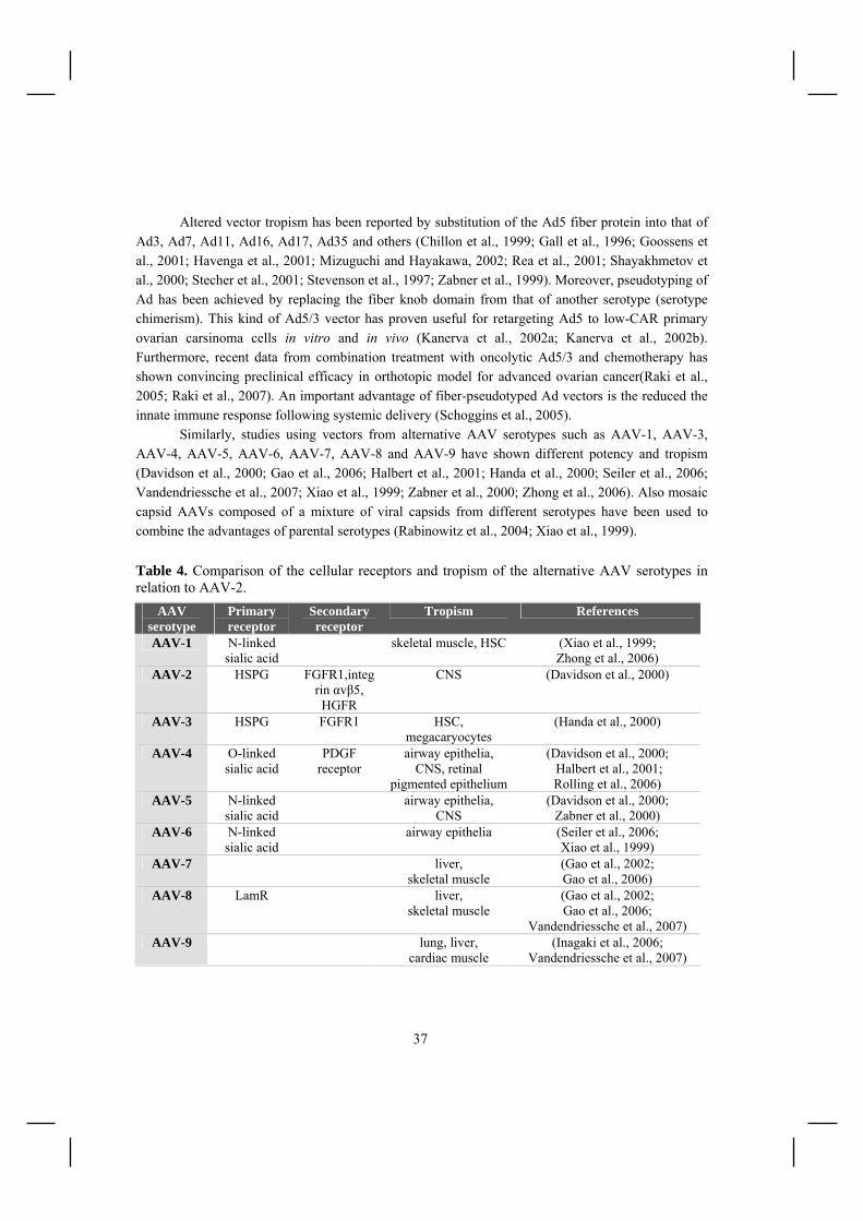
37
Altered vector tropism has been reported by substitution of the Ad5 fiber protein into that of Ad3, Ad7, Ad11, Ad16, Ad17, Ad35 and others (Chillon et al., 1999; Gall et al., 1996; Goossens et al., 2001; Havenga et al., 2001; Mizuguchi and Hayakawa, 2002; Rea et al., 2001; Shayakhmetov et al., 2000; Stecher et al., 2001; Stevenson et al., 1997; Zabner et al., 1999). Moreover, pseudotyping of Ad has been achieved by replacing the fiber knob domain from that of another serotype (serotype chimerism). This kind of Ad5/3 vector has proven useful for retargeting Ad5 to low-CAR primary ovarian carsinoma cells in vitro and in vivo (Kanerva et al., 2002a; Kanerva et al., 2002b). Furthermore, recent data from combination treatment with oncolytic Ad5/3 and chemotherapy has shown convincing preclinical efficacy in orthotopic model for advanced ovarian cancer(Raki et al., 2005; Raki et al., 2007). An important advantage of fiber-pseudotyped Ad vectors is the reduced the innate immune response following systemic delivery (Schoggins et al., 2005).
Similarly, studies using vectors from alternative AAV serotypes such as AAV-1, AAV-3, AAV-4, AAV-5, AAV-6, AAV-7, AAV-8 and AAV-9 have shown different potency and tropism (Davidson et al., 2000; Gao et al., 2006; Halbert et al., 2001; Handa et al., 2000; Seiler et al., 2006; Vandendriessche et al., 2007; Xiao et al., 1999; Zabner et al., 2000; Zhong et al., 2006). Also mosaic capsid AAVs composed of a mixture of viral capsids from different serotypes have been used to combine the advantages of parental serotypes (Rabinowitz et al., 2004; Xiao et al., 1999).
Table 4. Comparison of the cellular receptors and tropism of the alternative AAV serotypes in relation to AAV-2.
AAV serotype
Primary receptor
Secondary receptor
Tropism References
AAV-1 N-linked sialic acid
skeletal muscle, HSC (Xiao et al., 1999; Zhong et al., 2006)
AAV-2 HSPG FGFR1,integrin αvβ5, HGFR
CNS (Davidson et al., 2000)
AAV-3 HSPG FGFR1 HSC, megacaryocytes
(Handa et al., 2000)
AAV-4 O-linked sialic acid
PDGF receptor
airway epithelia, CNS, retinal
pigmented epithelium
(Davidson et al., 2000; Halbert et al., 2001; Rolling et al., 2006)
AAV-5 N-linked sialic acid
airway epithelia, CNS
(Davidson et al., 2000; Zabner et al., 2000)
AAV-6 N-linked sialic acid
airway epithelia (Seiler et al., 2006; Xiao et al., 1999)
AAV-7 liver, skeletal muscle
(Gao et al., 2002; Gao et al., 2006)
AAV-8 LamR liver, skeletal muscle
(Gao et al., 2002; Gao et al., 2006;
Vandendriessche et al., 2007) AAV-9 lung, liver,
cardiac muscle (Inagaki et al., 2006;
Vandendriessche et al., 2007)

38
Genetic targeting of adenoviruses. To date, modification of the Ad fiber protein has been the most common approach used to genetically alter adenoviral tropism. Two approaches have been used for this purpose: addition of foreign peptides to the HI loop or C-terminus of the fiber knob (Figure 8) or design of knobless viruses. Expanded tropism has been reported for Ad vectors with C-terminal integrin-binding RGD motifs and polylysine ligands whereas other peptide ligands were ineffective in this context due to steric hindrance (Wickham et al., 1997). Consequently the HI-loop, tolerating insertions up to 100 amino acids with minimal negative effects on virion integrity, became the preferential site for ligand incorporation (Belousova et al., 2002).Dmitriev et al. introduced an integrin-binding RGD peptide into the HI-loop, resulting in vector with enhanced gene delivery to ovarian cancer cell lines and primary tumors (Dmitriev et al., 1998; Hemminki et al., 2001; Hemminki et al., 2002). Recent studies have demonstrated the high potential of this vector for the treatment of ovarian cancer in murine ovarian cancer models (Mahasreshti et al., 2006). Other targeting peptides inserted in the HI loop of the fiber included ones having high affinity for vascular endothelial cells, cancer cells, transferrin receptor and vascular smooth muscle cells (Nicklin et al., 2000; Nicklin et al., 2003; Work et al., 2004; Xia et al., 2000).
Another approach to achieve CAR-independent tropism consisted of ablating the fiber and/or knob domain (Figure 8), hereby the fiber is re-trimerized by an alternative trimerization motif such as T4 fibritin protein (Krasnykh et al., 2001). A de-knobbing strategy was employed by Magnussen et al. wherein an RGD motif was utilized to achieve selective infection of integrin-expressing cell lines in vitro (Magnusson et al., 2001). In another approach the entire fiber was replaced with T4 fibritin fused with a trimeric CD40 ligand (Belousova et al., 2003). Notably, this vector provided CD40-specific gene delivery in vivo following systemic delivery (Izumi et al., 2005)
In addition to fiber modifications, capsid protein hexon as well as minor capsid protein polypeptide IX have been used as platforms for incorporation of targeting peptides (Figure 8). They are both attractive locales for peptide ligand incorporation due to their surface exposure and abundance (240 hexon/80 pIX homotrimers per virion). Vigne et al. exploited hexon hypervariable region 5 as a site for incorporation of an integrin-binding RGD motif, demonstrating an enhanced, fiber independent transduction to low-CAR vascular smooth muscle cells (Vigne et al., 1999). The C-terminus of pIX has been used to insert poly-lysine, FLAG and RGD motifs, resulting in augmented, CAR-independent gene transfer (Dmitriev et al., 2002; Vellinga et al., 2004).
Genetic targeting of AAVs. The first attempts to alter the tropism of AAV-2 genetically was based on the insertion of scFv or receptor-specific ligands at the N-terminus of VP proteins. Although successful targeting was achieved using CD34 scFv, serpin receptor ligand, human luteinizing hormone peptide, it was of low efficiency and virus titers were affected (Shi et al., 2001; Wu et al., 2000; Yang et al., 1998). The more successful demonstration of genetic capsid modifications were achieved by inserting targeting peptides in different sites inside VP1, VP2 and VP3 sequences determined by sequence alignment between AAV-2 and other parvoviruses or systematic insertional mutagenesis. Peptides specific for integrin receptors, endothelial cells, atherosclerotic plaques and tumors have been employed for targeting of AAV-2 with promising results (Grifman et al., 2001; Nicklin et al., 2001; Shi and Bartlett, 2003; White et al., 2007).

39
Combination of genetic and adaptor-based targeting of Ads and AAVs. Also more general targeting systems have been developed for Ad- and AAV-vectors embodying elements of both genetic modification and adapter-based targeting. These studies incorporated the IgG domain of Staphylococcus aureus protein A on viral capsids enabling these vectors to form stable complexes with a wide variety of targeting molecules containing the Fc region of Ig. Genetic incorporation of this ZZ domain on VP3 on AAV-2 vectors led to specific transduction of distinct human hematopoietic cell lines using targeting antibodies against CD29, c-kit receptor, and CXCR4 (Ried et al., 2002). Similarly, other research groups incorporated the protein A domain into the fiber C-terminus or HI-loop of Ad vectors (Henning et al., 2005; Korokhov et al., 2003; Volpers et al., 2003). The results demonstrated a successful targeting and activation of dendritic cells via an Fc-single-chain antibody directed against CD40 (Korokhov et al., 2005). In addition, this system was used to target ovarian cancer cells via an antibody directed against mesothelin, as well as the pulmonary endothelium in a rat model in vitro (Balyasnikova et al., 2005; Breidenbach et al., 2005). Despite the flexibility of this system for the screening of numerous targeting antibodies, these systems still face some issues regarding the stability of the virus-adaptor complex in vivo and the fact that polyclonal Igs in the bloodstream might replace the targeting molecule.
2.3.3 Targeting non-viral vectors The intrinsic properties of cationic lipoplexes and polyplexes complicate their systemic administration. For some applications the positive charge can be harnessed to target sites of increased vascular growth such as tumors, but for most applications this unspecific interaction leads to unwanted accumulation in the lung tissue associated with toxicity (Kircheis et al., 1999). Similarly, the size of the carrier can limit organ access and modulate biodistribution at the cellular level. Thus for successful targeting, these intrinsic properties of non-viral vectors need to be minimized. In this regard, the hydrophilic polymer polyethylene glycol (PEG) has been used to shield lipoplexes (Hofland et al., 2002; Monck et al., 2000) and polyplexes (Blessing et al., 2001; Ogris et al., 1999; Wolschek et al., 2002). PEG-shielding reduced gene transfer efficiency of complexes, but the efficiency was at least partly restored by incorporation of targeting ligands. Other approaches have used the serum protein transferrin to create negatively charged liposomes (Simoes et al., 1998) or to mask the positive charge of PEI-DNA complexes (Kircheis et al., 2001). Specific targeting of the shielded non-viral vectors has most often taken advantage of natural ligand-receptor interactions. On this basis, systemic targeting of tumors was demonstrated using the folic acid receptor (Hofland et al., 2002; Jeong et al., 2005a), transferrin receptor (Bartlett et al., 2007; Hildebrandt et al., 2003; Kircheis et al., 1999; Xu et al., 1999) or EGFR (Wolschek et al., 2002) as a target. However, a potential drawback of endogenous targeting ligands is the presence of circulating ligands and binding to soluble receptors and receptors in nontarget tissue. Antibodies or antibody fragments might help to avoid some of these problems. As an example, antibodies have been used to target pancreas islet beta cells (Jeong et al., 2005b), human ovarian carcinoma cells (Merdan

40
et al., 2003), breast cancer cells (Germershaus et al., 2006) and glioma cells (Zhang et al., 2002), even though the in vivo data is still limited. Despite these proof-of-principle studies, the transduction efficiencies of non-viral vectors remain low compared to viral vectors and particle shielding often adds to this problem. On this account, design of synthetic virus-like systems, combining the natural mechanisms exploited by viruses for membrane binding, endosomal escape and navigation through the nuclear pore, could improve the targeting potential of non-viral vector systems in the future (Wagner, 2004; Walker et al., 2005). 2.3.4 Avidin and streptavidin- biotin technology Chicken avidin and bacterial streptavidin are widely used tools in the field of life sciences due to their strong specific affinity for biotin (Kd ~1013-1015 M-1) and ability to bind 4 biotins per (strept)avidin molecule. There is no interaction between the carboxy-containing side chain of biotin and avidin, which allows the modification of both counterparts (Bayer and Wilchek, 1994): Biotin can be modified chemically and attached to a wide variety of probes and binders, whereas avidin can be derivatised with other molecular probes or reporter groups of different types, without interfering with their interaction together. Together these features form the crux of (strept)avidin-biotin technology. 2.3.4.1 Avidin and streptavidin Avidin is found in oviparous vertebrates including various birds, reptiles and amphibia, but no analogous protein has been detected in mammalian species (Elo, 1980). Chicken avidin, isolated from the hen egg white, and streptavidin, secreted by several species of Streptomyces, are functionally (Green, 1975; Green, 1990) and structurally (Livnah et al., 1993; Weber et al., 1989) analogous proteins. The main biological function of (strept)avidin is to bind biotin, vital enzymatic cofactor also known as vitamin H (Green, 1990). Both proteins form tetrameric complexes of approximately 60 kDa in which each subunit can bind one molecule of biotin with extremely high affinity (Kd ~1013-1015 M-1). This interaction is primarily thought to represent a natural defence mechanism against biotin requiring microbes (Board and Fuller, 1974; Elo et al., 1980) but also additional roles have been suggested for avidin (Elo and Korpela, 1984; Zerega et al., 2001). In addition to their exceptional ligand binding characteristics, avidin and streptavidin are exceptionally stable against high concentrations of denaturing agents, proteases, wide range of pH and temperature (Green, 1990). Despite similarities, avidin and streptavidin differ in their primary amino acid sequence (41 % similarity), glycosylation, pI (Green, 1990), immunological reactivity and pharmacokinetics. Each avidin monomer has a single oligosaccharide moiety whereas streptavidin is devoid of sugars. Avidin is a basic protein with a high isoelectric point (pI) 10.5 whereas streptavidin has a mildly acidic isoelectric point (pI) of ~6. Owing to these dissimilarities, streptavidin has the advantage of lower

41
nonspecific binding to lectin-like- and negatively charged molecules than avidin. This has been further shown to affect the pharmacokinetics of these proteins with avidin eliciting a shorter plasma half-life compared to streptavidin (Marshall et al., 1995; Rosebrough and Hartley, 1996; Rosebrough, 1993; Schechter et al., 1990). The glycosylation is responsible for avidin’s tendency to accumulate in the liver, whereas its accumulation in the kidneys is mainly due to high pI (Yao et al., 1999). Streptavidin shows accumulation to kidneys (Schechter et al., 1990). Both proteins are immunogenic (Knox et al., 2000; Subramanian and Adiga, 1997; Weiden and Breitz, 2001) 2.3.4.2 Modified avidins Streptavidin was hoped to eliminate the non-specific backround binding to extraneous material accounted with avidin due to high pI and the presence of oligosaccharide moiety. However, streptavidin has been found to interact in biotin-independent manner with the integrins and related cell surface receptors (Alon et al., 1990). To overcome these problems, and thus to improve the immunological and pharmacokinetic properties of (strept)avidin, a plethora of studies have focused on developing novel chemically and genetically engineered forms of avidin and streptavidin (Figure 10). Figure 10. Engineering approaches used to modify the structural and functional properties of (strept)avidin (Laitinen et al., 2006). Early attempts to reduce the charge of avidin consisted of formylation (Guesdon et al., 1979), acetylation (Kaplan et al., 1983) or succinylation (Finn et al., 1984) via covalent attachment to the available lysines of avidin. However, this blocked free amino groups which are often used in preparing avidin-conjugates.

42
The modification of avidin via arginines resolved the problem by leaving lysines available for subsequent interaction. Commercialized neutral avidin derivatives prepared this manner include ExtrAvidin (Sigma-Aldrich, St. Louis, MO, USA), NeutraLite Avidin (Belovo Chemicals, Bastogne, Belgium) and NeutrAvidin (Pierce, Rockford, IL, USA). Marttila et al, have further reported the construction of series of avidin charge mutants with pIs ranging from 4.7 to 9.4 by site-directed mutagenesis replacing arginines and lysines with neutral or acidic amino acids (Marttila et al., 1998). These mutants showed reduction of non-specific binding, while retaining their thermal stability and the ability to tightly bind biotin. To further reduce the non-specific interaction with extraneous macromolecules, the oligosaccharide moiety of avidin can be removed chemically or enzymatically (Bayer and Wilchek, 1994). Enzymatic deglycosylation was used to develop NeutraLite Avidin, which consequently lacks the carbohydrates, exhibits neutral pI and bears free lysines for potential attachment of probes. In a more recent study Marttila et al produced an avidin mutant lacking the oligosaccharide moiety by genetic means. They combined this sugarless mutant with the avidin charge mutant (pI 4.7) to further improve the non-specific binding characteristics of avidin (Marttila et al., 2000). Although most of the applications of (strept)avidin-biotin technology are based on the particularly high affinity between (strept)avidin and biotin, mutants with reduced affinity and reversible binding would be of great value for affinity-based separation of biotinylated molecules. To this end, two types of modifications have been extensively studied: binding-site mutants and interface mutants (Laitinen et al., 2006). To date, numerous binding-site mutants from streptavidin and avidin have been created with affinities for biotin ranging from near wild-type to 6 orders of magnitude weaker values (Chilkoti et al., 1995; Klumb et al., 1998; Marttila et al., 2003; Sano and Cantor, 1995). Modification of the interfaces of (strept)avidin monomers has additionally provided solutions to aggregation problems due to the cross-linking by tetravalent (strept)avidin. Laitinen et al demonstrated the production of dimeric (Laitinen et al., 1999) and monomeric biotin-binding forms which are tetramerized upon biotin binding (Laitinen et al., 2001) or remain in the monomeric state (Laitinen et al., 2003). The affinity for biotin binding was decreased to Kd ~ 10-8 M in the case of dimeric avidin and to Kd ~10-7 M for monomeric avidin known to remain as a monomer upon biotin binding. Following these studies, monomeric forms of streptavidin have been developed (Qureshi et al., 2001; Wu and Wong, 2005) Avidin has been further engineered for improved characteristics and this work continues to widen the field of applicability of avidin-biotin system. For example recent work by Kulomaa and co-workers has demonstrated the production of dual-and single-chain avidins or avidins with dual-affinity (Hytonen et al., 2005; Nordlund et al., 2004; Nordlund et al., 2005). In another case, Howarth et al contructed a streptavidin which binds biotin in monovalent fashion while retaining its high affinity (Howarth et al., 2006).

43
2.3.4.3 Biotin and biotinylation Biotin is a small (244 Da) water-soluble vitamin H synthetized by bacteria, yeasts, molds, algae and some plants, but required by all forms of life (Mock, 1996). Biotin has been shown to play an essential role in regulating gene expression in E.coli and in mammalian cells (Cronan, 1989; Rodriguez-Melendez and Zempleni, 2003). Moreover, in mammalians more than 2000 biotin-dependent genes have been identified and biotinylation of histones plays an essential role in cell proliferation, gene silencing and cellular response to DNA damage (Zempleni, 2005). Biotin is bound to cellular carboxylases and decarboxylases, which catalyze the transfer of CO2 to and between metabolites in gluconeogenesis, lipogenesis, amino acid degradation and energy transduction (Knowles, 1989; Samols et al., 1988). The attachment of biotin to the ε-amino group of a specific lysine moiety in carboxylases is catalyzed by biotin protein ligase in an ATP-dependent reaction (Figure 11) (Otsuka and Abelson, 1978). The number of biotinated carboxylases varies from species to species: In E.coli, the bacterial biotin ligase BirA biotinylates only one protein called acetyl-CoA carboxylase whereas in mammalian cells acetylCoA- carboxylase, methyl crotonyt-CoA carboxylase, propionyl-CoA carboxylase and puryvate carboxylase are biotinylated (Cronan, 1990). Figure 11. The biotin protein ligase reaction. In the first step, biotin protein ligase catalyses the attack of an oxygen atom of the biotin carboxylate on phosphate of ATP to form biotinoyl-AMP. Next, the nucleophilic ε-amino group of the lysine on apo-form of the biotin acceptor domain attacks the carbon atom on biotinoyl-AMP thus forming an amide bond between biotin and biotin accepting domain (Chapman-Smith and Cronan, 1999).

44
Biotinylation of proteins is an attractive alternative to epitope tagging due to the strong (strept)avidin-biotin interaction. While a wide range of chemical biotinylation techniques exist, they are limited by the fact that chemical biotinylation are not site-specific, require prior purification of the substrate and can lead to inactivation of the target protein (Stolz et al., 1998). Given the difficulties in chemical biotinylation, many efforts are now devoted to developing systems exploiting the natures own biotinylation machinery. Early studies using this approach took advantage of the 1.3S subunit of Propioni bacterium shermanii transcarboxylase (PSTCD) which is naturally biotinylated at lysine 89 (Cronan, 1990). The expression of PSTCD fusion protein led to its enzymatic biotinylation in E.coli and Saccharomyces cerevisiae. Later on, this approach has been expanded to mammalian cells and animals (Parrott and Barry, 2000; Parrott and Barry, 2001). Isolation of new shorter biotin acceptor peptide (BAP) substrates (ca. 13-20 residues) for BirA have further increased the appeal of this system for purification applications (Duffy et al., 1998; Schatz, 1993). Metabolically biotinylated gene therapy vectors will be discussed in the next chapter. 2.3.4.4 (Strept)avidin-biotin technology in gene therapy The specific characteristics of avidin have been shown to be of great advantage in drug targeting: The high positive charge of avidin augments the efficiency of cellular uptake of biotin-coated particles (Pardridge and Boado, 1991), whereas incubation of bioconjugated avidin with biotinylated cell lines results in rapid surface attachment and endocytosis with efficiencies approaching 100% (Wojda et al., 1999). In addition, (strept)avidin has been demonstrated to accumulate into specific tissues (Rosebrough and Hartley, 1996; Rosebrough, 1993; Schechter et al., 1990), especially to tumors in vivo (Yao et al., 1998). Consequently, avidin alone can enable some targeting of gene therapy vectors to specific tissues, while tissue-specificity can be altered by biochemical modification of the protein. The strong avidin-biotin interaction can be used to develop targeted therapies by the biotinylation of ligands or tissues in vivo (De La Fuente et al., 1997; Hoya et al., 2001; Singh et al., 2005). Several studies have shown promising results using monoclonal antibodies in targeting of biotinylated therapeutic or diagnostic compounds via avidin in experimental animals (Corti et al., 1998; Guttinger et al., 2000; Wu and Pardridge, 1999). One such approach, called pretargeted radioimmunotherapy, separates the administration of the monoclonal antibodies from that of a low-molecular-weight radionuclide ligand. This multistep (2 or 3 steps) approach has shown to improve tumor: normal tissue radiation dose ratios since the targeting molecule administered first is not radiolabeled. Subsequent administration of (strept)avidin removes the excess circulating antibodies or binds to tumor cells preparing them to receive radiolabelled biotin. This strategy has already been followed in clinical trials for the treatment of malignant glioma demontstrating impeding of cancer progression without significant toxicity (Grana et al., 2002; Paganelli et al., 1999; Paganelli et al., 2001; Paganelli et al., 2006). Other similar studies are being conducted for the treatment of colon cancer, B-cell lymphoma and gastrointestinal malignancies (Forero et al., 2004; Knox et al., 2000; Linden et al., 2005; Shen et al., 2005).

45
Another potential approach for targeting tumors or tissues is to introduce recombinant-avidin gene into specific tissues by local gene transfer which consequently can sequester and concentrate considerable amounts of therapeutic biotin conjugates in vitro and in vivo (Lehtolainen et al., 2002a; Lehtolainen et al., 2003; Walker et al., 1996) In addition, (strept)avidin-biotin technology can be adapted to improve vector targeting (Waehler et al., 2007). It provides substantial advantage over other adaptor systems by assuring sufficient stability of the vector-adaptor complex even under physiological conditions (Pereboeva et al., 2007). Early studies with retroviruses used streptavidin-bound antibodies specific for both viral and cell membrane epitopes provided the proof-of-principle even though only low transduction efficiencies were attained (Etienne-Julan et al., 1992; Roux et al., 1989). Since that, the (strept)avidin-biotin-based targeting has followed two different approaches depicted in figure 12. Firstly, gene therapy vectors can be biotinylated either chemically or metabolically, while bringing the biotinylated targeting molecule in conjunction to avidin (Barry et al., 2003). First studies by Smith and collegues were performed with a chemically biotinylated adenovirus vector. They demonstrated successfull vector targeting to hematopoetic cells through an avidin bridge carrying biotinylated c-Kit receptor ligand, resulting up to 2400-fold increase in reporter gene expression (Smith et al., 1999). Later on, chemically biotinylated retrovirus, AAV and vaccinia virus have been created, all of which exhibited significantly increased transduction of target cells (Ponnazhagan et al., 2002; Purow and Staveley-O'Carroll, 2005; Zhong et al., 2001). Figure 12. The principle of (strept)avidin-biotin technology in vector targeting. The vector can either carry avidin or biotin on its surface. This allows attachment of biotinylated or avidinylated targeting molecules (binders; e.g. cell surface ligands, antibodies).
Avidin BiotinB
cell
Binder
BinderB
B
B
B
Bvirus
Receptor
Receptor
BB
A
A
cell Binder virusReceptor
B A
A
A
A
A
A
A

46
However, chemical biotinylation often leads to non-specific labelling, inactivation of target proteins and require prior purification of the virus. Therefore, metabolic biotinylation, achieved directly in living cells, is considered a more promising approach (Cronan, 1990). A number of studies have shown that metabolic biotinylation provides an efficient means to target gene therapy vectors such as adenovirus (Campos and Barry, 2006; Campos et al., 2004; Maguire et al., 2006; Parrott et al., 2003) and AAV (Arnold et al., 2006; Stachler and Bartlett, 2006). A recent report by Pereboeva and coworkers also provided evidence on the applicability of metabolically biotinylated adenovirus vectors for in vivo retargeting (Pereboeva et al., 2007). Owing to the large availability of (strept)avidin-biotin-based purification methods, metabolical biotinylation of gene therapy vectors has also been applied for virus concentration (Arnold et al., 2006; Campos and Barry, 2006; Chan et al., 2005; Nesbeth et al., 2006; Stachler and Bartlett, 2006). An opposite targeting approach consists of avidin displaying vectors (Figure 12). To this end, chemically avidinylated PEI-vectors (Wojda and Miller, 2000) and adenoviruses (Park et al., 2008) together with genetically engineered avidin displaying baculoviruses have been described (Raty et al., 2004). In the latter study, Räty and collegues expressed an avidin-gp64 fusion protein on the baculovirus surface thus providing binding sites to biotinylated targeting ligands (Raty et al., 2004). This 2-step system is easier to control than the 3-step process using the avidin-bridge between desired biotinylated molecules and offers better valence for covering since all four biotin-binding sites are available for biotinylated ligands. Avidin displaying baculovirus showed a 5-fold increase in transduction efficiency of rat glioma cells and a 26-fold increase in rabbit aortic smooth muscle cells compared to wild-type virus without major cytotoxicity. Enhanced transduction was also observed with biotinylated cells and biotinylated EGF enabled targeting to EGFR expressing cells. In addition, the use of biotinylated paramagnetic particles allowed magnetic targeting. In addition to providing great potential for therapeutic purposes, the avidin-biotin technology has also been exploited for in vivo imaging purposes. The pretargeted radioimmunotherapy for cancer treatment described above, has been widely applied for simultaneous radioimmunoimaging (Cauchon et al., 2007; Hama et al., 2007; Li et al., 2005; Sharkey et al., 2005). Furthermore, conjugation of baculovirus vector to biotinylated iron oxide particles or radionuclides has enabled imaging of the viral particle biodistribution by magnetic resonance imaging and single photon emission computed tomography in vivo (Raty et al., 2006; Raty et al., 2007). Similarly, cells expressing metabolically biotinylated cell-surface receptors can be visualized with labeled streptavidin moieties (Tannous et al., 2006).
2.4 Detection of viral particles and gene expression
Molecular imaging techniques are defined as the visual characterization and quantification of biological processes at the cellular and subcellular levels within a living organism. The three main methodologies developed for noninvasive in vivo imaging include optical imaging, magnetic resonance imaging (MRI) and nuclear imaging [single photon emission tomography (SPECT), and

47
positron emission tomography (PET)]. Each of these technologies has its own advantages and drawbacks as listed in Table 5. Current imaging methods in gene therapy can be divided into biodistribution and reporter gene imaging. Reporter gene imaging visualizes the transgene expression whereas biodistribution demonstrates the vector location throughout the body. It is often desirable to obtain images with both imaging modalities to obtain conclusive data on the safety and efficacy of gene therapy. Table 5. Characteristics of different imaging modalities [Modified from (Massoud and Gambhir, 2003)]. Imaging
technique Electro-magnetic radiation
Spatial reso-lution
Temporal resolution
Sensiti-vity
Advantages Disadvantages
Fluores-cence
imaging
visible light or near-
infrared
2-3 mm seconds to minutes
10-9- 10-12 M
High sensitivity, low
cost
relatively low resolution, imaging
depth <1 cm
Biolumi-nescence imaging
visible light
3-5 mm seconds to minutes
10-15- 10-17 M
High sensitivity,
quick, low cost, high
throughput
low resolution, 2D imaging only, imaging
depth 1-2 mm
PET high-energy γ-
rays
1-2 mm 10 sec - minutes
10-11- 10-12 M
sensitive, isotopes can
substitute natural atoms
PET cyclotron or generator needed,
relatively low resolution, radiation
SPECT lower-energy γ-
rays
1-2 mm minutes 10-10- 10-11 M
many probes available,
simultaneous multiple probe
imaging
relatively low resolution, radiation
MRI radiowaves 25-100µm
minutes - hours
10-3- 10-5 M
high resolution, combines
morphological and functional
imaging
relatively low sensitivity, long scan
and posprocessing time
2.4.1 Optical imaging Optical imaging is a modality that is cost-effective, rapid, easy to use, and can be readily applied to studying transductional efficiency of gene therapy vectors in vivo. Optical methods can be divided into fluorescence- and bioluminescence-based reporter gene systems. A number of fluorescence reporter genes eg. different variants of the Aqueorea Victoria green fluorescent protein (GFP) (Contag et al., 2000; Ellenberg et al., 1999; Falk and Lauf, 2001; Hadjantonakis and Nagy, 2001) and red fluorescent proteins from Discosoma species (dsRed1 and dsRed2) (Campbell et al., 2002; Mathieu and El-Battari, 2003) have been described and applied in optical imaging of tumors and

48
metastases (Bharali et al., 2005; Bouvet et al., 2002; Yang et al., 2000). However, bioluminescence reporter imaging currently remains more sensitive for whole-body imaging: the most commonly used are the Firefly and Renilla luciferase genes (Choy et al., 2003; Gross and Piwnica-Worms, 2005; Wilson and Hastings, 1998; Yu et al., 2003). Expression of luciferase has been specifically used to image the growth kinetics of transformed tumor cells (Liang et al., 2004; Rehemtulla et al., 2002) and to monitor viral gene delivery (Berraondo et al., 2006) and replication (Yamamoto et al., 2006). 2.4.2 PET/SPECT Nuclear imaging is based on the administration and detection of decaying radioisotopes. The decay of a radioisotope emits a positron or gamma ray which produces detectable two (positron emission tomography, PET) or single high-energy photons (single photon emission tomography, SPECT). PET is based on the use of contrast agents (tracers) including radioisotopes 11C, 13N, 15O, 18F 64Cu, 68Ga, 76Br and 94mTc (Massoud and Gambhir, 2003). Positrons emitted by the tracers are annihilated in tissue by collision with an electron. It results in an emission of two high-energy photons detected by PET detector array. The sensitivity of PET is relatively high, in the range of 10-
11-10-12 M (Mandl et al., 2002). PET provides a spatial resolution of 1-2 mm enabling small animal micro-PET applications. SPECT is similar to PET, but acquires information on the concentration of gamma emitting radionucleotides like 111In, 133Xe, 99mTc 123I, 125I, and 131I (Rosenthal et al., 1995). These heavy isotopes have longer decay times than PET contrast agents and are more readily available (Mandl et al., 2002). However, SPECT tracers can also be taken up by other areas than their target area and simultaneous imaging by computer tomography (CT) may be required (Labbe, 2003). SPECT is at least a log order less sensitive than PET even though the spatial resolution is near the same (Massoud and Gambhir, 2003). Direct labeling of virus particles allows the systemic distribution of the virus to be detected by nuclear imaging. Labeling herpes simplex viruses with 111In enabled to trace the organ distribution of the viruses after intravenous administration (Schellingerhout et al., 1998). The same group also performed mass distribution studies of HSV vectors in an experimental brain tumor model to evaluate and compare different delivery modalities (Schellingerhout et al., 2000). Imaging techniques for other viral vectors have also been developed based on iodination (Frost, 1977; Markwell and Fox, 1978; Montelaro and Rueckert, 1975; Moore et al., 1974) and the biotin-avidin system (Skulstad et al., 1995). In several of these studies, however, the viral labeling was reported to affect the infectivity of the vectors. Recently, a less detrimental approach, based on the display of avidin on the baculovirus surface, was introduced (Raty et al., 2007). The avidin-display enabled baculovirus labeling with 99mTc-polylys-ser-DTPA-biotin, and visualization of viral biodistribution and kinetics after various administration routes. Reporter genes for nuclear imaging can encode for intracellular enzymes, extracellular or intracellular receptors or cell membrane transporters capable of irreversibly binding or transporting a
49
radiolabeled or paramagnetic probe. Wild-type HSV1 thymidine kinase (tk) (Tjuvajev et al., 1998) and its mutant version HSV-sr39tk (Gambhir et al., 2000), are the most commonly used enzyme-based marker genes in transductional imaging. Unlike human thymidine kinases, these enzymes have less substrate specificity and can selectively phosphorylate a wide range of compounds. Imaging of TK expression is based on the use of iodinated or fluorinated acycloguanosines (eg, ganciclovir ; penciclovir; 18F -labeled 9-[4-fluoro-(hydroxymethyl)butyl]guanine (Gambhir et al., 1999; Jacobs et al., 2001; Namavari et al., 2000) as well as thymidine analogues (eg, 2'-fluoro-2'-deoxy-1-beta-D-arabinofuranosyl-5-iodouracil, 2'-18F-fluoro-5-ethyl-1-beta-D-arabinofuranosyl-uracil) (Buursma et al., 2006; Yaghoubi et al., 2001). When HSV-TK is present in the cells, the substrates become phosphorylated and thus trapped. Cellular retention of radioactivity indicates the transfection and this has been widely used to monitor the gene therapy of cancer (Blasberg and Tjuvajev, 1999; Deng et al., 2006; Tseng et al., 2006; Yaghoubi et al., 2005). Alternative enzyme-based reporter systems are xanthine-phophoribotransferase (Doubrovin et al., 2003) and cytosine deaminase (Haberkorn et al., 1996). Use of extracellular receptors or cell membrane transporters as reporter genes eliminates the need for the probe to penetrate into a cell. The human dopamine receptor is an example of such reporter gene (hD2R). hD2R expression is largely limited to the striatal-nigral system of the brain, which makes it a good candidate for transductional imaging. hD2R expressing cells and tissues can be imaged through the accumulation of an established probe, 18F fluoroethyl spiperone (MacLaren et al., 1999). Somatostatin receptor subtype II is another receptor-based reporter gene, which has a naturally limited expression to carcinoid tumors (Rogers et al., 1999; Rogers et al., 2000). Both receptors can be imaged simultaneously with HSV-tk providing a platform for indirect imaging of therapeutic gene expression (Chen et al., 2004; Verwijnen et al., 2004). Human sodium iodine symporter (hNIS), iodine-transporter in thyroid follicular cells, provides an opportunity to image with both PET (124I-iodine) and gamma camera (123I-iodine or 99mTc-pertechnetate) (Haberkorn et al., 2001). hNIS has been used for imaging adenovirus biodistribution and gene therapy (Dwyer et al., 2006; Groot-Wassink et al., 2002; Lee et al., 2004; Yang et al., 2004). More recently it has been suggested as a potential candidate for monitoring cardiac gene therapy (Miyagawa et al., 2005a; Miyagawa et al., 2005b). Another transporter-based reporter system, norepinephrine transporter, has been utilized to image transduced tumors (Altmann et al., 2003; Buursma et al., 2005). 2.4.3 MRI Magnetic resonance imaging has the advantage of high spatial resolution (10-100µm) but the natural insensitivity for label detection requires robust amplification techniques. MRI is based on the detection of molecules that possess the property of nuclear spin, which align themselves when exposed to magnetic field. Following a radiofrequency pulse, the nuclear spins return to their baseline

50
orientation with a relaxation time corresponding to their physicochemical environment which can be measured (Massoud and Gambhir, 2003). Contrast agents used in MRI can be divided into two categories which modify either T1 or T2 relaxation time constants, proton density or nuclear polarization to attain improved sensitivity. T1 contrast agents produce a positive signal enhancement on MRI images and are usually based on paramagnetic ions or stable free radical molecules (Potter, 2002). T2 contrast agents, on the other hand, decrease the signal in T2 weighted MRI seen as the darkening of image. T2 contrast agents are generally based on ferromagnetic of superparamagnetic nanoparticles. These particles can differ in their core size, coating thickness and coating material, all of which affect their biodistribution (Wang et al., 2001). Most current transductional MRI strategies are based on coupling a reporter transgene with a specific probe. Initially, enzyme-based reporters were applied to monitor the changes in phosphor metabolism by arginine and creatine kinases (Koretsky et al., 1990; Walter et al., 2000) and fluorine metabolism by the enzyme activity of cytosine deaminase (Stegman et al., 1999) and β-galactosidase (Louie, 2006). Also approaches based on targeting the contrast agent to the cell surface using appropriate ligands have been developed. For example, after overexpression of inflammatory adhesion molecule or non-endogenous H2K(k) antigen, antibodies against these molecules conjugated to a superparamagnetic iron oxide particle, generated strong negative contrast (Bulte et al., 1998; So et al., 2005).
The most common reporter genes for MRI are based on proteins involved in iron metabolism, such as tyrosinase, transferrin receptor and ferritin. All cells use iron and they attain it from the circulating blood where it is bound to transferrin. Transferrin receptors throughout the body engulf and internalize both the protein and the iron attached to it. Once inside, the cell transfers the iron to ferritin, the internal iron storage molecule. Both these proteins can thus be used as reporter genes for MRI. Transferrin receptor overexpression can be probed with superparamagnetic agents (Moore et al., 2001; Weissleder et al., 2000) whereas ferritin is itself made superparamagnetic by the cell eliminating the need for an exogenous contrast agent (Genove et al., 2005). Ferritin is a ubiquitous intracellular iron storage protein that consists of 24 subunits of the heavy (H) and light (L) type (Arosio and Levi, 2002). This multi-subunit protein is capable of containing as many as 4,500 atoms of iron within a hydrous ferric oxide core. The H and L subunits have different functional specificity: the L-chain enhances the stability of the iron core while the H-chain has a metal-binding site which confers ferroxidase activity (Fe2+ oxidation to Fe3+) to the protein and accelerates iron incorporation (Levi et al., 1993; Levi et al., 1994). To date, ferritin expression has enabled the imaging of adenovirus transduction in mouse brain (Genove et al., 2005), and detection of transcriptional regulation of gene expression in glioma tumor models (Cohen et al., 2005) and transgenic animals (Cohen et al., 2007). The third approach for detecting transgenes by MRI relying on cellular accumulation of iron takes advantage of tyrosinase gene expression which leads to the production of metal-binding metabolite, melanin (Weissleder et al., 1997).

51
3. AIMS
The general objective of this study was to improve the efficiency, targetability and safety of baculo- and lentiviral vectors in order to extend the applicability of these viruses in the field gene therapy. A special emphasis was put on the development of generally applicable strategies for virus targeting, purification and non-invasive imaging based on avidin-biotin technology. More specifically, the aims of this study were: (I) To study if the VSV-GED could enhance the transduction efficiency of baculovirus vectors when coexpressed with gp64. (II) To develop metabolically biotinylated baculovirus vectors by displaying a biotin acceptor peptide (BAP) fused either to different sites in the baculovirus glycoprotein gp64 or VSV-GED and to evaluate the utility of these vectors for virus targeting and purification applications. (III) To create targetable lentivirus vectors displaying (strept)avidin-VSV-GED fusion proteins and to assess the utility of these vectors for in vivo imaging of virus particle biodistribution and transduction using SPECT/CT and MRI. (IV) To analyze the transcription and expression of baculoviral immediate early genes in human cells and to examine the interactions between viral components and subnuclear structures after viral transduction.

52
4. MATERIALS AND METHODS
4.1 Methods
Table 6. Methods used in the studies. Method Desription Study
No. DNA cloning Vector design and construction I- IV
Baculovirus production I,II,IV I,II,IV Virus concentration: Ultracentrifugation
Magnetic capture II
Production of baculovirus vectors
Baculovirus titering: End-point dilution I,II,IV Lentivirus production III Virus concentration: Ultracentrifugation III
Production of lentivirus vectors
Lentivirus titering: p24 ELISA Flow cytometry
III III
SDS-PAGE and Immunoblotting I, II Characterization of baculovirus vectors ELISA II
SDS-PAGE and Immunoblotting III Characterization of lentivirus vectors ELISA III
I, II I, II
Transduction β-galactosidase staining efficiency: Luminescent β-galactosidase assay Flow cytometry III Immunofluorescence labeling, confocal microscopy IV Cell targeting with ligands and antibodies II,III Magnetic targeting II Cytotoxicity assay I Endocytosis blocking I Syncutium formation assay I Magnetic resonance imaging III
In vitro experiments
RNA extraction, RT-PCR III, IV Stereotactic injections to rat brain I, III Intramuscular injections to rabbit hind limb I Magnetic resonance imaging III
In vivo experiments
SPECT/CT imaging III β-galactosidase staining I, III Antibody staining III
Histochemical analyzes
Iron staining III Mean SD I- IV Unpaired t test I- IV
Statistical methods
ANOVA II,III

53
4.2 Plasmids and DNA oligomers
The plasmids used to generate the recombinant baculo- and lentiviruses are summarized in Table 7. The DNA oligomers used in cloning and sequencing are listed in Table 8, whereas the individual cloning steps are described more thoroughly in the original publications. Table 7. Plasmids used in studies I-IV. Plasmid Reference Description Study pFastBac-1 Invitrogen, Carlsbad, CA, USA Backbone for baculovirus constructs I, II pBacSurf-1 Novagen, Madison, WI, USA Source of gp64 II Baavi (Raty et al., 2004) Source of avidin and LacZ III PFD27 (Laitinen et al., 2003) Source of monomeric avidin III pGEM-streptavidin
M. Kulomaa, University of Tampere, Finland
Source of streptavidin III
pDONR201 Invitrogen, Carlsbad, CA, USA Intermediate cloning of BirA II pCMV-VSVG T. Friedmann, UCSD, La Jolla,
CA, USA Source of VSV-G, Lentivirus production
I II, III
pRSV-Rev Tronolab, Lausanne, Switzerland
Lentivirus production III
pMDLg/pRRE Tronolab, Lausanne, Switzerland
Lentivirus production III
pLV-GFP (Makinen et al., 2006) Lentivirus production, transfer vector III pENTR™221-Ferritin
RZPD German Resource Center for Genome Research, Berlin , Germany
Source of ferritin III
pBOB-CAG I. Verma, Salk Institute, La Jolla, CA, USA
Lentivirus production, transfer vector backbone
III
H2B-EYFP J. Langowski, German Cancer Research Center, Heidelberg, Germany
Detection of human histone protein H2B expression
IV
pEGFP-sp100 G. Dellaire, The hospital for Sick Children, Toronto, Canada
Detection of promyelocytic nuclear bodies
IV
EGFP-PAB2 M. Carmo-Fonseca (Intituto de Medica Molecular, Lisbon, Portugal
Detection of nuclear speckles IV
NPM-EGFP M. Laiho, University of Helsinki, Finland
Detection of nucleophosmin IV

54
Table 8. DNA oligomers used in the studies.
Sequence Desription Usage Study GGGGTGATACTGGGCTATCCAA VSV-GED 5’forward Cloning I,II AGATCTTTACTTTCCAAGTCGGTTCA VSV-GED 3’reverse Cloning I,II GGAAGTTCACCATAGTTTTTCCAC VSV-G 5’forward Cloning I GAAGGAGATAACATGAGATCT AAGGATAACACCGTGCCACTG
BirA 5’forward Cloning II
TTTAGTGATGGTGATGGTGA TGTTTTTCTGCACTACGCAGGG
BirA 3’reverse Cloning II
GATCCGTAAGCGCTATTGTTTTATATGTGCTTTTGGCGGCGGCGGCGCATTCTGCCTTTGCGA
Gp64 signal sequence linker
Cloning II
GATCTCGCAAAGGCAGAATGCGCCGCCGCCGCCAAAAGCACATATAAAACAATAGCGCTTAG
Gp64 signal sequence linker
Cloning II
GTTCATGCCATTCAATTTTTTGTGCTT CAAAGATATCATTCAGGCCCTGCA
BAP linker Cloning II
GGGCCTGAATGATATCTTTGAAGCAC AAAAAATTGAATGGCATGAACTGCA
BAP linker Cloning II
GGTACCCCCGGGCGGAGCACTGC Gp64 5’forward Cloning II ATAACCCGGGTCTTTAATATTGTCTATTACGG Gp64 3’reverse Cloning II CTTGGCTCTAACGTTGTGGCGTTCATGCCATTCAATTTTTTGTGCTTCAAAGATATCATTCAGGCCCCAAGTGGGCGGCCGCTTC
BAP + compatible ends to gp64 site 283, 3’ reverse
Cloning II
AATGATAACCATCTCGCA 5’forw. pPolh-cassette Sequenc. I,II GGATGAAGTGGTTCGCATCC 3’rev. pPolh-cassette Sequenc. I,II GCAACGTGCTGGTCTGTGTGC 5’forw. CMV-cassette Sequenc. III CACACCAGCCACCACCTTCTG 3’rev. CMV-cassette Sequenc. III GGAACATGCTGAGAAACTGATGAAG 5’frw.human/rat ferritin RT-PCR III CACAGTCTGGTTTCTTGATATCCTGA 3’rev. human ferritin RT-PCR III CACGGTCAGGTTTCTTTATATCCTGC 3’rev. rat ferritin RT-PCR III TTAACGCGTCGTACACCAGCG 5’forw. ie-1 RT-PCR IV TTATAATAACTTAAATAGTCGTTGGG 3’rev. ie-1 RT-PCR IV ATGAGTCGCCAAATCAACGC 5’forw. ie-2 RT-PCR IV GGCTTCGGGAGATGTTGTAAAG 3’rev. ie-2 RT-PCR IV TCCGACTTGGGCAAATGG 5’forw. lef-3 RT-PCR IV GATTGAAATCCGCACATAGCTTT 3’rev. lef-3 RT-PCR IV

55
4.3 Vectors The characteristics of the recombinant vectors developed in the studies are listed in Table 9 and Table 10. Table 9. Baculoviral vectors constructed in studies I-IV. Baculoviral vectors (pseudotype)
Promoter-transgene
Surface Modification Study
VSV-GED CMV-LacZ Transmembrane anchor of VSV-G I VSV-G CMV-LacZ VSV-G I BAP-VSV-GED CMV-LacZ BAP fused to VSV-GED II BAP-N-gp64 CMV-LacZ BAP fused to N-terminus of gp64 II BAP-283-gp64 CMV-LacZ BAP inserted at aa. position 283 of gp64 II BAP-283+N-gp64 CMV-LacZ BAP fused to N-terminus and to site 283 of
gp64 II
Table 10. Lentiviral vectors constructed in studies I-IV. Lentiviral vectors (pseudotype)
Promoter-transgene
Surface Modification Refe-rence
Gp64 hPGK-GFP Baculovirus envelope glycoprotein, gp64 III AVD/Gp64 hPGK-GFP Avidin fused to VSV-GED, coexpressed
with gp64 III
SA/Gp64 hPGK-GFP/ CAG-Ferritin/ CAG-LacZ
Streptavidin fused to VSV-GED, coexpressed with gp64
III
MONO/Gp64 hPGK-GFP Monomeric avidin fused to VSV-GED, coexpressed with gp64
III

56
4.4 Antibodies and ligands
Table 11. Antibodies used in immunoblotting, immunofluorescence and targeting experiments. (* Conjugated to biotin) Antibody Source Descripition Study Anti-gp64 mAb Insight Biotechnology, Webley,
UK Gp64 detection I, II
Anti-vp39 pAb Loy Volkman, University of California, Berkeley, USA
Vp39 detection I, IV
Anti-avidin pAb (Laitinen et al., 2002) Avidin detection III Anti-streptavidin pAb (Bayer et al., 1986) Streptavidin detection III Anti-VSVG pAb Bethyl Laboratories Inc.,
Montgomery, TX, USA VSV-GED detection I- III
Streptavidin-HRP Vector laboratories, Burlingame, CA, USA
Biotin detection II
Anti-lamin Novocastra Laboratories Ltd, Newcastle, UK
Nuclear membrane detection IV
Anti-TfR mAb* Ancell Corporation, Bayport, MN, USA
Targeting to transferrin receptor
II, III
Cetuximab mAb* Merck, Darmstadt, Germany Targeting to EGFR receptor II, III Anti-CD46 mAb* Exbio, Prague, Czech Republic Targeting to CD46 receptor II, III Transferrin* Molecular Probes, Invitrogen,
Carlsbad, CA, USA Targeting to transferrin receptor
II, III
EGF* Molecular Probes, Invitrogen, Carlsbad, CA, USA
Targeting to EGFR receptor III
Anti-Ferritin rH02 Paolo Arosio, University of Brescia, Milan, Italy
Detection of human heavy chain ferritin
III
Anti-IE-2 Knebel-Mörsdorf,Max-Planck-Institute for Neurological Research, Germany
Baculovirus immediate early protein 2 detection
IV
Anti-GFP Molecular Probes, Invitrogen, Carlsbad, CA, USA
Green fluorescent protein detection
IV
Anti-SC-35 Abcam, Cambridge, UK Marker for nuclear speckles IV Anti-PML Abcam, Cambridge, UK Promyelotic leukaemia
protein detection IV
Anti-p80 A.Lamond, University of Dundee, Scotland, UK
Marker for Cajal bodies IV
Anti-rabbit IgG AP Bio-Rad, Richmond, CA, USA Detection of rabbit antibodies I-III Anti-rabbit IgG-HRP Sigma-Aldrich,St Louis,MO,USA Detection of rabbit antibodies IV Anti-rabbit IgG-alexa 488
Molecular Probes, Invitrogen, Carlsbad, CA, USA
Detection of rabbit antibodies IV
Anti-rabbit IgG-alexa 555
Molecular Probes, Invitrogen, Carlsbad, CA, USA
Detection of rabbit antibodies IV
Anti-mouse IgG AP Bio-Rad, Richmond, CA, USA Detection of mouse Abs I-III Anti-mouse IgG-HRP Silenus Laboratories,
Hawthorn, Australia Detection of mouse Abs II, III
Anti-mouse IgG-alexa 555
Molecular Probes, Invitrogen, Carlsbad, CA, USA
Detection of mouse antibodies
IV

57
4.5 Cell lines
Cell lines used for in vitro studies are presented in Table 12. All the mammalian cell lines were grown in +37 ºC under 5% CO2 in their recommended medium, whereas Sf9-insect cells were grown in +28 ºC. All the cell culture protocols are described with more detail in the cited articles. Table 12. Cell lines used in studies I-IV. Cell line Source Description Study 293 ATCC: CRL-1573 Fetal human kidney cell line IV 293T ATCC: CRL-11268 Fetal human kidney cell line I, III A549 ATCC: CCL-185 Human lung adenocarcinoma III D54 (Bigner et al., 1981) Human glioblastoma III BT4C (Laerum et al., 1977;
Sandmair et al., 1999) Rat glioma cell line I- III
EAHY-926 University of North-Carolina, Department of Pathology, NC, USA
Hybridoma of human airway epithelium and human umbilical vein endothelial cells
I
HeLa ATCC: CCL-2 Human cervical carcinoma I, III HepG2 ATCC: HB-8065 Human hepatocarcinoma I- IV Sf9 Invitrogen, Carlsbad, CA,
USA Spodoptera frugiperda IPLB-Sf-21-AE cells
I, II, IV
SKOV-3 ATCC: HTB-77 Human ovarian carcinoma I- III U118MG ATCC: HTB-15 Human glioblastoma III
4.6 In vivo experiments
Table 13. Animals used in the studies and the treatments they received. Animal n Virus Dose Admin.
route Sacri-fice
Study
BDIX female rat 10 VSV-GED/Ctrl baculovirus
20 µl (2x10⁸ pfu/ml)
Intra-cranial
d4 I
New Zealand white female rabbit
3 VSV-GED/Ctrl baculovirus
10 x 50 µl (1x10⁹ pfu/ml)
Intra-muscular
d6 I
Wistar male rat 22 SA/Gp64- lentivirus
15 µl (2 x10⁸ pg/ml)
Intra-cranial
d5-d63 III

58
The rats were anestetized with fentanyl-fluanisone-midazolam (Study I) or ketamine-medetomine (Study III). For intracranial injections, the virus was injected with Hamilton syringe and a 27-gaude needle into the right lateral ventricle with the following coordinates: 1.0 mm caudal to bregma, 1.5 mm right to sutura sagittalis, and depth of 3-3.5 mm using stereotaxic apparatus (Figure 13). The New Zealand white rabbits were anestetized with ketamine-metetomidine before the baculovirus vectors were injected in a volume of 50 µl into M. semimembranosus by 10 injections (Figure 14). All the performed studies were approved by the Animal Care and Use Committee of University of Kuopio.
Figure 13. The rat brain showing the stereotaxic coordinates for virus injections in studies I and III (gray dot) [modified from (Paxinos and Watson, 1986)].

59
Figure 14. Lateral view of the muscles of a rabbit thigh. The virus injections were done to musculus semimembranosus.[(www.hundezeitung.de/Ana11-Dateien/image004.jpg) with modification]
60
5. RESULTS AND DISCUSSION
5.1 VSV-GED-displaying baculovirus for improved gene delivery (I)
Baculoviruses are gaining popularity as potential vectors for gene transfer technology. Despite many promising aspects, some limitations regarding the efficacy and specificity of these vectors have slowed their widespread use. Although AcMNPV can enter to almost any cell from any origin, they are unable to reach the nucleus of many of these cells (Kukkonen et al., 2003; Volkman and Goldsmith, 1983). This might be due to the inability of the virus to escape from endosomes (Barsoum et al., 1997; Park et al., 2001) or blockage of the transport or entry into the nucleus (Kukkonen et al., 2003; van Loo et al., 2001). To enhance the escape of the baculovirus vectors from endosomes, a novel vector displaying a truncated form of VSV-G was designed. 5.1.1 Generation and characterization of VSV-GED pseudotyped virus To improve the efficacy of baculovirus vectors as a gene delivery tool, we developed surface-modified baculovirus displaying a truncated form of vesicular stomatitis virus G protein, (VSV-G). The truncated VSV-G construct contained an AcMNPV polyhedrin promoter, gp64 signal peptide, 21 amino acids from the C terminus of the VSV-G EctoDomain (positions 442 to 463), and the predicted TM domain and CTD of VSV-G (VSV-GED; I/Fig. 1).
The incorporation of VSV-GED into the virions was studied by immunoblotting using a VSV-G antibody that recognizes 15 carboxy-terminal amino acids of the VSV-G cytoplasmic tail. Bands positive for the VSV-G antibody, representing mainly monomeric (8.6 kDa) and trimeric forms (Robison and Whitt, 2000) of VSV-GED were identified (I/Fig.2). Results demonstrate that VSV-GED can be efficiently incorporated into the virions, in a manner similar to VSV-GED fusion proteins (Chapple and Jones, 2002; Makela et al., 2006; Ojala et al., 2004). A striking characteristic of VSV-G membrane anchor based fusions is their ability to incorporate also on the lateral virion surfaces (Chapple and Jones, 2002; Ojala et al., 2004), which is not possible with gp64-fusions (Boublik et al., 1995). This feature, together with the small size of VSV-GED, might contribute to the high-level of incorporation into the viral particles.
VSV-G is routinely used to pseudotype retroviruses, lentiviruses and baculoviruses (Cronin et al., 2005; Makela and Oker-Blom, 2006; Sandrin et al., 2003). Despite its wide application, there are also drawbacks limiting the production and use of VSV-G pseudotyped vectors. The most well-known problem is the cytotoxicity of VSV-G in packaging cell lines (Burns et al., 1993). In insect cell culture VSV-G causes large cell-cell fusions [I/Fig. 5b; (Park et al., 2001)], thus reducing the virus yields. In addition, incorporation of VSV-G into the viral envelope increases vector toxicity (Facciabene et al., 2004; Park et al., 2000; Watson et al., 2002). To study the effects of VSV-GED expression on the baculovirus production, we determined the ratio of the total particle number (TP) versus the number of infectious virus particles (IP) by immunoblotting. An equal amount of plaque

61
forming units (PFUs) was probed with gp64 and vp39 antibodies, revealing a similar TP/IP ratio between the control virus and the VSV-GED virus (I/Fig. 2). In line with this, the titers of VSV-GED virus stocks were repeatedly high (after 300 x concentration, a typical titer was 2.5 x 10¹⁰ PFU/ml), suggesting no adverse effects on the viral replication associated with the expression of VSV-GED. To evaluate the cytotoxicity of VSV-GED vectors, we performed an MTT assay to measure cellular proliferation after viral transduction. No cytotoxicity caused by the VSV-GED or the control virus was detected (I/Table 1). Altogether, these results suggest that VSV-GED-display circumvents the problems associated with the production and cytotoxicity of VSV-G pseudotyped vectors. 5.1.2 Improved transduction efficiency and serum stability in vitro To study if VSV-GED had an effect on the gp64-mediated gene delivery, the transduction efficiency of HeLa, SKOV-3, BT4C, HepG2, 293T and EAHY cells was determined by using CMV promoter-driven β-galactosidase as the reporter gene. The gene delivery efficiency of VSV-GED virus was compared to the non-surface-modified control virus using multiplicity of infection (MOI) ranging from 10 to 5000. The transduction efficiencies were assessed by counting the percentage of β-galactosidase positive cells (I/Fig. 3a) and by measuring the β-galactosidase enzyme activity from the cell lysates (I/Fig. 3b) 48 hours post-transduction. VSV-GED resulted in significant increase in gene transfer efficiency in all the cell lines except EAHY, where only negligible gene expression was detected (data not shown). The most notable increase in the transduction efficiency of VSV-GED was observed in BT4C cells transduced at MOI 10, where a 13-fold increase in the percentage of transduced cells was achieved corresponding to an almost 40-fold increase in β-galactosidase enzyme activity (I/Fig. 3). In other cell lines, the β-galactosidase enzyme activity was enhanced by 2-15-fold. In most cells, the highest increase in transduction efficiency was seen at low MOIs, indicating saturation of the virus uptake pathway with increasing MOIs (I/Fig. 4). In HepG2 cells, however, no saturation was detected even at MOI 1000, a phenomenon possibly characteristic to cell lines highly susceptible to baculovirus-mediated gene delivery. Taken together, VSV-GED was able to enhance the transduction of numerous cell lines, except the human endothelial cell line EAHY. This could be explained by a theory that transduction of EAHY cells is blocked at a stage after ensodomal release, during cytoplasmic trafficking or nuclear import (Kukkonen et al., 2003), whereas in other cell lines the escape from the endosomes is the limiting step for transduction (Barsoum et al., 1997; Park et al., 2001). VSV-G pseudotyping being the most widely adopted strategy to enhance baculovirus transduction; we next wanted to set the positive characteristics of VSV-G against those of VSV-GED. Since VSV-G baculovirus has also been reported to exhibit an enhanced ability to transduce mammalian cells, we first compared the transduction efficiency of these two vectors. The β-galactosidase enzyme activities measured from the transduced BT4C, 293T and HeLa cells (MOI 200) showed no significant differences between the viruses (Figure 15, unpublished results). This implies that VSV-GED functions in a manner similar to VSV-G in enhancing the gene delivery efficiency of baculovirus vectors.

62
Figure 15. Comparison of the transduction efficiencies of VSV-GED and VSV-G displaying baculoviruses in BT4C, 293T and HeLa cells measured by the β-galactosidase enzyme activity 48 hours p.t. No significant differences were detected between the viruses (n=3; Means ± SD).
Another advantage of the VSVG-modified baculovirus is suggested to be its greater resistance to animal serum inactivation compared to the unmodified control baculovirus (Tani et al., 2003). Barsoum et al. hypothesized that VSV-G pseudotyped baculovirus conferred resistance to complement, imparting the ability to perform gene transduction into mouse hepatocytes following tail vein injection (Barsoum et al., 1997). Pieroni et al. demonstrated increased gene delivery into mouse quadriceps after direct intramuscular injection of VSVG-modified baculovirus, partially bypassing the complement system (Pieroni et al., 2001). To test if the VSV-GED pseudotyping could confer protection from the complement response, we incubated baculoviruses with either untreated or heat-inactivated serum from mouse and rabbit (Figure 16, unpublished results). Residual infectivity was determined following inoculation into HepG2 cells. Significant reduction in LacZ expression following the control virus transduction was observed after incubation with serum representing 4-8 % residual infectivity. More moderate reductions in infectivity were observed following the VSV-G and VSV-GED virus transductions and the residual infectivities were 4-5-fold higher compared to the control virus. These results suggest that, similarly to VSV-G, VSV-GED exhibits resistance to complement inactivation, thereby enabling more efficient gene delivery also in vivo. In conclusion, the display of VSV-GED seems to circumvent the problems associated with the use of VSV-G, while retaining high transduction capacity and resistance to serum inactivation.

63
Figure 16. Survival of baculovirus variants determined as the percentage of β-galactosidase levels resulting from vector preincubation with untreated compared with heat-treated sera. The survival percentages were compared to the control virus using the unpaired t test with a two-tailed P value, *P<0.05 (n=3; Means ± SD). 5.1.3 Improved transduction efficiency in vivo To examine the ability of the VSV-GED baculovirus to mediate more efficient gene transduction in vivo, we injected VSV-GED and the control virus directly into the right ventricle of the rat brain. β-galactosidase expression was examined 4 days after injection. In line with the previous results, the control virus injection led to β-galactosidase expression in cuboidal epithelial cells of the choroids plexus, in endothelial cells of the microvessels and in the subarachnoidal space [I/Fig. 7a and 7b; (Lehtolainen et al., 2002b)]. In addition to this expected transduction pattern the VSV-GED virus showed a strong marker gene expression also in the epithelial lining of the lateral ventricles, epithelial lining of the cerebral aqueduct and subarachnoidal membrane (I/Fig. 7c-7f). The observed change in the tropism of VSV-GED vector resembles that of VSV-G pseudotyped lentiviruses (Watson et al., 2005), confirming the in vitro results.
We also inoculated VSV-GED and control virus into the rabbit semimembranosus muscle (Figure 13). Interestingly, while the control virus expression was mainly observed in non-muscle cells, for example pericytes, the VSV-GED virus showed an enhanced transduction of muscle cells (Figure 17). However, only modest transduction efficiency was detected. This could be due to the exposure of the viruses to the complement system or rabbit skeletal muscle being a poor target for baculovirus-mediated gene delivery.
Altogether, it seems that VSV-GED possesses a similar transduction pattern as the full-length VSV-G in vitro and in vivo and VSV-GED pseudotyping provides thus a simple means to increase the baculovirus transduction efficiency.

64
Figure 17. Gene delivery efficiency of VSV-GED virus or control baculovirus after direct injection to rabbit M. semimembranosus. The average amount of β-galactosidase expressing muscle cells was counted from 15-20 muscle sections from each animal (n=3; Means ± SD). 5.1.4 Mechanism of action of VSV-GED Previous studies with truncated forms of VSV-G have shown the role of membrane proximal stem domain in mediating efficient VSV budding with only 12 or more stem region residues together with the TM and CTD domains being sufficient for the production of near wild type levels of virus particles (Robison and Whitt, 2000). A recent report by Zhou and Blissard, demonstrated that the expression of VSV G-stem construct was able to rescue the budding deficient phenotype of gp64-null baculovirus thus resulting in an efficient production of virions (Zhou and Blissard, 2008). Another study by Jeetendra et al, revealed that 14 or more membrane proximal residues (G-stem; I/Fig. 1) can potentiate the membrane fusion activities of several heterologous viral glycoproteins, such as simian virus F protein, HIV-1 envelope proteins, when coexpressed (Jeetendra et al., 2002). The mechanism for this action was explained by the ability of VSV-G ectodomain to induce the formation of a hemifusion diaphragm, thus to reduce the energy barrier for membrane fusion. These results suggest that VSV-GED might potentiate the fusion activity of gp64 on the baculoviral membrane. To further study this hypothesis, we performed a series of experiments to examine the viral fusion process. To study if VSV-GED could relieve the normal low-pH activation step required for gp64-mediated membrane fusion as in the context of wild-type VSV-G, we determined the pH requirement for viral membrane fusion by a syncytium formation assay (I/Fig. 5). Wild-type baculovirus and VSV-G pseudotyped baculoviruses were used as controls. In line with the previous studies, a pH ≤ 5.5 was required to induce the gp64-mediated fusion, whereas the expression of VSV-G resulted in large syncytia formation in the pH 6.2 of the insect cell medium (I/Fig. 5b and 5c) (Blissard and Wenz, 1992; Carneiro et al., 2001; White et al., 1981). VSV-GED mediated infection resulted in

65
significant syncytia formation at pH under 5.5 (I/Fig 5f), indicating that VSV-GED does not relieve the requirement for gp64 to attain a low-pH trigger to obtain fusion-competent conformation. To study the efficiency of endosomal release of VSV-GED pseudotyped baculovirus compared to the wild-type virus, monensin and increasing concentrations of ammonium chloride were used to prevent endosomal acidification. Treatment of BT4C and HepG2 cells with monensin prevented the transduction by the control virus whereas the gene delivery of VSV-GED was partly retained (Figure 18; unpublished results). A progressive decrease in transduction efficiency was observed with increasing concentrations of ammonium chloride for both viruses (I/Fig. 6). The transgene delivery by both viruses was completely abolished at 8 mM ammonium chloride, as expected for viruses sharing the same pH for fusion. Altogether these results suggest that the VSV-GED augments baculovirus transduction by enhancing endosomal escape although the pH requirement for fusion remains unaltered. Proposed model for the fusion potentiation by VSV-GED is depicted in Figure 19 (Jeetendra et al., 2002). The pathway II of this model suggests that the VSV-GED –mediated membrane destabilization can cause outer leaflet mixing, which might result in the establishment of a hydrophobic environment leading to conformational changes in the fusion protein. In line with this hypothesis, synthetic peptides corresponding to the transmembrane domain of VSV-GED have been shown to promote PEG-mediated fusion of liposomes by enhancing the rate of formation of the initial lipid-mixed fusion intermediate and its subsequent conversion into a stable fusion pore (Dennison et al., 2002).
Figure 18. Effect of monensin treatment (0.5 µM) on baculovirus transduction efficiency (unpublished results) cells transduced with LacZ control virus (a) and VSV-GED virus (b) at MOI 200.
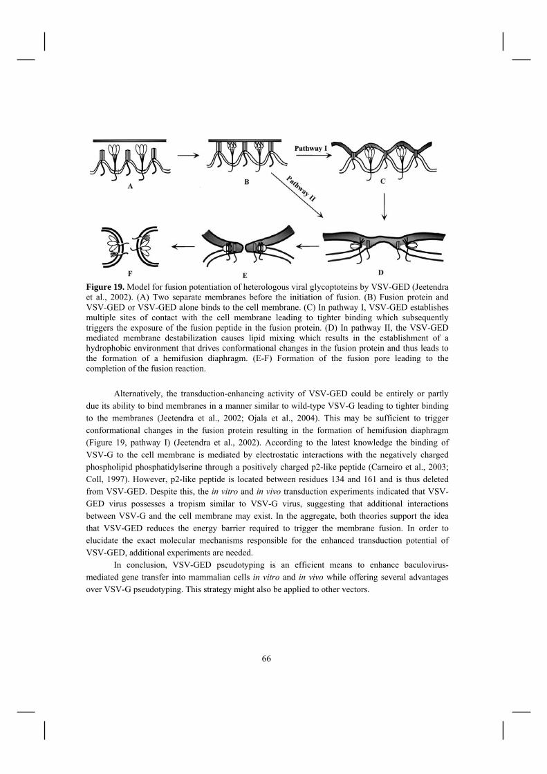
66
Figure 19. Model for fusion potentiation of heterologous viral glycoptoteins by VSV-GED (Jeetendra et al., 2002). (A) Two separate membranes before the initiation of fusion. (B) Fusion protein and VSV-GED or VSV-GED alone binds to the cell membrane. (C) In pathway I, VSV-GED establishes multiple sites of contact with the cell membrane leading to tighter binding which subsequently triggers the exposure of the fusion peptide in the fusion protein. (D) In pathway II, the VSV-GED mediated membrane destabilization causes lipid mixing which results in the establishment of a hydrophobic environment that drives conformational changes in the fusion protein and thus leads to the formation of a hemifusion diaphragm. (E-F) Formation of the fusion pore leading to the completion of the fusion reaction.
Alternatively, the transduction-enhancing activity of VSV-GED could be entirely or partly due its ability to bind membranes in a manner similar to wild-type VSV-G leading to tighter binding to the membranes (Jeetendra et al., 2002; Ojala et al., 2004). This may be sufficient to trigger conformational changes in the fusion protein resulting in the formation of hemifusion diaphragm (Figure 19, pathway I) (Jeetendra et al., 2002). According to the latest knowledge the binding of VSV-G to the cell membrane is mediated by electrostatic interactions with the negatively charged phospholipid phosphatidylserine through a positively charged p2-like peptide (Carneiro et al., 2003; Coll, 1997). However, p2-like peptide is located between residues 134 and 161 and is thus deleted from VSV-GED. Despite this, the in vitro and in vivo transduction experiments indicated that VSV-GED virus possesses a tropism similar to VSV-G virus, suggesting that additional interactions between VSV-G and the cell membrane may exist. In the aggregate, both theories support the idea that VSV-GED reduces the energy barrier required to trigger the membrane fusion. In order to elucidate the exact molecular mechanisms responsible for the enhanced transduction potential of VSV-GED, additional experiments are needed.
In conclusion, VSV-GED pseudotyping is an efficient means to enhance baculovirus-mediated gene transfer into mammalian cells in vitro and in vivo while offering several advantages over VSV-G pseudotyping. This strategy might also be applied to other vectors.

67
5.2 Targeting of metabolically biotinylated baculoviruses (II) Limitations regarding the efficacy and specificity of baculovirus-mediated transduction have slowed their widespread application. In this study we tested the utility of metabolical biotinylation of baculovirus for vector targeting. Given the exceptional affinity of (strept)avidin-biotin interactions (Kd~1013- 1015 M-1), this system qualifies for in vivo application where the stability of the vector-targeting molecule is of utmost importance. 5.2.1 Biotin display on the surface of baculoviruses To produce recombinant baculoviruses displaying biotin, the biotin acceptor peptide (BAP) was cloned either to different sites in the gp64 or to the N-terminus of VSV-GED (II/Fig. 1). In gp64-based constructs the BAP was either inserted after the signal sequence (BAP-N-gp64), at amino-acid position 283 (BAP-283-gp64) or both (BAP-283+N-gp64) (II/Fig. 1B). Given that BAP is not a substrate for enzymatic biotinylation in Spodoptera frugiperda (Sf9) cells (Duffy et al., 1998) and that the biotinylation of the secreted proteins does not naturally occur (Parrott and Barry, 2001), an Escherichia coli biotin holoenzyme synthetase, BirA, with gp64 signal sequence was coexpressed from baculovirus genome during the production of recombinant viruses to place its activity within the ER lumen where gp64 and VSV-GED are synthesized. To verify the expression of the biotin on the surface of baculovirus particles, an equivalent number of PFUs of the gradient purified viruses were subjected to western blot analysis with anti-gp64, anti-VSV-G or streptavidin-HRP. Incorporation of gp64 protein was detected in all the virions, confirming efficient production of viruses (II/Fig. 2A). The amount of gp64 was similar to that of control virus, indicating a normal ratio of total particle number versus infectious virus particles (TP/IP). Expression of BAP-VSV-GED (10 kDa) was identified with VSV-G antibodies, confirming successful incorporation (II/Fig. 2B). The incorporation of biotin was detected in all BAP-displaying virus samples using streptavidin-HRP (II/Fig. 2C). Of BAP-gp64 viruses, BAP-283-gp64 showed the highest level of biotin incorporation, BAP-N-gp64 the lowest, whereas BAP-283+N-gp64 settled between the two. As expected, no biotin was detected in the control virus sample. To assess the availability of biotin for streptavidin binding in solution and thus to assure of the accessibility of biotin for conjugation applications, a competition ELISA was performed. Binding of the biotinylated baculoviruses (1.5 x 10⁹ PFUs) on streptavidin coated plates was inhibited by increasing concentrations of free streptavidin and the amount of virus bound was detected with gp64 antibody. BAP-283-gp64 showed the highest binding to streptavidin and in the absence of free streptavidin the difference was 10-, 4- and 1.5-fold compared to BAP-VSV-GED, BAP-N-gp64 and BAP-283+N-gp64 viruses, respectively (II/Fig. 3). Of note, however, the ELISA underestimates the binding of BAP-VSV-GED since the bound virus is probed with gp64 antibody thus leading to increase of signal for BAP-gp64 vectors expressing two copies of gp64. Complete inhibition by

68
streptavidin was observed at 1µg/ml concentration of streptavidin. No obvious binding of the control virus to ELISA plates was observed. Together these experiments verify the incorporation of biotin on the surface of baculovirus vectors but also highlight the role of peptide insertion site in determining the efficiency of biotin display. These results are in line with a previous report where 283-site was described for the successful surface display of peptides ranging from 6 to 23 amino acids (Spenger et al., 2002). In the same study it was shown that the N-terminal insertions lead to weaker peptide display. It is hypothesized that hetero-oligomer formation is the basis of N-terminal fusion protein incorporation into virions, this being responsible for the low level of target protein display (Boublik et al., 1995). On this basis, the incorporation of peptides at site 283 is suggested to allow incorporation through homo-oligomerization of the fusion proteins thus allowing more efficient display (Spenger et al., 2002). To investigate the effects of BAP and biotin incorporation on the efficiency of baculovirus-mediated transduction, the gene delivery efficiency to HepG2, BT4C and SKOV-3 cells was studied using MOI 200. The transduction efficiency was assessed by measuring the β-galactosidase enzyme activity from the cell lysates 48h post-transduction (II/Fig. 4). As shown in Fig. 4, the BAP-VSV-GED transduced HepG2 and SKOV-3 cells with a similar efficiency to control virus, whereas a 4-fold increase in gene delivery efficiency to BT4C cells was detected. Interestingly, gene delivery by BAP-N-gp64 led to levels of β-galactosidase that were 5-, 6- and 100-fold lower in SKOV3, BT4C, and HepG2 cells, respectively, than those of the unmodified control virus. The most interesting finding was that transduction by BAP-283-gp64 and BAP-283+N-gp64 resulted in enhanced transduction efficiency in all the cell lines tested. BAP-283-gp64 showed 6 - 13- fold enhancement in transduction whereas for BAP-283+N-gp64 an increase of 12 - 25-fold was observed. Correlating with our results, previous reports have shown that N-terminal insertions to gp64 can in some cases affect the functionality of the vector (Huser et al., 2001; Riikonen et al., 2005; Spenger et al., 2002). We hypothesize that hetero-oligomerization of BAP-N-gp64 protein with the native gp64 impairs the assembly of stable gp64 trimers into multiprotein aggregates required for membrane fusion (Markovic et al., 1998; Guibinga et al., 2008) thus leading to poor transduction. On the other hand, the enhanced transduction efficiency by BAP-283-gp64 and BAP-283+N-gp64 viruses implies that the fusion proteins on these vectors could act as functional second copies of the gp64 thus eliciting a positive effect on the transduction efficiency as has been proposed for baculoviruses displaying an extra copy of wild-type gp64 (Tani et al., 2001). However, without further studies we cannot rule out the role of biotin or conformational changes caused by BAP insertion in increasing viral transduction. BAP-VSV-GED should not affect the functionality of the gp64 since no hetero-oligomerization with gp64 occurs and this was confirmed by the transduction experiments. In the study I we showed that VSV-GED alone is able to augment the transduction efficiency of baculovirus vector probably by potentiating the membrane fusion activity of baculovirus envelope protein gp64. However, VSV-GED fusions do not necessarily share this feature (Chapple and Jones, 2002; Ojala et al., 2004). Indeed, no significant differences were seen in the transduction efficiencies in HepG2 and SKOV3 cells although some increase was seen in BT4C cells.

69
5.2.2 Vector retargeting by biotinylated ligands and antibodies in vitro To determine whether we could redirect the baculoviral tropism, we tested biotinylated ligands and antibodies selectively binding to transferrin-, epidermal growth factor- and CD46- receptors expressed at high levels on tumor cells (BT4C, SKOV3). No increase of β-galactosidase expression was observed as a result of BAP-VSV-GED and BAP-N-gp64 retargeting to the receptors (II/Fig. 5). This may be due to less efficient biotin display subsequently reducing the extent of ligand binding. The tropism mediated by the wild-type gp64 can thus be predominant in these constructs. The targeted transduction of BAP-283-gp64 by conjugation of targeting molecules through streptavidin-biotin linker resulted in 15 - 40 % enhancement of transduction efficiency whereas for BAP-283+N-gp64 an increase of 30 - 60 % was attained (II/Fig. 5). This most probably resulted from the augmentation of virus binding to the target cell membrane since it was achieved after very short (15 min) exposure of the cells to the virus. In line with this, specific receptor-mediated binding (Kd ~ 10-7-10-11) and especially streptavidin-biotin binding (Kd = 2.5 x 10⁻¹³) have been shown to be faster than the attachment via multiple, low affinity bonds (Kd ~ 10-4-10-6) responsible for the binding of gp64 on the mammalian cell surface (Chilkoti and Stayton, 1995; Duisit et al., 1999; Tani et al., 2001). At present, however, the domains of gp64 involved in cell binding have not been elucidated (Duisit et al., 1999; Tani et al., 2001). The successful targeting of BAP-283-gp64 and BAP-283+N-gp64 led us to speculate that site 283 might be at the proximity of one such determinant, allowing redirection of the viral tropism when conjugated to targeting moieties. This is supported by recent reports suggesting that insertion of a hepatitis B peptide and RGD-motif at amino acid positions 278 and 283, respectively, of the single copy of gp64 alters the domains involved in cell recognition and uptake (Markusic et al., 2007; Riikonen et al., 2005). To further enhance the specificity of viral binding, it might be beneficial to generate baculoviruses lacking the wild-type gp64. Interestingly, the retargeting capacity of BAP-283-gp64 and BAP-283+N-gp64 was significantly lower compared to retargeted biotinylated adenovirus that showed 80-300-fold increase in reporter gene expression (Parrott et al., 2003). This might be due to the background binding mediated by the wild-type gp64 or other viral membrane proteins, whereas the tropism of adenovirus vectors is more strictly dependent on the initial recognition of coxsackie- and adenovirus receptor (CAR) on cells (Bergelson et al., 1997). However, differences in the used targeting protocols and in the sensitivity of the reporter gene assays, prevents further comparison. Nevertheless, targeting of this vector together with its overall increase in transduction efficiency has several advantages over non-targetable unmodified vectors. 5.2.3 Magnetic targeting In addition to conjugate-based targeting, the gene delivery location can be controlled spatially by the use of magnetic force. Magnetic targeting has already been used to target biotinylated retro- and adenoviruses (Campos et al., 2004; Hughes et al., 2001; Pandori et al., 2002). In order to examine the magnetic targeting of biotinylated baculoviruses, we conjugated the viruses to streptavidin conjugated

70
paramagnetic particles (SA-PMP) and transduced monolayer BT4C cells (II/Fig. 7). The results demonstrated successful guidance of PMP-conjugated metabolically biotinylated baculoviruses with local magnetic field indicating that magnetofection could become a choice for local gene delivery in vivo (Scherer et al., 2002). In addition, the PMP-conjugated vector was able to potentiate the gene delivery efficiency of metabolically biotinylated baculovirus vectors compared to sucrose gradient purified virus (II/Fig. 6C). This could be explained by the rapid settling vector-PMP complexes onto target cells, promotion of additional virus-cell interactions or enhanced endocytosis, as has been proposed for other vectors (Chan et al., 2005; Hughes et al., 2001; Pandori et al., 2002; Scherer et al., 2002). Indeed, due to their higher density, vector-particle complexes may allow safer local gene delivery by resisting the forces of diffusion in solutions (Pandori et al., 2002). In addition to physical targeting, these virus-PMP complexes can also be, by virtue of free binding sites on streptavidin, conjugated to targeting molecules and this way be directed to any cell, tissue or tumor in the body providing that the tissue specific markers have been identified (Gupta et al., 2007). Finally, PMPs or PMP-conjugated radionuclides can also be traced in vivo to allow imaging of vector biodistribution (Raty et al., 2006; Raty et al., 2007).
5.3 Purification of metabolically biotinylated baculoviruses (II)
For gene therapy studies and future clinical need, a simple and efficient purification method for baculovirus is also necessary. Conventional purification techniques for baculovirus involves a series of sucrose gradient ultracentrifugation steps, which is tedious, time-consuming and limited by the small volume capacity and loss of viral activity due to hydrodynamic stress and aggregation of viral particles (Barsoum, 1999; O’Reilly et al., 1994). More simple, effective and scalable methods based on chromatography have been developed for baculovirus purification and are represented in Table 14. One of the objectives of this work was to develop a novel method for baculovirus purification based on magnetic particle-dependent capture of biotinylated baculoviruses. Since biotin was incorporated and displayed on the viral surface, we used streptavidin conjugated paramagnetic particles for baculovirus purification. The efficiency of the method for 200-fold concentration of baculoviral stocks was evaluated by processing 10 ml of secondary viral preparations (Table 14). The collected PMP-virus pellet was washed and resuspended in 50µl of PBS. The total virus recoveries after end-point dilution titering were 30-100- fold higher compared to the starting preparation, demonstrating 15-50 % virus recovery, respectively (II/Fig. 6A). To test the functionality of the vector after PMP-purification we transduced HepG2 cells with equal volumes of the starting preparation and the concentrated virus (II/Fig. 6B). PMP-conjugated BAP-VSV-GED showed 200-fold increase in β-galactosidase expression upon transduction corresponding to the concentration factor. For BAP-N-gp64 the resultant gene delivery efficiency was only 20-fold, suggesting some loss of the transduction efficiency. This might be due to the overall decrease in transduction efficiency by BAP-N-gp64 virus (II/Fig. 4). The most surprising finding was that transduction by BAP-283-gp64 and BAP-283+N-gp64 resulted in 400-900- fold increase in β-
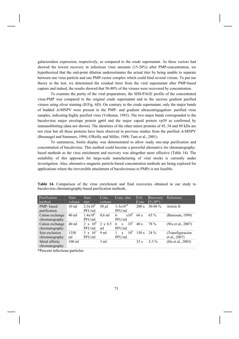
71
galactosidase expression, respectively, as compared to the crude supernatant. As these vectors had showed the lowest recovery in infectious virus amounts (15-20%) after PMP-concentration, we hypothesized that the end-point dilution underestimates the actual titer by being unable to separate between one virus particle and one PMP-vector complex which could bind several virions. To put our theory to the test, we determined the residual titers from the viral supernatant after PMP-based capture and indeed, the results showed that 50-80% of the viruses were recovered by concentration. To examine the purity of the viral preparations, the SDS-PAGE profile of the concentrated virus-PMP was compared to the original crude supernatant and to the sucrose gradient purified viruses using silver staining (II/Fig. 6D). On contrary to the crude supernatant, only the major bands of budded AcMNPV were present in the PMP- and gradient ultracentrigugation- purified virus samples, indicating highly purified virus (Volkman, 1983). The two major bands corresponded to the baculovirus major envelope protein gp64 and the major capsid protein vp39 as confirmed by immunoblotting (data not shown). The identities of the other minor proteins of 45, 54 and 89 kDa are not clear but all these proteins have been observed in previous studies from the purified AcMNPV (Braunagel and Summers, 1994; O'Reilly and Miller, 1988; Tani et al., 2001). To summarize, biotin display was demonstrated to allow ready one-step purification and concentration of baculovirus. This method could become a powerful alternative for chromatography-based methods as the virus enrichment and recovery was altogether more effective (Table 14). The suitability of this approach for large-scale manufacturing of viral stocks is currently under investigation. Also, alternative magnetic particle-based concentration methods are being explored for applications where the irreversible attachment of baculoviruses to PMPs is not feasible. Table 14. Comparison of the virus enrichment and final recoveries obtained in our study to baculovirus chromatography-based purification methods. Purification method
Start. volume
Start. titer
Conc. volume
Conc. titer Vol. Conc
Recovery (% IP*)
Reference
PMP- based purification
10 ml 2-5x10⁸ PFU/ml
50 µl 1-3x10¹⁰ PFU/ml
200 x 50-80 % Article II
Cation exchange chromatography
40 ml 1.4x10⁸
PFU/ml 0,6 ml 6 x10⁹
PFU/ml 66 x 65 % (Barsoum, 1999)
Cation exchange chromatography
40 ml 2 x 10⁸ PFU/ml
2 x 0.5 ml
6 x 10⁹ PFU/ml
40 x 78 % (Wu et al., 2007)
Size exclusion chromatography
1350 ml
3 x 10⁷
PFU/ml 9 ml 1 x 10⁹
PFU/ml 150 x 24 % (Transfiguracion
et al., 2007) Metal affinity chromatography
100 ml 3 ml 33 x 2-3 % (Hu et al., 2003)
*Percent infectious particles

72
5.4 (Strept)avidin-displaying lentiviruses for vector targeting (III)
One of the problems limiting the efficiency of lentiviral gene therapy is the lack of specificity in viral particle binding. More selective gene delivery would reduce the viral dose and consequently decrease the vector related side effects and ease clinical applications. In this study we describe the development of a flexible lentiviral vector platform for virus targeting based on (strept)avidin-display. 5.4.1 (Strept)avidin-VSV-GED incorporation on lentivirus surface To develop lentivirus vectors with targetable gene delivery, we designed novel gp64- pseudotyped vectors coexpressing avidin or streptavidin fused to the transmembrane anchor of VSV-G on the virus envelope. Separation of the targeting moiety and the envelope protein was hoped to leave the fusion protein intact for endosomal escape thus avoiding the common problem of decrease in virus infectivity by the modified envelope proteins (Martin et al., 1999; Zhao et al., 1999b). By transient transfection of five plasmids, we produced lentiviral vectors that efficiently incorporated gp64 and (strept)avidin fusion proteins into virus particles (III/Fig. 1). As the viral amounts were normalized by p24 antigen levels the SA/GP64 virions showed a higher level of fusion protein incorporation. Results of the titering and quantification of the p24 gag protein demonstrated no major differences in the infectivities of GP64 and AVD/GP64 lentiviruses, indicating a normal ratio of total particle number versus infectious virus particles (TP/IP) (III/Fig. 2a and b). Little decrease in the infectivity of SA/GP64 was detected which might be due to high level of streptavidin-VSV-GED incorporation. Optimization of the transfection conditions could help to avoid this. Nevertheless, we conclude that no major adverse effects were associated on (strept)avidin display on lentivirus surface. In line with the previous studies, SA/GP64 and AVD/GP64 were able to transduce a variety of cell types (BT4C, HeLa, HepG2, SKOV-3, D54, A549, U118MG; III/Fig. 2b and 4), indicating that these viruses share the wide cell tropism of gp64-pseudotyped viruses (Kumar et al., 2003; Schauber et al., 2004). However, the transduction of HeLa cells was several-fold lower compared to the results by Kumar et al (Kumar et al., 2003). This discrepancy is most likely due to differences in the old HeLa cell lines between the labs and to variation in the method of titering (Hughes et al., 2007). Next we wanted to assess if the (strept)avidin displaying lentiviruses were able to bind biotin and thereby assure of the ability of the virions to bind molecular conjugates. Increasing amount of viruses were allowed to attach to the biotin coated ELISA plates and the binding of the virus was detected by gp64 antibody. Results showed that AVD/GP64 and SA/GP64 viruses bound to ELISA plates in a dose-dependent manner but the extent of binding was markedly 3-times higher for SA/GP64 as measured from the slopes (III/ Fig. 3). This is in line with the western blot results demonstrating more efficient display of streptavidin-VSV-GED compared to avidin-VSV-GED.

73
GP64 control virus showed some unspecific binding to ELISA plates which was, however, significantly weaker compared to AVD/GP64 and SA/GP64. During this study, alternative vectors displaying streptavidin/avidin-VSV-GED together with VSV-G or avidin fused to the N-terminus of gp64 were also created (data not shown). In both cases, however, efficient (stept)avidin display was not achieved without compromising virus infectivity. In the former case, this might be due to heteromerization of streptavidin/avidin-VSV-GED fusion with the wild type VSV-G molecules leading to display of nonfunctional VSV-G molecules. The low infectivity of N-terminal gp64-fusions is likely explained by similar mechanisms as discussed for baculoviruses in study II.
5.4.2 Vector retargeting to tumor cells in vitro To redirect the specificity of infection of SA/GP64 and AVD/GP64, we used biotinylated ligands and antibodies selectively targeting receptors expressed at high levels on tumor cells. The efficiency of cell targeting was analyzed with BT4C, D54, SKOV-3, HepG2, A549 and U118MG cells by EGFP expression (III/Fig. 4). Biotinylated targeting molecules were incubated with the cells followed by washes and addition of pseudotyped viral particles for 15 minutes. The short virus incubation time was used to favor ligand specific binding events mediated by the high affinity streptavidin-biotin interaction. Targeting to transferrin receptor overexpressed on BT4C and D54 glioma cells led to 20-30% enhancement in transduction efficiency of AVD/GP64 whereas an increase of 50-60% was achieved with SA/GP64 (III/Fig. 4a). Redirection to EGFR led at best to 40% and 100% increase in percentage of EGFP-positive cells by AVD/GP64 and SA/GP64, respectively (III/Fig. 4b). The most successful retargeting was achieved to CD46 receptor-overexpressing U118MG cells as 2-3-fold increase in transduction efficiency was attained with (strept)avidin displaying viruses (III/Fig. 4c). The more efficient targeting of SA/GP64 was probably due to its more efficient biotin binding capacity but it has also been shown that biotin conjugates bind more stably to streptavidin than to avidin (Pazy et al., 2002). It remains to be studied if an optimization of the transfection conditions could further increase the level of avidin-VSV-GED display thus enhancing the retargeting capacity of AVD/GP64. No increase in EGFP expression was observed when the non-biotinylated targeting molecules were used. Targeting of GP64 control virus did not confer enhanced transduction of the cell lines. Altogether, the results demonstrate that target cell-specific transduction of SA/GP64 and AVD/GP64 can be increased by the use of biotinylated ligands and antibodies in vitro. It remains to be studied if precomplexed virus-ligand/Ab particles could favor faster and more efficient binding and internalization of the retargeted vector translating to more prominent transduction efficiencies. In addition, we need to consider the fact that SA/GP64 and AVD/GP64 still exhibit a similar host range to GP64 which limits the targeting efficiency. For applications aiming at transduction of liver, lung, skin or endothelium the presence of wild-type gp64 poses no problem but to achieve more stringent targeting this matter should be addressed (Schauber et al., 2004). In order to overcome this problem,

74
we could attempt to remove or greatly reduce the natural binding activity of gp64, without disturbing the fusion, by mutagenesis. Alternatively, pseudotyping with other binding-defective mutants, such as hemagglutinin (HA) of influenza A (Lin et al., 2001) and Sindbis virus (Morizono and Chen, 2005; Morizono et al., 2001; Yang et al., 2006) envelope proteins, could be considered. The approach described here for specific targeting of cells by lentiviral vector transduction overcomes many of the limitations of previous targeting strategies. The preparation of targeting vector is not limited by the introduction of modifications into the envelope proteins (VSV-G, gp64) that might result in substantial decreases in infectivity. This strategy could be applicable to other viruses e.g. to murine retroviral vectors. Finally, the approach described here should in theory be generally applicable to any cell surface molecule for which there are specific reagents that bind.
5.5 Imaging of streptavidin-displaying lentivirus (III)
One of the current limitations of preclinical trials in gene therapy is the difficulty in detecting virus location and gene expression upon administration of the delivery vector. Ideally, this detection should be noninvasive and repeatable over time to provide information about the location and magnitude of gene expression. In this study, we took advantage of (strept)avidin-display and MRI reporter gene, ferritin, to achieve non-invasive multi-modality imaging of virus biodistribution and transduction in vivo. 5.5.1 SPECT/CT imaging of virus biodistribution In vivo imaging techniques have the potential to provide critical information about the safety and kinetics of viral administration. Compared to the traditional immunological and histological methods, non-invasive imaging methods can follow virus localization with anatomical accuracy in real-time. Consequently, these techniques are crucial for elucidation of the specificity of new targeted vectors. In this regard, SPECT imaging has been previously utilized to measure changes in the liver tropism of adenovirus knob domain (Awasthi et al., 2004; Zinn et al., 1998), to image the biodistribution of herpes simplex viruses (Schellingerhout et al., 1998; Schellingerhout et al., 2000) and baculoviruses (Raty et al., 2007). In this study, we examined the biodistribution of SA/GP64 lentivirus in rats following injection into rat corpus callosum. This was achieved by radiolabeling SA/GP64 lentivirus with 111In labeled biotin-poly-Lys-DTPA and imaging the biodistribution with a dual modality SPECT/CT during three days after injection. Results from the day two when planar and 3D images were performed are shown in III/Fig. 6. In most of the virus injected rats the concentration of 111Indium was observed solely in the brain, indicating no viral leakage (III/Fig. 6a and 6d). This was further confirmed by RT-PCR, showing no transgene expression in the tissues which show lentivirus accumulation after systemic administration i.e. liver and spleen (data not shown) (Pan et al., 2002).

75
However, a signal from one rat, showing leakage to the circulation during the injection, was also seen in the abdominal area (III/Fig. 6b and 6e). Interestingly, no transgene expression was detected from the liver or the spleen of this rat either, suggesting a specific tropism of the labeled virus to the peritoneum. As expected, the small molecular weight 111In labeled biotin-poly-Lys-DTPA alone showed an accumulation to the brain followed by elimination through the kidneys and bladder (III/Fig. 6c and 6f). In conclusion, the SPECT/CT imaging provides an efficient means to measure specific targeting of streptavidin-displaying lentivirus vectors for gene therapy applications. Furthermore, the translation of this methodology into clinical applications should be feasible. 5.5.2 MRI imaging of viral gene delivery In addition to the knowledge about virus biodistribution, the information of the transgene expression is vital for the success of gene therapy. To achieve this, we generated SA/GP64 virus encoding the human heavy chain ferritin (hHF) cDNA. The underlying principle of visualizing ferritin expression on MRI is that this iron storage protein has a superparamagnetic core which disrupts the magnetic field and produces a lower (darker) signal on T2 weighted images (Drayer et al., 1986). As the transduced cells attain iron from the blood transferrin and subsequently transfer it to ferritin, no external contrast agents are needed. To date, ferritin has been utilized in gene therapy to image adenovirus-mediated gene delivery to rat brain (Genove et al., 2005) and to detect transcriptional regulation of gene expression in glioma tumors (Cohen et al., 2005) and in transgenic animals (Cohen et al., 2007). To investigate the efficiency of iron accumulation after SA/GP64-hHF virus transduction, we conducted in vitro studies using a HepG2 cell line. At 48 h post-transduction, the pelleted cells were analyzed by MRI (III/ Fig. 5). Transduced cells grown in their normal growth medium showed little increase in the relaxation rate R2 (=1/T2) compared to the control cells. Incubation of the cells in the iron supplemented medium (ferric ammonium citrate) resulted in 2-fold increase in the R2 of the virus transduced cells compared to the control, indicating that ferritin-transduced cells have an enhanced iron loading capacity (III/Fig. 5). These results are in line with a previous study, showing that the increased iron storage capacity can only be filled under iron supplemented conditions in vitro (Genove et al., 2005). To evaluate the potential of ferritin expression for the imaging of lentivirus-mediated gene delivery, we injected SA/GP64-hHF to the corpus callosum of the rat brain. To monitor the transgene expression, the animals underwent MRI scans at 4.7 T at days 3, 14, 28, 52 and 63 after SA/GP64-ferritin injection. Using gradient echo contrast, T2*-shortening was noted at the inoculation site, demonstrating a stable transduction of the target area by SA/GP64 lentivirus (III/Fig. 7a). As expected, the signal loss became more notable at later time points indicative of the increase in the cell’s ability to store iron. Some negative contrast was also detected in the needle track (data not shown). Control animals receiving 111In labeled biotin-poly-Lys-DTPA alone or SA/GP64-LacZ showed minor signal loss at the injection site at day 3 (III/Fig. 7b and c). This, however, resolved

76
before later measurements and was attributed to small hemorrhage caused by injection (Bradley, 1994). Histology and immunohistochemistry validated the MRI results showing transduction along the needle track, in the corpus callosum and in choroid plexus (III/Fig. 8). In concordance to the previous studies performed with baculovirus the transgene expression was also detected the cuboidal epithelial cells of the choroid plexus cells suggests that GP64 pseudotyped retain the same tropism (Lehtolainen et al., 2002b). However, the transduction of corpus callosum has not been seen with baculovirus. This may be explained by the differences in the membrane composition of these vectors. Nevertheless, these results provide evidence of the potential applicability of Gp64-pseudotyped lentivirus vectors as gene delivery vehicles for the treatment of CNS disorders.
The molecular imaging technology presented in this study can provide unique information about the success of virus administration and the resulting transduction efficiency. Further development of bicistronic vectors leading to coexpression of the therapeutic gene and ferritin may enable long-term monitoring of the status of therapeutic gene expression. Moreover, several reports have indicated that H subunit of ferritin may protect cells from the oxidative effects of iron (Cozzi et al., 2000; Epsztejn et al., 1999; Orino et al., 2001). In humans, oxidative cell and tissue damage has been linked to carcinogenesis, neurodegenerative disorders, autoimmune diseases, and atherosclerosis, among others (Berg and Youdim, 2006; Chau, 2000; Ong and Halliwell, 2004; Valko et al., 2006). Thus, the ferritin gene itself may have some therapeutic potential which should be taken into account when developing new therapies.
5.6 Characterization of baculovirus transduction in mammalian cells (IV) The fact that the baculovirus AcMNPV is used as a vector for many gene therapy studies makes research of the effects of viral transduction on the cellular machinery a high priority. In this study we investigated the expression profile of baculoviral genes in mammalian cells and the virus-induced alterations in the nuclear organization. 5.6.1 Baculovirus-mediated immediate early gene expression In general, it appears that baculoviruses are able to enter a phylogenetically broad range of insect cells but the expression of baculovirus genes appears to be blocked at an early gene expression stage during or after viral DNA replication (Morris and Miller, 1993). Similarly, AcMNPV can enter a myriad of vertebrate cells but appear to be unable to reach the nucleus of these cells, a notable exception being mammalian hepatocytes and osteosarcoma cells (Kukkonen et al., 2003; Song et al., 2003; Volkman and Goldsmith, 1983). In this work, we show that in HepG2 and 293 cells the virions accumulate in the nucleus as early as 4 hours p.t reaching the maximum at 8 hours p.t (IV/Fig. 1A-1C). In line with the previous reports, we also demonstrate that the nuclear entry of baculovirus is not dependent upon the disintegration of the nuclear membrane i.e. upon cell division (IV/Fig. 1D and 1E) (Lee et al., 2007; van Loo et al., 2001).

77
Recently, it was demonstrated that AcMNPV is able to transcribe at least few viral early genes in mammalian cells which are implicated in viral replication, namely ie-0, ie-1, pe-38, gp64, p35 and p6.9 (Fujita et al., 2006; Kitajima et al., 2006). Of these the ie-1 is the only gene essential for viral replication encoding the principal early transregulator protein IE-1 (Kool et al., 1994; Lu and Miller, 1995). DNA microarray analysis has also suggested the transcription of another essential gene, lef-3, and a stimulatory ie-2 gene but this has not been confirmed by RT-PCR studies (Fujita et al., 2006). IE-2 protein stimulates the expression of ie-1 (Yoo and Guarino, 1994) and pe-38 (Lu and Carstens, 1993) whereas LEF-3 is a single-strand binding protein which improves the strand displacement ability of viral DNA polymerase (Hang et al., 1995; McDougal and Guarino, 1999). In this study, we investigated the transcription of ie-1 and ie-2 in human HepG2 and 293 cells. Quantitative RT-PCR showed that both genes were expressed in a time-dependent manner, transcription starting at 4 h p.t and increasing at the last time point studied i.e. 48 h p.t. (IV/Fig 2A and 2B, data not shown at 48h). Both genes were expressed at higher levels in HepG2 cells than in 293 cells (IV/Fig. 2C), probably due to the differences in the transduction efficiency (data not shown). Furthermore, we demonstrated the expression of IE-2 protein first appearing at 4 h p.t. and continuing up to 48 h p.t. (IV/Fig. 3). In additional experiments, the transcription of lef-3 was also confirmed (Figure 20; unpublished results). Since IE-1, IE-2 and LEF-3 are found colocalized at viral replication sites in the nucleus of infected insect cells (Mainz et al., 2002; Okano et al., 1999), further studies regarding the localization of these proteins in baculovirus-transduced mammalian cells would be of great interest.
Figure 20. Transcription of baculoviral lef-3 gene measured by quantitative RT-PCR. (A) Relative gene expression values of control and transduced HepG2 cells 4-24 h p.t. (B) Comparison of the relative lef-3 expression in transduced HepG2 and 293 cells at 24h p.t. Taken together, these results confirm that AcMNPV is capable of expressing some viral genes in mammalian cells at the transcriptional and translational level. This is somewhat not surprising since the immediate early and delayed-early genes are transcribed by host RNA polymerase II, transcription mechanism of which is highly conserved among eukaryotes (Kornberg, 1999). The transcription initiation site in mammalian cells, however, may differ from the early viral transcription

78
site as demonstrated for pe-38 and p6.9, even though the transcription of ie-0, ie-1 and gp64 is shown to be initiated at the same site as in Sf9 cells (Fujita et al., 2006). Thus, the host RNA polymerase II and associated transcription factors dictate the outcome of early viral transcription in different species. On the contrary, late and very late genes are transcribed by viral RNA polymerase and thus it is unlikely that a late viral promoter would be activated in human cells. 5.6.2 Baculovirus induced nuclear reorganization All viruses have to interact with the cell nucleus consisting of different nuclear bodies (NBs) including cajal bodies, the nucleolus, perinucleolar and perichromatin regions, nuclear speckles and promyelotic nuclear bodies (PML NBs) (Zimber et al., 2004). In this work we investigated the interaction of baculovirus with PML NBs, Cajal bodies, nuclear speckles and chromatin after transduction of mammalian HepG2 and 293 cells. PML NBs are distinct subnuclear structures which appear as dense spherical particles, 0.3 to 0.5 µm in diameter, that are tightly associated with the nuclear matrix (Hodges et al., 1998). Although a number of proteins seem to transiently localize to PML NBs, two nuclear body antigens, PML and Sp100, are considered to build the framework of these structures (Sternsdorf et al., 1997). PML-NBs have been suggested to participate in transcriptional regulation, DNA damage response, regulation of apoptosis, senescence and neoangiogenesis (Bernardi and Pandolfi, 2007). The integrity of PML NBs is compromised in certain human diseases, including leukemia and neurodegenerative disorders but also during infection by a number of DNA viruses such as adenovirus (Carvalho et al., 1995; Doucas et al., 1996), herpes simplex virus (Everett and Maul, 1994; Everett et al., 1995), and cytomegalovirus (Ahn and Hayward, 1997; Kelly et al., 1995). Indeed, it appears to be a general tendency for DNA viruses to establish replication centers on the periphery of the PML NBs and first evidence of AcMNPV replication center association at close proximity of human PMLs has been provided by transient transfection experiments (Mainz et al., 2002). To address this issue in baculovirus transduced mammalian cells, we measured the colocalization of baculovirus with PML proteins and sp100 at 6h p.t. (IV/Fig. 4A and 5A). Consistent with the previous results, the virus foci was situated at the close proximity of PML NBs but no significant colocalization was detected at 6-24 h p.t. (data not shown) (Mainz et al., 2002). However, following viral transduction the size of PML NBs was increased by almost 2-fold together with an overall decrease in the number of PML NBs per cell (IV/ Fig. 4B and 4C). This may be a result of virus-induced cellular response or rearrangement of these structures into virus transcription or disassembly sites. An increased size of PML NBs has previously been shown to be involved in cell cycle, cellular stress and virus induced interferon response (IFN) (Buonamici et al., 2005; Djavani et al., 2001). PML NBs together with nucleophosmin are likely to play an important role, perhaps as sensors for cellular stress, during the DNA damage response (Dellaire and Bazett-Jones, 2004; Gjerset, 2006a; Wu and Yung, 2002). Several studies suggest that they function by regulating p53 stability (Coutts and La Thangue, 2005). Interestingly, the translocation of NPM from the nucleolus to nucleoplasm is indicative of cellular stress (Gjerset, 2006b). To determine the effects of baculovirus

79
transduction on cellular stress response, we monitored the localization of NPM-EGFP at 6-24 h p.t. No translocation of NPM was detected even at MOIs 1000-2000 (unpublished data), suggesting no evidence of the cytopathic effects in AcMNPV transduced cells. However, it remains to be studied if the reorganization of PML NBs was induced by IFN response as baculoviruses are shown to stimulate the expression of IFN-α/β in vitro and in vivo (Abe et al., 2005; Gronowski et al., 1999). Many viruses have also been found to interact with cajal bodies (CBs) and nuclear speckles; T-cell leukemia virus accumulated into nuclear speckles, whereas influenza virus alters their nuclear localization and adenoviral infection leads to the disruption of CBs. To address this matter in baculovirus transduced cells, we monitored the distribution of nuclear speckles markers (PAB2-EGFP and SC-35 Ab) and CB marker (p80coilin Ab) in relation to baculoviral capsids at 6 to 24 h p.t (IV/Fig. 5B-5C). Together, these data showed that baculovirus virions do not associate with nuclear speckles and CBs. Condensation, marginalization or dispersion of the chromatin, increase of the nucleoli and disruption of the nuclear lamina, has all been observed during infection of viruses. Similarly, baculovirus AcMNPV has been shown to disperse host cell chromatin of insect cells during infection. Here, we used chromatin label Drag5™ and human histone plasmid H2B-EYFP to study changes in host cell chromatin in HepG2 and 293 cells. In control cells the chromatin was detected around the periphery of the nuclear lamina and nucleoli (IV/Fig. 6A). In transduced cells, the chromatin label showed a more dispersed pattern resulting in less detectable lining of the nuclear lamina and nucleolus (IV/Fig. 6B). Similar results were obtained from aphidicolin synchronized G1/S-phase arrested cells (unpublished data). The altered chromatin distribution increased significantly over time (24-48 h p.t.) and with increasing viral load (MOI 200-1000) (IV/Fig. 6C). The effect of labeling of baculovirus genomes with Drag5™ in transduced cells was ruled out by flow cytometry studies which showed no difference in the chromatin fluorescence intensities in cells transduced at MOIs ranging from 10-1000. We also noticed that the peripheral heterochromatin lining the nucleoli was dispersed gradually during transduction, effect being more evident at higher MOIs (IV/Fig. 6D). These results were confirmed by monitoring changes in H2B-EYFP distribution which was significantly altered (IV/Fig. 7). Taken together, these results demonstrate that baculovirus virions or the products of early viral genes are able to induce alteration in the distribution of host cell chromatin. This chromatin remodeling may be mediated by viral interactions with nuclear actin, actin-related proteins or other histone-modifying factors but more work is required before this issue can be clarified (Chen and Shen, 2007; Lachner and Jenuwein, 2002; Simpson-Holley et al., 2005; Volkman, 2007). Baculoviruses have been in contact with humans since the emergence of species and there is no evidence that baculoviruses influence human health in any manner. The data provided here suggests that baculoviruses can induce viral gene expression and nuclear alterations in mammalian cells. Whether the expression of baculoviral proteins could induce immune responses or other physiological changes requires further investigation.

80
6. SUMMARY AND CONCLUSIONS
On the basis of the presented thesis studies, the following conclusions can be made.
1. VSV-GED pseudotyping augments baculovirus transduction in vitro and in vivo probably by reducing the energy barrier required to trigger the membrane fusion. In addition, VSV-GED display confers enhanced serum stability and offers several advantages over VSV-G pseudotyping such as low cytotoxicity.
2. Metabolic biotinylation of baculovirus membrane proteins allows viral targeting by an easy
exchange of surface molecules while enabling efficient capture, purification and concentration of baculovirus vectors. Significant differences in the extent of biotin incorporation and transduction efficiencies were revealed with respect to the site of biotin acceptor peptide on the envelope proteins; most efficient display was achieved by incorporation of BAP at site 283 on gp64 as this is suggested to allow incorporation through homo-oligomerization of the fusion proteins.
3. Targetable gp64-pseudotyped lentivirus vectors were developed by displaying streptavidin or
avidin fused to VSV-GED on the virus envelope. This strategy allows ligand- and antibody-mediated targeting of lentivirus to several tumor cell lines in vitro. Furthermore, streptavidin displaying lentivirus carrying a ferritin reporter gene enabled long-term non-invasive imaging of virus biodistribution and gene delivery by SPECT/CT and MRI. This study also provided pioneering information about the transduction pattern of gp64-pseudotyped lentiviruses in the central nervous system.
4. Baculovirus nuclear entry into mammalian cells was shown to be followed by the
transcription of early viral genes ie-1, ie-2 and lef-3. Moreover, the transduction was demonstrated to affect the size and number of PML bodies, which are often implicated in replication and transcription of viruses. Another effect was manifested by the remodeling of the peripheral host chromatin into disperse patterns. To address the safety issues of employing baculoviruses in gene therapy, expression of baculovirus endogenous genes and virus induced nuclear alterations, further studies are needed.
These results encourage further studies in characterization of the enhanced transduction capacity of VSV-GED and targeting potential of biotinylated baculoviruses and (strept)avidin-displaying lentiviruses in vivo. Also the use of streptavidin display for noninvasive imaging should allow the monitoring of virus biodistribution and gene delivery in the context of a therapeutic regimen. Finally, we provided significant new knowledge about the effects of baculovirus-mediated gene delivery in mammalian cells which poses new challenges for the future engineering of baculovirus vectors.
81
7. REFERENCES Abe, T., Hemmi, H., Miyamoto, H., Moriishi, K., Tamura, S., Takaku, H., Akira, S. and Matsuura, Y., (2005).
Involvement of the Toll-like receptor 9 signaling pathway in the induction of innate immunity by baculovirus. Journal of virology 79(5): 2847-2858.
Abe, T., Takahashi, H., Hamazaki, H., Miyano-Kurosaki, N., Matsuura, Y. and Takaku, H., (2003). Baculovirus induces an innate immune response and confers protection from lethal influenza virus infection in mice. Journal of immunology (Baltimore, Md.: 1950) 171(3): 1133-1139.
Afione, S.A., Conrad, C.K., Kearns, W.G., Chunduru, S., Adams, R., Reynolds, T.C., Guggino, W.B., Cutting, G.R., Carter, B.J. and Flotte, T.R., (1996). In vivo model of adeno-associated virus vector persistence and rescue. Journal of virology 70(5): 3235-3241.
Ahn, J.H. and Hayward, G.S., (1997). The major immediate-early proteins IE1 and IE2 of human cytomegalovirus colocalize with and disrupt PML-associated nuclear bodies at very early times in infected permissive cells. Journal of virology 71(6): 4599-4613.
Airenne, K.J., Hiltunen, M.O., Turunen, M.P., Turunen, A.M., Laitinen, O.H., Kulomaa, M.S. and Yla-Herttuala, S., (2000). Baculovirus-mediated periadventitial gene transfer to rabbit carotid artery. Gene therapy 7: 1499-1504.
Airenne, K.J., Mähönen, A.J., Laitinen, O.H. and Ylä-Herttuala, S. (2008). Baculovirus-mediated gene transfer: an emerging universal concept. In Gene and Cell Therapy: Therapeutic Mechanisms and Strategies, 3rd Edition. N.S. Templeton ed. (Boca Raton, FL: Taylor & Francis, CRC Press). pp. 263-291. In press
Alisky, J.M., Hughes, S.M., Sauter, S.L., Jolly, D., Dubensky, T.W.,Jr, Staber, P.D., Chiorini, J.A. and Davidson, B.L., (2000). Transduction of murine cerebellar neurons with recombinant FIV and AAV5 vectors. Neuroreport 11(12): 2669-2673.
Alon, R., Bayer, E.A. and Wilchek, M., (1990). Streptavidin contains an RYD sequence which mimics the RGD receptor domain of fibronectin. Biochemical and biophysical research communications 170(3): 1236-1241.
Altmann, A., Kissel, M., Zitzmann, S., Kubler, W., Mahmut, M., Peschke, P. and Haberkorn, U., (2003). Increased MIBG uptake after transfer of the human norepinephrine transporter gene in rat hepatoma. Journal of nuclear medicine : official publication, Society of Nuclear Medicine 44(6): 973-980.
Ambegia, E., Ansell, S., Cullis, P., Heyes, J., Palmer, L. and MacLachlan, I., (2005). Stabilized plasmid-lipid particles containing PEG-diacylglycerols exhibit extended circulation lifetimes and tumor selective gene expression. Biochimica et biophysica acta 1669(2): 155-163.
Arnold, G.S., Sasser, A.K., Stachler, M.D. and Bartlett, J.S., (2006). Metabolic biotinylation provides a unique platform for the purification and targeting of multiple AAV vector serotypes. MOLECULAR THERAPY; Molecular Therapy 14(1): 97-106.
Arosio, P. and Levi, S., (2002). Ferritin, iron homeostasis, and oxidative damage. Free radical biology & medicine 33(4): 457-463.
Auricchio, A., Kobinger, G., Anand, V., Hildinger, M., O'Connor, E., Maguire, A.M., Wilson, J.M. and Bennett, J., (2001). Exchange of surface proteins impacts on viral vector cellular specificity and transduction characteristics: the retina as a model. Human molecular genetics 10(26): 3075-3081.
Awasthi, V., Meinken, G., Springer, K., Srivastava, S.C. and Freimuth, P., (2004). Biodistribution of radioiodinated adenovirus fiber protein knob domain after intravenous injection in mice. Journal of virology 78(12):6431-6438.
Ayres, M.D., Howard, S.C., Kuzio, J., Lopez-Ferber, M. and Possee, R.D., (1994). The complete DNA sequence of Autographa californica nuclear polyhedrosis virus. Virology 202(2): 586-605.
Balyasnikova, I.V., Metzger, R., Visintine, D.J., Dimasius, V., Sun, Z.L., Berestetskaya, Y.V., McDonald, T.D., Curiel, D.T., Minshall, R.D. and Danilov, S.M., (2005). Selective rat lung endothelial targeting with a new set of monoclonal antibodies to angiotensin I-converting enzyme. Pulmonary pharmacology & therapeutics 18(4): 251-267.
Bank, A., Dorazio, R. and Leboulch, P., (2005). A phase I/II clinical trial of beta-globin gene therapy for beta-thalassemia. Annals of the New York Academy of Sciences 1054 308-316.
Barry, M.A., Campos, S.K., Ghosh, D., Adams, K.E., Mok, H., Mercier, G.T. and Parrott, M.B., (2003). Biotinylated gene therapy vectors. Expert opinion on biological therapy 3(6): 925-940.
Barsoum, J., (1999). Concentration of recombinant baculovirus by cation-exchange chromatography. BioTechniques 26(5): 834-6, 838, 840.
Barsoum, J., Brown, R., McKee, M. and Boyce, F.M., (1997). Efficient transduction of mammalian cells by a recombinant baculovirus having the vesicular stomatitis virus G glycoprotein. Human Gene Therapy 8(17): 2011-2018.

82
Bartlett, D.W., Su, H., Hildebrandt, I.J., Weber, W.A. and Davis, M.E., (2007). Impact of tumor-specific targeting on the biodistribution and efficacy of siRNA nanoparticles measured by multimodality in vivo imaging. Proceedings of the National Academy of Sciences of the United States of America 104(39): 15549-15554.
Bartlett, J.S., Kleinschmidt, J., Boucher, R.C. and Samulski, R.J., (1999). Targeted adeno-associated virus vector transduction of nonpermissive cells mediated by a bispecific F(ab'gamma)2 antibody. Nature biotechnology 17(2): 181-186.
Bayer, E.A. and Wilchek, M., (1994). Modified avidins for application in avidin-biotin technology: an improvement on nature. In: J.S. Sim and S. Nakai, eds, Egg Uses and Processing Technologies. Wallingford, UK: CAB International, pp. 158-176.
Bayer, E.A., Ben-Hur, H. and Wilchek, M., (1986). A sensitive enzyme assay for biotin, avidin, and streptavidin. Analytical Biochemistry 154(1): 367-370.
Belousova, N., Korokhov, N., Krendelshchikova, V., Simonenko, V., Mikheeva, G., Triozzi, P.L., Aldrich, W.A., Banerjee, P.T., Gillies, S.D., Curiel, D.T. and Krasnykh, V., (2003). Genetically targeted adenovirus vector directed to CD40-expressing cells. Journal of virology 77(21): 11367-11377.
Belousova, N., Krendelchtchikova, V., Curiel, D.T. and Krasnykh, V., (2002). Modulation of adenovirus vector tropism via incorporation of polypeptide ligands into the fiber protein. Journal of virology 76(17): 8621-8631.
Benedict, C.A., Tun, R.Y., Rubinstein, D.B., Guillaume, T., Cannon, P.M. and Anderson, W.F., (1999). Targeting retroviral vectors to CD34-expressing cells: binding to CD34 does not catalyze virus-cell fusion. Human Gene Therapy 10(4): 545-557.
Berg, D. and Youdim, M.B., (2006). Role of iron in neurodegenerative disorders. Topics in magnetic resonance imaging : TMRI 17(1): 5-17.
Bergelson, J.M., Cunningham, J.A., Droguett, G., Kurt-Jones, E.A., Krithivas, A., Hong, J.S., Horwitz, M.S., Crowell, R.L. and Finberg, R.W., (1997). Isolation of a common receptor for Coxsackie B viruses and adenoviruses 2 and 5. Science (New York, N.Y.) 275(5304): 1320-1323.
Bernardi, R. and Pandolfi, P.P., (2007). Structure, dynamics and functions of promyelocytic leukaemia nuclear bodies. Nature reviews.Molecular cell biology 8(12): 1006-1016.
Berraondo, P., Crettaz, J., Ochoa, L., Paneda, A., Prieto, J., Troconiz, I.F. and Gonzalez-Aseguinolaza, G., (2006). Intrahepatic injection of recombinant adeno-associated virus serotype 2 overcomes gender-related differences in liver transduction. Human Gene Therapy 17(6): 601-610.
Berto, E., Bozac, A. and Marconi, P., (2005). Development and application of replication-incompetent HSV-1-based vectors. Gene therapy 12 Suppl 1 S98-102.
Bharali, D.J., Klejbor, I., Stachowiak, E.K., Dutta, P., Roy, I., Kaur, N., Bergey, E.J., Prasad, P.N. and Stachowiak, M.K., (2005). Organically modified silica nanoparticles: a nonviral vector for in vivo gene delivery and expression in the brain. Proceedings of the National Academy of Sciences of the United States of America 102(32): 11539-11544.
Bigner, D.D., Bigner, S.H., Ponten, J., Westermark, B., Mahaley, M.S., Ruoslahti, E., Herschman, H., Eng, L.F. and Wikstrand, C.J., (1981). Heterogeneity of Genotypic and phenotypic characteristics of fifteen permanent cell lines derived from human gliomas. Journal of neuropathology and experimental neurology 40(3): 201-229.
Black, B.C., Brennan, L.A., Dierks, P.M. and Gard, I.E., (1997). Commercialization of Baculoviral Insecticides. In: L.K. Miller, ed, 1 edn. New York: Plenum Press, pp. 341-387.
Blasberg, R.G. and Tjuvajev, J.G., (1999). Herpes simplex virus thymidine kinase as a marker/reporter gene for PET imaging of gene therapy. The quarterly journal of nuclear medicine: official publication of the Italian Association of Nuclear Medicine [and] the International Association of Radiopharmacology 43(2): 163-169.
Blessing, T., Kursa, M., Holzhauser, R., Kircheis, R. and Wagner, E., (2001). Different strategies for formation of pegylated EGF-conjugated PEI/DNA complexes for targeted gene delivery. Bioconjugate chemistry 12(4):529-537.
Blissard, G.W. and Wenz, J.R., (1992). Baculovirus gp64 envelope glycoprotein is sufficient to mediate pH-dependent membrane fusion. Journal of virology 66(11): 6829-6835.
Blomer, U., Naldini, L., Kafri, T., Trono, D., Verma, I.M. and Gage, F.H., (1997). Highly efficient and sustained gene transfer in adult neurons with a lentivirus vector. Journal of virology 71(9): 6641-6649.
Board, R.G. and Fuller, R., (1974). Non-specific antimicrobial defences of the avian egg, embryo and neonate. Biological reviews of the Cambridge Philosophical Society 49(1): 15-49.
Boeckle, S. and Wagner, E., (2006). Optimizing targeted gene delivery: chemical modification of viral vectors and synthesis of artificial virus vector systems. The AAPS journal 8(4): E731-42.

83
Boublik, Y., Di Bonito, P. and Jones, I.M., (1995). Eukaryotic virus display: engineering the major surface glycoprotein of the Autographa californica nuclear polyhedrosis virus (AcNPV) for the presentation of foreign proteins on the virus surface. Bio/technology (Nature Publishing Company) 13(10): 1079-1084.
Boussif, O., Lezoualc'h, F., Zanta, M.A., Mergny, M.D., Scherman, D., Demeneix, B. and Behr, J.P., (1995). A versatile vector for gene and oligonucleotide transfer into cells in culture and in vivo: polyethylenimine. Proceedings of the National Academy of Sciences of the United States of America 92(16): 7297-7301.
Bouvet, M., Wang, J., Nardin, S.R., Nassirpour, R., Yang, M., Baranov, E., Jiang, P., Moossa, A.R. and Hoffman, R.M., (2002). Real-time optical imaging of primary tumor growth and multiple metastatic events in a pancreatic cancer orthotopic model. Cancer research 62(5): 1534-1540.
Bradley, W.G.,Jr, (1994). Hemorrhage and hemorrhagic infections in the brain. Neuroimaging clinics of North America 4(4): 707-732.
Braunagel, S.C. and Summers, M.D., (1994). Autographa californica nuclear polyhedrosis virus, PDV, and ECV viral envelopes and nucleocapsids: structural proteins, antigens, lipid and fatty acid profiles. Virology 202(1): 315-328.
Breidenbach, M., Rein, D.T., Everts, M., Glasgow, J.N., Wang, M., Passineau, M.J., Alvarez, R.D., Korokhov, N. and Curiel, D.T., (2005). Mesothelin-mediated targeting of adenoviral vectors for ovarian cancer gene therapy. Gene therapy 12(2): 187-193.
Bukrinskaya, A.G., (2004). HIV-1 assembly and maturation. Archives of Virology 149(6): 1067-1082. Bulte, J.W.M., Verkuyl, J.M., Herynek, V., Katsanis, E., Brocke, S., Holla, M. and Frank, J.A., (1998).
Magnetoimmunodetection of (transfected) ICAM-1 gene expression. Proceeding of the International Society of Magnetic Resonance Medicine 6 307.
Buonamici, S., Li, D., Mikhail, F.M., Sassano, A., Platanias, L.C., Colamonici, O., Anastasi, J. and Nucifora, G., (2005). EVI1 abrogates interferon-alpha response by selectively blocking PML induction. The Journal of biological chemistry 280(1): 428-436.
Burges, H.D., Croizier, G. and Huber, J., (1980). A review of safety tests on baculoviruses. BioControl 25: 329-339. Burns, J.C., Friedmann, T., Driever, W., Burrascano, M. and Yee, J.K., (1993). Vesicular stomatitis virus G
glycoprotein pseudotyped retroviral vectors: concentration to very high titer and efficient gene transfer into mammalian and nonmammalian cells. Proceedings of the National Academy of Sciences of the United States of America 90(17): 8033-8037.
Bushman, F.D., (1994). Tethering human immunodeficiency virus 1 integrase to a DNA site directs integration to nearby sequences. Proceedings of the National Academy of Sciences of the United States of America 91(20): 9233-9237.
Buursma, A.R., Beerens, A.M., de Vries, E.F., van Waarde, A., Rots, M.G., Hospers, G.A., Vaalburg, W. and Haisma, H.J., (2005). The human norepinephrine transporter in combination with 11C-m-hydroxyephedrine as a reporter gene/reporter probe for PET of gene therapy. Journal of nuclear medicine : official publication, Society of Nuclear Medicine 46(12): 2068-2075.
Buursma, A.R., Rutgers, V., Hospers, G.A., Mulder, N.H., Vaalburg, W. and de Vries, E.F., (2006). 18F-FEAU as a radiotracer for herpes simplex virus thymidine kinase gene expression: in-vitro comparison with other PET tracers. Nuclear medicine communications 27(1): 25-30.
Campbell, R.E., Tour, O., Palmer, A.E., Steinbach, P.A., Baird, G.S., Zacharias, D.A. and Tsien, R.Y., (2002). A monomeric red fluorescent protein. Proceedings of the National Academy of Sciences of the United States of America 99(12): 7877-7882.
Campos, S.K. and Barry, M.A., (2007). Current advances and future challenges in Adenoviral vector biology and targeting. Current gene therapy 7(3): 189-204.
Campos, S.K. and Barry, M.A., (2006). Comparison of adenovirus fiber, protein IX, and hexon capsomeres as scaffolds for vector purification and cell targeting. Virology 349(2): 453-462.
Campos, S.K., Parrott, M.B. and Barry, M.A., (2004). Avidin-based targeting and purification of a protein IX-modified, metabolically biotinylated adenoviral vector. Molecular therapy : the journal of the American Society of Gene Therapy 9(6): 942-954.
Carbonell, L.F., Klowden, M.J. and Miller, L.K., (1985). Baculovirus-mediated expression of bacterial genes in dipteran and mammalian cells. Journal of virology 56(1): 153-160.
Carneiro, F.A., Bianconi, M.L., Weissmuller, G., Stauffer, F. and Da Poian, A.T., (2002). Membrane recognition by vesicular stomatitis virus involves enthalpy-driven protein-lipid interactions. Journal of virology 76(8): 3756-3764.

84
Carneiro, F.A., Ferradosa, A.S. and Da Poian, A.T., (2001). Low pH-induced conformational changes in vesicular stomatitis virus glycoprotein involve dramatic structure reorganization. The Journal of biological chemistry 276(1): 62-67.
Carneiro, F.A., Lapido-Loureiro, P.A., Cordo, S.M., Stauffer, F., Weissmuller, G., Bianconi, M.L., Juliano, M.A., Juliano, L., Bisch, P.M. and Da Poian, A.T., (2006). Probing the interaction between vesicular stomatitis virus and phosphatidylserine. European biophysics journal : EBJ 35(2): 145-154.
Carneiro, F.A., Stauffer, F., Lima, C.S., Juliano, M.A., Juliano, L. and Da Poian, A.T., (2003). Membrane fusion induced by vesicular stomatitis virus depends on histidine protonation. The Journal of biological chemistry 278(16): 13789-13794.
Carneiro, F.A., Vandenbussche, G., Juliano, M.A., Juliano, L., Ruysschaert, J.M. and Da Poian, A.T., (2006). Charged residues are involved in membrane fusion mediated by a hydrophilic peptide located in vesicular stomatitis virus G protein. Molecular membrane biology 23(5): 396-406.
Carvalho, T., Seeler, J.S., Ohman, K., Jordan, P., Pettersson, U., Akusjarvi, G., Carmo-Fonseca, M. and Dejean, A., (1995). Targeting of adenovirus E1A and E4-ORF3 proteins to nuclear matrix-associated PML bodies. The Journal of cell biology 131(1): 45-56.
Cauchon, N., Langlois, R., Rousseau, J.A., Tessier, G., Cadorette, J., Lecomte, R., Hunting, D.J., Pavan, R.A., Zeisler, S.K. and van Lier, J.E., (2007). PET imaging of apoptosis with (64)Cu-labeled streptavidin following pretargeting of phosphatidylserine with biotinylated annexin-V. European journal of nuclear medicine and molecular imaging 34(2): 247-258.
Chadwick, M.P., Morling, F.J., Cosset, F.L. and Russell, S.J., (1999). Modification of retroviral tropism by display of IGF-I. Journal of Molecular Biology 285(2): 485-494.
Chai, N., Chang, H.E., Nicolas, E., Gudima, S., Chang, J. and Taylor, J., (2007). Assembly of hepatitis B virus envelope proteins onto a lentivirus pseudotype that infects primary human hepatocytes. Journal of virology 81(20): 10897-10904.
Chan, L., Nesbeth, D., Mackey, T., Galea-Lauri, J., Gaken, J., Martin, F., Collins, M., Mufti, G., Farzaneh, F. and Darling, D., (2005). Conjugation of lentivirus to paramagnetic particles via nonviral proteins allows efficient concentration and infection of primary acute myeloid leukemia cells. Journal of virology 79(20): 13190-13194.
Chapman-Smith, A. and Cronan, J.E.,Jr, (1999). In vivo enzymatic protein biotinylation. Biomolecular engineering 16(1-4): 119-125.
Chapple, S.D. and Jones, I.M., (2002). Non-polar distribution of green fluorescent protein on the surface of Autographa californica nucleopolyhedrovirus using a heterologous membrane anchor. Journal of Biotechnology 95(3): 269-275.
Chau, L.Y., (2000). Iron and atherosclerosis. Proceedings of the National Science Council, Republic of China.Part B, Life sciences 24(4): 151-155.
Chen, I.Y., Wu, J.C., Min, J.J., Sundaresan, G., Lewis, X., Liang, Q., Herschman, H.R. and Gambhir, S.S., (2004). Micro-positron emission tomography imaging of cardiac gene expression in rats using bicistronic adenoviral vector-mediated gene delivery. Circulation 109(11): 1415-1420.
Chen, M. and Shen, X., (2007). Nuclear actin and actin-related proteins in chromatin dynamics. Current opinion in cell biology 19(3): 326-330.
Chen, P., Kovesdi, I. and Bruder, J.T., (2000). Effective repeat administration with adenovirus vectors to the muscle. Gene therapy 7(7): 587-595.
Chilkoti, A. and Stayton, P.S., (1995). Molecular origins of the slow streptavidin-biotin dissociation kinetics. Journal of the American Chemical Society 117 10622-10628.
Chilkoti, A., Tan, P.H. and Stayton, P.S., (1995). Site-directed mutagenesis studies of the high-affinity streptavidin-biotin complex: contributions of tryptophan residues 79, 108, and 120. Proceedings of the National Academy of Sciences of the United States of America 92(5): 1754-1758.
Chillon, M., Bosch, A., Zabner, J., Law, L., Armentano, D., Welsh, M.J. and Davidson, B.L., (1999). Group D adenoviruses infect primary central nervous system cells more efficiently than those from group C. Journal of virology 73(3): 2537-2540.
Choi, V.W., McCarty, D.M. and Samulski, R.J., (2005). AAV hybrid serotypes: improved vectors for gene delivery. Current gene therapy 5(3): 299-310.
Chowdhury, S., Chester, K.A., Bridgewater, J., Collins, M.K. and Martin, F., (2004). Efficient retroviral vector targeting of carcinoembryonic antigen-positive tumors. Molecular therapy : the journal of the American Society of Gene Therapy 9(1): 85-92.

85
Choy, G., O'Connor, S., Diehn, F.E., Costouros, N., Alexander, H.R., Choyke, P. and Libutti, S.K., (2003). Comparison of noninvasive fluorescent and bioluminescent small animal optical imaging. BioTechniques 35(5): 1022-6, 1028-30.
Chroboczek, J., Bieber, F. and Jacrot, B., (1992). The sequence of the genome of adenovirus type 5 and its comparison with the genome of adenovirus type 2. Virology 186(1): 280-285.
Chuang, C.K., Sung, L.Y., Hwang, S.M., Lo, W.H., Chen, H.C. and Hu, Y.C., (2007). Baculovirus as a new gene delivery vector for stem cell engineering and bone tissue engineering. Gene therapy 14(19): 1417-1424.
Cleverley, D.Z. and Lenard, J., (1998). The transmembrane domain in viral fusion: essential role for a conserved glycine residue in vesicular stomatitis virus G protein. Proceedings of the National Academy of Sciences of the United States of America 95(7): 3425-3430.
Cohen, B., Dafni, H., Meir, G., Harmelin, A. and Neeman, M., (2005). Ferritin as an endogenous MRI reporter for noninvasive imaging of gene expression in C6 glioma tumors. Neoplasia (New York, N.Y.) 7(2): 109-117.
Cohen, B., Ziv, K., Plaks, V., Israely, T., Kalchenko, V., Harmelin, A., Benjamin, L.E. and Neeman, M., (2007). MRI detection of transcriptional regulation of gene expression in transgenic mice. Nature medicine 13(4): 498-503.
Coll, J.M., (1997). Synthetic peptides from the heptad repeats of the glycoproteins of rabies, vesicular stomatitis and fish rhabdoviruses bind phosphatidylserine. Archives of Virology 142(10): 2089-2097.
Condreay, J.P., Witherspoon, S.M., Clay, W.C. and Kost, T.A., (1999). Transient and stable gene expression in mammalian cells transduced with a recombinant baculovirus vector. Proceedings of the National Academy of Sciences of the United States of America 96(1): 127-132.
Contag, C.H., Jenkins, D., Contag, P.R. and Negrin, R.S., (2000). Use of reporter genes for optical measurements of neoplastic disease in vivo. Neoplasia (New York, N.Y.) 2(1-2): 41-52.
Conti, C., Mastromarino, P., Ciuffarella, M.G. and Orsi, N., (1988). Characterization of rat brain cellular membrane components acting as receptors for vesicular stomatitis virus. Brief report. Archives of Virology 99: 261-269.
Corti, A., Gasparri, A., Sacchi, A., Curnis, F., Sangregorio, R., Colombo, B., Siccardi, A.G. and Magni, F., (1998). Tumor targeting with biotinylated tumor necrosis factor alpha: structure-activity relationships and mechanism of action on avidin pretargeted tumor cells. Cancer research 58(17): 3866-3872.
Cosset, F.L., Morling, F.J., Takeuchi, Y., Weiss, R.A., Collins, M.K. and Russell, S.J., (1995). Retroviral retargeting by envelopes expressing an N-terminal binding domain. Journal of virology 69(10): 6314-6322.
Coura, R.D. and Nardi, N.B., (2007). The state of the art of adeno-associated virus-based vectors in gene therapy. Virol.J. 4(1): 99.
Coutts, A.S. and La Thangue, N.B., (2005). The p53 response: emerging levels of co-factor complexity. Biochemical and biophysical research communications 331(3): 778-785.
Cozzi, A., Corsi, B., Levi, S., Santambrogio, P., Albertini, A. and Arosio, P., (2000). Overexpression of wild type and mutated human ferritin H-chain in HeLa cells: in vivo role of ferritin ferroxidase activity. The Journal of biological chemistry 275(33): 25122-25129.
Cronan, J.E.,Jr, (1990). Biotination of proteins in vivo. A post-translational modification to label, purify, and study proteins. The Journal of biological chemistry 265(18): 10327-10333.
Cronan, J.E.,Jr, (1989). The E. coli bio operon: transcriptional repression by an essential protein modification enzyme. Cell 58(3): 427-429.
Cronin, J., Zhang, X.Y. and Reiser, J., (2005). Altering the tropism of lentiviral vectors through pseudotyping. Current gene therapy 5(4): 387-398.
Davidson, B.L., Stein, C.S., Heth, J.A., Martins, I., Kotin, R.M., Derksen, T.A., Zabner, J., Ghodsi, A. and Chiorini, J.A., (2000). Recombinant adeno-associated virus type 2, 4, and 5 vectors: transduction of variant cell types and regions in the mammalian central nervous system. Proceedings of the National Academy of Sciences of the United States of America 97(7): 3428-3432.
De La Fuente, E.K., Dawson, C.A., Nelin, L.D., Bongard, R.D., McAuliffe, T.L. and Merker, M.P., (1997). Biotinylation of membrane proteins accessible via the pulmonary circulation in normal and hyperoxic rats. The American Journal of Physiology 272(3 Pt 1): L461-70.
Delenda, C., (2004). Lentiviral vectors: optimization of packaging, transduction and gene expression. The journal of gene medicine 6 Suppl 1 S125-38.
Dellaire, G. and Bazett-Jones, D.P., (2004). PML nuclear bodies: dynamic sensors of DNA damage and cellular stress. BioEssays : news and reviews in molecular, cellular and developmental biology 26(9): 963-977.
Deng, W.P., Wu, C.C., Lee, C.C., Yang, W.K., Wang, H.E., Liu, R.S., Wei, H.J., Gelovani, J.G., Hwang, J.J., Yang, D.M., Fu, Y.K. and Wu, C.W., (2006). Serial in vivo imaging of the lung metastases model and gene therapy

86
using HSV1-tk and ganciclovir. Journal of nuclear medicine : official publication, Society of Nuclear Medicine 47(5): 877-884.
Dennison, S.M., Greenfield, N., Lenard, J. and Lentz, B.R., (2002). VSV transmembrane domain (TMD) peptide promotes PEG-mediated fusion of liposomes in a conformationally sensitive fashion. Biochemistry 41(50): 14925-14934.
Desmaris, N., Bosch, A., Salaun, C., Petit, C., Prevost, M.C., Tordo, N., Perrin, P., Schwartz, O., de Rocquigny, H. and Heard, J.M., (2001). Production and neurotropism of lentivirus vectors pseudotyped with lyssavirus envelope glycoproteins. Molecular therapy : the journal of the American Society of Gene Therapy 4(2):149-156.
Diaz, R.M., Bateman, A., Emiliusen, L., Fielding, A., Trono, D., Russell, S.J. and Vile, R.G., (2000). A lentiviral vector expressing a fusogenic glycoprotein for cancer gene therapy. Gene therapy 7(19): 1656-1663.
Djavani, M., Rodas, J., Lukashevich, I.S., Horejsh, D., Pandolfi, P.P., Borden, K.L. and Salvato, M.S., (2001). Role of the promyelocytic leukemia protein PML in the interferon sensitivity of lymphocytic choriomeningitis virus. Journal of virology 75(13): 6204-6208.
Dmitriev, I., Krasnykh, V., Miller, C.R., Wang, M., Kashentseva, E., Mikheeva, G., Belousova, N. and Curiel, D.T., (1998). An adenovirus vector with genetically modified fibers demonstrates expanded tropism via utilization of a coxsackievirus and adenovirus receptor-independent cell entry mechanism. Journal of virology 72(12): 9706-9713.
Dmitriev, I.P., Kashentseva, E.A. and Curiel, D.T., (2002). Engineering of adenovirus vectors containing heterologous peptide sequences in the C terminus of capsid protein IX. Journal of virology 76(14): 6893-6899.
Doms, R.W., Keller, D.S., Helenius, A. and Balch, W.E., (1987). Role for adenosine triphosphate in regulating the assembly and transport of vesicular stomatitis virus G protein trimers. The Journal of cell biology 105(5): 1957-1969.
Doms, R.W., Ruusala, A., Machamer, C., Helenius, J., Helenius, A. and Rose, J.K., (1988). Differential effects of mutations in three domains on folding, quaternary structure, and intracellular transport of vesicular stomatitis virus G protein. The Journal of cell biology 107(1): 89-99.
Doubrovin, M., Ponomarev, V., Serganova, I., Soghomonian, S., Myagawa, T., Beresten, T., Ageyeva, L., Sadelain, M., Koutcher, J., Blasberg, R.G. and Tjuvajev, J.G., (2003). Development of a new reporter gene system--dsRed/xanthine phosphoribosyltransferase-xanthine for molecular imaging of processes behind the intact blood-brain barrier. Molecular imaging : official journal of the Society for Molecular Imaging 2(2): 93-112.
Doucas, V., Ishov, A.M., Romo, A., Juguilon, H., Weitzman, M.D., Evans, R.M. and Maul, G.G., (1996). Adenovirus replication is coupled with the dynamic properties of the PML nuclear structure. Genes & development 10(2): 196-207.
Douglas, J.T., Rogers, B.E., Rosenfeld, M.E., Michael, S.I., Feng, M. and Curiel, D.T., (1996). Targeted gene delivery by tropism-modified adenoviral vectors. Nature biotechnology 14(11): 1574-1578.
Drayer, B., Burger, P., Darwin, R., Riederer, S., Herfkens, R. and Johnson, G.A., (1986). MRI of brain iron. AJR.American journal of roentgenology 147(1): 103-110.
Duffy, S., Tsao, K.L. and Waugh, D.S., (1998). Site-specific, enzymatic biotinylation of recombinant proteins in Spodoptera frugiperda cells using biotin acceptor peptides. Analytical Biochemistry 262(2): 122-128.
Duisit, G., Saleun, S., Douthe, S., Barsoum, J., Chadeuf, G. and Moullier, P., (1999). Baculovirus vector requires electrostatic interactions including heparan sulfate for efficient gene transfer in mammalian cells. The journal of gene medicine 1(2): 93-102.
Dwyer, R.M., Bergert, E.R., O'Connor, M.K., Gendler, S.J. and Morris, J.C., (2006). Adenovirus-mediated and targeted expression of the sodium-iodide symporter permits in vivo radioiodide imaging and therapy of pancreatic tumors. Human Gene Therapy 17(6): 661-668.
Ellenberg, J., Lippincott-Schwartz, J. and Presley, J.F., (1999). Dual-colour imaging with GFP variants. Trends in cell biology 9(2): 52-56.
Elo, H.A., (1980). Occurrence of avidin-like biotin-binding capacity in various vertebrate tissues and its induction by tissue injury. Comp. Biochem.Physiol. 67B 221-224.
Elo, H.A. and Korpela, J., (1984). The occurrence and production of avidin: a new conception of the high-affinity biotin-binding protein. Comparative biochemistry and physiology.B, Comparative biochemistry 78(1): 15-20.
Elo, H.A., Raisanen, S. and Tuohimaa, P.J., (1980). Induction of an antimicrobial biotin-binding egg white protein (avidin) in chick tissues in septic Escherichia coli infection. Experientia 36(3): 312-313.
Elouahabi, A. and Ruysschaert, J.M., (2005). Formation and intracellular trafficking of lipoplexes and polyplexes. Molecular therapy : the journal of the American Society of Gene Therapy 11(3): 336-347.
Emi, N., Friedmann, T. and Yee, J.K., (1991). Pseudotype formation of murine leukemia virus with the G protein of vesicular stomatitis virus. Journal of virology 65(3): 1202-1207.

87
Epsztejn, S., Glickstein, H., Picard, V., Slotki, I.N., Breuer, W., Beaumont, C. and Cabantchik, Z.I., (1999). H-ferritin subunit overexpression in erythroid cells reduces the oxidative stress response and induces multidrug resistance properties. Blood 94(10): 3593-3603.
Ernst, W., Schinko, T., Spenger, A., Oker-Blom, C. and Grabherr, R., (2006). Improving baculovirus transduction of mammalian cells by surface display of a RGD-motif. Journal of Biotechnology 126(2): 237-240.
Ernst, W.J., Spenger, A., Toellner, L., Katinger, H. and Grabherr, R.M., (2000). Expanding baculovirus surface display. Modification of the native coat protein gp64 of Autographa californica NPV. European journal of biochemistry / FEBS 267(13): 4033-4039.
Etienne-Julan, M., Roux, P., Carillo, S., Jeanteur, P. and Piechaczyk, M., (1992). The efficiency of cell targeting by recombinant retroviruses depends on the nature of the receptor and the composition of the artificial cell-virus linker. The Journal of general virology 73 ( Pt 12)(Pt 12): 3251-3255.
Everett, R., O'Hare, P., O'Rourke, D., Barlow, P. and Orr, A., (1995). Point mutations in the herpes simplex virus type 1 Vmw110 RING finger helix affect activation of gene expression, viral growth, and interaction with PML-containing nuclear structures. Journal of virology 69(11): 7339-7344.
Everett, R.D. and Maul, G.G., (1994). HSV-1 IE protein Vmw110 causes redistribution of PML. The EMBO journal 13(21): 5062-5069.
Facciabene, A., Aurisicchio, L. and La Monica, N., (2004). Baculovirus vectors elicit antigen-specific immune responses in mice. Journal of virology 78(16): 8663-8672.
Falk, M.M. and Lauf, U., (2001). High resolution, fluorescence deconvolution microscopy and tagging with the autofluorescent tracers CFP, GFP, and YFP to study the structural composition of gap junctions in living cells. Microscopy research and technique 52(3): 251-262.
Farhood, H., Serbina, N. and Huang, L., (1995). The role of dioleoyl phosphatidylethanolamine in cationic liposome mediated gene transfer. Biochimica et biophysica acta 1235(2): 289-295.
Felgner, P.L., Gadek, T.R., Holm, M., Roman, R., Chan, H.W., Wenz, M., Northrop, J.P., Ringold, G.M. and Danielsen, M., (1987). Lipofection: a highly efficient, lipid-mediated DNA-transfection procedure. Proceedings of the National Academy of Sciences of the United States of America 84(21): 7413-7417.
Fielding, A.K., Chapel-Fernandes, S., Chadwick, M.P., Bullough, F.J., Cosset, F.L. and Russell, S.J., (2000). A hyperfusogenic gibbon ape leukemia envelope glycoprotein: targeting of a cytotoxic gene by ligand display. Human Gene Therapy 11(6): 817-826.
Fielding, A.K., Maurice, M., Morling, F.J., Cosset, F.L. and Russell, S.J., (1998). Inverse targeting of retroviral vectors: selective gene transfer in a mixed population of hematopoietic and nonhematopoietic cells. Blood 91(5): 1802-1809.
Finn, F.M., Titus, G. and Hofmann, K., (1984). Ligands for insulin receptor isolation. Biochemistry 23:2554-2558. Forero, A., Weiden, P.L., Vose, J.M., Knox, S.J., LoBuglio, A.F., Hankins, J., Goris, M.L., Picozzi, V.J., Axworthy,
D.B., Breitz, H.B., Sims, R.B., Ghalie, R.G., Shen, S. and Meredith, R.F., (2004). Phase 1 trial of a novel anti-CD20 fusion protein in pretargeted radioimmunotherapy for B-cell non-Hodgkin lymphoma. Blood 104(1): 227-236.
Fraser, M., (1986). Ultrastructural observation of virion maturation. in Autographa californica nuclear polyhedrosis virus infected Spodoptera frugiperda cell cultures. Journal of ultrastructure and molecular structure research 95 189-195.
Freed, E.O. and Martin, M.A., (2007). HIVs and their replication. In: D.M. Knipe and P.M. Howley, eds, Fields Virology. 5th Ed. edn. Philadelphia.: Lippincott, Williams, and Wilkins, pp. 2107-2186.
Frost, E.H., (1977). Radioactive labelling of viruses: an iodination technique preserving biological properties. The Journal of general virology 35(1): 181-185.
Fujita, R., Matsuyama, T., Yamagishi, J., Sahara, K., Asano, S. and Bando, H., (2006). Expression of Autographa californica multiple nucleopolyhedrovirus genes in mammalian cells and upregulation of the host beta-actin gene. Journal of virology 80(5): 2390-2395.
Funk, C.J. and Consigli, R.A., (1993). Phosphate cycling on the basic protein of Plodia interpunctella granulosis virus. Virology 193(1): 396-402.
Funk, C.J., Braunagel, S.C. and Rohrmann, G.F., (1997). Baculovirus Structure. In: L.K. Miller, ed, The Baculoviruses. 1.ed. edn. New York: Plenum Press, pp. 7-32.
Gall, J., Kass-Eisler, A., Leinwand, L. and Falck-Pedersen, E., (1996). Adenovirus type 5 and 7 capsid chimera: fiber replacement alters receptor tropism without affecting primary immune neutralization epitopes. Journal of virology 70(4): 2116-2123.
Gambhir, S.S., Barrio, J.R., Phelps, M.E., Iyer, M., Namavari, M., Satyamurthy, N., Wu, L., Green, L.A., Bauer, E., MacLaren, D.C., Nguyen, K., Berk, A.J., Cherry, S.R. and Herschman, H.R., (1999). Imaging adenoviral-

88
directed reporter gene expression in living animals with positron emission tomography. Proceedings of the National Academy of Sciences of the United States of America 96(5): 2333-2338.
Gambhir, S.S., Bauer, E., Black, M.E., Liang, Q., Kokoris, M.S., Barrio, J.R., Iyer, M., Namavari, M., Phelps, M.E. and Herschman, H.R., (2000). A mutant herpes simplex virus type 1 thymidine kinase reporter gene shows improved sensitivity for imaging reporter gene expression with positron emission tomography. Proceedings of the National Academy of Sciences of the United States of America 97(6): 2785-2790.
Gao, G.P., Alvira, M.R., Wang, L., Calcedo, R., Johnston, J. and Wilson, J.M., (2002). Novel adeno-associated viruses from rhesus monkeys as vectors for human gene therapy. Proceedings of the National Academy of Sciences of the United States of America 99(18): 11854-11859.
Gao, G.P., Lu, Y., Sun, X., Johnston, J., Calcedo, R., Grant, R. and Wilson, J.M., (2006). High-level transgene expression in nonhuman primate liver with novel adeno-associated virus serotypes containing self-complementary genomes. Journal of virology 80(12): 6192-6194.
Gao, X., Kim, K.S. and Liu, D., (2007). Nonviral gene delivery: what we know and what is next. The AAPS journal 9(1): E92-104.
Gaspar, H.B., Parsley, K.L., Howe, S., King, D., Gilmour, K.C., Sinclair, J., Brouns, G., Schmidt, M., Von Kalle, C., Barington, T., Jakobsen, M.A., Christensen, H.O., Al Ghonaium, A., White, H.N., Smith, J.L., Levinsky, R.J., Ali, R.R., Kinnon, C. and Thrasher, A.J., (2004). Gene therapy of X-linked severe combined immunodeficiency by use of a pseudotyped gammaretroviral vector. Lancet 364(9452): 2181-2187.
Gelderblom, H.R., (1991). Assembly and morphology of HIV: potential effect of structure on viral function. AIDS (London, England) 5(6): 617-637.
Genove, G., DeMarco, U., Xu, H., Goins, W.F. and Ahrens, E.T., (2005). A new transgene reporter for in vivo magnetic resonance imaging. Nature medicine 11(4): 450-454.
Germershaus, O., Merdan, T., Bakowsky, U., Behe, M. and Kissel, T., (2006). Trastuzumab-polyethylenimine-polyethylene glycol conjugates for targeting Her2-expressing tumors. Bioconjugate chemistry 17(5):1190-1199.
Ghosh, S., Parvez, M.K., Banerjee, K., Sarin, S.K. and Hasnain, S.E., (2002). Baculovirus as mammalian cell expression vector for gene therapy: an emerging strategy. Molecular therapy : the journal of the American Society of Gene Therapy 6(1): 5-11.
Ghosh, S.S., Gopinath, P. and Ramesh, A., (2006). Adenoviral vectors: a promising tool for gene therapy. Applied Biochemistry and Biotechnology 133(1): 9-29.
Gjerset, R.A., (2006a). DNA damage, p14ARF, nucleophosmin (NPM/B23), and cancer. Journal of molecular histology 37(5-7): 239-251.
Gjerset, R.A., (2006b). DNA damage, p14ARF, nucleophosmin (NPM/B23), and cancer. Journal of molecular histology 37(5-7): 239-251.
Goff, S.P., (2001). Retroviridae: The Retroviruses and Their Replication. In: D.M. Knipe and P.M. Howley, eds, Fields Virology. 4th edn. Philadelphia, PA: Lippincott, Williams & Wilkins, pp. 1871-1940.
Goldman, C.K., Rogers, B.E., Douglas, J.T., Sosnowski, B.A., Ying, W., Siegal, G.P., Baird, A., Campain, J.A. and Curiel, D.T., (1997). Targeted gene delivery to Kaposi's sarcoma cells via the fibroblast growth factor receptor. Cancer research 57(8): 1447-1451.
Goossens, P.H., Havenga, M.J., Pieterman, E., Lemckert, A.A., Breedveld, F.C., Bout, A. and Huizinga, T.W., (2001). Infection efficiency of type 5 adenoviral vectors in synovial tissue can be enhanced with a type 16 fiber. Arthritis and Rheumatism 44(3): 570-577.
Gordon, E.M., Chen, Z.H., Liu, L., Whitley, M., Liu, L., Wei, D., Groshen, S., Hinton, D.R., Anderson, W.F., Beart, R.W.,Jr and Hall, F.L., (2001a). Systemic administration of a matrix-targeted retroviral vector is efficacious for cancer gene therapy in mice. Human Gene Therapy 12(2): 193-204.
Gordon, E.M., Zhu, N.L., Forney Prescott, M., Chen, Z.H., Anderson, W.F. and Hall, F.L., (2001b). Lesion-targeted injectable vectors for vascular restenosis. Human Gene Therapy 12(10): 1277-1287.
Grana, C., Chinol, M., Robertson, C., Mazzetta, C., Bartolomei, M., De Cicco, C., Fiorenza, M., Gatti, M., Caliceti, P. and Paganelli, G., (2002). Pretargeted adjuvant radioimmunotherapy with yttrium-90-biotin in malignant glioma patients: a pilot study. British journal of cancer 86(2): 207-212.
Granados, R.R., (1978). Early events in the infection of Hiliothis zea midgut cells by a baculovirus. Virology 90(1): 170-174.
Granados, R.R. and Lawler, K.A., (1981). In vivo pathway of Autographa californica baculovirus invasion and infection. Virology 108 297-308.
Grassi, G., Kohn, H., Dapas, B., Farra, R., Platz, J., Engel, S., Cjsareck, S., Kandolf, R., Teutsch, C., Klima, R., Triolo, G. and Kuhn, A., (2006). Comparison between recombinant baculo- and adenoviral-vectors as transfer system in cardiovascular cells. Archives of Virology 151(2): 255-271.

89
Green, N.M., (1990). Avidin and streptavidin. Methods in enzymology 184 51-67. Green, N.M., (1975). Avidin. Advances in Protein Chemistry 29 85-133. Grifman, M., Trepel, M., Speece, P., Gilbert, L.B., Arap, W., Pasqualini, R. and Weitzman, M.D., (2001).
Incorporation of tumor-targeting peptides into recombinant adeno-associated virus capsids. Molecular therapy : the journal of the American Society of Gene Therapy 3(6): 964-975.
Gronowski, A.M., Hilbert, D.M., Sheehan, K.C., Garotta, G. and Schreiber, R.D., (1999). Baculovirus stimulates antiviral effects in mammalian cells. Journal of virology 73(12): 9944-9951.
Groot-Wassink, T., Aboagye, E.O., Glaser, M., Lemoine, N.R. and Vassaux, G., (2002). Adenovirus biodistribution and noninvasive imaging of gene expression in vivo by positron emission tomography using human sodium/iodide symporter as reporter gene. Human Gene Therapy 13(14): 1723-1735.
Gross, S. and Piwnica-Worms, D., (2005). Spying on cancer: molecular imaging in vivo with genetically encoded reporters. Cancer cell 7(1): 5-15.
Gu, D.L., Gonzalez, A.M., Printz, M.A., Doukas, J., Ying, W., D'Andrea, M., Hoganson, D.K., Curiel, D.T., Douglas, J.T., Sosnowski, B.A., Baird, A., Aukerman, S.L. and Pierce, G.F., (1999). Fibroblast growth factor 2 retargeted adenovirus has redirected cellular tropism: evidence for reduced toxicity and enhanced antitumor activity in mice. Cancer research 59(11): 2608-2614.
Guesdon, J.L., Ternynck, T. and Avrameas, S., (1979). The use of avidin-biotin interaction in immunoenzymatic techniques. The journal of histochemistry and cytochemistry : official journal of the Histochemistry Society 27(8): 1131-1139.
Guibinga, G.H., Song, S., Loring, J. and Friedmann, T., (2008). Characterization of the gene delivery properties of baculoviral-based virosomal vectors. Journal of virological methods 148(1-2): 277-282.
Gupta, A.K., Naregalkar, R.R., Vaidya, V.D. and Gupta, M., (2007). Recent advances on surface engineering of magnetic iron oxide nanoparticles and their biomedical applications. Nanomedicine (London, England) 2(1): 23-39.
Guttinger, M., Guidi, F., Chinol, M., Reali, E., Veglia, F., Viale, G., Paganelli, G., Corti, A. and Siccardi, A.G., (2000). Adoptive immunotherapy by avidin-driven cytotoxic T lymphocyte-tumor bridging. Cancer research 60(15): 4211-4215.
Haberkorn, U., Henze, M., Altmann, A., Jiang, S., Morr, I., Mahmut, M., Peschke, P., Kubler, W., Debus, J. and Eisenhut, M., (2001). Transfer of the human NaI symporter gene enhances iodide uptake in hepatoma cells. Journal of nuclear medicine : official publication, Society of Nuclear Medicine 42(2): 317-325.
Haberkorn, U., Oberdorfer, F., Gebert, J., Morr, I., Haack, K., Weber, K., Lindauer, M., van Kaick, G. and Schackert, H.K., (1996). Monitoring gene therapy with cytosine deaminase: in vitro studies using tritiated-5-fluorocytosine. Journal of nuclear medicine : official publication, Society of Nuclear Medicine 37(1): 87-94.
Hacein-Bey-Abina, S., Von Kalle, C., Schmidt, M., McCormack, M.P., Wulffraat, N., Leboulch, P., Lim, A., Osborne, C.S., Pawliuk, R., Morillon, E., Sorensen, R., Forster, A., Fraser, P., Cohen, J.I., de Saint Basile, G., Alexander, I., Wintergerst, U., Frebourg, T., Aurias, A., Stoppa-Lyonnet, D., Romana, S., Radford-Weiss, I., Gross, F., Valensi, F., Delabesse, E., Macintyre, E., Sigaux, F., Soulier, J., Leiva, L.E., Wissler, M., Prinz, C., Rabbitts, T.H., Le Deist, F., Fischer, A. and Cavazzana-Calvo, M., (2003). LMO2-associated clonal T cell proliferation in two patients after gene therapy for SCID-X1. Science (New York, N.Y.) 302(5644): 415-419.
Hadjantonakis, A.K. and Nagy, A., (2001). The color of mice: in the light of GFP-variant reporters. Histochemistry and cell biology 115(1): 49-58.
Haeseleer, F., Imanishi, Y., Saperstein, D.A. and Palczewski, K., (2001). Gene transfer mediated by recombinant baculovirus into mouse eye. Investigative ophthalmology & visual science 42(13): 3294-3300.
Haisma, H.J., Pinedo, H.M., Rijswijk, A., der Meulen-Muileman, I., Sosnowski, B.A., Ying, W., Beusechem, V.W., Tillman, B.W., Gerritsen, W.R. and Curiel, D.T., (1999). Tumor-specific gene transfer via an adenoviral vector targeted to the pan-carcinoma antigen EpCAM. Gene therapy 6(8): 1469-1474.
Hakkarainen, T., Hemminki, A., Pereboev, A.V., Barker, S.D., Asiedu, C.K., Strong, T.V., Kanerva, A., Wahlfors, J. and Curiel, D.T., (2003). CD40 is expressed on ovarian cancer cells and can be utilized for targeting adenoviruses. Clinical cancer research : an official journal of the American Association for Cancer Research 9(2): 619-624.
Halbert, C.L., Allen, J.M. and Miller, A.D., (2001). Adeno-associated virus type 6 (AAV6) vectors mediate efficient transduction of airway epithelial cells in mouse lungs compared to that of AAV2 vectors. Journal of virology 75(14): 6615-6624.
Hall, F.L., Gordon, E.M., Wu, L., Zhu, N.L., Skotzko, M.J., Starnes, V.A. and Anderson, W.F., (1997). Targeting retroviral vectors to vascular lesions by genetic engineering of the MoMLV gp70 envelope protein. Human Gene Therapy 8(18): 2183-2192.

90
Hall, F.L., Liu, L., Zhu, N.L., Stapfer, M., Anderson, W.F., Beart, R.W. and Gordon, E.M., (2000). Molecular engineering of matrix-targeted retroviral vectors incorporating a surveillance function inherent in von Willebrand factor. Human Gene Therapy 11(7): 983-993.
Hama, Y., Urano, Y., Koyama, Y., Choyke, P.L. and Kobayashi, H., (2007). Activatable fluorescent molecular imaging of peritoneal metastases following pretargeting with a biotinylated monoclonal antibody. Cancer research 67(8): 3809-3817.
Handa, A., Muramatsu, S., Qiu, J., Mizukami, H. and Brown, K.E., (2000). Adeno-associated virus (AAV)-3-based vectors transduce haematopoietic cells not susceptible to transduction with AAV-2-based vectors. The Journal of general virology 81(Pt 8): 2077-2084.
Hang, X., Dong, W. and Guarino, L.A., (1995). The lef-3 gene of Autographa californica nuclear polyhedrosis virus encodes a single-stranded DNA-binding protein. Journal of virology 69(6): 3924-3928.
Harrap, K.A., (1972a). The structure of nuclear polyhedrosis viruses. I. The inclusion body. Virology 50(1):114-123. Harrap, K.A., (1972b). The structure of nuclear polyhedrosis viruses. II. The virus particle. Virology 50(1): 124-132. Havenga, M.J., Lemckert, A.A., Grimbergen, J.M., Vogels, R., Huisman, L.G., Valerio, D., Bout, A. and Quax, P.H.,
(2001). Improved adenovirus vectors for infection of cardiovascular tissues. Journal of virology 75(7): 3335-3342.
Havenga, M.J., Lemckert, A.A., Ophorst, O.J., van Meijer, M., Germeraad, W.T., Grimbergen, J., van Den Doel, M.A., Vogels, R., van Deutekom, J., Janson, A.A., de Bruijn, J.D., Uytdehaag, F., Quax, P.H., Logtenberg, T., Mehtali, M. and Bout, A., (2002). Exploiting the natural diversity in adenovirus tropism for therapy and prevention of disease. Journal of virology 76(9): 4612-4620.
Hefferon, K.L., Oomens, A.G., Monsma, S.A., Finnerty, C.M. and Blissard, G.W., (1999). Host cell receptor binding by baculovirus GP64 and kinetics of virion entry. Virology 258(2): 455-468.
Heideman, D.A., Snijders, P.J., Craanen, M.E., Bloemena, E., Meijer, C.J., Meuwissen, S.G., van Beusechem, V.W., Pinedo, H.M., Curiel, D.T., Haisma, H.J. and Gerritsen, W.R., (2001). Selective gene delivery toward gastric and esophageal adenocarcinoma cells via EpCAM-targeted adenoviral vectors. Cancer gene therapy 8(5):342-351.
Heise, C. and Kirn, D.H., (2000). Replication-selective adenoviruses as oncolytic agents. The Journal of clinical investigation 105(7): 847-851.
Hemminki, A., Belousova, N., Zinn, K.R., Liu, B., Wang, M., Chaudhuri, T.R., Rogers, B.E., Buchsbaum, D.J., Siegal, G.P., Barnes, M.N., Gomez-Navarro, J., Curiel, D.T. and Alvarez, R.D., (2001). An adenovirus with enhanced infectivity mediates molecular chemotherapy of ovarian cancer cells and allows imaging of gene expression. Molecular therapy : the journal of the American Society of Gene Therapy 4(3): 223-231.
Hemminki, A., Wang, M., Desmond, R.A., Strong, T.V., Alvarez, R.D. and Curiel, D.T., (2002). Serum and ascites neutralizing antibodies in ovarian cancer patients treated with intraperitoneal adenoviral gene therapy. Human Gene Therapy 13(12): 1505-1514.
Henning, P., Andersson, K.M., Frykholm, K., Ali, A., Magnusson, M.K., Nygren, P.A., Granio, O., Hong, S.S., Boulanger, P. and Lindholm, L., (2005). Tumor cell targeted gene delivery by adenovirus 5 vectors carrying knobless fibers with antibody-binding domains. Gene therapy 12(3): 211-224.
Hildebrandt, I.J., Iyer, M., Wagner, E. and Gambhir, S.S., (2003). Optical imaging of transferrin targeted PEI/DNA complexes in living subjects. Gene therapy 10(9): 758-764.
Ho, Y.C., Chen, H.C., Wang, K.C. and Hu, Y.C., (2004). Highly efficient baculovirus-mediated gene transfer into rat chondrocytes. Biotechnology and bioengineering 88(5): 643-651.
Hodges, M., Tissot, C., Howe, K., Grimwade, D. and Freemont, P.S., (1998). Structure, organization, and dynamics of promyelocytic leukemia protein nuclear bodies. American Journal of Human Genetics 63(2): 297-304.
Hofland, H.E., Masson, C., Iginla, S., Osetinsky, I., Reddy, J.A., Leamon, C.P., Scherman, D., Bessodes, M. and Wils, P., (2002). Folate-targeted gene transfer in vivo. Molecular therapy : the journal of the American Society of Gene Therapy 5(6): 739-744.
Hofmann, C., Sandig, V., Jennings, G., Rudolph, M., Schlag, P. and Strauss, M., (1995). Efficient gene transfer into human hepatocytes by baculovirus vectors. Proceedings of the National Academy of Sciences of the United States of America 92(22): 10099-10103.
Hofmann, C. and Strauss, M., (1998). Baculovirus-mediated gene transfer in the presence of human serum or blood facilitated by inhibition of the complement system. Gene therapy 5(4): 531-536.
Howarth, M., Chinnapen, D.J., Gerrow, K., Dorrestein, P.C., Grandy, M.R., Kelleher, N.L., El-Husseini, A. and Ting, A.Y., (2006). A monovalent streptavidin with a single femtomolar biotin binding site. Nature methods 3(4): 267-273.

91
Hoya, K., Guterman, L.R., Miskolczi, L. and Hopkins, L.N., (2001). A novel intravascular drug delivery method using endothelial biotinylation and avidin-biotin binding. Drug delivery 8(4): 215-222.
Hsu, C.S., Ho, Y.C., Wang, K.C. and Hu, Y.C., (2004). Investigation of optimal transduction conditions for baculovirus-mediated gene delivery into mammalian cells. Biotechnology and bioengineering 88(1): 42-51.
Hu, Y.C., (2006). Baculovirus vectors for gene therapy. Advances in Virus Research 68 287-320. Hu, Y.-., Tsai, C.-., Chung, Y.-., Lu, J.-. and Hsu, J.T.-., (2003). Generation of chimeric baculovirus with histidine-
tags displayed on the envelope and its purification using immobilized metal affinity chromatography. Enzyme and Microbial Technology 33 445-452.
Hughes, C., Galea-Lauri, J., Farzaneh, F. and Darling, D., (2001). Streptavidin paramagnetic particles provide a choice of three affinity-based capture and magnetic concentration strategies for retroviral vectors. MOLECULAR THERAPY; Molecular Therapy 3(4): 623-630.
Hughes, P., Marshall, D., Reid, Y., Parkes, H. and Gelber, C., (2007). The costs of using unauthenticated, over-passaged cell lines: how much more data do we need? BioTechniques 43(5): 575, 577-8, 581-2 passim.
Huser, A., Rudolph, M. and Hofmann, C., (2001). Incorporation of decay-accelerating factor into the baculovirus envelope generates complement-resistant gene transfer vectors. Nature biotechnology 19(5): 451-455.
Hytonen, V.P., Nordlund, H.R., Horha, J., Nyholm, T.K., Hyre, D.E., Kulomaa, T., Porkka, E.J., Marttila, A.T., Stayton, P.S., Laitinen, O.H. and Kulomaa, M.S., (2005). Dual-affinity avidin molecules. Proteins 61(3): 597-607.
Imren, S., Payen, E., Westerman, K.A., Pawliuk, R., Fabry, M.E., Eaves, C.J., Cavilla, B., Wadsworth, L.D., Beuzard, Y., Bouhassira, E.E., Russell, R., London, I.M., Nagel, R.L., Leboulch, P. and Humphries, R.K., (2002). Permanent and panerythroid correction of murine beta thalassemia by multiple lentiviral integration in hematopoietic stem cells. Proceedings of the National Academy of Sciences of the United States of America 99(22): 14380-14385.
Inagaki, K., Fuess, S., Storm, T.A., Gibson, G.A., Mctiernan, C.F., Kay, M.A. and Nakai, H., (2006). Robust systemic transduction with AAV9 vectors in mice: efficient global cardiac gene transfer superior to that of AAV8. Molecular therapy : the journal of the American Society of Gene Therapy 14(1): 45-53.
Izumi, M., Kawakami, Y., Glasgow, J.N., Belousova, N., Everts, M., Kim-Park, S., Yamamoto, S., Wang, M., Le, L.P., Reynolds, P.N. and Curiel, D.T., (2005). In vivo analysis of a genetically modified adenoviral vector targeted to human CD40 using a novel transient transgenic model. The journal of gene medicine 7(12): 1517-1525.
Jacobs, A., Voges, J., Reszka, R., Lercher, M., Gossmann, A., Kracht, L., Kaestle, C., Wagner, R., Wienhard, K. and Heiss, W.D., (2001). Positron-emission tomography of vector-mediated gene expression in gene therapy for gliomas. Lancet 358(9283): 727-729.
Jeetendra, E., Ghosh, K., Odell, D., Li, J., Ghosh, H.P. and Whitt, M.A., (2003). The membrane-proximal region of vesicular stomatitis virus glycoprotein G ectodomain is critical for fusion and virus infectivity. Journal of virology 77(23): 12807-12818.
Jeetendra, E., Robison, C.S., Albritton, L.M. and Whitt, M.A., (2002). The membrane-proximal domain of vesicular stomatitis virus G protein functions as a membrane fusion potentiator and can induce hemifusion. Journal of virology 76(23): 12300-12311.
Jeong, J.H., Kim, S.H., Kim, S.W. and Park, T.G., (2005a). In vivo tumor targeting of ODN-PEG-folic acid/PEI polyelectrolyte complex micelles. Journal of biomaterials science.Polymer edition 16(11): 1409-1419.
Jeong, J.H., Lee, M., Kim, W.J., Yockman, J.W., Park, T.G., Kim, Y.H. and Kim, S.W., (2005b). Anti-GAD antibody targeted non-viral gene delivery to islet beta cells. Journal of controlled release : official journal of the Controlled Release Society 107(3): 562-570.
Johanson, C.E., Duncan, J.A., Stopa, E.G. and Baird, A., (2005). Enhanced prospects for drug delivery and brain targeting by the choroid plexus-CSF route. Pharmaceutical research 22(7): 1011-1037.
Kahn, J.S., Schnell, M.J., Buonocore, L. and Rose, J.K., (1999). Recombinant vesicular stomatitis virus expressing respiratory syncytial virus (RSV) glycoproteins: RSV fusion protein can mediate infection and cell fusion. Virology 254(1): 81-91.
Kanerva, A., Mikheeva, G.V., Krasnykh, V., Coolidge, C.J., Lam, J.T., Mahasreshti, P.J., Barker, S.D., Straughn, M., Barnes, M.N., Alvarez, R.D., Hemminki, A. and Curiel, D.T., (2002a). Targeting adenovirus to the serotype 3 receptor increases gene transfer efficiency to ovarian cancer cells. Clinical cancer research : an official journal of the American Association for Cancer Research 8(1): 275-280.
Kanerva, A., Wang, M., Bauerschmitz, G.J., Lam, J.T., Desmond, R.A., Bhoola, S.M., Barnes, M.N., Alvarez, R.D., Siegal, G.P., Curiel, D.T. and Hemminki, A., (2002b). Gene transfer to ovarian cancer versus normal tissues

92
with fiber-modified adenoviruses. Molecular therapy : the journal of the American Society of Gene Therapy 5(6): 695-704.
Kang, Y., Stein, C.S., Heth, J.A., Sinn, P.L., Penisten, A.K., Staber, P.D., Ratliff, K.L., Shen, H., Barker, C.K., Martins, I., Sharkey, C.M., Sanders, D.A., McCray, P.B.,Jr and Davidson, B.L., (2002). In vivo gene transfer using a nonprimate lentiviral vector pseudotyped with Ross River Virus glycoproteins. Journal of virology 76(18): 9378-9388.
Kang, Y., Xie, L., Tran, D.T., Stein, C.S., Hickey, M., Davidson, B.L. and McCray, P.B.,Jr, (2005). Persistent expression of factor VIII in vivo following nonprimate lentiviral gene transfer. Blood 106(5): 1552-1558.
Kaplan, M.R., Calef, E., Bercovici, T. and Gitler, C., (1983). The selective detection of cell surface determinants by means of antibodies and acetylated avidin attached to highly fluorescent polymer microspheres. Biochimica et biophysica acta 728(1): 112-120.
Kashiwakura, Y., Tamayose, K., Iwabuchi, K., Hirai, Y., Shimada, T., Matsumoto, K., Nakamura, T., Watanabe, M., Oshimi, K. and Daida, H., (2005). Hepatocyte growth factor receptor is a coreceptor for adeno-associated virus type 2 infection. Journal of virology 79(1): 609-614.
Kawabata, K., Takakura, Y. and Hashida, M., (1995). The fate of plasmid DNA after intravenous injection in mice: involvement of scavenger receptors in its hepatic uptake. Pharmaceutical research 12(6): 825-830.
Kawakami, Y., Li, H., Lam, J.T., Krasnykh, V., Curiel, D.T. and Blackwell, J.L., (2003). Substitution of the adenovirus serotype 5 knob with a serotype 3 knob enhances multiple steps in virus replication. Cancer research 63(6): 1262-1269.
Kay, M.A., Glorioso, J.C. and Naldini, L., (2001). Viral vectors for gene therapy: the art of turning infectious agents into vehicles of therapeutics. Nature medicine 7(1): 33-40.
Kelly, C., Van Driel, R. and Wilkinson, G.W., (1995). Disruption of PML-associated nuclear bodies during human cytomegalovirus infection. The Journal of general virology 76 ( Pt 11)(Pt 11): 2887-2893.
Kelly, E. and Russell, S.J., (2007). History of oncolytic viruses: genesis to genetic engineering. Molecular therapy : the journal of the American Society of Gene Therapy 15(4): 651-659.
Kelly, F.J., Miller, C.R., Buchsbaum, D.J., Gomez-Navarro, J., Barnes, M.N., Alvarez, R.D. and Curiel, D.T., (2000). Selectivity of TAG-72-targeted adenovirus gene transfer to primary ovarian carcinoma cells versus autologous mesothelial cells in vitro. Clinical cancer research : an official journal of the American Association for Cancer Research 6(11): 4323-4333.
Kenoutis, C., Efrose, R.C., Swevers, L., Lavdas, A.A., Gaitanou, M., Matsas, R. and Iatrou, K., (2006). Baculovirus-mediated gene delivery into Mammalian cells does not alter their transcriptional and differentiating potential but is accompanied by early viral gene expression. Journal of virology 80(8): 4135-4146.
Kichler, A., (2004). Gene transfer with modified polyethylenimines. The journal of gene medicine 6 Suppl 1 S3-10. Kircheis, R., Schuller, S., Brunner, S., Ogris, M., Heider, K.H., Zauner, W. and Wagner, E., (1999). Polycation-
based DNA complexes for tumor-targeted gene delivery in vivo. The journal of gene medicine 1(2): 111-120. Kircheis, R., Wightman, L., Schreiber, A., Robitza, B., Rossler, V., Kursa, M. and Wagner, E., (2001).
Polyethylenimine/DNA complexes shielded by transferrin target gene expression to tumors after systemic application. Gene therapy 8(1): 28-40.
Kitajima, M., Hamazaki, H., Miyano-Kurosaki, N. and Takaku, H., (2006). Characterization of baculovirus Autographa californica multiple nuclear polyhedrosis virus infection in mammalian cells. Biochemical and biophysical research communications 343(2): 378-384.
Kitajima, M. and Takaku, H., (2008). Induction of antitumor acquired immunity by baculovirus Autographa californica multiple nuclear polyhedrosis virus infection in mice. Clinical and vaccine immunology : CVI 15(2): 376-378.
Klumb, L.A., Chu, V. and Stayton, P.S., (1998). Energetic roles of hydrogen bonds at the ureido oxygen binding pocket in the streptavidin-biotin complex. Biochemistry 37(21): 7657-7663.
Knipe, D.M., Baltimore, D. and Lodish, H.F., (1977). Maturation of viral proteins in cells infected with temperature-sensitive mutants of vesicular stomatitis virus. Journal of virology 21(3): 1149-1158.
Knowles, J.R., (1989). The mechanism of biotin-dependent enzymes. Annual Review of Biochemistry 58 195-221. Knowles, M.R., Hohneker, K.W., Zhou, Z., Olsen, J.C., Noah, T.L., Hu, P.C., Leigh, M.W., Engelhardt, J.F.,
Edwards, L.J. and Jones, K.R., (1995). A controlled study of adenoviral-vector-mediated gene transfer in the nasal epithelium of patients with cystic fibrosis. The New England journal of medicine 333(13): 823-831.
Knox, S.J., Goris, M.L., Tempero, M., Weiden, P.L., Gentner, L., Breitz, H., Adams, G.P., Axworthy, D., Gaffigan, S., Bryan, K., Fisher, D.R., Colcher, D., Horak, I.D. and Weiner, L.M., (2000). Phase II trial of yttrium-90-DOTA-biotin pretargeted by NR-LU-10 antibody/streptavidin in patients with metastatic colon cancer. Clinical cancer research : an official journal of the American Association for Cancer Research 6(2): 406-414.

93
Kobayashi, M., Iida, A., Ueda, Y. and Hasegawa, M., (2003). Pseudotyped lentivirus vectors derived from simian immunodeficiency virus SIVagm with envelope glycoproteins from paramyxovirus. Journal of virology 77(4): 2607-2614.
Kobinger, G.P., Deng, S., Louboutin, J.P., Vatamaniuk, M., Matschinsky, F., Markmann, J.F., Raper, S.E. and Wilson, J.M., (2004). Transduction of human islets with pseudotyped lentiviral vectors. Human Gene Therapy 15(2): 211-219.
Kobinger, G.P., Weiner, D.J., Yu, Q.C. and Wilson, J.M., (2001). Filovirus-pseudotyped lentiviral vector can efficiently and stably transduce airway epithelia in vivo. Nature biotechnology 19(3): 225-230.
Kool, M., Voeten, J.T., Goldbach, R.W. and Vlak, J.M., (1994). Functional mapping of regions of the Autographa californica nuclear polyhedrosis viral genome required for DNA replication. Virology 198(2): 680-689.
Kootstra, N.A. and Verma, I.M., (2003). Gene therapy with viral vectors. Annual Review of Pharmacology and Toxicology 43 413-439.
Kordower, J.H., Bloch, J., Ma, S.Y., Chu, Y., Palfi, S., Roitberg, B.Z., Emborg, M., Hantraye, P., Deglon, N. and Aebischer, P., (1999). Lentiviral gene transfer to the nonhuman primate brain. Experimental neurology 160(1): 1-16.
Koretsky, A.P., Brosnan, M.J., Chen, L.H., Chen, J.D. and Van Dyke, T., (1990). NMR detection of creatine kinase expressed in liver of transgenic mice: determination of free ADP levels. Proceedings of the National Academy of Sciences of the United States of America 87(8): 3112-3116.
Kornberg, R.D., (1999). Eukaryotic transcriptional control. Trends in cell biology 9(12): M46-9. Korokhov, N., de Gruijl, T.D., Aldrich, W.A., Triozzi, P.L., Banerjee, P.T., Gillies, S.D., Curiel, T.J., Douglas, J.T.,
Scheper, R.J. and Curiel, D.T., (2005). High efficiency transduction of dendritic cells by adenoviral vectors targeted to DC-SIGN. Cancer biology & therapy 4(3): 289-294.
Korokhov, N., Mikheeva, G., Krendelshchikov, A., Belousova, N., Simonenko, V., Krendelshchikova, V., Pereboev, A., Kotov, A., Kotova, O., Triozzi, P.L., Aldrich, W.A., Douglas, J.T., Lo, K.M., Banerjee, P.T., Gillies, S.D., Curiel, D.T. and Krasnykh, V., (2003). Targeting of adenovirus via genetic modification of the viral capsid combined with a protein bridge. Journal of virology 77(24): 12931-12940.
Kost, T.A. and Condreay, J.P., (2002). Innovations-Biotechnology: Baculovirus Vectors as Gene Transfer Vectors for Mammalian Cells: Biosafety Consideration. Applied Biosafety 7 167-169.
Kost, T.A., Condreay, J.P. and Jarvis, D.L., (2005). Baculovirus as versatile vectors for protein expression in insect and mammalian cells. Nature biotechnology 23(5): 567-575.
Krasnykh, V., Belousova, N., Korokhov, N., Mikheeva, G. and Curiel, D.T., (2001). Genetic targeting of an adenovirus vector via replacement of the fiber protein with the phage T4 fibritin. Journal of virology 75(9): 4176-4183.
Kreis, T.E. and Lodish, H.F., (1986). Oligomerization is essential for transport of vesicular stomatitis viral glycoprotein to the cell surface. Cell 46(6): 929-937.
Kukkonen, S.P., Airenne, K.J., Marjomaki, V., Laitinen, O.H., Lehtolainen, P., Kankaanpaa, P., Mahonen, A.J., Raty, J.K., Nordlund, H.R., Oker-Blom, C., Kulomaa, M.S. and Yla-Herttuala, S., (2003). Baculovirus capsid display: a novel tool for transduction imaging. Molecular therapy : the journal of the American Society of Gene Therapy 8(5): 853-862.
Kumar, M., Bradow, B.P. and Zimmerberg, J., (2003). Large-scale production of pseudotyped lentiviral vectors using baculovirus GP64. Human Gene Therapy 14(1): 67-77.
Labbe, J.P., (2003). SPECT/CT Emerges from the shadow of PET/CT. Biophotonics International 50-57. Lachner, M. and Jenuwein, T., (2002). The many faces of histone lysine methylation. Current opinion in cell biology
14(3): 286-298. Laerum, O.D., Rajewsky, M.F., Schachner, M., Stavrou, D., Haglid, K.G. and Haugen, A., (1977). Phenotypic
properties of neoplastic cell lines developed from fetal rat brain cells in culture after exposure to ethylnitrosourea in vivo. Zeitschrift fur Krebsforschung und klinische Onkologie.Cancer research and clinical oncology 89(3): 273-295.
Laitinen, O.H., Airenne, K.J., Marttila, A.T., Kulik, T., Porkka, E., Bayer, E.A., Wilchek, M. and Kulomaa, M.S., (1999). Mutation of a critical tryptophan to lysine in avidin or streptavidin may explain why sea urchin fibropellin adopts an avidin-like domain. FEBS letters 461(1-2): 52-58.
Laitinen, O.H., Hytonen, V.P., Ahlroth, M.K., Pentikainen, O.T., Gallagher, C., Nordlund, H.R., Ovod, V., Marttila, A.T., Porkka, E., Heino, S., Johnson, M.S., Airenne, K.J. and Kulomaa, M.S., (2002). Chicken avidin-related proteins show altered biotin-binding and physico-chemical properties as compared with avidin. The Biochemical journal 363(Pt 3): 609-617.

94
Laitinen, O.H., Hytonen, V.P., Nordlund, H.R. and Kulomaa, M.S., (2006). Genetically engineered avidins and streptavidins. Cellular and molecular life sciences : CMLS 63(24): 2992-3017.
Laitinen, O.H., Marttila, A.T., Airenne, K.J., Kulik, T., Livnah, O., Bayer, E.A., Wilchek, M. and Kulomaa, M.S., (2001). Biotin induces tetramerization of a recombinant monomeric avidin. A model for protein-protein interactions. The Journal of biological chemistry 276(11): 8219-8224.
Laitinen, O.H., Nordlund, H.R., Hytonen, V.P., Uotila, S.T., Marttila, A.T., Savolainen, J., Airenne, K.J., Livnah, O., Bayer, E.A., Wilchek, M. and Kulomaa, M.S., (2003). Rational design of an active avidin monomer. The Journal of biological chemistry 278(6): 4010-4014.
Lee, H.P., Chen, Y.L., Shen, H.C., Lo, W.H. and Hu, Y.C., (2007). Baculovirus transduction of rat articular chondrocytes: roles of cell cycle. The journal of gene medicine 9(1): 33-43.
Lee, W.W., Moon, D.H., Park, S.Y., Jin, J., Kim, S.J. and Lee, H., (2004). Imaging of adenovirus-mediated expression of human sodium iodide symporter gene by 99mTcO4 scintigraphy in mice. Nuclear medicine and biology 31(1): 31-40.
Lehtolainen, P., Taskinen, A., Laukkanen, J., Airenne, K.J., Heino, S., Lappalainen, M., Ojala, K., Marjomaki, V., Martin, J.F., Kulomaa, M.S. and Yla-Herttuala, S., (2002a). Cloning and characterization of Scavidin, a fusion protein for the targeted delivery of biotinylated molecules. The Journal of biological chemistry 277: 8545-8550.
Lehtolainen, P., Tyynela, K., Kannasto, J., Airenne, K.J. and Yla-Herttuala, S., (2002b). Baculoviruses exhibit restricted cell type specificity in rat brain: a comparison of baculovirus- and adenovirus-mediated intracerebral gene transfer in vivo. Gene therapy 9(24): 1693-1699.
Lehtolainen, P., Wirth, T., Taskinen, A.K., Lehenkari, P., Leppanen, O., Lappalainen, M., Pulkkanen, K., Marttila, A., Marjomaki, V., Airenne, K.J., Horton, M., Kulomaa, M.S. and Yla-Herttuala, S., (2003). Targeting of biotinylated compounds to its target tissue using a low-density lipoprotein receptor-avidin fusion protein. Gene therapy 10(25): 2090-2097.
Levi, S., Santambrogio, P., Albertini, A. and Arosio, P., (1993). Human ferritin H-chains can be obtained in non-assembled stable forms which have ferroxidase activity. FEBS letters 336(2): 309-312.
Levi, S., Santambrogio, P., Cozzi, A., Rovida, E., Corsi, B., Tamborini, E., Spada, S., Albertini, A. and Arosio, P., (1994). The role of the L-chain in ferritin iron incorporation. Studies of homo and heteropolymers. Journal of Molecular Biology 238(5): 649-654.
Li, G.P., Zhang, H., Zhu, C.M., Zhang, J. and Jiang, X.F., (2005). Avidin-biotin system pretargeting radioimmunoimaging and radioimmunotherapy and its application in mouse model of human colon carcinoma. World journal of gastroenterology : WJG 11(40): 6288-6294.
Li, H.J., Everts, M., Pereboeva, L., Komarova, S., Idan, A., Curiel, D.T. and Herschman, H.R., (2007). Adenovirus tumor targeting and hepatic untargeting by a coxsackie/adenovirus receptor ectodomain anti-carcinoembryonic antigen bispecific adapter. Cancer research 67(11): 5354-5361.
Li, Y., Drone, C., Sat, E. and Ghosh, H.P., (1993). Mutational analysis of the vesicular stomatitis virus glycoprotein G for membrane fusion domains. Journal of virology 67(7): 4070-4077.
Liang, Q., Yamamoto, M., Curiel, D.T. and Herschman, H.R., (2004). Noninvasive imaging of transcriptionally restricted transgene expression following intratumoral injection of an adenovirus in which the COX-2 promoter drives a reporter gene. Molecular imaging and biology : MIB : the official publication of the Academy of Molecular Imaging 6(6): 395-404.
Lin, A.H., Kasahara, N., Wu, W., Stripecke, R., Empig, C.L., Anderson, W.F. and Cannon, P.M., (2001). Receptor-specific targeting mediated by the coexpression of a targeted murine leukemia virus envelope protein and a binding-defective influenza hemagglutinin protein. Human Gene Therapy 12(4): 323-332.
Linden, O., Kurkus, J., Garkavij, M., Cavallin-Stahl, E., Ljungberg, M., Nilsson, R., Ohlsson, T., Sandberg, B., Strand, S.E. and Tennvall, J., (2005). A novel platform for radioimmunotherapy: extracorporeal depletion of biotinylated and 90Y-labeled rituximab in patients with refractory B-cell lymphoma. Cancer biotherapy & radiopharmaceuticals 20(4): 457-466.
Lingappa, V.R., Katz, F.N., Lodish, H.F. and Blobel, G., (1978). A signal sequence for the insertion of a transmembrane glycoprotein. Similarities to the signals of secretory proteins in primary structure and function. The Journal of biological chemistry 253(24): 8667-8670.
Liu, L., Anderson, W.F., Beart, R.W., Gordon, E.M. and Hall, F.L., (2000). Incorporation of tumor vasculature targeting motifs into moloney murine leukemia virus env escort proteins enhances retrovirus binding and transduction of human endothelial cells. Journal of virology 74(11): 5320-5328.
Livnah, O., Bayer, E.A., Wilchek, M. and Sussman, J.L., (1993). Three-dimensional structures of avidin and the avidin-biotin complex. Proceedings of the National Academy of Sciences of the United States of America 90(11): 5076-5080.
95
Long, G., Pan, X., Kormelink, R. and Vlak, J.M., (2006). Functional entry of baculovirus into insect and mammalian cells is dependent on clathrin-mediated endocytosis. Journal of virology 80(17): 8830-8833.
Louie, A., (2006). Design and characterization of magnetic resonance imaging gene reporters. Methods in Molecular Medicine 124 401-417.
Lu, A. and Carstens, E.B., (1993). Immediate-early baculovirus genes transactivate the p143 gene promoter of Autographa californica nuclear polyhedrosis virus. Virology 195(2): 710-718.
Lu, A. and Miller, L.K., (1995). The roles of eighteen baculovirus late expression factor genes in transcription and DNA replication. Journal of virology 69(2): 975-982.
Machamer, C.E. and Rose, J.K., (1988). Vesicular stomatitis virus G proteins with altered glycosylation sites display temperature-sensitive intracellular transport and are subject to aberrant intermolecular disulfide bonding. The Journal of biological chemistry 263(12): 5955-5960.
MacKenzie, T.C., Kobinger, G.P., Kootstra, N.A., Radu, A., Sena-Esteves, M., Bouchard, S., Wilson, J.M., Verma, I.M. and Flake, A.W., (2002). Efficient transduction of liver and muscle after in utero injection of lentiviral vectors with different pseudotypes. Molecular therapy : the journal of the American Society of Gene Therapy 6(3): 349-358.
MacLaren, D.C., Gambhir, S.S., Satyamurthy, N., Barrio, J.R., Sharfstein, S., Toyokuni, T., Wu, L., Berk, A.J., Cherry, S.R., Phelps, M.E. and Herschman, H.R., (1999). Repetitive, non-invasive imaging of the dopamine D2 receptor as a reporter gene in living animals. Gene therapy 6(5): 785-791.
Magnusson, M.K., Hong, S.S., Boulanger, P. and Lindholm, L., (2001). Genetic retargeting of adenovirus: novel strategy employing "deknobbing" of the fiber. Journal of virology 75(16): 7280-7289.
Maguire, C.A., Sapinoro, R., Girgis, N., Rodriguez-Colon, S.M., Ramirez, S.H., Williams, J. and Dewhurst, S., (2006). Recombinant adenovirus type 5 vectors that target DC-SIGN, ChemR23 and alpha(v)beta3 integrin efficiently transduce human dendritic cells and enhance presentation of vectored antigens. Vaccine 24:671-682.
Mahasreshti, P.J., Kataram, M., Wu, H., Yalavarthy, L.P., Carey, D., Fisher, P.B., Chada, S., Alvarez, R.D., Haisma, H.J., Dent, P. and Curiel, D.T., (2006). Ovarian cancer targeted adenoviral-mediated mda-7/IL-24 gene therapy. Gynecologic oncology 100(3): 521-532.
Mahato, R.I., Kawabata, K., Takakura, Y. and Hashida, M., (1995). In vivo disposition characteristics of plasmid DNA complexed with cationic liposomes. Journal of drug targeting 3(2): 149-157.
Mahonen, A.J., Airenne, K.J., Purola, S., Peltomaa, E., Kaikkonen, M.U., Riekkinen, M.S., Heikura, T., Kinnunen, K., Roschier, M.M., Wirth, T. and Yla-Herttuala, S., (2007). Post-transcriptional regulatory element boosts baculovirus-mediated gene expression in vertebrate cells. Journal of Biotechnology 131(1): 1-8.
Mainz, D., Quadt, I. and Knebel-Morsdorf, D., (2002). Nuclear IE2 structures are related to viral DNA replication sites during baculovirus infection. Journal of virology 76(10): 5198-5207.
Makela, A.R., Matilainen, H., White, D.J., Ruoslahti, E. and Oker-Blom, C., (2006). Enhanced baculovirus-mediated transduction of human cancer cells by tumor-homing peptides. Journal of virology 80(13): 6603-6611.
Makela, A.R. and Oker-Blom, C., (2006). Baculovirus display: a multifunctional technology for gene delivery and eukaryotic library development. Advances in Virus Research 68 91-112.
Mandl, S., Schimmelpfennig, C., Edinger, M., Negrin, R.S. and Contag, C.H., (2002). Understanding immune cell trafficking patterns via in vivo bioluminescence imaging. Journal of cellular biochemistry.Suppl. 39 239-248.
Markovic, I., Pulyaeva, H., Sokoloff, A. and Chernomordik, L.V., (1998). Membrane fusion mediated by baculovirus gp64 involves assembly of stable gp64 trimers into multiprotein aggregates. The Journal of cell biology 143(5): 1155-1166.
Markusic, D.M., Kanitz, A., Oude-Elferink, R.P. and Seppen, J., (2007). Preferential Gene Transfer of Lentiviral Vectors to Liver-Derived Cells, Using a Hepatitis B Peptide Displayed on GP64. Human Gene Therapy 18(7): 673-679.
Markwell, M.A. and Fox, C.F., (1978). Surface-specific iodination of membrane proteins of viruses and eucaryotic cells using 1,3,4,6-tetrachloro-3alpha,6alpha-diphenylglycoluril. Biochemistry 17(22): 4807-4817.
Marshall, D., Pedley, R.B., Melton, R.G., Boden, J.A., Boden, R. and Begent, R.H., (1995). Galactosylated streptavidin for improved clearance of biotinylated intact and F(ab')2 fragments of an anti-tumour antibody. British journal of cancer 71(1): 18-24.
Martin, F., Neil, S., Kupsch, J., Maurice, M., Cosset, F. and Collins, M., (1999). Retrovirus targeting by tropism restriction to melanoma cells. Journal of virology 73(8): 6923-6929.
Marttila, A.T., Airenne, K.J., Laitinen, O.H., Kulik, T., Bayer, E.A., Wilchek, M. and Kulomaa, M.S., (1998). Engineering of chicken avidin: a progressive series of reduced charge mutants. FEBS letters 441(2): 313-317.

96
Marttila, A.T., Hytonen, V.P., Laitinen, O.H., Bayer, E.A., Wilchek, M. and Kulomaa, M.S., (2003). Mutation of the important Tyr-33 residue of chicken avidin: functional and structural consequences. The Biochemical journal 369(Pt 2): 249-254.
Marttila, A.T., Laitinen, O.H., Airenne, K.J., Kulik, T., Bayer, E.A., Wilchek, M. and Kulomaa, M.S., (2000). Recombinant NeutraLite avidin: a non-glycosylated, acidic mutant of chicken avidin that exhibits high affinity for biotin and low non-specific binding properties. FEBS letters 467(1): 31-36.
Massoud, T.F. and Gambhir, S.S., (2003). Molecular imaging in living subjects: seeing fundamental biological processes in a new light. Genes & development 17(5): 545-580.
Mastromarino, P., Conti, C., Goldoni, P., Hauttecoeur, B. and Orsi, N., (1987). Characterization of membrane components of the erythrocyte involved in vesicular stomatitis virus attachment and fusion at acidic pH. The Journal of general virology 68 ( Pt 9)(Pt 9): 2359-2369.
Mathieu, S. and El-Battari, A., (2003). Monitoring E-selectin-mediated adhesion using green and red fluorescent proteins. Journal of immunological methods 272(1-2): 81-92.
Matilainen, H., Makela, A.R., Riikonen, R., Saloniemi, T., Korhonen, E., Hyypia, T., Heino, J., Grabherr, R. and Oker-Blom, C., (2006). RGD motifs on the surface of baculovirus enhance transduction of human lung carcinoma cells. Journal of Biotechnology 125(1): 114-126.
Matilainen, H., Rinne, J., Gilbert, L., Marjomaki, V., Reunanen, H. and Oker-Blom, C., (2005). Baculovirus entry into human hepatoma cells. Journal of virology 79(24): 15452-15459.
May, C., Rivella, S., Chadburn, A. and Sadelain, M., (2002). Successful treatment of murine beta-thalassemia intermedia by transfer of the human beta-globin gene. Blood 99(6): 1902-1908.
Mazarakis, N.D., Azzouz, M., Rohll, J.B., Ellard, F.M., Wilkes, F.J., Olsen, A.L., Carter, E.E., Barber, R.D., Baban, D.F., Kingsman, S.M., Kingsman, A.J., O'Malley, K. and Mitrophanous, K.A., (2001). Rabies virus glycoprotein pseudotyping of lentiviral vectors enables retrograde axonal transport and access to the nervous system after peripheral delivery. Human molecular genetics 10(19): 2109-2121.
McDougal, V.V. and Guarino, L.A., (1999). Autographa californica nuclear polyhedrosis virus DNA polymerase: measurements of processivity and strand displacement. Journal of virology 73(6): 4908-4918.
Mebatsion, T., Konig, M. and Conzelmann, K.K., (1996). Budding of rabies virus particles in the absence of the spike glycoprotein. Cell 84(6): 941-951.
Mehier-Humbert, S. and Guy, R.H., (2005). Physical methods for gene transfer: improving the kinetics of gene delivery into cells. Advanced Drug Delivery Reviews 57(5): 733-753.
Mei, Y.F., Segerman, A., Lindman, K., Hornsten, P., Wahlin, A. and Wadell, G., (2004). Human hematopoietic (CD34+) stem cells possess high-affinity receptors for adenovirus type 11p. Virology 328(2): 198-207.
Merdan, T., Callahan, J., Petersen, H., Kunath, K., Bakowsky, U., Kopeckova, P., Kissel, T. and Kopecek, J., (2003). Pegylated polyethylenimine-Fab' antibody fragment conjugates for targeted gene delivery to human ovarian carcinoma cells. Bioconjugate chemistry 14(5): 989-996.
Metsikko, K. and Simons, K., (1986). The budding mechanism of spikeless vesicular stomatitis virus particles. The EMBO journal 5(8): 1913-1920.
Miletic, H., Fischer, Y.H., Neumann, H., Hans, V., Stenzel, W., Giroglou, T., Hermann, M., Deckert, M. and Von Laer, D., (2004). Selective transduction of malignant glioma by lentiviral vectors pseudotyped with lymphocytic choriomeningitis virus glycoproteins. Human Gene Therapy 15(11): 1091-1100.
Miller, C.R., Buchsbaum, D.J., Reynolds, P.N., Douglas, J.T., Gillespie, G.Y., Mayo, M.S., Raben, D. and Curiel, D.T., (1998). Differential susceptibility of primary and established human glioma cells to adenovirus infection: targeting via the epidermal growth factor receptor achieves fiber receptor-independent gene transfer. Cancer research 58(24): 5738-5748.
Miller, L.K., ed, (1997). The Baculoviruses. 1 edn. New York: Plenum Press. Miller, N. and Whelan, J., (1997). Progress in transcriptionally targeted and regulatable vectors for genetic therapy.
Human Gene Therapy 8(7): 803-815. Mitchell, R.S., Beitzel, B.F., Schroder, A.R., Shinn, P., Chen, H., Berry, C.C., Ecker, J.R. and Bushman, F.D.,
(2004). Retroviral DNA integration: ASLV, HIV, and MLV show distinct target site preferences. PLoS biology 2(8): E234.
Mitrophanous, K., Yoon, S., Rohll, J., Patil, D., Wilkes, F., Kim, V., Kingsman, S., Kingsman, A. and Mazarakis, N., (1999). Stable gene transfer to the nervous system using a non-primate lentiviral vector. Gene therapy 6(11): 1808-1818.
Miyagawa, M., Anton, M., Wagner, B., Haubner, R., Souvatzoglou, M., Gansbacher, B., Schwaiger, M. and Bengel, F.M., (2005a). Non-invasive imaging of cardiac transgene expression with PET: comparison of the human

97
sodium/iodide symporter gene and HSV1-tk as the reporter gene. European journal of nuclear medicine and molecular imaging 32(9): 1108-1114.
Miyagawa, M., Beyer, M., Wagner, B., Anton, M., Spitzweg, C., Gansbacher, B., Schwaiger, M. and Bengel, F.M., (2005b). Cardiac reporter gene imaging using the human sodium/iodide symporter gene. Cardiovascular research 65(1): 195-202.
Miyoshi, H., Blomer, U., Takahashi, M., Gage, F.H. and Verma, I.M., (1998). Development of a self-inactivating lentivirus vector. Journal of virology 72(10): 8150-8157.
Miyoshi, H., Takahashi, M., Gage, F.H. and Verma, I.M., (1997). Stable and efficient gene transfer into the retina using an HIV-based lentiviral vector. Proceedings of the National Academy of Sciences of the United States of America 94(19): 10319-10323.
Mizuguchi, H. and Hayakawa, T., (2002). Adenovirus vectors containing chimeric type 5 and type 35 fiber proteins exhibit altered and expanded tropism and increase the size limit of foreign genes. Gene 285(1-2): 69-77.
Mock, D.M., (1996). Biotin. In: E.E. Ziegler and L.J. Filer, eds, Present Knowledge in Nutrition. 7th edn. Washington D.C.: ILSI Press, pp. 220-236.
Monck, M.A., Mori, A., Lee, D., Tam, P., Wheeler, J.J., Cullis, P.R. and Scherrer, P., (2000). Stabilized plasmid-lipid particles: pharmacokinetics and plasmid delivery to distal tumors following intravenous injection. Journal of drug targeting 7(6): 439-452.
Monsma, S.A., Oomens, A.G. and Blissard, G.W., (1996). The GP64 envelope fusion protein is an essential baculovirus protein required for cell-to-cell transmission of infection. Journal of virology 70(7): 4607-4616.
Montelaro, R.C. and Rueckert, R.R., (1975). On the use of chloramine-T to iodinate specifically the surface proteins of intact enveloped viruses. The Journal of general virology 29(1): 127-131.
Moore, A., Josephson, L., Bhorade, R.M., Basilion, J.P. and Weissleder, R., (2001). Human transferrin receptor gene as a marker gene for MR imaging. Radiology 221(1): 244-250.
Moore, N.F., Kelley, J.M. and Wagner, R.R., (1974). Envelope proteins of vesicular stomatitis virions: accessibility to iodination. Virology 61(1): 292-296.
Mori, S., Wang, L., Takeuchi, T. and Kanda, T., (2004). Two novel adeno-associated viruses from cynomolgus monkey: pseudotyping characterization of capsid protein. Virology 330(2): 375-383.
Morizono, K., Bristol, G., Xie, Y.M., Kung, S.K. and Chen, I.S., (2001). Antibody-directed targeting of retroviral vectors via cell surface antigens. Journal of virology 75(17): 8016-8020.
Morizono, K. and Chen, I.S., (2005). Targeted gene delivery by intravenous injection of retroviral vectors. Cell cycle (Georgetown, Tex.) 4(7): 854-856.
Morizono, K., Xie, Y., Ringpis, G.E., Johnson, M., Nassanian, H., Lee, B., Wu, L. and Chen, I.S., (2005). Lentiviral vector retargeting to P-glycoprotein on metastatic melanoma through intravenous injection. Nature medicine 11(3): 346-352.
Moroziewicz, D. and Kaufman, H.L., (2005). Gene therapy with poxvirus vectors. Current opinion in molecular therapeutics 7(4): 317-325.
Morris, T.D. and Miller, L.K., (1993). Characterization of productive and non-productive AcMNPV infection in selected insect cell lines. Virology 197(1): 339-348.
Moss, B., (2001). Poxviridae: The viruses and their replication. In: D.M. Knipe and P.M. Howley, eds, Fields Virology. 4th ed. edn. Philadelphia, PA: Lippincott Williams and Wilkins, pp. 2849-2883.
Mottershead, D.G., Alfthan, K., Ojala, K., Takkinen, K. and Oker-Blom, C., (2000). Baculoviral display of functional scFv and synthetic IgG-binding domains. Biochemical and biophysical research communications 275(1): 84-90.
Muzyczka, N. and Berns, K.I., (2001). Parvoviridae: the viruses and their replication. In: D.M. Knipe and P.M. Howley, eds, Fields Virology. 4th ed. edn. Philadelpia, PA: Lippincott Williams and Wilkins, pp. 2327-2359.
Nakai, H., Montini, E., Fuess, S., Storm, T.A., Grompe, M. and Kay, M.A., (2003). AAV serotype 2 vectors preferentially integrate into active genes in mice. Nature genetics 34(3): 297-302.
Nakai, H., Yant, S.R., Storm, T.A., Fuess, S., Meuse, L. and Kay, M.A., (2001). Extrachromosomal recombinant adeno-associated virus vector genomes are primarily responsible for stable liver transduction in vivo. Journal of virology 75(15): 6969-6976.
Naldini, L., Blomer, U., Gage, F.H., Trono, D. and Verma, I.M., (1996a). Efficient transfer, integration, and sustained long-term expression of the transgene in adult rat brains injected with a lentiviral vector. Proceedings of the National Academy of Sciences of the United States of America 93(21): 11382-11388.
Naldini, L., Blomer, U., Gallay, P., Ory, D., Mulligan, R., Gage, F.H., Verma, I.M. and Trono, D., (1996b). In vivo gene delivery and stable transduction of nondividing cells by a lentiviral vector. Science (New York, N.Y.) 272(5259): 263-267.

98
Namavari, M., Barrio, J.R., Toyokuni, T., Gambhir, S.S., Cherry, S.R., Herschman, H.R., Phelps, M.E. and Satyamurthy1, N., (2000). Synthesis of 8-[(18)F]fluoroguanine derivatives: in vivo probes for imaging gene expression with positron emission tomography. Nuclear medicine and biology 27(2): 157-162.
Nesbeth, D., Williams, S.L., Chan, L., Brain, T., Slater, N.K., Farzaneh, F. and Darling, D., (2006). Metabolic Biotinylation of Lentiviral Pseudotypes for Scalable Paramagnetic Microparticle-Dependent Manipulation. Molecular Therapy; Molecular Therapy 13(4): 814.
Ni, S., Gaggar, A., Di Paolo, N., Li, Z.Y., Liu, Y., Strauss, R., Sova, P., Morihara, J., Feng, Q., Kiviat, N., Toure, P., Sow, P.S. and Lieber, A., (2006). Evaluation of adenovirus vectors containing serotype 35 fibers for tumor targeting. Cancer gene therapy 13(12): 1072-1081.
Nicklin, S.A., Buening, H., Dishart, K.L., de Alwis, M., Girod, A., Hacker, U., Thrasher, A.J., Ali, R.R., Hallek, M. and Baker, A.H., (2001). Efficient and selective AAV2-mediated gene transfer directed to human vascular endothelial cells. Molecular therapy : the journal of the American Society of Gene Therapy 4(3): 174-181.
Nicklin, S.A., Dishart, K.L., Buening, H., Reynolds, P.N., Hallek, M., Nemerow, G.R., Von Seggern, D.J. and Baker, A.H., (2003). Transductional and transcriptional targeting of cancer cells using genetically engineered viral vectors. Cancer letters 201(2): 165-173.
Nicklin, S.A., White, S.J., Watkins, S.J., Hawkins, R.E. and Baker, A.H., (2000). Selective targeting of gene transfer to vascular endothelial cells by use of peptides isolated by phage display. Circulation 102(2): 231-237.
Niemialtowski, M.G., Toka, F.N., Malicka, E., Gierynska, Spohr de Faundez, I. and Schollenberger, A., (1996). Controlling orthopoxvirus infections--200 years after Jenner's revolutionary immunization. Archivum Immunologiae et Therapiae Experimentalis 44(5-6): 373-378.
Nishikawa, M. and Hashida, M., (2002). Nonviral approaches satisfying various requirements for effective in vivo gene therapy. Biological & pharmaceutical bulletin 25(3): 275-283.
Nisole, S. and Saib, A., (2004). Early steps of retrovirus replicative cycle. Retrovirology 1 9. Nordlund, H.R., Hytonen, V.P., Horha, J., Maatta, J.A., White, D.J., Halling, K., Porkka, E.J., Slotte, J.P., Laitinen,
O.H. and Kulomaa, M.S., (2005). Tetravalent single-chain avidin: from subunits to protein domains via circularly permuted avidins. The Biochemical journal 392(Pt 3): 485-491.
Nordlund, H.R., Laitinen, O.H., Hytonen, V.P., Uotila, S.T., Porkka, E. and Kulomaa, M.S., (2004). Construction of a dual chain pseudotetrameric chicken avidin by combining two circularly permuted avidins. The Journal of biological chemistry 279(35): 36715-36719.
Noureddini, S.C. and Curiel, D.T., (2005). Genetic targeting strategies for adenovirus. Molecular pharmaceutics 2(5): 341-347.
O’Reilly, D.R., Miller, L.K. and Luckow, V.A., eds, (1994). Baculovirus Expression Vectors: A Laboratory Manual. New York, NY: Oxford University Press, Inc.
Odell, D., Wanas, E., Yan, J. and Ghosh, H.P., (1997). Influence of membrane anchoring and cytoplasmic domains on the fusogenic activity of vesicular stomatitis virus glycoprotein G. Journal of virology 71(10): 7996-8000.
Ogris, M., Brunner, S., Schuller, S., Kircheis, R. and Wagner, E., (1999). PEGylated DNA/transferrin-PEI complexes: reduced interaction with blood components, extended circulation in blood and potential for systemic gene delivery. Gene therapy 6(4): 595-605.
Ojala, K., Koski, J., Ernst, W., Grabherr, R., Jones, I. and Oker-Blom, C., (2004). Improved display of synthetic IgG-binding domains on the baculovirus surface. Technology in cancer research & treatment 3(1): 77-84.
Ojala, K., Mottershead, D.G., Suokko, A. and Oker-Blom, C., (2001). Specific binding of baculoviruses displaying gp64 fusion proteins to mammalian cells. Biochemical and biophysical research communic. 284(3):777-784.
Okano, K., Mikhailov, V.S. and Maeda, S., (1999). Colocalization of baculovirus IE-1 and two DNA-binding proteins, DBP and LEF-3, to viral replication factories. Journal of virology 73(1): 110-119.
Oker-Blom, C., Airenne, K.J. and Grabherr, R., (2003). Baculovirus display strategies: Emerging tools for eukaryotic libraries and gene delivery. Briefings in functional genomics & proteomics 2(3): 244-253.
Ong, W.Y. and Halliwell, B., (2004). Iron, atherosclerosis, and neurodegeneration: a key role for cholesterol in promoting iron-dependent oxidative damage? Annals of the New York Academy of Sciences 1012 51-64.
Oomens, A.G. and Blissard, G.W., (1999). Requirement for GP64 to drive efficient budding of Autographa californica multicapsid nucleopolyhedrovirus. Virology 254(2): 297-314.
Oomens, A.G., Monsma, S.A. and Blissard, G.W., (1995). The baculovirus GP64 envelope fusion protein: synthesis, oligomerization, and processing. Virology 209(2): 592-603.
O'Reilly, D.R. and Miller, L.K., (1988). Expression and complex formation of simian virus 40 large T antigen and mouse p53 in insect cells. Journal of virology 62(9): 3109-3119.
Orino, K., Lehman, L., Tsuji, Y., Ayaki, H., Torti, S.V. and Torti, F.M., (2001). Ferritin and the response to oxidative stress. The Biochemical journal 357(Pt 1): 241-247.

99
Ory, D.S., Neugeboren, B.A. and Mulligan, R.C., (1996). A stable human-derived packaging cell line for production of high titer retrovirus/vesicular stomatitis virus G pseudotypes. Proceedings of the National Academy of Sciences of the United States of America 93(21): 11400-11406.
Otsuka, A. and Abelson, J., (1978). The regulatory region of the biotin operon in Escherichia coli. Nature 276(5689): 689-694.
Owens, R.J. and Rose, J.K., (1993). Cytoplasmic domain requirement for incorporation of a foreign envelope protein into vesicular stomatitis virus. Journal of virology 67(1): 360-365.
Paganelli, G., Bartolomei, M., Ferrari, M., Cremonesi, M., Broggi, G., Maira, G., Sturiale, C., Grana, C., Prisco, G., Gatti, M., Caliceti, P. and Chinol, M., (2001). Pre-targeted locoregional radioimmunotherapy with 90Y-biotin in glioma patients: phase I study and preliminary therapeutic results. Cancer biotherapy & radiopharmaceuticals 16(3): 227-235.
Paganelli, G., Bartolomei, M., Grana, C., Ferrari, M., Rocca, P. and Chinol, M., (2006). Radioimmunotherapy of brain tumor. Neurological research 28(5): 518-522.
Paganelli, G., Grana, C., Chinol, M., Cremonesi, M., De Cicco, C., De Braud, F., Robertson, C., Zurrida, S., Casadio, C., Zoboli, S., Siccardi, A.G. and Veronesi, U., (1999). Antibody-guided three-step therapy for high grade glioma with yttrium-90 biotin. European journal of nuclear medicine 26(4): 348-357.
Palombo, F., Monciotti, A., Recchia, A., Cortese, R., Ciliberto, G. and La Monica, N., (1998). Site-specific integration in mammalian cells mediated by a new hybrid baculovirus-adeno-associated virus vector. Journal of virology 72(6): 5025-5034.
Pan, D., Gunther, R., Duan, W., Wendell, S., Kaemmerer, W., Kafri, T., Verma, I.M. and Whitley, C.B., (2002). Biodistribution and toxicity studies of VSVG-pseudotyped lentiviral vector after intravenous administration in mice with the observation of in vivo transduction of bone marrow. Molecular therapy : the journal of the American Society of Gene Therapy 6(1): 19-29.
Pandori, M., Hobson, D. and Sano, T., (2002). Adenovirus-microbead conjugates possess enhanced infectivity: a new strategy for localized gene delivery. Virology 299(2): 204-212.
Pardridge, W.M. and Boado, R.J., (1991). Enhanced cellular uptake of biotinylated antisense oligonucleotide or peptide mediated by avidin, a cationic protein. FEBS letters 288(1-2): 30-32.
Pariente, N., Morizono, K., Virk, M.S., Petrigliano, F.A., Reiter, R.E., Lieberman, J.R. and Chen, I.S., (2007). A Novel Dual-targeted Lentiviral Vector Leads to Specific Transduction of Prostate Cancer Bone Metastases In Vivo After Systemic Administration. Molecular therapy : the journal of the American Society of Gene Therapy 15(11): 1973-1981.
Park, F., (2003). Correction of bleeding diathesis without liver toxicity using arenaviral-pseudotyped HIV-1-based vectors in hemophilia A mice. Human Gene Therapy 14(15): 1489-1494.
Park, F., Ohashi, K. and Kay, M.A., (2000). Therapeutic levels of human factor VIII and IX using HIV-1-based lentiviral vectors in mouse liver. Blood 96(3): 1173-1176.
Park, J.W., Mok, H. and Park, T.G., (2008). Epidermal growth factor (EGF) receptor targeted delivery of PEGylated adenovirus. Biochemical and biophysical research communications 366(3): 769-774.
Park, S.W., Lee, H.K., Kim, T.G., Yoon, S.K. and Paik, S.Y., (2001). Hepatocyte-specific gene expression by baculovirus pseudotyped with vesicular stomatitis virus envelope glycoprotein. Biochemical and biophysical research communications 289(2): 444-450.
Parrott, M.B., Mok, H., Campos, S.K., Adams, K.E., Mercier, G.T. and Barry, M.A., (2003). Metabolically biotinylated adenovirus for cell targeting, ligand screening, and vector purification. Mol Ther 8(4): 688-700.
Parrott, M.B. and Barry, M.A., (2001). Metabolic biotinylation of secreted and cell surface proteins from mammalian cells. Biochemical and biophysical research communications 281(4): 993-1000.
Parrott, M.B. and Barry, M.A., (2000). Metabolic biotinylation of recombinant proteins in mammalian cells and in mice. Molecular therapy : the journal of the American Society of Gene Therapy 1(1): 96-104.
Pawliuk, R., Westerman, K.A., Fabry, M.E., Payen, E., Tighe, R., Bouhassira, E.E., Acharya, S.A., Ellis, J., London, I.M., Eaves, C.J., Humphries, R.K., Beuzard, Y., Nagel, R.L. and Leboulch, P., (2001). Correction of sickle cell disease in transgenic mouse models by gene therapy. Science (New York, N.Y.) 294(5550): 2368-2371.
Paxinos, G. and Watson, C., eds, (1986). The Rat Brain in Stereotaxic Coordinates. 2nd edn. San Diego, California: Academic Press, Inc.
Pazy, Y., Kulik, T., Bayer, E.A., Wilchek, M. and Livnah, O., (2002). Ligand exchange between proteins. Exchange of biotin and biotin derivatives between avidin and streptavidin. The Journal of biological chemistry 277(34): 30892-30900.
Peng, K.W., Morling, F.J., Cosset, F.L., Murphy, G. and Russell, S.J., (1997). A gene delivery system activatable by disease-associated matrix metalloproteinases. Human Gene Therapy 8(6): 729-738.

100
Peng, K.W., Vile, R., Cosset, F.L. and Russell, S., (1999). Selective transduction of protease-rich tumors by matrix-metalloproteinase-targeted retroviral vectors. Gene therapy 6(9): 1552-1557.
Pereboeva, L., Komarova, S., Roth, J., Ponnazhagan, S. and Curiel, D.T., (2007). Targeting EGFR with metabolically biotinylated fiber-mosaic adenovirus. Gene therapy 14(8): 627-637.
Philpott, N.J. and Thrasher, A.J., (2007). Use of nonintegrating lentiviral vectors for gene therapy. Human Gene Therapy 18(6): 483-489.
Pieroni, L., Maione, D. and La Monica, N., (2001). In vivo gene transfer in mouse skeletal muscle mediated by baculovirus vectors. Human Gene Therapy 12(8): 871-881.
Ponnazhagan, S., Mahendra, G., Kumar, S., Thompson, J.A. and Castillas, M., (2002). Conjugate-based targeting of recombinant adeno-associated virus type 2 vectors by using avidin-linked ligands. Journal of virology 76(24): 12900-12907.
Potter, K., (2002). Magnetic resonance microscopy approaches to molecular imaging: sensitivity vs. specificity. Journal of cellular biochemistry.Supplement 39 147-153.
Purow, B. and Staveley-O'Carroll, K., (2005). Targeting of vaccinia virus using biotin-avidin viral coating and biotinylated antibodies. J Surg Res 123(1): 49-54.
Qing, K., Mah, C., Hansen, J., Zhou, S., Dwarki, V. and Srivastava, A., (1999). Human fibroblast growth factor receptor 1 is a co-receptor for infection by adeno-associated virus 2. Nature medicine 5(1): 71-77.
Qureshi, M.H., Yeung, J.C., Wu, S.C. and Wong, S.L., (2001). Development and characterization of a series of soluble tetrameric and monomeric streptavidin muteins with differential biotin binding affinities. The Journal of biological chemistry 276(49): 46422-46428.
Rabinowitz, J.E., Bowles, D.E., Faust, S.M., Ledford, J.G., Cunningham, S.E. and Samulski, R.J., (2004). Cross-dressing the virion: the transcapsidation of adeno-associated virus serotypes functionally defines subgroups. Journal of virology 78(9): 4421-4432.
Raki, M., Kanerva, A., Ristimaki, A., Desmond, R.A., Chen, D.T., Ranki, T., Sarkioja, M., Kangasniemi, L. and Hemminki, A., (2005). Combination of gemcitabine and Ad5/3-Delta24, a tropism modified conditionally replicating adenovirus, for the treatment of ovarian cancer. Gene therapy 12(15): 1198-1205.
Raki, M., Sarkioja, M., Desmond, R.A., Chen, D.T., Butzow, R., Hemminki, A. and Kanerva, A., (2007). Oncolytic adenovirus Ad5/3-Delta24 and chemotherapy for treatment of orthotopic ovarian cancer. Gynecologic oncology
Raty, J.K., Airenne, K.J., Marttila, A.T., Marjomaki, V., Hytonen, V.P., Lehtolainen, P., Laitinen, O.H., Mahonen, A.J., Kulomaa, M.S. and Yla-Herttuala, S., (2004). Enhanced gene delivery by avidin-displaying baculovirus. Molecular therapy : the journal of the American Society of Gene Therapy 9(2): 282-291.
Raty, J.K., Liimatainen, T., Huhtala, T., Kaikkonen, M.U., Airenne, K.J., Hakumaki, J.M., Narvanen, A. and Yla-Herttuala, S., (2007). SPECT/CT imaging of baculovirus biodistribution in rat. Gene therapy 14(12): 930-938.
Raty, J.K., Liimatainen, T., Wirth, T., Airenne, K.J., Ihalainen, T.O., Huhtala, T., Hamerlynck, E., Vihinen-Ranta, M., Narvanen, A., Yla-Herttuala, S. and Hakumaki, J.M., (2006). Magnetic resonance imaging of viral particle biodistribution in vivo. Gene therapy 13(20): 1440-1446.
Rea, D., Havenga, M.J., van Den Assem, M., Sutmuller, R.P., Lemckert, A., Hoeben, R.C., Bout, A., Melief, C.J. and Offringa, R., (2001). Highly efficient transduction of human monocyte-derived dendritic cells with subgroup B fiber-modified adenovirus vectors enhances transgene-encoded antigen presentation to cytotoxic T cells. Journal of immunology (Baltimore, Md.: 1950) 166(8): 5236-5244.
Rehemtulla, A., Hall, D.E., Stegman, L.D., Prasad, U., Chen, G., Bhojani, M.S., Chenevert, T.L. and Ross, B.D., (2002). Molecular imaging of gene expression and efficacy following adenoviral-mediated brain tumor gene therapy. Molecular imaging : official journal of the Society for Molecular Imaging 1(1): 43-55.
Reiser, J., Harmison, G., Kluepfel-Stahl, S., Brady, R.O., Karlsson, S. and Schubert, M., (1996). Transduction of nondividing cells using pseudotyped defective high-titer HIV type 1 particles. Proceedings of the National Academy of Sciences of the United States of America 93(26): 15266-15271.
Reynolds, P.N., Zinn, K.R., Gavrilyuk, V.D., Balyasnikova, I.V., Rogers, B.E., Buchsbaum, D.J., Wang, M.H., Miletich, D.J., Grizzle, W.E., Douglas, J.T., Danilov, S.M. and Curiel, D.T., (2000). A targetable, injectable adenoviral vector for selective gene delivery to pulmonary endothelium in vivo. Molecular therapy : the journal of the American Society of Gene Therapy 2(6): 562-578.
Ried, M.U., Girod, A., Leike, K., Buning, H. and Hallek, M., (2002). Adeno-associated virus capsids displaying immunoglobulin-binding domains permit antibody-mediated vector retargeting to specific cell surface receptors. Journal of virology 76(9): 4559-4566.
Riikonen, R., Matilainen, H., Rajala, N., Pentikainen, O., Johnson, M., Heino, J. and Oker-Blom, C., (2005). Functional display of an alpha2 integrin-specific motif (RKK) on the surface of baculovirus particles. Technology in cancer research & treatment 4(4): 437-445.

101
Robison, C.S. and Whitt, M.A., (2000). The membrane-proximal stem region of vesicular stomatitis virus G protein confers efficient virus assembly. Journal of virology 74(5): 2239-2246.
Rodriguez-Melendez, R. and Zempleni, J., (2003). Regulation of gene expression by biotin (review). The Journal of nutritional biochemistry 14(12): 680-690.
Roesler, J., Brenner, S., Bukovsky, A.A., Whiting-Theobald, N., Dull, T., Kelly, M., Civin, C.I. and Malech, H.L., (2002). Third-generation, self-inactivating gp91(phox) lentivector corrects the oxidase defect in NOD/SCID mouse-repopulating peripheral blood-mobilized CD34+ cells from patients with X-linked chronic granulomatous disease. Blood 100(13): 4381-4390.
Rogers, B.E., McLean, S.F., Kirkman, R.L., Della Manna, D., Bright, S.J., Olsen, C.C., Myracle, A.D., Mayo, M.S., Curiel, D.T. and Buchsbaum, D.J., (1999). In vivo localization of [(111)In]-DTPA-D-Phe1-octreotide to human ovarian tumor xenografts induced to express the somatostatin receptor subtype 2 using an adenoviral vector. Clinical cancer research : an official journal of the American Association for Cancer Research 5(2): 383-393.
Rogers, B.E., Zinn, K.R. and Buchsbaum, D.J., (2000). Gene transfer strategies for improving radiolabeled peptide imaging and therapy. The quarterly journal of nuclear medicine : official publication of the Italian Association of Nuclear Medicine (AIMN) [and] the International Association of Radiopharmacology (IAR) 44(3): 208-223.
Rohr, O., Marban, C., Aunis, D. and Schaeffer, E., (2003). Regulation of HIV-1 gene transcription: from lymphocytes to microglial cells. Journal of leukocyte biology 74(5): 736-749.
Romano, G., (2005). Current development of lentiviral-mediated gene transfer. Drug news & perspectives 18(2): 128-134.
Rose, J.K., Adams, G.A. and Gallione, C.J., (1984). The presence of cysteine in the cytoplasmic domain of the vesicular stomatitis virus glycoprotein is required for palmitate addition. Proceedings of the National Academy of Sciences of the United States of America 81(7): 2050-2054.
Rose, J.K. and Bergmann, J.E., (1983). Altered cytoplasmic domains affect intracellular transport of the vesicular stomatitis virus glycoprotein. Cell 34(2): 513-524.
Rosebrough, S.F., (1993). Pharmacokinetics and biodistribution of radiolabeled avidin, streptavidin and biotin. Nuclear medicine and biology 20(5): 663-668.
Rosebrough, S.F. and Hartley, D.F., (1996). Biochemical modification of streptavidin and avidin: in vitro and in vivo analysis. Journal of nuclear medicine : official publication, Society of Nuclear Medicine 37(8): 1380-1384.
Rosenthal, M.S., Cullom, J., Hawkins, W., Moore, S.C., Tsui, B.M. and Yester, M., (1995). Quantitative SPECT imaging: a review and recommendations by the Focus Committee of the Society of Nuclear Medicine Computer and Instrumentation Council. Journal of nuclear medicine : official publication, Society of Nuclear Medicine 36(8): 1489-1513.
Roux, P., Jeanteur, P. and Piechaczyk, M., (1989). A versatile and potentially general approach to the targeting of specific cell types by retroviruses: application to the infection of human cells by means of major histocompatibility complex class I and class II antigens by mouse ecotropic murine leukemia virus-derived viruses. Proceedings of the National Academy of Sciences of the United States of America 86(23): 9079-9083.
Russell, S.J., Hawkins, R.E. and Winter, G., (1993). Retroviral vectors displaying functional antibody fragments. Nucleic acids research 21(5): 1081-1085.
Salminen, M., Airenne, K.J., Rinnankoski, R., Reimari, J., Valilehto, O., Rinne, J., Suikkanen, S., Kukkonen, S., Yla-Herttuala, S., Kulomaa, M.S. and Vihinen-Ranta, M., (2005). Improvement in nuclear entry and transgene expression of baculoviruses by disintegration of microtubules in human hepatocytes. Journal of virology 79(5): 2720-2728.
Samols, D., Thornton, C.G., Murtif, V.L., Kumar, G.K., Haase, F.C. and Wood, H.G., (1988). Evolutionary conservation among biotin enzymes. The Journal of biological chemistry 263(14): 6461-6464.
Samulski, R.J., Zhu, X., Xiao, X., Brook, J.D., Housman, D.E., Epstein, N. and Hunter, L.A., (1991). Targeted integration of adeno-associated virus (AAV) into human chromosome 19. The EMBO journal 10:3941-3950.
Sandig, V., Hofmann, C., Steinert, S., Jennings, G., Schlag, P. and Strauss, M., (1996). Gene transfer into hepatocytes and human liver tissue by baculovirus vectors. Human Gene Therapy 7(16): 1937-1945.
Sandmair, A.M., Loimas, S., Poptani, H., Vainio, P., Vanninen, R., Turunen, M., Tyynela, K., Vapalahti, M. and Yla-Herttuala, S., (1999). Low efficacy of gene therapy for rat BT4C malignant glioma using intra-tumoural transduction with thymidine kinase retrovirus packaging cell injections and ganciclovir treatment. Acta Neurochirurgica 141(8): 867-72; discussion 872-3.
Sandrin, V., Boson, B., Salmon, P., Gay, W., Negre, D., Le Grand, R., Trono, D. and Cosset, F.L., (2002). Lentiviral vectors pseudotyped with a modified RD114 envelope glycoprotein show increased stability in sera and augmented transduction of primary lymphocytes and CD34+ cells derived from human and nonhuman primates. Blood 100(3): 823-832.

102
Sandrin, V., Russell, S.J. and Cosset, F.L., (2003). Targeting retroviral and lentiviral vectors. Current topics in microbiology and immunology 281 137-178.
Sano, T. and Cantor, C.R., (1995). Intersubunit contacts made by tryptophan 120 with biotin are essential for both strong biotin binding and biotin-induced tighter subunit association of streptavidin. Proceedings of the National Academy of Sciences of the United States of America 92(8): 3180-3184.
Sarkis, C., Serguera, C., Petres, S., Buchet, D., Ridet, J.L., Edelman, L. and Mallet, J., (2000). Efficient transduction of neural cells in vitro and in vivo by a baculovirus-derived vector. Proceedings of the National Academy of Sciences of the United States of America 97(26): 14638-14643.
Schatz, P.J., (1993). Use of peptide libraries to map the substrate specificity of a peptide-modifying enzyme: a 13 residue consensus peptide specifies biotinylation in Escherichia coli. Bio/technology (Nature Publishing Company) 11(10): 1138-1143.
Schauber, C.A., Tuerk, M.J., Pacheco, C.D., Escarpe, P.A. and Veres, G., (2004). Lentiviral vectors pseudotyped with baculovirus gp64 efficiently transduce mouse cells in vivo and show tropism restriction against hematopoietic cell types in vitro. Gene therapy 11(3): 266-275.
Schechter, B., Silberman, R., Arnon, R. and Wilchek, M., (1990). Tissue distribution of avidin and streptavidin injected to mice. Effect of avidin carbohydrate, streptavidin truncation and exogenous biotin. European journal of biochemistry / FEBS 189(2): 327-331.
Schellingerhout, D., Bogdanov, A.,Jr, Marecos, E., Spear, M., Breakefield, X. and Weissleder, R., (1998). Mapping the in vivo distribution of herpes simplex virions. Human Gene Therapy 9(11): 1543-1549.
Schellingerhout, D., Rainov, N.G., Breakefield, X.O. and Weissleder, R., (2000). Quantitation of HSV mass distribution in a rodent brain tumor model. Gene therapy 7(19): 1648-1655.
Scherer, F., Anton, M., Schillinger, U., Henke, J., Bergemann, C., Kruger, A., Gansbacher, B. and Plank, C., (2002). Magnetofection: enhancing and targeting gene delivery by magnetic force in vitro and in vivo. Gene therapy 9(2): 102-109.
Schlegel, R., Tralka, T.S., Willingham, M.C. and Pastan, I., (1983). Inhibition of VSV binding and infectivity by phosphatidylserine: is phosphatidylserine a VSV-binding site? Cell 32(2): 639-646.
Schlegel, R., Willingham, M.C. and Pastan, I.H., (1982). Saturable binding sites for vesicular stomatitis virus on the surface of Vero cells. Journal of virology 43(3): 871-875.
Schmidt, M.F. and Schlesinger, M.J., (1979). Fatty acid binding to vesicular stomatitis virus glycoprotein: a new type of post-translational modification of the viral glycoprotein. Cell 17(4): 813-819.
Schnell, M.J., Buonocore, L., Boritz, E., Ghosh, H.P., Chernish, R. and Rose, J.K., (1998). Requirement for a non-specific glycoprotein cytoplasmic domain sequence to drive efficient budding of vesicular stomatitis virus. The EMBO journal 17(5): 1289-1296.
Schnell, M.J., Buonocore, L., Kretzschmar, E., Johnson, E. and Rose, J.K., (1996). Foreign glycoproteins expressed from recombinant vesicular stomatitis viruses are incorporated efficiently into virus particles. Proceedings of the National Academy of Sciences of the United States of America 93(21): 11359-11365.
Schnell, M.J., Johnson, J.E., Buonocore, L. and Rose, J.K., (1997). Construction of a novel virus that targets HIV-1-infected cells and controls HIV-1 infection. Cell 90(5): 849-857.
Schoggins, J.W., Nociari, M., Philpott, N. and Falck-Pedersen, E., (2005). Influence of fiber detargeting on adenovirus-mediated innate and adaptive immune activation. Journal of virology 79(18): 11627-11637.
Schroder, A.R., Shinn, P., Chen, H., Berry, C., Ecker, J.R. and Bushman, F., (2002). HIV-1 integration in the human genome favors active genes and local hotspots. Cell 110(4): 521-529.
Seelamgari, A., Maddukuri, A., Berro, R., de la Fuente, C., Kehn, K., Deng, L., Dadgar, S., Bottazzi, M.E., Ghedin, E., Pumfery, A. and Kashanchi, F., (2004). Role of viral regulatory and accessory proteins in HIV-1 replication. Frontiers in bioscience : a journal and virtual library 9 2388-2413.
Seiler, M.P., Miller, A.D., Zabner, J. and Halbert, C.L., (2006). Adeno-associated virus types 5 and 6 use distinct receptors for cell entry. Human Gene Therapy 17(1): 10-19.
Sevier, C.S., Weisz, O.A., Davis, M. and Machamer, C.E., (2000). Efficient export of the vesicular stomatitis virus G protein from the endoplasmic reticulum requires a signal in the cytoplasmic tail that includes both tyrosine-based and di-acidic motifs. Molecular biology of the cell 11(1): 13-22.
Shan, L., Wang, L., Yin, J., Zhong, P. and Zhong, J., (2006). An OriP/EBNA-1-based baculovirus vector with prolonged and enhanced transgene expression. The journal of gene medicine 8(12): 1400-1406.
Sharkey, R.M., Karacay, H., Cardillo, T.M., Chang, C.H., McBride, W.J., Rossi, E.A., Horak, I.D. and Goldenberg, D.M., (2005). Improving the delivery of radionuclides for imaging and therapy of cancer using pretargeting methods. Clinical cancer research : an official journal of the American Association for Cancer Research 11(19 Pt 2): 7109s-7121s.

103
Shayakhmetov, D.M., Papayannopoulou, T., Stamatoyannopoulos, G. and Lieber, A., (2000). Efficient gene transfer into human CD34(+) cells by a retargeted adenovirus vector. Journal of virology 74(6): 2567-2583.
Shen, H.C., Lee, H.P., Lo, W.H., Yang, D.G. and Hu, Y.C., (2007). Baculovirus-mediated gene transfer is attenuated by sodium bicarbonate. The journal of gene medicine 9(6): 470-478.
Shen, S., Forero, A., LoBuglio, A.F., Breitz, H., Khazaeli, M.B., Fisher, D.R., Wang, W. and Meredith, R.F., (2005). Patient-specific dosimetry of pretargeted radioimmunotherapy using CC49 fusion protein in patients with gastrointestinal malignancies. Journal of nuclear medicine : official publication, Society of Nuclear Medicine 46(4): 642-651.
Shen, Y. and Nemunaitis, J., (2006). Herpes simplex virus 1 (HSV-1) for cancer treatment. Cancer gene therapy 13(11): 975-992.
Shenk, T.E., (2001). Adenoviridae: The Viruses and Their Replication. In: D.M. Knipe and P.M. Howley, eds, Fields Virology. 4th edn. Philadelphia, PA: Lippincott, Williams & Wilkins, pp. 2265-2300.
Sherman, M.P. and Greene, W.C., (2002). Slipping through the door: HIV entry into the nucleus. Microbes and infection / Institut Pasteur 4(1): 67-73.
Shi, W., Arnold, G.S. and Bartlett, J.S., (2001). Insertional mutagenesis of the adeno-associated virus type 2 (AAV2) capsid gene and generation of AAV2 vectors targeted to alternative cell-surface receptors. Human Gene Therapy 12(14): 1697-1711.
Shi, W. and Bartlett, J.S., (2003). RGD inclusion in VP3 provides adeno-associated virus type 2 (AAV2)-based vectors with a heparan sulfate-independent cell entry mechanism. Molecular therapy : the journal of the American Society of Gene Therapy 7(4): 515-525.
Shokralla, S., He, Y., Wanas, E. and Ghosh, H.P., (1998). Mutations in a carboxy-terminal region of vesicular stomatitis virus glycoprotein G that affect membrane fusion activity. Virology 242(1): 39-50.
Simoes, S., Slepushkin, V., Gaspar, R., de Lima, M.C. and Duzgunes, N., (1998). Gene delivery by negatively charged ternary complexes of DNA, cationic liposomes and transferrin or fusigenic peptides. Gene therapy 5(7): 955-964.
Simpson-Holley, M., Colgrove, R.C., Nalepa, G., Harper, J.W. and Knipe, D.M., (2005). Identification and functional evaluation of cellular and viral factors involved in the alteration of nuclear architecture during herpes simplex virus 1 infection. Journal of virology 79(20): 12840-12851.
Singh, N.P., Yolcu, E.S., Askenasy, N. and Shirwan, H., (2005). ProtEx: a novel technology to display exogenous proteins on the cell surface for immunomodulation. Annals of the New York Academy of Sciences 1056 344-358.
Sinn, P.L., Burnight, E.R., Hickey, M.A., Blissard, G.W. and McCray, P.B.,Jr, (2005). Persistent gene expression in mouse nasal epithelia following feline immunodeficiency virus-based vector gene transfer. Journal of virology 79(20): 12818-12827.
Sinn, P.L., Hickey, M.A., Staber, P.D., Dylla, D.E., Jeffers, S.A., Davidson, B.L., Sanders, D.A. and McCray, P.B.,Jr, (2003). Lentivirus vectors pseudotyped with filoviral envelope glycoproteins transduce airway epithelia from the apical surface independently of folate receptor alpha. Journal of virology 77(10): 5902-5910.
Sinn, P.L., Penisten, A.K., Burnight, E.R., Hickey, M.A., Williams, G., McCoy, D.M., Mallampalli, R.K. and McCray, P.B., (2005a). Gene transfer to respiratory epithelia with lentivirus pseudotyped with Jaagsiekte sheep retrovirus envelope glycoprotein. Human Gene Therapy 16(4): 479-488.
Sinn, P.L., Sauter, S.L. and McCray, P.B.,Jr, (2005b). Gene therapy progress and prospects: development of improved lentiviral and retroviral vectors--design, biosafety, and production. Gene therapy 12(14): 1089-1098.
Skog, J., Edlund, K., Bergenheim, A.T. and Wadell, G., (2007). Adenoviruses 16 and CV23 Efficiently Transduce Human Low-passage Brain Tumor and Cancer Stem Cells. Molecular therapy : the journal of the ASGT.
Skulstad, S., Rodahl, E., Jakobsen, K., Langeland, N. and Haarr, L., (1995). Labeling of surface proteins of herpes simplex virus type 1 using a modified biotin-streptavidin system. Virus research 37(3): 253-270.
Smith, J.S., Keller, J.R., Lohrey, N.C., McCauslin, C.S., Ortiz, M., Cowan, K. and Spence, S.E., (1999). Redirected infection of directly biotinylated recombinant adenovirus vectors through cell surface receptors and antigens. Proceedings of the National Academy of Sciences of the United States of America 96(16): 8855-8860.
Snitkovsky, S., Niederman, T.M., Carter, B.S., Mulligan, R.C. and Young, J.A., (2000). A TVA-single-chain antibody fusion protein mediates specific targeting of a subgroup A avian leukosis virus vector to cells expressing a tumor-specific form of epidermal growth factor receptor. Journal of virology 74(20): 9540-9545.
Snitkovsky, S., Niederman, T.M., Mulligan, R.C. and Young, J.A., (2001). Targeting avian leukosis virus subgroup A vectors by using a TVA-VEGF bridge protein. Journal of virology 75(3): 1571-1575.
Snitkovsky, S. and Young, J.A., (2002). Targeting retroviral vector infection to cells that express heregulin receptors using a TVA-heregulin bridge protein. Virology 292(1): 150-155.

104
Snitkovsky, S. and Young, J.A., (1998). Cell-specific viral targeting mediated by a soluble retroviral receptor-ligand fusion protein. Proceedings of the National Academy of Sciences of the United States of America 95(12): 7063-7068.
So, P.W., Hotee, S., Herlihy, A.H. and Bell, J.D., (2005). Generic method for imaging transgene expression. Magnetic resonance in medicine : official journal of the Society of Magnetic Resonance in Medicine / Society of Magnetic Resonance in Medicine 54(1): 218-221.
Solly, S.K., Nguyen, T.H., Weber, A. and Horellou, P., (2005). Targeting of c-Met and urokinase expressing human glioma cell lines by retrovirus vector displaying single-chain variable fragment antibody. Cancer biology & therapy 4(9): 987-992.
Song, L.Y., Ahkong, Q.F., Rong, Q., Wang, Z., Ansell, S., Hope, M.J. and Mui, B., (2002). Characterization of the inhibitory effect of PEG-lipid conjugates on the intracellular delivery of plasmid and antisense DNA mediated by cationic lipid liposomes. Biochimica et biophysica acta 1558(1): 1-13.
Song, S.U. and Boyce, F.M., (2001). Combination treatment for osteosarcoma with baculoviral vector mediated gene therapy (p53) and chemotherapy (adriamycin). Experimental & molecular medicine 33(1): 46-53.
Song, S.U., Shin, S.H., Kim, S.K., Choi, G.S., Kim, W.C., Lee, M.H., Kim, S.J., Kim, I.H., Choi, M.S., Hong, Y.J. and Lee, K.H., (2003). Effective transduction of osteogenic sarcoma cells by a baculovirus vector. The Journal of general virology 84(Pt 3): 697-703.
Spenger, A., Ernst, W., Condreay, J.P., Kost, T.A. and Grabherr, R., (2004). Influence of promoter choice and trichostatin A treatment on expression of baculovirus delivered genes in mammalian cells. Protein expression and purification 38(1): 17-23.
Spenger, A., Grabherr, R., Tollner, L., Katinger, H. and Ernst, W., (2002). Altering the surface properties of baculovirus Autographa californica NPV by insertional mutagenesis of the envelope protein gp64. European journal of biochemistry / FEBS 269(18): 4458-4467.
Stachler, M.D. and Bartlett, J.S., (2006). Mosaic vectors comprised of modified AAV1 capsid proteins for efficient vector purification and targeting to vascular endothelial cells. Gene Ther 13(11): 926-31.
Stecher, H., Shayakhmetov, D.M., Stamatoyannopoulos, G. and Lieber, A., (2001). A capsid-modified adenovirus vector devoid of all viral genes: assessment of transduction and toxicity in human hematopoietic cells. Molecular therapy : the journal of the American Society of Gene Therapy 4(1): 36-44.
Steffens, S., Tebbets, J., Kramm, C.M., Lindemann, D., Flake, A. and Sena-Esteves, M., (2004). Transduction of human glial and neuronal tumor cells with different lentivirus vector pseudotypes. Journal of neuro-oncology 70(3): 281-288.
Stegman, L.D., Rehemtulla, A., Beattie, B., Kievit, E., Lawrence, T.S., Blasberg, R.G., Tjuvajev, J.G. and Ross, B.D., (1999). Noninvasive quantitation of cytosine deaminase transgene expression in human tumor xenografts with in vivo magnetic resonance spectroscopy. Proceedings of the National Academy of Sciences of the United States of America 96(17): 9821-9826.
Stein, C.S., Martins, I. and Davidson, B.L., (2005). The lymphocytic choriomeningitis virus envelope glycoprotein targets lentiviral gene transfer vector to neural progenitors in the murine brain. Molecular therapy : the journal of the American Society of Gene Therapy 11(3): 382-389.
Sternsdorf, T., Jensen, K. and Will, H., (1997). Evidence for covalent modification of the nuclear dot-associated proteins PML and Sp100 by PIC1/SUMO-1. The Journal of cell biology 139(7): 1621-1634.
Stevenson, S.C., Rollence, M., Marshall-Neff, J. and McClelland, A., (1997). Selective targeting of human cells by a chimeric adenovirus vector containing a modified fiber protein. Journal of virology 71(6): 4782-4790.
Stolz, J., Ludwig, A. and Sauer, N., (1998). Bacteriophage lambda surface display of a bacterial biotin acceptor domain reveals the minimal peptide size required for biotinylation. FEBS letters 440(1-2): 213-217.
Stone, D. and Lieber, A., (2006). New serotypes of adenoviral vectors. Current opinion in molecular therapeutics 8(5): 423-431.
Strang, B.L., Takeuchi, Y., Relander, T., Richter, J., Bailey, R., Sanders, D.A., Collins, M.K. and Ikeda, Y., (2005). Human immunodeficiency virus type 1 vectors with alphavirus envelope glycoproteins produced from stable packaging cells. Journal of virology 79(3): 1765-1771.
Subramanian, N. and Adiga, P.R., (1997). Mapping the common antigenic determinants in avidin and streptavidin. Biochemistry and molecular biology international 43(2): 375-382.
Summerford, C. and Samulski, R.J., (1998). Membrane-associated heparan sulfate proteoglycan is a receptor for adeno-associated virus type 2 virions. Journal of virology 72(2): 1438-1445.
Summers, M.D., (1971). Electron microscopic observations on granulosis virus entry, uncoating and replication processes during infection of the midgut cells of Trichoplusia ni. Journal of ultrastructure research 35: 606-625.

105
Sung, L.Y., Lo, W.H., Chiu, H.Y., Chen, H.C., Chung, C.K., Lee, H.P. and Hu, Y.C., (2007). Modulation of chondrocyte phenotype via baculovirus-mediated growth factor expression. Biomaterials 28(23): 3437-3447.
Takada, A., Robison, C., Goto, H., Sanchez, A., Murti, K.G., Whitt, M.A. and Kawaoka, Y., (1997). A system for functional analysis of Ebola virus glycoprotein. Proceedings of the National Academy of Sciences of the United States of America 94(26): 14764-14769.
Tan, W., Dong, Z., Wilkinson, T.A., Barbas, C.F.,3rd and Chow, S.A., (2006). Human immunodeficiency virus type 1 incorporated with fusion proteins consisting of integrase and the designed polydactyl zinc finger protein E2C can bias integration of viral DNA into a predetermined chromosomal region in human cells. Journal of virology 80(4): 1939-1948.
Tani, H., Limn, C.K., Yap, C.C., Onishi, M., Nozaki, M., Nishimune, Y., Okahashi, N., Kitagawa, Y., Watanabe, R., Mochizuki, R., Moriishi, K. and Matsuura, Y., (2003). In vitro and in vivo gene delivery by recombinant baculoviruses. Journal of virology 77(18): 9799-9808.
Tani, H., Nishijima, M., Ushijima, H., Miyamura, T. and Matsuura, Y., (2001). Characterization of cell-surface determinants important for baculovirus infection. Virology 279(1): 343-353.
Tannous, B.A., Grimm, J., Perry, K.F., Chen, J.W., Weissleder, R. and Breakefield, X.O., (2006). Metabolic biotinylation of cell surface receptors for in vivo imaging. Nature methods 3(5): 391-396.
Thorne, S.H., Hwang, T.H. and Kirn, D.H., (2005). Vaccinia virus and oncolytic virotherapy of cancer. Current opinion in molecular therapeutics 7(4): 359-365.
Thornhill, S.I., Schambach, A., Howe, S.J., Ulaganathan, M., Grassman, E., Williams, D., Schiedlmeier, B., Sebire, N.J., Gaspar, H.B., Kinnon, C., Baum, C. and Thrasher, A.J., (2008). Self-inactivating gammaretroviral vectors for gene therapy of X-linked severe combined immunodeficiency. Molecular therapy : the journal of the American Society of Gene Therapy 16(3): 590-598.
Tillman, B.W., de Gruijl, T.D., Luykx-de Bakker, S.A., Scheper, R.J., Pinedo, H.M., Curiel, T.J., Gerritsen, W.R. and Curiel, D.T., (1999). Maturation of dendritic cells accompanies high-efficiency gene transfer by a CD40-targeted adenoviral vector. Journal of immunology (Baltimore, Md.: 1950) 162(11): 6378-6383.
Tjuvajev, J.G., Avril, N., Oku, T., Sasajima, T., Miyagawa, T., Joshi, R., Safer, M., Beattie, B., DiResta, G., Daghighian, F., Augensen, F., Koutcher, J., Zweit, J., Humm, J., Larson, S.M., Finn, R. and Blasberg, R., (1998). Imaging herpes virus thymidine kinase gene transfer and expression by positron emission tomography. Cancer research 58(19): 4333-4341.
Transfiguracion, J., Jorio, H., Meghrous, J., Jacob, D. and Kamen, A., (2007). High yield purification of functional baculovirus vectors by size exclusion chromatography. Journal of virological methods 142(1-2): 21-28.
Tseng, J.C., Zanzonico, P.B., Levin, B., Finn, R., Larson, S.M. and Meruelo, D., (2006). Tumor-specific in vivo transfection with HSV-1 thymidine kinase gene using a Sindbis viral vector as a basis for prodrug ganciclovir activation and PET. Journal of nuclear medicine : official publication, Society of Nuclear Medicine 47(7): 1136-1143.
Ulasov, I.V., Rivera, A.A., Han, Y., Curiel, D.T., Zhu, Z.B. and Lesniak, M.S., (2007). Targeting adenovirus to CD80 and CD86 receptors increases gene transfer efficiency to malignant glioma cells. Journal of neurosurgery 107(3): 617-627.
Vacek, M.M., Ma, H., Gemignani, F., Lacerra, G., Kafri, T. and Kole, R., (2003). High-level expression of hemoglobin A in human thalassemic erythroid progenitor cells following lentiviral vector delivery of an antisense snRNA. Blood 101(1): 104-111.
Valko, M., Rhodes, C.J., Moncol, J., Izakovic, M. and Mazur, M., (2006). Free radicals, metals and antioxidants in oxidative stress-induced cancer. Chemico-biological interactions 160(1): 1-40.
van Loo, N.D., Fortunati, E., Ehlert, E., Rabelink, M., Grosveld, F. and Scholte, B.J., (2001). Baculovirus infection of nondividing mammalian cells: mechanisms of entry and nuclear transport of capsids. Journal of virology 75(2): 961-970.
Vandendriessche, T., Thorrez, L., Acosta-Sanchez, A., Petrus, I., Wang, L., Ma, L., DE Waele, L., Iwasaki, Y., Gillijns, V., Wilson, J.M., Collen, D. and Chuah, M.K., (2007). Efficacy and safety of adeno-associated viral vectors based on serotype 8 and 9 vs. lentiviral vectors for hemophilia B gene therapy. Journal of thrombosis and haemostasis : JTH 5(1): 16-24.
Vellinga, J., Rabelink, M.J., Cramer, S.J., van den Wollenberg, D.J., Van der Meulen, H., Leppard, K.N., Fallaux, F.J. and Hoeben, R.C., (2004). Spacers increase the accessibility of peptide ligands linked to the carboxyl terminus of adenovirus minor capsid protein IX. Journal of virology 78(7): 3470-3479.
Venkaiah, B., Viswanathan, P., Habib, S. and Hasnain, S.E., (2004). An additional copy of the homologous region (hr1) sequence in the Autographa californica multinucleocapsid polyhedrosis virus genome promotes hyperexpression of foreign genes. Biochemistry 43(25): 8143-8151.

106
Verhoeyen, E. and Cosset, F.L., (2004). Surface-engineering of lentiviral vectors. The journal of gene medicine 6 Suppl 1 S83-94.
Verwijnen, S.M., Sillevis Smith, P.A., Hoeben, R.C., Rabelink, M.J., Wiebe, L., Curiel, D.T., Hemminki, A., Krenning, E.P. and de Jong, M., (2004). Molecular imaging and treatment of malignant gliomas following adenoviral transfer of the herpes simplex virus-thymidine kinase gene and the somatostatin receptor subtype 2 gene. Cancer biotherapy & radiopharmaceuticals 19(1): 111-120.
Vigne, E., Mahfouz, I., Dedieu, J.F., Brie, A., Perricaudet, M. and Yeh, P., (1999). RGD inclusion in the hexon monomer provides adenovirus type 5-based vectors with a fiber knob-independent pathway for infection. Journal of virology 73(6): 5156-5161.
Volkman, L.E., (2007). Baculovirus infectivity and the actin cytoskeleton. Current Drug Targets 8(10): 1075-1083. Volkman, L.E., (1983). Occluded and Budded Autographa californica Nuclear Polyhedrosis Virus: Immunological
Relatedness of Structural Proteins. Journal of virology 46(1): 221-229. Volkman, L.E. and Goldsmith, P.A., (1985). Mechanism of neutralization of budded Autographa californica nuclear
polyhedrosis virus by monoclonal antibody: inhibition of entry by adsorbtive endocytosis. Virology 143: 185-195.
Volkman, L.E. and Goldsmith, P.A., (1983). In Vitro Survey of Autographa californica Nuclear Polyhedrosis Virus Interaction with Nontarget Vertebrate Host Cells. Applied and Environmental Microbiology 45(3): 1085-1093.
Volpers, C., Thirion, C., Biermann, V., Hussmann, S., Kewes, H., Dunant, P., von der Mark, H., Herrmann, A., Kochanek, S. and Lochmuller, H., (2003). Antibody-mediated targeting of an adenovirus vector modified to contain a synthetic immunoglobulin g-binding domain in the capsid. Journal of virology 77(3): 2093-2104.
Von Seggern, D.J., Huang, S., Fleck, S.K., Stevenson, S.C. and Nemerow, G.R., (2000). Adenovirus vector pseudotyping in fiber-expressing cell lines: improved transduction of Epstein-Barr virus-transformed B cells. Journal of virology 74(1): 354-362.
Waehler, R., Russell, S.J. and Curiel, D.T., (2007). Engineering targeted viral vectors for gene therapy. Nature reviews.Genetics 8(8): 573-587.
Wagner, E., (2004). Strategies to improve DNA polyplexes for in vivo gene transfer: will "artificial viruses" be the answer? Pharmaceutical research 21(1): 8-14.
Walker, G.F., Fella, C., Pelisek, J., Fahrmeir, J., Boeckle, S., Ogris, M. and Wagner, E., (2005). Toward synthetic viruses: endosomal pH-triggered deshielding of targeted polyplexes greatly enhances gene transfer in vitro and in vivo. Molecular therapy : the journal of the American Society of Gene Therapy 11(3): 418-425.
Walker, L., Kulomaa, M.S., Bebok, Z., Parker, W.B., Allan, P., Logan, J., Huang, Z., Reynolds, R.C., King, S. and Sorscher, E.J., (1996). Development of drug targeting based on recombinant expression of the chicken avidin gene. Journal of drug targeting 4(1): 41-49.
Walter, G., Barton, E.R. and Sweeney, H.L., (2000). Noninvasive measurement of gene expression in skeletal muscle. Proceedings of the National Academy of Sciences of the United States of America 97(10): 5151-5155.
Wang, C.Y., Li, F., Yang, Y., Guo, H.Y., Wu, C.X. and Wang, S., (2006). Recombinant baculovirus containing the diphtheria toxin A gene for malignant glioma therapy. Cancer research 66(11): 5798-5806.
Wang, C.Y. and Wang, S., (2005). Adeno-associated virus inverted terminal repeats improve neuronal transgene expression mediated by baculoviral vectors in rat brain. Human Gene Therapy 16(10): 1219-1226.
Wang, J., Li, B., Cai, C., Zhang, Y., Wang, S., Hu, S., Tian, X. and Zhang, M., (2007). Efficient transduction of spiral ganglion neurons in vitro by baculovirus vectors. Neuroreport 18(13): 1329-1333.
Wang, P., Hammer, D.A. and Granados, R.R., (1997). Binding and fusion of Autographa californica nucleopolyhedrovirus to cultured insect cells. The Journal of general virology 78 ( Pt 12)(Pt 12): 3081-3089.
Wang, W.K., Chen, M.Y., Chuang, C.Y., Jeang, K.T. and Huang, L.M., (2000). Molecular biology of human immunodeficiency virus type 1. Journal of microbiology, immunology, and infection = Wei mian yu gan ran za zhi 33(3): 131-140.
Wang, Y.X., Hussain, S.M. and Krestin, G.P., (2001). Superparamagnetic iron oxide contrast agents: physicochemical characteristics and applications in MR imaging. European radiology 11(11): 2319-2331.
Watson, D.J., Kobinger, G.P., Passini, M.A., Wilson, J.M. and Wolfe, J.H., (2002). Targeted transduction patterns in the mouse brain by lentivirus vectors pseudotyped with VSV, Ebola, Mokola, LCMV, or MuLV envelope proteins. Molecular therapy : the journal of the American Society of Gene Therapy 5(5 Pt 1): 528-537.
Watson, D.J., Passini, M.A. and Wolfe, J.H., (2005). Transduction of the choroid plexus and ependyma in neonatal mouse brain by vesicular stomatitis virus glycoprotein-pseudotyped lentivirus and adeno-associated virus type 5 vectors. Human Gene Therapy 16(1): 49-56.
Weber, P.C., Ohlendorf, D.H., Wendoloski, J.J. and Salemme, F.R., (1989). Structural origins of high-affinity biotin binding to streptavidin. Science (New York, N.Y.) 243(4887): 85-88.

107
Weiden, P.L. and Breitz, H.B., (2001). Pretargeted radioimmunotherapy (PRIT) for treatment of non-Hodgkin's lymphoma (NHL). Critical reviews in oncology/hematology 40(1): 37-51.
Weissleder, R., Moore, A., Mahmood, U., Bhorade, R., Benveniste, H., Chiocca, E.A. and Basilion, J.P., (2000). In vivo magnetic resonance imaging of transgene expression. Nature medicine 6(3): 351-355.
Weissleder, R., Simonova, M., Bogdanova, A., Bredow, S., Enochs, W.S. and Bogdanov, A.,Jr, (1997). MR imaging and scintigraphy of gene expression through melanin induction. Radiology 204(2): 425-429.
White, J., Matlin, K. and Helenius, A., (1981). Cell fusion by Semliki Forest, influenza, and vesicular stomatitis viruses. The Journal of cell biology 89(3): 674-679.
White, K., Buning, H., Kritz, A., Janicki, H., McVey, J., Perabo, L., Murphy, G., Odenthal, M., Work, L.M., Hallek, M., Nicklin, S.A. and Baker, A.H., (2007). Engineering adeno-associated virus 2 vectors for targeted gene delivery to atherosclerotic lesions. Gene therapy .
Whitley, R.J., (2001). Herpes simplex virus. In: D.M. Knipe and P.M. Howley, eds, Fields Virology. 4th ed. edn. Philadelphia, PA: Lippincott Williams and Wilkins, pp. 2461-2509.
Whitt, M.A., Buonocore, L., Prehaud, C. and Rose, J.K., (1991). Membrane fusion activity, oligomerization, and assembly of the rabies virus glycoprotein. Virology 185(2): 681-688.
Whitt, M.A., Chong, L. and Rose, J.K., (1989). Glycoprotein cytoplasmic domain sequences required for rescue of a vesicular stomatitis virus glycoprotein mutant. Journal of virology 63(9): 3569-3578.
Whitt, M.A. and Rose, J.K., (1991). Fatty acid acylation is not required for membrane fusion activity or glycoprotein assembly into VSV virions. Virology 185(2): 875-878.
Wickham, T.J., Granados, R.R., Wood, H.A., Hammer, D.A. and Shuler, M.L., (1990). General analysis of receptor-mediated viral attachment to cell surfaces. Biophysical journal 58(6): 1501-1516.
Wickham, T.J., Mathias, P., Cheresh, D.A. and Nemerow, G.R., (1993). Integrins alpha v beta 3 and alpha v beta 5 promote adenovirus internalization but not virus attachment. Cell 73(2): 309-319.
Wickham, T.J., Tzeng, E., Shears, L.L.,2nd, Roelvink, P.W., Li, Y., Lee, G.M., Brough, D.E., Lizonova, A. and Kovesdi, I., (1997). Increased in vitro and in vivo gene transfer by adenovirus vectors containing chimeric fiber proteins. Journal of virology 71(11): 8221-8229.
Williams, G.V. and Faulkner, P., (1997). Cytological changes ans Viral Morphogenesis during Baculovirus infection. In: L.K. Miller, ed, The Baculoviruses. 1. ed. edn. New York: Plenum Press, pp. 61-107.
Wilson, J.M., (2008). Adverse Events in Gene Transfer Trials and an Agenda for the New Year. Human Gene Therapy 19(1): 1-2.
Wilson, M.E. and Consigli, R.A., (1985). Functions of a protein kinase activity associated with purified capsids of the granulosis virus infecting Plodia interpunctella. Virology 143 526-535.
Wilson, T. and Hastings, J.W., (1998). Bioluminescence. Annual Review of Cell and Developmental Biology 14 197-230.
Wojda, U., Goldsmith, P. and Miller, J.L., (1999). Surface membrane biotinylation efficiently mediates the endocytosis of avidin bioconjugates into nucleated cells. Bioconjugate chemistry 10(6): 1044-1050.
Wojda, U. and Miller, J.L., (2000). Targeted transfer of polyethylenimine-avidin-DNA bioconjugates to hematopoietic cells using biotinylated monoclonal antibodies. Journal of pharmaceutical sciences 89: 674-681.
Wolschek, M.F., Thallinger, C., Kursa, M., Rossler, V., Allen, M., Lichtenberger, C., Kircheis, R., Lucas, T., Willheim, M., Reinisch, W., Gangl, A., Wagner, E. and Jansen, B., (2002). Specific systemic nonviral gene delivery to human hepatocellular carcinoma xenografts in SCID mice. Hepatology (Baltimore, Md.) 36(5): 1106-1114.
Wong, L.F., Azzouz, M., Walmsley, L.E., Askham, Z., Wilkes, F.J., Mitrophanous, K.A., Kingsman, S.M. and Mazarakis, N.D., (2004). Transduction patterns of pseudotyped lentiviral vectors in the nervous system. Molecular therapy : the journal of the American Society of Gene Therapy 9(1): 101-111.
Wong, L.F., Goodhead, L., Prat, C., Mitrophanous, K.A., Kingsman, S.M. and Mazarakis, N.D., (2006). Lentivirus-mediated gene transfer to the central nervous system: therapeutic and research applications. Human Gene Therapy 17(1): 1-9.
Work, L.M., Nicklin, S.A., Brain, N.J., Dishart, K.L., Von Seggern, D.J., Hallek, M., Buning, H. and Baker, A.H., (2004). Development of efficient viral vectors selective for vascular smooth muscle cells. Molecular therapy : the journal of the American Society of Gene Therapy 9(2): 198-208.
Wu, C., Soh, K.Y. and Wang, S., (2007). Ion-exchange membrane chromatography method for rapid and efficient purification of recombinant baculovirus and baculovirus gp64 protein. Human Gene Therapy 18(7): 665-672.
Wu, D. and Pardridge, W.M., (1999). Neuroprotection with noninvasive neurotrophin delivery to the brain. Proceedings of the National Academy of Sciences of the United States of America 96(1): 254-259.

108
Wu, G.Y. and Wu, C.H., (1987). Receptor-mediated in vitro gene transformation by a soluble DNA carrier system. The Journal of biological chemistry 262(10): 4429-4432.
Wu, M.H. and Yung, B.Y., (2002). UV stimulation of nucleophosmin/B23 expression is an immediate-early gene response induced by damaged DNA. The Journal of biological chemistry 277(50): 48234-48240.
Wu, P., Xiao, W., Conlon, T., Hughes, J., Agbandje-McKenna, M., Ferkol, T., Flotte, T. and Muzyczka, N., (2000). Mutational analysis of the adeno-associated virus type 2 (AAV2) capsid gene and construction of AAV2 vectors with altered tropism. Journal of virology 74(18): 8635-8647.
Wu, S.C. and Wong, S.L., (2005). Engineering soluble monomeric streptavidin with reversible biotin binding capability. The Journal of biological chemistry 280(24): 23225-23231.
Wu, X., Li, Y., Crise, B. and Burgess, S.M., (2003). Transcription start regions in the human genome are favored targets for MLV integration. Science (New York, N.Y.) 300(5626): 1749-1751.
Xia, H., Anderson, B., Mao, Q. and Davidson, B.L., (2000). Recombinant human adenovirus: targeting to the human transferrin receptor improves gene transfer to brain microcapillary endothelium. Journal of virology 74(23): 11359-11366.
Xiao, W., Chirmule, N., Berta, S.C., McCullough, B., Gao, G. and Wilson, J.M., (1999). Gene therapy vectors based on adeno-associated virus type 1. Journal of virology 73(5): 3994-4003.
Xie, Q., Bu, W., Bhatia, S., Hare, J., Somasundaram, T., Azzi, A. and Chapman, M.S., (2002). The atomic structure of adeno-associated virus (AAV-2), a vector for human gene therapy. Proceedings of the National Academy of Sciences of the United States of America 99(16): 10405-10410.
Xu, L., Pirollo, K.F., Tang, W.H., Rait, A. and Chang, E.H., (1999). Transferrin-liposome-mediated systemic p53 gene therapy in combination with radiation results in regression of human head and neck cancer xenografts. Human Gene Therapy 10(18): 2941-2952.
Yaghoubi, S., Barrio, J.R., Dahlbom, M., Iyer, M., Namavari, M., Satyamurthy, N., Goldman, R., Herschman, H.R., Phelps, M.E. and Gambhir, S.S., (2001). Human pharmacokinetic and dosimetry studies of [(18)F]FHBG: a reporter probe for imaging herpes simplex virus type-1 thymidine kinase reporter gene expression. Journal of nuclear medicine : official publication, Society of Nuclear Medicine 42(8): 1225-1234.
Yaghoubi, S.S., Barrio, J.R., Namavari, M., Satyamurthy, N., Phelps, M.E., Herschman, H.R. and Gambhir, S.S., (2005). Imaging progress of herpes simplex virus type 1 thymidine kinase suicide gene therapy in living subjects with positron emission tomography. Cancer gene therapy 12(3): 329-339.
Yamamoto, S., Deckter, L.A., Kasai, K., Chiocca, E.A. and Saeki, Y., (2006). Imaging immediate-early and strict-late promoter activity during oncolytic herpes simplex virus type 1 infection and replication in tumors. Gene therapy 13(24): 1731-1736.
Yang, C.L., Stetler, D.A. and Weaver, R.F., (1991). Structural comparison of the Autographa californica nuclear polyhedrosis virus-induced RNA polymerase and the three nuclear RNA polymerases from the host, Spodoptera frugiperda. Virus research 20(3): 251-264.
Yang, H.S., Lee, H., Kim, S.J., Lee, W.W., Yang, Y.J., Moon, D.H. and Park, S.W., (2004). Imaging of human sodium-iodide symporter gene expression mediated by recombinant adenovirus in skeletal muscle of living rats. European journal of nuclear medicine and molecular imaging 31(9): 1304-1311.
Yang, J., Zhou, W., Zhang, Y., Zidon, T., Ritchie, T. and Engelhardt, J.F., (1999). Concatamerization of adeno-associated virus circular genomes occurs through intermolecular recombination. Journal of virology 73(11): 9468-9477.
Yang, L., Bailey, L., Baltimore, D. and Wang, P., (2006). Targeting lentiviral vectors to specific cell types in vivo. Proceedings of the National Academy of Sciences of the United States of America 103(31): 11479-11484.
Yang, M., Baranov, E., Jiang, P., Sun, F.X., Li, X.M., Li, L., Hasegawa, S., Bouvet, M., Al-Tuwaijri, M., Chishima, T., Shimada, H., Moossa, A.R., Penman, S. and Hoffman, R.M., (2000). Whole-body optical imaging of green fluorescent protein-expressing tumors and metastases. Proceedings of the National Academy of Sciences of the United States of America 97(3): 1206-1211.
Yang, Q., Mamounas, M., Yu, G., Kennedy, S., Leaker, B., Merson, J., Wong-Staal, F., Yu, M. and Barber, J.R., (1998). Development of novel cell surface CD34-targeted recombinant adenoassociated virus vectors for gene therapy. Human Gene Therapy 9(13): 1929-1937.
Yao, Z., Zhang, M., Sakahara, H., Nakamoto, Y., Higashi, T., Zhao, S., Sato, N., Arano, Y. and Konishi, J., (1999). The relationship of glycosylation and isoelectric point with tumor accumulation of avidin. Journal of nuclear medicine : official publication, Society of Nuclear Medicine 40(3): 479-483.
Yao, Z., Zhang, M., Sakahara, H., Saga, T., Arano, Y. and Konishi, J., (1998). Avidin targeting of intraperitoneal tumor xenografts. Journal of the National Cancer Institute 90(1): 25-29.

109
Yei, S., Mittereder, N., Wert, S., Whitsett, J.A., Wilmott, R.W. and Trapnell, B.C., (1994). In vivo evaluation of the safety of adenovirus-mediated transfer of the human cystic fibrosis transmembrane conductance regulator cDNA to the lung. Human Gene Therapy 5(6): 731-744.
Yoo, S. and Guarino, L.A., (1994). Functional dissection of the ie2 gene product of the baculovirus Autographa californica nuclear polyhedrosis virus. Virology 202(1): 164-172.
Yotnda, P., Onishi, H., Heslop, H.E., Shayakhmetov, D., Lieber, A., Brenner, M. and Davis, A., (2001). Efficient infection of primitive hematopoietic stem cells by modified adenovirus. Gene therapy 8(12): 930-937.
Yu, Y.A., Timiryasova, T., Zhang, Q., Beltz, R. and Szalay, A.A., (2003). Optical imaging: bacteria, viruses, and mammalian cells encoding light-emitting proteins reveal the locations of primary tumors and metastases in animals. Analytical and bioanalytical chemistry 377(6): 964-972.
Zabner, J., Chillon, M., Grunst, T., Moninger, T.O., Davidson, B.L., Gregory, R. and Armentano, D., (1999). A chimeric type 2 adenovirus vector with a type 17 fiber enhances gene transfer to human airway epithelia. Journal of virology 73(10): 8689-8695.
Zabner, J., Seiler, M., Walters, R., Kotin, R.M., Fulgeras, W., Davidson, B.L. and Chiorini, J.A., (2000). Adeno-associated virus type 5 (AAV5) but not AAV2 binds to the apical surfaces of airway epithelia and facilitates gene transfer. Journal of virology 74(8): 3852-3858.
Zempleni, J., (2005). Uptake, localization, and noncarboxylase roles of biotin. Annual Review of Nutrition 25 175-196.
Zeng, J., Du, J., Zhao, Y., Palanisamy, N. and Wang, S., (2007). Baculoviral vector-mediated transient and stable transgene expression in human embryonic stem cells. Stem cells (Dayton, Ohio) 25(4): 1055-1061.
Zerega, B., Camardella, L., Cermelli, S., Sala, R., Cancedda, R. and Descalzi Cancedda, F., (2001). Avidin expression during chick chondrocyte and myoblast development in vitro and in vivo: regulation of cell proliferation. Journal of cell science 114(Pt 8): 1473-1482.
Zhang, L. and Ghosh, H.P., (1994). Characterization of the putative fusogenic domain in vesicular stomatitis virus glycoprotein G. Journal of virology 68(4): 2186-2193.
Zhang, L.Q., Mei, Y.F. and Wadell, G., (2003). Human adenovirus serotypes 4 and 11 show higher binding affinity and infectivity for endothelial and carcinoma cell lines than serotype 5. The Journal of general virology 84(Pt 3): 687-695.
Zhang, Y., Jeong Lee, H., Boado, R.J. and Pardridge, W.M., (2002). Receptor-mediated delivery of an antisense gene to human brain cancer cells. The journal of gene medicine 4(2): 183-194.
Zhang, Y., Qian, H., Love, Z. and Barklis, E., (1998). Analysis of the assembly function of the human immunodeficiency virus type 1 gag protein nucleocapsid domain. Journal of virology 72(3): 1782-1789.
Zhao, Y., Zhu, L., Lee, S., Li, L., Chang, E., Soong, N.W., Douer, D. and Anderson, W.F., (1999a). Identification of the block in targeted retroviral-mediated gene transfer. Proceedings of the National Academy of Sciences of the United States of America 96(7): 4005-4010.
Zhao, Y., Zhu, L., Lee, S., Li, L., Chang, E., Soong, N.W., Douer, D. and Anderson, W.F., (1999b). Identification of the block in targeted retroviral-mediated gene transfer. Proceedings of the National Academy of Sciences of the United States of America 96(7): 4005-4010.
Zhong, L., Li, W., Li, Y., Zhao, W., Wu, J., Li, B., Maina, N., Bischof, D., Qing, K., Weigel-Kelley, K.A., Zolotukhin, I., Warrington, K.H.,Jr, Li, X., Slayton, W.B., Yoder, M.C. and Srivastava, A., (2006). Evaluation of primitive murine hematopoietic stem and progenitor cell transduction in vitro and in vivo by recombinant adeno-associated virus vector serotypes 1 through 5. Human Gene Therapy 17(3): 321-333.
Zhong, Q., Kolls, J.K. and Schwarzenberger, P., (2001). Retrovirus molecular conjugates. A novel, high transduction efficiency, potentially safety-improved, gene transfer system. The Journal of biological chemistry 276(27): 24601-24607.
Zhou, J. and Blissard, G.W., (2008). Display of Heterologous Proteins on gp64null Baculovirus Virions and Enhanced Budding Mediated by a Vesicular Stomatitis Virus G-Stem Construct. Journal of virology 82(3): 1368-1377.
Zimber, A., Nguyen, Q.D. and Gespach, C., (2004). Nuclear bodies and compartments: functional roles and cellular signalling in health and disease. Cellular signalling 16(10): 1085-1104.
Zinn, K.R., Douglas, J.T., Smyth, C.A., Liu, H.G., Wu, Q., Krasnykh, V.N., Mountz, J.D., Curiel, D.T. and Mountz, J.M., (1998). Imaging and tissue biodistribution of 99mTc-labeled adenovirus knob (serotype 5). Gene therapy 5(6): 798-808.
Zoltick, P.W., Chirmule, N., Schnell, M.A., Gao, G.P., Hughes, J.V. and Wilson, J.M., (2001). Biology of E1-deleted adenovirus vectors in nonhuman primate muscle. Journal of virology 75(11): 5222-5229.

Kuopio University Publications G. - A.I.Virtanen Institute G 43. Nairismägi, Jaak. Magnetic resonance imaging study of induced epileptogenesis in animal models of epilepsy. 2006. 77 p. Acad. Diss. G 44. Niiranen, Kirsi. Consequences of spermine synthase or spermidine/spermine N1-acetyltransferase deficiency in polyamine metabolism - Studies with gene-disrupted embryonic stem cells and mice. 2006. 72 p. Acad. Diss. G 45. Roy, Himadri. Vascular Endothelial Growth (VEGFs) - Role in Perivascular Therapeutic Angiogenesis and Diabetic Macrovascular Disease. 2006. 81 p. Acad. Diss. G 46. Räty, Jani. Baculovirus surface modifications for enhanced gene delivery and biodistribution imaging. 2006. 86 p. Acad. Diss. G 47. Tyynelä, Kristiina. Gene therapy of malignant glioma. Experimental and clinical studies. 2006. 114 p. Acad. Diss. G 48. Malm, Tarja. Glial Cells in Alzheimer's Disease Models. 2006. 118 p. Acad. Diss. G 49. Tuunanen, Pasi. Sensory Processing by Functional MRI. Correlations with MEG and the Role of Oxygen Availability. 2006. 118 p. Acad. Diss. G 50. Liimatainen, Timo. Molecular magnetic resonance imaging of gene therapy-induced apoptosis and gene transfer: a role for 1H spectroscopic imaging and iron oxide labelled viral particles. 2007. 81 p. Acad. Diss. G 51. Keinänen, Riitta et al. (eds.). The first annual post-graduate symposium of the graduate school of molecular medicine: winter school 2007. 2007. 65 p. Abstracts. G 52. Vartiainen, Suvi. Caenorhabditis elegans as a model for human synucleopathies. 2007. 94 p. Acad. Diss. G 53. Määttä, Ann-Marie. Development of gene and virotherapy against non-small cell lung cancer. 2007. 75 p. Acad. Diss. G 54. Rautsi, Outi. Hurdles and Improvements in Therapeutic Gene Transfer for Cancer. 2007. 79 p. Acad. Diss. G 55. Pehkonen, Petri. Methods for mining data from genome wide high-throughput technologies. 2007. 91 p. Acad. Diss. G 56. Hyvönen, Mervi T. Regulation of spermidine/spermine N'-acetyltransferase and its involvement in cellular proliferation and development of acute pancreatitis. 2007. 79 p. Acad. Diss. G 57. Gurevicius, Kestutis. EEG and evoked potentials as indicators of interneuron pathology in mouse models of neurological diseases. 2007. 76 p. Acad. Diss. G 58. Leppänen, Pia. Mouse models of atherosclerosis, vascular endothelial growth factors and gene therapy. 2007. 91 p. Acad. Diss.