Endoscopic Paranasal Sinus Surgery: Radiographic Evaluation of ...
7
Endoscopic Paranasal Sinus Surgery: Radiographic Evaluation of Severe Complications Patricia A. Hudgins, 1 Donald G. Browning, 1 ' 3 Jeffrey Gallups, 2 GeraldS. Gussack, 2 Susan B. Peterman, 1 Patricia C. Davis, 1 Andrew M. Silverstein, 1 William W. Beckett, 1 ' 4 and James C. Hoffman, Jr. 1 Purpose: To report our experience with the radiographic evaluation of severe complications resulting from the functional endoscopic sinus surgery (FESS) procedure. Patients: Ten major complications were reviewed retrospectively. Findings: Ten major complications occurred. Eight of 10 had injury to the floor of the anterior cranial fossa, fovea ethmoidalis (roof of the ethmoid sinus), or roof of the sphenoid sinus. Six patients presented with meningitis or rhinorrhea, two presented with headache and massive pneumocephalus; one patient who presented with meningitis had a large nasal frontal encephalocele. Noncontrast brain CT that included the paranasal sinuses adequately evaluated the source of pneumocephalus. Thin-section coronal CT accurately predicted the site of leak in five patients. Both coronal sinus CT and MR imaging were useful to confirm the nasal encephalocele. Two of 10 had vascular injury secondary to FESS. One patient presented with subarachnoid hemorrhage seen on noncontrast CT and cerebral angiography demonstrated an aneurysm of the anterior cerebral artery. The second patient suffered severe intraoperative hemorrhage. Emergency angiography revealed a pseudoaneurysm of the cavernous carotid artery, and balloon occlusion of the artery was performed. No deaths occurred in this series. Conclusion: Radiologists should be familiar with the rare, but potential complications of this commonly performed procedure in order to help direct the work-up in an efficacious manner. Index terms: Paranasal sinuses, abnormalities and anomalies; Surgery, complications; Para nasal sinuses, computed tomography; Paranasal sinuses, magnetic resonance AJNR 13: 1161-1167, Jui/Aug 1992 Functional endoscopic sinus surgery (FESS) has become the technique of choice for evaluat- ing and treating benign inflammatory disease of the paranasal sinuses refractory to medical ther- apy (1-3). Previously, it was felt that the diseased mucosa was the primary lesion, and treatment was directed to removing the abnormal mucosa in an effort to reduce the inflammatory disease. Kennedy et al (1) coined the phrase "functional Received May 17, 1991; accepted and revision requested August 14; revision received September 23. Presented at the American Society of Head and Neck Radiology, 24th Annual Meeting, Boston, MA, April 3-6 , 1991. Emory University School of Medicine, 1 Department of Radiology, Section of Neuroradiology, and 2 Department of Otorhinolaryngology, 1364 Clifton Road, N.E., Atlanta, GA 30322. Address reprint requests to Patricia A. Hudgins, MD. Present address: 3 Gwinnett Medical Center, 600 Professional Drive, Suite 120, Lawrenceville, GA 30245 and 4 Department of Radiology, Children's Hospital Medical Center, Eiland and Bethesda Avenues, Cinn- cinati, OH 45229. AJNR 13:1161-1167 , Jui/Aug 1992 0195-6108/ 92/ 1304-1161 © American Society of Neuroradiology endoscopic sinus surgery," and suggested that the primary lesion is the obstructed sinus ostium, which leads to retained secretions, infection, and diseased mucosa as a secondary process. The goal of the functional surgery is to endoscopically open the obstructed sinus ostia, restore normal mucociliary function , and sinus drainage. The mucosa will subsequently return to its normal nondiseased state. The paranasal sinuses are bound, in close prox- imity, by the internal carotid arteries and cavern- ous sinuses, the cribriform plate, olfactory groove, anterior cranial fossa, and the orbits. Injury to any of these structures may occur during the endoscopic procedure (4-12). We report our experience with the radiographic evaluation and work-up of 10 patients with severe complications following FESS . Methods and Materials 1161 A retrospective chart review was performed over a 3- year period (January 1988-December 1990) on approxi-
-
Upload
truongkhanh -
Category
Documents
-
view
221 -
download
3
Transcript of Endoscopic Paranasal Sinus Surgery: Radiographic Evaluation of ...
Endoscopic Paranasal Sinus Surgery: Radiographic Evaluation of Severe Complications
Patricia A. Hudgins, 1 Donald G. Browning, 1' 3 Jeffrey Gallups,2 GeraldS. Gussack,2 Susan B. Peterman, 1 Patricia C. Davis, 1
Andrew M. Silverstein, 1 William W. Beckett, 1' 4 and James C. Hoffman, Jr. 1
Purpose: To report our experience with the radiographic evaluation of severe complications resulting from the functional endoscopic sinus surgery (FESS) procedure. Patients: Ten major complications were reviewed retrospectively. Findings: Ten major complications occurred. Eight
of 10 had injury to the floor of the anterior cranial fossa, fovea ethmoidalis (roof of the ethmoid sinus), or roof of the sphenoid sinus. Six patients presented with meningitis or rhinorrhea , two presented with headache and massive pneumocephalus; one patient who presented with meningitis had a large nasal frontal encephalocele. Noncontrast brain CT that included the paranasal sinuses adequately evaluated the source of pneumocephalus. Thin-section coronal CT accurately predicted the site of leak in five patients. Both coronal sinus CT and MR imaging were useful to confirm the
nasal encephalocele. Two of 10 had vascular injury secondary to FESS. One patient presented with subarachnoid hemorrhage seen on noncontrast CT and cerebral angiography demonstrated an aneurysm of the anterior cerebral artery. The second patient suffered severe intraoperative
hemorrhage. Emergency angiography revealed a pseudoaneurysm of the cavernous carotid artery, and balloon occlusion of the artery was performed. No deaths occurred in this series. Conclusion: Radiologists should be familiar with the rare, but potential complications of this commonly
performed procedure in order to help direct the work-up in an efficacious manner.
Index terms: Paranasal sinuses, abnormalities and anomalies; Surgery , complications; Para nasal sinuses, computed tomography; Paranasal sinuses, magnetic resonance
AJNR 13:1161-1167, Jui/Aug 1992
Functional endoscopic sinus surgery (FESS) has become the technique of choice for evaluating and treating benign inflammatory disease of the paranasal sinuses refractory to medical therapy (1-3). Previously, it was felt that the diseased mucosa was the primary lesion, and treatment was directed to removing the abnormal mucosa in an effort to reduce the inflammatory disease. Kennedy et al (1) coined the phrase "functional
Received May 17, 1991; accepted and revision requested August 14; revision received September 23.
Presented at the American Society of Head and Neck Radiology, 24th
Annual Meeting, Boston, MA, April 3-6, 1991. Emory University School of Medicine, 1 Department of Radiology,
Section of Neuroradiology, and 2 Department of Otorhinolaryngology, 1364 Clifton Road, N.E., Atlanta, GA 30322. Address reprint requests to
Patricia A. Hudgins, MD. Present address: 3 Gwinnett Medical Center, 600 Professional Drive,
Suite 120, Lawrenceville, GA 30245 and 4 Department of Radiology, Children 's Hospital Medical Center, Eiland and Bethesda Avenues, Cinn
cinati, OH 45229.
AJNR 13:1161-1167, Jui/Aug 1992 0195-6108/ 92/ 1304-1161
© American Society of Neuroradiology
endoscopic sinus surgery," and suggested that the primary lesion is the obstructed sinus ostium, which leads to retained secretions, infection, and diseased mucosa as a secondary process. The goal of the functional surgery is to endoscopically open the obstructed sinus ostia, restore normal mucociliary function , and sinus drainage. The mucosa will subsequently return to its normal nondiseased state.
The paranasal sinuses are bound, in close proximity, by the internal carotid arteries and cavernous sinuses, the cribriform plate, olfactory groove, anterior cranial fossa, and the orbits. Injury to any of these structures may occur during the endoscopic procedure (4-12). We report our experience with the radiographic evaluation and work-up of 10 patients with severe complications following FESS.
Methods and Materials
1161
A retrospective chart review was performed over a 3-year period (January 1988-December 1990) on approxi-
1162
mately 150 patients who had undergone FESS at our institution . All complications were noted . Minor complications such as orbital ecchymosis or emphysema without vision loss , intraoperative bleeding that resolved spontaneously or after nasal packing, or cerebrospinal fluid (CSF) leak that resolved spontaneously were not included in this series. Two serious complications were noted, and an additional seven patients were referred to our institution following FESS at other institutions for care of their complications only. A single patient had postoperative complications cared for at another institution. His films, detailed clinical history , and postoperative course were available to us for review, and he is included in this series. These 10 patients comprise the study population. The clinical histories , intraoperative findings and procedures performed, postoperative history , and both pre- and postoperative radiographs were available for review, except in three cases where the preoperative films and FESS findings were not available .
All patients had FESS complications refractory to conservative management, and all had radiographic evaluation as part of the work-up for their FESS complication. Because most of the patients in this study were referred to our tertiary care hospital for care of their complications only, they had a variety of different initial radiographic studies obtained on a variety of different machines; studies varied considerably in quality. The imaging modality used to investigate the complication was based on the clinical presentation and suspected source of the complication. Computed tomography (CT) of the brain and paranasal sinuses was performed with either a Philips LX (Shelton, CT), Picker Synerview 1200SX (Highland Heights, OH), or General Electric 9800 Hi Lite (Milwaukee, WI) . Three of 10 were studied initially with CT of the brain only. Brain scans were performed parallel to the supraorbital meatal line with 5-mm section thickness through the posterior fossa and 10 mm through the remainder of the brain. Three of 10 had both brain and sinus CT as the initial evaluation. Postoperative sinus CT scans were performed in both the axial (5-mm section thickness) and coronal planes (3- or 5-mm section thickness) without intravenous contrast. The axial scan plane was roughly parallel to a line drawn from the back of the hard palate to the foramen magnum ("0 degree angle"). Two of 10 had brain and sinus CT scans performed after intravenous administration of iodinated contrast (100 ml diatrizoate meglumine or Hypaque 60%; Winthrop; New York, NY).
Two of 10 underwent contrast-enhanced magnetic resonance (MR) imaging of the brain and sinuses on either a Philips Gyroscan operating at 1.5 T, or a Siemens Magnetom 63SP (Erlangen, Germany) at 1.5 T. Gadopentetate dimeglumine (Gd-DTPA) (Berlex; Wayne, NJ) was administered, after informed written consent, in a dose of 0.1 mmol/kg over 1-2 min. Sequences included sagittal and coronal T1-weighted images (600/ 20/1 (TRITE/acquisitions) , 24 em field of view, 5-mm section thickness), T2-weighted axial and coronal images (2000/ 30-90/1, 5-mm section thickness), and contrast-enhanced T1-weighted coronal scans (600/20/1) through the anterior cranial
AJNR: 13, July/ August 1992
fossa/ cribriform plate region. Two of 1 0 underwent cerebral angiography to evaluate the postoperative complication . Both were performed using standard Seldinger technique via the femoral artery, common carotid artery injections, and cut-film angiography.
Five of 10 had thin-section CT and CT-cisternography as the primary method for evaluation of their complication. Cisternogram technique consisted of noncontrast axial and coronal CT scans of the paranasal sinuses at 5-mm section increments. Coronal scans were performed in the prone position with the neck extended and axial scans were performed in the supine position in neutral position. The patient was then taken to the fluoroscopy suite where 5 ml of nonionic contrast (Omnipaque 300 or lohexol; Winthrop) was placed in the subarachnoid space via a lumbar puncture with a 22-gauge spinal needle. The fluoroscopy table was tilted head down for 90 seconds with confirmation of the cranial flow of contrast. The patient then returned to the CT scanner and the paranasal sinus scans were repeated with the same technique as precontrast, except the coronal scans were performed at 3-mm section increments. All images of the paranasal sinuses were magnified 1.5 times and photographed at both soft-tissue and bone windows. Patients were observed in a holding area for 2 hours, and then discharged from the hospital. There were no complications from the CT -cisternograms.
Results
Seven of the patients were women, three were men. Ages ranged from 40 to 80 years with an average age of 48 years. All had chronic symptoms of sinusitis and had been treated with a variety of antibiotics and decongestants with no permanent relief of their inflammatory sinus symptoms.
All but three patients had axial and coronal preoperative CT scans of the paranasal sinuses available for review. Four of seven had findings of sinusitis with mucoperiosteal thickening, sinus opacification, or air-fluid levels, one of seven had sinusitis and nasal polyps, and two of seven had high-density intrasinus contents on the preoperative scan, suggesting chronic tenacious sinus contents or fungal sinusitis. No preoperative scan showed dehiscence of the cribriform plate, lamina papyrecea, or fovea ethmoidalis. Expansion of the sphenoid sinus with thinning of its walls was noted in one patient (patient 5).
All ' patients underwent endoscopic sphenoethmoidectomy. Additionally, five of 10 also had maxillary sinus antrostomies. Four of 10 had a septoplasty as part of the endoscopic surgical procedure.
Eight of the 10 patients presented with symptoms suggesting defects of the cribriform plate,
AJNR: 13, July/ August 1992
anterior cranial fossa floor, or fovea ethmoidalis (Table 1). Time of presentation of the complication following surgery was variable, ranging from immediate to 18 months postoperatively.
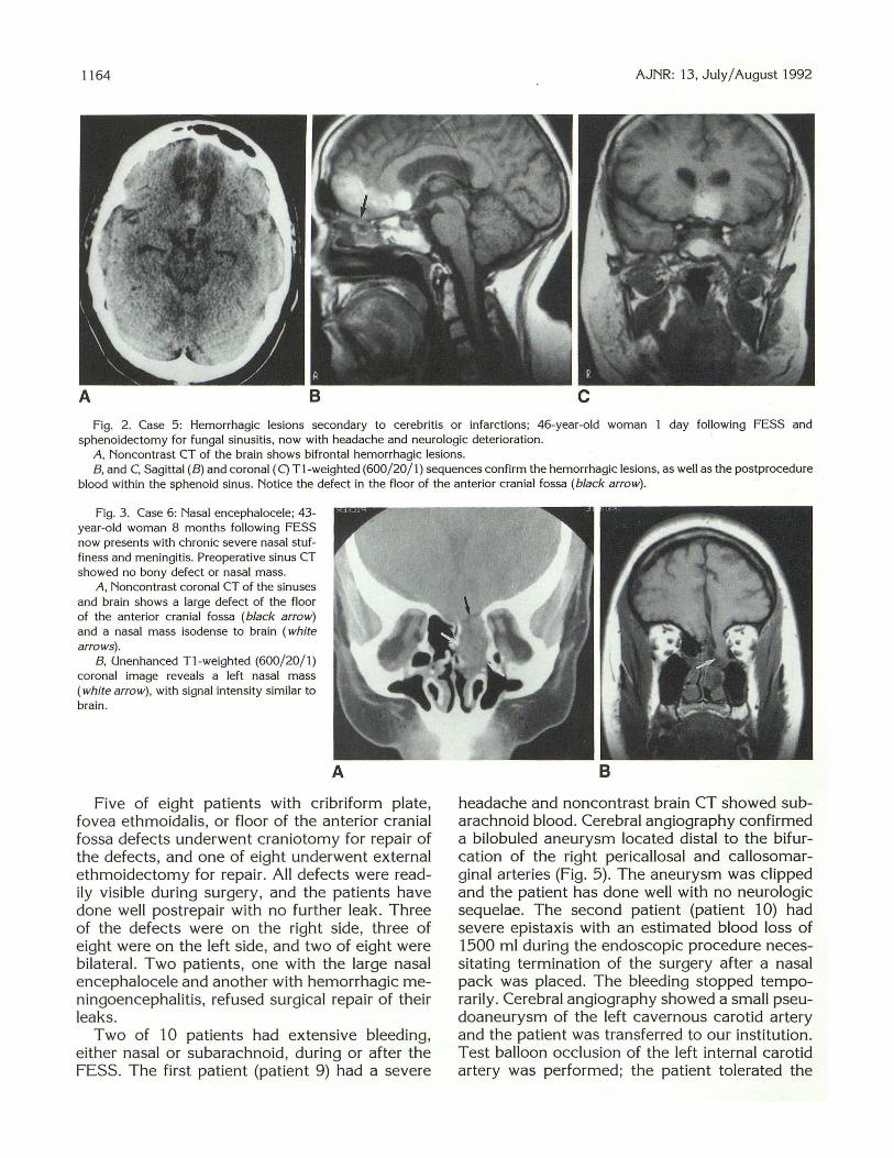
Five of eight who had findings suspicious for defects in the skull base underwent thin-section coronal sinus CT scans and a CT -cisternogram. The site of bony dehiscence was seen on the precontrast scan in all five patients (Fig. 1 ). Contrast within a sinus, best seen on magnified images photographed at bone-window technique, confirmed the site of leak. A sing.le patient with CSF leak (patient 5) had preoperative studies suggesting fungal sphenoid sinusitis with associated marked thinning of the walls of the sinus, and cultures obtained during surgery confirmed a Bipolaris species. The immediate postoperative course was complicated by headaches, nausea, fever, and a change in mentation. Both CT and MR revealed large bifrontal hemorrhagic lesions consistent with either hemorrhagic strokes or cerebritis (Fig. 2). The patient refused angiography.
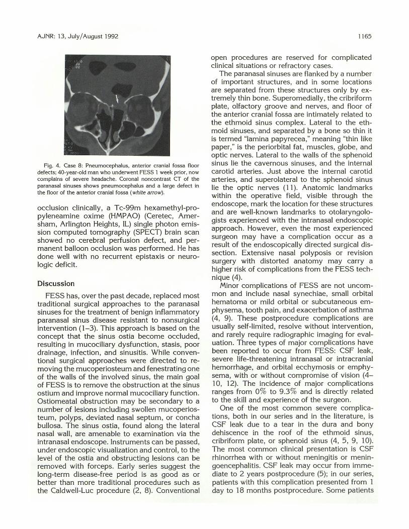
One of eight patients who presented with meningitis also reported severe nasal stuffiness (patient 6). A defect in the floor of the anterior
1163
Fig. 1. Case 4: CSF leak , fovea ethmoidalis defect; 47-yearold woman 18 months following FESS for chronic sinusitis presents with uncomplicated rhinorrhea. Coronal CT photographed at bone window detail , confirms dehiscent fovea ethmoidalis (white arrow). Notice the bilateral maxillary antrostomies.
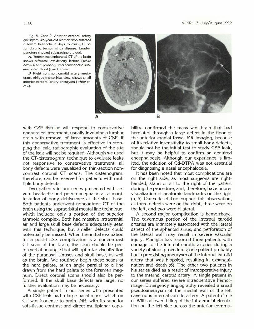
cranial fossa and a nasal encephalocele (Fig. 3) were seen on CT and MR. Two of eight patients with skull base defects presented with severe headache without meningitis (patients 7 and 8), and both had CT or plain radiographs that revealed massive intracranial air (Fig. 4).
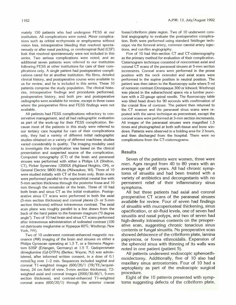
TABLE 1: Clinical findings for 10 patients with complications after undergoing FESS
Patient Complication FESS Procedure Clinical Presentation Time since Work-up (CT. Cgrm,
Findings Surgery MR, Angio)
Treatment
CSF leak Sph-eth. Rt max antra, Meningitis 15 days Cgrm Bony defect, Rt crib Craniotomy, Rt defect
septoplasty plate, contrast in Rt repair
ethmoid
2 CSF leak Sph-eth , max antra Rhinorrhea 4 mo Cgrm Bony defect. Rt crib Ext. ethmoid, Rt defect
plate, contrast in Rt repair
ethmoid
3 CSF leak Sph-eth Rhinorrhea I mo Cgrm Bony defect, Rt crib Craniotomy, Rt defect
plate, contrast in Rt repair
ethmoid
4 CSF leak Sph-eth, septoplasty Rhinorrhea 18 mo Cgrm Bony defect, Lt crib Craniotomy, Lt defect re-
plate, contrast in Lt pair
ethmoid
5 CSF leak Sph-eth, sphenoidectomy Meningoencephalitis 2 days Brain CT, MR, Bifrontal hemorrhagic Antifungal therapy
Hemorrhagic cerebritis Fungal sinusitis Cgrm lesions Refused surgery to repair
Bony defects, bil crib defects
plate
6 CSF leak Sph-eth, max antra Meningitis 8 mo CT, MR of brain Bony defect, Lt crib Refused surgery
Nasal encephalocele and sinuses plate
Encephalocele
7 Tension pneumocephalus Sph-eth, max antra Severe headache I day Brain CT Tens ion pneumoceph- Craniotomy, Lt crib de-
a Ius feet repair
8 Pneumocephalus Sph-eth, septoplasty Severe headache I wk Brain , sinus CT Pneumocephalus, air in Craniotomy, bil defects
ventricles, bil crib repair
defects
9 Aneurysm, ACA Sph-eth, max antra Subarachnoid hem- 5 days Brain CT, Anglo Subarachnoid blood Craniotomy, aneurysm
orrhage Aneurysm cl ipped
10 Aneurysm, Lt ICA Sph-eth, septoplasty Severe epistaxis Intraoperative Brain, sinus CT, Ai r-fluid level , Lt sphe- Permanent balloon oc-
Anglo noid sinus; Lt JCA elusion
aneurysm
Note-Cgrm, cisternogram; Anglo, cerebral angiogram; Sph-eth, sphenoethmoidectomy; antra, maxillary antrostomy; crib, cribriform; Lt, left; Rt, right; Ext. ethmoid,
external ethmoidectomy; ACA, anterior cerebral artery; ICA, internal carotid artery; bil , bilateral.
1164 AJNR: 13, July/ August 1992
A 8 c Fig. 2. Case 5: Hemorrhagic lesions secondary to cerebritis or infarctions; 46-year-old woman 1 day following FESS and
sphenoidectomy for fungal sinusitis, now with headache and neurologic deterioration. A, Noncontrast CT of the brain shows bifrontal hemorrhagic lesions. B, and C, Sagittal (B) and coronal (C) T1-weighted (600/20/1) sequences confirm the hemorrhagic lesions, as well as the postprocedure
blood within the sphenoid sinus. Notice the defect in the floor of the anterior cranial fossa (black arrow).
Fig. 3. Case 6: Nasal encephalocele; 43-year-old woman 8 months following FESS now presents with chronic severe nasal stuffiness and meningitis. Preoperative sinus CT showed no bony defect or nasal mass.
A, Noncontrast coronal CT of the sinuses and brain shows a large defect of the floor of the anterior cranial fossa (black arrow) and a nasal mass isodense to brain (white arrows).
B, Unenhanced T1-weighted (600/20/1) coronal image reveals a left nasal mass (white arrow), with signal intensity similar to brain.
A
Five of eight patients with cribriform plate, fovea ethmoidalis, or floor of the anterior cranial fossa defects underwent craniotomy for repair of the defects, and one of eight underwent external ethmoidectomy for repair. All defects were readily visible during surgery, and the patients have done well postrepair with no further leak. Three of the defects were on the right side, three of eight were on the left side, and two of eight were bilateral. Two patients, one with the large nasal encephalocele and another with hemorrhagic meningoencephalitis, refused surgical repair of their leaks.
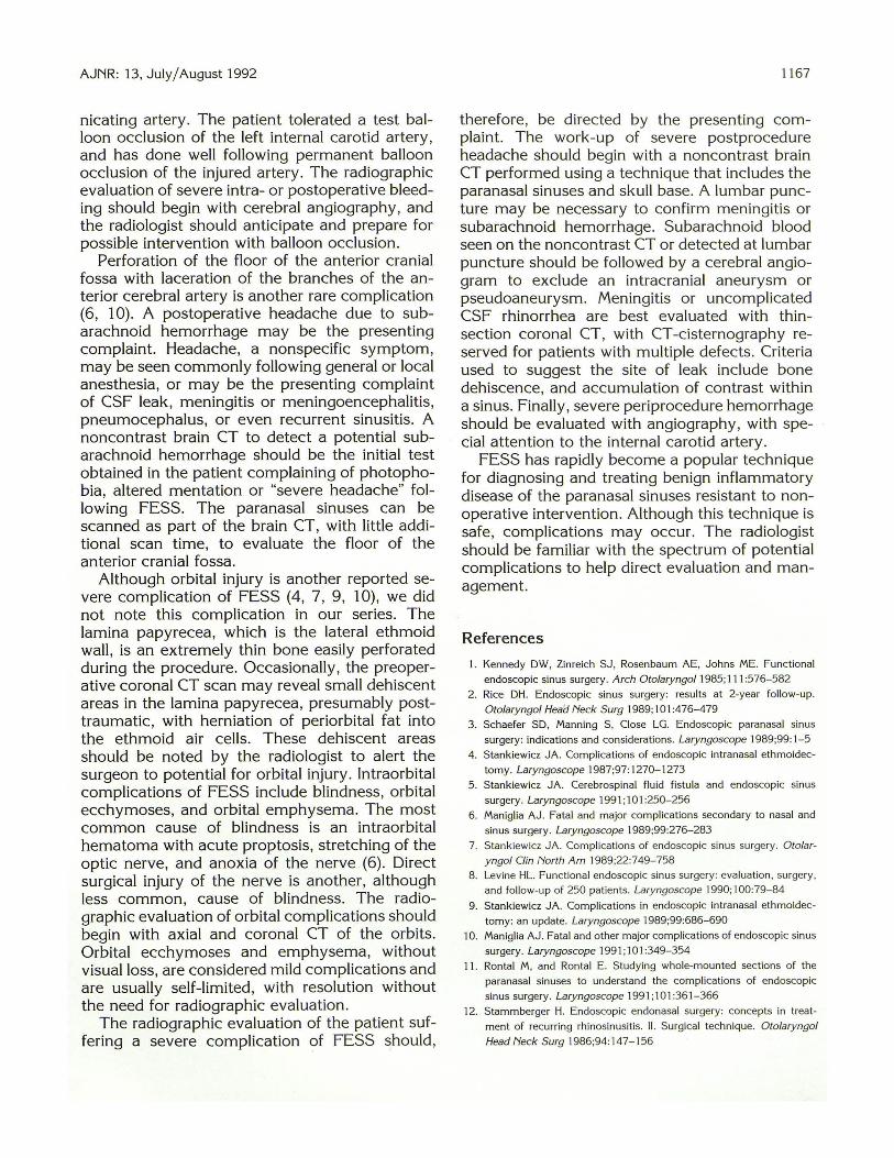
Two of 10 patients had extensive bleeding, either nasal or subarachnoid, during or after the FESS. The first patient (patient 9) had a severe
8
headache and noncontrast brain CT showed subarachnoid blood. Cerebral angiography confirmed a bilobuled aneurysm located distal to the bifurcation of the right pericallosal and callosomarginal arteries (Fig. 5). The aneurysm was clipped and the patient has done well with no neurologic sequelae. The second patient (patient 1 0) had severe epistaxis with an estimated blood loss of 1500 ml during the endoscopic procedure necessitating termination of the surgery after a nasal pack was placed. The bleeding stopped temporarily. Cerebral angiography showed a small pseudoaneurysm of the left cavernous carotid artery and the patient was transferred to our institution. Test balloon occlusion of the left internal carotid artery was performed; the patient tolerated the
AJNR: 13, July/August 1992
Fig. 4. Case 8: Pneumocephalus, anterior cranial fossa floor defects; 40-year-old man who underwent FESS 1 week prior, now complains of severe headache. Coronal noncontrast CT of the paranasal sinuses shows pneumocephalus and a large defect in the floor of the anterior cranial fossa (white arrow).
occlusion clinically, a Tc-99m hexamethyl-propyleneamine oxime (HMPAO) (Ceretec, Amersham, Arlington Heights, IL) single photon emission computed tomography (SPECT) brain scan showed no cerebral perfusion defect, and permanent balloon occlusion was performed. He has done well with no recurrent epistaxis or neurologic deficit.
Discussion
FESS has, over the past decade, replaced most traditional surgical approaches to the paranasal sinuses for the treatment of benign inflammatory paranasal sinus disease resistant to nonsurgical intervention (1-3). This approach is based on the concept that the sinus ostia become occluded, resulting in mucociliary dysfunction, stasis, poor drainage, infection, and sinusitis. While conventional surgical approaches were directed to removing the mucoperiosteum and fenestrating one of the walls of the involved sinus, the main goal of FESS is to remove the obstruction at the sinus ostium and improve normal mucociliary function. Ostiomeatal obstruction may be secondary to a number of lesions including swollen mucoperiosteum, polyps, deviated nasal septum, or concha bullosa. The sinus ostia, found along the lateral nasal wall , are amenable to examination via the intranasal endoscope. Instruments can be passed, under endoscopic visualization and control , to the level of the ostia and obstructing lesions can be removed with forceps . Early series suggest the long-term disease-free period is as good as or better than more traditional procedures such as the Caldwell-Luc procedure (2, 8). Conventional
1165
open procedures are reserved for complicated clinical situations or refractory cases.
The paranasal sinuses are flanked by a number of important structures, and in some locations are separated from these structures only by extremely thin bone. Superomedially, the cribriform plate, olfactory groove and nerves, and floor of the anterior cranial fossa are intimately related to the ethmoid sinus complex. Lateral to the ethmoid sinuses, and separated by a bone so thin it is termed "lamina papyrecea ," meaning "thin like paper," is the periorbital fat, muscles, globe, and optic nerves. Lateral to the walls of the sphenoid sinus lie the cavernous sinuses, and the internal carotid arteries. Just above the internal carotid arteries, and superolateral to the sphenoid sinus lie the optic nerves (11) . Anatomic landmarks within the operative field, visible through the endoscope, mark the location for these structures and are well-known landmarks to otolaryngologists experienced with the intranasal endoscopic approach. However, even the most experienced surgeon may have a complication occur as a result of the endoscopically directed surgical dissection. Extensive nasal polyposis or revision surgery with distorted anatomy may carry a higher risk of complications from the FESS technique (4).
Minor complications of FESS are not uncommon and include nasal synechiae, small orbital hematoma or mild orbital or subcutaneous emphysema, tooth pain, and exacerbation of asthma (4, 9). These postprocedure complications are usually self-limited, resolve without intervention, and rarely require radiographic imaging for evaluation. Three types of major complications have been reported to occur from FESS: CSF leak, severe life-threatening intranasal or intracranial hemorrhage, and orbital ecchymosis or emphysema, with or without compromise of vision (4-10, 12). The incidence of major complications ranges from 0% to 9.3 % and is directly related to the skill and experience of the surgeon.
One of the most common severe complications, both in our series and in the literature, is CSF leak due to a tear in the dura and bony dehiscence in the roof of the ethmoid sinus, cribriform plate, or sphenoid sinus (4, 5, 9, 10). The most common clinical presentation is CSF rhinorrhea with or without meningitis or meningoencephalitis. CSF leak may occur from immediate to 2 years postprocedure (5); in our series, patients with this complication presented from 1 day to 18 months postprocedure. Some patients
1166
Fig . 5. Case 9: Anterior cerebral artery cmeurysm; 45-year-old woman who suffered a severe headache 5 days following FESS for chronic benign sinus disease. Lumbar puncture showed subarachnoid blood.
A, Non contrast enhanced CT of the brain shows bifrontal low-density lesions (white arrows) and probably interhemispheric subarachnoid blood (black arrow).
B, Right common carotid artery angiogram, oblique transorbital view, shows small anterior cerebral artery aneurysm (white arrow).
A
with CSF fistulae will respond to conservative nonsurgical treatment, usually involving a lumbar drain with removal of large amounts of CSF. If this conservative treatment is effective in stopping the leak, radiographic evaluation of the site of the leak will not be required. Although we used the CT -cisternogram technique to evaluate leaks not responsive to conservative treatment, all bony defects were visualized on thin-section noncontrast coronal CT scans. The cisternogram, therefore, can be reserved for patients with multiple bony defects.
Two patients in our series presented with severe headache and pneumocephalus as a manifestation of bony dehiscence at the skull base. Both patients underwent noncontrast CT of the brain using the supraorbital meatal line technique, which included only a portion of the superior ethmoid complex . Both had massive intracranial air and large skull base defects adequately seen with this technique, but smaller defects could potentially be missed. When the initial evaluation for a post-FESS complication is a noncontrast CT scan of the brain, the scan should be performed at an angle that will optimize visualization of the paranasal sinuses and skull base, as well as the brain. We routinely begin these scans at the hard palate , at an angle parallel to a line drawn from the hard palate to the foramen magnum. Direct coronal scans should also be performed. If the skull base defects are large, no further evaluation may be necessary.
A single patient in our series who presented with CSF leak had a large nasal mass, which on CT was isodense to brain. MR, with its superior soft-tissue contrast and direct multiplanar capa-
AJNR: 13, July/ August 1992
bility, confirmed the mass was brain that had herniated through a large defect in the floor of the anterior cranial fossa. MR imaging, because of its relative insensitivity to small bony defects, should not be the initial test to study CSF leak, but it may be helpful to confirm an acquired encephalocele. Although our experience is limited, the addition of Gd-DTPA was not essential for diagnosing a nasal encephalocele.
It has been noted that most complications are on the right side, as most surgeons are righthanded, stand or sit to the right of the patient during the procedure, and , therefore, have poorer visualization of anatomic landmarks on the right (5 , 6). Our series did not support this observation, as three defects were on the right, three were on the left, and two were bilateral.
A second major complication is hemorrhage. The cavernous portion of the internal carotid arteries are intimately associated with the lateral aspect of the sphenoid sinus, and perforation of the lateral wall may result in severe vascular injury. Maniglia has reported three patients with damage to the internal carotid arteries during a variety of sinus procedures; one patient probably had a preexisting aneurysm of the internal carotid artery that was biopsied, resulting in exsanguination and death (6). The other two patients in his series died as a result of intraoperative injury to the internal carotid artery. A single patient in our series suffered severe intraoperative hemorrhage . Emergency angiography revealed a small pseudoaneurysm of the medial wall of the left cavernous internal carotid artery. A patent circle of Willis allowed filling of the intracranial circulation on the left side across the anterior commu-
AJNR: 13, July/August 1992
nicating artery. The patient tolerated a test balloon occlusion of the left internal carotid artery, and has done well following permanent balloon occlusion of the injured artery. The radiographic evaluation of severe intra- or postoperative bleeding should begin with cerebral angiography, and the radiologist should anticipate and prepare for possible intervention with balloon occlusion.
Perforation of the floor of the anterior cranial fossa with laceration of the branches of the anterior cerebral artery is another rare complication (6, 1 0). A postoperative headache due to subarachnoid hemorrhage may be the presenting complaint. Headache, a nonspecific symptom, may be seen commonly following general or local anesthesia, or may be the presenting complaint of CSF leak, meningitis or meningoencephalitis, pneumocephalus, or even recurrent sinusitis. A noncontrast brain CT to detect a potential subarachnoid hemorrhage should be the initial test obtained in the patient complaining of photophobia, altered mentation or "severe headache" following FESS. The paranasal sinuses can be scanned as part of the brain CT, with little additional scan time, to evaluate the floor of the anterior cranial fossa.
Although orbital injury is another reported severe complication of FESS (4, 7, 9, 10), we did not note this complication in our series. The lamina papyrecea, which is the lateral ethmoid wall, is an extremely thin bone easily perforated during the procedure. Occasionally, the preoperative coronal CT scan may reveal small dehiscent areas in the lamina papyrecea, presumably posttraumatic, with herniation of periorbital fat into the ethmoid air cells. These dehiscent areas should be noted by the radiologist to alert the surgeon to potential for orbital injury. Intraorbital complications of FESS include blindness, orbital ecchymoses, and orbital emphysema. The most common cause of blindness is an intraorbital hematoma with acute proptosis, stretching of the optic nerve, and anoxia of the nerve (6). Direct surgical injury of the nerve is another, although less common, cause of blindness. The radiographic evaluation of orbital complications should begin with axial and coronal CT of the orbits. Orbital ecchymoses and emphysema, without visual loss, are considered mild complications and are usually self-limited, with resolution without the need for radiographic evaluation.
The radiographic evaluation of the patient suffering a severe complication of FESS should,
1167
therefore, be directed by the presenting complaint. The work-up of severe postprocedure headache should begin with a noncontrast brain CT performed using a technique that includes the paranasal sinuses and skull base. A lumbar puncture may be necessary to confirm meningitis or subarachnoid hemorrhage. Subarachnoid blood seen on the noncontrast CT or detected at lumbar puncture should be followed by a cerebral angiogram to exclude an intracranial aneurysm or pseudoaneurysm. Meningitis or uncomplicated CSF rhinorrhea are best evaluated with thinsection coronal CT, with CT -cisternography reserved for patients with multiple defects . Criteria used to suggest the site of leak include bone dehiscence, and accumulation of contrast within a sinus. Finally, severe peri procedure hemorrhage should be evaluated with angiography, with special attention to the internal carotid artery .
FESS has rapidly become a popular technique for diagnosing and treating benign inflammatory disease of the paranasal sinuses resistant to nonoperative intervention. Although this technique is safe, complications may occur. The radiologist should be familiar with the spectrum of potential complications to help direct evaluation and management.
References
1. Kennedy DW, Zinreich SJ, Rosenbaum AE, Johns ME. Functional
endoscopic sinus surgery . Arch Oto/ary ngo/1985; 111 :576-582
2. Rice DH. Endoscopic sinus surgery : results at 2-year follow-up.
Otolaryngol Hea'd Neck Surg 1989; 1 01 :476-479
3. Schaefer SD, Manning S, Close LG. Endoscopic paranasal sinus
surgery: indications and considerations. Lary ngoscope 1989;99: 1-5
4. Stankiewicz JA. Complications of endoscopic intranasal ethmoidec
tomy. Lary ngoscope 1987 ;97: 1270-1 273
5. Stankiewicz JA. Cerebrospinal fluid fistula and endoscopic sinus
surgery. Lary ngoscope 199 1;101:250-256
6. Maniglia AJ . Fatal and major complications secondary to nasal and
sinus surgery . Lary ngoscope 1989;99:276-283
7. Stankiewicz JA. Complica tions of endoscopic sinus surgery . Oto/ar
y ngol Clin North A m 1989;22: 7 49- 758
8. Levine HL. Functional endoscopic sinus surgery : evaluation, surgery,
and follow-up of 250 patients . Lary ngoscope 1990; 100:79-84
9. Stankiewicz JA. Complications in endoscopic intranasal ethmoidec
tomy: an update. Lary ngoscope 1989;99:686-690
10. Maniglia AJ . Fatal and other major complications of endoscopic sinus
surgery. Lary ngoscope 1991 ; 1 01 :349- 354
11 . Rontal M, and Rontal E. Study ing whole-mounted sections of the
paranasal sinuses to understand the complications of endoscopic
sinus surgery. Lary ngoscope 1991;101:361-366
12. Stammberger H. Endoscopic endonasal surgery: concepts in treat
ment of recurring rhinosinusitis. II . Surgical technique. Otolary ngol
Head Neck Surg 1986;94:147-156