
Endoscopic closure devices - ASGE ON EMERGING TECHNOLOGY Endoscopic closure devices The ASGE...
8
REPORT ON EMERGING TECHNOLOGY Endoscopic closure devices The ASGE Technology Committee provides reviews of new or emerging endoscopic technologies that have the potential to affect the practice of GI endoscopy. Evidence- based methodology is used, with a MEDLINE literature search to identify pertinent preclinical and clinical studies on the topic and a MAUDE (U.S. Food and Drug Admin- istration Center for Devices and Radiological Health) da- tabase search to identify the reported complications of a given technology. Both are supplemented by accessing the “related articles” feature of PubMed and by scrutinizing pertinent references cited by the identified studies. Con- trolled clinical trials are emphasized, but in many cases, data from randomized, controlled trials are lacking. In such cases, large case series, preliminary clinical studies, and expert opinions are used. Technical data are gathered from traditional and Web-based publications, proprietary publications, and informal communications with perti- nent vendors. For this review, the MEDLINE database was searched through October 2011 using the keywords “en- terotomy,” “gastrotomy,” “colostomy,” “perforation,” “fis- tula,” “natural orifice transluminal endoscopic surgery,” “closure,” “endoscopic suturing,” “endoscopic clipping,” and “placating.” Reports on Emerging Technologies are drafted by 1 or 2 members of the ASGE Technology Committee, reviewed and edited by the committee as a whole, and approved by the Governing Board of the ASGE. These reports are scien- tific reviews provided solely for educational and informa- tional purposes. Reports on Emerging Technologies are not rules and should not be construed as establishing a legal standard of care or as encouraging, advocating, requir- ing, or discouraging any particular treatment or payment for such treatment. BACKGROUND Nonsurgical closure of the GI wall may be desired in the setting of inflammatory or neoplastic fistulae, dehis- cence of surgical anastomoses, and spontaneous or iatro- genic perforations. Closure is also necessary after natural orifice translumenal endoscopic surgery (NOTES ® ). Sev- eral devices and techniques are being developed to allow endoscopic closure of these GI wall defects. The ideal closure device should be inexpensive, safe, and easy to use. It should provide rapid, reliable closure, which is both robust and durable, and should also be effective in the closure of larger defects. Among the first endoscopic devices used for the closure of small perforations were endoscopic clips. 1 Clips were discussed in a previous technology committee document. 2 Although endoscopic clips may provide an adequate clo- sure solution for small defects, they are less useful for larger defects because of the restricted opening distance between their jaws, low closure force, and inability to accomplish deep-tissue capture. These deficiencies are particularly troublesome when trying to use endoscopic clips in the setting of the inflamed, indurated, and fibrotic tissue associated with chronic fistulae. Case reports and small case series also describe successful closure of per- forations, fistulae, and anastomotic leaks by using tissue glues and covered self-expandable metal stents. 3-5 How- ever, these too provide an inadequate and unreliable so- lution. An unmet need has therefore persisted for a reliable and robust endoscopic solution for the closure of mural defects, both spontaneous and iatrogenic. With the devel- opment of NOTES as a new and potentially viable surgical platform, there has been an increasing interest and imme- diacy in the need to develop dedicated devices and tech- niques that allow closure of enterotomies. Although many endoscopic closure devices have undergone testing and evaluation in bench and animal models, only a few have been used in human subjects and only 2 are being actively marketed in the United States at this time. EMERGING TECHNOLOGY Over-the-scope clip An over-the-scope clip (OTSC) (Ovesco, Tübingen, Germany) has been developed for the closure of small mural defects and bleeding ulcers. 6 In animal models, reliable full-thickness closure of defects of as large as 27 mm was achieved with this device. 7 The OTSC produces more durable closure than standard endoclips 8 because of its ability to grasp more tissue, include the entire thickness of the visceral wall, and apply a greater compressive force. The OTSC received Conformite Europeene certification in Europe in 2009 and 510(k) clearance by the U.S. Food and Drug Administration (FDA) in 2010. Description of the device. The device includes an applicator cap, a nitinol clip, and a hand wheel (Fig. 1). Copyright © 2012 by the American Society for Gastrointestinal Endoscopy 0016-5107/$36.00 doi:10.1016/j.gie.2012.02.028 244 GASTROINTESTINAL ENDOSCOPY Volume 76, No. 2 : 2012 www.giejournal.org
Transcript of Endoscopic closure devices - ASGE ON EMERGING TECHNOLOGY Endoscopic closure devices The ASGE...
e
REPORT ON EMERGING TECHNOLOGY
Endoscopic closure devices
ecurc
odAslbapctsfgeladopdneebm
E
O
GmrmmioTED
The ASGE Technology Committee provides reviews ofnew or emerging endoscopic technologies that have thepotential to affect the practice of GI endoscopy. Evidence-based methodology is used, with a MEDLINE literaturesearch to identify pertinent preclinical and clinical studieson the topic and a MAUDE (U.S. Food and Drug Admin-istration Center for Devices and Radiological Health) da-tabase search to identify the reported complications of agiven technology. Both are supplemented by accessing the“related articles” feature of PubMed and by scrutinizingpertinent references cited by the identified studies. Con-trolled clinical trials are emphasized, but in many cases,data from randomized, controlled trials are lacking. Insuch cases, large case series, preliminary clinical studies,and expert opinions are used. Technical data are gatheredfrom traditional and Web-based publications, proprietarypublications, and informal communications with perti-nent vendors. For this review, the MEDLINE database wassearched through October 2011 using the keywords “en-terotomy,” “gastrotomy,” “colostomy,” “perforation,” “fis-tula,” “natural orifice transluminal endoscopic surgery,”“closure,” “endoscopic suturing,” “endoscopic clipping,”and “placating.”
Reports on Emerging Technologies are drafted by 1 or 2members of the ASGE Technology Committee, reviewedand edited by the committee as a whole, and approved bythe Governing Board of the ASGE. These reports are scien-tific reviews provided solely for educational and informa-tional purposes. Reports on Emerging Technologies are notrules and should not be construed as establishing a legalstandard of care or as encouraging, advocating, requir-ing, or discouraging any particular treatment or paymentfor such treatment.
BACKGROUND
Nonsurgical closure of the GI wall may be desired inthe setting of inflammatory or neoplastic fistulae, dehis-cence of surgical anastomoses, and spontaneous or iatro-genic perforations. Closure is also necessary after naturalorifice translumenal endoscopic surgery (NOTES®). Sev-ral devices and techniques are being developed to allow
Copyright © 2012 by the American Society for Gastrointestinal Endoscopy0016-5107/$36.00doi:10.1016/j.gie.2012.02.028
a
244 GASTROINTESTINAL ENDOSCOPY Volume 76, No. 2 : 2012
ndoscopic closure of these GI wall defects. The ideallosure device should be inexpensive, safe, and easy tose. It should provide rapid, reliable closure, which is bothobust and durable, and should also be effective in thelosure of larger defects.
Among the first endoscopic devices used for the closuref small perforations were endoscopic clips.1 Clips wereiscussed in a previous technology committee document.2
lthough endoscopic clips may provide an adequate clo-ure solution for small defects, they are less useful forarger defects because of the restricted opening distanceetween their jaws, low closure force, and inability toccomplish deep-tissue capture. These deficiencies arearticularly troublesome when trying to use endoscopiclips in the setting of the inflamed, indurated, and fibroticissue associated with chronic fistulae. Case reports andmall case series also describe successful closure of per-orations, fistulae, and anastomotic leaks by using tissuelues and covered self-expandable metal stents.3-5 How-ver, these too provide an inadequate and unreliable so-ution. An unmet need has therefore persisted for a reliablend robust endoscopic solution for the closure of muralefects, both spontaneous and iatrogenic. With the devel-pment of NOTES as a new and potentially viable surgicallatform, there has been an increasing interest and imme-iacy in the need to develop dedicated devices and tech-iques that allow closure of enterotomies. Although manyndoscopic closure devices have undergone testing andvaluation in bench and animal models, only a few haveeen used in human subjects and only 2 are being activelyarketed in the United States at this time.
MERGING TECHNOLOGY
ver-the-scope clipAn over-the-scope clip (OTSC) (Ovesco, Tübingen,
ermany) has been developed for the closure of smallural defects and bleeding ulcers.6 In animal models,
eliable full-thickness closure of defects of as large as 27m was achieved with this device.7 The OTSC producesore durable closure than standard endoclips8 because of
ts ability to grasp more tissue, include the entire thicknessf the visceral wall, and apply a greater compressive force.he OTSC received Conformite Europeene certification inurope in 2009 and 510(k) clearance by the U.S. Food andrug Administration (FDA) in 2010.Description of the device. The device includes an
pplicator cap, a nitinol clip, and a hand wheel (Fig. 1).
www.giejournal.org
ps
woocsmiapcrLi
a
cci
gtcawt2smsiattsb
csa3tciraO
O
dd
Endoscopic closure devices
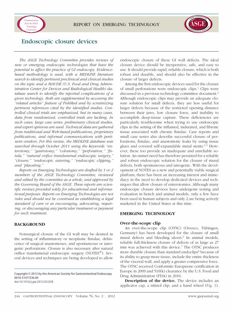
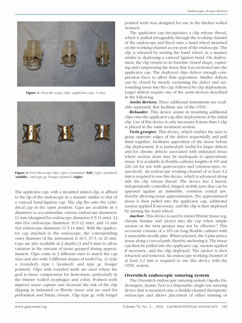
The applicator cap, with a mounted nitinol clip, is affixedto the tip of the endoscope in a manner similar to that ofa variceal band-ligation cap. The clip fits onto the cylin-drical cap in the open position. Caps are available in 3diameters to accommodate various endoscope diameters:11 mm (designed for endoscope diameters 9.5-11 mm), 12mm (for endoscope diameters 10.5-12 mm), and 14 mm(for endoscope diameters 11.5-14 mm). With the applica-tor cap attached to the endoscope, the correspondingouter diameter of the instrument is 16.5, 17.5, or 21 mm.Caps are also available in 2 depths (3 and 6 mm) to allowvariation in the amount of tissue grasped during approx-imation. Clips come in 3 different sizes to match the capsizes and also with 3 different shapes of teeth (Fig. 2): typea (rounded), type t (pointed), and type gc (longerpointed). Clips with rounded teeth are used where thegoal is tissue compression for hemostasis, particularly inthe thinner walled esophagus and colon. Pointed teethimprove tissue capture and decrease the risk of the clipslipping in indurated or fibrotic tissue and are used for
Figure 1. Over-the-scope clips: applicator caps; 3 sizes.
Figure 2. Over-the-scope clips: type a (rounded) (left), type t (pointed)(middle), and type gc (longer pointed) (right).
perforation and fistula closure. Clip type gc with longer e
www.giejournal.org V
ointed teeth was designed for use in the thicker walledtomach.
The applicator cap incorporates a clip release thread,hich is pulled retrogradely through the working channelf the endoscope and fixed onto a hand wheel mountedn the working-channel access port of the endoscope. Thelip is released by turning the hand wheel, in a mannerimilar to deploying a variceal ligation band. On deploy-ent, the clip returns to its baseline closed shape, captur-
ng and compressing the tissue that was suctioned into thepplicator cap. The deployed clips deliver enough com-ression force to allow firm apposition. Smaller defectsan be closed by merely suctioning the defect and sur-ounding tissue into the cap, followed by clip deployment.arger defects require one of the assist devices describedn the following.
Assist devices. Three additional instruments are avail-ble separately that facilitate use of the OTSC.
Reloader. This device assists in mounting additionallips onto the applicator cap after deployment of the initiallip. Use of this device is only necessary if more than 1 clips placed in the same treatment session.
Twin grasper. This device, which enables the user torasp opposite edges of the defect sequentially and pullhem together, facilitates apposition of the tissue beforelip deployment. It is particularly useful for larger defectsnd for chronic defects associated with indurated tissuehere suction alone may be inadequate to approximate
issue. It is available in flexible catheter lengths of 165 and20 cm for use with gastroscopes and colonoscopes, re-pectively. An endoscope working channel of at least 3.2m is required to use this device, which is advanced along
ide the clip release thread. The device has 2 lateral,ndependently controlled, hinged, mobile jaws that can bepposed against an immobile, common central jaw,hereby allowing tissue approximation. The approximatedissue is then pulled into the applicator cap, additionaluction applied if necessary, and the clip is then deployedy turning the hand wheel.
Anchor. This device is used to retract fibrotic tissue (eg,hronic fistulae and ulcers) into the cap when simpleuction or the twin grasper may not be effective.9 Thisccessory consists of a 165-cm long flexible catheter withretractable needle pins. When released, the 3 pins pierce
issue along a curved path, thereby anchoring it. The tissuean then be pulled into the applicator cap, suction appliedf necessary, and the clip deployed. The anchor is thenetracted and removed. An endoscope working channel oft least 3.2 mm is required to use this device with theTSC system.
verstitch endoscopic suturing systemThe Overstitch endoscopic suturing system (Apollo En-
osurgery, Austin, Tex) is a disposable, single-use suturingevice that is mounted onto a double-channel therapeutic
ndoscope and allows placement of either running orolume 76, No. 2 : 2012 GASTROINTESTINAL ENDOSCOPY 245
pdtwus
checchisaaaTt
pat
D
tdHsswcoetdstitwrcb
Ca
C
O
tmafpacgwrurfetc
saci
Endoscopic closure devices
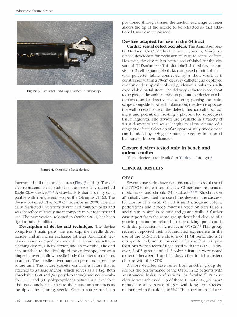
interrupted full-thickness sutures (Figs. 3 and 4). The de-vice represents an evolution of the previously describedEagle Claw device.10,11 A drawback is that it is only com-atible with a single endoscope, the Olympus 2T160. Theevice obtained FDA 510(k) clearance in 2008. The ini-ially marketed Overstitch device had multiple parts andas therefore relatively more complex to put together andse. The new version, released in October 2011, has beenignificantly simplified.
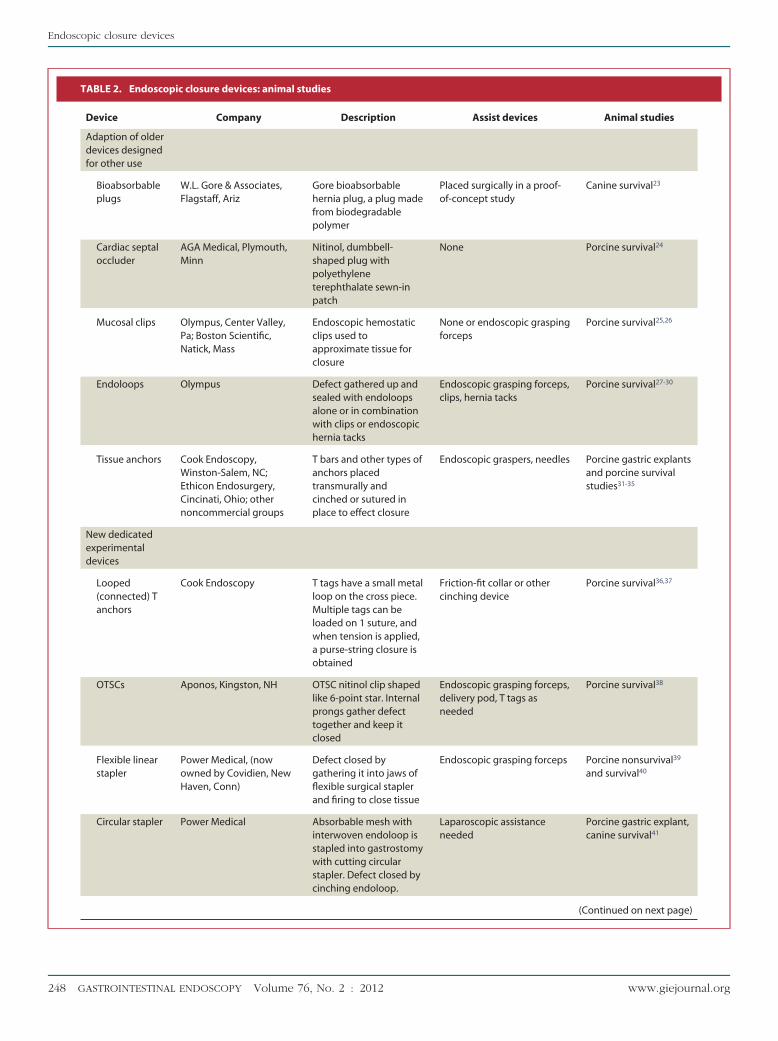
Description of device and technique. The deviceomprises 3 main parts: the end cap, the needle driverandle, and an anchor exchange catheter. Additional nec-ssary assist components include a suture cassette, ainching device, a helix device, and an overtube. The endap, attached to the distal tip of the endoscope, houses ainged, curved, hollow needle body that opens and closesn an arc. The needle driver handle opens and closes theuture arm. The suture cassette contains a suture that isttached to a tissue anchor, which serves as a T tag. Bothbsorbable (2-0 and 3-0 polydioxanone) and nonabsorb-ble (2-0 and 3-0 polypropylene) sutures are available.he tissue anchor attaches to the suture arm and acts as
Figure 3. Overstitch: end cap attached to endoscope.
Figure 4. Overstitch: helix device.
he tip of the suturing needle. Once a suture has been m
246 GASTROINTESTINAL ENDOSCOPY Volume 76, No. 2 : 2012
ositioned through tissue, the anchor exchange catheterllows the tip of the needle to be retracted so that addi-ional tissue can be pierced.
evices adapted for use in the GI tractCardiac septal defect occluders. The Amplatzer Sep-
al Occluder (AGA Medical Group, Plymouth, Minn) is aevice developed for occlusion of cardiac septal defects.owever, the device has been used off-label for the clo-
ure of GI fistulae.12-15 This dumbbell-shaped device con-ists of 2 self-expandable disks composed of nitinol meshith polyester fabric connected by a short waist. It isonstrained within a 70-cm delivery catheter and deployedver an endoscopically placed guidewire similar to a self-xpandable metal stent. The delivery catheter is too shorto be passed through an endoscope, but the device can beeployed under direct visualization by passing the endo-cope alongside it. After implantation, the device apposeshe wall on each side of the defect, mechanically occlud-ng it and potentially creating a platform for subsequentissue ingrowth. The devices are available in a variety ofaist diameters and waist lengths to allow closure of a
ange of defects. Selection of an appropriately sized devicean be aided by sizing the mural defect by inflation ofalloons of known diameter.
losure devices tested only in bench andnimal studiesThese devices are detailed in Tables 1 through 3.
LINICAL RESULTS
TSCSeveral case series have demonstrated successful use of
he OTSC in the closure of acute GI perforations, anasto-otic leaks, and chronic GI fistulae.6,9,50-55 Kirschniak et
l6 initially described the use of this device in the success-ul closure of 2 small (4 and 8 mm) iatrogenic colonicerforations and 2 deep mucosal resection sites (also 4nd 8 mm in size) in colonic and gastric walls. A furtherase report from the same group described closure of aastric perforation related to necrotizing pancreatitisith the placement of 2 adjacent OTSCs.56 This group
ecently reported their accumulated experience in these of the OTSC in the closure of 11 GI perforations (4etroperitoneal) and 8 chronic GI fistulae.54 All GI per-orations were successfully closed with the OTSC. How-ver, 2 of 5 gastric and all 3 colonic fistulae were notedo recur between 5 and 11 days after initial transientlosure with the OTSC.
A more detailed case series from another group de-cribes the performance of the OTSC in 12 patients withnastomotic leaks, perforations, or fistulae.57 Primarylosure was achieved in 9 of these 12 patients, giving anmmediate success rate of 75%, with long-term success
aintained in 8 patients (66%). The 4 treatment failureswww.giejournal.org

cge
gs
ipd
wstwpaos
ic
O
do(tab
C
oa
S
Ohtgwp
Endoscopic closure devices
included a patient in whom clip placement was notsuccessful because of the inability to approximate tis-sue, a patient with a chronic gastrocutaneous fistula inwhom the clip detached within 1 day of placement, and2 patients with chronic enterocutaneous fistulae forwhich the OTSC deployed, but only incomplete closurewas achieved. Successful closure was invariablyachieved (100%) if the defects were closed within aweek of diagnosis, compared with a 57% closure ratewhen defects were closed more than 4 weeks afterdiagnosis. Another European group described their ex-perience closing perforations and anastomotic leakswith the OTSC.55 Successful closure was achieved andsurgery avoided in 4 of 7 patients. In a further caseseries of patients with perforations, fistulae, and anas-tomotic leaks, successful closure was achieved in 8 of 10patients.52 Although the OTSC is cleared by the FDA forlosure of defects as large as 20 mm, closure of largerastric defects by using 2 clips deployed adjacent toach other has been reported.56,57
The induration and fibrosis associated with chronicfistulae may result in failure of adequate tissue appositionand a consequent inability to deploy the OTSC satisfacto-rily.9 This may result in challenging, protracted proce-dures. In a recent small case series, OTSC placement wassuccessful in only 2 of 4 patients, despite procedure timesranging from 24 to 93 minutes.9 Cauterization of the mar-ins of chronic fistulae has been advocated to facilitateubsequent closure with the OTSC.51
Initial results of an ongoing prospective multicenterEuropean study evaluating the performance of the OTSCin iatrogenic perforations have been published in abstractform.58 OTSC placement was attempted in 14 patients withatrogenic GI tract perforation in a variety of locations. Therocedure was abandoned in 1 patient because of a
TABLE 1. Endoscopic closure techniques
Technique Description
Trial of no closure of defect Gastric puncture dilated with balBecause there is dilation only, thmuscularis approximates edges acloses defect.
Self-approximating tunnel/flap
Submucosal tunnel with exit sitedistant from mucosal entry site;mucosa closed with clips.
Gastropexy method Gastrotomy is pulled up to anterabdominal wall and sutured in pwith transmural T tags or othersutures.
Omentoplasty method Omental patch is pulled into gasdefect to plug hole, and is fixed iplace with clips.
evice-related esophageal mucosal laceration. The OTSC d
www.giejournal.org V
as successfully deployed in 13 patients. Closure wasuccessful and achieved rapidly, with a median closureime of 4 minutes, 30 seconds. Contrast studies performedithin 24 hours indicated no contrast extravasation in anyatient. Twelve patients did well and resumed an oral diett a median of 1 day post-procedure. One patient deteri-rated because of incomplete closure and died despiteubsequent surgery.
Overall, the initial data suggest that OTSCs are effectiven closing acute perforations and to a lesser extent in thelosure of chronic fistulae.
verstitchOnly a single case report has currently been published
ocumenting use of the Overstitch device for the closuref a refractory gastrocutaneous fistula.59 A small case series6 patients) published only in abstract form describes ini-ial closure of rectovaginal and gastromediastinal fistulaes well as gastrogastric fistulae after Roux-en-Y gastricypass; no long-term follow-up is available.60
ardiac septal defect occludersCase reports describe the use of cardiac septal defect
ccluders in the successful initial closure of tracheoesoph-geal, tracheogastric, and gastrocolonic fistulae.12-15
AFETY
A single perforation was reported after placement of theTSC for a bleeding duodenal ulcer.57 Mucosal lacerationsave been reported after advancement of the OTSChrough the upper esophageal sphincter and esopha-us.55,58 Device malfunction resulting in partial releaseith the clip stuck to the applicator cap has been re-orted.9 This report describes premature complete release
Assist devices Animal studies
None Porcine,16 canine17
survival
Endoscopic hemostatic clips Porcine gastric,18,19 andesophageal20 closure
Endoscopic graspingforceps, T tags
Porcine survival21
Endoscopic hemostatic clipsand forceps
Porcine survival22
loon.end
iorlace
tricn
uring a training session in an animal laboratory, resulting
olume 76, No. 2 : 2012 GASTROINTESTINAL ENDOSCOPY 247
Endoscopic closure devices
TABLE 2. Endoscopic closure devices: animal studies
Device Company Description Assist devices Animal studies
Adaption of olderdevices designedfor other use
Bioabsorbableplugs
W.L. Gore & Associates,Flagstaff, Ariz
Gore bioabsorbablehernia plug, a plug madefrom biodegradablepolymer
Placed surgically in a proof-of-concept study
Canine survival23
Cardiac septaloccluder
AGA Medical, Plymouth,Minn
Nitinol, dumbbell-shaped plug withpolyethyleneterephthalate sewn-inpatch
None Porcine survival24
Mucosal clips Olympus, Center Valley,Pa; Boston Scientific,Natick, Mass
Endoscopic hemostaticclips used toapproximate tissue forclosure
None or endoscopic graspingforceps
Porcine survival25,26
Endoloops Olympus Defect gathered up andsealed with endoloopsalone or in combinationwith clips or endoscopichernia tacks
Endoscopic grasping forceps,clips, hernia tacks
Porcine survival27-30
Tissue anchors Cook Endoscopy,Winston-Salem, NC;Ethicon Endosurgery,Cincinati, Ohio; othernoncommercial groups
T bars and other types ofanchors placedtransmurally andcinched or sutured inplace to effect closure
Endoscopic graspers, needles Porcine gastric explantsand porcine survivalstudies31-35
New dedicatedexperimentaldevices
Looped(connected) Tanchors
Cook Endoscopy T tags have a small metalloop on the cross piece.Multiple tags can beloaded on 1 suture, andwhen tension is applied,a purse-string closure isobtained
Friction-fit collar or othercinching device
Porcine survival36,37
OTSCs Aponos, Kingston, NH OTSC nitinol clip shapedlike 6-point star. Internalprongs gather defecttogether and keep itclosed
Endoscopic grasping forceps,delivery pod, T tags asneeded
Porcine survival38
Flexible linearstapler
Power Medical, (nowowned by Covidien, NewHaven, Conn)
Defect closed bygathering it into jaws offlexible surgical staplerand firing to close tissue
Endoscopic grasping forceps Porcine nonsurvival39
and survival40
Circular stapler Power Medical Absorbable mesh withinterwoven endoloop isstapled into gastrostomywith cutting circularstapler. Defect closed bycinching endoloop.
Laparoscopic assistanceneeded
Porcine gastric explant,canine survival41
(Continued on next page)
248 GASTROINTESTINAL ENDOSCOPY Volume 76, No. 2 : 2012 www.giejournal.org
ttP
tthpawotem
l.
Endoscopic closure devices
in the OTSC inadvertently attaching a twin grasper deviceto the GI wall, tethering the endoscope. Clips that arepartially dislodged over the cap before deployment shouldtherefore be removed and replaced; care should also betaken to avoid inadvertent deployment of the clip over thetwin grasper.
Significant postprocedural pain was reported in a singlepatient after application of the OTSC.55 Clip detachmentwithin a day of successful placement was reported,57 andhis possibility should be considered in patients who de-eriorate clinically after initial successful clip deployment.
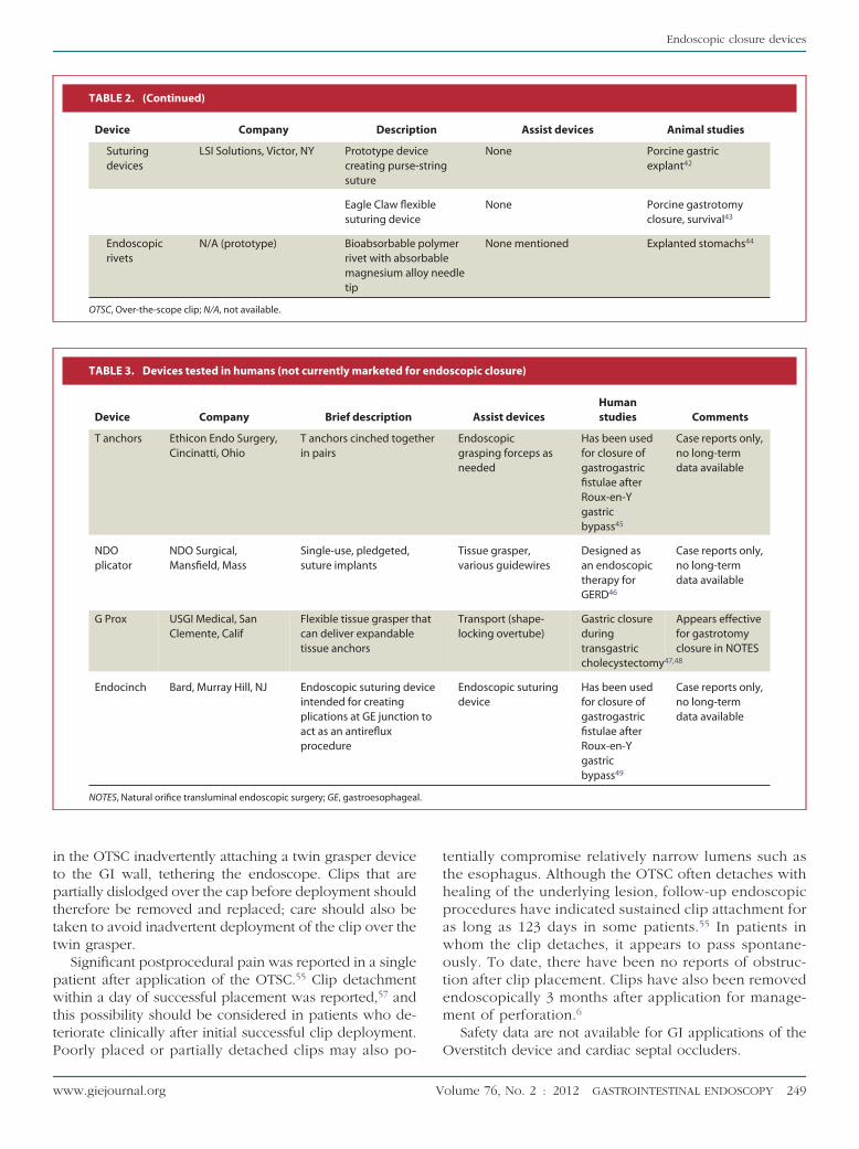
TABLE 2. (Continued)
Device Company Descript
Suturingdevices
LSI Solutions, Victor, NY Prototype devicreating purse-suture
Eagle Claw flexsuturing device
Endoscopicrivets
N/A (prototype) Bioabsorbablerivet with absomagnesium allotip
OTSC, Over-the-scope clip; N/A, not available.
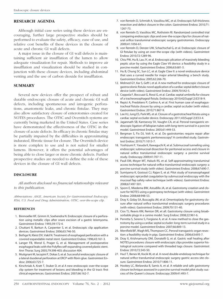
TABLE 3. Devices tested in humans (not currently marketed for
Device Company Brief description
T anchors Ethicon Endo Surgery,Cincinatti, Ohio
T anchors cinched togetin pairs
NDOplicator
NDO Surgical,Mansfield, Mass
Single-use, pledgeted,suture implants
G Prox USGI Medical, SanClemente, Calif
Flexible tissue grasper thcan deliver expandabletissue anchors
Endocinch Bard, Murray Hill, NJ Endoscopic suturing devintended for creatingplications at GE junctionact as an antirefluxprocedure
NOTES, Natural orifice transluminal endoscopic surgery; GE, gastroesophagea
oorly placed or partially detached clips may also po- O
www.giejournal.org V
entially compromise relatively narrow lumens such ashe esophagus. Although the OTSC often detaches withealing of the underlying lesion, follow-up endoscopicrocedures have indicated sustained clip attachment fors long as 123 days in some patients.55 In patients inhom the clip detaches, it appears to pass spontane-usly. To date, there have been no reports of obstruc-ion after clip placement. Clips have also been removedndoscopically 3 months after application for manage-ent of perforation.6
Safety data are not available for GI applications of the
Assist devices Animal studies
None Porcine gastricexplant42
None Porcine gastrotomyclosure, survival43
er
edle
None mentioned Explanted stomachs44
scopic closure)
Assist devicesHumanstudies Comments
Endoscopicgrasping forceps asneeded
Has been usedfor closure ofgastrogastricfistulae afterRoux-en-Ygastricbypass45
Case reports only,no long-termdata available
Tissue grasper,various guidewires
Designed asan endoscopictherapy forGERD46
Case reports only,no long-termdata available
Transport (shape-locking overtube)
Gastric closureduringtransgastriccholecystectomy47,48
Appears effectivefor gastrotomyclosure in NOTES
Endoscopic suturingdevice
Has been usedfor closure ofgastrogastricfistulae afterRoux-en-Ygastricbypass49
Case reports only,no long-termdata available
ion
cestring
ible
polymrbable
y ne
endo
her
at
ice
to
verstitch device and cardiac septal occluders.
olume 76, No. 2 : 2012 GASTROINTESTINAL ENDOSCOPY 249
1
1
1
1
1
1
1
1
1
1
2
2
2
2
2
2
2
2
2
Endoscopic closure devices
RESEARCH AGENDA
Although initial case series using these devices are en-couraging, further large prospective studies should beperformed to evaluate the safety, efficacy, ease of use, andrelative cost benefits of these devices in the closure ofacute and chronic GI wall defects.
A major issue in the closure of GI wall defects is main-taining sufficient air insufflation of the lumen to allowadequate visualization for repair. Methods to improve airinsufflation and visualization should be studied in con-junction with these closure devices, including abdominalventing and the use of carbon dioxide for insufflation.
SUMMARY
Several new devices offer the prospect of robust anddurable endoscopic closure of acute and chronic GI walldefects, including spontaneous and iatrogenic perfora-tions, anastomotic leaks, and chronic fistulae. They mayalso allow satisfactory closure of enterotomies created forNOTES procedures. The OTSC and Overstitch systems arecurrently being marketed in the United States. Case serieshave demonstrated the effectiveness of the OTSC in theclosure of acute defects. Its efficacy in chronic fistulae maybe partially impaired by the difficulties in approximatingindurated, fibrotic tissue for closure. The Overstitch deviceis more complex to use and is not suited for smallerlumens. However, it offers the potential advantages ofbeing able to close larger or more chronic defects. Furtherprospective studies are needed to define the role of thesedevices in the closure of GI wall defects.
DISCLOSURE
All authors disclosed no financial relationships relevantto this publication.
Abbreviations: ASGE, American Society for Gastrointestinal Endoscopy;FDA, U.S. Food and Drug Administration; OTSC, over-the-scope clip.
REFERENCES
1. Binmoeller KF, Grimm H, Soehendra N. Endoscopic closure of a perfora-tion using metallic clips after snare excision of a gastric leiomyoma.Gastrointest Endosc 1993;39:172-4.
2. Chuttani R, Barkun A, Carpenter S, et al. Endoscopic clip applicationdevices. Gastrointest Endosc 2006;63:746-50.
3. Bethge N, Kleist DV, Vakil N. Treatment of esophageal perforation with acovered expandable metal stent. Gastrointest Endosc 1996;43:161-3.
4. Langer FB, Wenzl E, Prager G, et al. Management of postoperativeesophageal leaks with the Polyflex self-expanding covered plastic stent.Ann Thorac Surg 2005;79:398-403; discussion 404.
5. Mutignani M, Iacopini F, Dokas S, et al. Successful endoscopic closure ofa lateral duodenal perforation at ERCP with fibrin glue. Gastrointest En-dosc 2006;63:725-7.
6. Kirschniak A, Kratt T, Stuker D, et al A new endoscopic over-the-scopeclip system for treatment of lesions and bleeding in the GI tract: first
clinical experiences. Gastrointest Endosc 2007;66:162-7.250 GASTROINTESTINAL ENDOSCOPY Volume 76, No. 2 : 2012
7. von Renteln D, Schmidt A, Vassiliou MC, et al. Endoscopic full-thicknessresection and defect closure in the colon. Gastrointest Endosc 2010;71:1267-73.
8. von Renteln D, Vassiliou MC, Rothstein RI. Randomized controlled trialcomparing endoscopic clips and over-the-scope clips for closure of nat-ural orifice transluminal endoscopic surgery gastrotomies. Endoscopy2009;41:1056-61.
9. von Renteln D, Denzer UW, Schachschal G, et al. Endoscopic closure ofGI fistulae by using an over-the-scope clip (with videos). GastrointestEndosc 2010;72:1289-96.
0. Chiu PW, Hu B, Lau JY, et al. Endoscopic plication of massively bleedingpeptic ulcer by using the Eagle Claw VII device: a feasibility study in aporcine model. Gastrointest Endosc 2006;63:681-5.
1. Hu B, Chung SC, Sun LC, et al. Eagle Claw II: a novel endosuture devicethat uses a curved needle for major arterial bleeding: a bench study.Gastrointest Endosc 2005;62:266-70.
2. Melmed GY, Kar S, Geft I, et al. A new method for endoscopic closure ofgastrocolonic fistula: novel application of a cardiac septal defect closuredevice (with video). Gastrointest Endosc 2009;70:542-5.
3. Coppola F, Boccuzzi G, Rossi G, et al. Cardiac septal umbrella for closureof a tracheoesophageal fistula. Endoscopy 2010;42(Suppl 2):E318-9.
4. Repici A, Presbitero P, Carlino A, et al. First human case of esophagus-tracheal fistula closure by using a cardiac septal occluder (with video).Gastrointest Endosc 2010;71:867-9.
5. Lee HJ, Jung ES, Park MS, et al. Closure of a gastrotracheal fistula using acardiac septal occluder device. Endoscopy 2011;43(Suppl 2):E53-4.
6. Jagannath SB, Kantsevoy SV, Vaughn CA, et al. Peroral transgastric en-doscopic ligation of fallopian tubes with long-term survival in a porcinemodel. Gastrointest Endosc 2005;61:449-53.
7. Bergman S, Fix DJ, Volt K, et al. Do gastrotomies require repair afterendoscopic transgastric peritoneoscopy? A controlled study. Gastroin-test Endosc 2010;71:1013-7.
8. Yoshizumi F, Yasuda K, Kawaguchi K, et al. Submucosal tunneling usingendoscopic submucosal dissection for peritoneal access and closure innatural orifice transluminal endoscopic surgery: a porcine survivalstudy. Endoscopy 2009;41:707-11.
9. Pauli EM, Moyer MT, Haluck RS, et al. Self-approximating transluminalaccess technique for natural orifice transluminal endoscopic surgery: aporcine survival study (with video). Gastrointest Endosc 2008;67:690-7.
0. Sumiyama K, Gostout CJ, Rajan E, et al. Pilot study of transesophagealendoscopic epicardial coagulation by submucosal endoscopy with themucosal flap safety valve technique (with videos). Gastrointest Endosc2008;67:497-501.
1. Sporn E, Miedema BW, Astudillo JA, et al. Gastrotomy creation and clo-sure for NOTES using a gastropexy technique (with video). GastrointestEndosc 2008;68:948-53.
2. Dray X, Giday SA, Buscaglia JM, et al. Omentoplasty for gastrotomy clo-sure after natural orifice transluminal endoscopic surgery procedures(with video). Gastrointest Endosc 2009;70:131-40.
3. Cios TJ, Reavis KM, Renton DR, et all. Gastrotomy closure using bioab-sorbable plugs in a canine model. Surg Endosc 2008;22:961-6.
4. Perretta S, Sereno S, Forgione A, et al. A new method to close the gas-trotomy by using a cardiac septal occluder: long-term survival study in aporcine model. Gastrointest Endosc 2007;66:809-13.
5. Merrifield BF, Wagh MS, Thompson CC. Peroral transgastric organ resec-tion: a feasibility study in pigs. Gastrointest Endosc 2006;63:693-7.
6. Dray X, Krishnamurty DM, Donatelli G, et al. Gastric wall healing afterNOTES procedures: closure with endoscopic clips provides superior his-tological outcome compared with threaded tags closure. GastrointestEndosc 2010;72:343-50.
7. Hucl T, Benes M, Kocik M, et al. A novel double-endoloop technique fornatural orifice transluminal endoscopic surgery gastric access site clo-sure. Gastrointest Endosc 2010;71:806-11.
8. Hookey LC, Bielawska B, Samis A, et al. A reliable and safe gastrotomyclosure technique assessed in a porcine survival model pilot study: suc-
cess of the Queen’s closure. Endoscopy 2009;41:493-7.www.giejournal.org
4
5
5
5
5
5
5
5
5
5
5
6
PASBYDKJPDUJALS
TC
Endoscopic closure devices
29. Lee SS, Oelschlager BK, Wright AS, et al. Assessment of a simple, novelendoluminal method for gastrotomy closure in NOTES. Surg Endosc2011;25:3448-52.
30. Bhat YM, Hegde S, Knaus M, et al. Transluminal endosurgery: novel useof endoscopic tacks for the closure of access sites in natural orificetransluminal endoscopic surgery (with videos). Gastrointest Endosc2009;69:1161-6.
31. Dray X, Gabrielson KL, Buscaglia et al. Air and fluid leak tests after NOTESprocedures: a pilot study in a live porcine model (with videos). Gastro-intest Endosc 2008;68:513-9.
32. Sumiyama K, Gostout CJ, Rajan E, et al. Endoscopic full-thickness closureof large gastric perforations by use of tissue anchors. Gastrointest En-dosc 2007;65:134-9.
33. Park PO, Bergstrom M, Rothstein R, et al. Endoscopic sutured closure ofa gastric natural orifice transluminal endoscopic surgery access gastrot-omy compared with open surgical closure in a porcine model. A ran-domized, multicenter controlled trial. Endoscopy 2010;42:311-7.
34. Guarner-Argente C, Cordova H, et al. Gastrotomy closure with a newtissue anchoring device: a porcine survival study. World J Gastroenterol2011;17:1732-8.
35. Trunzo JA, Cavazzola LT, Elmunzer BJ, et al. Facilitating gastrotomy clo-sure during natural-orifice transluminal endoscopic surgery using tis-sue anchors. Endoscopy 2009;41:487-92.
36. Romanelli JR, Desilets DJ, Chapman CN, et al. Loop-anchor purse-stringclosure of gastrotomy in NOTES(R) procedures: survival studies in a por-cine model. Surg Innov 2010;17:312-7.
37. Willingham FF, Turner BG, Gee DW, et al. Leaks and endoscopic assess-ment of break of integrity after NOTES gastrotomy: the LEAKING study,a prospective, randomized, controlled trial. Gastrointest Endosc 2010;71:1018-24.
38. Desilets DJ, Romanelli JR, Earle DB, et al. Gastrotomy closure with thelock-it system and the Padlock-G clip: a survival study in a porcinemodel. J Laparoendosc Adv Surg Tech A 2010;20:671-6.
39. Meireles OR, Kantsevoy SV, Assumpcao LR, et al. Reliable gastric closureafter natural orifice translumenal endoscopic surgery (NOTES) using anovel automated flexible stapling device. Surg Endosc 2008;22:1609-13.
40. Magno P, Giday SA, Dray X, et al. A new stapler-based full-thicknesstransgastric access closure: results from an animal pilot trial. Endoscopy2007;39:876-80.
41. Sherwinter DA, Gupta A, Cummings L, et al. Evaluation of a modifiedcircular stapler for use as a viscerotomy formation and closure device innatural orifice surgery. Surg Endosc 2010;24:1456-61.
42. Ryou M, Pai RD, Sauer JS, et al. Evaluating an optimal gastric closuremethod for transgastric surgery. Surg Endosc 2007;21:677-80.
43. Chiu PW, Lau JY, Ng EK, et al. Closure of a gastrotomy after transgastrictubal ligation by using the Eagle Claw VII: a survival experiment in aporcine model (with video). Gastrointest Endosc 2008;68:554-9.
44. Hausmann U, Feussner H, Ahrens P, et al. Endoluminal endosurgery:rivet application in flexible endoscopy. Gastrointest Endosc 2006;64:101-3.
45. Spaun GO, Martinec DV, Kennedy TJ, et al. Endoscopic closure of gastro-gastric fistulae by using a tissue apposition system (with videos). Gas-trointest Endosc 2010;71:606-11.
46. von Renteln D, Schmidt A, Riecken B, et al. Gastric full-thickness suturingduring EMR and for treatment of gastric-wall defects (with video). Gas-trointest Endosc 2008;67:738-44.
47. Swanstrom LL, Whiteford M, Khajanchee Y. Developing essential toolsto enable transgastric surgery. Surg Endosc 2008;22:600-4.
48. Sclabas GM, Swain P, Swanstrom LL. Endoluminal methods for gastrot-omy closure in natural orifice transenteric surgery (NOTES). Surg Innov
2006;13:23-30.B
www.giejournal.org V
9. Fernandez-Esparrach G, Lautz DB, Thompson CC. Endoscopic repair ofgastrogastric fistula after Roux-en-Y gastric bypass: a less-invasive ap-proach. Surg Obes Relat Dis 2010;6:282-8.
0. Repici A, Arezzo A, De Caro G, et al. Clinical experience with a new en-doscopic over-the-scope clip system for use in the GI tract. Dig Liver Dis2009;41:406-10.
1. Iacopini F, Di Lorenzo N, Altorio F, et al. Over-the-scope clip closure oftwo chronic fistulas after gastric band penetration. World J Gastroen-terol 2010;16:1665-9.
2. Parodi A, Repici A, Pedroni A, et al. Endoscopic management of GI per-forations with a new over-the-scope clip device (with videos). Gastroin-test Endosc 2010;72:881-6.
3. Pohl J, Borgulya M, Lorenz D, et al. Endoscopic closure of postoperativeesophageal leaks with a novel over-the-scope clip system. Endoscopy2010;42:757-9.
4. Kirschniak A, Subotova N, Zieker D, et al. The Over-The-Scope Clip(OTSC) for the treatment of gastrointestinal bleeding, perforations, andfistulas. Surg Endosc 2011;25:2901-5.
5. Seebach L, Bauerfeind P, Gubler C. “Sparing the surgeon”: clinical expe-rience with over-the-scope clips for gastrointestinal perforation. Endos-copy 2010;42:1108-11.
6. Kirschniak A, Traub F, Kueper MA, et al. Endoscopic treatment of gastricperforation caused by acute necrotizing pancreatitis using over-the-scope clips: a case report. Endoscopy 2007;39:1100-2.
7. Albert JG, Friedrich-Rust M, Woeste G, et al. Benefit of a clipping devicein use in intestinal bleeding and intestinal leakage. Gastrointest Endosc2011;74:389-97.
8. Voermans RP, Deprez PH, Le Moine O, et al. Endoscopic closure of iatro-genic perforations of the gastrointestinal tract using the over-the-scope-clip: a prospective multicenter human trial [abstract]. Gastroin-test Endosc 2010;71:AB132, 683n.
9. Kantsevoy SV, Thuluvath PJ. Successful closure of a chronic refractorygastrocutaneous fistula with a new endoscopic suturing device (withvideo). Gastrointest Endosc 2012;75:688-90.
0. Watson RR, Jirapinyo P, Thompson CC. Endoscopic repair of post-operative gastrointestinal fistulae using a novel endoscopic suturingdevice: technical feasibility and safety. Gastrointest Endosc 2011;73:653.
repared by:SGE TECHNOLOGY COMMITTEEubhas Banerjee, MDradley A. Barth, MD, NASPGHAN Representativeasser M. Bhat, MDavid J. Desilets, MDlaus T. Gottlieb, MDohn T. Maple, DOatrick R. Pfau, MDouglas K. Pleskow, MDzma D. Siddiqui, MD
effrey L. Tokar, MDmy Wang, MDouis-Michel Wong Kee Song, MDarah A. Rodriguez, MD, Committee Chair
his document is a product of the ASGE Technology Assessmentommittee. This document was reviewed and approved by the Governing
oard of the ASGE.olume 76, No. 2 : 2012 GASTROINTESTINAL ENDOSCOPY 251


![Endosurgical Talk [Read-Only] › › resource › ... · Endoscopic closure of fistula Endoscopic closure of perforations Within short time interval, hours Stable patients Location](https://static.fdocuments.net/doc/165x107/5f2606b120c80529d772c57a/endosurgical-talk-read-only-a-a-resource-a-endoscopic-closure-of-fistula.jpg)


