Endoscopes and optics -principles, utilization, energy...
27
Endoscopes and optics - principles, utilization, energy sources, types and latest development Dr. Sivaprakasam
-
Upload
nguyennguyet -
Category
Documents
-
view
226 -
download
2
Transcript of Endoscopes and optics -principles, utilization, energy...

Endoscopes and optics -principles, utilization, energy
sources, types and latest development
Dr. Sivaprakasam

Endoscopy - EVOLUTION • 1806 - first endoscope 'Lichtleiter‘ for bladder inspection by
Philipp Bozzini (1773–1809)
• 1826 - 'Lichtleiter‘ improved by Pierre Salomon Segalas (1792–1875) – better lumination (candle) and materials
• 1853 - term ‘endoscope’ introduced by Antonin Jean Désormeaux (1815–1894) – replaced 'Lichtleiter‘ candle with turpentine lamp and improved
focusing optics
– first to report endoscopic excision of a urethral papilloma and urethral strictures

• 1876 - Maximilian Nitze (1848–1906) developed modern cystoscope – better optics with prisms and lenses
– better illumination with water-cooled electric platinum filament lamp
• 1878 - vacuum lamp (Mignon lamp) introduced
• 1883 - incorporation of incandescent electric bulb (David Newman)
• 1926 - development of resectoscope by Maximilian Stern(1877–1946)

• 1931 - moving cutting loop into the resectoscope
• 1956 - use of video cameras in endoscope
• 1963 - first use of a flexible-fiber ureterorenoscope
• 1966 - Hopkins rod lens developed by Prof Harold Hopkins & Dr Karl Storz

Endoscopy in urology • components
– endoscope – light source – camera system
• key characteristics
– optical fidelity • adequate light at the target site • provide a high-quality representation of the target
• urological endoscope types – rod lens – flexible – semi-rigid – digital, distal-tip chip
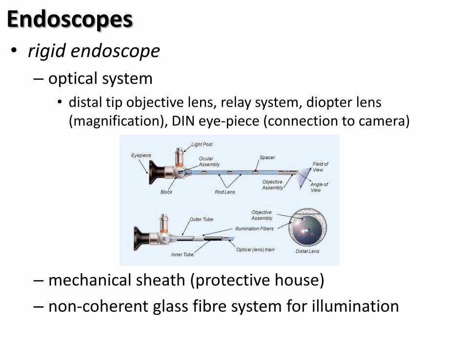
Endoscopes • rigid endoscope
– optical system
• distal tip objective lens, relay system, diopter lens (magnification), DIN eye-piece (connection to camera)
– mechanical sheath (protective house)
– non-coherent glass fibre system for illumination

– instrument/flow channel
– distal objective lens can be angled off 0⁰ to give oblique view
• common 12⁰, 30⁰, 70⁰
• rotation on their axis given differential field of view without much physical trauma to surrounding

• flexible endoscope
– consists of
• combined coherent glass fibres each stretched to 1/100mm in diameter – transmit image – typical flexible URS scope has 4000 fibres and 270⁰ of deflection
• non-coherent bundle – light transmission
• instrument/irrigation channel
• control wires/cables for tip deflection control mechanism

– benefits
• flexibility; telescope conforms to the anatomy
• deflecting tip allows scope to view and access areas not reachable by rigid scope
– limits
• image quality; limitation by mechanics and physics of individual fibre size and number
• complex and fine manufacturing processes; expensive to purchase and maintain
• choice of construction materials; non heat resistant – can only be decontaminated and not sterilised with pressurised steam

• semi-rigid endoscopes
– coherent image bundle and non-coherent illumination bundle housed semi-rigid metal skin
– no tip deflection
– longer scopes and smaller diameters
– typically paediatric endoscopes
• digital, distal-tip chip (discussed later)

Endoscopy - light sources • diagnostic light source
– white light endoscopy
• power sources 100-300W
• light intensity controlled manually/auto
• ideally automatic regulation to match varying situation
• inbuilt cooling fan
• high intensity light can burn surgeon, patient or surgical drapes
• use standby mode when not in active use
• illumination; halogen bulb - soft light or xenon bulb – brighter white light

– narrow band imaging (NBI)
• optical filter technology (based on CCD system)
• improve visibility of subtle tissue structure – optimising absorbance/scatter characteristics of light
• 2 discrete bands: – blue (414nm), highlight superficial capillary network
– green (540nm), highlight sub-epithelial tissue

– photodynamic diagnosis (PDD)
• primarily used in bladder cancer detection
• bladder tissue can be photosensitised by instillation of intravesical delta-aminolevulinic acid (ALA) or its derivative hexaminolevulinate (HAL, Hexvix) – prodrugs; initiate a series of biochemical reactions that result in a
transient though significant accumulation of protoporphyrin IX (PpIX) or photoactive porphyrins (PAPs)
– accumulation of protoporphyrin IX (PpIX) or photoactive porphyrins (PAPs) preferentially occurs in cancer tissue
• bladder wall is illuminated (excited) by blue light (~380–470 nm), the PpIX/PAPs entrapped in cancer cells emit red fluorescence (~693 nm), whereas normal bladder wall tissue appears blue-green

• therapeutic light sources
– photodynamic therapy(PDT)
• photo-sensitising chemicals injected intravesically or intravenously
• light source used to stimulate apoptosis
• red visible light (400-1064nm) – penetrate well into tissue
– induce singlet O2 which induces apoptosis in sensitised tissue
• light source options halogen lamp, LED, lasers; choice depends on light dose and geometry of area

Endoscopy – digital camera systems • 1970 – Boyle & Smith created the charge couple
device (CCD) – record images as a grid of pixels
– linearity of response, high sensitivity and instant image production
– mounted external, detachable camera
– integrated into end of videoscope
• CMOS (complementary metal oxide semiconductor ) device – a low-cost alternative
– offered reductions in cost and device size
– more suitable than CCDs for mass production

• CCD and CMOS
– digital sensors composed of millions of photodiodes
– convert photons into electric current that is later transformed into voltage, amplified and converted to a digital form
– CCD chip
• a photoactive region and a transmission region
• colour accuracy of the CCD camera allows enhanced visualization of reproduced images
• improved resolution and quality of the images achieved when three chips are present (one each for red, blue, and green light)
• “global” shutter
• prone to sensor artefact - smear

• CMOS chip
– electronic registration of images projected onto their surface through the lens of a camera
– provide image capture to a grid of pixels formed by photoelectric elements
– each pixel acts as its own independent amplifier of the image signal
– limits noise as the charge-to-voltage conversion takes place within each pixel
– rolling shutter
– prone to sensor artefact – skew, wobble and partial exposure

• CMOS vs CCD
– improved integration (more functions on the chip)
– lower energy consumption
– less heat production
– requires fewer electronic components; allows such chips to be smaller and more compact
– advantages of CMOS chips often come at the expense of image quality and device cost
– require companion chips to optimize image quality, which sometimes increases cost

Endoscopy – beyond imaging • endoscope protection system (EPS)
– exploit the ability of the CMOS sensor to detect colours in different portions of the optical field in order to detect the blue cladding of laser fibre
– when EPS is active, the CMOS chip continuously detects the presence of the laser fibre as a blue colour in a specific portion of the optical field • laser fibre is retracted (or obscured), information is
transmitted from the CMOS chip to the computer control unit
• control unit is connected to the laser and is able to initiate shutdown of the system within milliseconds
• prevents the laser from functioning 'blind' and reduces endoscope damage and probably procedure costs

EPS
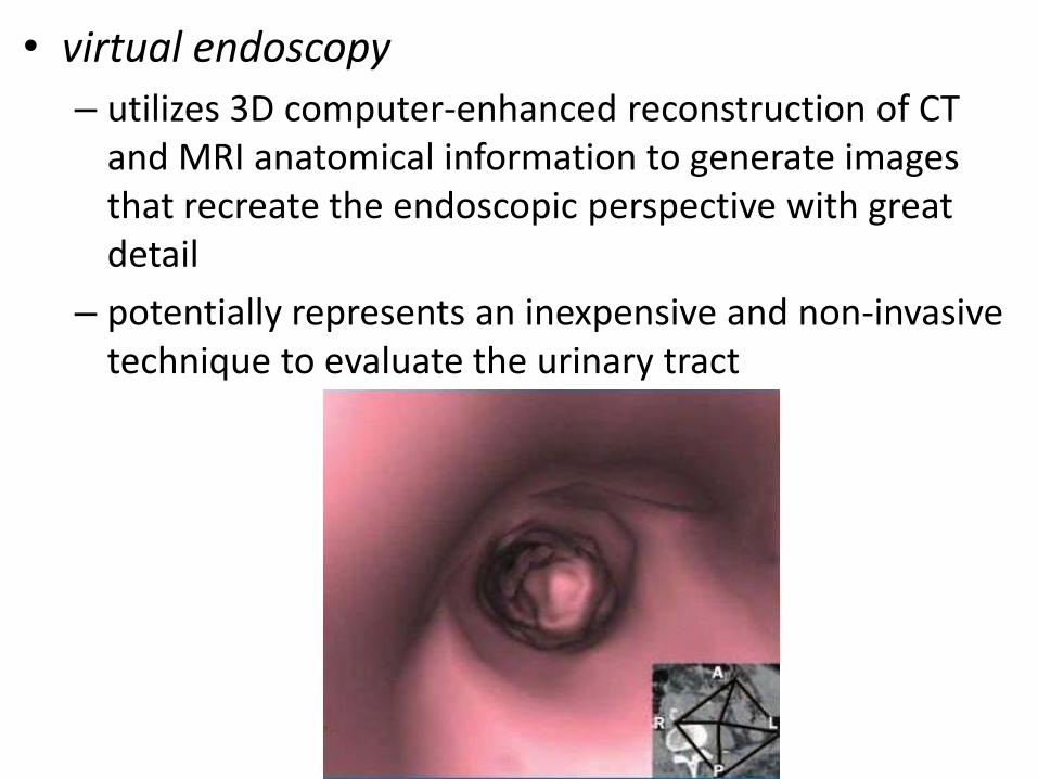
• virtual endoscopy
– utilizes 3D computer-enhanced reconstruction of CT and MRI anatomical information to generate images that recreate the endoscopic perspective with great detail
– potentially represents an inexpensive and non-invasive technique to evaluate the urinary tract
– at present insufficient sensitivity to replace conventional endoscopic evaluation
– virtual cystoscopy can be successfully used for non-invasive detection of bladder lesions with a sensitivity of 90% and specificity of 94%; Kirvak et al
– Lopes and co-workers reported similar results, with a sensitivity of 95.1% and specificity of 91.2% • limitation; virtual cystoscopy will not be able to detect flat lesions such as
carcinoma in situ, and does not afford the surgeon the opportunity to biopsy suspicious lesions
– virtual nephroscopy (a modality based on CT urography) was reported by Takebayashi and colleagues, who assessed 32 patients with suspected renal pelvic tumor • CT nephroscopy revealed 22 of 24 tumors (92%) while axial CT detected
only 20 (83%) lesions • virtual nephroscopy was also superior to axial CT in detecting
pedunculated and infiltrating lesions
– virtual ureteroscopy successfully detected the presence of ureteral tumour, with a sensitivity and specificity of 81% and 100%, respectively; Takebayashi et al

• wireless capsular endoscopy (WCE)
– only been used experimentally in urologic disorders
– successfully used to evaluate the urinary tract in a porcine model, where it produced continuous images and identified specific landmarks
– the device could be manipulated by means of an external magnet; Gettman et al

Endoscopy – the future • imaging devices will certainly continue to decrease in
size, such that 'microendoscopy' will be feasible in the near future
• integration between nanotechnology and endoscopy • Raman endoscopy (Hanchanel et al)
– powerful light-scattering technique used to identify the internal structure of molecules and crystals
– might greatly improve real-time histologic tissue diagnosis by measuring the molecular components of tissue in a qualitative and quantitative way
– light scattered by each tissue type has a characteristic spectrum, which can be used to generate a pseudocolour map
– eventually determine if tissue composition is of a normal or a pathologic nature
– might enable real-time decisions

• optical coherence tomography (OCT) – capable of producing high-resolution, cross-sectional,
subsurface tomographic imaging of the microstructure in biologic systems
– low-power infrared light with a wavelength of 750–1,300 nm
– images are generated from measuring the echo time delay and the intensity of back-scattered light
– depth of penetration of OCT imaging is approximately 1–3 mm depending upon tissue structure, depth of focus, and pressure applied to the tissue surface
– layers of the bladder (urothelium, lamina propria, and muscularis propria) can be individually visualized by use of this technology according to their different light-reflecting properties, Carberg et al
– might help in real time decision making

Endoscopy - utilisations in urology
ORGAN BENIGN MALIGNANT
KIDNEY Stone Stricture or obstruction Hematuria
Selected superficial urothelial tumors
URETER Stone Stricture
Selected superficial urothelial tumors
BLADDER Stone Selected diverticula
Superficial urothelial tumors
PROSTATE Hyperplasia Obstruction due to prostate cancer
URETHRA Stone Stricture
Superficial neoplasms

The End