
Handwritten Text Segmentation via End-to-End Learning of ...

End-to-End Fully Automatic Segmentation ofVertebrae in Spinal X-Ray Images
Abdullah-Al-Zubaer Imran2,1, Chao Huang1, Hui Tang1, Wei Fan1,Kenneth M.C. Cheung3, Michael To3, Zhen Qian1, Demetri Terzopoulos2,4
1Tencent Medical AI Lab, Palo Alto, CA, USA2University of California, Los Angeles, CA, USA
3The University of Hong Kong, China4VoxelCloud, Inc., Los Angeles, CA, USA
Abstract
Accurate vertebral identification and labeling is essential in image-guided spinaldisease diagnosis and treatment planning. Unfortunately, spinal assessments tra-ditionally rely on tedious and time-consuming manual measurement subject toobserver variability. In particular, the measurement of scoliosis requires the seg-mentation of individual vertebrae, but an automatic method that can accuratelyidentify and segment vertebrae is unavailable in the literature. We introduce anend-to-end fully automatic segmentation method that leverages a carefully-adjustedU-Net model with progressive side outputs in order to provide reliable segmenta-tions of the vertebrae associated with scoliosis measurement. Our experimentalresults with X-ray images of scoliosis patients indicate that our method, whichachieves an average Dice score of 0.993, promises to be an effective tool in theidentification and labeling of vertebrae for the reliable estimation of scoliosis.
1 Introduction
Conventional spine image analysis tasks, particularly for the measurement of scoliosis, an abnormallateral curve to the vertebral column, involve tedious manual labor subject to imprecision andvariability. In addition to the landmark detection approach [4], vertebrae segmentation methodsare available in literature. Existing computer-aided vertebral segmentation methods rely on manualinteraction [7], hand-crafted feature engineering [10], patch-based approaches that lose full spatialcontext [8, 1], limited scope that fails to consider all the required vertebrae at once [6], etc. As adeparture from previous vertebral segmentation approaches, we introduce a method that involves nomanual intervention and is end-to-end, avoiding any pre-/post-processing steps.
2 Method
Following the progressive side-outputs in segmentation model [2, 5, 3], we propose a progressiveU-Net with some careful adjustments in the U-Net. As in a U-Net [9], our model has an encoderand a decoder with skip connections. In each encoder layer, two 3 × 3 convolutions are followedby instance normalization, ReLU activation, and a 2× 2 max-pooling. A dropout of 0.25 is appliedin every encoder and decoder stage of the network. We generate side-outputs in every stage of thedecoder. Progressively adding one side-output to the next improves the segmentation performancecompared to collecting the output from the final decoder stage [3]. The progressive side-outputs alsoensure that micro-structure is preserved at all levels of the decoder.
Medical Imaging Meets NeurIPS (Med-NeurIPS 2019), Vancouver, Canada.
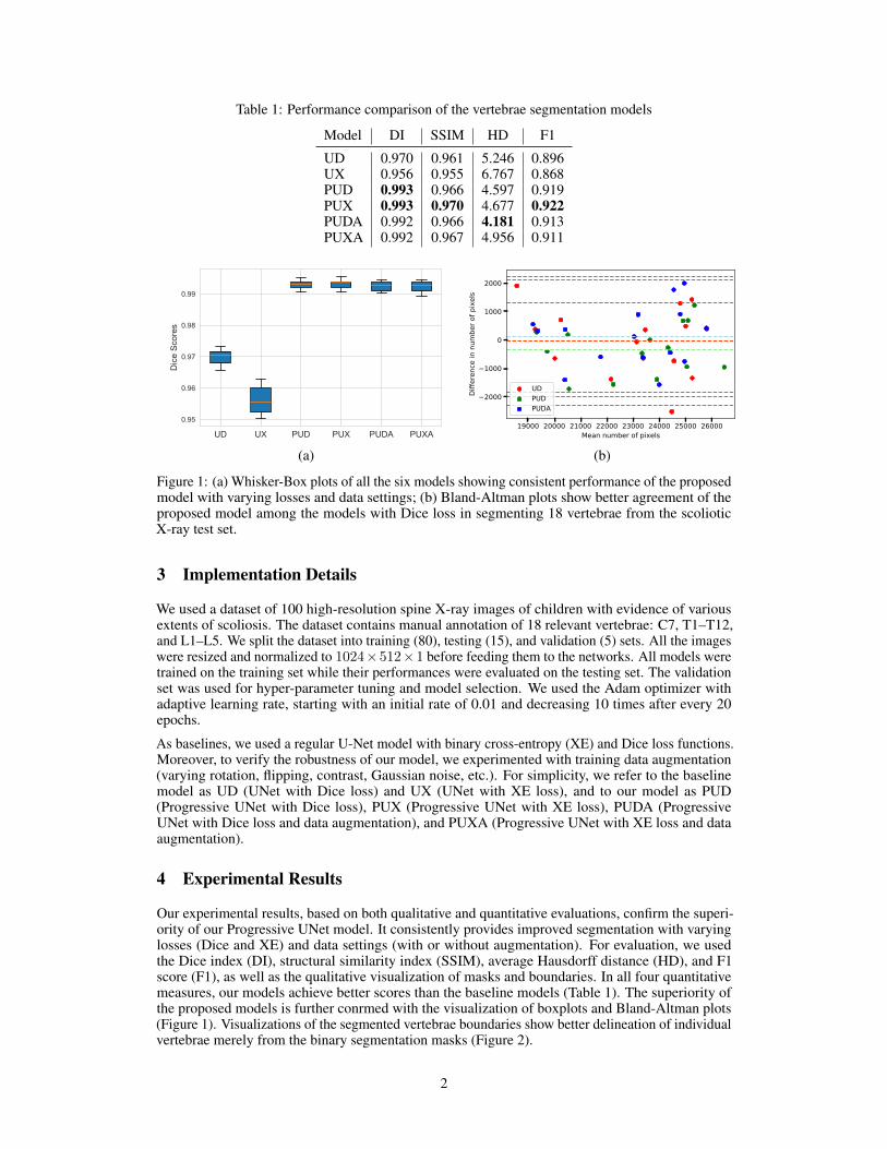
Table 1: Performance comparison of the vertebrae segmentation models
Model DI SSIM HD F1
UD 0.970 0.961 5.246 0.896UX 0.956 0.955 6.767 0.868PUD 0.993 0.966 4.597 0.919PUX 0.993 0.970 4.677 0.922PUDA 0.992 0.966 4.181 0.913PUXA 0.992 0.967 4.956 0.911
UD UX PUD PUX PUDA PUXA
0.95
0.96
0.97
0.98
0.99
Dic
e Sc
ores
19000 20000 21000 22000 23000 24000 25000 26000Mean number of pixels
2000
1000
0
1000
2000
Diffe
renc
e in
num
ber o
f pix
els
UDPUDPUDA
(a) (b)
Figure 1: (a) Whisker-Box plots of all the six models showing consistent performance of the proposedmodel with varying losses and data settings; (b) Bland-Altman plots show better agreement of theproposed model among the models with Dice loss in segmenting 18 vertebrae from the scolioticX-ray test set.
3 Implementation Details
We used a dataset of 100 high-resolution spine X-ray images of children with evidence of variousextents of scoliosis. The dataset contains manual annotation of 18 relevant vertebrae: C7, T1–T12,and L1–L5. We split the dataset into training (80), testing (15), and validation (5) sets. All the imageswere resized and normalized to 1024×512×1 before feeding them to the networks. All models weretrained on the training set while their performances were evaluated on the testing set. The validationset was used for hyper-parameter tuning and model selection. We used the Adam optimizer withadaptive learning rate, starting with an initial rate of 0.01 and decreasing 10 times after every 20epochs.
As baselines, we used a regular U-Net model with binary cross-entropy (XE) and Dice loss functions.Moreover, to verify the robustness of our model, we experimented with training data augmentation(varying rotation, flipping, contrast, Gaussian noise, etc.). For simplicity, we refer to the baselinemodel as UD (UNet with Dice loss) and UX (UNet with XE loss), and to our model as PUD(Progressive UNet with Dice loss), PUX (Progressive UNet with XE loss), PUDA (ProgressiveUNet with Dice loss and data augmentation), and PUXA (Progressive UNet with XE loss and dataaugmentation).
4 Experimental Results
Our experimental results, based on both qualitative and quantitative evaluations, confirm the superi-ority of our Progressive UNet model. It consistently provides improved segmentation with varyinglosses (Dice and XE) and data settings (with or without augmentation). For evaluation, we usedthe Dice index (DI), structural similarity index (SSIM), average Hausdorff distance (HD), and F1score (F1), as well as the qualitative visualization of masks and boundaries. In all four quantitativemeasures, our models achieve better scores than the baseline models (Table 1). The superiority ofthe proposed models is further conrmed with the visualization of boxplots and Bland-Altman plots(Figure 1). Visualizations of the segmented vertebrae boundaries show better delineation of individualvertebrae merely from the binary segmentation masks (Figure 2).
2
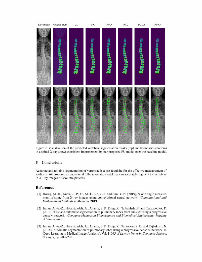
Raw Image Ground Truth UD UX PUD PUX PUDA PUXA
Figure 2: Visualization of the predicted vertebrae segmentation masks (top) and boundaries (bottom)in a spinal X-ray shows consistent improvement by our proposed PU model over the baseline model.
5 Conclusions
Accurate and reliable segmentation of vertebrae is a pre-requisite for the effective measurement ofscoliosis. We proposed an end-to-end fully automatic model that can accurately segment the vertebraein X-Ray images of scoliotic patients.
References[1] Horng, M.-H., Kuok, C.-P., Fu, M.-J., Lin, C.-J. and Sun, Y.-N. [2019], ‘Cobb angle measure-
ment of spine from X-ray images using convolutional neural network’, Computational andMathematical Methods in Medicine 2019.
[2] Imran, A.-A.-Z., Hatamizadeh, A., Ananth, S. P., Ding, X., Tajbakhsh, N. and Terzopoulos, D.[2019], ‘Fast and automatic segmentation of pulmonary lobes from chest ct using a progressivedense v-network’, Computer Methods in Biomechanics and Biomedical Engineering: Imaging& Visualization .
[3] Imran, A.-A.-Z., Hatamizadeh, A., Ananth, S. P., Ding, X., Terzopoulos, D. and Tajbakhsh, N.[2018], Automatic segmentation of pulmonary lobes using a progressive dense V-network, in‘Deep Learning in Medical Image Analysis’, Vol. 11045 of Lecture Notes in Computer Science,Springer, pp. 282–290.
3

[4] Imran, A.-A.-Z., Huang, C., Tang, H., Fan, W., Cheung, K. M., To, M., Qian, Z. and Terzopoulos,D. [2019], Bipartite distance for shape-aware landmark detection in spinal X-ray images, in‘Medical Imaging Meets NeurIPS Workshop’, Vancouver, Canada.
[5] Imran, A.-A.-Z. and Terzopoulos, D. [2019], Semi-supervised multi-task learning with chestx-ray images, in ‘MICCAI Machine Learning in Medical Imaging’, Vol. 11861, p. 151.
[6] Lessmann, N., van Ginneken, B., de Jong, P. A. and Išgum, I. [2019], ‘Iterative fully convolu-tional neural networks for automatic vertebra segmentation and identification’, Medical ImageAnalysis 53, 142–155.
[7] Mateusiak, M. and Mikolajczyk, K. [2019], Semi-automatic spine segmentation method of CTdata, in R. Szewczyk, J. Krejsa, M. Nowicki and A. Ostaszewska-Lizewska, eds, ‘Mechatronics2019: Recent Advances Towards Industry 4.0’.
[8] Qadri, S. F., Zhao, Z., Ai, D., Ahmad, M. and Wang, Y. [2019], Vertebrae segmentationvia stacked sparse autoencoder from computed tomography images, in ‘11th InternationalConference on Digital Image Processing (ICDIP 2019)’, Vol. 11179 of Proceedings of the SPIE,pp. 4K:160–166.
[9] Ronneberger, O., Fischer, P. and Brox, T. [2015], U-net: Convolutional networks for biomedicalimage segmentation, in ‘International Conference on Medical Image Computing and Computer-Assisted Intervention’, Springer, pp. 234–241.
[10] Taghizadeh, E., Terrier, A., Becce, F., Farron, A. and Büchler, P. [2019], ‘Automated CT bonesegmentation using statistical shape modelling and local template matching’, Computer Methodsin Biomechanics and Biomedical Engineering 0(0), 1–8.
4


![Columna vertebralis (canis) - Állatorvostudományi Egyetem · 2016. 2. 7. · Extremitas caudalis [=Fossa vertebrae] Arcus vertebrae. Vertebrae. Processus articularis cranialis.](https://static.fdocuments.net/doc/165x107/61216d37ae072938fd5b60ac/columna-vertebralis-canis-llatorvostudomnyi-egyetem-2016-2-7-extremitas.jpg)


