End-capped HyBeacon Probes for the Analysis of Human Genetic
12
Supporting information End-capped HyBeacon Probes for the Analysis of Human Genetic Polymorphisms Related to Warfarin Metabolism Nouha Ben Gaied a , James A. Richardson a , Daniel G. Singleton a , Zhengyun Zhao a , David French b , Tom Brown a * a School of Chemistry, University of Southampton, Highfield, SO17 1BJ, Southampton, UK b Innovation and Development, LGC, Queens Road, Teddington, TW11 0LY. UK. * To whom correspondence should be sent: Tel: +44(0)2380 592974, fax: +44(0)2380 592991, E-mail: [email protected] Table S1. Oligonucleotide sequences used Table S2. Mass spectra of oligonucleotides Figure S1. Capillary electrophoresis analysis of oligonucleotides Figure S2. UV/visible spectra of end cap modifications Experimental. Determination of fluorescence properties and extinction coefficients of 3’-modifications Figure S3. Fluorescence melting curves Figure S4. UV melting curves.
Transcript of End-capped HyBeacon Probes for the Analysis of Human Genetic

Supporting information
End-capped HyBeacon Probes for the Analysis of Human Genetic Polymorphisms Related to Warfarin Metabolism
Nouha Ben Gaieda, James A. Richardsona, Daniel G. Singletona, Zhengyun Zhaoa, David Frenchb, Tom Browna*
aSchool of Chemistry, University of Southampton, Highfield, SO17
1BJ, Southampton, UK
bInnovation and Development, LGC, Queens Road, Teddington, TW11
0LY. UK.
*To whom correspondence should be sent:
Tel: +44(0)2380 592974, fax: +44(0)2380 592991, E-mail: [email protected]
Table S1. Oligonucleotide sequences used
Table S2. Mass spectra of oligonucleotides
Figure S1. Capillary electrophoresis analysis of oligonucleotides
Figure S2. UV/visible spectra of end cap modifications
Experimental. Determination of fluorescence properties and extinction
coefficients of 3’-modifications
Figure S3. Fluorescence melting curves
Figure S4. UV melting curves.
Supplementary Material (ESI) for Organic and Biomolecular ChemistryThis journal is © The Royal Society of Chemistry 2010

Modification Oligonucleotide sequence 1 Matched sequence 3’-CCTCTTGAACACGGTCCTCAATGCTCC-5’ 2 Mismatched sequence 3’-CCTCTTGAACACAGTCCTCAATGCTCC-5’ 3 3’-P 5’-CGATFGAGGACCGFGTTCAAG-P-3’ 4 5’-TMS-3’-P 5’-TMS-CGATFGAGGACCGFGTTCAAG-P-3’ 5 3’-AnthdR 5’-CGATFGAGGACCGFGTTCAAG-AnthdR-3’ 6 5’-TMS-3’-AnthdR 5’-TMS-CGATFGAGGACCGFGTTCAAG-AnthdR-3’ 7 3’-AnthdRNH2 5’-CGATFGAGGACCGFGTTCAAG-AnthdRNH2-3’ 8 5’-TMS-3’-AnthdRNH2 5’-TMS-CGATFGAGGACCGFGTTCAAG-AnthdRNH2-3’ 9 3’-AmBuPyr 5’-CGATFGAGGACCGFGTTCAAG-AmBuPyr-3’ 10 5’-TMS-3’-AmBuPyr 5’-TMS-CGATFGAGGACCGFGTTCAAG-AmBuPyr-3’ 11 3’-ThrPyr 5’-CGATFGAGGACCGFGTTCAAG-ThrPyr-3’ 12 5’-TMS-3’-ThrPyr 5’-TMS-CGATFGAGGACCGFGTTCAAG-ThrPyr-3’ Table S1: List of oligonucleotides. Twelve oligonucleotides were prepared: two
targets and ten probes labelled with two internal fluorescein-dT residues (F), a 3’-
modification and/or a 5’-trimethoxystilbene (TMS). P: phosphate.
Sequence Modification MW calc. MW found ODN1 Matched sequence 8128 8135 ODN2 Mismatched sequence 8113 8125 ODN3 3’-P 7589 7600 ODN4 5’-TMS/ 3’-P 8021 8031 ODN5 AnthdR 8067 8099 ODN6 5’-TMS-AnthdR 8489 8494 ODN7 AnthdRNH2 8168 8169 ODN8 5’-TMS-3’-AnthdRNH2 8603 8606 ODN9 3’-AmBuPyr 8079 8078/8100 ODN10 5’-TMS-3’-AmBuPyr 8513 8514 ODN11 3’-ThrPyr 7948 7950 ODN12 5’-TMS-3’-ThrPyr 8383 8386
Table S2: Mass spectra. Mass spectra of all targets and HyBeacons® were recorded
by negative mode electrospray on a Fisons VG platform spectrometer in water with
Triisopropylamine (2%).
Supplementary Material (ESI) for Organic and Biomolecular ChemistryThis journal is © The Royal Society of Chemistry 2010

ODN1
ODN2
ODN3
ODN4
ODN5
ODN6
ODN7
ODN8
Supplementary Material (ESI) for Organic and Biomolecular ChemistryThis journal is © The Royal Society of Chemistry 2010

ODN9
ODN10
ODN11
ODN12
Figure S1: Capillary electrophoresis (CE) analysis of modified oligonucleotides after
HPLC purification.
The purity of oligonucleotides was confirmed by injection (0.4 OD/100 µL) of each
sample individually on ssDNA 100-R Gel. Tris-Borate 7M Urea were used (kit N°
477480) on a Beckman coulter P/ACE™ MDQ Capillary Electrophoresis system
using 32 Karat software. UV-254, inject voltage 10.0 kV and separate voltage 9.0 kV
(45.0 min duration). The x-axis shows time in min and the y-axis is UV absorbance at
254 nm.
Supplementary Material (ESI) for Organic and Biomolecular ChemistryThis journal is © The Royal Society of Chemistry 2010

Figure S2: UV spectra
A) UV spectra of 3’-caps
-0.2
0
0.2
0.4
0.6
0.8
1
1.2
1.4
1.6
200 250 300 350 400 450 500 550 600
Wavelength (nm)
Abs
orpt
ion
OHO
HO
NHO
HN
O O
0
0.05
0.1
0.15
0.2
0.25
0.3
0.35
0.4
0.45
200 300 400 500 600 700Wavelength (nm)
Abs
orpt
ion
OHO
HO
NHO
HN
O O
NH(CH2)6NHCOCF3
0
0.1
0.2
0.3
0.4
0.5
0.6
0.7
0.8
200 220 240 260 280 300 320 340 360 380 400
Wavelength (nm)
Abs
orpt
ion
HN
OOH
OH
Supplementary Material (ESI) for Organic and Biomolecular ChemistryThis journal is © The Royal Society of Chemistry 2010
0
0.2
0.4
0.6
0.8
1
1.2
1.4
1.6
1.8
2
200 220 240 260 280 300 320 340 360 380 400
Wavelength
Abs
orpt
ion
OHO
HOO
HN
O
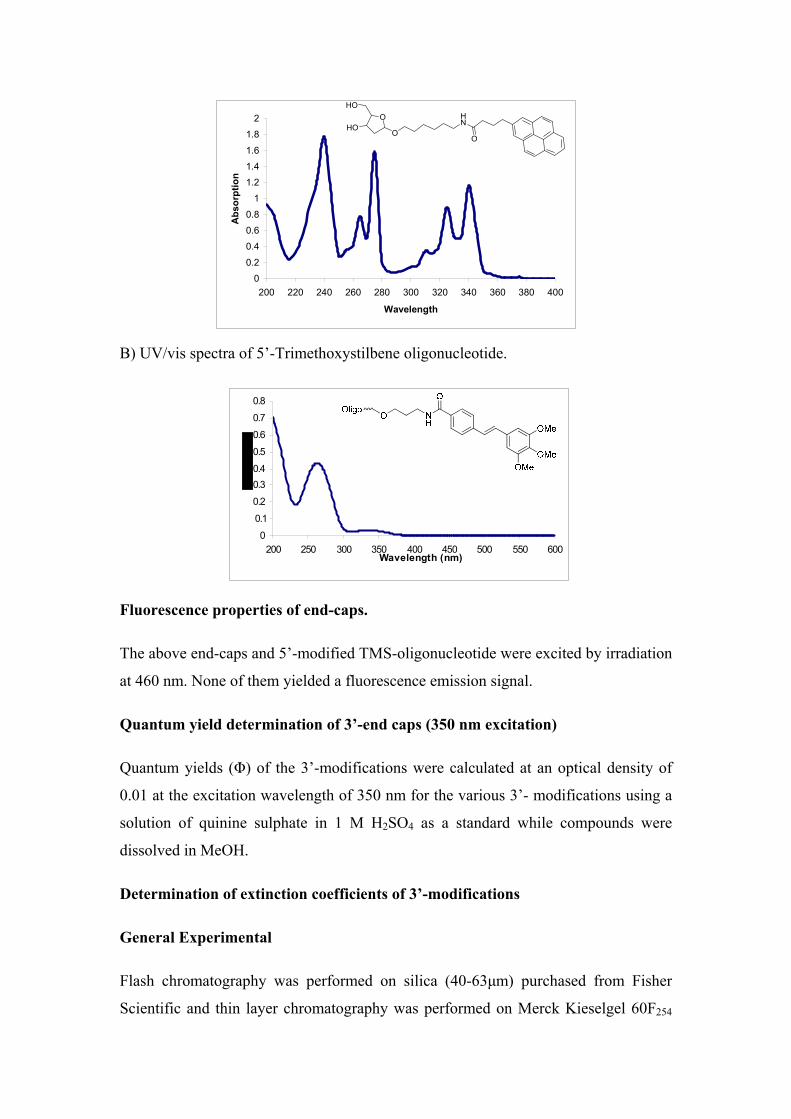
B) UV/vis spectra of 5’-Trimethoxystilbene oligonucleotide.
Fluorescence properties of end-caps.
The above end-caps and 5’-modified TMS-oligonucleotide were excited by irradiation
at 460 nm. None of them yielded a fluorescence emission signal.
Quantum yield determination of 3’-end caps (350 nm excitation)
Quantum yields (Φ) of the 3’-modifications were calculated at an optical density of
0.01 at the excitation wavelength of 350 nm for the various 3’- modifications using a
solution of quinine sulphate in 1 M H2SO4 as a standard while compounds were
dissolved in MeOH.
Determination of extinction coefficients of 3’-modifications
General Experimental
Flash chromatography was performed on silica (40-63μm) purchased from Fisher
Scientific and thin layer chromatography was performed on Merck Kieselgel 60F254
0
0.1
0.2
0.30.4
0.5
0.6
0.7
0.8
200 250 300 350 400 450 500 550 600Wavelength (nm)
Supplementary Material (ESI) for Organic and Biomolecular ChemistryThis journal is © The Royal Society of Chemistry 2010

coated plates (0.22 mm thickness, aluminum backed). Compounds were visualized by
staining with vanillin (2g of vanillin in 100 mL of EtOH/ H2SO4 98:2) and by
ultraviolet absorbance at 254nm. 1H and 13C NMR spectra were recorded on a Bruker
AV300 or a Bruker DPX 400 spectrometer. All spectra were internally referenced to
the appropriate residual undeuterated solvent signal. Chemical shifts are given in ppm
relative to tetramethylsilane. J values are given in Hz and corrected to within 0.5 Hz.
Multiplicities of 13C signals were determined using the DEPT spectral editing
technique. High-resolution mass spectra were recorded in acetonitrile, methanol or
water (HPLC grade) using the electrospray technique on a Bruker APEX III FT-ICR
mass spectrometer.
3’-Oligonucleotide modifications
Anthraquinone and Pyrene analogues for 3’-oligonucleotide modification and the
corresponding oligonucleotide synthesis resins were prepared as described previously1
These compounds were detritylated by the general procedure outlined below to obtain
extinction coefficients for calculating oligonucleotide concentrations. DMTr protected
compounds (1-4) were dissolved in a solution of 3% trichloroacetic acid (TCA) in
CH2Cl2 (oligonucleotide grade) at a concentration of 0.03 M. The mixture was stirred
at room temperature until detritylation was complete (TLC CH2Cl2: MeOH 90:10).
The reaction mixture was then quenched with a saturated solution of NaHCO3 and
CH2Cl2 was added. The organic layer was washed with H2O and brine, dried over
Na2SO4 and evaporated to dryness. The crude product was purified by silica gel flash
chromatography using an Isolute cartridge (Biotage) eluting with a gradient of MeOH
in CH2Cl2.
N-(6-(9,10-Dihydro-9,10-dioxoanthracen-1-ylamino)hexyl)-2’-deoxy-D-
ribofuranose-1-β-acetamide (1). The reaction was performed on 84 mg (0.107 mmol)
in 3.5 mL of TCA 3% in CH2Cl2. 50 mg, 97%.
Rf CH2Cl2: MeOH (90:10) 0.31. NMR 1H (400 MHz, DMSO-d6) δH 9.73-9.71 (2H, t,
J= 5 Hz, NH), 8.26-8.18 (2H, dd, J= 7.5 Hz, H-Ar), 7.97-7.88 (3H, m, H-Ar), 7.71-
7.67 (1H, t, J= 8.5 Hz, H-Ar), 7.29-7.27 (1H, d, J= 8.5 Hz, H-Ar), 4.99-4.98 (1H, d,
J= 4 Hz, OH), 4.74-4.71 (1H, m, OH), 4.43-4.36 (1H, m, H1’), 4.16 (1H, d, J= 2.48
Hz, H3’), 3.72-3.69 (1H, m, H4’), 3.46-3.37 (4H, m, CH2), 3.21-3.10 (2H, m, CH2),
2.48-2.29 (2H, ddd, J1= 7 Hz, J2= 6.5 Hz, J3= 33.1 Hz, H5’), 1.92-1.88 (1H, m, H2’),
Supplementary Material (ESI) for Organic and Biomolecular ChemistryThis journal is © The Royal Society of Chemistry 2010

1.77-1.69 (3H, m, CH2, H2’), 1.54-1.44 (6H, m, CH2). NMR 13C (100 MHz, DMSO-
d6) δC 183.77, 182.66 (CO), 180.24 (CO-NH), 169.49 (C-Ar), 151.18 (C-Ar), 135.41,
134.26 (CH-Ar), 134.20 (C-Ar), 133.72 (CH-Ar), 133.21 (C-Ar), 128.19, 126.25
(CH-Ar), 126.06 (C-Ar), 118.35, 114.79 (CH-Ar), 111.70 (C-Ar), 87.15 (C1’), 74.72
(C4’), 71.88 (C3’), 62.26 (CH2-NH), 42.6 (CH2-CO), 40.74 (C2’), 38.85 (C5’), 29.53,
29.01, 26.76, 26.62 (CH2). UV-Vis λmax (MeOH)/nm 510 (ε/dm3.mol-1.cm-1 2360). Φ
(MeOH, 350 nm) 0.04. m/z LRMS [ES+, MeOH] 503.3 (M+Na+, 100%), 983.8
(2M+Na+, 50%). m/z HRMS (M+Na+) (C27H32N2O6Na): calc. 503.2158 found
503.2153.
N-(6-(9,10-Dihydro-9,10-dioxoanthracen-1-ylamino)-5-yl-(6-aminohexyl)hexyl)-
2-deoxy-D-ribofuranose-1-β-acetamide (2). The reaction was performed on 0.114 g
(0.115 mmol) in 4 mL of TCA 3% in CH2Cl2. Purple solid 75 mg, 95%.
Rf CH2Cl2: MeOH (90:10) 0.33. NMR 1H (400 MHz, DMSO-d6) δH 9.77-9.74 (2H,
t, J= 5 Hz, NH), 9.51 (1H, bs, NH), 7.89-7.86 (1H, t, J= 5.2 Hz, NH), 7.73-7.69 (2H,
t, J= 7.7 Hz, H-Ar), 7.53-7.51 (2H, d, J= 7.5 Hz, H-Ar), 7.24-7.22 (2H, d, J= 8.5 Hz,
H-Ar), 4.97 (1H, bs, OH), 4.72 (1H, bs, OH), 4.42-4.35 (1H, m, H1’), 4.15-4.14 (1H,
m, H4’), 3.71-3.68 (1H, m, H3’), 3.58-3.42 (12H, m, CH2), 3.33-3.28 (2H, q, J= 6.5
Hz, CH2-CO), 2.47-2.28 (2H, ddd, J1= 6.5 Hz, J2= 14.0 Hz, J3= 33.1 Hz, H5’), 1.91-
1.86 (1H, m, H2’), 1.78-1.45 (13H, m, CH2, H2’). NMR 13C (100 MHz, DMSO-d6)
δC 185.2 (CO), 181.2 (COCF3), 170.5 (C-CO), 152.0 (C-NH), 136.5 (CH-Ar), 117.9
(CH-Ar), 115.2 (CH-Ar), 112.8 (CF3), 88.2 (C1’), 75.8 (C4’), 72.9 (C3’), 63.3 (CH2-
CO), 43.0 (C2’), 39.3 (C5’), 29.4, 29.0, 28.9, 28.6, 26.7, 26.6, 26.3 (CH2). UV-Vis
λmax (MeOH)/nm 525 (ε/ dm3.mol-1.cm-1 1077). Φ (MeOH, 350 nm) 0.04. m/z LRMS
[ES+, MeOH] 713.5 (M+Na+, 100%). m/z HRMS (M+Na+) (C35H45N4O7F3Na): calc.
713.3138. found 713.3133.
1-β-O-(hexylamidopropylpyrene-1-yl)-2-deoxy-D-ribofuranose (3). The reaction
was performed on 0.200 g (0.25 mmol) in 8 mL TCA 3% in CH2Cl2. Colourless foam,
30 mg, 24 %.
Rf CH2Cl2: MeOH (90:10) 0.51. NMR 1H (400 MHz, DMSO-d6) δH 8.39-8.36 (1H, d,
J= 9.1 Hz, NH), 8.28-8.19 (4H, m, H-Ar), 8.15-8.09 (2H, dd, J1= 8.8 Hz, J2= 10.2 Hz,
H-Ar), 8.07-8.02 (1H, t, J= 7.7 Hz, H-Ar), 7.94-7.91 (1H, d, J= 8.0 Hz, H-Ar), 7.83-
Supplementary Material (ESI) for Organic and Biomolecular ChemistryThis journal is © The Royal Society of Chemistry 2010

7.79 (1H, t, J= 5.8 Hz, H-Ar), 4.97-4.95 (1H, dd, J1= 2.6 Hz, J2= 5.8 Hz, H1’), 4.84-
4.82 (1H, d, J= 5.1 Hz, H4’), 4.65-4.62 (1H, t, J= 5.5 Hz, H3’), 3.95-3.86 (1H, m,
OH), 3.71-3.66 (1H, m, OH), 3.60-3.47 (2H, m, CH2), 3.41-3.24 (6H, m, CH2), 3.09-
3.02 (2H, q, J= 6.3 Hz, CH2), 2.29-2.20 (3H, m, CH2, H2’), 2.06-1.96 (2H, q, J= 7.4
Hz, H5’), 1.66-1.58 (1H, m, H2’), 1.47-1.37 (6H, m, CH2). NMR 13C (100 MHz,
DMSO-d6) δC 172.1 (CO), 137.0, 131.3, 130.9, 129.7 (C-Ar), 128.8, 127.9, 127.6,
126.9, 126.6, 125.4, 125.2 (CH-Ar), 124.6 (C-Ar), 123.9 (C-Ar), 103.3 (C1’), 85.1
(C4’), 70.5 (C3’), 67.2 (CH2), 61.7 (C5’), 41.5 (C2’), 38.8, 35.5, 32.7, 29.6, 28.0,
26.7, 25.9 (CH2). UV-Vis λmax (MeOH)/nm 340 (ε/ dm3.mol-1.cm-1 161523), 324 nm
(124267), 274 (219444) and 264 (108333). Φ (MeOH, 350 nm) 0.12. m/z LRMS
[ES+, MeOH] 526.3 (M+Na+, 100%), 1029.7 (2M+Na+, 10%). m/z HRMS (M+Na+)
(C31H37NO5Na): calc. 526.2569 found 526.2564.
L-threoninolamidoprop-1-yl pyrene (4). The reaction was performed on 200 mg
(0.295 mmol) in 10 mL of TCA 3% in CH2Cl2. Colourless solid 75 mg, 68%.
Rf CH2Cl2: MeOH (90:10) 0.46. NMR 1H (400 MHz, DMSO-d6) δH 8.51-8.47 (1H,
dd, J1= 4.3 Hz, J2= 9.3 Hz, NH), 8.37-8.11 (7H, m, H-Ar), 8.04-8.01 (1H, dd, J1=4.5
Hz, J2= 8 Hz, H-Ar), 7.56-7.53 (1H, m, H-Ar), 4.73 (1H, bs, OH), 4.05 (1H, bs, CH-
Thr), 3.89 (1H, bs, CH-Thr), 3.66 (1H, bs, OH), 3.53 (2H, bs, CH2), 3.46-3.41 (2H, m,
CH2), 2.47-2.46 (2H, m, CH2), 2.15 (2H, bs, CH2), 1.20-1.17 (3H, t, J= 5.2 Hz, CH3-
Thr). NMR 13C (100 MHz, DMSO-d6) δC 180.2 (CO), 172.2 (C-Ar), 136.6, 130.8,
130.3, 129.2, 128.1 (C-Ar), 127.4, 127.3, 127.1, 126.3, 126.0, 124.8, 124.7, 124.6
(CH-Ar), 124.0 (C-Ar), 123.4 (CH-Ar), 64.4 (CH-Thr), 60.6 (CH2), 55.6 (CH-Thr),
35.1, 32.2, 27.8 (CH2), 20.1 (CH3). UV-Vis λmax (MeOH)/nm 340 (ε/dm3.mol-1.cm-1
29864), 324 (23000), 274 (40889) and 264 (20665). Φ (MeOH, 350 nm) 0.1. m/z
LRMS [ES+, MeOH] 398.3 (M+Na+, 100%), 773.6 (2M+Na+, 50%). m/z HRMS
(M+Na+) (C24H25NO3Na): calc. 398.1732 found 398.1729.
All compounds were detritylated efficiently except 1-β-O-(hexylamidopropylpyrene-
1-yl)-2-deoxy-D-ribofuranose 3 for which an impurity corresponding to 6-
hydroxyhexylamidopropylpyrene was identified by MS and NMR analysis. The same
side-reaction was also observed during oligonucleotide synthesis to yield modest
amounts of oligonucleotide products without 3’-pyrene. These impurities were easily
removed by reversed-phase HPLC.
Supplementary Material (ESI) for Organic and Biomolecular ChemistryThis journal is © The Royal Society of Chemistry 2010

Temperature (oC) Temperature (oC)
DMTr protected compounds 1-4 were attached via a succinyl linkage to long chain
aminoalkyl silica resin (92 μmol/g amino-loading, Link Technologies Ltd) and used
in oligonucleotide synthesis.1
Fluorescence melting curves
A) Hybeacons® vs matched sequence B) Hybeacons® vs mismatched sequence
C)
Figure S3: A) Fluorescence melting curves of all HyBeacon® analogues against matched sequence. B) Fluorescence melting curves of all HyBeacon® analogues against mismatch sequence. C) Fluorescence melting curves of ThrPyr analogue in presence and in absence of 5’-TMS in comparison to control HyBeacons®. Fluorescence melting was recorded in TaKaRa buffer 1x, 1M NaCl.
Temperatue
-dF/dT
-dF/dT
ODN5 ODN7 ODN9 ODN11 ODN6 ODN8 ODN10 ODN12
ODN1-ODN3 ODN1-ODN11 ODN1-ODN12 ODN2-ODN3 ODN2-ODN11 ODN2-ODN12
-dF/dT
Supplementary Material (ESI) for Organic and Biomolecular ChemistryThis journal is © The Royal Society of Chemistry 2010

UV melting curves
0
0.002
0.004
0.006
0.008
0.01
0.012
30 40 50 60 70
Temperature (degres celsius)
dA/d
T
0
0.002
0.004
0.006
0.008
0.01
0.012
30 40 50 60 70
Temperature (degres celcius)dA
/dT
C)
0
0.002
0.004
0.006
0.008
0.01
0.012
30 40 50 60 70
Temperature (degres celcius)
dA/d
T
Figure S4: UV melting analysis. A) Pyrene analogues and control HyBeacons® against matched sequence (ODN1). B) Pyrene analogues and control HyBeacons® against mismatched sequence (ODN2). C) Direct comparison of 5’-TMS-3’ThrPyr (ODN12) and 3’-P HyBeacons® (ODN3) towards matched and mismatched sequence. UV melting recorded in phosphate buffer, 200 mM NaCl, pH 7.0. 1. N. Ben Gaied, Z. Y. Zhao, S. R. Gerrard, K. R. Fox and T. Brown, Chembiochem, 2009, 10, 1839-1851.
ODN2-ODN3
ODN2-ODN12
ODN1-ODN12
ODN1-ODN3
ODN3 ODN4 ODN9 ODN10 ODN11 ODN12
ODN3 ODN4 ODN9 ODN10 ODN11 ODN12
Supplementary Material (ESI) for Organic and Biomolecular ChemistryThis journal is © The Royal Society of Chemistry 2010

Supplementary Material (ESI) for Organic and Biomolecular ChemistryThis journal is © The Royal Society of Chemistry 2010