Emotion, Decision Making and the Antoine Bechara, Hanna ...
13
The somatic marker hypothesis provides a systems-level neuro- anatomical and cognitive framework for decision making and the influence on it by emotion. The key idea of this hypothesis is that decision making is a process that is influenced by marker signals that arise in bioregulatory processes, including those that express themselves in emotions and feelings. This influence can occur at multiple levels of operation, some of which occur consciously and some of which occur non-consciously. Here we review studies that confirm various predictions from the hypothesis. The orbitofrontal cortex represents one critical structure in a neural system sub- serving decision making. Decision making is not mediated by the orbitofrontal cortex alone, but arises from large-scale systems that include other cortical and subcortical components. Such structures include the amygdala, the somatosensory/insular cortices and the peripheral nervous system. Here we focus only on the role of the orbitofrontal cortex in decision making and emotional processing, and the relationship between emotion, decision making and other cognitive functions of the frontal lobe, namely working memory. It has long been known that different sectors of the human prefrontal cortex are involved in distinctive cognitive and behavioral operations. The insight regarding this functional specialization came from the clinical observation of neurological patients in whom damage to different sectors of the frontal lobe caused remarkably different neuropsychological defects, and this insight was the springboard for systematic attempts to characterize the defects in relation to cognitive processes defined in terms of their components. The ensuing results, based on experimental work in both humans with brain lesions as well as non-human primates, has generally supported the notion of functional specialization within the prefrontal cortices and has yielded a large body of work. The examples, in relation to dorsolateral cortex, encompass the work of Goldman-Rakic and her group (Goldman-Rakic, 1992), of Milner and Petrides (Milner et al., 1985; Petrides, 1996) and of Fuster (Fuster, 1990). In this article we survey recent progress in relation to another prefrontal sector, the ventromedial, as studied in humans with brain lesions. The ventromedial sector includes both the gyrus rectus and mesial half of the orbital gyri, as well as the inferior half of the medial prefrontal surface, from its most caudal aspect to its most rostral in the frontal pole. Areas 11, 12, 13, 25, 32 and 10 of Brodmann are included in this sector, as is the white matter subjacent to all of these areas [see Fig. 1, areas marked in red; see also pp. 24–25 of (Damasio, 1995)]. Damage to the ventro- medial sector disrupts social behavior profoundly. Previously well-adapted individuals become unable to observe social conventions and unable to decide advantageously on matters pertaining to their own lives. Remarkably, the patient’s intel- lectual abilities are generally well preserved, in the sense that they have normal learning and memory, language and attention, and they even perform normally on many so-called executive function tests, such as the Wisconsin Card Sorting Test. Equally remarkably, these patients have an abnormality in their pro- cesses of emotion and feeling. The abnormality is such that they do not engage emotions in relation to complex situations and events, e.g. the emotion and ensuing feeling of embarrassment which are induced by specific social contexts (Damasio et al., 1991; Damasio and Anderson, 1993). The intriguing nature of these defects and the fact that they could not be accounted for by a primary problem with the availability of the pertinent social knowledge, with the ability to apply logic to such knowledge or with general defects of attention or language led to the develop- ment of an account known as the somatic marker hypothesis (Damasio, 1994, 1996). The somatic marker hypothesis proposes that a defect in emotion and feeling plays an important role in impaired decision making. The hypothesis also specifies a number of structures and operations required for the normal operation of decision making. Because emotion is most importantly expressed through changes in the representation of body state, though not solely, and because the results of emotion are primarily repres- ented in the brain in the form of transient changes in the activity pattern of somatosensory structures, the emotional changes are designated under the umbrella term ‘somatic state’. The term ‘somatic’ thus refers to internal milieu, visceral and musculo- skeletal, of the soma rather than just to the musculoskeletal aspects. It should also be noted that although somatic signals are based on structures which represent the body and its states, from the brainstem and hypothalamus to the cerebral cortex, the ‘somatic’ signals do not need to originate in the body in every instance and can be generated intracerebrally (Damasio, 1994, 1995b). The summary of the proposal is presented below. Background Assumptions The somatic marker hypothesis is based on the following main assumptions: (i) that human reasoning and decision making depend on many levels of neural operation, some of which are conscious and overtly cognitive, some of which are not; conscious, overtly cognitive operations depend on sensory images based on the activity of early sensory cortices; (ii) that cognitive operations, regardless of their content, depend on support processes such as attention, working memory and emotion; (iii) that reasoning and decision making depend on the availability of knowledge about situations, actors, options for action and outcomes; such knowledge is stored in ‘dispositional’ form throughout higher-order cortices and some subcortical nuclei (the term dispositional is synonymous with implicit and non-topographically organized) [details on dispositional knowledge and the convergence zone framework are presented elsewhere (Damasio, 1989a,b, 1994; Damasio and Damasio, 1994)]; dispositional knowledge can be made explicit in the form of (a) motor responses of varied types and complexity Emotion, Decision Making and the Orbitofrontal Cortex Antoine Bechara, Hanna Damasio and Antonio R. Damasio Department of Neurology, Division of Behavioral Neurology and Cognitive Neuroscience, University of Iowa College of Medicine, Iowa City, IA 52242, USA Cerebral Cortex Mar 2000;10:295–307; 1047–3211/00/$4.00 © Oxford University Press 2000
Transcript of Emotion, Decision Making and the Antoine Bechara, Hanna ...

The somatic marker hypothesis provides a systems-level neuro-anatomical and cognitive framework for decision making and theinfluence on it by emotion. The key idea of this hypothesis is thatdecision making is a process that is influenced by marker signals thatarise in bioregulatory processes, including those that expressthemselves in emotions and feelings. This influence can occur atmultiple levels of operation, some of which occur consciously andsome of which occur non-consciously. Here we review studies thatconfirm various predictions from the hypothesis. The orbitofrontalcortex represents one critical structure in a neural system sub-serving decision making. Decision making is not mediated by theorbitofrontal cortex alone, but arises from large-scale systems thatinclude other cortical and subcortical components. Such structuresinclude the amygdala, the somatosensory/insular cortices and theperipheral nervous system. Here we focus only on the role of theorbitofrontal cortex in decision making and emotional processing,and the relationship between emotion, decision making and othercognitive functions of the frontal lobe, namely working memory.
It has long been known that different sectors of the human
prefrontal cortex are involved in distinctive cognitive and
behavioral operations. The insight regarding this functional
specialization came from the clinical observation of neurological
patients in whom damage to different sectors of the frontal lobe
caused remarkably different neuropsychological defects, and
this insight was the springboard for systematic attempts to
characterize the defects in relation to cognitive processes
defined in terms of their components. The ensuing results, based
on experimental work in both humans with brain lesions as well
as non-human primates, has generally supported the notion
of functional specialization within the prefrontal cortices and
has yielded a large body of work. The examples, in relation to
dorsolateral cortex, encompass the work of Goldman-Rakic and
her group (Goldman-Rakic, 1992), of Milner and Petrides (Milner
et al., 1985; Petrides, 1996) and of Fuster (Fuster, 1990). In
this article we survey recent progress in relation to another
prefrontal sector, the ventromedial, as studied in humans with
brain lesions.
The ventromedial sector includes both the gyrus rectus and
mesial half of the orbital gyri, as well as the inferior half of the
medial prefrontal surface, from its most caudal aspect to its
most rostral in the frontal pole. Areas 11, 12, 13, 25, 32 and 10
of Brodmann are included in this sector, as is the white matter
subjacent to all of these areas [see Fig. 1, areas marked in red;
see also pp. 24–25 of (Damasio, 1995)]. Damage to the ventro-
medial sector disrupts social behavior profoundly. Previously
well-adapted individuals become unable to observe social
conventions and unable to decide advantageously on matters
pertaining to their own lives. Remarkably, the patient’s intel-
lectual abilities are generally well preserved, in the sense that
they have normal learning and memory, language and attention,
and they even perform normally on many so-called executive
function tests, such as the Wisconsin Card Sorting Test. Equally
remarkably, these patients have an abnormality in their pro-
cesses of emotion and feeling. The abnormality is such that they
do not engage emotions in relation to complex situations and
events, e.g. the emotion and ensuing feeling of embarrassment
which are induced by specific social contexts (Damasio et al.,
1991; Damasio and Anderson, 1993). The intriguing nature of
these defects and the fact that they could not be accounted for
by a primary problem with the availability of the pertinent social
knowledge, with the ability to apply logic to such knowledge or
with general defects of attention or language led to the develop-
ment of an account known as the somatic marker hypothesis
(Damasio, 1994, 1996).
The somatic marker hypothesis proposes that a defect in
emotion and feeling plays an important role in impaired decision
making. The hypothesis also specifies a number of structures
and operations required for the normal operation of decision
making. Because emotion is most importantly expressed through
changes in the representation of body state, though not solely,
and because the results of emotion are primarily repres- ented in
the brain in the form of transient changes in the activity pattern
of somatosensory structures, the emotional changes are
designated under the umbrella term ‘somatic state’. The term
‘somatic’ thus refers to internal milieu, visceral and musculo-
skeletal, of the soma rather than just to the musculoskeletal
aspects. It should also be noted that although somatic signals
are based on structures which represent the body and its states,
from the brainstem and hypothalamus to the cerebral cortex, the
‘somatic’ signals do not need to originate in the body in every
instance and can be generated intracerebrally (Damasio, 1994,
1995b). The summary of the proposal is presented below.
Background AssumptionsThe somatic marker hypothesis is based on the following main
assumptions: (i) that human reasoning and decision making
depend on many levels of neural operation, some of which are
conscious and overtly cognitive, some of which are not;
conscious, overtly cognitive operations depend on sensory
images based on the activity of early sensory cortices; (ii) that
cognitive operations, regardless of their content, depend on
support processes such as attention, working memory and
emotion; (iii) that reasoning and decision making depend on the
availability of knowledge about situations, actors, options for
action and outcomes; such knowledge is stored in ‘dispositional’
form throughout higher-order cortices and some subcortical
nuclei (the term dispositional is synonymous with implicit
and non-topographically organized) [details on dispositional
knowledge and the convergence zone framework are presented
elsewhere (Damasio, 1989a,b, 1994; Damasio and Damasio,
1994)]; dispositional knowledge can be made explicit in the
form of (a) motor responses of varied types and complexity
Emotion, Decision Making and theOrbitofrontal Cortex
Antoine Bechara, Hanna Damasio and Antonio R. Damasio
Department of Neurology, Division of Behavioral Neurology
and Cognitive Neuroscience, University of Iowa College of
Medicine, Iowa City, IA 52242, USA
Cerebral Cortex Mar 2000;10:295–307; 1047–3211/00/$4.00© Oxford University Press 2000

(some combinations of which are part of emotions) and (b)
images. The results of motor responses, including those that are
not generated consciously, can be represented in images; and
(iv) that knowledge can be classified as follows: (a) innate and
acquired knowledge concerning bioregulatory processes and
body states and actions, including those which are made explicit
as emotions; (b) knowledge about entities, facts (e.g. relations,
rules), actions and action-complexes (stories), which are usually
made explicit as images; (c) knowledge about the linkages
between (b) and (a) items, as ref lected in individual experience;
and (d) knowledge resulting from the categorizations of items in
(a), (b) and (c).
Specifics of the HypothesisThe ventromedial prefrontal cortex is a repository of dis-
positionally recorded linkages between factual knowledge and
bioregulatory states. Structures in ventromedial prefrontal
cortex provide the substrate for learning an association between
certain classes of complex situation, on the one hand, and the
type of bioregulatory state (including emotional state) usually
associated with that class of situation in past individual ex-
perience. The ventromedial sector holds linkages between the
facts that compose a given situation, and the emotion previously
paired with it in an individual’s contingent experience. The
linkages are ‘dispositional’ in the sense that they do not hold the
representation of the facts or of the emotional state explicitly,
but hold rather the potential to reactivate an emotion by acting
on the appropriate cortical or subcortical structures (Damasio,
1989a,b, 1994; Damasio and Damasio, 1994). The experience
we acquire regarding a complex situation and its components—a
certain configuration of actors and actions requiring a response;
a set of response options; a set of immediate and long-term
outcomes for each response option—is processed in sensory
imagetic and motor terms and is then recorded in dispositional
and categorized form. But because the experience of some
of those components has been associated with emotional
responses, which were triggered from cortical and subcortical
sites that are dispositionally prepared to respond, it is proposed
that the ventromedial prefrontal cortex establishes a linkage
between the disposition for a certain aspect of a situation (for
Figure 1. Overlap of lesions in the VM patients (n = 13). Red indicates an overlap of four or more patients.
296 Emotion, Decision Making and the Orbitofrontal Cortex • Bechara et al.
instance, the long-term outcome for a type of response option),
and the disposition for the type of emotion that in past experi-
ence has been associated with the situation.
When subjects face a situation for which some factual aspects
have been previously categorized, the pertinent dispositions are
activated in higher-order association cortices. This leads to the
recall of pertinently associated facts which are experienced
in imagetic form. At the same time, the related ventromedial
prefrontal linkages are also activated, and the emotional disposi-
tion apparatus is competently activated as well. The result of
those combined actions is the reconstruction of a previously
learned factual–emotional set.
The re-activation described above can be carried out via a
‘body loop’, in which the soma actually changes in response to
the activation and the ensuing changes are relayed to somato-
sensory cortices; or via an ‘as-if body loop’, in which the body
is bypassed and re-activation signals are conveyed to the
somatosensory structures which then adopt the appropriate
pattern. From both evolutionary and ontogenetic perspectives,
the ‘body loop’ is the original mechanism but has been
superseded by the ‘as-if body loop’ and is possibly used less
frequently than it. The results of either the ‘body loop’ or the
‘as-if body loop’ may become overt (conscious) or remain covert
(non-conscious).
The establishment of a somatosensory pattern appropriate
to the situation, via the ‘body loop’ or via the ‘as-if body loop’,
either overtly or covertly, is co-displayed with factual evocations
pertinent to the situation and qualifies those factual evocations.
This constrains the process of reasoning over multiple options
and multiple future outcomes. For instance, when the somato-
sensory image which defines a certain emotional response is
juxtaposed to the images which describe a related scenario of
future outcome, and which triggered the emotional response via
the ventromedial linkage, the somatosensory pattern marks the
scenario as good or bad.
When this process is overt, the somatic state operates as
an alarm or incentive signal. The somatic state is alerting you
to the goodness or badness of a certain option–outcome pair.
The device produces its result at the openly cognitive level.
When the process is covert the somatic state constitutes a
biasing signal. Using a non-conscious inf luence, e.g. through
a non-specific neurotransmitter system, the device inf luences
cognitive processing.
Certain option–outcome pairs can be rapidly rejected or
endorsed, and pertinent facts can be more effectively processed.
The hypothesis thus suggests that somatic markers normally
help constrain the decision-making space by making that space
manageable for logic-based, cost–benefit analyses. In situations
in which there is remarkable uncertainty about the future and in
which the decision should be inf luenced by previous individual
experience, such constraints permit the organism to decide
efficiently within short time intervals.
In this article we review a number of findings related to
the investigation of the somatic marker hypothesis in human
subjects with ventromedial prefrontal cortex (VM) damage.
The lesions of some of the subjects who participated in the
experiments described below are presented in Figure 1.
The Role of the VM in Decision Making
The Gambling Task
The study of the decision-making impairment of patients with
VM lesions required an instrument for the detection and
measurement of such impairments in the laboratory. The
development of a card task known as ‘the gambling task’
(Bechara et al., 1994) provided this tool. The essential feature of
this task is that it mimics real-life situations in the way it factors
uncertainty, reward and punishment. The task involves four
decks of cards, named A, B, C and D. The goal is to maximize
profit on a loan of play money. Subjects are required to make a
series of 100 card selections, but are not told ahead of time how
many card selections they are going to be allowed to make. Cards
can be selected one at a time, from any deck, and subjects are
free to switch from any deck to another, at any time and as often
as they wish. The decision to select from one deck or another
is largely inf luenced by schedules of reward and punishment.
These schedules are pre-programmed and known to the exam-
iner, but not to the subject (Bechara et al., 1994, 1999a). They
are arranged in such a way that every time the subject selects a
card from deck A or B, s/he gets $100, and every time deck C or
D is selected, the subject gets $50. However, in each of the four
decks, subjects encounter unpredictable money loss (punish-
ment). The punishment is set to be higher in the high-paying
decks A and B, and lower in the low-paying decks C and D. In
decks A and B the subject encounters a total loss of $1250 in
every 10 cards. In decks C and D the subject encounters a total
loss of $250 in every 10 cards. In the long term, decks A and B
are disadvantageous because they cost more, a loss of $250 in
every 10 cards. Decks C and D are advantageous because they
result in an overall gain in the end, a gain of $250 in every 10
cards.
Insensitivity to Future Consequences following Bilateral
Damage of the Prefrontal Cortex
A large sample of normal control subjects (n = 82, balanced in
terms of gender, with 8–20 years of education, and between the
ages of 20 and 64) has been tested with the original card version
of the gambling task described above. Patients with lesions in
different sectors of the frontal lobe (n = 45), or with lesions in
areas of the lateral temporal cortex or occipital cortex (n = 35)
have also been tested. Since the original manual version of the
gambling task was described, a new computer version has been
devised, and similar numbers of control subjects and patients
have been tested with the new computer version. The results
from either version of the gambling task are interchangeable.
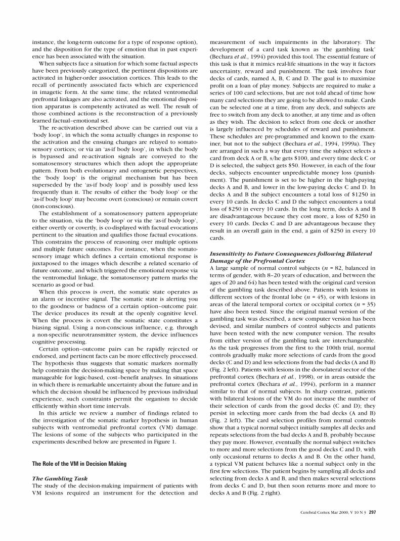
As the task progresses from the first to the 100th trial, normal
controls gradually make more selections of cards from the good
decks (C and D) and less selections from the bad decks (A and B)
(Fig. 2 left). Patients with lesions in the dorsolateral sector of the
prefrontal cortex (Bechara et al., 1998), or in areas outside the
prefrontal cortex (Bechara et al., 1994), perform in a manner
similar to that of normal subjects. In sharp contrast, patients
with bilateral lesions of the VM do not increase the number of
their selection of cards from the good decks (C and D); they
persist in selecting more cards from the bad decks (A and B)
(Fig. 2 left). The card selection profiles from normal controls
show that a typical normal subject initially samples all decks and
repeats selections from the bad decks A and B, probably because
they pay more. However, eventually the normal subject switches
to more and more selections from the good decks C and D, with
only occasional returns to decks A and B. On the other hand,
a typical VM patient behaves like a normal subject only in the
first few selections. The patient begins by sampling all decks and
selecting from decks A and B, and then makes several selections
from decks C and D, but then soon returns more and more to
decks A and B (Fig. 2 right).
Cerebral Cortex Mar 2000, V 10 N 3 297

In the normal population, performance on the gambling task
does not seem to depend on education or gender, although a few
preliminary reports suggest that males perform slightly better
than females (LeLand et al., 1998; Reavis et al., 1998). Most
intriguing is that, as a group, older adults (above 64 years of age)
perform poorly on this task relative to younger adults (i.e. age
26–56) (Denburg et al., 1999). It should be noted, however, that
performance on the gambling task in older adults is dichoto-
mous, i.e. some perform very well and some perform very
poorly. This finding raises an important question as to why
this happens in some older adults and not others, the answer to
which may help explain why some older adults are especially
vulnerable to advertising fraud in real life.
In the VM patient population, the decision-making impair-
ment, as measured by the gambling task, is stable over time.
When a sample of six VM frontal patients and five normal
controls were re-tested after various time intervals (1 month after
the first test, 24 h later and for the fourth time, 6 months later),
the performance of VM patients did not improve. On the other
hand, the performance of normal controls improved
significantly over time (Fig. 3).
These results demonstrate that the VM patients’ performance
profile is comparable to their real-life inability to learn from their
previous mistakes. This is especially true in personal and social
matters, a domain for which in life, as in the gambling task,
an exact calculation of the future outcome is not possible and
choices must be based on approximations.
Biases Guide Decisions
The results described above prompted the following question:
what is the basis for the ‘myopia for the future’ that plagues VM
frontal patients? For many years, many theorists of decision
making assumed that the feelings triggered when making a
decision or a risky choice were not integral to the decision-
making process. In this sense, the decision-making theorists
assumed that risky decision making was essentially a cognitive
activity devoid of an emotional component. These theories
suggest that people assess the possible outcomes of their actions
through some type of cost–benefit analysis. However, several
authors have proposed an alternative theoretical account which
highlights the role of the affect experienced during the time
of deliberation prior to making decisions (Schwartz and Clore,
1983; Zajonc, 1984; Damasio et al., 1990).
Evidence in support of this idea comes from studies of normal
control subjects and patients with bilateral VM frontal damage
during their performance on the gambling task, and the analysis
of their psychophysiological activity during task performance
(Bechara et al., 1996). Skin conductance response (SCR) activity
has been recorded so far in a large sample of normal subjects
(n = 55) and VM patients (n = 15) during the performance of
the gambling task. Despite some variations in the methods for
collecting the SCR data (Bechara et al., 1996, 1999a), the general
principles remain the same. Every time the subject picks a card,
the deck from which that card was picked is recorded, and the
magnitude of the SCR in the time window (∼ 5 s) right before the
subject picked the card is measured. In addition, the magnitude
of the SCR in the time window (∼ 5 s) after the card was picked is
also measured. Thus, three types of responses are identified. (i)
The reward SCRs, those occurring after turning cards with
reward only. (ii) The punishment SCRs, those occurring after
turning cards with reward and punishment. (iii) The antici-
patory SCRs, those occurring before turning a card from a deck,
during the time the subject ponders from which deck to choose
(Bechara et al., 1996).
Figure 2. (Left panels) Card selection on the gambling task as a function of group (normal control, VM patients), deck type (disadvantageous versus advantageous), and trial block.Normal control subjects (n = 82) shifted their selection of cards towards the advantageous decks. The VM frontal patients (n = 15) opted for the disadvantageous decks. (Rightpanels) Profiles of card selections (from the first to the 100th selection) obtained from a typical control and a typical VM patient. Although the VM patient made numerous switches,he returned more often to the disadvantageous decks.
298 Emotion, Decision Making and the Orbitofrontal Cortex • Bechara et al.

The results from the psychophysiological experiments
conducted so far reveal that normal controls and VM patients
generate SCRs as a reaction to reward or punishment. Normal
controls, however, as they become experienced with the task,
also begin to generate SCRs before the selection of any card. The
anticipatory SCRs generated by normal controls: (i) develop over
time (i.e. after selecting several cards from each deck, and thus
encountering several instances of reward and punishment); and
(ii) actually become more pronounced before selecting cards
from the disadvantageous decks (A and B). These anticipatory
SCRs are absent in the VM patients (Fig. 4). This suggests that VM
patients have a specific impairment in their ability to generate
anticipatory SCRs in response to a possible outcome of their
action. Since SCRs are physiological indices of an autonomically
controlled change in somatic state, it seems reasonable to
conclude that the absence of anticipatory SCRs is an indication
that these patients’ ability to change somatic states in response to
an imagined scenario is severely compromised. In this
perspective, the failure to enact a somatic state appropriate to
the consequences of a response would be a correlate of their
inability to choose advantageously.
Risk Taking versus Impaired Decision Making
None of the bilateral VM patients tested so far have performed
advantageously on the gambling task. However, not every
normal control subject performs advantageously. Approximately
20% of normal adults who describe themselves as high-risk takers
in real life end up selecting more cards from the bad decks
relative to the good ones (Bechara et al., 1999a). When looking
at the anticipatory SCRs in these normal individuals, it is often
found that the magnitudes of the anticipatory SCRs in relation to
the bad decks are slightly lower than those in relation to the good
decks (Bechara et al., 1999a). The opposite is true (i.e. higher
anticipatory SCRs with the bad decks relative to the good decks)
in normal individuals who play advantageously. The most critical
distinction between these normal individuals and the VM
patients, however, is that these normal individuals do generate
anticipatory SCRs. The VM patients, on the other hand, do not
Figure 3. A learning curve revealing the level of performance of normal control (n = 5) and VM patients (n = 6) on the gambling task, as a function of repetition over time. The VMpatients failed to show a significant improvement as a function of repeated testing.
Figure 4. Magnitudes of anticipatory SCRs as a function of group [normal control (A) (n = 12) versus VM patients (B) (n = 7)], deck and card position within each deck. Note thatcontrol subjects gradually began to generate high-amplitude SCRs to the disadvantageous decks. The VM patients failed to do so.
Cerebral Cortex Mar 2000, V 10 N 3 299

generate anticipatory SCRs at all. These physiological results are
very important because they separate individuals with high-
risk-taking behavior from individuals with VM frontal lobe
dysfunction. When taking a risk, the somatic states signaling the
possible negative consequences of the outcome are enacted.
However, the individual can override these biases by higher
cognitive processes. In the case of VM damage, these biases
are never enacted and never enter the decision-making process.
This suggests that taking a risk is not the same as having poor
judgement and impaired decision making. The issue of risk
taking versus decision making was addressed in a previous study
that showed that orbitofrontal patients risked significantly less
of their accumulated reward than controls, thus suggesting a
pattern of conservative behavior (Rogers et al., 1999). Yet, these
same patients made suboptimal choices and spent more time
deliberating their choices (Rogers et al., 1999). Another study
suggested that while frontal patients were impulsive and made
poor decisions, they did not express a high-risk-taking behavior
(Miller, 1992). This evidence suggests that risk-taking behavior
and impaired decision making are not synonymous.
Biases Do Not Need To Be Conscious
Given the important role that biases play in decision making, it is
important to determine if these anticipatory responses (biases)
develop after the subject knows which decks are good or bad,
or if they precede such explicit knowledge. This question was
addressed in an experiment in which ten normal subjects and six
VM frontal patients were tested on the gambling task, while their
SCRs were being recorded as before. However, in this
experiment, every time a subject had picked ten cards the
game was stopped brief ly and the subject was asked to describe
whatever s/he knew was going on in the game (Bechara et
al., 1997). The analysis of the subjects’ answers suggested that
they went through four distinct periods across the task. The
first was a pre-punishment period, when subjects sampled
the decks, before they encountered any punishment. The second
was a pre-hunch period, when subjects began to encounter
punishment, but had no clue about what was going on in the
game. The third was a hunch period, when subjects began to
express a hunch about the decks that were riskier, even if they
were not sure about their guess. The fourth was a conceptual
period, when subjects knew very well the contingencies in the
task, which decks were the good ones, which decks were the
bad ones and why this was so (Fig. 5). It is interesting that 30% of
the control subjects did not reach the fourth, or conceptual,
period in this experiment, yet they performed normally on the
gambling task.
In normal controls, when the anticipatory SCRs from each of
these four periods were examined, it was found that there was
no significant anticipatory activity during the pre-punishment
period. There was a substantial rise in anticipatory responses
during the pre-hunch period, i.e. before any conscious know-
ledge developed. This anticipatory SCR activity was sustained for
the rest of the task. When the type of choice from the different
decks was examined for each period, the results revealed that
there was a preference for the high-paying decks (A and B)
during the pre-punishment period. There was a hint of a shift in
the pattern of card selection, away from the bad decks, as early
as in the pre-hunch period. This preference for the good decks
became more pronounced during the hunch and conceptual
periods. Even those 30% of controls who did not reach a full
conceptual knowledge of the relative goodness or badness of
the decks ended up playing advantageously. The VM frontal
patients, on the other hand, never reported a hunch. They also
never developed anticipatory SCRs, and continued to choose
more cards from decks A and B relative to C and D. However, 50%
of VM frontal patients did reach the conceptual period, in which
Figure 5. Anticipatory SCRs and behavioral responses (card selection) as a function of four periods (pre-punishment, pre-hunch, hunch and conceptual) from normal control subjects(n = 10) and VM patients (n = 6).
300 Emotion, Decision Making and the Orbitofrontal Cortex • Bechara et al.
they were able to recognize and identify the bad decks. Even so,
they still performed disadvantageously (Bechara et al., 1997).
These results show that VM frontal patients continue to
choose disadvantageously in the gambling task, even after
realizing the consequences of their action. This suggests that
the anticipatory SCRs represent unconscious biases, probably
derived from prior experiences with reward and punishment.
These biases help deter the normal subject from pursuing a
course of action that is disadvantageous in the future. This
biasing effect occurs even before the subject becomes aware of
the goodness or badness of the choice s/he is about to make.
Even without these biases, the knowledge of what is right and
what is wrong may become available, as happened in 50% of the
VM patients. However, by itself, such knowledge is not sufficient
to ensure an advantageous behavior. Although the frontal patient
may be fully aware of what is right and what is wrong, s/he still
fails to act accordingly. These patients may ‘say’ the right thing,
but ‘do’ the wrong thing.
Relationship between Emotion, Memory and Decision MakingIt has been established that the memory of facts is improved
when the facts are learned in connection with an emotion (Cahill
et al., 1995; Roozendaal et al., 1996), although under extreme
conditions (e.g. intense arousal) emotions can actually impair
memory (Easterbrook, 1959). The prefrontal cortex, especially
its dorsolateral (DL) sector, has been linked to the ability to
remember facts for a short period of time, i.e. working memory
(Goldman-Rakic, 1987; Fuster, 1991; D’Esposito et al., 1995;
Smith et al., 1995; Courtney et al., 1997). Thus, it became
pertinent to ask whether cognitive functions related to working
memory were distinct from those related to decision making. In
other words, do working memory and decision making depend
at least in part on separate anatomical substrates? Furthermore,
it seemed pertinent to investigate whether the mechanism by
which emotion boosts working memory (or improve the short
term memory for certain facts) is the same as, or different from,
the mechanism through which emotion biases decision making.
Decision Making and Working Memory are Distinct
Operations of the Prefrontal Cortex
The rationale for the notion that working memory and decision
making are distinct functions comes from the observations that
VM frontal patients suffer from impairments in decision making,
while preserving a normal level of memory and intellect. On
the other hand, although some DL frontal patients complain
of memory impairments, they do not appear to suffer from
impairments in decision making, as judged from their behavior
in real life. Using modified delay-task procedures (delayed
response and delayed non-matching to sample) to measure
working memory (Goldman-Rakic, 1987; Fuster, 1991), and
the gambling task to measure decision making, the following
experiment was performed. A group of 21 normal control
subjects, nine patients with bilateral VM frontal lesions and ten
patients with right or left lesions of the DL sector of the pre-
frontal cortex were tested on the delay and gambling tasks
(Bechara et al., 1998). The gambling task was the same task
mentioned previously, and the delay task procedures were
modifications of classical delay tasks.
Delay tasks that are used in non-human primates are too
simple for use with humans. Therefore, a distracter was intro-
duced during the delay between the cue and the response. The
purpose of the distracter was to interfere with the ability of the
subject to rehearse the position or the color of the cues during
the delay, and thus to increase the demands of the tasks on
working memory. In the delayed response experiment, four
cards appeared for 2 s on a computer screen, with two of the
cards face down and the other two face up, showing red or
black colors. The cards disappeared for one, 10, 30 or 60 s and
then reappeared, but this time all the cards were face down. The
correct response was to select the two cards that were initially
face up. During the delay, the subject had to read aloud a series
of semantically meaningless sentences. Scores were calculated as
the percent of correct choices made by the subject at the 10, 30
and 60 s delays. Impaired performance on the delayed response
task was defined as achieving a percent correct score of 80
or less at the 60 s delay, a cutoff score below which no normal
control ever performed (Bechara et al., 1998). In the delayed
non-matching to sample experiment, the task was similar to
the delayed response task except that only one card appeared
initially on the computer screen for 2 s. The card was face up and
was either red or black. After the card disappeared for 1, 10,
30 or 60 s, four cards appeared on the screen, all face up, two
of which were red and two black. The correct response was to
select the two cards that were opposite in color (non-matching)
to the initial sample card.
In this experiment we used two types of delay tasks because
studies in non-human primates show that different areas of
the DL frontal cortex are associated with different domains of
working memory. The inferior areas of the dorsolateral sector
have been associated with object memory, whereas the superior
areas have been associated with spatial memory (Goldman-
Rakic, 1987, 1992; Wilson et al., 1993). Similar dissociations
were found in humans (Courtney et al., 1996). The delayed
response tasks have been designed to tax the spatial (where)
domain of working memory, whereas the delayed non-matching
to sample tasks are supposed to tax the object (what) domain
of working memory (Fuster, 1990; Wilson et al., 1993). Since
the lesions in the patients we studied were not restricted to
the inferior or superior regions, and the lesions spanned a wide
area of DL frontal cortices, we used both types of delayed tasks
because we anticipated that both domains of working memory
(spatial and object) may be affected. In other words, our attempt
was not to sort out differences between different types of work-
ing memory, but rather to cover a range of working memory
with one task. Therefore, the results we report here are an
average of the results obtained from both delay tasks. In the next
section, we use the term ‘delay tasks’ to refer to both procedures
(delayed response and delayed non-matching to sample) [results
obtained with each individual delay task are given elsewhere
(Bechara et al., 1998)].
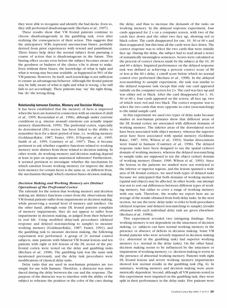
This experiment revealed two intriguing findings. First,
working memory is not dependent on the intactness of decision
making, i.e. subjects can have normal working memory in the
presence or absence of deficits in decision making. Some VM
frontal patients who were severely impaired in decision making
(i.e. abnormal in the gambling task) had superior working
memory (i.e. normal in the delay tasks). On the other hand,
decision making seems to be inf luenced by the intactness or
impairment of working memory, i.e. decision making is worse in
the presence of abnormal working memory. Patients with right
DL frontal lesions and severe working memory impairments
showed low normal results in the gambling task (Fig. 6). In
summary, working memory and decision making were asym-
metrically dependent. Second, although all VM patients tested in
this experiment were impaired on the gambling task, they were
split in their performance in the delay tasks. Five patients were
Cerebral Cortex Mar 2000, V 10 N 3 301

abnormal in the delay tasks (Abnormal Gambling/Abnormal
Delay) and four were normal in the delay tasks (Abnormal
Gambling/Normal Delay) (Fig. 7, graphs). The most important
finding is that all patients in the Abnormal Gambling/Abnormal
Delay group had lesions that extended posteriorly, possibly
involving the basal forebrain region. However, the other group
(Abnormal Gambling/Normal Delay) had lesions that were more
anterior and did not involve the basal forebrain (Fig. 7, anatomy).
It is important to note that in this experiment only the
patients with right DL lesions were impaired on these working
memory tasks. All patients with left DL frontal lesions had
normal working memory. The absence of a working memory
impairment in left DL patients is not surprising because, during
the delay, the verbal memorization of cues was probably avoided
by the interference procedure, thus rendering the task primarily
non-verbal. This is consistent with several functional neuroimag-
ing studies in humans that showed higher activation in the right
DL frontal cortex, relative to the left, during the performance
of similar delay tasks (Jonides et al., 1993; Petrides et al., 1993;
McCarthy et al., 1994; D’Esposito et al., 1995a,b; Smith et al.,
1995; Swartz et al., 1995).
These findings reveal a double dissociation (cognitive and
anatomic) between deficits in decision making (anterior VM)
and working memory (right DL). They reinforce the special
importance of the VM region in decision making, independently
of a direct role in working memory.
The Emotional Mechanism that Biases Decision Making
is Distinct from the Emotional Mechanism that
Improves Memory
The previous discussion leads to the question of whether the
mechanism by which emotion improves memory is the same as,
or different from, the mechanism through which emotion biases
decisions. The amygdala has been found to be necessary for
emotions to improve memory (Cahill et al., 1995). Our own
work has also shown that the amygdala is important in the
creation of biases and in decision making (Bechara et al., 1999a).
This suggests that in the amygdala, the mechanisms through
which emotion modulates memory and decision making may be
inseparable. The remaining question is whether these mech-
anisms might be separable in the VM cortex. In order to answer
this last question, we tested 12 normal control subjects and six
VM patients with anterior lesions that spared the basal forebrain
for their memory of a series of neutral and emotionally charged
pictures. The series of pictures involved four sets, with four
pictures in each set. Each set of four pictures contained two
neutral (e.g. farm scenes) and two emotional (e.g. raped and
mutilated bodies’) pictures. The pictures in set 1 were presented
once each; those in set 2 were presented twice each; in set 3,
four times each; and in set 4, eight times each. Five minutes after
viewing all the pictures, subjects were tested for their recall of
each picture they saw, and for the overall content of the picture.
The recall of picture content was calculated for each subject as a
function of repetition times and emotional content.
As might be expected, both normal controls and VM patients
showed improved memory as a result of repetition. The most
important finding, however, was that both groups showed a
response to the emotion manipulation, producing a better
memory curve for pictures with emotional content than for
neutral pictures (Fig. 8). Thus, this experiment actually
Figure 6. Means ± SEM of the average of percent correct responses from the twodelay tasks, or the total number of cards selected from the good decks, that were madeby VM patients (n = 4) with more anterior lesions and by patients with right DL lesions(n = 4). Note that the VM patients were severely impaired on the gambling task (lownumber of choices from the good decks), but normal on the delay tasks (high % correctresponse). On the other hand, the right DL patients were impaired on the delay tasksand, although their gambling task performance is considered advantageous, it falls in thelow normal range.
Figure 7 (Graphs) The behavioral results on the gambling task and the delay tasks fromthe groups of patients shown in Figure 7 (Anatomy).
302 Emotion, Decision Making and the Orbitofrontal Cortex • Bechara et al.

separated the memory curve that is a function of repetition from
the curve that is a function of emotional content. The results
indicate that the VM patients are able to use emotional content in
order to enhance their memory, suggesting that the mechanism
through which emotion modulates decision making is different
from that through which emotion modulates memory. These
Figure 7. (Anatomy) Separate mapping of VM lesions for the group with Abnormal Gambling/Abnormal delay (A) (n = 5) and the group with Abnormal Gambling/Normal Delay (B)(n = 4). Red indicates an overlap of two subjects or more. The maximal overlap of lesions in (A) is seen spanning the whole extent of the mesial orbital surface of the frontal lobe. Itreaches the posterior sector (coronal slice 4), where basal forebrain structures are found. However, in (B) the maximal overlap is mostly anterior extending only to slice 1 and 2. Slices4 does not show any lesion. Coronal sections are arranged according to radiological convention, i.e. right is left, and vice versa.
Cerebral Cortex Mar 2000, V 10 N 3 303

results also support the conclusion that the decision-making
impairment of VM patients cannot be explained by a deficit in
the recall of emotional events.
Why Do VM Frontal Patients Fail to Trigger Somatic States whenThey Contemplate Decisions?The series of studies outlined earlier helped establish that
decision making is critically dependent on the generation of
somatic states or biases. Why do VM patients fail to generate
these biases or emotional signals? Is it because they no longer can
re-experience emotions? Is it because they need a much higher
threshold for triggering an emotion? Is it because they no longer
can attach emotional significance to a neutral event, as, for in-
stance, in conditioning? Our research addressing these questions
is still in its preliminary phase. The nature of the mechanism
responsible for the failure of VM patients to trigger somatic states
when pondering decisions remains unspecified. The following is
a preliminary search for the answer.
Emotional Conditioning
We have tested whether one reason for VM frontal patients to fail
to trigger anticipatory biases (anticipatory SCRs) when contem-
plating a decision in the gambling task is due to a failure to
couple an exteroceptive stimulus (or event) with the somatic
state of a punishment. These patients may have a defect in
acquiring fear conditioning. To test this possibility directly, we
used a fear-conditioning procedure, which consisted of using
four different colors of monochrome slides as conditioned
stimuli (CS) and a startlingly aversive loud sound (100 db) as the
unconditioned stimulus (US) (Bechara et al., 1995). Electro-
dermal activity (SCR) served as the dependent measure of
autonomic conditioning. The emotional conditioning of each
subject included three phases: (i) a habituation phase; (ii) a
conditioning phase. In this phase, only one of the colors was
paired with the US. These CS slides were presented at random
among the other colors; and (iii) an extinction phase.
A group of ten VM patients and ten matched control subjects
were tested in the experiment just mentioned. The VM patients
did condition to the loud noise (Tranel et al., 1996; Bechara et
al., 1999a). This suggests that the VM cortex is not essential for
emotional conditioning. Consequently, it is reasonable to assume
that the failure of VM patients to acquire anticipatory SCRs in the
gambling task, and their decision-making impairment, cannot
be explained by a failure to acquire conditioned emotional
responses.
The Experience of Emotions
Recently, we have started to address the question of whether one
reason that VM frontal patients fail to trigger anticipatory biases
(anticipatory SCRs) is the inability to re-experience the
emotional state associated with punishment when recalling pre-
vious instances of punishment. A variety of procedures can be
used to induce emotions in human subjects, such as watching
emotional film clips, looking at pictures charged with emotions
or recalling highly emotional personal events. In this prelim-
inary study, emotional imagery was used as a method to induce
emotional states. The subjects are asked to think about and
describe a situation in their lives in which they felt each of the
following emotions: happiness, sadness, fear and anger. After a
brief description of each story is obtained, the subject is asked to
imagine and re-experience each emotional situation while their
physiological activity (SCR, heart rate, respiratory frequency,
skin temperature and facial EMG) is monitored. As a control
condition, the subject is also asked to recall and imagine a non-
emotional situation, e.g. getting up that morning, showering,
dressing up, having breakfast and then going to work.
We tested eight VM patients with this procedure. They were
all able to retrieve previous emotional experiences. Most import-
antly, they generated higher physiological activity (e.g. SCR and
heart rate) during the imagery of the angry situations than
during the neutral situations (Damasio et al., 1997; Tranel et
al., 1998). Although all the VM patients reliably re-experienced
anger, the re-experience of fear was less reliable, i.e. some could
not experience it at all, and those who could did so with a less
intense response. Whereas the VM patients were able to re-
experience anger and in some cases fear, most of them had
difficulties conjuring up a happy or sad emotion. This suggests
that damage to the VM cortex weakens the ability to re-experi-
ence an emotion from the recall of an appropriate emotional
event. Consequently, it is reasonable to assume that the failure
of VM patients to acquire anticipatory SCRs, coupled to their
decision-making impairment, is in part due to their inability
to re-experience the emotion of a previously fearful situation.
Obviously, this is a preliminary finding that requires further
investigation.
The same is also true for the induction of an emotion by
external stimuli such as the viewing of emotionally charged
pictures. Indeed, earlier studies showed that VM patients failed
to generate SCRs to emotionally charged pictures when they
viewed these pictures passively (Damasio et al., 1990). However,
the same patients generated normal magnitude SCRs to the same
target pictures when they were asked to view and describe
the content of the pictures (Damasio et al., 1990). During the
gambling task, VM patients would generate SCRs when they
lost a large sum of money, but the magnitude of these SCRs was
never as high as that of normal controls (Bechara et al., 1999a).
Together, these results suggest that these patients may have a
weakened ability to process the affective attribute of an emo-
tional stimulus or to actually experience the emotion associated
with that stimulus. This weakness may contribute to the failure
to trigger somatic states when deliberating about options for
Figure 8. Recall scores in normal control (n = 12) and VM patients with anteriorlesions that spare the basal forebrain (n = 4) as a function of repetition and emotionalcontent. The VM patients showed a strong improvement in recall as a function of theemotional manipulation. We note that although in VM patients with basal forebrainlesions the overall recall is somewhat lower than in normal controls (not shown in thefigure), these patients still show a strong improvement in recall as a function of theemotional manipulation.
304 Emotion, Decision Making and the Orbitofrontal Cortex • Bechara et al.

a decision. However, the fact that these VM patients are not
completely emotionless suggests that this weakness is not the
sole factor responsible for the failure. Nonetheless, it is an
intriguing thought that if the experience of punishment would
be more intense, the VM patients might overcome the failure to
re-experience the emotional state of punishment, and therefore
improve the decision-making impairment. For instance, in the
gambling task, if the punishment was made several times the
amount that is effective in normal subjects, it may become
effective and VM patients might begin to choose advantageously.
The Issue of Impulsiveness and Response InhibitionThe notion of impulsiveness is often linked to the function of
the prefrontal cortex (Miller, 1992; Fuster, 1996), and is usually
understood as a lack of response inhibition. In other words, the
subject is unable to suppress or withhold a previously rewarding
response, and the behavior appears impulsive. It is important to
address this issue of impulsiveness in relation to the foregoing
studies.
First, it is important to distinguish between motor and
cognitive impulsiveness. Motor impulsiveness is usually studied
in animals under the umbrella of ‘response inhibition’. After
establishing a habit to respond to a stimulus that predicts a
reward, there is a sudden change in the contingencies of the task
that requires the inhibition of the previously rewarded response.
Go/no-go tasks, delayed alternation and response shifting are
examples of experimental design that measure this type of
impulsive behavior (Mishkin, 1964; Freedman, 1986; Diamond,
1990; Fuster, 1990; Stuss, 1992; Dias et al., 1996; Freedman et
al., 1998). In humans, impulsive behavior is often uncovered in
neuropsychological tasks that detect ‘perseverative errors’, such
as the Wisconsin Card Sorting Task, or in experimental tasks
such as delayed alternation (Freedman, 1986; Freedman et al.,
1998). Cognitive impulsiveness, on the other hand, which can
be seen as related to an inability to delay gratification, is a more
complex form of disinhibited behavior. Cognitive impulsiveness
may be illustrated with the example in which a child sees a piece
of a candy on the table and is told by the parent ‘no, you must
wait 30 min before you can have the candy; otherwise, you face
punishment’. The child understands the information and holds
for a short while but after 2 min can no longer delay the
gratification, resist the temptation and inhibit the response to
reach for the candy.
Previous studies have shown that VM patients with lesions
that spare the basal forebrain do not show motor impulsiveness
and do not perseverate in conventional neuropsychological tests,
although those with lesions that involve the basal forebrain may
(Bechara et al., 1998). We can also state that VM patients during
their performance of the gambling task switch decks whenever
they receive punishment, just as normal controls do. Such a
performance does not suggest a lack of inhibition to a previously
rewarding response (Fig. 2). VM patients are also unimpaired in
delay task procedures (i.e. delayed non-matching to sample)
considered sensitive to deficits in response inhibition (Bechara
et al., 1998).
On the other hand, the behavior of VM patients in the
gambling task, and in real life, can be viewed as similar to the
cognitive impulsiveness of the child with the candy. That is to
say, when the patients are presented with a deck of cards which
yields a large immediate reward, even if it can cost a large loss in
the future, the patients seem unable to delay the gratification of
the reward for too long. Their tendency to return quickly and
more often to the decks that yield high immediate reward seems
to suggest such a mechanism. The question, however, is ‘who’
decides when to suppress, or not to suppress, such a response as
the seeking of a large immediate reward? Somatic states may
indeed serve as the decision maker in such a situation. The
following is a proposal of how somatic states may interact with
mechanisms of response inhibition.
Using the impulsive behavior of the child with the candy as an
illustrative example, one can see the conf lict created by the
decision to reach or not to reach for the candy. There are positive
somatic states generated by the immediate and available reward
(the candy), or the large sum of money in the gambling task. On
the other hand, there are negative somatic states generated by
the delayed punishment threatened by the parent or the possible
loss of a large sum of money in the gambling task. If the threat
of punishment were severe enough, then the evoked negative
somatic states generated by that threat would counteract the
positive somatic states produced by the immediate reward. The
choice to seek the reward would thus be marked with a negative
value, and the response to reach for the immediate reward
might be inhibited. However, if the situation were that of a mild
punishment, which would let the immediate reward outweigh
the future punishment, the negative somatic states triggered
by the possible punishment might not be sufficiently strong
to counteract the positive states triggered by the immediate
reward. In this case, the choice would be marked with a positive
value. In our example, the child reaches for the candy and the
behavior may be considered normal, advantageous and not
impulsive. This example illustrates two different readings of the
same situation involving an immediate reward and a future
punishment. The difference, however, is that in one situation the
inhibition of the action to seek the reward should be inhibited
because the delayed punishment outweighs the immediate
reward. In the other situation, the action to seek the reward
should not be inhibited because the immediate reward out-
weighs the delayed punishment. The construct of impulsiveness
and response inhibition by itself does not explain when to
inhibit a given response or not. The activation of somatic states
provides the important signals leading to whether to inhibit the
response under consideration or not.
ConclusionWith the exception of a few theories on decision making
(Janis and Mann, 1977; Mann, 1992), most current theories of
choice use a cognitive perspective. These theories assume that
decisions derive from an assessment of the future outcomes of
various options and alternatives through some type of cost–
benefit analyses. Some of these theories have addressed emotion
as a factor in decision making, but mostly as a consequence of
a decision (e.g. the disappointment or regret experienced after
some risky decision) rather than as the reactions arising directly
from the decision itself at the time of deliberation. The somatic
marker hypothesis proposes that individuals make judgements
not only by assessing the severity of outcomes and their
probability of occurrence, but also and primarily in terms of
their emotional quality. Lesions of the VM prefrontal cortex
interfere with the normal processing of somatic or emotional
signals, but leave other cognitive functions minimally affected.
This damage leads to pathological impairments in the decision-
making process which seriously compromise the efficency of
everyday-life decisions. The somatic marker proposal is consist-
ent with the views of others who invoke a primary role for
mood, affect and emotion in decision making (Schwartz and
Clore, 1983; Zajonc, 1984; LeDoux, 1996). However, it differs
Cerebral Cortex Mar 2000, V 10 N 3 305

from the view that body signals only introduce noise into the
decision-making system (Rolls, 1999). Shallice has proposed a
model for decision making that invokes the idea of marking
various options with a value (Shallice, 1993). However, the
nature of these markers is not specified, and it is implied that
they are cognitive in nature. Thus, the views of both Rolls
and Shallice are more consistent with the ‘as-if body loop’
component of the somatic marker hypothesis. The fundamental
notion of the somatic marker hypothesis is that bioregulatory
signals, including those that constitute feeling and emotion,
provide the principal guide for decisions and are the basis for the
development of the ‘as-if body loop’ mode of operation.
The somatic marker hypothesis and the experimental
strategies used to study decision making in neurological patients
provide parallels and direct implications for understanding the
nature of several psychiatric disorders. For instance, substance
abusers are similar to VM patients in that when faced with a
choice that brings some immediate reward (i.e. taking a drug),
at the risk of incurring a loss of reputation, job, home and
family, they choose the immediate reward and ignore the future
consequences. Using the gambling task (Grant et al., 1997; Petry
et al., 1998; Bechara et al., 1999b) or related decision-making
tasks (Rogers et al., 1999), recent studies have indicated that
impairment in decision making may stand at the core of the
problem of substance abuse. Similarly, the personality profile of
VM patients bears some striking similarities to psychopathic (or
sociopathic) personality, so much so that we have used the term
‘acquired sociopathy’ to describe the condition of patients with
VM damage (Damasio et al., 1990). The qualifier ‘acquired’
signifies that the condition in VM patients follows the onset of
brain injury, and occurs in persons whose personalities and
social conduct were previously normal. The patients are usually
not destructive or harmful to others, a feature that tends to
distinguish the ‘acquired’ form of the disorder from the standard
‘developmental’ form. Indeed, recent evidence has indicated
that the earlier the onset of VM damage, the more severe the
antisocial behavior, suggesting that early dysfunction in the
prefrontal cortex may, by itself, cause abnormal development of
social and moral behavior (Anderson et al., 1999). Recent studies
have begun to look at the possibility that the psychopathic
behavior seen in cases in which no neurological history has been
identified may be linked to abnormal operation of the neural
system involving the VM (Schmitt et al., 1999). Finally, in
addition to the disorders mentioned above, applications of the
somatic marker hypothesis may extend to psychiatric disorders
that include schizophrenia (Wilder et al., 1998), pathological
gambling, depression, and attention deficit and hyperactivity
disorders (ADHD).
NotesThis work was supported in part by NIH Program Project Grant PO1
NS19632, and the Mathers Foundation. We are indebted to Daniel Tranel
and Ralph Adolphs for their valuable suggestions.
Address correspondence to Antonio R. Damasio, Department of
Neurology, University of Iowa Hospitals and Clinics, Iowa City, IA 52242,
USA. Email: [email protected] ([email protected].
uiowa.edu).
ReferencesAnderson SW, Bechara A, Damasio H, Tranel D, Damasio AR (1999)
Impairment of social and moral behavior related to early damage in
the human prefrontal cortex. Nature Neurosci 2:1032–1037.
Bechara A, Damasio AR, Damasio H, Anderson SW (1994) Insensitivity to
future consequences following damage to human prefrontal cortex.
Cognition 50:7–15.
Bechara A, Tranel D, Damasio H, Adolphs R, Rockland C, Damasio AR
(1995) Double dissociation of conditioning and declarative
knowledge relative to the amygdala and hippocampus in humans.
Science 269: 1115–1118.
Bechara A, Tranel D, Damasio H, Damasio AR (1996) Failure to respond
autonomically to anticipated future outcomes following damage to
prefrontal cortex. Cereb Cortex 6:215–225.
Bechara A, Damasio H, Tranel D, Damasio AR (1997) Deciding
advantageously before knowing the advantageous strategy. Science
275:1293–1295.
Bechara A, Damasio H, Tranel D, Anderson SW (1998) Dissociation of
working memory from decision making within the human prefrontal
cortex. J Neurosci 18:428–437.
Bechara A, Damasio H, Damasio AR, Lee GP (1999a) Different
contributions of the human amygdala and ventromedial prefrontal
cortex to decision-making. J Neurosci 19:5473–5481.
Bechara A, Dolan S, Hindes A, Anderson SW, Nathan P (1999b) Decision-
making deficits, linked to a dysfunctional orbitofrontal cortex,
revealed in alcohol and stimulant abusers. Soc Neurosci Abstr (in
press).
Cahill L, Babinsky R, Markowitsch HJ, McGaugh JL (1995) The amygdala
and emotional memory. Nature 377:295–296.
Courtney SM, Ungerleider LG, Keil K, Haxby JV (1996) Object and spatial
visual working memory activate separate neural systems in human
cortex. Cereb Cortex 6:39–49.
Courtney SM, Ungerleider LG, Keil K, Haxby JV (1997) Transient and
sustained activity in a distributed neural system for human working
memory. Nature 386:608–611.
Damasio AR (1989a) The brain binds entities and events by multiregional
activation from convergence zones. Neural Computat 1:123–132.
Damasio AR (1989b) Time-locked multiregional retroactivation: a
systems-level proposal for the neural substrates of recall and
recognition. Cognition 33:25–62.
Damasio AR (1994) Descartes’ error: emotion, reason, and the human
brain. New York: Grosset/Putnam.
Damasio H (1995) Human brain anatomy in computerized images. New
York: Oxford University Press.
Damasio AR (1996) The somatic marker hypothesis and the possible
functions of the prefrontal cortex. Phil Trans R Soc Lond B 351:
1413–1420.
Damasio AR, Anderson SW (1993) The frontal lobes. In: Clinical neuro-
psychology (Heilman KM, Valenstein E, eds), pp. 409–460. New York:
Oxford University Press.
Damasio AR, Damasio H (1994) Cortical systems for retrieval of concrete
knowledge: the convergence zone framework. In: Large scale
neuronal theories of the brain (Koch C, ed.), pp. 61–74. Cambridge,
MA: MIT Press.
Damasio AR, Tranel D, Damasio H (1990) Individuals with sociopathic
behavior caused by frontal damage fail to respond autonomically to
social stimuli. Behav Brain Res 41:81–94.
Damasio AR, Tranel D, Damasio H (1991) Somatic markers and the
guidance of behavior: theory and preliminary testing. In: Frontal lobe
function and dysfunction (Levin HS, Eisenberg HM, Benton AL, eds),
pp. 217–229. New York: Oxford University Press.
Damasio H, Bechara A, Tranel D, Damasio AR (1997) Double dissociation
of emotional conditioning and emotional imagery relative to the
amygdala and right somatosensory cortex. Soc Neurosci Abstr 23:
1318.
Denburg NL, Bechara A, Tranel D, Hindes AR, Damasio AR (1999)
Neuropsychological evidence for why the ability to decide advan-
tageously weakens with advancing age. Soc Neurosci Abstr 25:32.
D’Esposito M, Detre JA, Alsop DC, Shin RK, Atlas S, Grossman M (1995a)
The neural basis of central execution systems of working memory.
Nature 378:279–281.
D’Esposito M, Shin RK, Detre JA, Incledon S, Annis D, Aguirre GK,
Grossman M, Alsop DC (1995b) Object and spatial working memory
activates dorsolateral prefrontal cortex: a functional MRI study. Soc
Neurosci Abstr 21:1498.
Diamond A (1990) The development and neural bases of higher cognitive
functions. New York: New York Academy of Sciences.
Dias R, Robbins TW, Roberts AC (1996) Dissociation in prefrontal cortex
of affective and attentional shifts. Nature 380:69–72.
Easterbrook JA (1959) The effect of emotion on cue utilization and the
organization of behavior. Psychol Rev 66:183–201.
306 Emotion, Decision Making and the Orbitofrontal Cortex • Bechara et al.

Freedman M (1986) Bilateral frontal lobe disease and selective delayed
response deficits in humans. Behav Neurosci 100:337–342.
Freedman M, Black S, Ebert P, Binns M (1998) Orbitofrontal function,
object alternation and perseveration. Cereb Cortex 8:18–27.
Fuster JM (1990) Prefrontal cortex and the bridging of temporal gaps in
the perception–action cycle. In: The development and neural bases
of higher cognitive functions. (Diamond A, ed.), pp. 318–336. New
York: Annals of the New York Academy of Science.
Fuster JM (1991) The prefrontal cortex and its relation to behavior. In:
Progress in brain research (Holstege G, ed.), pp. 201–211. New York:
Elsevier Science Publishers.
Fuster JM (1996) The prefrontal cortex. Anatomy, physiology, and
neuropsychology of the frontal lobe. New York: Raven Press.
Goldman-Rakic PS (1987) Circuitry of primate prefrontal cortex and
regulation of behavior by representational memory. In: Handbook of
physiology; the nervous system (Plum F, ed.), pp. 373–401. Bethesda,
MD: American Physiological Society.
Goldman-Rakic PS (1992) Working memory and the mind. Scient Am
267:111–117.
Grant S, Contoreggi C, London ED (1997) Drug abusers show impaired
performance on a test of orbitofrontal function. Soc Neurosci Abstr
23:1943.
Janis IL, Mann L (1977) Decision-making: a psychological analysis of
conf lict, choice, and commitment. New York: Free Press.
Jonides J, Smith EE, Koeppe RA, Awh E, Minoshima S, Mintun MA (1993)
Spatial working memory in humans as revealed by PET. Nature
363:623–625.
LeDoux J (1996) The emotional brain: the mysterious underpinnings of
emotional life. New York: Simon and Schuster.
LeLand DS, Richardson JS, Vankov A, Grant SJ, Pineda JA (1998)
Decision-making and associated ERPS in low- and high-dependence
smokers performing the Iowa gambling task. Soc Neurosci Abstr
24:1175.
Mann L (1992) Stress, affect, and risk taking. In: Risk-taking behavior
(Frank YJ, ed.), pp. 202–230. Chichester: Johm Wiley & Sons.
McCarthy G, Blamire AM, Puce A, Nobre AC, Boch G, Hyder F,
Goldman-Rakic P, Shulman RG (1994) Functional magnetic resonance
imaging of human prefrontal cortex activation during a spatial
working memory task. Proc Natl Acad Sci USA 91:8690–8694.
Miller LA (1992) Impulsivity, risk-taking, and the ability to synthesize
fragmented information after frontal lobectomy. Neuropsychologia
30:69–79.
Milner B, Petrides M, Smith ML (1985) Frontal lobes and the temporal
organization of memory. Hum Neurobiol 4:137–142.
Mishkin M (1964) Perseveration of central sets after frontal lesions in
monkeys. In: The frontal granular cortex and behavior (Akert JM,
Wa K, eds), pp. 219–241. New York: McGraw-Hill.
Petrides M (1996) Specialized systems for the processing of mnemonic
information within the primate frontal cortex. Phil Trans R Soc Lond
351:1445–1457.
Petrides M, Alivisatos B, Meyer E, Evans AC (1993) Functional activation
of the human frontal cortex during the performance of verbal
working memory tasks. Proc Natl Acad Sci USA 90:878–882.
Petry NM, Bickel WK, Arnett M (1998) Shortened time horizons and
insensitivity to future consequences in heroin addicts. Addiction
93:729–738.
Reavis R, Overman WH, Hendrix S, Exposito W, Dezio-Cottle C (1998)
Possible double dissociation of function between adult males and
females in two brain system. Soc Neurosci Abstr 24:1177.
Rogers RD, Everitt BJ, Baldacchino A, Blackshaw AJ, Swainson R,
Wynne K, Baker NB, Hunter J, Carthy T, Booker E, London M,
Deakin JFW, Sahakian BJ, Robbins TW (1999) Dissociable deficits
in the decision-making cognition of chronic amphetamine abusers,
opiate abusers, patients with focal damage to prefrontal cortex, and
tryptophan-depleted normal volunteers: evidence for monoaminergic
mechanisms. Neuropsychopharmacology 20:322–339.
Rolls ET (1999) The brain and emotion. Oxford: Oxford University Press.
Roozendaal B, Cahill L, McGaugh JL (1996) Interaction of emotionally
activated neuromodulatory systems in regulating memory storage. In:
Brain processes and memory (Ishikawa K, McGaugh JL, Sakata H,
eds), pp. 39–54. Amsterdam: Elsevier.
Schmitt WA, Brinkley CA, Newman JP (1999) Testing Damasio’s somatic
marker hypothesis with psychopathic individuals: risk-takers or
risk-averse? J Abnorm Psychol (in press).
Schwartz N, Clore GL (1983) Mood, misattribution, and judgements
of well-being: information and directive functions of affective states.
J Person Soc Psychol 45:513–523.
Shallice T (1993) From neuropsychology to mental structure. Cambridge:
Cambridge University Press.
Smith EE, Jonides J, Koeppe RA, Awh E, Schumacher EH, Minoshima S
(1995) Spatial versus object working memory: PET investigations.
J Cogn Neurosci 7:337–356.
Stuss DT (1992) Biological and psychological development of executive
functions. Brain Cogn 20:8–23.
Swartz BE, Halgren E, Fuster JM, Simpkins F, Gee M, Mandelkern M
(1995) Cortical metabolic activation in humans during a visual
memory task. Cereb Cortex 3:205–214.
Tranel D, Bechara A, Damasio H, Damasio AR (1996) Fear conditioning
after ventromedial frontal lobe damage in humans. Soc Neurosci Abstr
22:1108.
Tranel D, Bechara A, Damasio H, Damasio AR (1998) Neural correlates of
emotional imagery. Int J Psychophys 30:107.
Wilder KE, Weinberger DR, Goldberg TE (1998) Operant conditioning
and the orbitofrontal cortex in schizophrenic patients: unexpected
evidence for intact functioning. Schizophr Res 30:169–174.
Wilson FAW, Scalaidhe SPO, Goldman-Rakic PS (1993) Dissociation of
object and spatial processing domains in primate prefrontal cortex.
Science 260:1955–1958.
Zajonc RB (1984) On the primacy of affect. Am Psychol 39:117–123.
Cerebral Cortex Mar 2000, V 10 N 3 307


![Emotion, Decision Making and the Antoine Bechara, Hanna ...elsewhere (Damasio, 1989a,b, 1994; Damasio and Damasio, 1994)]; dispositional knowledge can be made explicit in the form](https://static.fdocuments.net/doc/165x107/5ed2efb95beba44fdc53235d/emotion-decision-making-and-the-antoine-bechara-hanna-elsewhere-damasio.jpg)