Emerging Kidney Models to Investigate Metabolism...
55
DMD # 82958 1 1. Title page Emerging Kidney Models to Investigate Metabolism, Transport and Toxicity of Drugs and Xenobiotics Piyush Bajaj, Swapan K. Chowdhury, Robert Yucha, Edward J. Kelly, Guangqing Xiao Drug Safety Research and Evaluation, Takeda Pharmaceutical International Co., Cambridge, Massachusetts (P.B.); Drug Metabolism and Pharmacokinetics Department, Takeda Pharmaceutical International Co., Cambridge, Massachusetts (S.K.C., R.Y., G. X.); Department of Pharmaceutics, University of Washington, Seattle, Washington (E.J.K.) This article has not been copyedited and formatted. The final version may differ from this version. DMD Fast Forward. Published on August 3, 2018 as DOI: 10.1124/dmd.118.082958 at ASPET Journals on January 2, 2020 dmd.aspetjournals.org Downloaded from
Transcript of Emerging Kidney Models to Investigate Metabolism...

DMD # 82958
1
1. Title page
Emerging Kidney Models to Investigate Metabolism, Transport and Toxicity
of Drugs and Xenobiotics
Piyush Bajaj, Swapan K. Chowdhury, Robert Yucha, Edward J. Kelly, Guangqing Xiao
Drug Safety Research and Evaluation, Takeda Pharmaceutical International Co., Cambridge,
Massachusetts (P.B.); Drug Metabolism and Pharmacokinetics Department, Takeda
Pharmaceutical International Co., Cambridge, Massachusetts (S.K.C., R.Y., G. X.); Department
of Pharmaceutics, University of Washington, Seattle, Washington (E.J.K.)
This article has not been copyedited and formatted. The final version may differ from this version.DMD Fast Forward. Published on August 3, 2018 as DOI: 10.1124/dmd.118.082958
at ASPE
T Journals on January 2, 2020
dmd.aspetjournals.org
Dow
nloaded from

DMD # 82958
2
2. Running title page
Running title: Emerging kidney models for investigating transport and toxicity
Corresponding Author:
Piyush Bajaj, PhD
Global Investigative Toxicology
Drug Safety Research and Evaluation
35 Landsdowne Street,
Cambridge, MA 02139 USA
Phone: 617-679-7287
Email: [email protected]
Document Statistics:
Number of text pages: 27
Number of tables: 2
Number of figures: 2
Number of references: 114
Number of words in Abstract: 227
Number of words in Introduction: 665
Nonstandard abbreviations used in the paper:
3,4,5-trichloroaniline (TCA)
American Type Culture Collection (ATCC)
Aminopeptidase N (CD13)
Aquaporin 1 (AQP-1)
Area under the receiver operating characteristic curve (AUC-ROC)
Aristolochic acid (AA)
ATP-binding cassette (ABC)
Breast cancer resistance protein (BCRP)
Chinese Hamster Ovary (CHO)
Conditionally immortalized proximal tubule epithelial cells (ciPTEC)
Conditionally immortalized proximal tubule epithelial cells overexpressing OAT-1 (ciPTEC -
OAT1)
This article has not been copyedited and formatted. The final version may differ from this version.DMD Fast Forward. Published on August 3, 2018 as DOI: 10.1124/dmd.118.082958
at ASPE
T Journals on January 2, 2020
dmd.aspetjournals.org
Dow
nloaded from

DMD # 82958
3
Conditionally immortalized proximal tubule epithelial cells overexpressing OAT-3 (ciPTEC -
OAT3)
Cyclic guanosine monophosphate (cGMP)
Cytochromes P450 (CYPs)
Drug-drug interactions (DDIs)
European Medicines Agency (EMA)
Extracellular matrix (ECM)
Flavin-containing monooxygenase (FMO)
Food and Drug Administration (FDA)
Gamma glutamyltransferase (GGT)
Glutathione (GSH)
Glutathione disulfide (GSSG)
Glutathione-s-transferase (GST)
Heme oxygenase – 1 (HO-1)
Human embryonic kidney (HEK)
Human primary tubular cells (HPTCs)
Human primary tubular epithelial cells (HPTECs)
Human umbilical cord endothelial cells (HUVECs)
in vitro to in vivo extrapolation (IVIVE)
Induced pluripotent stem cells (iPSC)
Interleukin 6 (IL-6)
Interleukin 8 (IL-8)
International consortium for innovation and quality in pharmaceutical development (IQ)
International Transporter Consortium (ITC)
Investigational new drug (IND)
Kidney injury molecule – 1 (KIM-1)
Lactate dehydrogenase (LDH)
Macrophage colony-stimulating factor (M-CSF)
Madin-Darby Canine Kidney (MDCK)
Metabolites in safety testing (MIST)
Microphysiological systems (MPS)
Monocarboxylate transporter (MCT)
Multidrug and toxin extrusion protein 1 (MATE-1)
Multidrug and toxin extrusion protein 2K (MATE-2K)
Multidrug resistance-associated protein 2 (MRP2)
Multidrug resistance-associated protein 4 (MRP4)
Neutrophil gelatinase-associated lipocalin (NGAL)
New chemical entities (NCE)
Organic anion transporter 1 (OAT1)
Organic anion transporter 2 (OAT2)
Organic anion transporter 3 (OAT3)
Organic anion transporter 4 (OAT4)
Organic cation transporter 2 (OCT2)
organic cation transporter, novel, type 1 (OCTN1)
organic cation transporter, novel, type 2 (OCTN2)
p-aminohippuric acid (PAH)
This article has not been copyedited and formatted. The final version may differ from this version.DMD Fast Forward. Published on August 3, 2018 as DOI: 10.1124/dmd.118.082958
at ASPE
T Journals on January 2, 2020
dmd.aspetjournals.org
Dow
nloaded from
DMD # 82958
4
Peptide transporter 1 (PEPT1)
Peptide transporter 2 (PEPT2)
P-glycoprotein (P-gp)
Pharmaceuticals and medical devices agency (PMDA)
pharmacokinetic/pharmacodynamic (PK/PD)
Pharmacokinetics (PK)
Pig Kidney Epithelial cells (LLC-PK1)
Polycystic kidney disease (PKD)
Polydimethylsiloxane (PDMS)
Proximal tubule cell (PTC)
Renal Proximal Tubule Cells (RPTEC)
Simian virus 40 large T antigen (SV40T)
Sodium-glucose co-transporter-2 (SGLT2)
Solute carrier (SLC)
Sulfotransferases (SULTs)
Telomerase Reverse Transcriptase (TERT1)
Three-dimensional (3D)
Trans-epithelial electrical resistance (TEER)
Two-dimensional (2D)
Urate Anion Exchanger 1 (URAT1)
Uridine-di phosphate (UDP)
Uridine-di phosphate-glucuronosyltransferases (UGTs)
Zonula occludens 1 (ZO-1)
Zonula occludens 3 (ZO-3)
This article has not been copyedited and formatted. The final version may differ from this version.DMD Fast Forward. Published on August 3, 2018 as DOI: 10.1124/dmd.118.082958
at ASPE
T Journals on January 2, 2020
dmd.aspetjournals.org
Dow
nloaded from

DMD # 82958
5
3. Abstract
Kidney is a major clearance organ of the body responsible for the elimination of many xenobiotics
and prescription drugs. With its multitude of uptake and efflux transporters, and metabolizing
enzymes, the proximal tubule cell (PTC) in the nephron plays a key role in the disposition of
xenobiotics and is also a primary site for toxicity. In this mini-review, we first provide an overview
of the major transporters and metabolizing enzymes in the PTCs responsible for biotransformation
and disposition of drugs. Next, we discuss different cell sources that have been used to model the
PTCs in vitro, their pros and cons, and their characterization. As current technology is inadequate
to reliably evaluate drug disposition and toxicity in the kidney, the review then discusses recent
advancements in kidney microphysiological systems (MPS) and the need to develop robust in vitro
platforms that could be routinely used by pharmaceutical companies to screen compounds. Finally,
we discuss the new and exciting field of stem cell derived kidney models as potential cell sources
for future kidney MPS. Given the push from both regulatory agencies and pharma companies to
use more predictive “human-like” in vitro systems in the early stages of drug development to
reduce attrition, these emerging models have the potential to be a game changer and may
revolutionize how renal disposition and kidney toxicity in drug discovery is evaluated in the future.
This article has not been copyedited and formatted. The final version may differ from this version.DMD Fast Forward. Published on August 3, 2018 as DOI: 10.1124/dmd.118.082958
at ASPE
T Journals on January 2, 2020
dmd.aspetjournals.org
Dow
nloaded from

DMD # 82958
6
4. Introduction
The kidneys perform essential functions in humans by maintaining the composition of blood, its
pH, preventing the buildup of waste products, and keeping levels of electrolytes such as sodium,
potassium, and phosphate stable. In normal adults, the two kidneys daily filter about 150 - 180 L
of blood to produce 1 to 2 L of urine, composed of wastes and extra fluid. The kidneys are also
responsible for the elimination of numerous drugs, endogenous metabolites important to maintain
physiological homeostasis, exogenous and endogenous toxins, nutrients, etc. The elimination of
exogenous and endogenous compounds through kidney occurs as a net result of glomerular
filtration, tubular secretion, kidney metabolism, and reabsorption. Evaluation of the mechanisms
involved in the elimination of drugs and other exo- and endogenous molecules can provide
valuable understanding of their clearance, potential for drug-drug interactions (DDIs), potential
for development of kidney and other organ toxicity, and thus on the effect of the elimination of an
investigational drug on its pharmacokinetics (PK) in patients with compromised kidney functions.
In the past several decades, there has been a considerable progress made in our understanding of
the mechanisms by which the drugs and xenobiotics are eliminated by kidneys. The discovery and
identification of several important tubular apical and basolateral transporters and their role in the
elimination and reabsorption of xenobiotics and endogenous substrates have spearheaded this
renaissance (Morrissey et al., 2013; Nigam et al., 2015; Miners et al., 2017). Furthermore,
metabolizing enzymes in kidneys also play an important role in the clearance of xenobiotics and
endogenous compounds (Lash, 1994; Lock and Reed, 1998; Lohr et al., 1998; Knights et al., 2013).
Therefore, it is important to evaluate early in the development a) the mechanism of clearance of
new chemical entities (NCE), b) DDI as a victim if the metabolism and transport is modulated by
co-administered drugs, c) DDI as a perpetrator and d) potentials for toxicity. In a recent DDI
This article has not been copyedited and formatted. The final version may differ from this version.DMD Fast Forward. Published on August 3, 2018 as DOI: 10.1124/dmd.118.082958
at ASPE
T Journals on January 2, 2020
dmd.aspetjournals.org
Dow
nloaded from
DMD # 82958
7
guidance, FDA has recommended to perform clinical DDI studies to understand if the NCE could
be a victim of kidney transporter inhibition if it undergoes active renal secretion or there are
concerns about renal toxicity. The guidance further recommends DDI studies as a perpetrator if in
vitro studies demonstrated that the NCE has potential for inhibition of cytochrome P450 (CYP)
enzymes and transporters including kidney transporters regardless of the investigational drug’s
route of elimination (FDA 2017a; FDA 2017b).
It should be noted that besides liver, kidney is one of the most frequent targets for drug-induced
toxicity. Of the top 200 prescribed drugs, 32% of them undergo renal elimination (Morrissey et
al., 2013). About 20 to 30% of intensive care unit patients and ~5% of hospitalized patients develop
acute kidney toxicity and nearly 20% of these toxicities were attributed to nephrotoxic drugs (Li
et al., 2014). This may be because kidney is an organ that is exposed to a lot of drugs, metabolites
and endogenous compounds by being the recipient of the 25% of cardiac output and an organ of
elimination of many of these compounds (Tiong et al., 2014). Unfortunately, the nephrotoxicity is
identified late in the development programs with only 2% of drug attritions happen in preclinical
studies but 19% during phase 3 studies (Redfern et al., 2010). This inability to successfully remove
nephrotoxic compounds from development early in the program could be attributed to a lack of
appropriate preclinical models to investigate kidney toxicity (Li et al., 2014; Tiong et al., 2014).
Due to accumulating evidences indicating an important role of kidney in the metabolism, transport
and clearance of xenobiotics, proteins and hormones, and endogenous compounds, there has been
an accelerated growth in the past decade on the development of technologies to investigate the
disposition of NCEs targeted to treat human diseases and potential for toxicities. This article
highlights the evolution of novel technologies developed to investigate the mechanism of
This article has not been copyedited and formatted. The final version may differ from this version.DMD Fast Forward. Published on August 3, 2018 as DOI: 10.1124/dmd.118.082958
at ASPE
T Journals on January 2, 2020
dmd.aspetjournals.org
Dow
nloaded from

DMD # 82958
8
disposition of drugs by kidneys and potential for toxicities together with an overview of the role
of kidney metabolism and transport and their involvement in kidney toxicity.
Overview of metabolism and transport of drugs in kidney and their role in kidney toxicity
The metabolism of drugs in kidney has been described extensively (Lash, 1994; Lock and Reed,
1998; Lohr et al., 1998; Knights et al., 2013; Gundert-Remy et al., 2014; Miners et al., 2017).
Numerous enzymes play a role in the metabolism and clearance of endo- and exogenous
compounds. These include CYP enzymes, non-CYP enzymes such as uridine-di phosphate-
glucuronosyltransferases (UGTs), esterases, glutathione-s-transferases (GSTs), sulfotransferases
(SULTs), and some other enzymes highlighted in Table 1. Therefore, kidneys can execute a diverse
array of metabolic reactions, such as oxidation, reduction, hydrolysis, and conjugation. Many of
these reactions facilitate the elimination of drugs. While these reactions are considered a
detoxification mechanism, there are certain metabolism reactions that lead to the formation of
reactive species resulting in kidney toxicities. For example, glutathione (GSH) S-conjugates can
undergo further metabolism to cysteine S-conjugates that can be eliminated as non-toxic
mercapturic acids or catalyzed by β-lyase to unstable thiols. These thiols are highly reactive and
can covalently bind to cellular macromolecules causing cytotoxicity and carcinogenicity. In
addition to β-lyase, cysteine conjugate S-oxidase and other enzymes can also bioactivate chemicals
to produce nephrotoxic species (Lash, 1994).
Nephrotoxicity due to biotransformation in the kidney has been well documented. Chloroanilines
are commonly used as chemical intermediates to manufacture dyes, agricultural chemicals, drugs
and industrial compounds. 3,4,5-trichloroaniline is the most potent nephrotoxicant among
trichloroanilines (TCA). Using isolated renal cortical cells from rats Racine et al. demonstrated
that pretreatment with CYP inhibitor piperonyl butoxide, cyclooxygenase inhibitor indomethacin
This article has not been copyedited and formatted. The final version may differ from this version.DMD Fast Forward. Published on August 3, 2018 as DOI: 10.1124/dmd.118.082958
at ASPE
T Journals on January 2, 2020
dmd.aspetjournals.org
Dow
nloaded from

DMD # 82958
9
or peroxidase inhibitor mercaptosuccinate reduced TCA mediated cytotoxicity. However, no effect
was seen by the pretreatment with flavin-containing monooxygenase (FMO) inhibitors, indicating
that bioactivation of TCA to toxic metabolites by CYPs, cyclooxygenase and peroxidase
contributed to the TCA cytotoxicity (Racine et al., 2014). Acyclovir and other antiviral drug
induced nephrotoxicities have caused concerns in humans (Izzedine et al., 2005). In vitro studies
using cultured human proximal tubular cells (PTCs) suggested that the acyclovir induced toxicity
was associated with the formation of acyclovir aldehyde in the kidney by alcohol dehydrogenase
(Gunness et al., 2011). Cisplatin causes dose-limiting nephrotoxicity in rodents and humans
(Wainford et al., 2008). Inhibition of gamma glutamyltransferase (GGT) prevented cisplatin
induced nephrotoxicity in vivo in rats and C57BL6 mice indicating that this enzyme plays a critical
role in cisplatin nephrotoxicity. In vitro studies using isolated rat and human renal PTCs
demonstrated that amino peptidase-N, renal dipeptidase, and C-S lyase were not involved in
cisplatin induced toxicity.
Renal transporters also play an important role in the disposition of drugs and development of
kidney toxicity by accumulation of compounds and/or their metabolites. These transporters are
part of two super-families of carrier proteins, ATP-binding cassette (ABC) and solute carrier
(SLC) (Morrissey et al., 2013; Nigam et al., 2015; Miners et al., 2017). The SLCs generally
transport substances either down their concentration gradient or against their concentration
gradient coupled with movement of a second substance down its concentration gradient. In the
kidney, the most multi-specific SLC transporters appear to be organic anion transporters: OAT1
(SLC22A6) and OAT3 (SLC22A8) and organic cation transporter (OCT2, SLC22A2). A recent
publication suggests that another less commonly cited transporter, OAT2, expressed both in the
liver and kidney is involved in the elimination of several drugs including creatinine and cGMP in
This article has not been copyedited and formatted. The final version may differ from this version.DMD Fast Forward. Published on August 3, 2018 as DOI: 10.1124/dmd.118.082958
at ASPE
T Journals on January 2, 2020
dmd.aspetjournals.org
Dow
nloaded from
DMD # 82958
10
the kidney (Shen et al., 2017). Accumulating evidence further indicates the importance of several
other SLC families of transporters, such as multidrug and toxin extrusion proteins (MATEs,
SLC37), peptide transporters (PEPT, SLC15) and organic carnitine/zwitterionic transporters
(OCTN, SLC22A4 and SLC22A5). ABC transporters use energy generated by the hydrolysis of
ATP to transport molecules across cell membranes. The most commonly linked ABC transporters
of significance for pharmaceuticals’ transport are P-glycoprotein (P-gp, ABCB1), breast cancer
resistance protein (BCRP, ABCG2), and multidrug resistance proteins (MRP, ABCC), such as
MRP2 and MRP4. Table 2 shows the nomenclature, common names, and the localization of key
transporters located in the PTCs. OAT1, OAT3, OCT2 on the basolateral membrane and P-gp,
MATEs, MRP2, MRP4 on the apical membrane are the major PTC transporters known to interact
with many renally secreted drugs (Morrissey et al., 2013). Renal transporter mediated elimination
of drugs or metabolites has been extensively investigated, and the results indicate that in general,
drugs with molecular weight less than 400 Da are substrates of several renal transporters (Varma
et al., 2017).
While renal transporters generally enhance renal elimination, certain transporters, such as OAT4,
PEPT2, and SGLT2 located on the apical side of the PTCs facilitate reabsorption of their substrates
(Burckhardt, 2012; DeFronzo et al., 2017; Tchernitchko et al., 2017). As renal elimination of
endogenous and xenobiotic compounds is determined by its net vectorial transport, the PK
parameters will be affected when a NCE and/or its metabolites inhibit any of the transporters
responsible for the elimination of these molecules. The subsequent imbalance can result in adverse
effect on the kidney or any other organ. Because of the significance of the several kidney
transporters in regulating drug clearance, regulatory authorities that govern the approval of
pharmaceuticals for humans require evaluation of the potential for a NCE to be a perpetrator of
This article has not been copyedited and formatted. The final version may differ from this version.DMD Fast Forward. Published on August 3, 2018 as DOI: 10.1124/dmd.118.082958
at ASPE
T Journals on January 2, 2020
dmd.aspetjournals.org
Dow
nloaded from

DMD # 82958
11
DDI and if renal active secretion is significant (>25% of total clearance), to evaluate the potential
to be a victim of DDI (EMA, 2012; FDA, 2017b; FDA, 2017a). Inhibition of these transporters
can not only affect the exposure to the co-administered drugs but also affect the elimination of
endo- and exogenous toxins, therefore posing significant risks to patients. More recently the
International Transporter Consortium (ITC) published recommendations to help guide clinical
studies on the currently accepted drug transporter interactions (The International Transporter,
2010). The probe substrates and inhibitors for in vitro and in vivo DDI assessment can be found
in FDA and PMDA DDI guidance’s (EMA, 2012; FDA, 2017b; FDA, 2017a).
Emerging data associate membrane transporters to kidney disease and toxicity, if a drug is
accumulated in the kidney to a certain level (Dresser et al., 2001; Nies et al., 2011; Miners et al.,
2017). Accumulation in the kidney can arise when the uptake of the drug into the kidney PTCs
is faster than the efflux out of the kidney, a process generally mediated by transporters.
Nephrotoxicity occurs when a NCE is a substrate of one or multiple transporters or inhibits the
transport of toxicants or co-administered drug(s) resulting in the increase in intracellular and/or
circulating concentration of substrate(s) of the transporter involved. For example, the
nephrotoxicity of cisplatin has been associated with renal accumulation, determined by the
substrate specificity of the OCT2 and MATE families of transporters (Yokoo et al., 2007). OCT2
mediates the transport of cisplatin into PTC from blood; whereas, MATE1/2K transport cisplatin
from PTC to urine. Several genetic variants with reduced functionality of OCT2 can lower
uptake/accumulation of cisplatin in the PTCs, thus reducing cisplatin induced nephrotoxicity.
Tenofovir, is primarily a substrate of OAT1 and to a lesser extent OAT3, which transports the
nucleotide antiviral from blood into the PTC. The drug is subsequently transported out of PTC into
urine by MRP4. The nephrotoxicity of tenofovir was shown to be due to accumulation of the drug
This article has not been copyedited and formatted. The final version may differ from this version.DMD Fast Forward. Published on August 3, 2018 as DOI: 10.1124/dmd.118.082958
at ASPE
T Journals on January 2, 2020
dmd.aspetjournals.org
Dow
nloaded from

DMD # 82958
12
in the PTC (Moss et al., 2014). Ritonavir, an inhibitor of multiple transporters expressed in the
kidney including MRPs, increases the propensity for developing nephrotoxicity in patients who
are taking ritonavir in combination with tenofovir compared to patients treated with tenofovir alone
(Moss et al., 2014). Cidofovir, a substrate for OATs, has been associated with dose-limiting
nephrotoxicity (Ho et al., 2000). Probenecid, an inhibitor of OAT-mediated uptake of cidofovir
by PTCs decreases the prevalence of cidofovir-induced nephrotoxicity. OAT1 mediated uptake
and accumulation of cephaloridine, a beta-lactam antibiotic has been connected to the cytotoxicity
to the PTC. The toxicity could be completely prevented by probenecid (Tune and Fravert, 1980).
While small molecules are excreted via active transport with the aid of transporters, glomerular
filtration and passive diffusion, the overall excretion of proteins involves an interplay between
excretion and reabsorption. Many large, soluble biomolecules are reabsorbed in the PTCs by
endocytosis as part of the renal physiology that deals with the retrieval of filtered proteins, thus
preventing them from disappearing from the body through the urine. This is also essential for the
recovery of vitamins, hormones, enzymes and drugs. The endocytic receptors megalin and cubilin
have been identified as essential receptors responsible for the reabsorption of proteins (Christensen
and Gburek, 2004; Nielsen et al., 2016). Megalin is a 600 kDa glycosylated receptor containing a
single transmembrane domain (23 amino acids) and an intracellular C-terminal cytoplasmic tail
(209 amino acids). It is predominantly expressed in the apical membranes of the PTCs. The
cytoplasmic domain of megalin regulates receptor trafficking and endocytosis. Cubilin is a 460
kDa glycosylated extracellular protein that interacts with other membrane proteins for membrane
localization and endocytosis. In the PTCs, cubilin is shown to interact with megalin, forming a
multi-receptor complex with megalin driving the internalization of the complex and bound ligand.
This has been supported by in vitro uptake studies of the cubilin ligands transferrin and
This article has not been copyedited and formatted. The final version may differ from this version.DMD Fast Forward. Published on August 3, 2018 as DOI: 10.1124/dmd.118.082958
at ASPE
T Journals on January 2, 2020
dmd.aspetjournals.org
Dow
nloaded from

DMD # 82958
13
Apolipoprotein A-I showing that the uptake was inhibited by anti-megalin antibodies and megalin
anti-sense nucleotides (Kozyraki et al., 2001; Nielsen et al., 2016). It has been further shown that
cubilin also depends on a single transmembrane protein, amnionless, for membrane localization
and endocytosis. Neilsen et al. extensively reviewed the role of megalin and cubilin in the
reabsorption of proteins in the kidney and provided an extensive list of ligands of these receptors
(Nielsen et al., 2016). Once the proteins are reabsorbed, they are subsequently degraded in
lysosomes. The free amino acids are transported across the basolateral membrane by amino acid
transporters.
Since reabsorption of the filtered proteins in the proximal tubule is an important physiological and
pathophysiological function by regulating biologically important substances like vitamins,
hormones, enzymes, etc., the importance of the proper functioning of megalin and cubilin cannot
be overstated. If not duly reabsorbed, the excess proteins in the tubular fluid irrespective of their
discrete biological activities are sufficient to initiate a cascade of events leading to tubular injury,
interstitial inflammation, fibrosis, and eventual renal scarring (Kozyraki et al., 2001; Christensen
and Gburek, 2004).
While all kidney transporters have their own importance, and can play a vital role in the disposition
of drugs, an ideal PTC model should at the least demonstrate functionality of OAT1, OAT3, OCT2
on the basolateral side and MATEs, P-gp, BCRP, MRP2, MRP4 on the apical side to be used in
assessing the nephrotoxic liabilities of NCEs. In addition, and especially if biologics or large
molecules will be assessed, it should also demonstrate megalin and cubilin mediated uptake
potential of the NCE. GGT enzyme which is one of PTCs antioxidant defense mechanism should
also be present at physiological levels in the model. Finally, many of the metabolic enzymes listed
in Table 1 should also be a part of an ideal in vitro PTC model.
This article has not been copyedited and formatted. The final version may differ from this version.DMD Fast Forward. Published on August 3, 2018 as DOI: 10.1124/dmd.118.082958
at ASPE
T Journals on January 2, 2020
dmd.aspetjournals.org
Dow
nloaded from

DMD # 82958
14
Common Sources of Kidney Cells
In the context of drug development, regulatory agencies have specified prominent transport
proteins of which assessment of a drug’s inhibitory and substrate potentials are required (EMA,
2012; FDA, 2017b; FDA, 2017a). Often, in vitro transporter DDI studies are performed using
single transporter transfected cell lines, such as human embryonic kidney (HEK)-293 or Madin-
Darby Canine Kidney (MDCK). Singly transfected cells allow for specific interactions to be
elucidated, but correlation to in vivo interactions is not always straight forward. Primary renal cell
cultures provide a more realistic model, but are limited by donor availability and variability.
Recently, the abundances of renal transporters, with the exceptions of BCRP and MATE-2K,
within the human kidney cortex have been quantified (Prasad et al., 2016). Although large donor-
to-donor and site-specific variabilities were seen, this information helps fill a large gap in the
understanding of renal transporters and may be used to better characterize in vitro cell lines and
inform more accurate in vitro to in vivo extrapolation (IVIVE) models. The use of kidney cell
lines to evaluate transporter interactions in the context of nephrotoxicity have been previously
reviewed (Fisel et al., 2014; George et al., 2017). This section will highlight common sources of
renal models with respect to drug transporters, nephrotoxicity, and drug disposition.
Renal Proximal Tubule Cells /Telomerase Reverse Transcriptase (RPTEC/TERT1): One major
challenge with human derived cells is their limited capacity to be expanded in culture. Renal
Proximal Tubule Epithelial Cells (RPTEs/PTCs) with ectopic expression of Telomerase reverse
transcriptase (hTERT) has been shown to immortalize human derived RPTECs for up to 90
population doublings (Wieser et al., 2008). RPTEC/TERT1 cells stably express RNA and protein
for OCT2, OCT3, OCTN2, OAT1, OAT3, OAT4, MATE1, and MATE2K. Functional transport
of the fluorescent cation 4-Di-1-ASP by OCT/MATE was displayed in RPTEC/TERT1 cells,
This article has not been copyedited and formatted. The final version may differ from this version.DMD Fast Forward. Published on August 3, 2018 as DOI: 10.1124/dmd.118.082958
at ASPE
T Journals on January 2, 2020
dmd.aspetjournals.org
Dow
nloaded from

DMD # 82958
15
however, OAT function was questionable due to inability to transport p-aminohippuric acid (PAH)
(Aschauer et al., 2015a). The lack in OAT function may be overcome by stably transfected lines
available through ATCC (ATCC, 2016) though currently limited data is available.
RPTEC/TERT1 cells have been utilized for in vitro kidney toxicity assessments via genetic
analysis (Aschauer et al., 2015b), and of environmental toxicants (Simon et al., 2014; Simon-Friedt
et al., 2015). Recently, highly differentiated 3D tubules of RPTEC/TERT1 cells were developed
by sandwiching them between Matrigel layers leading to a branched network of cell free lumen.
However, OAT1 expression was negligible even though other characteristics of a well-
differentiated epithelium such as polar expression of Na+/K+ ATPase and ZO-3 were present
(Secker et al., 2017). Although RPTEC/TERT1 cells provide an advantage of reduced culture
time, they are limited by low OAT1 expression/function and require specialized culture media
conditions. RPTEC/TERT1 cells can be licensed through Evercyte GmBH.
Conditionally immortalized human PTEC (ciPTEC): Researchers have generated conditionally
immortalized PTCs by non-invasively obtaining renal material from the urine and transfecting
those cells by using a temperature sensitive mutant U19tsA58 of simian virus 40 large T antigen
(SV40T) and hTERT vectors (Wilmer et al., 2010). Transfection with SV40T allows the cells to
proliferate at lower temperatures of 33oC and these cells can be cultured for up to 10 days at 37oC.
These cells were rigorously characterized and demonstrated multiple characteristics of the kidney
epithelium such as presence of tight junction proteins (ZO-1), PTC specific brush border enzyme
(aminopeptidase N (CD13)), AQP1, megalin/cubilin endocytic receptors, P-gp, MRP4, and OCT2.
However, these cells were lacking the organic anion transporters, OAT1 and OAT3. Later
Nieskens et al., generated organic anion transporter overexpressing lines namely, ciPTEC-OAT1
and ciPTEC-OAT3 (Nieskens et al., 2016). These overexpressing ciPTECs demonstrated
This article has not been copyedited and formatted. The final version may differ from this version.DMD Fast Forward. Published on August 3, 2018 as DOI: 10.1124/dmd.118.082958
at ASPE
T Journals on January 2, 2020
dmd.aspetjournals.org
Dow
nloaded from

DMD # 82958
16
functionality of OATs by showing dose dependent cytotoxicity to the antivirals tenofovir,
cidofovir, and adefovir and inhibition of fluorescein uptake when the cells were incubated with
prototypic substrates and inhibitors of these transporters. Very recently, Fedecostante et al.,
developed 3D kidney models on decellularized rat scaffolds by re-cellularizing it with ciPTEC-
OAT1 overexpressing cells (Fedecostante et al., 2018). These cells showed a strong mRNA
expression of a multitude of renal transporters such as OAC2, OAT1, P-gp, BCRP, MATEs, and
MRP4. More importantly, the expression levels of the recellularized scaffold was always stronger
than its 2D counterpart. When challenged with nephrotoxicants such as cisplatin, tenofovir, and
cyclosporine A, the recellularized scaffold showed dose dependent cytotoxicity and for cisplatin
the 3D platform showed a higher sensitivity than the 2D monolayer because of higher levels of
OCT2 and MATEs in the 3D model. While this is indeed a novel platform, more studies with
different types of nephrotoxicants is needed to really show its potential for drug discovery.
Transfected Cell Lines: HEK-293, MDCK, pig kidney-derived cells (LLC-PK1) and Chinese
Hamster Ovary (CHO) cell lines are commonly transfected with a single transporter for use in in
vitro testing. These cell lines have been transfected with all relevant drug transporters to determine
uptake and inhibition parameters of developmental compounds. Transfected cell lines are easily
obtained, simple to maintain, and provide relatively reproducible data. These cell lines could be
used to characterize substrate selectivity and species differences explaining renal toxicity (Zou et
al., 2018). Hence, transfected cell lines are the most common tools used to evaluate transporter
interactions in vitro in the kidney. However, lack of physiology limits their utility in toxicity
testing or for predictive screens.
Epithelial cell lines from normal adult human kidney (HK-2): In 1994, human renal cortex
derived cells have been transfected with the human papilloma virus 16 (HPV-16) E6/E7 genes to
This article has not been copyedited and formatted. The final version may differ from this version.DMD Fast Forward. Published on August 3, 2018 as DOI: 10.1124/dmd.118.082958
at ASPE
T Journals on January 2, 2020
dmd.aspetjournals.org
Dow
nloaded from

DMD # 82958
17
create the immortalized HK-2 cell line (Ryan et al., 1994). HK-2 cells have since been used to
show that P-gp is suppressed by vancomycin (Im et al., 2017), elucidate biotransformation and
toxicity of acyclovir (Gunness et al., 2011), assess in vitro biomarkers of cisplatin nephrotoxicity
(Sohn et al., 2013), and evaluate general nephrotoxicity of compounds (Wu et al., 2009; Li et al.,
2017). P-gp (Tramonti et al., 2001) and monocarboxylate transporter (MCT) (Wang et al., 2006)
have been fully characterized in HK-2 cells. However, an extensive genetic analysis of HK-2 cells
revealed that OAT1, OAT2, OAT3, OCT2, MRP2, and BCRP were not expressed by HK-2 cells,
bringing into question their overall utility in assessing renal transporter function or transporter
related toxicities (Jenkinson et al., 2012). Thus, HK-2 cells may be limited in future nephrotoxicity
assessments as they were shown to be inferior to primary RPTECs/PTCs for in vitro nephrotoxic
biomarker (KIM-1, NGAL, and M-CSF) production (Huang et al., 2015). Additionally, the authors
are unaware of any reports regarding expression or function of MATEs in HK-2 cells.
RPTEC: RPTECs or PTCs, whether cryopreserved or freshly isolated, are often thought of as the
‘gold standard’ for in vitro kidney work. Freshly isolated RPTECs have been shown to express
mRNA for all the major renal transporters, predominantly in the proximal tubule. Function of P-
gp, BCRP, MRP2, OCT2 and OAT1/3 were also established (Brown et al., 2008). It should be
noted that determination of specific transporter effects is challenging. For instance, functional
studies confirm contributions of both OAT1 and OAT3 mediated transport of substrates such as
PAH and statins (Burckhardt and Burckhardt, 2003; Windass et al., 2007; Brown et al., 2008).
Commercial entities, such as Solvo Biotechnology, ATCC, etc., offer a wide range of
cryopreserved and freshly isolated proximal tubule cells. As large donor-to-donor and site-
specific variabilities of renal transporters were shown in isolated healthy kidneys (Prasad et al.,
2016) and large differences in native tissue versus cell lines (Hilgendorf et al., 2007), it can be
This article has not been copyedited and formatted. The final version may differ from this version.DMD Fast Forward. Published on August 3, 2018 as DOI: 10.1124/dmd.118.082958
at ASPE
T Journals on January 2, 2020
dmd.aspetjournals.org
Dow
nloaded from

DMD # 82958
18
assumed that large variability in transporter expression and function exists within available
RPTECs. This may be further exacerbated by disease states (Motohashi et al., 2002; Habu et al.,
2003; Feng et al., 2010). Cryopreserved and freshly isolated RPTECs each come with their own
challenges. The availability of primary tissue is limited and many protocols exist for purifying
proximal tubule from other renal cells (Elnaz and Katherine, 2018). Cryopreservation can assist
with availability issues and cryopreserved RPTECs have been shown to express kidney tubule
specific markers (Adler et al., 2016) and are more differentiated than cell lines like HK-2 (David
et al., 2004). However, cryopreserved RPTECs lack strong transporter mRNA expression
compared to isolated renal tissue (Van der Hauwaert et al., 2014). RPTECs have been used
extensively in nephrotoxicity investigations and were reviewed recently (Elnaz and Katherine,
2018). RPTECs were used to describe biotransformation related nephrotoxicity of cisplatin
(Wainford et al., 2008) and TCA (Racine et al., 2014). Figure 1 shows phase contrast images of
few different cell types discussed to show minor variations in their morphology. Although, both
fresh and cryopreserved PTCs shows a higher physiological relevance to the native kidney
compared to cell lines in terms of the transporters, their expression can quickly downregulate in
conventional static culture models. Hence, advanced dynamic models are needed.
In general, the limitations associated with these in vitro models to predict kidney toxicity are
associated with the cell types because not all necessary transporters, metabolizing enzymes or
biomarkers are expressed at physiological levels. Therefore, selecting the right end points becomes
essential for a particular in vitro model. Many of the available PTC cell types discussed herein
have also been utilized for nephrotoxicity prediction, in both 2-D and 3-D systems. Recently,
Tiong et. al. reviewed the applicability of PTC cell types and culture systems for nephrotoxicity
testing (Tiong et al., 2014). One striking limitation is that few studies include more than 10
This article has not been copyedited and formatted. The final version may differ from this version.DMD Fast Forward. Published on August 3, 2018 as DOI: 10.1124/dmd.118.082958
at ASPE
T Journals on January 2, 2020
dmd.aspetjournals.org
Dow
nloaded from

DMD # 82958
19
compounds. Thus, it becomes extremely hard to generate any reasonable statistics related to the
predictive performance of the assay and for researchers to properly assess the utility of the model.
To our knowledge, only two groups have looked at a large enough compound set to generate the
different parameters related to the predictive performance of the assay such as sensitivity,
specificity, area under the receiver operating characteristic curve (AUC-ROC), positive predictive
and negative predictive values meaningful. Few different reports were published in evaluating the
nephrotoxic potential of over 40 compounds (Li et al., 2013; Li et al., 2014; Kandasamy et al.,
2015). The researchers compared the predictive performance of HK-2 cells, LLC-PK1 cells,
primary PTCs (HPTCs) and stem cell derived PTCs (HPTC-like). They showed that the primary
PTCs cells which retains multiple characteristics of its in vivo counterpart showed the highest
accuracy (AUC-ROC – 0.85) while HK-2 cells which have limited transporter expression
(Jenkinson et al., 2012) showed the lowest accuracy (AUC-ROC – 0.71). Another report recently
compared the nephrotoxic potential of 39 mechanically distinct nephrotoxicants using primary
PTCs (HPTECs) and showed that heme oxygenase – 1 (HO-1) combined with cell count yielded
the highest predictive performance in their assay (AUC-ROC -0.92) (Adler et al., 2016). However,
tenofovir could not be detected as nephrotoxic in their model likely because of the lack of apical
to basolateral polarity which was seen when primary PTCs are grown on a flat surface.
Another limitation, particularly of 2D cultures, is the lack of appropriate toxicity biomarkers.
Commonly nonspecific cell health markers, such as ATP, apoptosis/necrosis, mitochondrial
function, trans-epithelial electrical resistance (TEER), are utilized. 2D cultures of stem cell derived
PTCs (HPTC-like) showed that more specific endpoints such as inflammatory cytokines (IL-6) or
chemokines (IL-8) demonstrated better predictive performance (AUC-ROC – 0.94) than a
This article has not been copyedited and formatted. The final version may differ from this version.DMD Fast Forward. Published on August 3, 2018 as DOI: 10.1124/dmd.118.082958
at ASPE
T Journals on January 2, 2020
dmd.aspetjournals.org
Dow
nloaded from

DMD # 82958
20
commonly used cytotoxic endpoint such as ATP depletion endpoints (AUC-ROC – 0.65) in
primary PTCs (Li et al., 2013; Li et al., 2014).
Importance of fluid shear stress for kidney proximal tubule cells
In vivo, there is a constant flow of glomerular filtrate over the PTCs. In addition to providing
several different growth factors and hormones to the PTCs, the filtrate also subjects the PTCs to
shear stress which is detected by the mechanosensory cilia located on the apical side of the cells.
Although the exact mechanism of this mechanotransduction phenomenon is somewhat
controversial (Delling et al., 2016), it has been convincingly shown in numerous studies that fluid
induced shear stress leads to enhanced endocytosis in renal cells,(Raghavan et al., 2014; Long et
al., 2017) cytoskeletal re-organization (Duan et al., 2008), and presence of continuous tight
junction (ZO-1) and adherens junction proteins (E-cadherin) (Duan et al., 2007), characteristics
which suggest improved cellular maturity. Hence, over the past decade there has been a great
interest to mimic the complex in vivo architecture and microenvironment of the kidney. By using
the techniques initially developed for the semiconductor fabrication industry, numerous research
groups and startup companies have now developed microfluidic devices using novel materials
which can provide biomechanical cues to the kidney cells in vitro. In addition to kidney, numerous
other organs such as liver (Domansky et al., 2010), heart (Mathur et al., 2015), brain (Park et al.,
2015), lung (Huh et al., 2010), etc. are also being developed.
Kidney microphysiological system (MPS) technologies for drug discovery
Microfluidic kidney MPS
One of the first kidney-on-a-chip models with PTCs was developed by Ingber’s group where a
polyester membrane coated with Collagen IV was sandwiched between two polydimethylsiloxane
This article has not been copyedited and formatted. The final version may differ from this version.DMD Fast Forward. Published on August 3, 2018 as DOI: 10.1124/dmd.118.082958
at ASPE
T Journals on January 2, 2020
dmd.aspetjournals.org
Dow
nloaded from

DMD # 82958
21
(PDMS) slabs as shown in Figure 2A (Jang et al., 2013). The bottom surface served as a medium
or drug reservoir while human primary PTCs were seeded on the top surface. After reaching
confluence, a fluidic shear stress of 0.2 dyn/cm2 was applied to the cells on the top surface which
lead to their maturation as demonstrated by increased expression of AQP1 and Na+/K+ ATPase,
and increased number of primary cilia compared to the static Transwell model. Furthermore, there
was a marked increase in the expression of SGLT2 which lead to increased glucose transport.
Enhanced uptake of albumin was also reported possibly by enhanced megalin-cubilin
functionality. They were also able to model cisplatin toxicity which is transported to the PTCs via
OCT2. PTCs seeded in the microfluidic chips showed increased fidelity to cisplatin by showing
reduced LDH release and apoptosis. Additionally, cimetidine, a known inhibitor of OCT2 offered
significant protection to the PTCs from cisplatin mediated toxicity in the microfluidic chip while
only partial protection was offered in the static model. Increased functionality of P-gp was also
noted in the dynamic models compared to the static one. Thus, this work showed that shear stress
helped the PTCs achieve a more mature phenotype and a kidney-on-a-chip could better model
nephrotoxicity by improved expression of both uptake and efflux transporters. Recently, the
development of a glomerulus-on-a-chip from human induced pluripotent stem was also reported
by the same group (Musah et al., 2017).
University of Washington researchers collaborated with Nortis Inc. to develop a novel kidney-on-
a-chip platform where a single straight microfluidic channel was molded inside a collagen I matrix
using microfiber as shown in Figure 2B 1-2 (Weber et al., 2016). Primary human PTCs were
seeded inside this collagen-IV coated channel, allowed to adhere for 24 hours and then perfusion
of media was initiated inside the lumen. Cells inside the lumen displayed characteristic features of
mature PTCs with ZO-1 staining localized to the apical surface and basolateral expression of
This article has not been copyedited and formatted. The final version may differ from this version.DMD Fast Forward. Published on August 3, 2018 as DOI: 10.1124/dmd.118.082958
at ASPE
T Journals on January 2, 2020
dmd.aspetjournals.org
Dow
nloaded from

DMD # 82958
22
Na+/K+ ATPase suggesting polarized epithelia. KIM-1, an important kidney injury biomarker was
present at low levels on the kidney-on-a-chip device but is often expressed at high levels in a 2D
static model suggesting that the architecture of the device coupled with flow helped with a
quiescent phenotype of the cells and prevented epithelial mesenchymal transition. Other
characteristics such as strong GGT activity and strong functionality of SGLT2 were also noted in
the kidney MPS. The researchers also demonstrated metabolism potential of the MPS by showing
bioactivation of vitamin D. Calcifediol, a vitamin D pre-hormone produced in the liver was
converted to the active form of vitamin D, calcitriol, through the action of CYP27B1 and to its
inactive metabolites via CYP24A1. Finally, the researchers demonstrated that in the MPS device,
probenecid, a competitive inhibitor of OAT1/3 and MRP2/4 decreased the apparent permeability
of PAH, while no change in the apparent permeability was seen in the static 2D Transwell model.
The researchers then coupled the above kidney MPS with a liver MPS to elucidate the nephropathy
of a commonly used Chinese herb, aristolochic acid (AA) (Chang et al., 2017). AA is a potent
nephrotoxin, however, it often requires bioactivation and formation of reactive metabolites, a
process that happens in the liver. AA showed modest cell death and KIM-1 expression when
directly added to the kidney MPS before hepatic metabolism. However, when AA was first added
to the liver MPS which was coupled to the kidney MPS, increased cell death and KIM-1 expression
was seen. Metabolites of AA were actively secreted out of the liver MPS via the action of MRPs
and taken up by the PTCs via the action of OAT4 located on the apical side of the kidney MPS to
cause nephrotoxicity. This and similar MPS systems could be used to model the toxicity of drugs
in the future, develop PK/PD relationships, and be used to investigate organ-organ interactions.
While the above-mentioned kidney MPS platforms show increased maturation of kidney cells
compared to their static counterparts, majority of them are currently being fabricated out of PDMS,
This article has not been copyedited and formatted. The final version may differ from this version.DMD Fast Forward. Published on August 3, 2018 as DOI: 10.1124/dmd.118.082958
at ASPE
T Journals on January 2, 2020
dmd.aspetjournals.org
Dow
nloaded from
DMD # 82958
23
a polymer notorious in absorption of small, lipid soluble hydrophobic compounds. Hence, for the
wide-spread use of these kidney MPS platforms, alternative materials need to be used for their
fabrication which will prevent or at least minimize drug absorption. van Midwound et al.,
demonstrated that UV-ozone treated polycarbonate and cyclic olefin copolymer showed excellent
biocompatibility, ease of device fabrication, and transparency, characteristics required for
microfluidic devices, moreover, these thermoplastics significantly minimized adsorption of
compounds and thus could be used as alternatives for PDMS in the next generation of kidney MPS
(van Midwoud et al., 2012). Furthermore, majority of these kidney MPS platforms currently lack
kidney microvasculature. Kidney vasculature plays an extremely important role in vivo by
delivering nutrients to the tubular cells and maintenance of a healthy kidney epithelium (Jen et al.,
2011). As they also participate in tubular secretion and re-absorption of drugs and xenobiotics they
are prone to their insults as well (Basile, 2007). Thus, to accurately model drug clearance and
toxicity to the kidney cells, it is vital that the next generation of kidney MPS incorporate kidney
vasculature. Some other challenges such as lab-to-lab variability in the fabrication of the device,
its robustness, and the need for experienced personnel to assemble the device should also be
addressed for the MPS technology to gain a strong foothold in research in industry. Finally, the
current throughput of these devices prevents its widespread use, especially in a pharma setting
where hundreds of compounds are routinely screened. To increase the throughput of these devices
while still allowing flow-based studies for kidney, Mimetas, a Dutch startup company recently
launched an SBS compatible, free of PDMS, microtiter 384-well plate MPS called OrganoPlates.
These OrganoPlates employ the use of phaseguide technology which enables patterning of liquids
and gels (Vulto et al., 2011). Four wells of the plate together form one microfluidic chip thus
leading to 96-microfludic chips (2-channel) on a single plate. An extracellular matrix (ECM) is
This article has not been copyedited and formatted. The final version may differ from this version.DMD Fast Forward. Published on August 3, 2018 as DOI: 10.1124/dmd.118.082958
at ASPE
T Journals on January 2, 2020
dmd.aspetjournals.org
Dow
nloaded from

DMD # 82958
24
first pipetted in the gel-channel which gets patterned because of the phaseguide. This matrix serves
as the support layer for the cells in the liquid channel where they can adhere and proliferate to
form a tube-like structure. The liquid-channel can be perfused using a gravity driven flow
eliminating the need for costly pumps. The resulting tubule of cells can be used for several
functions such as investigating barrier integrity after a chemical insult, transporter based drug
clearance and toxicity, etc. making it an extremely versatile platform. Based on this unique
platform, Mimetas along with several collaborators from EU were part of a Crack-iT NephroTube
challenge sponsored by GSK, Pfizer, and Roche to develop new models for assessing
nephrotoxicity in vitro. Furthermore, by reducing the number of devices in the plate a third channel
could be added to grow the kidney endothelial cells thereby mimicking the vasculature for the
kidney (Figure 3C 1-2). Although, this new platform could offer a lot of potential and will be
useful for testing the nephrotoxic liability of compound, its validation is critical to show its strength
over current existing 2D static models. Majority of the above models have focused on
characterizing few uptake and efflux transporters. However, a better characterization of the
functionality of transporters is needed in each of these models so that the end user knows the
strengths and shortcomings of these models in relation to xenobiotic handling and thus can use
these platforms accordingly. A large dataset of mechanistically distinct nephrotoxicants also need
to be validated in these models. In general, MPS technologies have been shown to exhibit more
physiologically relevant phenotypes for many kidney makers compared to their static 2D
counterparts, however, currently limited information exists involving the use of kidney specific
biomarkers in assessing nephrotoxic liabilities of drugs in MPS to allow for IVIVE. Also, as
majority of these MPS are fabricated using some form of lithography or other types of cleanroom
based manufacturing processes, inclusion of sensors/biosensors which can allow different readouts
This article has not been copyedited and formatted. The final version may differ from this version.DMD Fast Forward. Published on August 3, 2018 as DOI: 10.1124/dmd.118.082958
at ASPE
T Journals on January 2, 2020
dmd.aspetjournals.org
Dow
nloaded from

DMD # 82958
25
in real-time as per user needs would also be a great addition to the next generation of devices to
obtain an efficient in vitro kidney model.
Bioprinted kidney MPS
A new emerging technology of bioprinting is now being increasingly used to develop complex 3D
in vitro models (Bajaj et al., 2014; Murphy and Atala, 2014). Recently, Lewis’s group
demonstrated biofabrication of a 3D human renal PTC model with complex geometry and an open
lumen lined by PTCs which allowed active perfusion allowing their maintenance in the construct
for over two months (Figure 2D 1-2) (Homan et al., 2016). The PTCs in the 3D chip showed
enhanced cell height, microvilli length and density, improved albumin uptake and megalin
expression compared to their 2D controls with and without perfusion. Additionally, the researchers
showed dose dependent toxicity of a common nephrotoxin, cyclosporine A in their 3D PTC model.
As this model was bioprinted, it can be batch produced and can also incorporate user defined size
and geometry. Other relevant cell types of the kidney such as endothelial cells can be cultured in
conjunction with the PTCs to accurately model the kidney barrier. Thus, this platform has the
potential to be developed into a truly unique in vitro system for nephrotoxicity screening and
xenobiotic handling. Another example of a 3D bioprinted kidney model was demonstrated by
Organovo, a San Diego based company which specializes in the development of 3D printed tissues
(King et al., 2017). The unique aspect of their model was the incorporation of kidney interstitial
cells such as renal fibroblasts and endothelial cells in addition to PTCs thus allowing development
of a diseased model when challenged with TGF-β, a master regulator of fibrosis. In addition, the
researchers also demonstrated improved kidney phenotype which could be maintained for up to a
month in culture and strong expression of important kidney markers including renal transporters.
Importantly, expression of the four transporters most relevant for xenobiotic handling in the
This article has not been copyedited and formatted. The final version may differ from this version.DMD Fast Forward. Published on August 3, 2018 as DOI: 10.1124/dmd.118.082958
at ASPE
T Journals on January 2, 2020
dmd.aspetjournals.org
Dow
nloaded from

DMD # 82958
26
kidney, OAT1/3 (low), OCT2, and P-gp was shown to be maintained over the course of a month
and was verified to be similar in comparison to that in the kidney cortex by LC-MS/MS. The ability
of this model to be used for nephrotoxicity screening would require its validation with a large
library of compounds to demonstrate its potential.
Currently, we believe that there is no “perfect” in vitro kidney model available for investigating
nephrotoxicity. However, continued development of these advanced novel in vitro platforms offers
a great promise for assessment of nephrotoxicity and understanding of drug clearance for new
molecular entities, especially now when more and more research groups in academia and industry
have started to work with both small and large molecules many of which cannot be accurately
modeled using current in vitro kidney platforms.
Current challenges and future perspectives
In comparison to other tissue/cell types, the use of human renal cells by the pharmaceutical
industry is rather limited. In contrast, the use of cryopreserved and/or fresh human hepatocytes is
quite common, with multiple companies involved in sourcing and providing hepatocytes. The
reasons for this are multiple, but one driving force was the search for alternatives to whole organ
liver transplants. While the first successful cadaveric organ transplant occurred in 1950 and the
first living twin organ transplant was in 1954, these both involved the kidney (Lawler et al., 1950;
Watson and Dark, 2012). In the case of the liver, the first attempted transplant was in 1963 by Dr.
Thomas Starzl, but was unsuccessful resulting in death of the patient (Starzl et al., 1964). Thus,
greater efforts were expended on isolation and propagation of functional (and transplantable)
hepatocytes versus kidney cell types. Using rats, Berry and Friend developed methodologies which
resulted in high-yield isolates of liver parenchymal cells using a collagenase perfusion method
This article has not been copyedited and formatted. The final version may differ from this version.DMD Fast Forward. Published on August 3, 2018 as DOI: 10.1124/dmd.118.082958
at ASPE
T Journals on January 2, 2020
dmd.aspetjournals.org
Dow
nloaded from

DMD # 82958
27
(Berry and Friend, 1969). Thus, with decades of experience, robust protocols now exist for
isolating human parenchymal (and non-parenchymal) liver cells. The availability of highly
characterized hepatocytes from of multiple vendors (e.g. Sekisui Xenotech, TRL/Lonza &
ThermoFisher) has created an industry for supplying these cells to the pharmaceutical companies
for studying metabolism and safety testing of NCEs. The demand of these cells is driven in part
by FDA (and other regulatory agencies) guidance, including the 2016 Safety Testing of Drug
Metabolites Guidance for Industry (or MIST), which states “In vitro studies can use liver
microsomes, liver slices, or hepatocytes from animals and humans and generally should be
conducted before initiation of clinical trials (FDA, 2016)”
The early successes of kidney transplantation likely hindered research on isolation and propagation
of cells in vitro. However, it is worth noting that, in comparison to the liver, a relatively simple
organ containing only 4 basic cell types (parenchyma, endothelial, stellate and Kupffer cells), the
kidney is a very complex organ composed of dozens of specialized cell types (Kriz and Bankir,
1988; Baer et al., 1997). Because of this complexity, attempts to restore organ function in vivo
utilizing in vitro isolation techniques for purified cell types are simply not feasible.
In the field of preclinical toxicity, the pharmaceutical industry has historically relied on various
proximal tubule cell lines such as LLC-PK1, MDCK and HK2 (Rezzani et al., 2002; Gunness et
al., 2010). However, as discussed, these cell lines are not adequate to be used as predictive models.
Thus, to address these issues, investigators have turned to isolating primary PTCs (Pizzonia et al.,
1991; Baer et al., 1997; Qi et al., 2007). There are now several commercial entities which sell
human proximal tubule epithelial cells (e.g. ATCC, Lonza & Biopredic). But the use of these cells
is not as widespread or as common as hepatocytes, likely due to many reasons, including lack of
specific regulatory guidance on their use, limited characterization, as well as limited availability
This article has not been copyedited and formatted. The final version may differ from this version.DMD Fast Forward. Published on August 3, 2018 as DOI: 10.1124/dmd.118.082958
at ASPE
T Journals on January 2, 2020
dmd.aspetjournals.org
Dow
nloaded from

DMD # 82958
28
of human tissue. In the academic sector, their use is more commonly associated with access to
tissue from a linked medical center, given the high costs charged by cell vendors.
To overcome or bypass the hurdle of access to primary kidney cell types, the field has turned to
directed differentiation of human induced pluripotent stem cells (iPSCs). However, in comparison
to other cells types (e.g. cardiomyocytes, neuronal/oligodendrocytes and hepatocytes) research on
the kidney has lagged. This changed in 2012 with the establishment of a differentiation protocol
resulting in the derivation of renal podocyte-like cells (Song et al., 2012) with a series of other
publications in quick succession (Araoka et al., 2014; Lam et al., 2014; Takasato et al., 2014).
Recent research has described generation of kidney “organoids” from iPSCs (Morizane et al.,
2015; Takasato et al., 2016; Takasato and Little, 2017) and utilizing organoids to model kidney
disease (Freedman et al., 2015; Cruz et al., 2017; Kim et al., 2017). More recently, standardized
protocols have been published, detailing the sequential steps and proportions of growth factors and
timing for creating kidney organoids. In one protocol, a high efficiency (80-90%) system is
described which generates nephron progenitor cells within 9 days of differentiation (Morizane &
Bonventre, 2017) and a further 12 days is required to generate kidney organoids with “high
reproducibility”. This protocol does make use of a 96 well format, lending it to potential utility in
high throughput screening systems, but the authors do note that “careful attention to morphological
changes indicative of differentiation” is necessary. Another slightly more recent protocol,
describes first inducing pluripotent stem cells into a cell type akin to the posterior primitive streak,
followed by subsequent programming into cells representing posterior and anterior intermediate
mesoderm (Takasato et al., 2017). These two cell types are then aggregated and undergo self-
organization into kidney organoids. The resultant organoids are composed of all the cell types of
the nephron including the glomerulus, proximal/distal tubules and endothelial network. In a very
This article has not been copyedited and formatted. The final version may differ from this version.DMD Fast Forward. Published on August 3, 2018 as DOI: 10.1124/dmd.118.082958
at ASPE
T Journals on January 2, 2020
dmd.aspetjournals.org
Dow
nloaded from

DMD # 82958
29
recent publication, a system has been described which incorporates a robust differentiation
protocol with little user input (Czerniecki et al., 2018). This system takes human pluripotent stem
cells in 96 or 384 well format plates and completes the entire differentiation process using a robotic
platform. This system yields kidney organoids composed of both glomerular tissue as well as
proximal and distal tubule cells. This has tremendous potential given the relative ease of use, the
high degree of throughput and the efficiency of the differentiation protocol. Potential applications
include preclinical toxicity screening of lead compounds on the resultant organoids as well as
applications in developmental toxicity screening. For the latter, one could envision perturbing the
differentiation process with drugs or chemicals at any point in the protocol. Developmental and
reproductive chemical screening is a critical component of the Tox21 Initiative and this technology
could refine the current animal testing approaches currently used. As the authors also note, this
technology also has application in the drug discovery world when applied to kidney disease e.g.
polycystic kidney disease (PKD). Given there is only one FDA-approved drug for PKD (with ill-
defined mechanism(s) of action), this is clearly an unmet need for a highly prevalent genetic
disorder. With recent advances in single cell transcriptomics it might also be possible to further
characterize these kidney organoids similar to the work done by Park et al. on healthy mouse
kidneys, to discover new cellular targets for kidney disease and to learn more about kidney
development (Park et al., 2018). While great strides are being made in the generation of kidney
organoids, this technology and expertise are still quite limited and currently seen only in a handful
of academic research labs. Before kidney organoids are adopted as part of the preclinical repertoire
on NCE screening, several issues need to be addressed. First, kidney organoids are not a structure
amenable to facilitated transport studies. Given that the primary reason a NCE induces
nephrotoxicity is accumulation in the proximal tubule (Weber et al., 2017), protocols need to be
This article has not been copyedited and formatted. The final version may differ from this version.DMD Fast Forward. Published on August 3, 2018 as DOI: 10.1124/dmd.118.082958
at ASPE
T Journals on January 2, 2020
dmd.aspetjournals.org
Dow
nloaded from

DMD # 82958
30
established for isolation of pure tubule epithelial cells from organoids. Then, these cell isolates
could be used to populate 3D MPS as previously described. Second, the differentiation potential
towards formation of a kidney organoid will vary depending on the cell source which was used to
generate the iPSCs and thus would require some optimization.
The IQ-Pharma consortium, in collaboration with NCATS, has pushed for generation of preclinical
species as a cell source for generating MPS for toxicity testing. The reason being the existence of
extensive data sets on unanticipated toxicity of lead compounds, with species disparities. This
leaves open the question, in the case of “killed” compounds, which species is predictive of clinical
trial outcomes? Having available human MPS, in conjunction with appropriate preclinical species
MPS, allows one to address this question. The availability of iPSC-derived kidney cells will be a
big advance in their adoption in the pharmaceutical industry, as they represent an unlimited cell
supply and, with gene editing, will facilitate disease modeling as well as the potential role(s) of
polymorphic variants using isogenic control iPSC lines. For drug transport/toxicity, this latter
application is particularly useful when applied to the transporters facilitating active uptake and
secretion in the proximal tubule. The current caveat is that these cells, much like iPSC-derived
cell types for other organs, are immature in phenotype, resembling fetal cells at best.
In the end, what will drive adoption of MPS technologies, including kidney MPS, will be the
regulatory agencies world-wide. Given the widespread agreement that animals are not always
reliable predictors of clinical trial outcomes, this paradigm shift will occur, it is simply a matter of
time. In the United States, the FDA has recognized the potential of this technology and recently
signed an agreement with Emulate in 2017 to evaluate this company’s MPS technologies at the
agency’s Center for Food Safety and Applied Nutrition. So, depending on the outcomes of these
This article has not been copyedited and formatted. The final version may differ from this version.DMD Fast Forward. Published on August 3, 2018 as DOI: 10.1124/dmd.118.082958
at ASPE
T Journals on January 2, 2020
dmd.aspetjournals.org
Dow
nloaded from

DMD # 82958
31
efforts, we could see adoption of this new technology as standard practice as part of future IND
filings.
This article has not been copyedited and formatted. The final version may differ from this version.DMD Fast Forward. Published on August 3, 2018 as DOI: 10.1124/dmd.118.082958
at ASPE
T Journals on January 2, 2020
dmd.aspetjournals.org
Dow
nloaded from
DMD # 82958
32
5. Acknowledgments
The views expressed in this document are solely those of the authors and do not necessarily reflect
those of the agency or the company. Takeda and EPA do not endorse any products or commercial
services mentioned in this publication.
This article has not been copyedited and formatted. The final version may differ from this version.DMD Fast Forward. Published on August 3, 2018 as DOI: 10.1124/dmd.118.082958
at ASPE
T Journals on January 2, 2020
dmd.aspetjournals.org
Dow
nloaded from

DMD # 82958
33
6. Authorship contributions
Wrote or contributed to the writing of the manuscript: Bajaj, Chowdhury, Yucha, Kelly, Xiao
This article has not been copyedited and formatted. The final version may differ from this version.DMD Fast Forward. Published on August 3, 2018 as DOI: 10.1124/dmd.118.082958
at ASPE
T Journals on January 2, 2020
dmd.aspetjournals.org
Dow
nloaded from

DMD # 82958
34
7. References
Adler M, Ramm S, Hafner M, Muhlich JL, Gottwald EM, Weber E, Jaklic A, Ajay AK, Svoboda
D, Auerbach S, Kelly EJ, Himmelfarb J, and Vaidya VS (2016) A Quantitative Approach
to Screen for Nephrotoxic Compounds In Vitro. J Am Soc Nephrol 27:1015-1028.
Araoka T, Mae S, Kurose Y, Uesugi M, Ohta A, Yamanaka S, and Osafune K (2014) Efficient
and rapid induction of human iPSCs/ESCs into nephrogenic intermediate mesoderm
using small molecule-based differentiation methods. PLoS One 9:e84881.
Aschauer L, Carta G, Vogelsang N, Schlatter E, and Jennings P (2015a) Expression of
xenobiotic transporters in the human renal proximal tubule cell line RPTEC/TERT1.
Toxicol In Vitro 30:95-105.
Aschauer L, Limonciel A, Wilmes A, Stanzel S, Kopp-Schneider A, Hewitt P, Lukas A, Leonard
MO, Pfaller W, and Jennings P (2015b) Application of RPTEC/TERT1 cells for
investigation of repeat dose nephrotoxicity: A transcriptomic study. Toxicol In Vitro
30:106-116.
ATCC (2016) RPTEC/TERT1 OAT1 (ATCC® CRL-4031-OAT1™), 2016.
Baer PC, Nockher WA, Haase W, and Scherberich JE (1997) Isolation of proximal and distal
tubule cells from human kidney by immunomagnetic separation. Technical note. Kidney
Int 52:1321-1331.
Bajaj P, Schweller RM, Khademhosseini A, West JL, and Bashir R (2014) 3D Biofabrication
Strategies for Tissue Engineering and Regenerative Medicine. Annu Rev of Biomed Eng
16:247-276.
Basile DP (2007) The endothelial cell in ischemic acute kidney injury: implications for acute and
chronic function. Kidney Int 72:151-156.
This article has not been copyedited and formatted. The final version may differ from this version.DMD Fast Forward. Published on August 3, 2018 as DOI: 10.1124/dmd.118.082958
at ASPE
T Journals on January 2, 2020
dmd.aspetjournals.org
Dow
nloaded from

DMD # 82958
35
Berry MN and Friend DS (1969) High-yield preparation of isolated rat liver parenchymal cells: a
biochemical and fine structural study. J Cell Biol 43:506-520.
Brown CDA, Sayer R, Windass AS, Haslam IS, De Broe ME, D'Haese PC, and Verhulst A
(2008) Characterisation of human tubular cell monolayers as a model of proximal tubular
xenobiotic handling. Toxicol Appl Pharmacol 233:428-438.
Burckhardt BC and Burckhardt G (2003) Transport of organic anions across the basolateral
membrane of proximal tubule cells, in: Reviews of Physiology, Biochemistry and
Pharmacology, pp 95-158, Springer Berlin Heidelberg, Berlin, Heidelberg.
Burckhardt G (2012) Drug transport by Organic Anion Transporters (OATs). Pharmacol Ther
136:106-130.
Chang SY, Weber EJ, Sidorenko VS, Chapron A, Yeung CK, Gao C, Mao Q, Shen D, Wang J,
Rosenquist TA, Dickman KG, Neumann T, Grollman AP, Kelly EJ, Himmelfarb J, and
Eaton DL (2017) Human liver-kidney model elucidates the mechanisms of aristolochic
acid nephrotoxicity. JCI insight 2. doi.org/10.1172/jci.insight.95978.
Christensen EI and Gburek J (2004) Protein reabsorption in renal proximal tubule—function and
dysfunction in kidney pathophysiology. Pediatr Nephrol 19:714-721.
Cruz NM, Song X, Czerniecki SM, Gulieva RE, Churchill AJ, Kim YK, Winston K, Tran LM,
Diaz MA, Fu H, Finn LS, Pei Y, Himmelfarb J, and Freedman BS (2017) Organoid
cystogenesis reveals a critical role of microenvironment in human polycystic kidney
disease. Nat Mater 16:1112-1119.
Czerniecki SM, Cruz NM, Harder JL, Menon R, Annis J, Otto EA, Gulieva RE, Islas LV, Kim
YK, Tran LM, Martins TJ, Pippin JW, Fu H, Kretzler M, Shankland SJ, Himmelfarb J,
Moon RT, Paragas N, and Freedman BS (2018) High-Throughput Screening Enhances
This article has not been copyedited and formatted. The final version may differ from this version.DMD Fast Forward. Published on August 3, 2018 as DOI: 10.1124/dmd.118.082958
at ASPE
T Journals on January 2, 2020
dmd.aspetjournals.org
Dow
nloaded from

DMD # 82958
36
Kidney Organoid Differentiation from Human Pluripotent Stem Cells and Enables
Automated Multidimensional Phenotyping. Cell Stem Cell 22: 929-940.
David EO, Philip GW, Robert JK, Margaret BF, Wenwu J, Frisa PS, Chee-Keong C, Chung-Fai
Y, Kwok-Wah C, Martin IR, Douglas JG, Edwards JC, James WJ, and Hopfer U (2004)
Growth, Immortalization, and Differentiation Potential of Normal Adult Human Proximal
Tubule Cells. In Vitro Cell Dev - An 40:22-34.
DeFronzo RA, Norton L, and Abdul-Ghani M (2017) Renal, metabolic and cardiovascular
considerations of SGLT2 inhibition. Nat Rev Nephrol 13:11-26
Delling M, Indzhykulian AA, Liu X, Li Y, Xie T, Corey DP, and Clapham DE (2016) Primary
cilia are not calcium-responsive mechanosensors. Nature 531:656-660.
Domansky K, Inman W, Serdy J, Dash A, Lim MH, and Griffith LG (2010) Perfused multiwell
plate for 3D liver tissue engineering. Lab Chip 10:51-58.
Dresser MJ, Leabman MK, and Giacomini KM (2001) Transporters involved in the elimination
of drugs in the kidney: Organic anion transporters and organic cation transporters. J
Pharm Sci 90:397-421.
Duan Y, Du Z, Yan Q, Weinstein A, Weinbaum S, and Wang T (2007) Role of fluid shear stress
in cytoskeleton reorganization of mouse proximal tubule epithelium. FASEB J 21:A915-
A915.
Duan Y, Gotoh N, Yan Q, Du Z, Weinstein AM, Wang T, and Weinbaum S (2008) Shear-
induced reorganization of renal proximal tubule cell actin cytoskeleton and apical
junctional complexes. P Natl Acad Sci USA 105:11418-11423.
Elnaz G and Katherine SF (2018) Current State of In vitro Cell-Based Renal Models. Curr Drug
Metab 19:310-326.
This article has not been copyedited and formatted. The final version may differ from this version.DMD Fast Forward. Published on August 3, 2018 as DOI: 10.1124/dmd.118.082958
at ASPE
T Journals on January 2, 2020
dmd.aspetjournals.org
Dow
nloaded from

DMD # 82958
37
EMA (2012) Guideline on the investigation of drug interactions (Products CfHM ed, European
Medicines Agency Science Medicines Health, London, UK.
FDA (2017a) Draft Guidance for Industry: Clinical drug Interaction Studies- study Design, Data
Analysis, and Clinical Implications (US Department of Health and Human Services FDA,
Silver Spring, MD).
FDA (2017b) Draft Guidance for Industry: In Vitro Metabolism and Transporter-Mediated Drug-
Drug Interaction Studies (US Department of Health and Human Services FDA, Silver
Spring, MD).
FDA (2016) Safety Testing for Drug Metabolites: Guidance for Industry (US Department of
Health and Human Services FDA, Silver Spring, MD).
Fedecostante M, Westphal K, Buono M, Sanchez Romero N, Wilmer MJ, Kerkering J, Baptista
PM, Hoenderop JG, and Masereeuw R (2018) Recellularized native kidney scaffolds as a
novel tool in nephrotoxicity screening. Drug Metab Disp doi: 10.1124/dmd.118.080721
Feng B, LaPerle JL, Chang G, and Varma MVS (2010) Renal clearance in drug discovery and
development: molecular descriptors, drug transporters and disease state. Expert Opin
Drug Met 6:939-952.
Fisel P, Renner O, Nies AT, Schwab M, and Schaeffeler E (2014) Solute carrier transporter and
drug-related nephrotoxicity: the impact of proximal tubule cell models for preclinical
research. Expert Opin Drug Met 10:395-408.
Freedman BS, Brooks CR, Lam AQ, Fu H, Morizane R, Agrawal V, Saad AF, Li MK, Hughes
MR, Werff RV, Peters DT, Lu J, Baccei A, Siedlecki AM, Valerius MT, Musunuru K,
McNagny KM, Steinman TI, Zhou J, Lerou PH, and Bonventre JV (2015) Modelling
This article has not been copyedited and formatted. The final version may differ from this version.DMD Fast Forward. Published on August 3, 2018 as DOI: 10.1124/dmd.118.082958
at ASPE
T Journals on January 2, 2020
dmd.aspetjournals.org
Dow
nloaded from
DMD # 82958
38
kidney disease with CRISPR-mutant kidney organoids derived from human pluripotent
epiblast spheroids. Nat Commun 6:8715.
George B, You D, Joy M, and M. Aleksunes L (2017) Xenobiotic transporters and kidney injury.
Gundert-Remy U, Bernauer U, Blömeke B, Döring B, Fabian E, Goebel C, Hessel S, Jäckh C,
Lampen A, Oesch F, Petzinger E, Völkel W, and Roos PH (2014) Extrahepatic
metabolism at the body's internal–external interfaces. Drug Metab Rev 46:291-324.
Gunness P, Aleksa K, Bend J, and Koren G (2011) Acyclovir-induced nephrotoxicity: the role of
the acyclovir aldehyde metabolite. Transl Res 158:290-301.
Gunness P, Aleksa K, Kosuge K, Ito S, and Koren G (2010) Comparison of the novel HK-2
human renal proximal tubular cell line with the standard LLC-PK1 cell line in studying
drug-induced nephrotoxicity. Can J Physiol Pharm 88:448-455.
Habu Y, Yano I, Takeuchi A, Saito H, Okuda M, Fukatsu A, and Inui K-i (2003) Decreased
activity of basolateral organic ion transports in hyperuricemic rat kidney: roles of organic
ion transporters, rOAT1, rOAT3 and rOCT2. Biochem Pharmacol 66:1107-1114.
Hilgendorf C, Ahlin G, Seithel A, Artursson P, Ungell A-L, and Karlsson J (2007) Expression of
Thirty-six Drug Transporter Genes in Human Intestine, Liver, Kidney, and Organotypic
Cell Lines. Drug Metab Disp 35:1333-1340.
Ho ES, Lin DC, Mendel DB, and Cihlar T (2000) Cytotoxicity of Antiviral Nucleotides Adefovir
and Cidofovir Is Induced by the Expression of Human Renal Organic Anion Transporter
1. J Am Soc Nephrol 11:383-393.
Homan KA, Kolesky DB, Skylar-Scott MA, Herrmann J, Obuobi H, Moisan A, and Lewis JA
(2016) Bioprinting of 3D Convoluted Renal Proximal Tubules on Perfusable Chips. Sci
Rep 6:34845.
This article has not been copyedited and formatted. The final version may differ from this version.DMD Fast Forward. Published on August 3, 2018 as DOI: 10.1124/dmd.118.082958
at ASPE
T Journals on January 2, 2020
dmd.aspetjournals.org
Dow
nloaded from
DMD # 82958
39
Huang JX, Kaeslin G, Ranall MV, Blaskovich MA, Becker B, Butler MS, Little MH, Lash LH,
and Cooper MA (2015) Evaluation of biomarkers for in vitro prediction of drug-induced
nephrotoxicity: comparison of HK-2, immortalized human proximal tubule epithelial, and
primary cultures of human proximal tubular cells. Pharmacol Res Perspect 3:e00148.
Huh D, Matthews BD, Mammoto A, Montoya-Zavala M, Hsin HY, and Ingber DE (2010)
Reconstituting Organ-Level Lung Functions on a Chip. Science 328:1662-1668.
Im Ds, Shin Hj, Yang KJ, Jung SY, Song Hy, Hwang HS, and Gil H-W (2017) Cilastatin
attenuates vancomycin-induced nephrotoxicity via P-glycoprotein. Toxicol Lett 277:9-17.
Izzedine H, Launay-Vacher V, and Deray G (2005) Antiviral drug-induced nephrotoxicity. Am J
Kidney Dis 45:804-817.
Jang KJ, Mehr AP, Hamilton GA, McPartlin LA, Chung S, Suh KY, and Ingber DE (2013)
Human kidney proximal tubule-on-a-chip for drug transport and nephrotoxicity
assessment. Integr Biol 5:1119-1129.
Jen KY, Haragsim L, and Laszik ZG (2011) Kidney microvasculature in health and disease.
Contrib Nephrol 169:51-72.
Jenkinson SE, Chung GW, van Loon E, Bakar NS, Dalzell AM, and Brown CD (2012) The
limitations of renal epithelial cell line HK-2 as a model of drug transporter expression
and function in the proximal tubule. Pflugers Arch 464:601-611.
Kandasamy K, Chuah JKC, Su R, Huang P, Eng KG, Xiong S, Li Y, Chia CS, Loo L-H, and
Zink D (2015) Prediction of drug-induced nephrotoxicity and injury mechanisms with
human induced pluripotent stem cell-derived cells and machine learning methods. Sci
Rep 5:12337.
This article has not been copyedited and formatted. The final version may differ from this version.DMD Fast Forward. Published on August 3, 2018 as DOI: 10.1124/dmd.118.082958
at ASPE
T Journals on January 2, 2020
dmd.aspetjournals.org
Dow
nloaded from
DMD # 82958
40
Kim YK, Refaeli I, Brooks CR, Jing P, Gulieva RE, Hughes MR, Cruz NM, Liu Y, Churchill AJ,
Wang Y, Fu H, Pippin JW, Lin LY, Shankland SJ, Vogl AW, McNagny KM, and
Freedman BS (2017) Gene-Edited Human Kidney Organoids Reveal Mechanisms of
Disease in Podocyte Development. Stem Cells 35:2366-2378.
King SM, Higgins JW, Nino CR, Smith TR, Paffenroth EH, Fairbairn CE, Docuyanan A, Shah
VD, Chen AE, Presnell SC, and Nguyen DG (2017) 3D Proximal Tubule Tissues
Recapitulate Key Aspects of Renal Physiology to Enable Nephrotoxicity Testing. Front
Physiol 8. doi: 10.3389/fphys.2017.00123.
Knights KM, Rowland A, and Miners JO (2013) Renal drug metabolism in humans: the potential
for drug-endobiotic interactions involving cytochrome P450 (CYP) and UDP-
glucuronosyltransferase (UGT). Brit J Clin Pharmacol 76:587-602.
Kozyraki R, Fyfe J, Verroust PJ, Jacobsen C, Dautry-Varsat A, Gburek J, Willnow TE,
Christensen EI, and Moestrup SK (2001) Megalin-dependent cubilin-mediated
endocytosis is a major pathway for the apical uptake of transferrin in polarized epithelia.
P Natl Acad Sci USA 98:12491-12496.
Kriz W and Bankir L (1988) A standard nomenclature for structures of the kidney. The Renal
Commission of the International Union of Physiological Sciences (IUPS). Kidney Int
33:1-7.
Lam AQ, Freedman BS, Morizane R, Lerou PH, Valerius MT, and Bonventre JV (2014) Rapid
and efficient differentiation of human pluripotent stem cells into intermediate mesoderm
that forms tubules expressing kidney proximal tubular markers. J Am Soc Nephrol
25:1211-1225.
This article has not been copyedited and formatted. The final version may differ from this version.DMD Fast Forward. Published on August 3, 2018 as DOI: 10.1124/dmd.118.082958
at ASPE
T Journals on January 2, 2020
dmd.aspetjournals.org
Dow
nloaded from

DMD # 82958
41
Lash LH (1994) Role of renal metabolism in risk to toxic chemicals. Environ Health Persp
102:75-79.
Lawler RH, West JW, Mc NP, Clancy EJ, and Murphy RP (1950) Homotransplantation of the
kidney in the human. J Am Med Assoc 144:844-845.
Li S, Zhao J, Huang R, Steiner T, Bourner M, Mitchell M, Thompson DC, Zhao B, and Xia M
(2017) Development and application of human renal proximal tubule epithelial cells for
assessment of compound toxicity. Curr Chem Genom Trans Med 11:19-30.
Li Y, Kandasamy K, Chuah JK, Lam YN, Toh WS, Oo ZY, and Zink D (2014) Identification of
nephrotoxic compounds with embryonic stem-cell-derived human renal proximal tubular-
like cells. Mol Pharm 11:1982-1990.
Li Y, Oo ZY, Chang SY, Huang P, Eng KG, Zeng JL, Kaestli AJ, Gopalan B, Kandasamy K,
Tasnim F, and Zink D (2013) An in vitro method for the prediction of renal proximal
tubular toxicity in humans. Toxicol Res 2:352-365.
Lock EA and Reed CJ (1998) Xenobiotic Metabolizing Enzymes of the Kidney. Toxicol Pathol
26:18-25.
Lohr JW, Willsky GR, and Acara MA (1998) Renal Drug Metabolism. Pharmacol Rev 50:107-
142.
Long KR, Shipman KE, Rbaibi Y, Menshikova EV, Ritov VB, Eshbach ML, Jiang Y, Jackson
EK, Baty CJ, and Weisz OA (2017) Proximal tubule apical endocytosis is modulated by
fluid shear stress via an mTOR-dependent pathway. Mol Biol Cell 28:2508-2517.
Mathur A, Loskill P, Shao K, Huebsch N, Hong S, Marcus SG, Marks N, Mandegar M, Conklin
BR, Lee LP, and Healy KE (2015) Human iPSC-based Cardiac Microphysiological
System For Drug Screening Applications. Sci Rep 5:8883.
This article has not been copyedited and formatted. The final version may differ from this version.DMD Fast Forward. Published on August 3, 2018 as DOI: 10.1124/dmd.118.082958
at ASPE
T Journals on January 2, 2020
dmd.aspetjournals.org
Dow
nloaded from

DMD # 82958
42
Miners JO, Yang X, Knights KM, and Zhang L (2017) The Role of the Kidney in Drug
Elimination: Transport, Metabolism, and the Impact of Kidney Disease on Drug
Clearance. Clin Pharmacol Ther 102:436-449.
Morizane R, Lam AQ, Freedman BS, Kishi S, Valerius MT, and Bonventre JV (2015) Nephron
organoids derived from human pluripotent stem cells model kidney development and
injury. Nat Biotechnol 33:1193-1200.
Morrissey KM, Stocker SL, Wittwer MB, Xu L, and Giacomini KM (2013) Renal transporters in
drug development. Annu Rev Pharmacol Toxicol 53:503-529.
Moss DM, Neary M, and Owen A (2014) The role of drug transporters in the kidney: lessons
from tenofovir. Front Pharmacol 5:248 doi: 10.3389/fphar.2014.00248.
Motohashi H, Sakurai Y, Saito H, Masuda S, Urakami Y, Goto M, Fukatsu A, Ogawa O, and
Inui K-i (2002) Gene Expression Levels and Immunolocalization of Organic Ion
Transporters in the Human Kidney. J Am Soc Nephrol 13:866-874.
Murphy SV and Atala A (2014) 3D bioprinting of tissues and organs. Nat Biotechnol 32:773.
Musah S, Mammoto A, Ferrante TC, Jeanty SSF, Hirano-Kobayashi M, Mammoto T, Roberts K,
Chung S, Novak R, Ingram M, Fatanat-Didar T, Koshy S, Weaver JC, Church GM, and
Ingber DE (2017) Mature induced-pluripotent-stem-cell-derived human podocytes
reconstitute kidney glomerular-capillary-wall function on a chip. Nat Biomed Eng
1:0069.
Nielsen R, Christensen EI, and Birn H (2016) Megalin and cubilin in proximal tubule protein
reabsorption: from experimental models to human disease. Kidney Int 89:58-67.
Nies AT, Koepsell H, Damme K, and Schwab M (2011) Organic Cation Transporters (OCTs,
MATEs), In Vitro and In Vivo Evidence for the Importance in Drug Therapy, in: Drug
This article has not been copyedited and formatted. The final version may differ from this version.DMD Fast Forward. Published on August 3, 2018 as DOI: 10.1124/dmd.118.082958
at ASPE
T Journals on January 2, 2020
dmd.aspetjournals.org
Dow
nloaded from

DMD # 82958
43
Transporters (Fromm MF and Kim RB eds), pp 105-167, Springer Berlin Heidelberg,
Berlin, Heidelberg.
Nieskens TT, Peters JG, Schreurs MJ, Smits N, Woestenenk R, Jansen K, van der Made TK,
Roring M, Hilgendorf C, Wilmer MJ, and Masereeuw R (2016) A Human Renal
Proximal Tubule Cell Line with Stable Organic Anion Transporter 1 and 3 Expression
Predictive for Antiviral-Induced Toxicity. AAPS J 18:465-475.
Nigam SK, Wu W, Bush KT, Hoenig MP, Blantz RC, and Bhatnagar V (2015) Handling of
Drugs, Metabolites, and Uremic Toxins by Kidney Proximal Tubule Drug Transporters.
Clin J Am Soc Nephro 10:2039-2049.
Park J, Lee BK, Jeong GS, Hyun JK, Lee CJ, and Lee SH (2015) Three-dimensional brain-on-a-
chip with an interstitial level of flow and its application as an in vitro model of
Alzheimer's disease. Lab Chip 15:141-150.
Park J, Shrestha R, Qiu C, Kondo A, Huang S, Werth M, Li M, Barasch J, and Suszták K (2018)
Single-cell transcriptomics of the mouse kidney reveals potential cellular targets of
kidney disease. Science DOI: 10.1126/science.aar2131.
Pizzonia JH, Gesek FA, Kennedy SM, Coutermarsh BA, Bacskai BJ, and Friedman PA (1991)
Immunomagnetic separation, primary culture, and characterization of cortical thick
ascending limb plus distal convoluted tubule cells from mouse kidney. In Vitro Cell Dev
Biol 27A:409-416.
Prasad B, Johnson K, Billington S, Lee C, Chung GW, Brown CDA, Kelly EJ, Himmelfarb J,
and Unadkat JD (2016) Abundance of Drug Transporters in the Human Kidney Cortex as
Quantified by Quantitative Targeted Proteomics. Drug Metab Disp 44:1920-1924.
This article has not been copyedited and formatted. The final version may differ from this version.DMD Fast Forward. Published on August 3, 2018 as DOI: 10.1124/dmd.118.082958
at ASPE
T Journals on January 2, 2020
dmd.aspetjournals.org
Dow
nloaded from

DMD # 82958
44
Qi W, Johnson DW, Vesey DA, Pollock CA, and Chen X (2007) Isolation, propagation and
characterization of primary tubule cell culture from human kidney. Nephrology 12:155-
159.
Racine C, Ward D, Anestis DK, Ferguson T, Preston D, and Rankin GO (2014) 3,4,5-
Trichloroaniline nephrotoxicity in vitro: potential role of free radicals and renal
biotransformation. Int J Mol Sci 15:20900-20912.
Raghavan V, Rbaibi Y, Pastor-Soler NM, Carattino MD, and Weisz OA (2014) Shear stress-
dependent regulation of apical endocytosis in renal proximal tubule cells mediated by
primary cilia. P Natl Acad Sci USA 111:8506-8511.
Redfern W, Ewart L, Hammond T, Bialecki R, Kinter L, and Lindgren S (2010) Impact and
frequency of different toxicities throughout the pharmaceutical life cycle. Toxicologist
114: 1081-1085
Rezzani R, Angoscini P, Borsani E, Rodella L, and Bianchi R (2002) Cyclosporine A-induced
toxicity in two renal cell culture models (LLC-PK1 and MDCK). Histochem J 34:27-33.
Ryan MJ, Johnson G, Kirk J, Fuerstenberg SM, Zager RA, and Torok-Storb B (1994) HK-2: an
immortalized proximal tubule epithelial cell line from normal adult human kidney.
Kidney Int 45:48-57.
Secker P, Luks L, Schlichenmaier N, and Dietrich D (2017) RPTEC/TERT1 cells form highly
differentiated tubules when cultured in a 3D matrix. ALTEX 35:223-234
Shen H, Lai Y, and Rodrigues AD (2017) Organic Anion Transporter 2: An Enigmatic Human
Solute Carrier. Drug Metab Disp 45:228-236.
This article has not been copyedited and formatted. The final version may differ from this version.DMD Fast Forward. Published on August 3, 2018 as DOI: 10.1124/dmd.118.082958
at ASPE
T Journals on January 2, 2020
dmd.aspetjournals.org
Dow
nloaded from

DMD # 82958
45
Simon-Friedt BR, Wilson MJ, Blake DA, Yu H, Eriksson Y, and Wickliffe JK (2015) The
RPTEC/TERT1 Cell Line as an Improved Tool for In Vitro Nephrotoxicity Assessments.
Biol Trace Elem Res 166:66-71.
Simon BR, Wilson MJ, and Wickliffe JK (2014) The RPTEC/TERT1 cell line models key renal
cell responses to the environmental toxicants, benzo[a]pyrene and cadmium. Toxicol Rep
1:231-242.
Sohn S-J, Kim SY, Kim HS, Chun Y-J, Han SY, Kim SH, and Moon A (2013) In vitro
evaluation of biomarkers for cisplatin-induced nephrotoxicity using HK-2 human kidney
epithelial cells. Toxicol Lett 217:235-242.
Song B, Smink AM, Jones CV, Callaghan JM, Firth SD, Bernard CA, Laslett AL, Kerr PG, and
Ricardo SD (2012) The directed differentiation of human iPS cells into kidney podocytes.
PLoS One 7:e46453.
Starzl TE, Marchioro TL, Huntley RT, Rifkind D, Rowlands DT, Jr., Dickinson TC, and
Waddell WR (1964) Experimental and Clinical Homotransplantation of the Liver. Ann N
Y Acad Sci 120:739-765.
Takasato M, Er PX, Becroft M, Vanslambrouck JM, Stanley EG, Elefanty AG, and Little MH
(2014) Directing human embryonic stem cell differentiation towards a renal lineage
generates a self-organizing kidney. Nat Cell Biol 16:118-126.
Takasato M, Er PX, Chiu HS, and Little MH (2016) Generation of kidney organoids from human
pluripotent stem cells. Nat Protoc 11:1681-1692.
Takasato M and Little MH (2017) Making a Kidney Organoid Using the Directed Differentiation
of Human Pluripotent Stem Cells. Method Mol Biol 1597:195-206.
This article has not been copyedited and formatted. The final version may differ from this version.DMD Fast Forward. Published on August 3, 2018 as DOI: 10.1124/dmd.118.082958
at ASPE
T Journals on January 2, 2020
dmd.aspetjournals.org
Dow
nloaded from
DMD # 82958
46
Tchernitchko D, Tavernier Q, Lamoril J, Schmitt C, Talbi N, Lyoumi S, Robreau AM, Karim Z,
Gouya L, Thervet E, Karras A, Puy H, and Pallet N (2017) A Variant of Peptide
Transporter 2 Predicts the Severity of Porphyria-Associated Kidney Disease. J Am Soc
Nephrol 28:1924-1932.
The International Transporter C (2010) Membrane transporters in drug development. Nat Rev
Drug Discov 9:215.
Tiong HY, Huang P, Xiong S, Li Y, Vathsala A, and Zink D (2014) Drug-Induced
Nephrotoxicity: Clinical Impact and Preclinical in Vitro Models. Mol Pharm 11:1933-
1948.
Tramonti G, Romiti N, Norpoth M, and Chieli E (2001) P-glycoprotein in HK-2 proximal tubule
cell line. Renal failure 23:331-337.
Tune BM and Fravert D (1980) Mechanisms of cephalosporin nephrotoxicity: A comparison of
cephaloridine and cephaloglycin. Kidney Int 18:591-600.
Van der Hauwaert C, Savary G, Buob D, Leroy X, Aubert S, Flamand V, Hennino M-F, Perrais
M, Lo-Guidice J-M, Broly F, Cauffiez C, and Glowacki F (2014) Expression profiles of
genes involved in xenobiotic metabolism and disposition in human renal tissues and renal
cell models. Toxicol Appl Pharmacol 279:409-418.
van Midwoud PM, Janse A, Merema MT, Groothuis GM, and Verpoorte E (2012) Comparison
of biocompatibility and adsorption properties of different plastics for advanced
microfluidic cell and tissue culture models. Anal Chem 84:3938-3944.
Varma MV, Lai Y, and El-Kattan AF (2017) Molecular properties associated with transporter-
mediated drug disposition. Adv Drug Deliver Rev 116:92-99.
This article has not been copyedited and formatted. The final version may differ from this version.DMD Fast Forward. Published on August 3, 2018 as DOI: 10.1124/dmd.118.082958
at ASPE
T Journals on January 2, 2020
dmd.aspetjournals.org
Dow
nloaded from

DMD # 82958
47
Vulto P, Podszun S, Meyer P, Hermann C, Manz A, and Urban GA (2011) Phaseguides: a
paradigm shift in microfluidic priming and emptying. Lab Chip 11:1596-1602.
Wainford RD, Weaver RJ, Stewart KN, Brown P, and Hawksworth GM (2008) Cisplatin
nephrotoxicity is mediated by gamma glutamyltranspeptidase, not via a C-S lyase
governed biotransformation pathway. Toxicology 249:184-193.
Wang Q, Lu Y, Yuan M, Darling IM, Repasky EA, and Morris ME (2006) Characterization of
Monocarboxylate Transport in Human Kidney HK-2 Cells. Mol Pharm 3:675-685.
Watson CJ and Dark JH (2012) Organ transplantation: historical perspective and current
practice. Br J Anaesth 108:i29-42.
Weber EJ, Chapron A, Chapron BD, Voellinger JL, Lidberg KA, Yeung CK, Wang Z, Yamaura
Y, Hailey DW, Neumann T, Shen DD, Thummel KE, Muczynski KA, Himmelfarb J, and
Kelly EJ (2016) Development of a microphysiological model of human kidney proximal
tubule function. Kidney Int 90:627-637.
Weber EJ, Himmelfarb J, and Kelly EJ (2017) Concise Review: Current and Emerging
Biomarkers of Nephrotoxicity. Curr Opin Toxicol 4:16-21.
Wieser M, Stadler G, Jennings P, Streubel B, Pfaller W, Ambros P, Riedl C, Katinger H, Grillari
J, and Grillari-Voglauer R (2008) hTERT alone immortalizes epithelial cells of renal
proximal tubules without changing their functional characteristics. Am J Physiol-Renal
295:F1365-F1375.
Wilmer MJ, Saleem MA, Masereeuw R, Ni L, van der Velden TJ, Russel FG, Mathieson PW,
Monnens LA, van den Heuvel LP, and Levtchenko EN (2010) Novel conditionally
immortalized human proximal tubule cell line expressing functional influx and efflux
transporters. Cell Tissue Res 339:449-457.
This article has not been copyedited and formatted. The final version may differ from this version.DMD Fast Forward. Published on August 3, 2018 as DOI: 10.1124/dmd.118.082958
at ASPE
T Journals on January 2, 2020
dmd.aspetjournals.org
Dow
nloaded from

DMD # 82958
48
Windass AS, Lowes S, Wang Y, and Brown CDA (2007) The Contribution of Organic Anion
Transporters OAT1 and OAT3 to the Renal Uptake of Rosuvastatin. J Pharmacol Exp
Ther 322:1221-1227.
Wu Y, Connors D, Barber L, Jayachandra S, Hanumegowda UM, and Adams SP (2009)
Multiplexed assay panel of cytotoxicity in HK-2 cells for detection of renal proximal
tubule injury potential of compounds. Toxicol In Vitro 23:1170-1178.
Yokoo S, Yonezawa A, Masuda S, Fukatsu A, Katsura T, and Inui K-I (2007) Differential
contribution of organic cation transporters, OCT2 and MATE1, in platinum agent-
induced nephrotoxicity. Biochem Pharmacol 74:477-487.
Zou L, Stecula A, Gupta A, Prasad B, Chien HC, Yee SW, Wang L, Unadkat JD, Stahl SH,
Fenner KS, and Giacomini KM (2018) Molecular Mechanisms for Species Differences in
Organic Anion Transporter 1, OAT1: Implications for Renal Drug Toxicity. Mol
Pharmacol 94: 689-699.
This article has not been copyedited and formatted. The final version may differ from this version.DMD Fast Forward. Published on August 3, 2018 as DOI: 10.1124/dmd.118.082958
at ASPE
T Journals on January 2, 2020
dmd.aspetjournals.org
Dow
nloaded from

DMD # 82958
49
8. Footnotes
P.B., S.K.C., R.Y., G.X. were compensated employees of Takeda Pharmaceutical Company
Limited. E.J.K. was supported in part by National Institutes of Health grants [UH3TR000504],
[UG3TR002158] and [ES070033] and by Environmental Protection Agency Assistance
Agreement [83573801].
This article has not been copyedited and formatted. The final version may differ from this version.DMD Fast Forward. Published on August 3, 2018 as DOI: 10.1124/dmd.118.082958
at ASPE
T Journals on January 2, 2020
dmd.aspetjournals.org
Dow
nloaded from

DMD # 82958
50
9. Legend for figures
Figure 1: Morphological comparisons of commonly used kidney cell lines and primary cells
derived from induced pluripotent stems cells or human tissue. A) LLC-PK1; B) MDCK; C) HK-
2; D) iPSC-derived tubule epithelial cells; E) Proximal tubule epithelial cells grown from human
kidney biopsy. (scale bar in box E is 100 µm)
Figure 2: Examples of kidney microfluidic devices (A-C) and bioprinted device (D). (A) Bi-layer
PDMS device with a sandwiched porous ECM-coated polyester membrane creating 2
compartments – top compartment for cell seeding with physiological flow and bottom
compartment serves as a reservoir. Reproduced with permission from the Royal Society of
Chemistry and Jang et al., 2013 (B-1) Single channel 3D MPS platform showing phase contrast
and Live/Dead image of PTCs at day 28 within the microfluidic channel. Reproduced with
permission from Elsevier and Weber et al., 2016 (B-2) Three channel device of the 3D MPS
platfrom shown in B-1. Image kindly provided by Nortis Bio (C-1) Overview of the platform for
epithelium and endothelial tubule co-culture. The 3-lane OrganoPlate® contains 40 microfluidic
chips in a 384-well plate. Photos show top and bottom of the 3-lane plate with zoom of a single
chip. An ECM is patterned by two PhaseGuides. (C-2) A 3D reconstruction of a confocal stack
shows the tubular morphology of the RPTEC and HUVEC culture alongside the ECM gel. The
HUVEC cells express RFP (purple), the tubes are stained for ZO-1 (green) and ezrin (red). DNA
in blue. Scale bars: 100µm. Image kindly provided by Mimetas (D-1) Schematics and images
showing the different steps of fabricating a 3D convoluted PTC channel. (D-2) A confocal 3D
rendering of PTCs in the channel, actin (red) and nuclei (blue). Reproduced with permission from
Homan et al., 2016. The work was published under a CC BY license (Creative Commons
This article has not been copyedited and formatted. The final version may differ from this version.DMD Fast Forward. Published on August 3, 2018 as DOI: 10.1124/dmd.118.082958
at ASPE
T Journals on January 2, 2020
dmd.aspetjournals.org
Dow
nloaded from

DMD # 82958
51
Attribution 4.0 International License) Link: https://creativecommons.org/licenses/by/4.0/. No
changes were made to the original figure.
This article has not been copyedited and formatted. The final version may differ from this version.DMD Fast Forward. Published on August 3, 2018 as DOI: 10.1124/dmd.118.082958
at ASPE
T Journals on January 2, 2020
dmd.aspetjournals.org
Dow
nloaded from

DMD # 82958
52
10. Tables
Table 1: A list of drug metabolizing enzymes commonly associated with biotransformation of
drugs and xenobiotics in the kidney (Lash, 1994; Lock and Reed, 1998; Lohr et al., 1998; Knights
et al., 2013)
Phase I enzymes Conjugating enzymes Hydrolytic and other
metabolizing enzymes
Cytochrome P450a
CYP2B6, CYP3A5, CYP4A11,
CYP4F2, CYP4F8, CYP4F11,
CYP4F12
UDP-Glucuronocyltransferasesb
UGT1A5, UGT1A6, UGT1A7,
UGT1A9, UGt2B7
Epoxide hydrolase
Microsomal Flavin adenine
dinucleotide-containing
monooxygenase
N-acetyl transferase Catalase
Alcohol and aldehyde
dehydrogenase
N-methyl transferase NADPH-quinone
oxidoreductase
Prostaglandin synthetase Glutathione S-transferase Superoxide dismutase
Monoamine oxidase GSSG reductasec
Steroid 21 hydroxylase Sulfotransferases Esterases,
Carboxyesterases
Thiol S-methyl transferase
Cysteine conjugate β-
lyase
GSH peroxidase
Glutamyl transferase,
cysteinylglycinase
a: Major CYP450 enzymes identified in the kidney; b: UGT enzymes expressed in human
kidney; c: GSSG- glutathione disulfide
This article has not been copyedited and formatted. The final version may differ from this version.DMD Fast Forward. Published on August 3, 2018 as DOI: 10.1124/dmd.118.082958
at ASPE
T Journals on January 2, 2020
dmd.aspetjournals.org
Dow
nloaded from
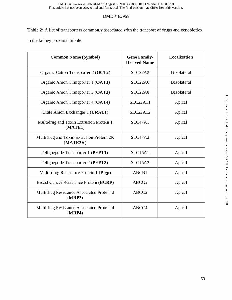
DMD # 82958
53
Table 2: A list of transporters commonly associated with the transport of drugs and xenobiotics
in the kidney proximal tubule.
Common Name (Symbol) Gene Family-
Derived Name
Localization
Organic Cation Transporter 2 (OCT2) SLC22A2 Basolateral
Organic Anion Transporter 1 (OAT1) SLC22A6 Basolateral
Organic Anion Transporter 3 (OAT3) SLC22A8 Basolateral
Organic Anion Transporter 4 (OAT4) SLC22A11 Apical
Urate Anion Exchanger 1 (URAT1) SLC22A12 Apical
Multidrug and Toxin Extrusion Protein 1
(MATE1)
SLC47A1 Apical
Multidrug and Toxin Extrusion Protein 2K
(MATE2K)
SLC47A2 Apical
Oligoeptide Transporter 1 (PEPT1) SLC15A1 Apical
Oligoeptide Transporter 2 (PEPT2) SLC15A2 Apical
Multi-drug Resistance Protein 1 (P-gp) ABCB1 Apical
Breast Cancer Resistance Protein (BCRP) ABCG2 Apical
Multidrug Resistance Associated Protein 2
(MRP2)
ABCC2 Apical
Multidrug Resistance Associated Protein 4
(MRP4)
ABCC4 Apical
This article has not been copyedited and formatted. The final version may differ from this version.DMD Fast Forward. Published on August 3, 2018 as DOI: 10.1124/dmd.118.082958
at ASPE
T Journals on January 2, 2020
dmd.aspetjournals.org
Dow
nloaded from

54
11. Figures
Figure 1
This article has not been copyedited and formatted. The final version may differ from this version.DMD Fast Forward. Published on August 3, 2018 as DOI: 10.1124/dmd.118.082958
at ASPE
T Journals on January 2, 2020
dmd.aspetjournals.org
Dow
nloaded from

55
Figure 2
This article has not been copyedited and formatted. The final version may differ from this version.DMD Fast Forward. Published on August 3, 2018 as DOI: 10.1124/dmd.118.082958
at ASPE
T Journals on January 2, 2020
dmd.aspetjournals.org
Dow
nloaded from