Emergency Gastrointestinal Bleeding in Dialysis...
4
Urol Nephrol Open Access J 2015, 2(1): 00021 Submit Manuscript | http://medcraveonline.com Urology & Nephrology Open Access Journal Abbreviations GI: Gastro Intestinal; APC: Argon Plasma Coagulation; CAPD: Continuous Ambulatory Peritoneal Dialysis; HD: Hemodialysis; ESRD: End-Stage Renal Disease Introduction Although continuous progress has been made in dialysis therapy, a substantial increase in the number of elderly and long- term dialysis patients has drawn attention to various types of complications [1]. One such complication is gastrointestinal (GI) bleeding, which is frequently found in emergency cases, generally because of several contributing risk factors such as uremic toxins, coagulation disturbances, secondary hyperparathyroidism, and the use of anticoagulant drugs [2-4]. In the present study, we analyzed the clinical characteristics of dialysis patients, who were admitted as emergency cases due to GI bleeding, in an urban, university affiliated hospital in Japan over a recent, 5-year period. Subjects and Methods Consecutive dialysis patients, hospitalized at Saitama Medical University due to emergency GI bleeding between April 1, 2007 and March 31, 2012, were evaluated. This is a 1000 bed university Emergency Gastrointestinal Bleeding in DialysisPatients:Five Years’ Experience in a Single Dialysis Center Research Article Volume 2 Issue 1 - 2015 Naofumi Ikeda, Tsutomu Inoue, Kazuhiro Kobayashi, Yusuke Watanabe and Hiromichi Suzuki* Department of Nephrology, Saitama Medical University, Japan *Corresponding author: Hiromichi Suzuki, Department of Nephrology, Saitama Medical University, 38 Moroyama-machi, Iruma-gun, Saitama, 350-0495, Japan, Tel: +81-49276-1620; Fax: +81-49295-7338; Email: Received: December 01, 2014| Published: January 03, 2015 hospital serving the northern part of Saitama prefecture with a population of half a million people. To be eligible for the study, emergency patients admitted to hospital due to GI bleeding had to meet the following criteria: I. The presence of hematemesis II. A GI lesion identified by either endoscopy or surgery. All endoscopic examinations were performed by an experienced physician in the Department of Gastroenterology. Patients were excluded if GI bleeding was found on routine examination. All patients received regular Hemodialysis (HD) therapy or continuous ambulatory peritoneal dialysis (CAPD). For diagnoses of Helicobacter pylori (H. pylori), non-invasive examinations, including serum anti-Hp-IgG antibody tests and 13C urea breath tests, were employed. The study was approved by the Saitama Medical University Ethical Committee, and written informed consent was obtained for all endoscopic procedures. Details of each patient’s demographics, medical history, physical examination findings, prior and current medications, co morbid conditions, laboratory and evaluation findings, and diagnostic information were collected at the time of admission. Abstract Background and Study aims: Dialysis patients can develop a number of gastrointestinal (GI) lesions that can result in bleeding in emergency situations. Our aim was to investigate the nature of GI bleeding that needed emergency endoscopy. Patients and Methods: Over a 5-year period (April 1, 2007-March 31, 2012), we studied consecutive dialysis patients with GI bleeding who underwent emergency endoscopic procedures. Demographic and clinical data were also collected. Results: Fifty-two patients were hospitalized for emergency GI bleeding and underwent a subsequent endoscopy. The locations of lesions involved in GI bleeding were: upper GI (n=34), lower GI (n=14), and unknown (n=4). Duodenal ulcer (26%) and gastric antral vascular ectasia (26%) were the most frequently identified causes of upper GI bleeding, followed by gastric ulcer (14%). In patients with lower GI bleeding, colorectal cancer, angiodysplasia and ischemic enteritis were identified in equal numbers (21%). Two thirds of patients with gastric antral vascular ectasia had recurrence in spite of treatment by argon plasma coagulation. Conclusion: The causes of GI bleeding in dialysis patients in emergency situations may differ from those in non-dialysis patients with GI bleeding. Keywords Hemodialysis; Peritoneal dialysis; Endoscopy; Gastric antral vascular ectasia; Argon plasma coagulation
Transcript of Emergency Gastrointestinal Bleeding in Dialysis...
Urol Nephrol Open Access J 2015, 2(1): 00021Submit Manuscript | http://medcraveonline.com
Urology & Nephrology Open Access Journal
Abbreviations GI: Gastro Intestinal; APC: Argon Plasma Coagulation; CAPD:
Continuous Ambulatory Peritoneal Dialysis; HD: Hemodialysis; ESRD: End-Stage Renal Disease
IntroductionAlthough continuous progress has been made in dialysis
therapy, a substantial increase in the number of elderly and long-term dialysis patients has drawn attention to various types of complications [1]. One such complication is gastrointestinal (GI) bleeding, which is frequently found in emergency cases, generally because of several contributing risk factors such as uremic toxins, coagulation disturbances, secondary hyperparathyroidism, and the use of anticoagulant drugs [2-4]. In the present study, we analyzed the clinical characteristics of dialysis patients, who were admitted as emergency cases due to GI bleeding, in an urban, university affiliated hospital in Japan over a recent, 5-year period.
Subjects and Methods
Consecutive dialysis patients, hospitalized at Saitama Medical University due to emergency GI bleeding between April 1, 2007 and March 31, 2012, were evaluated. This is a 1000 bed university
Emergency Gastrointestinal Bleeding in DialysisPatients:Five Years’ Experience in a Single
Dialysis Center
Research Article
Volume 2 Issue 1 - 2015
Naofumi Ikeda, Tsutomu Inoue, Kazuhiro Kobayashi, Yusuke Watanabe and Hiromichi Suzuki*Department of Nephrology, Saitama Medical University, Japan
*Corresponding author: Hiromichi Suzuki, Department of Nephrology, Saitama Medical University, 38 Moroyama-machi, Iruma-gun, Saitama, 350-0495, Japan, Tel: +81-49276-1620; Fax: +81-49295-7338; Email:
Received: December 01, 2014| Published: January 03, 2015
hospital serving the northern part of Saitama prefecture with a population of half a million people. To be eligible for the study, emergency patients admitted to hospital due to GI bleeding had to meet the following criteria:
I. The presence of hematemesis
II. A GI lesion identified by either endoscopy or surgery. All endoscopic examinations were performed by an experienced physician in the Department of Gastroenterology. Patients were excluded if GI bleeding was found on routine examination. All patients received regular Hemodialysis (HD) therapy or continuous ambulatory peritoneal dialysis (CAPD). For diagnoses of Helicobacter pylori (H. pylori), non-invasive examinations, including serum anti-Hp-IgG antibody tests and 13C urea breath tests, were employed. The study was approved by the Saitama Medical University Ethical Committee, and written informed consent was obtained for all endoscopic procedures. Details of each patient’s demographics, medical history, physical examination findings, prior and current medications, co morbid conditions, laboratory and evaluation findings, and diagnostic information were collected at the time of admission.
Abstract
Background and Study aims: Dialysis patients can develop a number of gastrointestinal (GI) lesions that can result in bleeding in emergency situations. Our aim was to investigate the nature of GI bleeding that needed emergency endoscopy. Patients and Methods: Over a 5-year period (April 1, 2007-March 31, 2012), we studied consecutive dialysis patients with GI bleeding who underwent emergency endoscopic procedures. Demographic and clinical data were also collected.
Results: Fifty-two patients were hospitalized for emergency GI bleeding and underwent a subsequent endoscopy. The locations of lesions involved in GI bleeding were: upper GI (n=34), lower GI (n=14), and unknown (n=4). Duodenal ulcer (26%) and gastric antral vascular ectasia (26%) were the most frequently identified causes of upper GI bleeding, followed by gastric ulcer (14%). In patients with lower GI bleeding, colorectal cancer, angiodysplasia and ischemic enteritis were identified in equal numbers (21%). Two thirds of patients with gastric antral vascular ectasia had recurrence in spite of treatment by argon plasma coagulation.
Conclusion: The causes of GI bleeding in dialysis patients in emergency situations may differ from those in non-dialysis patients with GI bleeding.
Keywords
Hemodialysis; Peritoneal dialysis; Endoscopy; Gastric antral vascular ectasia; Argon plasma coagulation
Emergency Gastrointestinal Bleeding in Dialysis Patients:Five Years’ Experience in a Single Dialysis Center
Citation: Ikeda N,Inoue T,Kobayashi K,Watanabe Y,Suzuki H (2015) Emergency Gastrointestinal Bleeding in Dialysis Patients:Five Years’ Experience in a Single Dialysis Center. Urol Nephrol Open Access J 2(1): 00021. DOI: 10.15406/unoaj.2015.02.00021
Copyright: 2015 Ikeda et al. 2/4
ResultsCharacteristics of GI bleeding
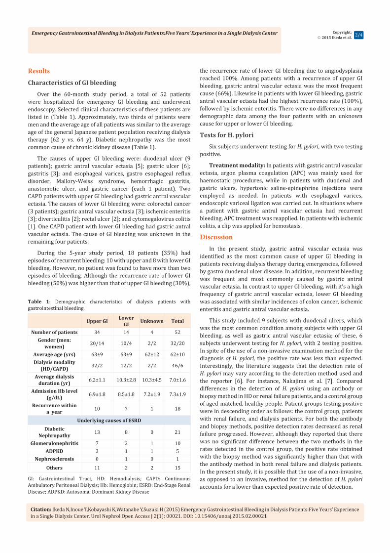
Over the 60-month study period, a total of 52 patients were hospitalized for emergency GI bleeding and underwent endoscopy. Selected clinical characteristics of these patients are listed in (Table 1). Approximately, two thirds of patients were men and the average age of all patients was similar to the average age of the general Japanese patient population receiving dialysis therapy (62 y vs. 64 y). Diabetic nephropathy was the most common cause of chronic kidney disease (Table 1).
The causes of upper GI bleeding were: duodenal ulcer (9 patients); gastric antral vascular ectasia [5]; gastric ulcer [6]; gastritis [3]; and esophageal varices, gastro esophageal reflux disorder, Mallory-Weiss syndrome, hemorrhagic gastritis, anastomotic ulcer, and gastric cancer (each 1 patient). Two CAPD patients with upper GI bleeding had gastric antral vascular ectasia. The causes of lower GI bleeding were: colorectal cancer (3 patients); gastric antral vascular ectasia [3]; ischemic enteritis [3]; diverticulitis [2]; rectal ulcer [2]; and cytomegalovirus colitis [1]. One CAPD patient with lower GI bleeding had gastric antral vascular ectasia. The cause of GI bleeding was unknown in the remaining four patients.
During the 5-year study period, 18 patients (35%) had episodes of recurrent bleeding: 10 with upper and 8 with lower GI bleeding. However, no patient was found to have more than two episodes of bleeding. Although the recurrence rate of lower GI bleeding (50%) was higher than that of upper GI bleeding (30%),
the recurrence rate of lower GI bleeding due to angiodysplasia reached 100%. Among patients with a recurrence of upper GI bleeding, gastric antral vascular ectasia was the most frequent cause (66%). Likewise in patients with lower GI bleeding, gastric antral vascular ectasia had the highest recurrence rate (100%), followed by ischemic enteritis. There were no differences in any demographic data among the four patients with an unknown cause for upper or lower GI bleeding.
Tests for H. pylori
Six subjects underwent testing for H. pylori, with two testing positive.
Treatment modality: In patients with gastric antral vascular ectasia, argon plasma coagulation (APC) was mainly used for haemostatic procedures, while in patients with duodenal and gastric ulcers, hypertonic saline-epinephrine injections were employed as needed. In patients with esophageal varices, endoscopic variceal ligation was carried out. In situations where a patient with gastric antral vascular ectasia had recurrent bleeding, APC treatment was reapplied. In patients with ischemic colitis, a clip was applied for hemostasis.
DiscussionIn the present study, gastric antral vascular ectasia was
identified as the most common cause of upper GI bleeding in patients receiving dialysis therapy during emergencies, followed by gastro duodenal ulcer disease. In addition, recurrent bleeding was frequent and most commonly caused by gastric antral vascular ectasia. In contrast to upper GI bleeding, with it’s a high frequency of gastric antral vascular ectasia, lower GI bleeding was associated with similar incidences of colon cancer, ischemic enteritis and gastric antral vascular ectasia.
This study included 9 subjects with duodenal ulcers, which was the most common condition among subjects with upper GI bleeding, as well as gastric antral vascular ectasia; of these, 6 subjects underwent testing for H. pylori, with 2 testing positive. In spite of the use of a non-invasive examination method for the diagnosis of H. pylori, the positive rate was less than expected. Interestingly, the literature suggests that the detection rate of H. pylori may vary according to the detection method used and the reporter [6]. For instance, Nakajima et al. [7]. Compared differences in the detection of H. pylori using an antibody or biopsy method in HD or renal failure patients, and a control group of aged-matched, healthy people. Patient groups testing positive were in descending order as follows: the control group, patients with renal failure, and dialysis patients. For both the antibody and biopsy methods, positive detection rates decreased as renal failure progressed. However, although they reported that there was no significant difference between the two methods in the rates detected in the control group, the positive rate obtained with the biopsy method was significantly higher than that with the antibody method in both renal failure and dialysis patients. In the present study, it is possible that the use of a non-invasive, as opposed to an invasive, method for the detection of H. pylori accounts for a lower than expected positive rate of detection.
Upper GI LowerGI Unknown Total
Number of patients 34 14 4 52Gender (men:
women) 20/14 10/4 2/2 32/20
Average age (yrs) 63±9 63±9 62±12 62±10Dialysis modality
(HD/CAPD) 32/2 12/2 2/2 46/6
Average dialysis duration (yr) 6.2±1.1 10.3±2.8 10.3±4.5 7.0±1.6
Admission Hb level (g/dL) 6.9±1.8 8.5±1.8 7.2±1.9 7.3±1.9
Recurrence within a year 10 7 1 18
Underlying causes of ESRDDiabetic
Nephropathy 13 8 0 21
Glomerulonephritis 7 2 1 10ADPKD 3 1 1 5
Nephrosclerosis 0 1 0 1
Others 11 2 2 15
Table 1: Demographic characteristics of dialysis patients with gastrointestinal bleeding.
GI: Gastrointestinal Tract, HD: Hemodialysis; CAPD: Continuous Ambulatory Peritoneal Dialysis; Hb: Hemoglobin; ESRD: End-Stage Renal Disease; ADPKD: Autosomal Dominant Kidney Disease
Emergency Gastrointestinal Bleeding in Dialysis Patients:Five Years’ Experience in a Single Dialysis Center
Citation: Ikeda N,Inoue T,Kobayashi K,Watanabe Y,Suzuki H (2015) Emergency Gastrointestinal Bleeding in Dialysis Patients:Five Years’ Experience in a Single Dialysis Center. Urol Nephrol Open Access J 2(1): 00021. DOI: 10.15406/unoaj.2015.02.00021
Copyright: 2015 Ikeda et al. 3/4
It is postulated that an increased rate of gastric antral vascular ectasia occurs in dialysis patients [8] because uremia induced platelet dysfunction [9-12] and heparin administration for HD have both been implicated in an increased risk of bleeding from this disease [12]. In support of this hypothesis, a retrospective study of 482 patients with upper GI bleeding reported that gastric antral vascular ectasia of the stomach or duodenum was the most common cause of bleeding in patients with chronic renal failure [13]. However, gastric antral vascular ectasia is rarely identified in patients receiving dialysis who are undergoing endoscopy for occult GI bleeding [14]. Yorioka et al. [15] reported on a patient with end-stage renal disease (ESRD) and bleeding from watermelon stomach (gastric antral vascular ectasia) who improved after the dialysis modality was changed from HD to CAPD. However, other reports exist of watermelon stomach and bleeding in CAPD patients [16,17] which may counter the suggestion of haemostatic risk factors, except uremic toxins, contributing to the occurrence of watermelon stomach in CAPD patients. In the present study, two CAPD patients with upper GI bleeding and one CAPD patient with lower GI bleeding were diagnosed as having gastric antral vascular ectasia. The exact pathogenesis of this condition is unknown; however, prior studies have indicated an association with advanced age, Rendu-Osler-Weber syndrome, bowel irradiation and collagen disease [18-21].
The high prevalence of gastric antral vascular ectasia in patients receiving dialysis therapy indicates that uremia and the bleeding diathesis of these patients play major roles in the pathogenesis of these vascular changes. Chalasani et al. [8] reported that the most common cause of upper GI bleeding in patients with chronic renal failure was peptic ulcer disease. Gastric antral vascular ectasia was more frequent in patients with chronic renal failure than in those with normal renal function and its prevalence seemed to be related to the duration and severity of renal disease [8]. The results from this present study lend support to the Chalasani et al. [8] study, as we found that the duration of dialysis in patients with gastric antral vascular ectasia was longer than for the average Japanese dialysis patient (7 vs. 3, respectively).
In terms of treatment for gastric antral vascular ectasia, in the past surgery was common; however, endoscopic treatment, which is minimally invasive, is now the preferred option. Furthermore, many institutions have recently attempted to achieve hemostasis using the APC method [14,22]. This relatively new method, in the field of therapeutic endoscopy, enables extensive and relatively shallow cauterization and allows for treatment at a fixed depth at a coagulated site. Further advantages over other endoscopic treatments are that it is a relatively simple and inexpensive technique. According to a report by Grund, et al. [23,24], the successful treatment rate for hemostasis using the APC method for various hemorrhagic lesions was 98%. In fact, such a method was also chosen for all subjects undergoing hemostatic procedures in this study, and no particular complications were observed. It is believed that APC is currently the most minimally invasive and safest hemostatic procedure available for treating
capillary telangiectasia, regardless of whether the target is a dialysis patient or not.
In spite of hemostatic therapy with APC in dialysis patients with gastric antral vascular ectasia, an inexplicably high recurrence rate was observed. Several studies [3,4,24] have reported recurrence rates for gastric antral vascular ectasia of between 30 to 40% in 66% of dialysis patients who were admitted to the emergency room. Moreover, in the present study, the recurrence rate of lower GI bleeding due to angiodysplasia reached 100%. Studies examining the incidence of angiodysplasia in the lower GI tract in patients receiving dialysis therapy are currently lacking. Akhtar reported that the incidence of angiodysplasia producing lower GI bleeding was 27% in elderly patients [22]. In the present study, 3 out of 4 patients were found to have angiodysplasia and all had recurrence, an event that might be specifically attributable to dialysis patients.
Limitations of the studyThis study was retrospective and did not include all
dialysis patients admitted as emergency cases with GI bleeding. Moreover, all patients who elected for emergency endoscopic procedures presented with melena, hematemesis, and rectal bleeding. Under these circumstances, the causes of GI bleeding may differ from those of the general population. Moreover, in the area where our university is located, patients not on dialysis and experiencing emergency GI bleeding are frequently admitted to a general hospital instead of a university hospital. Under these circumstances, it is not easy to evaluate the true incidence of emergency GI bleeding. Additionally, statistical procedures were not carried out, highlighting the descriptive nature of this paper and thus reducing the value of this study.
Conclusion The causes of GI bleeding in patients admitted to hospital
as emergency cases may differ for dialysis as opposed to non-dialysis patients.
References1. Nakai S, Shinzato T, Nagura Y, Masagane O, Kitaoka T, et al. (2004)
Overview of dialysis treatment in Japan (as of December 31, 2002). Ther Apher Dial 8(5): 358-382.
2. Kuo CC, Kuo HW, Lee IM, Lee CT, Yang CY (2013) The risk of upper gastrointestinal bleeding in patients treated with hemodialysis: a population-based cohort study. BMC Nephrol 14: 15.
3. Liang CC, Wang SM, Kuo HL, Chang CT, Liu JH, et al. (2014) Upper gastrointestinal bleeding in patients with CKD. Clin J Am Soc Nephrol 9(8): 1354-13529.
4. Trivedi H, Yang J, Szabo A (2014) Gastrointestinal bleeding in patients on long-term dialysis. J Nephrol [Epub ahead of print]
5. Escolar G, Diaz-Ricart M, Cases A (2005) Uremic platelet dysfunction: past and present. Curr Hematol Rep 4(5): 359-367.
6. Fabrizi F, Martin P (2000) Helicobacter pylori infection in patients with end-stage renal disease. Int J Artif Organs 23(3): 157-164.
7. Nakajima F, Sakaguchi M, Oka H, Kawase Y, Shibahara N, et al. (2004) Prevalence of Helicobacter pylori antibodies in long-term dialysis
Emergency Gastrointestinal Bleeding in Dialysis Patients:Five Years’ Experience in a Single Dialysis Center
Citation: Ikeda N,Inoue T,Kobayashi K,Watanabe Y,Suzuki H (2015) Emergency Gastrointestinal Bleeding in Dialysis Patients:Five Years’ Experience in a Single Dialysis Center. Urol Nephrol Open Access J 2(1): 00021. DOI: 10.15406/unoaj.2015.02.00021
Copyright: 2015 Ikeda et al. 4/4
patients. Nephrology (Carlton) 9(2): 73-76.
8. Chalasani N, Cotsonis G, Wilcox CM (1996) Upper gastrointestinal bleeding in patients with chronic renal failure: role of vascular ectasia. Am J Gastroenterol 91(11): 2329-2332.
9. Brophy DF, Martin EJ, Carr SL, Kirschbaum B, Carr ME (2007) The effect of uremia on platelet contractile force, clot elastic modulus and bleeding time in hemodialysis patients. Thromb Res 119(6): 723-729.
10. Sohal AS, Gangji AS, Crowther MA, Treleaven D (2006) Uremic bleeding: pathophysiology and clinical risk factors. Thromb Res 118(3): 417-422.
11. Boccardo P, Remuzzi G, Galbusera M (2004) Platelet dysfunction in renal failure. Semin Thromb Hemost 30(5): 579-589.
12. Janssen MJ, van der Meulen J (1996) The bleeding risk in chronic hemodialysis: preventive strategies in high-risk patients. Neth J Med 48(5): 198-207.
13. Zuckerman GR, Cornette GL, Clouse RE, Harter HR (1985) Upper gastrointestinal bleeding in patients with chronic renal failure. Ann Intern Med 102(5): 588-592.
14. Sebastian S, O'Morain CA, Buckley MJ (2003) Review article: current therapeutic options for gastric antral vascular ectasia. Aliment Pharmacol Ther 18(2): 157-165.
15. Yorioka N, Hamaguchi N, Taniguchi Y, Asakimori Y, Nishiki T, et al. (1996) Gastric antral vascular ectasia in a patient on hemodialysis improved with CAPD. Perit Dial Int 16(2): 177-178.
16. Moreiras M, Rodriguez Goyanes G, Cuina L, Gonzalez Pineiro A, Perez AJ (1998) More about watermelon stomach: a case report in a CAPD patient. Nephrol Dial Transplant 13(1): 230-231.
17. Tomori K, Nakamoto H, Kotaki S, Ishida Y, Takane H, et al. (2003) Gastric angiodysplasia in patients undergoing maintenance dialysis. Adv Perit Dial 19: 136-142.
18. Dalle I, Geboes K (2002) Vascular lesions of the gastrointestinal tract. Acta Gastroenterol Belg 65(4): 213-219.
19. Burak KW, Lee SS, Beck PL (2001) Portal hypertensive gastropathy and gastric antral vascular ectasia (GAVE) syndrome. Gut 49(6): 866-872.
20. Lorenzi AR, Johnson AH, Davies G, Gough A (2001) Gastric antral vascular ectasia in systemic sclerosis: complete resolution with methylprednisolone and cyclophosphamide. Ann Rheum Dis 60(8): 796-798.
21. Gordon FH, Watkinson A, Hodgson H (2001) Vascular malformations of the gastrointestinal tract. Best Pract Res Clin Gastroenterol 15(1): 41-58.
22. Akhtar AJ (2003) Lower gastrointestinal bleeding in elderly patients. J Am Med Dir Assoc 4(6): 320-322.
23. Jensen DM, Chaves DM, Grund KE (2004) Endoscopic diagnosis and treatment of watermelon stomach. Endoscopy 36(7): 640-647.
24. Grund KE, Straub T, Farin G (1999) New haemostatic techniques: argon plasma coagulation. Baillieres Best Pract Res Clin Gastroenterol 13(1): 67-84.