Embryonic diffrentiation of the Jejunum of one Humped...
7
Scholarly Journal of Biological Science Vol. 4(5), pp. 33-39, December 2015 Available online http:// www.scholarly-journals.com/SJBS ISSN 2315-6147 © 2015 Scholarly-Journals Full Length Research Paper Embryonic diffrentiation of the Jejunum of one Humped Camel (Camelus dromedarius): A Histomorphology Bello, A. 1 *, Onyeanusi, B.I. 2 , Umaru, M.A 3 , Onu, J.E. 4 , Mahmud, M.A. 5 , Maryam, S.A. 6 1 Department of Veterinary Anatomy, Usmanu Danfodiyo University, Sokoto; 2 Department of Veterinary Anatomy, Ahmadu Bello University Zaria; 3 Department of Theriogenology and Animal production, Usmanu Danfodiyo University, Sokoto, 4 Department of Animal Health and Production Technology, Niger State College of Agriculture, P.M.B. 109, Mokwa, Niger State; 5,6 Department of Biology, Isa Kaita College of Education, Dutsin-ma. Nigeria. Accepted 22 December, 2015 A histomorphological differentiation study was conducted on the jejunum of 35 foetuses (both sex) of the one-humped camel collected from the Sokoto metropolitan abattoir, over a period of five months at different gestational ages. The approximate age of the fetuses was estimated and categorised into first, second and third trimester. Gross observation of the small intestine insitu was recorded. The jejunum was characterised by having an extensive coiling in second and third trimester, with no evidence of segmental differentiation in the first trimesters. Histological observation of the tissues in this study revealed a complete structure of the tubular organ. The jejunum was found to consist of four layers namely: Tunica mucosa, Tunica sub mucosa, Tunica muscularis and Tunica serosa. The epithelium of the Tunica mucosa was stratified squamous epithelium with varying degree of stratification at first trimester and transformed to low columnar /cuboidal epithelium with prominent villi and crypt of lieberkhum at second trimester. At third trimester, the epithelium was simple columnar epithelium with prominent developed villi, microvilli, and crypt of lieberkhum in the entire tunica mucosa. The lamina propria mucosa was found absent at first trimester but prominent at second and third trimester. The Lamina muscularis mucosa was found prominent at third trimester but not identified at first and second trimester. At first trimester of age tunica submucosa was prominent while at second trimester, it consisted of connective tissue cells and fibres scattered all over the layers with preliminary blood vessels. The cells and fibres were undifferentiated at this stage. There was evidence of scattered blood vessels within the layer. At third trimester of age, the connective tissues and blood vessels were found prominent throughout the length of the jejunum. The tunica muscularis of camel jejunum consist of inner circular and outer longitudinal smooth muscle layers. At first trimester this layer did not differentiate into these two zones but only longitudinal orientation of smooth muscle layer. At second trimester, the layers of two zones with clear demarcation were observed. A thin layer of connective tissue comprising of undifferentiated cells lined the jejunum externally was observed in all the stages of development. Based on the above findings, it showed that development of the camels’ jejunum shown an early gross and histologically differentiation compared to other segments of the intestine. Key words: Camel, Jejunum, Embryonic differenciation, Histomorphology. INTRODUCTION Camels are similar to ruminants in several aspects, which include regurgitation of ingesta, and active microbial *Corresponding author e-mail: [email protected]. fermentation in the stomach (Frandson, 1981).The digestive anatomy and physiology of dromedarian camel at embryonic level is least understood when compared to Llama, Guanaco, Cattle, Sheep, Goat and Pig (Yagil and Ezion, 1980, Al-Tarazi, 2001; Bello et al.,2012). The description of dromedarian camel is usually made as if it
Transcript of Embryonic diffrentiation of the Jejunum of one Humped...
Scholarly Journal of Biological Science Vol. 4(5), pp. 33-39, December 2015 Available online http:// www.scholarly-journals.com/SJBS
ISSN 2315-6147 © 2015 Scholarly-Journals
Full Length Research Paper
Embryonic diffrentiation of the Jejunum of one Humped Camel (Camelus dromedarius): A Histomorphology
Bello, A.1*, Onyeanusi, B.I.2, Umaru, M.A3, Onu, J.E.4, Mahmud, M.A.5, Maryam, S.A.6
1Department of Veterinary Anatomy, Usmanu Danfodiyo University, Sokoto;
2Department of Veterinary Anatomy, Ahmadu Bello University Zaria;
3Department of Theriogenology and Animal production, Usmanu Danfodiyo University, Sokoto,
4Department of Animal Health and Production Technology, Niger State College of Agriculture,
P.M.B. 109, Mokwa, Niger State; 5,6
Department of Biology, Isa Kaita College of Education, Dutsin-ma. Nigeria.
Accepted 22 December, 2015
A histomorphological differentiation study was conducted on the jejunum of 35 foetuses (both sex) of the one-humped camel collected from the Sokoto metropolitan abattoir, over a period of five months at different gestational ages. The approximate age of the fetuses was estimated and categorised into first, second and third trimester. Gross observation of the small intestine insitu was recorded. The jejunum was characterised by having an extensive coiling in second and third trimester, with no evidence of segmental differentiation in the first trimesters. Histological observation of the tissues in this study revealed a complete structure of the tubular organ. The jejunum was found to consist of four layers namely: Tunica mucosa, Tunica sub mucosa, Tunica muscularis and Tunica serosa. The epithelium of the Tunica mucosa was stratified squamous epithelium with varying degree of stratification at first trimester and transformed to low columnar /cuboidal epithelium with prominent villi and crypt of lieberkhum at second trimester. At third trimester, the epithelium was simple columnar epithelium with prominent developed villi, microvilli, and crypt of lieberkhum in the entire tunica mucosa. The lamina propria mucosa was found absent at first trimester but prominent at second and third trimester. The
Lamina muscularis mucosa was found prominent at third trimester but not identified at first and second trimester. At first trimester of age tunica submucosa was prominent while at second trimester, it consisted of connective tissue cells and fibres scattered all over the layers with preliminary blood vessels. The cells and fibres were undifferentiated at this stage. There was evidence of scattered blood vessels within the layer. At third trimester of age, the connective tissues and blood vessels were found prominent throughout the length of the jejunum. The tunica muscularis of camel jejunum consist of inner circular and outer longitudinal smooth muscle layers. At first trimester this layer did not differentiate into these two zones but only longitudinal orientation of smooth muscle layer. At second trimester, the layers of two zones with clear demarcation were observed. A thin layer of connective tissue comprising of undifferentiated cells lined the jejunum externally was observed in all the stages of development. Based on the above findings, it showed that development of the camels’ jejunum shown an early gross and histologically differentiation compared to other segments of the intestine. Key words: Camel, Jejunum, Embryonic differenciation, Histomorphology.
INTRODUCTION Camels are similar to ruminants in several aspects, which include regurgitation of ingesta, and active microbial *Corresponding author e-mail: [email protected].
fermentation in the stomach (Frandson, 1981).The digestive anatomy and physiology of dromedarian camel at embryonic level is least understood when compared to Llama, Guanaco, Cattle, Sheep, Goat and Pig (Yagil and Ezion, 1980, Al-Tarazi, 2001; Bello et al.,2012). The description of dromedarian camel is usually made as if it

Scholarly J. Biol. Sci. 34
Table 1: The CVRL and weight of fetuses at various trimesters (mean±SEM).
Parameters First Trimester Second Trimester Third Trimester Number of sample (N) 13 11 11 CVRL (cm) 20.06 ± 3.0 60.27 ± 4.0 103.83 ± 6.0 Fetal weight (Kg) 1.40 ± 0. 6 6.10 ± 0.5 17.87 ± 0.6
is identical with Llama specie (Bello et al., 2013). Though, they are pseudo-ruminants that possess a three-chambered stomach, lacking the omasum that is part of the four-chambered stomach of the order Ruminantia (Al-Tarazi, 2001). The true camels (Camelus dromedarius and Camelus bacterianus) are closely related anatomically to the South American Camelids (Wilson, et al. 1990; Marzook, 1996; Reece, 1997; Ghaji et al, 1982; Adogwa, et al. 1987; Ghaji, et al. 1989; Bello et al., 2013).
Research work dealing with morphology, physiology, pathology, gross and developmental anatomy of various organs and system of dromedarian camel has been carried out in many countries using foetal and adult camel (Wilson, et al. 1990; Marzook, 1996; Reece, 1997; Bustinza ,1999; Ghaji et al, 1982; Adogwa, et al. 1987; Ghaji, et al. 1989; Bello et al., 2012; Hena et al., 2012) but little attentions have been paid for the developmental changes of the cranial part of the small intestine of the camel fetus. Thus, paucity of information on the prenatal development of camel jejunum exists; hence the present study was undertaken to bridge the information gap. MATERIALS AND METHODS The study was carried out on 35 foetuses of the one-humped camel collected from the metropolitan abattoir, Sokoto using standard animal ethics approved by the government, at different gestational ages. The collected foetuses were then taken to the Veterinary Anatomy laboratory of Usmanu Danfodiyo University; where the weight and age of the foetuses were determined. The foetal body weight was measured using electrical (digital) weighing balance for the smaller foetuses and compression spring balance (AT-1422), size C-1, sensitivity of 20kg X 50g in Kilogram for the bigger foetuses. The approximate age of the foetuses was estimated by using the following formula adopted by El-wishy et al. (1981) and Bello et al. (2013): GA = (CVRL + 23.99)/0.366, Where GA is age in days and CVRL is the Crown Vertebral Rump Length. Fetuses below 130 days were designated as first trimester, 131- 260 days as second trimester and 261 - 390 days as third trimester (Bello et al. 2013). Crown Vertebral Rump Length (CVRL) was measured (cm) as a
curved line along the vertebral column from the point of the anterior fontanel or the frontal bone following the vertebral curvature to the base of the tail. Based on this, foetal samples were divided into 3 main groups as described by Bello et al. (2013). The digestive tract of each fetus was collected by placing the fetus on dorsal recumbency and a mid-ventral skin incision was made via the abdomino-pelvic region down to the thoracic, to the neck up to the inter-mandibular space in order to remove the entire digestive tract. 1cm
2 thick of sample from each group was collected and
fixed in 10% formalin solution. After fixation was achieved, the tissue sample was processed for paraffin blocks preparation. The sections of 5µm were subjected to haematoxylin and eosin for routine morphology (Luiz and Jose, 2005). The standard sections were examined under light microscope and micrographs taken using motic cam camera with 12.1 mega pixel. RESULTS AND DISCUSSION The current study attempted to contribution to the developmental Anatomy of the dromedarian camel at fetal level. Result of the investigation shown that, there was an increase in the body weight, organ weight and individual segments of the small intestine in the fetuses with advancement in gestation period (Table 1). This is in agreement with the observations of Jamdar and Ema (1982), Bello et al. (2013), Luciano et al. (1979) and Sonfada (2006), who observed obvious body weight increase with advancement of gestation period in different species of animals. Bello et al. (2012) suggested that nutritional status and health condition of the dam played a vital role in the development of the fetus hence increase in weight of the fetus.
Grossly, the color of the small intestine was whitish at first trimester and grayish in second and third trimester. The jejunum was characterised by having an extensive coiling in second and third trimester, with no evidence of segmental differentiation in the first trimesters as shown in Plate 1. Similar findings were reported on the gross features of small intestine of various animals at different gestational ages; Llama (Smuts and Benzuidenhout, 1987; Luciano et al., 1979) and nutria (Perez et al., 2008a). On the other hand, the divisions were not reported in sheep (Sisson et al., 1975), cattle (Dyce et al., 2002) and pampas deer (Perez et al., 2008a).
The coil part of the jejunum was not divided at first trimester but differentiated into five parts (descending
Bello et al. 35
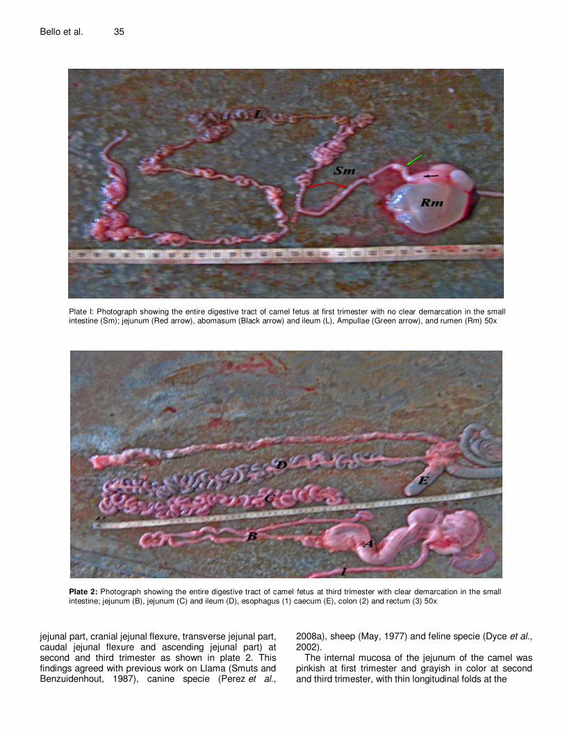
Plate I: Photograph showing the entire digestive tract of camel fetus at first trimester with no clear demarcation in the small intestine (Sm); jejunum (Red arrow), abomasum (Black arrow) and ileum (L), Ampullae (Green arrow), and rumen (Rm) 50x
Plate 2: Photograph showing the entire digestive tract of camel fetus at third trimester with clear demarcation in the small intestine; jejunum (B), jejunum (C) and ileum (D), esophagus (1) caecum (E), colon (2) and rectum (3) 50x
jejunal part, cranial jejunal flexure, transverse jejunal part, caudal jejunal flexure and ascending jejunal part) at second and third trimester as shown in plate 2. This findings agreed with previous work on Llama (Smuts and Benzuidenhout, 1987), canine specie (Perez et al.,
2008a), sheep (May, 1977) and feline specie (Dyce et al., 2002).
The internal mucosa of the jejunum of the camel was pinkish at first trimester and grayish in color at second and third trimester, with thin longitudinal folds at the
Scholarly J. Biol. Sci. 36
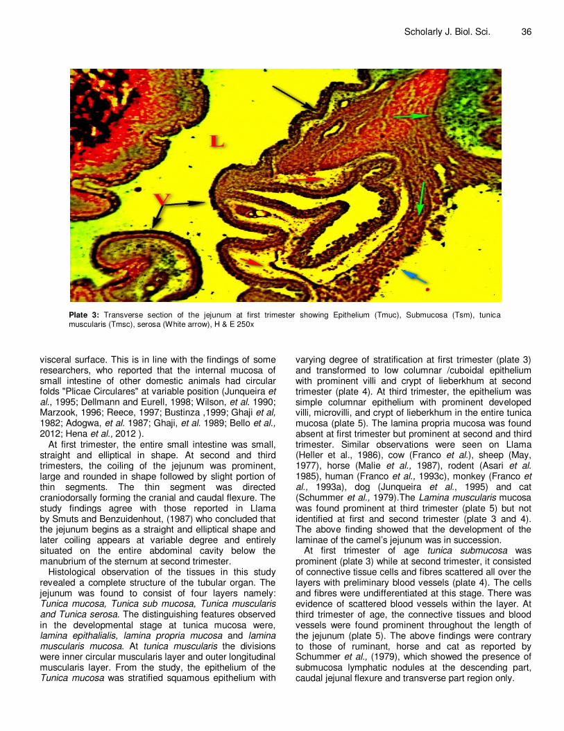
Plate 3: Transverse section of the jejunum at first trimester showing Epithelium (Tmuc), Submucosa (Tsm), tunica muscularis (Tmsc), serosa (White arrow), H & E 250x
visceral surface. This is in line with the findings of some researchers, who reported that the internal mucosa of small intestine of other domestic animals had circular folds "Plicae Circulares" at variable position (Junqueira et al., 1995; Dellmann and Eurell, 1998; Wilson, et al. 1990; Marzook, 1996; Reece, 1997; Bustinza ,1999; Ghaji et al, 1982; Adogwa, et al. 1987; Ghaji, et al. 1989; Bello et al., 2012; Hena et al., 2012 ).
At first trimester, the entire small intestine was small, straight and elliptical in shape. At second and third trimesters, the coiling of the jejunum was prominent, large and rounded in shape followed by slight portion of thin segments. The thin segment was directed craniodorsally forming the cranial and caudal flexure. The study findings agree with those reported in Llama by Smuts and Benzuidenhout, (1987) who concluded that the jejunum begins as a straight and elliptical shape and later coiling appears at variable degree and entirely situated on the entire abdominal cavity below the manubrium of the sternum at second trimester.
Histological observation of the tissues in this study revealed a complete structure of the tubular organ. The jejunum was found to consist of four layers namely: Tunica mucosa, Tunica sub mucosa, Tunica muscularis and Tunica serosa. The distinguishing features observed in the developmental stage at tunica mucosa were, lamina epithalialis, lamina propria mucosa and lamina muscularis mucosa. At tunica muscularis the divisions were inner circular muscularis layer and outer longitudinal muscularis layer. From the study, the epithelium of the Tunica mucosa was stratified squamous epithelium with
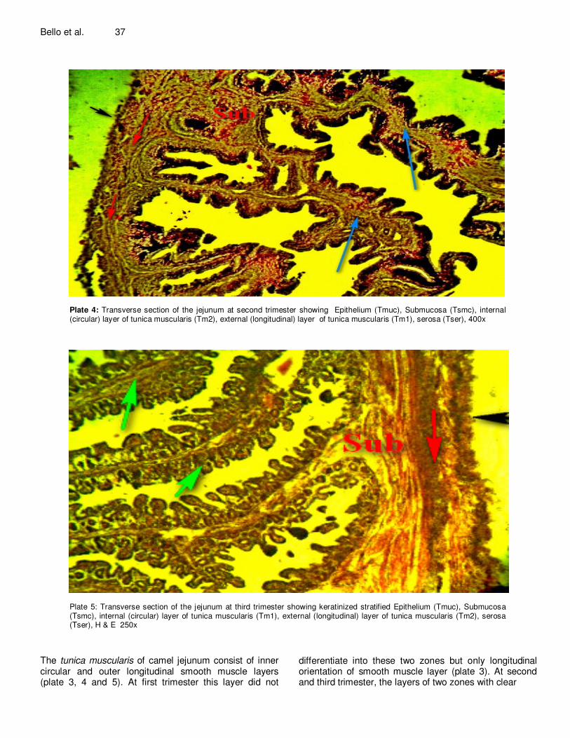
varying degree of stratification at first trimester (plate 3) and transformed to low columnar /cuboidal epithelium with prominent villi and crypt of lieberkhum at second trimester (plate 4). At third trimester, the epithelium was simple columnar epithelium with prominent developed villi, microvilli, and crypt of lieberkhum in the entire tunica mucosa (plate 5). The lamina propria mucosa was found absent at first trimester but prominent at second and third trimester. Similar observations were seen on Llama (Heller et al., 1986), cow (Franco et al.), sheep (May, 1977), horse (Malie et al., 1987), rodent (Asari et al. 1985), human (Franco et al., 1993c), monkey (Franco et al., 1993a), dog (Junqueira et al., 1995) and cat (Schummer et al., 1979).The Lamina muscularis mucosa was found prominent at third trimester (plate 5) but not identified at first and second trimester (plate 3 and 4). The above finding showed that the development of the laminae of the camel’s jejunum was in succession.
At first trimester of age tunica submucosa was prominent (plate 3) while at second trimester, it consisted of connective tissue cells and fibres scattered all over the layers with preliminary blood vessels (plate 4). The cells and fibres were undifferentiated at this stage. There was evidence of scattered blood vessels within the layer. At third trimester of age, the connective tissues and blood vessels were found prominent throughout the length of the jejunum (plate 5). The above findings were contrary to those of ruminant, horse and cat as reported by Schummer et al., (1979), which showed the presence of submucosa lymphatic nodules at the descending part, caudal jejunal flexure and transverse part region only.
Bello et al. 37
Plate 4: Transverse section of the jejunum at second trimester showing Epithelium (Tmuc), Submucosa (Tsmc), internal (circular) layer of tunica muscularis (Tm2), external (longitudinal) layer of tunica muscularis (Tm1), serosa (Tser), 400x
Plate 5: Transverse section of the jejunum at third trimester showing keratinized stratified Epithelium (Tmuc), Submucosa (Tsmc), internal (circular) layer of tunica muscularis (Tm1), external (longitudinal) layer of tunica muscularis (Tm2), serosa (Tser), H & E 250x
The tunica muscularis of camel jejunum consist of inner circular and outer longitudinal smooth muscle layers (plate 3, 4 and 5). At first trimester this layer did not
differentiate into these two zones but only longitudinal orientation of smooth muscle layer (plate 3). At second and third trimester, the layers of two zones with clear
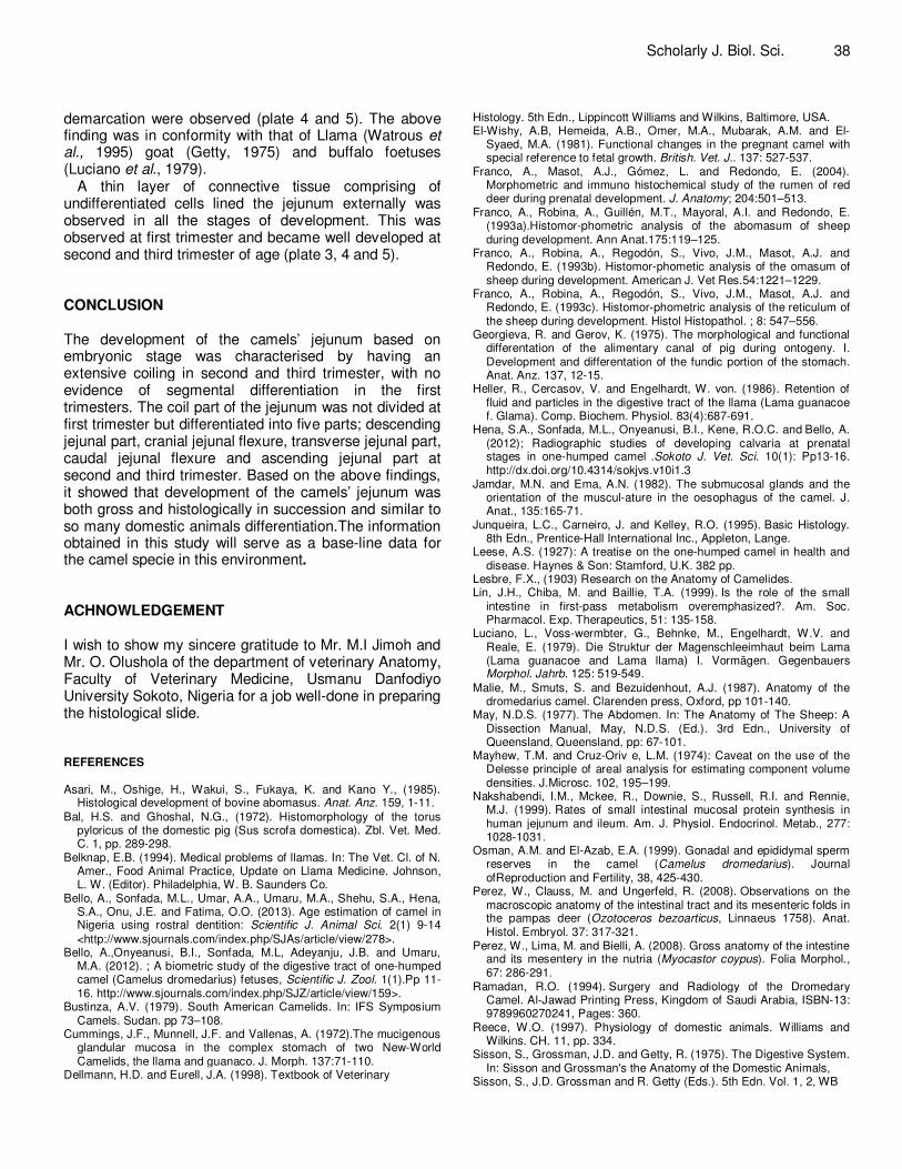
demarcation were observed (plate 4 and 5). The above finding was in conformity with that of Llama (Watrous et al., 1995) goat (Getty, 1975) and buffalo foetuses (Luciano et al., 1979).
A thin layer of connective tissue comprising of undifferentiated cells lined the jejunum externally was observed in all the stages of development. This was observed at first trimester and became well developed at second and third trimester of age (plate 3, 4 and 5). CONCLUSION The development of the camels’ jejunum based on embryonic stage was characterised by having an extensive coiling in second and third trimester, with no evidence of segmental differentiation in the first trimesters. The coil part of the jejunum was not divided at first trimester but differentiated into five parts; descending jejunal part, cranial jejunal flexure, transverse jejunal part, caudal jejunal flexure and ascending jejunal part at second and third trimester. Based on the above findings, it showed that development of the camels’ jejunum was both gross and histologically in succession and similar to so many domestic animals differentiation.The information obtained in this study will serve as a base-line data for the camel specie in this environment. ACHNOWLEDGEMENT I wish to show my sincere gratitude to Mr. M.I Jimoh and Mr. O. Olushola of the department of veterinary Anatomy, Faculty of Veterinary Medicine, Usmanu Danfodiyo University Sokoto, Nigeria for a job well-done in preparing the histological slide. REFERENCES Asari, M., Oshige, H., Wakui, S., Fukaya, K. and Kano Y., (1985).
Histological development of bovine abomasus. Anat. Anz. 159, 1-11.
Bal, H.S. and Ghoshal, N.G., (1972). Histomorphology of the torus pyloricus of the domestic pig (Sus scrofa domestica). Zbl. Vet. Med. C. 1, pp. 289-298.
Belknap, E.B. (1994). Medical problems of llamas. In: The Vet. Cl. of N. Amer., Food Animal Practice, Update on Llama Medicine. Johnson, L. W. (Editor). Philadelphia, W. B. Saunders Co.
Bello, A., Sonfada, M.L., Umar, A.A., Umaru, M.A., Shehu, S.A., Hena, S.A., Onu, J.E. and Fatima, O.O. (2013). Age estimation of camel in Nigeria using rostral dentition: Scientific J. Animal Sci. 2(1) 9-14
<http://www.sjournals.com/index.php/SJAs/article/view/278>. Bello, A.,Onyeanusi, B.I., Sonfada, M.L, Adeyanju, J.B. and Umaru,
M.A. (2012). ; A biometric study of the digestive tract of one-humped camel (Camelus dromedarius) fetuses, Scientific J. Zool. 1(1).Pp 11-16. http://www.sjournals.com/index.php/SJZ/article/view/159>.
Bustinza, A.V. (1979). South American Camelids. In: IFS Symposium Camels. Sudan. pp 73–108.
Cummings, J.F., Munnell, J.F. and Vallenas, A. (1972).The mucigenous glandular mucosa in the complex stomach of two New-World Camelids, the llama and guanaco. J. Morph. 137:71-110.
Dellmann, H.D. and Eurell, J.A. (1998). Textbook of Veterinary
Scholarly J. Biol. Sci. 38 Histology. 5th Edn., Lippincott Williams and Wilkins, Baltimore, USA. El-Wishy, A.B, Hemeida, A.B., Omer, M.A., Mubarak, A.M. and El-
Syaed, M.A. (1981). Functional changes in the pregnant camel with special reference to fetal growth. British. Vet. J.. 137: 527-537.
Franco, A., Masot, A.J., Gómez, L. and Redondo, E. (2004). Morphometric and immuno histochemical study of the rumen of red deer during prenatal development. J. Anatomy; 204:501–513.
Franco, A., Robina, A., Guillén, M.T., Mayoral, A.I. and Redondo, E. (1993a).Histomor-phometric analysis of the abomasum of sheep during development. Ann Anat.175:119–125.
Franco, A., Robina, A., Regodón, S., Vivo, J.M., Masot, A.J. and Redondo, E. (1993b). Histomor-phometic analysis of the omasum of sheep during development. American J. Vet Res.54:1221–1229.
Franco, A., Robina, A., Regodón, S., Vivo, J.M., Masot, A.J. and Redondo, E. (1993c). Histomor-phometric analysis of the reticulum of the sheep during development. Histol Histopathol. ; 8: 547–556.
Georgieva, R. and Gerov, K. (1975). The morphological and functional differentation of the alimentary canal of pig during ontogeny. I. Development and differentation of the fundic portion of the stomach. Anat. Anz. 137, 12-15.
Heller, R., Cercasov, V. and Engelhardt, W. von. (1986). Retention of fluid and particles in the digestive tract of the llama (Lama guanacoe f. Glama). Comp. Biochem. Physiol. 83(4):687-691.
Hena, S.A., Sonfada, M.L., Onyeanusi, B.I., Kene, R.O.C. and Bello, A.
(2012); Radiographic studies of developing calvaria at prenatal stages in one-humped camel
.Sokoto J. Vet. Sci. 10(1): Pp13-16.
http://dx.doi.org/10.4314/sokjvs.v10i1.3
Jamdar, M.N. and Ema, A.N. (1982). The submucosal glands and the orientation of the muscul-ature in the oesophagus of the camel. J. Anat., 135:165-71.
Junqueira, L.C., Carneiro, J. and Kelley, R.O. (1995). Basic Histology. 8th Edn., Prentice-Hall International Inc., Appleton, Lange.
Leese, A.S. (1927): A treatise on the one-humped camel in health and disease. Haynes & Son: Stamford, U.K. 382 pp.
Lesbre, F.X., (1903) Research on the Anatomy of Camelides. Lin, J.H., Chiba, M. and Baillie, T.A. (1999). Is the role of the small
intestine in first-pass metabolism overemphasized?. Am. Soc. Pharmacol. Exp. Therapeutics, 51: 135-158.
Luciano, L., Voss-wermbter, G., Behnke, M., Engelhardt, W.V. and Reale, E. (1979). Die Struktur der Magenschleeimhaut beim Lama (Lama guanacoe and Lama llama) I. Vormägen. Gegenbauers Morphol. Jahrb. 125: 519-549.
Malie, M., Smuts, S. and Bezuidenhout, A.J. (1987). Anatomy of the dromedarius camel. Clarenden press, Oxford, pp 101-140.
May, N.D.S. (1977). The Abdomen. In: The Anatomy of The Sheep: A Dissection Manual, May, N.D.S. (Ed.). 3rd Edn., University of Queensland, Queensland, pp: 67-101.
Mayhew, T.M. and Cruz-Oriv e, L.M. (1974): Caveat on the use of the Delesse principle of areal analysis for estimating component volume densities. J.Microsc. 102, 195–199.
Nakshabendi, I.M., Mckee, R., Downie, S., Russell, R.I. and Rennie, M.J. (1999). Rates of small intestinal mucosal protein synthesis in human jejunum and ileum. Am. J. Physiol. Endocrinol. Metab., 277: 1028-1031.
Osman, A.M. and El-Azab, E.A. (1999). Gonadal and epididymal sperm reserves in the camel (Camelus dromedarius). Journal ofReproduction and Fertility, 38, 425-430.
Perez, W., Clauss, M. and Ungerfeld, R. (2008). Observations on the macroscopic anatomy of the intestinal tract and its mesenteric folds in the pampas deer (Ozotoceros bezoarticus, Linnaeus 1758). Anat. Histol. Embryol. 37: 317-321.
Perez, W., Lima, M. and Bielli, A. (2008). Gross anatomy of the intestine and its mesentery in the nutria (Myocastor coypus). Folia Morphol., 67: 286-291.
Ramadan, R.O. (1994). Surgery and Radiology of the Dromedary Camel. Al-Jawad Printing Press, Kingdom of Saudi Arabia, ISBN-13: 9789960270241, Pages: 360.
Reece, W.O. (1997). Physiology of domestic animals. Williams and Wilkins. CH. 11, pp. 334.
Sisson, S., Grossman, J.D. and Getty, R. (1975). The Digestive System. In: Sisson and Grossman's the Anatomy of the Domestic Animals,
Sisson, S., J.D. Grossman and R. Getty (Eds.). 5th Edn. Vol. 1, 2, WB
Bello et al. 39 Saunders Company, Philadelphia, USA., Pages: 2095. Smuts, M.M.S. and Benzuidenhout, A.J. (1987). The Anatomy of the
Dromedary. Clarendon Press, Oxford, Pages: 230. Sonfada, M.L. (2008): Age related changes in musculoskeletal Tissues
of one-humped camel (Camelus dromedarius) from foetal period to two years old. A Ph.D Thesis, Department of Veterinary Anatomy, Faculty of Veterinary Medicine, Usmanu Danfodiyo University, Sokoto, Nigeria.
Sukon, P. (2009). The Physiology and Anatomy of the Digestive tract of Normal Llamas. PhD Thesis, Oregon State University, Corvallis.
Umaru, M.A and Bello, A. (2012.). A study of the biometric of the reproductive tract of the one-humped camel (Camelus dromedarius) in orthern Nigeria. Scientific J. Zool., North America, Available at: <http://www.sjournals.com/index.php/SJZ/article/view/395>.
Vallenas, A., Cummings, J.F. and Munnell, J.F.,(1972) A gross study of the compartmenta-lised stomach of two new-world camelids, the llama and guanaco. J. Morph. 134: 399–424.
Watrous, B.J., Pearson, E.G., Smith, B.B., Snyder, S.P., Blythe, L.L., Riebold, T.W. and Hedstrom, O.R. (1995). Megaesophagus in 15 llamas: a retrospective study (1985-1993). J.Vet. Intern. Med., 9:92-9.
Weisbradt, N.W. (1987). Motility of the Small Intestine. In: Physiology of
the Gastrointestinal Tract, Johnson L.R., J. Christense, M.J. Jackson, E.D. Jacobson and J.H. Walsh (Eds.). Vol. 2. Raven Press, New York, USA., pp: 631-663.
Williamson, G. and Payne, W.J.A. (1978). An Introduction to Animal Husbandry in the Tropics. 3rd Edn., Longman, London, UK., ISBN-13:9780582468139, pp: 484-498.
Wilson R.T. (1995). Studies on the livestock of Southern Darfur. Sudan V. Notes on camels. Tropical Animal Health Production, 10:19-25.
Wilson, R.T., Araya, A. and Melaku, A, (1990). The One-Humped Camel: An Analytical and Annotated Bibliography 1980-1989. UNSO Technical Publication. Bartridge Partners, Umberleigh, North Devon EX379AS U.K.