Embryology of the central Nervous System (2) Prof ... · 2- The special visceral efferent group...
33
E-mail: [email protected] E. mail: [email protected] Prof. Abdulameer Al-Nuaimi Embryology of the central Nervous System (2)
Transcript of Embryology of the central Nervous System (2) Prof ... · 2- The special visceral efferent group...

E-mail: [email protected] E. mail: [email protected]
Prof. Abdulameer Al-Nuaimi
Embryology of the central Nervous System (2)

In the 7th week of development. The Rhombencephalon (early hindbrain) folds posteriorly forming the pontine flexure. This flexure is a bend in the axis of the embryological central nervous system. Consequently, the alar plates expand and form the fourth ventricle of the brain. Pontine flexure marks the junction between the upper part of the rhombencephalon, which is the metencephalon (pons & cerebellum) and its lower part, the myelencephalon (Medulla).

Development of the myelencephalon (medulla oblongata) in many respects, the medulla serves as a tube for tracts that link the brain with the spinal cord, it also contains centres for the regulation of the heart beat and respiration. The fundamental arrangement of alar and basal plates with an intervening sulcus limitans is retained almost unchanged in the myelencephalon. The major topographical change from the spinal cord is a pronounced expansion of the roof plate to form the characteristic thin roof overlying the expanded central canal, which is called the fourth ventricle. Special visceral afferent and efferent columns of nuclei appear in the myelencephalon to accommodate structures derived from the pharyngeal arches.
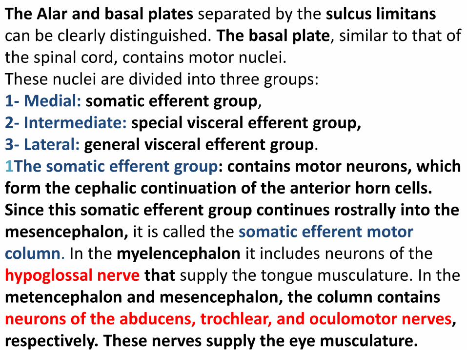
The Alar and basal plates separated by the sulcus limitans can be clearly distinguished. The basal plate, similar to that of the spinal cord, contains motor nuclei. These nuclei are divided into three groups: 1- Medial: somatic efferent group, 2- Intermediate: special visceral efferent group, 3- Lateral: general visceral efferent group. 1The somatic efferent group: contains motor neurons, which form the cephalic continuation of the anterior horn cells. Since this somatic efferent group continues rostrally into the mesencephalon, it is called the somatic efferent motor column. In the myelencephalon it includes neurons of the hypoglossal nerve that supply the tongue musculature. In the metencephalon and mesencephalon, the column contains neurons of the abducens, trochlear, and oculomotor nerves, respectively. These nerves supply the eye musculature.
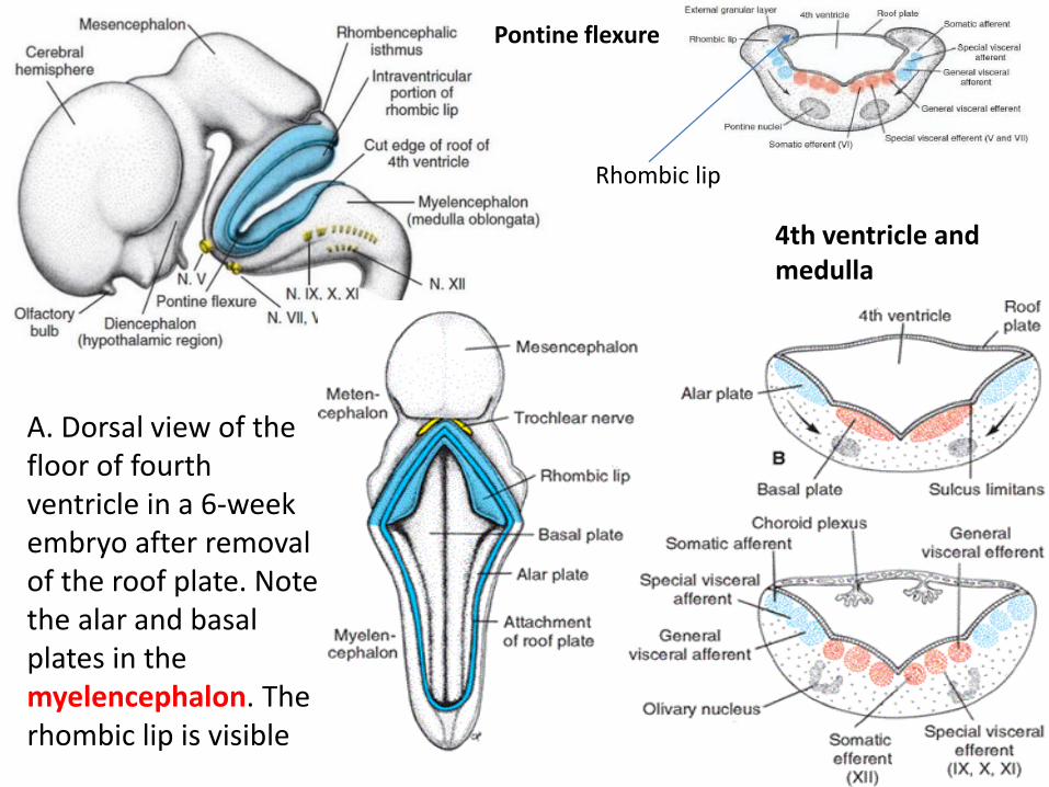
4th ventricle and medulla
Pontine flexure
A. Dorsal view of the floor of fourth ventricle in a 6-week embryo after removal of the roof plate. Note the alar and basal plates in the myelencephalon. The rhombic lip is visible
Rhombic lip

2- The special visceral efferent group extends into the metencephalon, forming the special visceral efferent motor column. It is motor neurons supply striated muscles of the pharyngeal arches. In the myelencephalon the column is represented by neurons of the accessory, vagus, and glossopharyngeal nerves. 3- The general visceral efferent group contains motor neurons that supply involuntary musculature of the respiratory tract, intestinal tract, and heart.

The alar plate contains three groups of sensory relay nuclei. 1. The most lateral of these, the somatic afferent (sensory) group,
receives impulses from the ear and surface of the head by way of the vestibulocochlear and trigeminal nerves.
2. The intermediate, or speacial visceral afferent, group receives impulses from taste buds of the tongue and from the palate, oropharynx, and epiglottis (VII, IX, X).
3. The medial, or general visceral afferent group, receives interoceptive information from the gastrointestinal tract and heart (sense of the internal state of the body).
1
2
3
VIII, V
(Taste VII, IX, X)
(GIT, Heart)

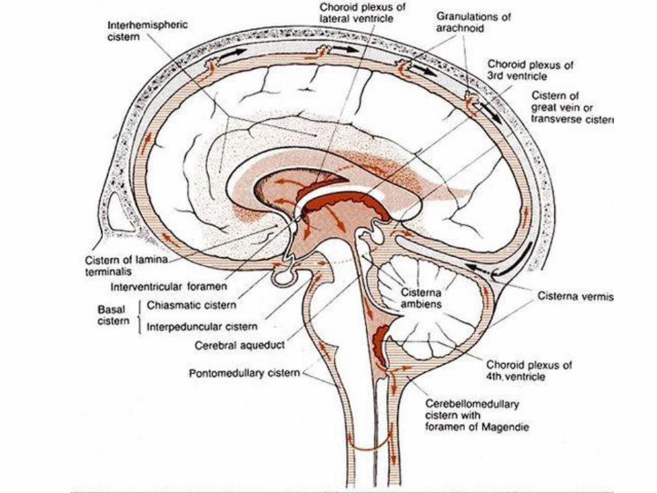
Choroid plexus in the 4th ventricle
The roof plate of the myelencephalon consists of a single layer of ependymal cells covered by vascular mesenchyme, the pia mater. The two combined are known as the tela choroidea. Because of active proliferation of the vascular mesenchyme, a number of saclike invaginations project into the underlaying ventricular cavity. These tuft form the choroid plexus, which produces cerebrospinal fluid.

Development of the Metencephalon The metencephalon, similar to the myelencephalon, is characterized by basal and alar plates. Metencephalon gives rise to two structures: 1. The cerebellum 2. The pons Each basal plate of the metencephalon contains three groups of motor neurons: 1) The medial somatic efferent group, which gives
rise to the nucleus of the abducent nerve; 2) The special visceral efferent group, containing
nuclei of the trigeminal and facial nerves, which innervate the musculature of the first and second pharyngeal arches;
3) The general visceral efferent group, whose axons supply the submandibular and sublingual glands. (facial nerve via the chorda tympani)
VI V, VI
Saliv glands

Cross section in the Rhombencephalon
Figure 19.20 A. Dorsal view of the mesencephalon and rhombencephalon in an 8 –week embryo. The roof of the fourth ventricle has been removed. Allowing a view of its floor. B. Similar view in a 4-month embryo. Note the choroidal fissure and the lateral and medial apertures in the roof of the fourth ventricle.
Rhombic lip

The marginal layer of the basal plates of the metencephalon expands as it makes a bridge for nerve fibers connecting the cerebral cortex and cerebellar cortex with the spinal cord.
The alar plates of the metencephalon contain three groups of sensory nuclei:
1- Lateral group : somatic afferent group which contains neurons of the trigeminal nerve and a small portion of the vestibulocochlear complex
2- Intermediate group:The special visceral afferent group. 3- Medial group: The general visceral afferent group.
V, VIII

At dorsolateral parts of metenceph, the alar plates bend medially and form the rhombic lips. In the caudal portion of the metencephalon, the rhombic lips are widely separated, but immediately below the mesencephalon they approach each other in the midline. As a result of a further deepening of the pontine flexure, the rhombic lips compress cephalocaudally and form the cerebellar plate. In a 12-week embryo, this plate shows a small midline portions, the vermis, and two lateral portions, the hemipheres. A transverse fissure soon separates the nodule from the vermis and the lateral flocculus from the hemipheres.
Rhombic lip
Pons Med

This flocculonodular lobe is phylogenetically the most primitive part of the cerebellum. Initially, the cerebellar plate consists of neuroepithelial, mantle, and marginal layers. During further development, a number of cells formed by the neuroepithelium migrate to the surface of the cerebellum to form the external granular layer (molecular layer). Cells of this layer retain their ability to divide and form a proliferative zone on the surface of the cerebellum.

Dorsal view
Cross section in the pons
Rhombic lip

Sagittal sections through the roof of the metencephalon showing development of the cerebellum. A.8 weeks (approximately 30mm). B. 12 weeks (70mm). C. 13 weeks. D. 15 weeks. Note formation of the external granular layer on the surface of the cerebellar plate (B and C). During later stages, cells of the external granular layer migrate inward to mingle with Purkinje cells and form the definitive cortex of the cerebellum. The dentate nucleus is one of the deep cerebellar nuclei. Note the anteror and posterior velum.

•In the sixth month of development, the external layer (Molecular layer) gives rise to various cell types. • Molecular cells migrate toward the differentiating Purkinje cells and give rise to granule cells (the internal granular layer) . •The cortex of the cerebellum contains: Golgi II neurons (stellate and basket cells), Purkinje cells, and granule cells. It reaches its definitive size after birth. •The deep cerebellar nuclei, such as the dentate nucleus, reach their final position before birth.

A.The external granular layer on the surface of the cerebellum forms a proliferative layer from which granule
cells arise. They migrate inward from the surface (arrows) to settle down underneath the purkinje cell layer
postnatal cerebellar cortex showing differentiated Purkinje
cells, the molecular layer on the surface, and the internal granular layer beneath the Purkinje cells.

Development of the Mesencephalon In the mesencephalon Each basal plate contains two groups of motor nuclei: 1. Medial somatic efferent group, represented by the
oculomotor and trochlear nerves, which innervates the eye musculature,
2. Small general visceral efferent group, represented by the nucleus of Edinger-Westphal, which innervates the sphincter pupillary muscle.
The marginal layer of each basal plate enlarges and forms the crus cerebri. These crura serve as pathways for nerve fibers descending from the cerebral cortex to lower centers in the pons and spinal cord.
The alar plates of the mesencephalon appear as two longitudinal elevations separated by a shallow midline depression. With further development, a transverse groove divides each elevation into an anterior (superior) and posterior (inferior) colliculus. The posterior colliculi serve as synaptic relay stations for auditory reflexes.
The anterior colliculi function as correlation and reflex centers for visual impulses.
The colliculi are formed by waves of neuroblasts migrating into the overlying marginal zone.

A and B. Position and differentiation of the basal and alar plates in the mesencephalon at various stages of development. Arrows in A indicate the path followed by cells of the alar plate to form the nucleus ruber and substantia nigra. Note the various motor nuclei in the basal plate.
nucleus of Edinger-Westphal

Development of the forebrain Above the mesencephalon is the prosencephalon vesicle. the alar plate forms two secondary vesicles, the cerebral hemispheres (Telencephalon). The basal plates (motor area) become the Diencephalon at the floor and ventral region of the forebrain vesicle. The cavity of the diencephalon forms the 3rd ventricle and the rostral end of the prosencephalon vesicle forms the lamina terminalis. Two secondary vesicles develop as an outgrowths from the sides of the forebrain (prosencephalon) vesicle. Each vesicle distend to form a cerebral hemisphere. The cavities inside these new vesicles form the lateral ventricle which is connected to the 3rd ventricle through an interventricular foramen. The lateral wall of the secondary vesicle is greatly thickened and the cavity of the lateral ventricle is pushed medially near the midline.

Neurons develops in the region of the diencephalon, forming the thalamus and hypothalamus. Neurons of the hemisphere develops in the floor of the lateral ventricle, forming the basal nuclei. Neurons of the hemispheres migrate to their surfaces and forming the cortical gray mater. The enlarging surface of each cerebral hemisphere is thrown into folds, forming the sulci and gyri. The cleft between the thalamus medially and the hemisphere laterally is filled up. The Hippocampus develops in the medial surface of the hemisphere dorsal to the interventricular foramen. On the ventral side of that foramen, the piriform cortex is formed. The hemispheres enlarge laterally, caudally and then ventrally. For that reason the lateral ventricle change to C-shaped, the Hippocampus curves around its convexity

Medial surface of cerebral hemisphere showing the hippocampus
Fornix

The lumen of the spinal cord, the central canal, is continuous with that of the brain vesicles.
The cavity of the rhombencephalon is the fourth ventricle. The cavity of the diencephalon is the third ventricle. The cavities of the cerebral hemispheres are the lateral ventricles. The lumen of the mesencephalon connects the third and fourth
ventricles.This lumen becomes very narrow and is then known as the aqueduct of sylvius.
The lateral ventricles communicate with the third ventricle through the interventricular foramina of Monro.

Choroid plexus of the 3rd ventricle is formed; it is made by the ependymal roof of the 3rd ventricle and invaginating network of blood vessels. Choroid plexus of the lateral ventricle is also formed, it passes through the choroid fissure. Small anterior diverticula develops from the diencephalon forming the optic stalks, it give rise to the optic nerves, retina and iris. Pineal gland develops dorsally from the diencephalon. Infundibular part of the pituitary gland develops ventrally from the diencephalon

Development of fibres 1- Projecting fibres: pass down from the cerebral cortex to lower centres, as well as some ascending fibres projecting from the thalamus to the cerebral cortex (ascending and descending fibres) . a- The internal capsule: Fibres of the internal capsule form a well defined bundle pass between the basal nuclei, the lentiform nucleus lies laterally and caudate nucleus and thalamus medially. The caudate nucleus lies adjacent to the thalamus. 2- Commissural fibres (Commissures): consist of fibres joining corresponding areas in the two sides of the brain across the midline.
a- Anterior commissure: is a ventrally placed commissural fibres passing through the lamina terminalis between the temporal poles (piriform cortex) b- Fornix commissure: a dorsally placed in the lamina terminalis develops between the two hippocampal areas. c- Corpus callosum: is a large, wide and flat bundle of commissural fibres, connects the left and right cerebral hemispheres, and enables communication between them. It develops in the fornix commissure and soon spreads over it to covers the roof of the diencephalon. As the corpus callosum increases in size, the dorsally placed hippocampus becomes much thinner and remains only as a thin layer of cells on top of the corpus callosum. The hippocampus is directed backwards and then ventrally, it lies in the floor of the inferior horn of the lateral ventricle

Coronal section of the brain

Horizontal section in the forebrain showing the internal capsule

3- Association fibres: These fibres join different areas of the same hemisphere. Most of these fibres are short.

Thank You