Elucidation of the Molecular Interaction between Cisplatin and Flavonol(s) and their Effect on DNA...
8
Elucidation of the Molecular Interaction between Cisplatin and Flavonol(s) and their Effect on DNA Binding Theodore J. Zwang, Kavisha Singh, Malkiat S. Johal, and Cynthia R. Selassie* Department of Chemistry, Pomona College, 645 North College Avenue, Claremont, California 91711, United States ABSTRACT: Combination therapy of cisplatin with flavonols is a promising treatment for increasing the efficacy of cisplatin when combating cancer. However, little is known about the molecular interactions between cisplatin and flavonols. The data herein helps to elucidate this interaction. Spectrophotometric data in the UV− visible range indicates that hydroxyl groups on the B-ring of flavonols are essential for reactivity with cisplatin. The use of a quartz crystal microbalance with dissipation monitoring approach clearly supports the critical role played by B-ring hydroxyls in their interactions with a cisplatin-bound double-stranded DNA surface; an increase in the number of hydroxyl groups on the B-ring of flavonols parallels the increase in their reaction rates with cisplatin and correlates well with their reported effects on leukemia cell apoptosis efficacy. This study underscores the importance of B-ring hydroxyls in cisplatin’s toxicity and may be used to better understand and improve combination therapies of flavonols with cisplatin. ■ INTRODUCTION Despite advances in the multimodal management of a wide spectrum of human cancers, the full clinical utility of cis- diaminedichloro-platinum(II) (cisplatin, cis-[PtCl 2 (NH 3 ) 2 ]) is limited because of acute and chronic nephrotoxicity, ototox- icity, and peripheral neuropathy. 1−7 In addition to these side effects, one of cisplatin’s greatest setbacks is the development of resistance that significantly reduces the efficacy of continued treatment. 8−12 Combination therapy with other drugs has been proposed to overcome early onset resistance to cisplatin. This has recently been explored through various studies combining cisplatin with palitaxel/SN-38, 13 new classes of platinum drugs, 14 multi- nuclear platinum compounds, 15 and various flavonols. 16 In some cases, combinations have shown increased efficacy over individual treatments 17 and reduced toxicity in cultured renal cells. 18 The combination of cisplatin with flavonols is particularly promising in light of recent studies which found an increased level of cell apoptosis upon treatment with cisplatin−flavone derivatives. 16,19,20 The polyphenolic structure of flavonols enhances their ability to accept free radicals or complex with metal ions. 21 Studies have also shown that flavonols can intercalate into deoxy- ribonucleic acid (DNA) as well as covalently bind to DNA and other proteins. 22,23 As a result of these characteristics, flavonols have complex biological interactions, and combinations of them with cisplatin are poorly understood, although multiple theories currently exist that try to explain the interactions between cisplatin and flavonols. 24−29 While some flavonols such as quercetin and luteolin have been found to improve the efficacy and cytotoxicity of cisplatin, other flavonols, such as apigenin, galangin, and chrysin, have been shown to have a negative effect on cisplatin’s cytotoxicity. 24,25 The mechanistic underpinnings of this change in efficacy is not known but presents a potentially interesting avenue for modulating cisplatin’sefficacy. The data shows a trend between the hydroxylation of the flavonol and increases in the efficacy of cisplatin. Unfortunately, the structures of cisplatin−flavonol complexes have not been well characterized, which makes it difficult to understand the structure−activity relationships. Some of the most studied flavonol−metal complexes are those of quercetin (Figure 1) and metal(II) centers. Quercetin forms complexes with copper(II), nickel(II), zinc(II), cobalt- (II), manganese(II), and lead(II). 30−32 One study suggests that the quercetin acts as a bidentate ligand for metal centers, tentatively forming an octahedral complex that involves its Received: July 23, 2012 Published: January 29, 2013 Figure 1. (A) Structure of flavonols with changes in hydroxylation in the B-ring. (B) Possible cisplatin binding modes for quercetin. 30−34 Article pubs.acs.org/jmc © 2013 American Chemical Society 1491 dx.doi.org/10.1021/jm3016798 | J. Med. Chem. 2013, 56, 1491−1498
Transcript of Elucidation of the Molecular Interaction between Cisplatin and Flavonol(s) and their Effect on DNA...

Elucidation of the Molecular Interaction between Cisplatin andFlavonol(s) and their Effect on DNA BindingTheodore J. Zwang, Kavisha Singh, Malkiat S. Johal, and Cynthia R. Selassie*
Department of Chemistry, Pomona College, 645 North College Avenue, Claremont, California 91711, United States
ABSTRACT: Combination therapy of cisplatin with flavonols is a promisingtreatment for increasing the efficacy of cisplatin when combating cancer. However,little is known about the molecular interactions between cisplatin and flavonols. Thedata herein helps to elucidate this interaction. Spectrophotometric data in the UV−visible range indicates that hydroxyl groups on the B-ring of flavonols are essential forreactivity with cisplatin. The use of a quartz crystal microbalance with dissipationmonitoring approach clearly supports the critical role played by B-ring hydroxyls intheir interactions with a cisplatin-bound double-stranded DNA surface; an increase inthe number of hydroxyl groups on the B-ring of flavonols parallels the increase in theirreaction rates with cisplatin and correlates well with their reported effects on leukemiacell apoptosis efficacy. This study underscores the importance of B-ring hydroxyls incisplatin’s toxicity and may be used to better understand and improve combinationtherapies of flavonols with cisplatin.
■ INTRODUCTIONDespite advances in the multimodal management of a widespectrum of human cancers, the full clinical utility of cis-diaminedichloro-platinum(II) (cisplatin, cis-[PtCl2(NH3)2]) islimited because of acute and chronic nephrotoxicity, ototox-icity, and peripheral neuropathy.1−7 In addition to these sideeffects, one of cisplatin’s greatest setbacks is the development ofresistance that significantly reduces the efficacy of continuedtreatment.8−12
Combination therapy with other drugs has been proposed toovercome early onset resistance to cisplatin. This has recentlybeen explored through various studies combining cisplatin withpalitaxel/SN-38,13 new classes of platinum drugs,14 multi-nuclear platinum compounds,15 and various flavonols.16 Insome cases, combinations have shown increased efficacy overindividual treatments17 and reduced toxicity in cultured renalcells.18 The combination of cisplatin with flavonols isparticularly promising in light of recent studies which foundan increased level of cell apoptosis upon treatment withcisplatin−flavone derivatives.16,19,20
The polyphenolic structure of flavonols enhances their abilityto accept free radicals or complex with metal ions.21 Studieshave also shown that flavonols can intercalate into deoxy-ribonucleic acid (DNA) as well as covalently bind to DNA andother proteins.22,23 As a result of these characteristics, flavonolshave complex biological interactions, and combinations of themwith cisplatin are poorly understood, although multiple theoriescurrently exist that try to explain the interactions betweencisplatin and flavonols.24−29 While some flavonols such asquercetin and luteolin have been found to improve the efficacyand cytotoxicity of cisplatin, other flavonols, such as apigenin,galangin, and chrysin, have been shown to have a negative effecton cisplatin’s cytotoxicity.24,25 The mechanistic underpinningsof this change in efficacy is not known but presents a potentially
interesting avenue for modulating cisplatin’s efficacy. The datashows a trend between the hydroxylation of the flavonol andincreases in the efficacy of cisplatin. Unfortunately, thestructures of cisplatin−flavonol complexes have not been wellcharacterized, which makes it difficult to understand thestructure−activity relationships.Some of the most studied flavonol−metal complexes are
those of quercetin (Figure 1) and metal(II) centers. Quercetinforms complexes with copper(II), nickel(II), zinc(II), cobalt-(II), manganese(II), and lead(II).30−32 One study suggests thatthe quercetin acts as a bidentate ligand for metal centers,tentatively forming an octahedral complex that involves its
Received: July 23, 2012Published: January 29, 2013
Figure 1. (A) Structure of flavonols with changes in hydroxylation inthe B-ring. (B) Possible cisplatin binding modes for quercetin.30−34
Article
pubs.acs.org/jmc
© 2013 American Chemical Society 1491 dx.doi.org/10.1021/jm3016798 | J. Med. Chem. 2013, 56, 1491−1498

CO group and the 3-OH oxygen atoms coordinating withthe platinum.30 Lemanska et al., have shown that the low pKa(7.03) of the 4′-OH moiety in the B-ring results in easydeprotonation that is facilitated by charge delocalization of theanion to the C4 keto moiety. The 7-OH in the A-ring is alsosusceptible to deprotonation at physiological pH.33 Thisimplies that these two sites are also capable of forming acomplex with cisplatin. Another study on the absorptionspectra of metal complexes of quercetin suggests that themetal(II) center complexes with the 3′-OH and 4′-OH sites ofthe B-ring of quercetin.34
To determine which sites of flavonols play a role in cisplatincomplexation, we examined the UV−vis spectra of cisplatinwith four flavonols that have different hydroxylation patterns onthe B-ring, only. We also used a quartz crystal microbalancewith dissipation monitoring (QCM-D) approach to assess theeffects of hydroxylation patterns on the flavonols abilities tocomplex with cisplatin that is bound to double-stranded (ds)DNA. This study also examines how exposure of DNA toquercetin and cisplatin in series differs from using a cisplatin−quercetin complex concurrently.QCM-D is a gravimetric technique that involves the
measurement of changes in resonance frequency and energydissipation upon the adsorption of a material to a sensorsurface. Essentially, the sensor vibrates with some constantamount of energy. Because the amount of energy in the systemremains constant, the frequency of the oscillations changes anytime mass is added to the surface. Small changes in the amountof adsorbed mass can be measured by relating changes infrequency to the amount of mass adsorbed by the Sauerbreyequation.35 However, a decrease in frequency does not alwaysindicate an increase in mass on the surface. Energy can be lostin other ways that would also decrease the frequency at whichthe sensor oscillates. This is measured by changes indissipation, which give a qualitative measure of how rigidlybound the adsorbent is. Increases in dissipation indicate moreenergy is being lost to the surrounding environment due to arelatively more “floppy” layer. Formation of a DNA surface onthe QCM-D sensor allows for the measurement of relativeamounts of flavonol−cisplatin combinations that bind to DNAas well as the rigidity of that binding. QCM-D has previouslybeen used to study interactions of molecules with DNAsurfaces that are similar to those explored herein, includingquercetin−copper(II) induced DNA damage.36
The ability of flavonols to modulate cisplatin’s efficacysuggests that an understanding of the interactions of flavonolswith cisplatin could greatly benefit the development ofimproved chemotherapy regimens. This study elucidateswhether cisplatin and various flavonols undergo complexation,the types of interactions that are involved, and their effects onbinding to dsDNA at the molecular level. UV−vis spectropho-tometry in combination with a QCM-D approach allows us toexamine the molecular interactions of quercetin and itsanalogues, shown in Figure 1, with cisplatin that is both freeand bound to DNA in a noncellular environment.
■ MATERIALS AND METHODSMaterials. All chemicals, unless otherwise stated, were purchased
from commercial sources and used without further purification. cis-Diaminedichloro-platinum(II) was purchased from Sigma-Aldrich andwas 99.9+%, metals basis. The flavanols, kaempferol, quercetin, andmyricetin, and galangin are ≥95% pure by HPLC, as certified bySigma-Aldrich. High molecular weight poly(ethylenimine) (PEI)
(MW = 25000 g/mol, mixture of linear and branched chains, water≤1%), silver nitrate (AgNO3: ACS reagent grade, ≥99%),dimethylsulfoxide (DMSO, ≥99%), and calf thymus DNA sodiumsalt were obtained from Sigma Aldrich and used without furthertreatment. Phosphate buffered saline (PBS) at pH 7.4 was obtainedfrom Fischer. PEI solutions were prepared at a concentration of 10mM (using the molecular weight of the polymer’s repeat unit) withultrapure water (resistivity >18 MΩ cm, Milli-Q) at pH 7. DNAsolutions were also prepared at a concentration of 0.1 mg/mL in a PBSsolution with 5% DMSO by volume following the procedure of Rawleet al.36 The flavonol solutions were all prepared at a concentration of0.1 mM PBS with 5% DMSO by volume because DMSO was requiredto ensure the solubility of the polyphenolic compounds. The cisplatinsolutions were prepared at 2.56 mM in PBS with 5% DMSO byvolume. Activated cisplatin ([Pt(NH3)2(H2O)2]
+2) solutions wereprepared by adding twice the molar amount of AgNO3 and allowingthe precipitation reaction to complete over a time period of 4−7 days.The precipitate of AgCl was removed by multiple vacuum filtrationsover this period of time, and finally a clear solution of activatedcisplatin was obtained. Minimal HCl or NaOH was added as neededto flavonol or cisplatin solutions to maintain pH 7.4 before use.
UV−vis Experiments. All UV−vis spectroscopic work was carriedout on a Cary 300 UV−visible spectrophotometer (Varian). Thecisplatin and flavonols were mixed in 50 mL centrifuge tubes at anequal molar ratio of cisplatin to flavonol and then incubated at 37 °C.The spectra were taken in quartz cuvettes and the solutions offlavonols, cisplatin, and cisplatin−flavonol mixtures were directlyanalyzed by pipetting 2 mL into the cuvettes at various time points.The reference solution used for these measurements was a solution ofPBS with 5% DMSO by volume.
QCM-D Experiments. A quartz crystal microbalance withdissipation monitoring (QCM-D) (E4, Q-Sense, Gothenburg,Sweden) was used to obtain all the frequency (F) and dissipation(D) data of the DNA, cisplatin, flavonols, and PEI surfaces. The QCM-D sensor consisted of an AT-cut piezoelectric quartz crystal disk,coated with an Au electrode (100 nm thick) on the back and an activesurface layer of SiO2 (∼50 nm thick). The QCM-D sensor crystal (14mm × 0.3 mm, active area of 0.2 cm2) operated at a frequency of 4.95MHz ± 50 kHz. The crystals were optically polished with a surfaceroughness of less than 3 nm (root-mean-square). The active side wasin contact with the solutions of PEI, DNA, and the experimentalsolutions. The crystal was mounted in a flow cell with a total volume of40 μL. Multiple overtone values were collected, but only the fifthovertone values for frequency and dissipation are reported herein. Atthis overtone, the frequency values were obtained with accuracy of±0.2 Hz or less, and the dissipation factor was obtained with accuracyof ±0.2 × 10⁻⁶ or less. F and D values were measured relative to abaseline obtained in ultrapure water.
Before usage, the SiO2-coated quartz crystals were exposed to a 2%Hellmanex solution (Hellma Co.) for 30 min, followed by an ethanolrinse and an ultrapure water rinse. The crystals were dried with N2 andtreated by UV/ozone for 10 min and then placed in the QCM-D flowcells. A stable baseline for frequency and dissipation was obtained byflushing the QCM-D flow cells with ultrapure water. The sensorsurface was then exposed to PEI in order to form a substrate ontowhich the DNA surface could form. The various other solutions werepassed through the QCM-D flow cells as indicated. In betweenexperimental solutions, the sensor surface was rinsed to remove anyweakly attached adsorbents and to ensure that the observed changes inF and D were not due to changes in the solutions to which the surfaceswere being exposed. The flow rate for each experiment was 100 μL/min, and all QCM-D experiments were conducted at 23 °C.
■ RESULTS
Flavonol−Cisplatin Spectra. Upon the addition ofcisplatin, the quercetin solution gradually changes color. Ascan be seen in Figure 2, this change in color coincides with adecrease in the characteristic absorbance of quercetin at 374and 274 nm as well as the appearance of a new peak at 293 nm
Journal of Medicinal Chemistry Article
dx.doi.org/10.1021/jm3016798 | J. Med. Chem. 2013, 56, 1491−14981492
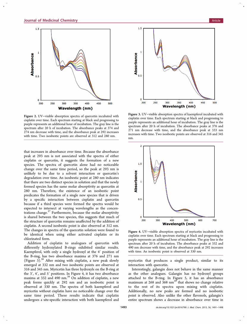
that increases in absorbance over time. Because the absorbancepeak at 293 nm is not associated with the spectra of eithercisplatin or quercetin, it suggests the formation of a newspecies. The spectra of quercetin alone had no noticeablechange over the same time period, so the peak at 293 nm isunlikely to be due to a solvent interaction or quercetin’sdegradation over time. An isosbestic point at 280 nm indicatesthat there are two distinct species in solution and that the newlyformed species has the same molar absorptivity as quercetin at280 nm. Therefore, the existence of an isosbestic pointpredicates the formation of a single new species that is drivenby a specific interaction between cisplatin and quercetinbecause if a third species were formed the spectra would beexpected to intersect at varying wavelengths as the concen-trations change.37 Furthermore, because the molar absorptivityis shared between the two species, this suggests that much ofthe structure of quercetin remains unaffected by the addition ofcisplatin. A second isosbestic point is also observed at 312 nm.The changes in spectra of the quercetin solution were found tobe identical when using either activated cisplatin or itschlorinated form.Addition of cisplatin to analogues of quercetin with
differently hydroxylated B-rings exhibited similar results.Kaempferol, with only a single hydroxyl at the 4′ position ofthe B-ring, has two absorbance maxima at 376 and 271 nm(Figure 3).38 After mixing with cisplatin, a new peak slowlyemerged at 332 nm and two isosbestic points are observed at316 and 345 nm. Myricetin has three hydroxyls on the B-ring atthe 3′, 4′, and 5′ positions. In Figure 4, it has two absorbancemaxima at 332 and 490 nm.38 On addition of cisplatin, a newpeak forms quickly at 292 nm and an isosbestic point isobserved at 330 nm. The spectra of both kaempferol andmyricetin without cisplatin have no noticeable change over thesame time period. These results indicate that cisplatinundergoes a site-specific interaction with both kaempferol and
myricetin that produces a single product, similar to itsinteraction with quercetin.Interestingly, galangin does not behave in the same manner
as the other analogues. Galangin has no hydroxyl groupsattached to the B-ring. In Figure 5, it has an absorbancemaximum at 268 and 368 nm39 that shows no change relativeto the rest of its spectra upon mixing with cisplatin.Additionally, no new peaks are formed and no isosbesticpoint is observed. Also unlike the other flavonols, galangin’sentire spectrum shows a decrease in absorbance over time in
Figure 2. UV−visible absorption spectra of quercetin incubated withcisplatin over time. Each spectrum starting at black and progressing topurple represents an additional hour of incubation. The gray line is thespectrum after 20 h of incubation. The absorbance peaks at 374 and274 nm decrease with time, and the absorbance peak at 292 increaseswith time. Two isosbestic points are observed at 312 and 280 nm.
Figure 3. UV−visible absorption spectra of kaempferol incubated withcisplatin over time. Each spectrum starting at black and progressing topurple represents an additional hour of incubation. The gray line is thespectrum after 20 h of incubation. The absorbance peaks at 376 and271 nm decrease with time, and the absorbance peak at 333 nmincreases with time. Two isosbestic points are observed at 316 and 345nm.
Figure 4. UV−visible absorption spectra of myricetin incubated withcisplatin over time. Each spectrum starting at black and progressing topurple represents an additional hour of incubation. The gray line is thespectrum after 20 h of incubation. The absorbance peaks at 332 and490 nm decrease with time, and the absorbance peak at 292 increaseswith time. An isosbestic point is observed at 330 nm.
Journal of Medicinal Chemistry Article
dx.doi.org/10.1021/jm3016798 | J. Med. Chem. 2013, 56, 1491−14981493

the absence or presence of cisplatin. This is likely due to itsdecreased solubility in water, which is consistent with itshydrophobicity; the other flavonols are more hydrophilic (seeTable 1). The absence of a new absorbance peak suggests thatthe production of a second species/complex does not occurwhen galangin is mixed with cisplatin.Drug Interactions with dsDNA Surface. In Figure 6, a
dsDNA surface was formed on the QCM-D sensor and firstexposed to cisplatin, resulting in a frequency decrease of 5 Hzand a corresponding increase in dissipation. This indicatesbinding of cisplatin to the DNA. Subsequent addition ofquercetin causes a further 10 Hz decrease in frequency andincrease in dissipation. In Figure 7, the quercetin solution wasflowed over a dsDNA surface prior to cisplatin binding; nomass deposition was observed upon exposure to quercetin andthe same 5 Hz decrease in frequency and increase in dissipationwas observed for the following addition of cisplatin. Thenonbinding of quercetin with a dsDNA surface agrees withprevious data.36 These results indicate that the degree ofquercetin deposited in Figure 6 can be attributed entirely to itsinteractions with cisplatin that was already bound to the DNAsurface.In Figure 8, activated ([Pt(NH3)2(H2O)2]
+2) cisplatin wasflowed over the dsDNA surface followed by quercetin.Exposure of the dsDNA to activated cisplatin results in a 30Hz decrease in frequency, which indicates that approximately 6-fold more activated cisplatin bound to the dsDNA surface thanchlorinated ([Pt(NH3)2(Cl)2]) cisplatin. It should be noted
that subsequent addition of quercetin increased the frequencyand decreased the dissipation; the complexation of quercetin in
Figure 5. UV−visible absorption spectra of galangin incubated withcisplatin over time. Each spectrum starting at black and progressing topurple represents an additional hour of incubation. The gray line is thespectrum after 20 h of incubation. The absorbance decreases over the20 h incubation. No new peaks were formed, nor were any peaks’absorbance diminished relative to the rest of the spectrum.
Table 1. UV−Vis Spectroscopy Results from Incubation of Various Flavonols with an Equimolar Concentration of Cisplatin
flavonoidno. of hydroxyls on B-
ringinitial peaks
(nm)isosbestic point
(nm)new peaks(nm)
ΔAbs ((abs/mmol−1)/(20 h−1))
relative initialrate ClogP
myrcetin 3 332, 490 330 292 0.569 3.31 0.84quercetin 2 374, 274 280, 312 293 0.192 2.02 1.50kaempferol 1 376, 271 316, 345 332 0.114 1 2.10galangin 0 368, 268 0 0 2.76
Figure 6. QCM-D response to the binding of cisplatin to a dsDNAsurface, with subsequent exposure to quercetin. Dotted lines representPBS rinses, which remove weakly adhered molecules. The black line isthe change in frequency, which corresponds to the left y-axis. The grayline is the change in the energy dissipation, which corresponds to theright y-axis. The asterisk indicates the end of the activated cisplatin−quercetin sequence, which resulted in a 13 Hz decrease. Thesubsequent rinse did not result in any significant change in frequencyor dissipation.
Figure 7. QCM-D response to the binding of quercetin to a dsDNAsurface, with subsequent exposure to cisplatin. Dotted lines representPBS rinses, which remove weakly adhered molecules. The black line isthe change in frequency, which corresponds to the left y-axis. The grayline is the change in the energy dissipation, which corresponds to theright y-axis.
Journal of Medicinal Chemistry Article
dx.doi.org/10.1021/jm3016798 | J. Med. Chem. 2013, 56, 1491−14981494

solution to bound cisplatin appears to be tearing some of thecisplatin away from the dsDNA.It is prudent to compare Figures 8 and 6 to see that the total
decrease in frequency caused by the deposition of activatedcisplatin followed by quercetin is the same as the decrease infrequency caused by the deposition of chlorinated cisplatinfollowed by quercetin. However, the subsequent rinse increasesthe frequency and decreases the dissipation for the dsDNAexposed to the activated cisplatin−quercetin sequence but doesnot affect the dsDNA exposed to the cisplatin−quercetinsequence. The increase in frequency and decrease in dissipationare characteristic of the loss of weakly adsorbed molecules fromthe surface.This suggests that activated cisplatin binds to dsDNA in both
high- and low-affinity modes while chlorinated cisplatin onlybinds in high-affinity modes. This is reflected by the respectivechanges in dissipation during the initial deposition of thecisplatins: the larger increase in dissipation for activatedcisplatin binding suggests floppiness or some incidence ofweaker binding. The access to low-affinity binding sites ofactivated cisplatin but not the chlorinated form is likely causedby the activated form’s positive charge, whereas the chlorinatedform does not have access to these sites because it is neutral.The electrostatic forces between the positively charged cisplatinand the negatively charged dsDNA surface could alter theaffinity of activated cisplatin and allow it access to otherwiseunfavorable binding sites. It appears that when cisplatin bindsto a high-affinity binding site, quercetin binds to it and thecomplex remains bound to the dsDNA surface. However, whencisplatin is at a low-affinity binding site, quercetin binds to itand pulls it away from the dsDNA. On subsequent rinsing, thedissipation drops dramatically and the frequency increases butremains slightly depressed, indicating the removal of weaklybound cisplatin while only the tightly bound cisplatin remainsattached to the dsDNA. Figure 9 supports this claim, that
quercetin is effectively tearing the activated cisplatin away fromthe surface by binding to it rather than to the DNA, bydemonstrating that none of the activated cisplatin is rinsed offin the quercetin-activated cisplatin sequence.Incubated solutions of cisplatin−quercetin (Figure 10) and
activated cisplatin−quercetin (Figure 11) were also exposed to
a dsDNA surface because the UV−vis data indicates that thesecompounds might be undergoing complexation over time.Indeed, this resulted in a significant frequency decrease of 15Hz with no change in dissipation for the chlorinated form and adecrease of 20 Hz with a significant drop in dissipation for theactivated form. This difference in frequency indicates that thecomplexes formed between the two forms of cisplatin andquercetin both possess the ability to bind to dsDNA and thatthe binding causes a frequency change similar to that whichoccurred when cisplatin and quercetin were exposed to thesurface, in series. However, the change in dissipation is
Figure 8. QCM-D response to the binding of activated cisplatin to adsDNA surface, with subsequent exposure to quercetin. Dotted linesrepresent PBS rinses, which remove weakly adhered molecules. Theblack line is the change in frequency, which corresponds to the left y-axis. The gray line is the change in the energy dissipation, whichcorresponds to the right y-axis. The asterisk indicates the end of theactivated cisplatin−quercetin sequence, which resulted in a 12 Hzdecrease. The subsequent rinse resulted in a 6 Hz increase infrequency and a significant decrease in dissipation, which suggests theloss of weakly adsorbed molecules from the surface.
Figure 9. QCM-D response to the binding of quercetin to a dsDNAsurface, with subsequent exposure to activated cisplatin. Dotted linesrepresent PBS rinses, which remove weakly adhered molecules. Theblack line is the change in frequency, which corresponds to the left y-axis. The gray line is the change in the energy dissipation, whichcorresponds to the right y-axis.
Figure 10. QCM-D response to the binding of the quercetin−cisplatincomplex to a dsDNA surface. Dotted lines represent PBS rinses, whichremove weakly adhered molecules. The black line is the change infrequency, which corresponds to the left y-axis. The gray line is thechange in the energy dissipation, which corresponds to the right y-axis.
Journal of Medicinal Chemistry Article
dx.doi.org/10.1021/jm3016798 | J. Med. Chem. 2013, 56, 1491−14981495

distinctly different when the incubated samples are usedtogether compared to the change in dissipation when thecompounds are not incubated and used in series. Usually, thedeposition of mass on a crystal surface is accompanied by anincrease in floppiness and dissipation, but this is not observedwith the incubated solutions. This suggests that the mixturesbind very tightly to the dsDNA surface, and in the case of theactivated cisplatin complex, its binding appears to be makingthe overall film significantly more rigid. This is consistent withthe rigidification of DNA that would be expected from anintercalative binding mode.To further explore the effect of hydroxyl groups on the B-
ring of flavonols, quercetin analogues were exposed to a dsDNAsurface with bound cisplatin. Myricetin (Figure 12) andkaempferol (Figure 13), with three hydroxyl groups and one
hydroxyl group on the B-ring, respectively, resulted in asignificant mass deposition and increase in dissipationcomparable to those for quercetin. Galangin (Figure 14),
with no hydroxyl groups on the B-ring, resulted in nosignificant mass deposition. This further strengthens theargument that phenolic, hydroxyl groups on the B-ring areessential for the binding of flavonols to cisplatin and thatincludes cisplatin that is bound to dsDNA.
■ DISCUSSIONDNA is regarded as the primary biological target of cisplatin,although alternative targets have been investigated.40−43 Oncecisplatin enters a cell, the low chloride concentration results inthe displacement of chlorine ligands by water to generate thediaqua active form of cisplatin, [Pt(NH3)2(H2O)2]
+2, which
Figure 11. QCM-D response to the binding of the activated cisplatin−quercetin complex to a dsDNA surface. Dotted lines represent PBSrinses, which remove weakly adhered molecules. The black line is thechange in frequency, which corresponds to the left y-axis. The gray lineis the change in the energy dissipation, which corresponds to the righty-axis.
Figure 12. QCM-D response to the binding of cisplatin to a dsDNAsurface, with subsequent exposure to myricetin. Dotted lines representPBS rinses, which remove weakly adhered molecules. The black line isthe change in frequency, which corresponds to the left y-axis. The grayline is the change in the energy dissipation, which corresponds to theright y-axis.
Figure 13. QCM-D response to the binding of cisplatin to a dsDNAsurface, with subsequent exposure to kaempferol. Dotted linesrepresent PBS rinses, which remove weakly adhered molecules. Theblack line is the change in frequency, which corresponds to the left y-axis. The gray line is the change in the energy dissipation, whichcorresponds to the right y-axis.
Figure 14. QCM-D response to the binding of cisplatin to a dsDNAsurface with subsequent exposure to galangin. Dotted lines representPBS rinses, which remove weakly adhered molecules. The black line isthe change in frequency, which corresponds to the left y-axis. The grayline is the change in the energy dissipation, which corresponds to theright y-axis.
Journal of Medicinal Chemistry Article
dx.doi.org/10.1021/jm3016798 | J. Med. Chem. 2013, 56, 1491−14981496

binds to DNA more easily than the chlorine-bound form.44 Theplatinum core binds to the N7 nitrogens of guanosine andadenosine, forming intrastrand adducts that distort the doubleDNA helix. This is further stabilized by hydrogen bondsbetween the amine ligands of cisplatin and the phosphategroups of DNA.41 The binding of cisplatin bends DNA anddisrupts mitosis by inhibiting the function of replication andtranscription factors. This induces early apoptosis and isadvantageous for the destruction of tumor cells.45,46
Flavonols have the well-established ability to complex withmetal ions.30−32,34 There are multiple possible complexing siteswithin a flavonol. Our data indicates that only flavonols with B-ring hydroxyls complex with cisplatin. This is strong evidencethat B-ring hydroxyls are the site of cisplatin complexation. Bymeasuring the absorbance spectrum of a mixture of cisplatinwith different flavonols, we have noted that the speed of thisreaction increases with the number of hydroxyl groups on theB-ring. Over the first 4 h, the relative rates at which the newpeak is formed are 3.31:2.02:1:0 for myricetin:quercetin:-kaempferol:galangin. This ratio changes slightly to 3.31:2.35:1:0if the rates are calculated over the first 8 h. The ratio of relativerates appears to mirror the ratio of hydroxyl groups on theflavonols’ B-ring. This, combined with the presence of only onecomplex in each reaction mixture, suggests that the complexbeing formed clearly involves the interaction of hydroxyls onthe B-ring with the platinum metal center, which is a structurethat shares similarities with a promising new DNA-bindingplatinum anticancer drug candidate.47
QCM-D results suggest that the enhanced cytotoxicity of theflavonol−cisplatin combination could result either from bindingof the flavonols to cisplatin already bound to dsDNA or fromthe binding of a flavonol−cisplatin complex to the dsDNA. It isworth noting that differences in the rate at which the flavonolsbind to cisplatin in dsDNA is much smaller than the differencesin the rate at which the flavonols bind to cisplatin in solution.Our results allow us to elucidate the relationship between
structure and activity for the phenomena reported by Cipak etal.24 In an elegant study, it was reported that flavonols candifferentially modulate the efficacy of cisplatin in the inductionof leukemia cell apoptosis. Quercetin and luteolin, both with 2catecholic-hydroxyls on the B-ring, potentiate the cytotoxicityof cisplatin by increasing cisplatin-induced apoptotic cell death.Apigenin, with a single hydroxyl, and chrysin and galingin, withno hydroxyls, have either minimal effects or antagonistic effectson the cytotoxicity of cisplatin. The effects on apoptotic DNAfragmentation in leukemia cells caused by these flavonols, indecreasing order of magnitude, are quercetin, luteolin, and thenapigenin, with chrysin and galingin having no effect.24 Theseflavonols are also listed in order of decreasing number of B-ringhydroxyls.Our data indicates that a decrease in the number of B-ring
hydroxyls will reduce the rate at which flavonols can complexwith cisplatin and that flavonol−cisplatin complexes can bind todsDNA, albeit at different rates. This strongly suggests that theenhanced cytotoxicity may be attributed to the effects of thecomplex and that increasing the rate of cisplatin−flavonolcomplex formation by using flavonols such as myricetin withmore B-ring hydroxyls may be able to further increase thesynergistic effects of flavonols on leukemia cell apoptosis. Thepotentiation caused by the complex appears to be competingwith a chemopreventative effect of flavonols, as observed by theantagonistic effects of apigenin, chrysin, and galingin oncisplatin cytotoxicity,24 which takes precedence when the
cytotoxicity-enhancing reaction between the flavonol andcisplatin is too slow. However, the inability of quercetin andthe other flavonols to bind to the cisplatin-free dsDNA on thesurface may be confounding this part of the analysis.
■ CONCLUSIONOur data indicates that hydroxyl groups on the B-ring offlavanols are essential for interactions between cisplatin andcertain flavonols. Increasing the rate of formation of thiscomplex potentiates the efficacy of cisplatin in the induction ofleukemia cell apoptosis. This study enhances understanding ofthe flavonol-modulated efficacy of cisplatin that will aid in therational discovery of more effective chemotherapeutics.Furthermore, these results indicate that previously reportedmetal(II) complexes of flavonols are not adequate models forunderstanding the interaction of flavonols and cisplatin.
■ AUTHOR INFORMATIONCorresponding Author*Phone: (909) 621-8446. Fax: (909) 607-7726. E-mail:[email protected] authors declare no competing financial interest.
■ ACKNOWLEDGMENTSThis research was supported in part by the Pomona CollegeUndergraduate Research Program.
■ ABBREVIATIONS USEDcisplatin, cis-diaminedichloro-platinum(II); DNA, deoxyribonu-cleic acid; QCMD, quartz crystal microbalance with dissipationmonitoring; dsDNA, double-stranded DNA; PEI, poly-(ethyleneimine); AgNO3; silver nitrate; DMSO, dimethylsulf-oxide; PBS, phosphate buffered saline; F, frequency; D,dissipation
■ REFERENCES(1) Rosenberg, B.; Vancamp, L.; Trosko, James, E.; Mansour,Virginia, H. Platinum Compounds: A New Class of Potent AntitumourAgents. Nature 1969, 222, 385−386.(2) Rose, P. G.; Bundy, B. N.; Watkins, E. B.; Thigpen, J. T.; Deppe,G.; Maiman, M. A.; Clarke-Pearson, D. L.; Insalaco, S. Concurrentcisplatin-based radiotherapy and chemotherapy for locally advancedcervical cancer. N. Engl. J. Med. 1999, 340, 1144−1153.(3) Crino, L.; Scagliotti, G.; Ricci, S.; De Marinis, F.; Rinaldi, M.;Gridelli, C.; Ceribelli, A.; Bianco, R.; Marangolo, M.; Di Costanzo, F.Gemcitabine and cisplatin versus mitomycin, ifosfamide, and cisplatinin advanced non-small-cell lung cancer: A randomized phase III studyof the Italian Lung Cancer Project. J. Clin. Oncol. 1999, 17, 3522−3530.(4) McGuire, W. P.; Hoskins, W. J.; Brady, M. F.; Kucera, P. R.;Partridge, E. E.; Look, K. Y.; Clarke-Pearson, D. L.; Davidson, M.Cyclophosphamide and cisplatin compared with paclitaxel andcisplatin in patients with stage III and stage IV ovarian cancer. NEngl. J. Med. 1996, 334, 1−6.(5) Burtness, B.; Goldwasser, M. A.; Flood, W.; Mattar, B.;Forastiere, A. A. Phase III randomized trial of cisplatin plus placebocompared with cisplatin plus cetuximab in metastatic/recurrent headand neck cancer: an Eastern Cooperative Oncology Group study. J.Clin. Oncol. 2005, 23, 8646−8654.(6) Treskes, M.; Vijgh, W. J. F. WR-2721 as a modulator of cisplatin-and carboplatin-induced side effects in comparison with otherchemoprotective agents: a molecular approach. Cancer Chemother.Pharmacol. 1993, 33, 93−106.
Journal of Medicinal Chemistry Article
dx.doi.org/10.1021/jm3016798 | J. Med. Chem. 2013, 56, 1491−14981497

(7) Hamers, F.; Gispen, W.; Neijt, J. Neurotoxic side-effects ofcisplatin. Eur. J. Cancer 1991, 27, 372−376.(8) Gately, D.; Howell, S. Cellular accumulation of the anticanceragent cisplatin. Br. J. Cancer 1993, 67, 1171−1176.(9) Aebi, S.; Kuri-Haidar, B.; Gordon, R.; Cenni, B.; Zheng, H.; Fink,D.; Christen, R. D.; Boland, C. R.; Koi, M.; Fishel, R. Loss of DNAmismatch repair in acquired resistance to cisplatin. Cancer Res. 1996,56, 3087−3090.(10) Godwin, A.; Meister, A.; O’Dwyer, P.; Huang, C.; Hamilton, T.;Anderson, M. High resistance to cisplatin in human ovarian cancer celllines is associated with marked increase in glutathione synthesis. Proc.Natl. Acad. Sci. U. S. A. 1992, 89, 3070−3074.(11) Kartalou, M.; Essigmann, J. M. Recognition of cisplatin adductsby cellular proteins. Mutat. Res., Fundam. Mol. Mech. Mutagen. 2001,478, 23−43.(12) Gore, M.; Fryatt, I.; Wiltshaw, E.; Dawson, T.; Robinson, B.;Calvert, A. Cisplatin/carboplatin cross-resistance in ovarian cancer. Br.J. Cancer 1989, 60, 767−769.(13) Komuro, Y.; Udagawa, Y.; Susumu, N.; Aoki, D.; Kubota, T.;Nozawa, S. Paclitaxel and SN-38 Overcome Cisplatin Resistance ofOvarian Cancer Cell Lines by Down-Regulating the Influx and EffluxSystem of Cisplatin. Cancer Sci. 2001, 92, 1242−1250.(14) Kelland, L, R.; Sharp, S. Y.; O’Neill, C. F.; Raynaud, F. I.; Beale,P. J.; Judson, I. R. Discovery and development of platinum complexesdesigned to circumvent cisplatin resistance. J. Inorg. Biochem. 1999, 77,111−115.(15) Perego, P.; Caserini, C.; Gatti, L.; Carenini, N.; Romanelli, S.;Supino, R.; Colangelo, D.; Viano, I.; Leone, R.; Spinelli, S. A noveltrinuclear platinum complex overcomes cisplatin resistance in anosteosarcoma cell system. Mol. Pharmacol. 1999, 55, 528−534.(16) Kosmider, B.; Osiecka, R. Flavonoid compounds: a review ofanticancer properties and interactions with cis-diamminedichloropla-tinum. Drug Dev. Res. 2004, 63, 200−211.(17) Neelam, S. S.; Bernabei, A.; Freedland, C.; Thompson, R.;Coebett, T. H.; Luk, G. D. Combination of flavone acetic acid (FAA)with adriamycin, cis-platinum and difluoromethylornithine (DFMO)in vitro against human colon cancer cells. Invest. New Drugs 1990, 8,263−268.(18) Kuhlman, M. K.; Horsch, E.; Burkhardt, G.; Wagner, M.;Kohler, H. Reduction of cisplatin toxicity in cultured renal tubular cellsby the bioflavonoid quercetin. Arch. Toxicol. 1998, 72, 536−540.(19) Kosmider, B.; Wyszynska, K.; Janik, Spiechowicz, E.; Osiecka,R.; Zyner, E.; Ochoki, J.; Ciesielska, E.; Wasowicz, W. Evaluation ofthe genotoxicity of cis-bis(3-aminoflavone) dichloroplatinum(II) incomparison with cis-DDP. Mutat. Res., Genet. Toxicol. Environ.Mutagen. 2004, 558, 93−110.(20) Choi, J. A.; Kim, J. Y.; Lee, J. Y.; Kang, C. M.; Kwon, H. J.; Yoo,Y. D.; Kim, T. W.; Lee, Y. S.; Lee, S. J. Induction of cell cycle arrestand apoptosis in human breast cancer cells by quercetin. Int. J. Oncol.2001, 19, 837−844.(21) Dehghan, G.; Dolatabadi, J. E. N.; Jouyban, A.; Zeynali, K. A.;Ahmadi, S. M.; Kashanian, S. Spectroscopic studies on the interactionof quercetin−terbium(III) complex with calf thymus DNA. DNA CellBiol. 2011, 30, 195−201.(22) Kanakis, C. D.; Tarantilis, P. A.; Polissiou; Diamantoglou, S.;Tajmir-Riahi, H. A. An overview of DNA and RNA bindings toantioxidant flavonoids. Cell Biochem. Biophys. 2007, 49, 29−36.(23) Janjua, N. K.; Siddiqa, A.; Yaqub, A.; Sabahat, S.; Qureshi, R.;Haque, S. Spectrophotometric analysis of flavonoid−DNA bindinginteractions at physiological conditions. Spectrochim. Acta, Part A 2009,74, 1135−1137.(24) Cipak, L.; Novotny, L.; Cipakova, I.; Rauko, P. Differentialmodulation of cisplatin and doxorubicin efficacies in leukemia cells byflavonoids. Nutr. Res. (N. Y., NY, U. S.) 2003, 23, 1045−1057.(25) Cipak, L.; Rauko, P.; Miadokova, E.; Cipakova, I.; Novotny, L.Effects of flavonoids on cisplatin-induced apoptosis of HL-60 andL1210 leukemia cells. Leuk. Res. 2003, 27, 65−72.(26) Ahmed, M. S.; Ramesh, V.; Nagaraja, V.; Parish, J.; Hadi, S.Mode of binding of quercetin to DNA. Mutagenesis 1994, 9, 193−197.
(27) Cross, H. J.; Tilby, M.; Chipman, J. K.; Ferry, D. R.; Gescher, A.Effect of quercetin on the genotoxic potential of cisplatin. Int. J. Cancer1996, 66, 404−408.(28) Behling, E. B.; Sendao, M. C.; Francescato, H. D. C.; Antunes,L. M. G.; Costa, R. S.; de Lourdes, P.; Bianchi, M. Comparative studyof multiple dosage of quercetin against cisplatin-induced nephrotox-icity and oxidative stress in rat kidneys. Pharmacol. Rep. 2006, 58,526−532.(29) Devi, P.; Shyamala, D. Protective effect of quercetin in cisplatininduced cell injury in the rat kidney. Indian J. Pharmacol 2009, 31,422−426.(30) Zhou, J.; Wang, L.; Wang, J.; Tang, N. Antioxidant and anti-tumour activities of solid quercetin zinc(II) complex. Transition Met.Chem. (Dordrecht, Neth.) 2001, 26, 57−63.(31) Tan, J.; Zhu, L.; Wang, B. DNA binding and cleavage activity ofquercetin nickel(II) complex. Dalton Trans. 2009, 24, 4722−4728.(32) Tan, J.; Wang, B.; Zhu, L. DNA binding and oxidative DNAdamage induced by a quercetin copper(II) complex: potentialmechanism of its antitumor properties. J. Biol. Inorg. Chem. 2009,14, 727−739.(33) Lemanska, K.; Szymudiak, H.; Tyrakowska, B.; Zielinski, R.;Soffers, A. E. M. F.; Rietjens, I. M. C. M. The influence of pH onantioxidant properties and the mechanism of antioxidant action ofhydroxyflavones. Free Radical Biol. Med. 2001, 31, 869−881.(34) Jurd, L.; Geissman, T. Absorption spectra of metal complexes offlavonoid compound. J. Org. Chem. 1956, 21, 1395−1401.(35) Sauerbrey, G. Verwendung von Schwingquarzen zur Wag̈ungdunner Schichten und zur Mikrowag̈ung. Z. Phys. 1959, 155, 206−222.(36) Rawle, R. J.; Johal, M. S.; Selassie, C. R. D. A real-time QCM-Dapproach to monitoring mammalian DNA damage using DNAadsorbed to a polyelectrolyte surface. Biomacromolecules 2007, 9, 9−12.(37) Moore, J. W.; Pearson, R. G. Kinetics and Mechanism, 3rd ed.;John Wiley & Sons: New York, 1981.(38) Szostek, B.; Orsaka-Gawrys, J.; Surowiec, I.; Trojanowicz, M.Investigation of natural dyes occurring in historical Coptic textiles byhigh-performance liquid chromatography with UV−vis and massspectrometric detection. J. Chromatogr., A 2003, 1012, 179−192.(39) Ahmad, M. S.; Fazal, F.; Rahman, A.; Hadi, S.; Parish, J. H.Activities of flavonoids for the cleavage of DNA in the presence ofCu(II): correlation with generation of active oxygen species. J. . 1992,13, 605−608.(40) Bose, R. Biomolecular targets for platinum antitumor drugs.Mini-Rev. Med. Chem. 2002, 2, 103−111.(41) Jamieson, E. R.; Lippard, S. J. Structure, recognition andprocessing of cisplatin DNA adducts. Chem. Rev. 1999, 99, 2467−2498.(42) Reed, E.; Ozols, R. F.; Tarone, R.; Yuspa, S. H.; Poirier, M. C.The measurement of cisplatin−DNA adduct levels in testicular cancerpatients. Carcinogenesis 1988, 9, 1909−1911.(43) Reed, E.; Yuspa, S.; Zwelling, L.; Ozols, R.; Poirier, M.Quantitation of cis-diamminedichloroplatinum II (cisplatin)−DNA-intrastrand adducts in testicular and ovarian cancer patients receivingcisplatin chemotherapy. J. Clin. Invest. 1986, 77, 545−550.(44) Ziegler, C. J.; Silverman, A. P.; Lippard, S. J. High-throughputsynthesis and screening of platinum drug candidates. J. Biol Inorg.Chem. 2000, 5, 774−783.(45) Trimmer, E. E.; Essigmann, J. M. Cisplatin. Essays Biochem.1999, 34, 191−211.(46) Sorenson, C. M.; Barry, M. A.; Eastman, A. Analysis of EventsAssociated with Cell Cycle Arrest at G2 Phase and Cell Death Inducedby Cisplatin. J. Natl. Cancer Inst. 1990, 82, 749−755.(47) Park, G. Y.; Wilson, J. J.; Song, Y.; Lippard, S. J.Phenanthriplatin, a monofunctional DNA-binding platinum anticancerdrug candidate with unusual potency and cellular activity profile. Proc.Natl. Acad. Sci. U. S. A. 2012, 109, 11987−11992.
Journal of Medicinal Chemistry Article
dx.doi.org/10.1021/jm3016798 | J. Med. Chem. 2013, 56, 1491−14981498