Elucidating the Dual Mode of Action of Dipeptidyl Enoates ...
20
1 Elucidating the Dual Mode of Action of Dipeptidyl Enoates in the Inhibition of Rhodesain Cysteine Proteases Kemel Arafet, 1 Florenci V. González, 2 Vicent Moliner 1,* 1. Departament de Química Física i Analítica, Universitat Jaume I, 12071 Castelló, Spain 2. Departament de Química Inorgànica i Orgànica, Universitat Jaume I, 12071 Castelló, Spain AUTHOR INFORMATION Corresponding Authors: *V.M.: e-mail, [email protected]; tel, +34964728084. ACADEMIC TITLES: Dr. Kemel Arafet, Prof. Florenci V. González, Prof. Vicent Moliner ORCID Vicent Moliner: 0000-0002-3665-3391 Kemel Arafet: 0000-0002-0569-7332 Florenci V. González: 0000-0001-5709-734X Twitter handle @BioComp_UJI; @CruzKemel @FlorenGonzlez; @vicente_moliner Keywords: rhodesain, free energy surfaces, inhibition mechanism, molecular dynamics, dipeptidyl enoates, QM/MM.
Transcript of Elucidating the Dual Mode of Action of Dipeptidyl Enoates ...

1
Elucidating the Dual Mode of Action of Dipeptidyl Enoates in the Inhibition of
Rhodesain Cysteine Proteases
Kemel Arafet,1 Florenci V. González,2 Vicent Moliner1,*
1. Departament de Química Física i Analítica, Universitat Jaume I, 12071 Castelló, Spain
2. Departament de Química Inorgànica i Orgànica, Universitat Jaume I, 12071 Castelló,
Spain
AUTHOR INFORMATION
Corresponding Authors:
*V.M.: e-mail, [email protected]; tel, +34964728084.
ACADEMIC TITLES:
Dr. Kemel Arafet, Prof. Florenci V. González, Prof. Vicent Moliner
ORCID
Vicent Moliner: 0000-0002-3665-3391
Kemel Arafet: 0000-0002-0569-7332
Florenci V. González: 0000-0001-5709-734X
Twitter handle
@BioComp_UJI; @CruzKemel @FlorenGonzlez; @vicente_moliner
Keywords: rhodesain, free energy surfaces, inhibition mechanism, molecular dynamics,
dipeptidyl enoates, QM/MM.
2
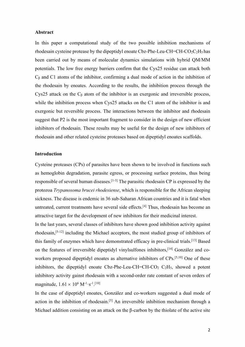
Abstract
In this paper a computational study of the two possible inhibition mechanisms of
rhodesain cysteine protease by the dipeptidyl enoate Cbz-Phe-Leu-CH=CH-CO2C2H5 has
been carried out by means of molecular dynamics simulations with hybrid QM/MM
potentials. The low free energy barriers confirm that the Cys25 residue can attack both
Cβ and C1 atoms of the inhibitor, confirming a dual mode of action in the inhibition of
the rhodesain by enoates. According to the results, the inhibition process through the
Cys25 attack on the Cβ atom of the inhibitor is an exergonic and irreversible process,
while the inhibition process when Cys25 attacks on the C1 atom of the inhibitor is and
exergonic but reversible process. The interactions between the inhibitor and rhodesain
suggest that P2 is the most important fragment to consider in the design of new efficient
inhibitors of rhodesain. These results may be useful for the design of new inhibitors of
rhodesain and other related cysteine proteases based on dipeptidyl enoates scaffolds.
Introduction
Cysteine proteases (CPs) of parasites have been shown to be involved in functions such
as hemoglobin degradation, parasite egress, or processing surface proteins, thus being
responsible of several human diseases.[1-3] The parasitic rhodesain CP is expressed by the
protozoa Trypanosoma brucei rhodesiense, which is responsible for the African sleeping
sickness. The disease is endemic in 36 sub-Saharan African countries and it is fatal when
untreated, current treatments have several side effects.[4] Thus, rhodesain has become an
attractive target for the development of new inhibitors for their medicinal interest.
In the last years, several classes of inhibitors have shown good inhibition activity against
rhodesain,[5-12] including the Michael acceptors, the most studied group of inhibitors of
this family of enzymes which have demonstrated efficacy in pre-clinical trials.[13] Based
on the features of irreversible dipeptidyl vinylsulfones inhibitors,[14] González and co-
workers proposed dipeptidyl enoates as alternative inhibitors of CPs.[5,10] One of these
inhibitors, the dipeptidyl enoate Cbz-Phe-Leu-CH=CH-CO2 C2H5, showed a potent
inhibitory activity gainst rhodesain with a second-order rate constant of seven orders of
magnitude, 1.61 ´ 106 M-1·s-1.[10]
In the case of dipeptidyl enoates, González and co-workers suggested a dual mode of
action in the inhibition of rhodesain.[5] An irreversible inhibition mechanism through a
Michael addition consisting on an attack on the β-carbon by the thiolate of the active site
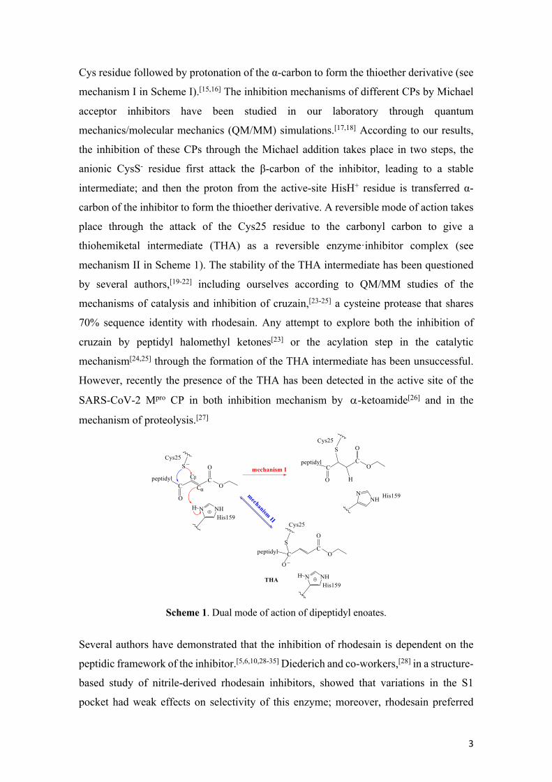
3
Cys residue followed by protonation of the α-carbon to form the thioether derivative (see
mechanism I in Scheme I).[15,16] The inhibition mechanisms of different CPs by Michael
acceptor inhibitors have been studied in our laboratory through quantum
mechanics/molecular mechanics (QM/MM) simulations.[17,18] According to our results,
the inhibition of these CPs through the Michael addition takes place in two steps, the
anionic CysS- residue first attack the β-carbon of the inhibitor, leading to a stable
intermediate; and then the proton from the active-site HisH+ residue is transferred α-
carbon of the inhibitor to form the thioether derivative. A reversible mode of action takes
place through the attack of the Cys25 residue to the carbonyl carbon to give a
thiohemiketal intermediate (THA) as a reversible enzyme·inhibitor complex (see
mechanism II in Scheme 1). The stability of the THA intermediate has been questioned
by several authors,[19-22] including ourselves according to QM/MM studies of the
mechanisms of catalysis and inhibition of cruzain,[23-25] a cysteine protease that shares
70% sequence identity with rhodesain. Any attempt to explore both the inhibition of
cruzain by peptidyl halomethyl ketones[23] or the acylation step in the catalytic
mechanism[24,25] through the formation of the THA intermediate has been unsuccessful.
However, recently the presence of the THA has been detected in the active site of the
SARS-CoV-2 Mpro CP in both inhibition mechanism by a-ketoamide[26] and in the
mechanism of proteolysis.[27]
Scheme 1. Dual mode of action of dipeptidyl enoates.
Several authors have demonstrated that the inhibition of rhodesain is dependent on the
peptidic framework of the inhibitor.[5,6,10,28-35] Diederich and co-workers,[28] in a structure-
based study of nitrile-derived rhodesain inhibitors, showed that variations in the S1
pocket had weak effects on selectivity of this enzyme; moreover, rhodesain preferred
β
α
4
extended hydrophobic for its S2 pocket, while the S3 pocket shows clear preference for
aromatic substituents. Later, Schirmeister and co-workers confirm these results,[31] and
concluded that the interaction between the S2 pocket of rhodesain and the P2 residue of
the inhibitor is an important specificity determinant. In addition, they showed that the
absence of hydrogen bond interactions with the residues Gly66 and Asp158 is related
with a lower affinity of the inhibitor. Recently, Ettari and co-workers in a structural study
with novel peptide-based Michael acceptor inhibitors arrived to some conclusions based
on structural variations on the P3, P2, and P1′ sites in the inhibitor.[33] The substitution
pattern on the phenyl at the P3 position has a remarkable impact on the inhibition of the
target protease, being the optimal substitution pattern the presence of electron
withdrawing fluorine atom or CF3 group located at 2 or 4 positions. With respect to the
P2 site, the substitution of the Phe residue with a homophenylalanine decreased the
inhibitory properties. They observed the same behaviour when the methyl group of the
vinyl ketone warhead at the P1′ site of the inhibitor was replaced with an ethyl group. In
the case of dipeptidyl enoate inhibitors, rhodesain exhibits a preference for the Phe
residue at the P2 site[5] and for the Leu residue in the P1 site[10] of the inhibitor. More
recently, Neuweiler and co-workers found in the irreversible vinylsulfones inhibitors, a
highly reactive warhead increases the efficiency of the rhodesain inhibition.[36]
Herein, we reported a detailed QM/MM study of the inhibition mechanisms of rhodesain
cysteine protease by the dipeptidyl enoate Cbz-Phe-Leu-CH=CH-CO2C2H5. The analysis
of the free energy surface (FES) of every single chemical step, computed in terms of the
potential of mean force (PMF), and the interactions between the inhibitor and the protein
afford describing how dipeptidyl enoate inhibits this CP at atomistic level, and proposing
suggestions for the design of new inhibitors of cysteine proteases with increasing affinity
and selectivity.
Results and Discussion
As mentioned in the previous section, the inhibition mechanism of the rhodesain CP by
the dipeptidyl enoate Cbz-Phe-Leu-CH=CH-CO2C2H5 has been studied by the generation
of the FESs corresponding to two possible mechanisms (see Scheme 1): the nucleophilic
attack of Cys25 on the Cβ of the inhibitor (Mechanism I) or on the carbonyl carbon C1 of
the inhibitor (Mechanism II).

5
Inhibition Mechanism I. The M06-2X:AM1d/MM FESs corresponding to the full
inhibition mechanism I is shown in Figure 1. The corresponding PMFs at the AM1d/MM
level is deposited in the Supporting Information (see Figure S3). As observed, the FESs
confirms the step-wise character of the mechanism: first the Cys25 of the protein attacks
on the Cβ atom of the inhibitor, leading to a stable intermediate Int-Cβ (see Scheme 2) and
then the proton from His159 is transferred to the Cα atom of the inhibitor forming the PS-
Cβ. According to the results, the attack of Cys25 on the Cβ, through TS1-Cβ, proceeds
with a free energy barrier of 4.2 kcal·mol-1 (see Figure 1a). Then, the proton transfer from
His159 to the Cα atom of the inhibitor takes place in a barrierless fashion leading to a
stable PS-Cβ. The formation of the intermediate Int-Cβ is the rate-limiting step of the
inhibition process. The structure of the TS1-Cβ was optimized at M06-2X/6-
31+G(d,p)/MM level and the minimum energy path, computed as the intrinsic reaction
coordinate (IRC) path, confirms the predictions derived from the M06-2X:AM1d/MM
FES (see Table S1).
From a thermodynamic point of view, an important difference is observed between the
two steps of the inhibition process (see Figure 1), while the formation of the intermediate
Int-Cβ is a reversible process the second step has a clear irreversible character, being the
full inhibition process exergonic with a value of the reaction free energy of -40.3
kcal·mol-1.
Figure 1. M06-2X/6-31+G(d,p):AM1d/MM FES for the inhibition mechanism I. a) Attack of sulfur on Cβ. b) Protonation of the Int-Cβ intermediate.
Representative snapshots of the key states involved in the inhibition mechanism I are
presented in Figure 2, while the average values of the key interatomic distances are listed
in Table S2 (see Supporting Information). The geometrical analysis indicates that while
∆PMF/kcal·mol-1
a)
Int-Cβ
TS1-Cβ
RS5.5
4.2
d(SG-Cβ)/Å
∆PMF/kcal·mol-1
b)
Int-Cβ
d(N3-H3)-d(Cα-H3)/Å
PS-Cβ34.8

6
the SG-Cβ bond is forming (2.39 ± 0.03 Å) in TS1, the double bond between Cβ and Cα
remains almost unaltered (1.34 ± 0.02 Å and 1.36 ± 0.02 Å in RS and TS1-Cβ,
respectively). The value of the Cβ-Cα distance in the PS is typical of a single C-C bond
(1.53 ± 0.03 Å). It is important to note how in the RS complex, the transferring hydrogen
atom of His159 is far from the Cα of the inhibitor, d(Cα-H3) = 5.59 ± 0.34 Å. Nevertheless,
as the double Cβ-Cα bond is elongating, the His159 is approaching to a more reactive
position being the distance Cα-H3 equal to 2.09 ± 0.22 Å in the intermediate Int-Cβ.
Figure 2. Representative snapshots of the key states of the inhibition mechanism I.
As mentioned before, a QM/MM study of the inhibition mechanism of cysteine protease
rhodesain by a dipeptidyl nitroalkene, Cbz-Phe-Ala-CH=CH-NO2, was recently carried
out in our laboratory.[17] Thus, a comparative analysis between the reactivity of the two
families of inhibitors (dipeptidyl nitroalkenes vs dipeptidyl enoates) toward rhodesain can
be based on the obtained free energy profiles. In this sense, the reactivity of the dipeptidyl
enoate Cbz-Phe-Leu-CH=CH-CO2C2H5 is greater than that of the dipeptidyl nitroalkene
Cbz-Phe-Ala-CH=CH-NO2 because the free energy barrier of the inhibition with the
enoate is significantly lower than the one required to form the covalent linkage between
the protein and the nitroalkene (4.2 kcal·mol-1 vs 20.4 kcal·mol-1).
RS
Inhibitor
Gly66
H2 Cys25
His159
Asn175
H4
SG
Cβ
H3
Gly66
H2
Cys25
His159
Asn175
H4
SGH3
Inhibitor
Inhibitor
Gly66
H2 Cys25
His159
Asn175
H4H3 Inhibitor
Gly66
H2Cys25
His159
Asn175
H4H3
Int-Cβ
TS1-Cβ
PS-Cβ
Cβ
Cβ
Cβ
Cα
SG SG
Cα
N3
N3

7
Scheme 2. Proposed inhibition mechanisms of rhodesain by dipeptidyl enoates as deduced from M06-2X/6-31+G(d,p):AM1d/MM calculations.
Inhibition mechanism II. The second proposed inhibition mechanism consists on the
sulfur attack on the carbonyl carbon C1 (see Scheme 2). The M06-2X:AM1d/MM 2D-
FES corresponding to the full inhibition process is shown in Figure 3. The corresponding
PMF at the AM1d/MM level, and the average values of key interatomic distances are
deposited in the Supporting Information (see Figure S6b and Table S3, respectively).
Representative snapshots of some key states involved in the inhibition process are
presented in Figure 4. The first conclusion derived from the FES shown in Figure 3 is
that the inhibition mechanism takes place in two steps, first the Cys25 of the protein
attacks on the C1 atom of the inhibitor through TS1-C1, with a free energy barrier of 2.0
kcal·mol-1, leading to the stable intermediate THA. Later, the proton from His159 is
transferred to the O1 atom of the inhibitor forming the PS-C1. This second step takes
place through TS2-C1, with a low free energy barrier of 0.9 kcal·mol-1. The whole
inhibition process is exergonic with a value of the reaction free energy of -21.8 kcal·mol-
1. An interesting point in this second mode of action of the enoate inhibitors is the
presence of the meta-stable THA intermediate, 2.9 kcal·mol-1 more stable than the RS
complex. As commented in the Introduction section, any previous attempt to explore the
inhibition[23] or the acylation step in the catalytic mechanism[24,25] of the cruzain cysteine
protease through the formation of the THA intermediate was unsuccessful. The structure
β
α
βα
β β
β
α

8
of both TS1-C1 and TS2-C1 were optimized at M06-2X/6-31+G(d,p)/MM level and the
minimum energy path, computed as the IRC path, confirms the predictions derived from
the M06-2X:AM1d/MM FES (see Table S4).
Figure 3. M06-2X/6-31+G(d,p):AM1d/MM FES of the inhibition mechanism II. Iso-energetic lines are displayed every 1.0 kcal·mol-1.
Figure 4. Representative snapshots of the key states of the inhibition mechanism II.
2 2.5 3
-1
-0.5
0
0.5
1
1.5
d(N3-H3)-d(O1-H3)/Å
d(SG-C1)/Å
RS
PS-C1
TS2-C1
THA
TS1-C1
TS1-C1
PS-C1TS2-C1
THA
Inhibitor
Gly66
H2
Cys25His159
Asn175H4SG
C1
H3O1
Gly66
H2
Cys25
His159
Asn175H4SG
C1
H3O1
Inhibitor
Inhibitor
Gly66
H2
Cys25
His159
Asn175H4
H3O1
Inhibitor
Gly66
H2
Cys25His159
Asn175H4
H3O1C1
SG
C1
SG
9
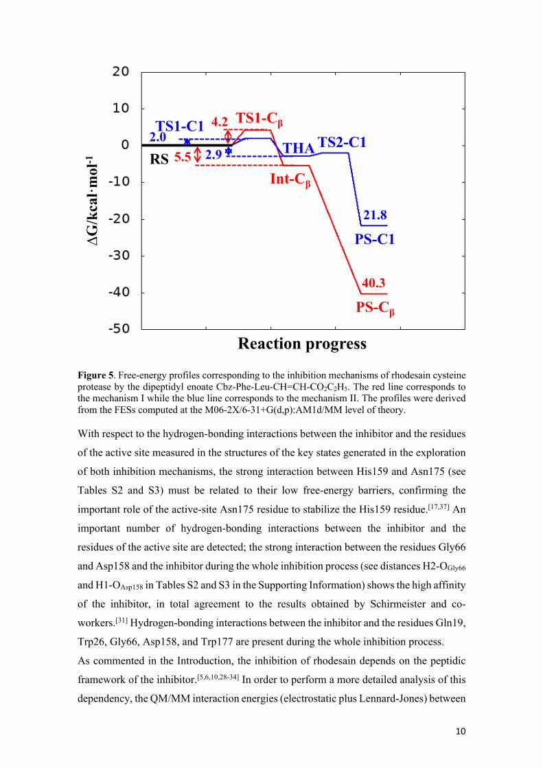
Comparison between Mechanism I and Mechanism II. Figure 5 displays the free
energy profiles of the two inhibition mechanisms of rhodesain by the dipeptidyl enoate
Cbz-Phe-Leu-CH=CH-CO2C2H5. The free energy profiles clearly indicate that the Cys25
can attack both Cβ and C1 atoms, confirming a dual mode of action in the inhibition of
rhodesain by the enoates, as previously suggested by González and co-workers.[5]
The first step in both cases is reversible and is related to the formation of the SG-C bond,
forming two stable intermediates Int-Cβ and THA, being the Int-Cβ intermediate slightly
more stable than the THA. From a kinetic point of view, the low free energy barrier
indicates that the formation of both intermediates Int-Cβ and THA is a favourable process.
The low activation free energies obtained in both inhibition mechanisms are in total
agreement with the second-order rate constant of seven orders of magnitude determined
by González and co-workers.[10] Regarding the second step of the inhibition process, the
more important difference is related to its reversibility; while the formation of the PS-C1
complex is a reversible process with a reaction free energy of -18.9 kcal·mol-1, the
formation of the PS-Cβ complex is clearly irreversible with a reaction free energy of -34.8
kcal·mol-1. The confirmation of the dual mode of action of this family of inhibitors is an
important issue that can be used to design improved inhibitors. Thus, according to our
results, the reversible or irreversible character of the inhibitor could be modulated by
structural improvements to favour the attack of Cys residue to the carbonyl carbon or the
β-carbon. More potent Michael acceptor inhibitors of rhodesain can be based on
compounds with more than one reactive position.
10
Figure 5. Free-energy profiles corresponding to the inhibition mechanisms of rhodesain cysteine protease by the dipeptidyl enoate Cbz-Phe-Leu-CH=CH-CO2C2H5. The red line corresponds to the mechanism I while the blue line corresponds to the mechanism II. The profiles were derived from the FESs computed at the M06-2X/6-31+G(d,p):AM1d/MM level of theory. With respect to the hydrogen-bonding interactions between the inhibitor and the residues
of the active site measured in the structures of the key states generated in the exploration
of both inhibition mechanisms, the strong interaction between His159 and Asn175 (see
Tables S2 and S3) must be related to their low free-energy barriers, confirming the
important role of the active-site Asn175 residue to stabilize the His159 residue.[17,37] An
important number of hydrogen-bonding interactions between the inhibitor and the
residues of the active site are detected; the strong interaction between the residues Gly66
and Asp158 and the inhibitor during the whole inhibition process (see distances H2-OGly66
and H1-OAsp158 in Tables S2 and S3 in the Supporting Information) shows the high affinity
of the inhibitor, in total agreement to the results obtained by Schirmeister and co-
workers.[31] Hydrogen-bonding interactions between the inhibitor and the residues Gln19,
Trp26, Gly66, Asp158, and Trp177 are present during the whole inhibition process.
As commented in the Introduction, the inhibition of rhodesain depends on the peptidic
framework of the inhibitor.[5,6,10,28-34] In order to perform a more detailed analysis of this
dependency, the QM/MM interaction energies (electrostatic plus Lennard-Jones) between
Reaction progress
∆G/kcal·mol-1 RS
PS-C1
TS2-C1THA
Int-Cβ
TS1-Cβ
PS-Cβ
4.2
5.5
40.3
21.8
2.9
TS1-C12.0
11
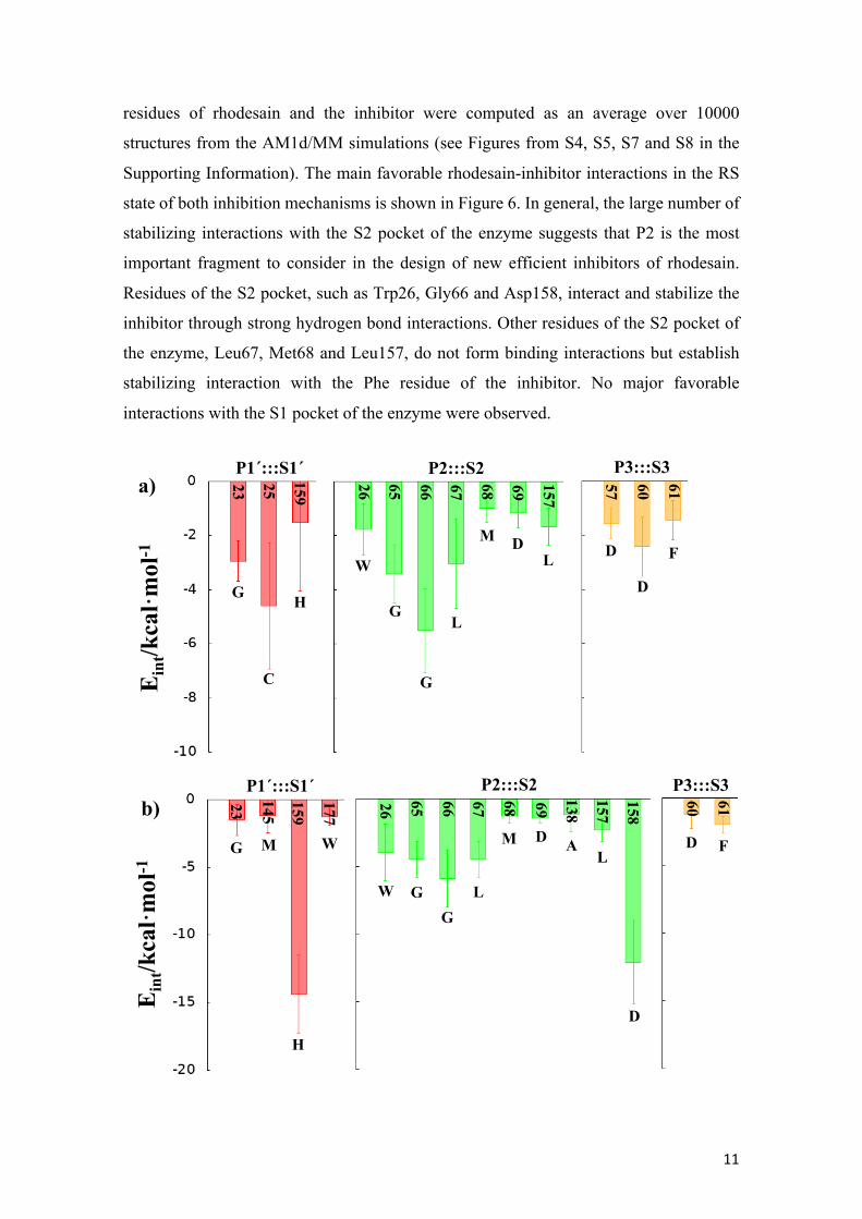
residues of rhodesain and the inhibitor were computed as an average over 10000
structures from the AM1d/MM simulations (see Figures from S4, S5, S7 and S8 in the
Supporting Information). The main favorable rhodesain-inhibitor interactions in the RS
state of both inhibition mechanisms is shown in Figure 6. In general, the large number of
stabilizing interactions with the S2 pocket of the enzyme suggests that P2 is the most
important fragment to consider in the design of new efficient inhibitors of rhodesain.
Residues of the S2 pocket, such as Trp26, Gly66 and Asp158, interact and stabilize the
inhibitor through strong hydrogen bond interactions. Other residues of the S2 pocket of
the enzyme, Leu67, Met68 and Leu157, do not form binding interactions but establish
stabilizing interaction with the Phe residue of the inhibitor. No major favorable
interactions with the S1 pocket of the enzyme were observed.
Eint/kcal·mol-1
Eint/kcal·mol-1
a)
b)
P1´:::S1´ P2:::S2 P3:::S3
P1´:::S1´ P2:::S2 P3:::S3
23 25 159
H
C
G
26 65 66 67 68 69 157
57 60 61
W
G
G
L
M DL D
D
F
23
145
159
H
MG
26 65 66 67 68 69
157
138
60 61
W GG
L
M DL
A
D
FW
177
158
D

12
Figure 6. Main favorable average interaction energies (electrostatic plus Lennard-Jones) between residues of rhodesain and each fragment of the inhibitor computed in the RS state of both inhibition mechanism I (a) and mechanism II (b). Results obtained as an average over 10000 structures of the AM1d/MM MD simulations.
Conclusions A computational study of the two possible inhibition mechanisms of rhodesain cysteine
protease by the dipeptidyl enoate Cbz-Phe-Leu-CH=CH-CO2C2H5 has been carried out
by means of MD simulations with hybrid QM/MM potentials, the nucleophilic attack of
Cys25 on the Cβ of the inhibitor (Mechanism I) or the carbonyl carbon C1 of the inhibitor
(Mechanism II). The first conclusion derived from our study is that the Cys25 residue can
attack both Cβ and C1 atoms of the inhibitor, confirming a dual mode of action in the
inhibition of the rhodesain by the enoates, suggested by one of us.[5] According to our
results, both inhibition mechanisms take place in two steps; the first step is related to the
formation of the SG-C bond, forming stable intermediates, Int-Cβ and THA in mechanism
I and II, respectively. From both the kinetic and thermodynamic point of views, the
formation of these intermediates is a favourable and reversible process. The low
activation free energies obtained in both alkylation process are consistent with the second-
order rate constant determined by González and co-workers.[10] The second step of the
two inhibition mechanisms consist in the protonation of the intermediates, Int-Cβ and
THA, by means of a proton transfer from His159 to the Cα atom (Int-Cβ) or the O1 atom
(THA) of the inhibitor leading to the formation of the PS-Cβ and PS-C1 complexes,
respectively. According to the energy required for the decomposition of these
intermediates into products, this second step is kinetically irrelevant and THA can be
considered as a metastable intermediate. Nevertheless, the significant difference in the
second step of both inhibition mechanisms is related to its reversibility; while the
formation of the PS-Cβ complex is an irreversible process, the formation of the PS-C1
complex is a reversible process. In summary, the full inhibition process through the Cys25
attack on the Cβ atom of the inhibitor is exergonic and irreversible with a value of the
reaction free energy of -40.3 kcal·mol-1; and full inhibition process through the Cys25
attack on the C1 atom of the inhibitor is exergonic and reversible with a value of the
reaction free energy of -21.8 kcal·mol-1, confirming a dual mode of action of this family
of inhibitors, as previously suggested by González and co-workers.[5]

13
Concerning the interactions between the residues of the active site and the inhibitor, an
important number of hydrogen-bonding interactions are detected. In particular,
interactions between residues Gln19, Trp26, Gly66, Asp158, and Trp177 appear to be
important during the whole inhibition process and confirm the great affinity of the
inhibitor.[31] In addition, the strong interaction between the His159 and Asn175 observed
in both inhibition mechanisms can be related to their low free-energy barriers, confirming
the important role of the active-site Asn175 residue to stabilize the His159 residue.[17,37]
Analysis of the QM/MM interaction energies between the peptidic framework of the
inhibitor and the residues of rhodesain, suggest that P2 is the most important fragment to
consider in the design of new efficient inhibitors of rhodesain. The interactions between
the inhibitor and rhodesain are clearly dominated by those in the P2:::S2 site. Residues
that interact with the inhibitor through strong hydrogen-bonding interactions, as Trp26,
Gly66, and Asp158, form stabilizing interactions with the P2 pocket of the inhibitor.
Other residues of the S2 pocket of the enzyme, Leu67, Met68 and Leu157, do not form
binding interactions but establish stabilizing interaction with the Phe residue of the
inhibitor. These results may be useful for the design of new inhibitors of rhodesain with
greater selectivity and affinity, by increasing the interaction with the S2 pocket of
rhodesain. The confirmation of the dual mode of action of this family of inhibitors is an
important issue that can be used to design improved inhibitors whose reversible or
irreversible character could be modulated by structural improvements to favour the attack
of Cys residue to the carbonyl carbon or the β-carbon. More potent Michael acceptor
inhibitors of rhodesain can be based on compounds with more than one reactive position.
These conclusions suggest that dipeptidyl enoates derivatives can be also used to inhibit
other cysteine proteases related with human diseases.
Computational Methods
The starting structure for the construction of the rhodesain-inhibitor model was the X-ray
crystal structure of rhodesain from Trypanosoma brucei rhodesiense with PDB code
2P7U.[38] The bound inhibitor K777 was replaced with the enoate inhibitor Cbz-Phe-Leu-
CH=CH-CO2C2H5. This particular X-ray structures was already used in our laboratory to
carry out computational studies on the inhibition of rhodesain cysteine proteases by
dipeptidyl nitroalkenes.[17]
14
In the molecular model, the missing hydrogen atoms of the X-ray structure were added at
pH 7 using the tLEAP module[39] of Amber Tools program within the pKa values of the
titratable residues calculated within the empirical PROPKA 3.1 program.[40] Missing
force field parameters for the inhibitor were computed using Antechamber software.[41]
Neutralization of the system was achieved by adding 16 Na+ counterions in the
electrostatically most favourable positions. Finally, the system was solvated in an
orthorhombic box of TIP3P[42] water molecules with the following size: 75.5 ´ 79.4 ´
77.1 Å3. 10 ns of classical MD simulations were required to equilibrate the system using
the AMBER force field[43] implemented in NAMD software.[44] The MD simulations were
carried out at 300 K using the NVT ensemble with a time step of 1fs. The temperature
during the MD simulation was controlled using the Langevin thermostat.[45] Analysis of
the time evolution of the root-mean-square deviation of the backbone atoms of the protein
models confirms that the system was equilibrated (see Figure S1 in the Supporting
Information). Analysis was done using cpptraj software.[46]
Once the molecular model was set up and equilibrated, potential energy surfaces (PESs)
corresponding to every possible chemical step of the inhibition mechanisms were
explored. The reactants, products, intermediates and transition state structures were
optimized by means of a micro-macro iterations scheme.[47] Frequency calculations were
carried out in all located structures to characterize them as minima or saddle points of
order one. The fDYNAMO library[48] was used for the QM/MM calculations. The QM
region was initially described with the AM1d semiempirical Hamiltonian,[49] and contains
the full inhibitor, the residue Cys25, the imidazole ring of His159 (see Figure 7a). This is
the Hamiltonian already employed in different computational studies related to the
cysteine proteases carried out in our laboratory.[17,23-25,50,51] The rest of the system, protein
and water molecules, were described by the OPLS-AA[52] and TIP3P[42] force fields,
respectively. Hydrogen link atoms were used to saturate the valence of the QM-MM
frontier bonds (see Figure 7a).[53] All residues further than 25 Å from the Cβ atom of the
inhibitor were kept frozen during the simulations. A force switching function with a cutoff
distance in the range of 14.5 to 16 Å and periodic boundary conditions were employed to
treat the non-bonding interactions.

15
Figure 7. a) Details of the atoms of the active site treated quantum mechanically (blue region). The link atoms between the QM and MM regions are indicated as black dots. b) Representative rhodesain-inhibitor model. Once the PESs were obtained (see Figures S2 and S6a in the Supporting Information),
FESs were generated in terms of the PMF using umbrella sampling and the weighted
histogram analysis method to recover the probabilities.[54,55] The error associated to this
method, when properly carried out, is usually accepted to be around 1 kcal·mol-1.[56] The
harmonic umbrella sampling force constants was 2500 kJ·mol-1·Å-2. 20 ps of equilibration
and 40 ps of production, with a time step of 1 fs, were used in every window of the PMFs.
For the study of the mechanism I, a monodimensional PMF (1D-PMF) was generated
using the bond-forming distance, d(SG-Cβ), as the reaction coordinate, z, to monitor the
attack of the sulfur atom of Cys25 on the Cβ of the inhibitor (see Figure S3a). This
required series of 68 simulation windows. This step leads to an intermediate called Int-
Cβ. For the protonation of the Int-Cβ intermediate, a 1D-PMF (see Figure S3b) was
generated using as z the antisymmetric combination of two distances defining the
hydrogen transfer from the His159 to the Cα of the inhibitor, d(N3-H3)-d(Cα-H3). This
step required series of 63 simulation windows. And finally, for the study of the
mechanism II, a 2D-PMF was generated with the bond-forming distance between Cys25
and C1 carbonyl carbon atom of the inhibitor, d(SG-C1) as z1, and the antisymmetric
combination of the two distances defining the hydrogen transfer from the His159 to the
O1 of the inhibitor, d(N3-H3)-d(O1-H3), as z2 (see Figure S6b). This required series of
2211 simulation windows centered on the values of the previously generated PES. The
QM sub-set of atoms in the QM/MM FESs were restricted to a low-level Hamiltonian,
AM1d, because of computer limitations. In order to improve the level of theory, an
interpolated correction scheme with a higher level Hamiltonian, developed in our
laboratory,[57] was applied as explained in detail in our previous papers.[17,23-25,50,51] The
β α
a) b)
Asn175
His159
Cys25
Inhibitor
S1´ pocket
S2 pocket
S3 pocketS1 pocket

16
M06-2X functional[58] with the standard 6-31+G(d,p) basis set,[59] following Truhlar and
co-workers suggestions,[58,60] was used employing the Gaussian09 program,[61] combined
with the fDYNAMO library.[48]
Finally, in order to obtain averaged interaction energies between the enzymatic
environment and the inhibitor, 1 ns of AM1d/MM MD simulations of the windows
corresponding to the different states were performed.
Contribution of each residue of the protein to the interaction energy with defined part of
inhibitor was computer using the following expression:
𝐸!"/""$%& = ∑ $Ψ& '!!(",!!
&Ψ' + ∑∑ )$!'!!($!,!!
+ 𝐸!"/""*+, (1)
This interaction energy can be exactly decomposed in a sum over residues provided that
the polarized wave function (Ψ) is employed to evaluate this energy contribution. The
global polarization effect can be obtained from the gas phase energy difference between
the polarized, Ψ, and non-polarized, Ψ0, wave functions.
Supporting Information
RMSD computed along the classical MD simulation for the backbone atoms of the
protein; PESs and FESs computed at AM1d/MM level corresponding to every single step
of the different inhibition mechanisms; key average inter-atomic distances for the key
states located along the different inhibition mechanisms, average interaction energies by
residues for some key states for the reaction mechanisms, and Cartesian coordinates of
the QM subset of atoms of the TSs, RSs, Int-Cβ, THA and PS-C1 optimized at M06-2X/6-
31+G(d,p)/MM level.
Acknowledgements
This work was supported by the Spanish Ministerio de Ciencia, Innovación y
Universidades (PGC2018-094852-B-C21), Generalitat Valenciana (AICO/2019/195)
Universitat Jaume I (UJI-B2020-03). K.A. thanks Generalitat Valenciana
(APOSTD/2020/015) for post-doctoral contract. The authors thankfully acknowledge the
local computational resources of the Servei d’Informàtica of Universitat Jaume I.

17
References [1] H. A. Chapman, R. J. Riese, G.-P. Shi, Annu. Rev. Physiol. 1997, 59, 63-88. [2] J. H. McKerrow, J. C. Engel, C. R. Caffrey, Bioorg. Med. Chem. 1999, 7, 639-644. [3] V. Olga, R. Thomas, P. Christoph, T. Dusan, T. Vito, T. Boris, Curr. Pharm. Des. 2007, 13, 387-403. [4] https://www.who.int/news-room/fact-sheets/detail/trypanosomiasis-human-african-(sleeping-sickness)/ (accessed March 2021). [5] S. Royo, S. Rodriguez, T. Schirmeister, J. Kesselring, M. Kaiser, F. V. Gonzalez, ChemMedChem 2015, 10, 1484-1487. [6] A. Latorre, T. Schirmeister, J. Kesselring, S. Jung, P. Johe, U. A. Hellmich, A. Heilos, B. Engels, R. L. Krauth-Siegel, N. Dirdjaja, L. Bou-Iserte, S. Rodriguez, F. V. Gonzalez, ACS Med. Chem. Lett. 2016, 7, 1073-1076. [7] S. F. P. Braga, L. C. Martins, E. B. da Silva, P. A. Sales, S. M. F. Murta, A. J. Romanha, W. T. Soh, H. Brandstetter, R. S. Ferreira, R. B. de Oliveira, Bioorg. Med. Chem. 2017, 25, 1889-1900. [8] M. Giroud, B. Kuhn, S. Saint-Auret, C. Kuratli, R. E. Martin, F. Schuler, F. Diederich, M. Kaiser, R. Brun, T. Schirmeister, W. Haap, J. Med. Chem. 2018, 61, 3370-3388. [9] D. A. Rocha, E. B. Silva, I. S. Fortes, M. S. Lopes, R. S. Ferreira, S. F. Andrade, Eur. J. Med. Chem. 2018, 157, 1426-1459. [10] S. Royo, T. Schirmeister, M. Kaiser, S. Jung, S. Rodriguez, J. Manuel Bautista, F. V. Gonzalez, Bioorg. Med. Chem. 2018, 26, 4624-4634. [11] P. Klein, P. Johe, A. Wagner, S. Jung, J. Kuhlborn, F. Barthels, S. Tenzer, U. Distler, W. Waigel, B. Engels, U. A. Hellmich, T. Opatz, T. Schirmeister, Molecules 2020, 25. [12] D. Steverding, Molecules 2020, 25, 143. [13] P. S. Doyle, Y. M. Zhou, J. C. Engel, J. H. McKerrow, Antimicrob. Agents Chemother. 2007, 51, 3932-3939. [14] J. T. Palmer, D. Rasnick, J. L. Klaus, D. Bromme, J. Med. Chem. 1995, 38, 3193-3196. [15] R. P. Hanzlik, S. A. Thompson, J. Med. Chem. 1984, 27, 711-712. [16] J. C. Powers, J. L. Asgian, O. D. Ekici, K. E. James, Chem. Rev. 2002, 102, 4639-4750. [17] K. Arafet, F. V. González, V. Moliner, Chem. Eur. J. 2020, 26, 2002-2012. [18] K. Arafet, N. Serrano-Aparicio, A. Lodola, A. J. Mulholland, F. V. González, K. Świderek, V. Moliner, Chem. Sci. 2021, 12, 1433-1444. [19] A. E. Howard, P. A. Kollman, J. Am. Chem. Soc. 1988, 110, 7195-7200. [20] D. Arad, R. Langridge, P. A. Kollman, J. Am. Chem. Soc. 1990, 112, 491-502. [21] K. Byun, J. L. Gao, J. Mol. Graphics Modell. 2000, 18, 50-55. [22] D. H. Wei, X. Q. Huang, J. J. Liu, M. S. Tang, C. G. Zhan, Biochemistry 2013, 52, 5145-5154. [23] K. Arafet, S. Ferrer, V. Moliner, Biochemistry 2015, 54, 3381-3391. [24] K. Arafet, S. Ferrer, V. Moliner, ACS Catalysis 2017, 7, 1207-1215. [25] K. Arafet, K. Świderek, V. Moliner, ACS Omega 2018, 3, 18613-18622. [26] L. Zhang, D. Lin, X. Sun, U. Curth, C. Drosten, L. Sauerhering, S. Becker, K. Rox, R. Hilgenfeld, Science 2020, 368, 409-412. [27] K. Świderek, V. Moliner, Chem. Sci. 2020, 11, 10626-10630. [28] V. Ehmke, E. Winkler, D. W. Banner, W. Haap, W. B. Schweizer, M. Rottmann, M. Kaiser, C. Freymond, T. Schirmeister, F. Diederich, ChemMedChem 2013, 8, 967-975. [29] R. Ettari, S. Previti, S. Cosconati, J. Kesselring, T. Schirmeister, S. Grasso, M. Zappala, J. Enzyme Inhib. Med. Chem. 2016, 31, 1184-1191. [30] S. Previti, R. Ettari, S. Cosconati, G. Arnendola, K. Chouchene, A. Wagner, U. A. Hellmich, K. Ulrich, R. L. Krauth-Siegel, P. R. Wich, I. Schmid, T. Schirmeister, J. Gut, P. J. Rosenthal, S. Grasso, M. Zappala, J. Med. Chem. 2017, 60, 6911-6923. [31] T. Schirmeister, J. Schmitz, S. Jung, T. Schmenger, R. L. Krauth-Siegel, M. Guetschow, Bioorg. Med. Chem. Lett. 2017, 27, 45-50.
18
[32] L. H. Santos, B. J. Waldner, J. E. Fuchs, G. A. N. Pereira, K. R. Liedl, E. R. Caffarena, R. S. Ferreira, J. Chem. Inf. Model. 2019, 59, 137-148. [33] R. Ettari, S. Previti, S. Maiorana, G. Amendola, A. Wagner, S. Cosconati, T. Schirmeister, U. A. Hellmich, M. Zappalà, J. Med. Chem. 2019, 62, 10617-10629. [34] S. Maiorana, R. Ettari, S. Previti, G. Amendola, A. Wagner, S. Cosconati, U. A. Hellmich, T. Schirmeister, M. Zappalà, ChemMedChem 2020, 15, 1552-1561. [35] H. Zhang, J. Collins, R. Nyamwihura, O. Crown, O. Ajayi, I. V. Ogungbe, Bioorg. Med. Chem. Lett. 2020, 30. [36] P. Johe, S. Jung, E. Endres, C. Kersten, C. Zimmer, W. Ye, C. Sönnichsen, U. A. Hellmich, C. Sotriffer, T. Schirmeister, H. Neuweiler, ACS Chem. Biol. 2021. [37] T. Vernet, D. C. Tessier, J. Chatellier, C. Plouffe, T. S. Lee, D. Y. Thomas, A. C. Storer, R. Ménard, J. Biol. Chem. 1995, 270, 16645-16652. [38] I. D. Kerr, J. H. Lee, C. J. Farady, R. Marion, M. Rickert, M. Sajid, K. C. Pandey, C. R. Caffrey, J. Legac, E. Hansell, J. H. McKerrow, C. S. Craik, P. J. Rosenthal, L. S. Brinen, J. Biol. Chem. 2009, 284, 25697-25703. [39] C. E. A. F. Schafmeister, W. S. Ross, V. Romanovski, University of California, San Francisco 1995. [40] M. H. M. Olsson, C. R. Sondergaard, M. Rostkowski, J. H. Jensen, J. Chem. Theory Comput. 2011, 7, 525-537. [41] J. Wang, W. Wang, P. A. Kollman, D. A. Case, J. Mol. Graphics Modell. 2006, 25, 247-260. [42] W. L. Jorgensen, J. Chandrasekhar, J. D. Madura, R. W. Impey, M. L. Klein, J. Chem. Phys. 1983, 79, 926-935. [43] Y. Duan, C. Wu, S. Chowdhury, M. C. Lee, G. M. Xiong, W. Zhang, R. Yang, P. Cieplak, R. Luo, T. Lee, J. Caldwell, J. M. Wang, P. Kollman, J. Comput. Chem. 2003, 24, 1999-2012. [44] J. C. Phillips, R. Braun, W. Wang, J. Gumbart, E. Tajkhorshid, E. Villa, C. Chipot, R. D. Skeel, L. Kalé, K. Schulten, J. Comput. Chem. 2005, 26, 1781-1802. [45] G. S. Grest, K. Kremer, Phys. Rev. A 1986, 33, 3628-3631. [46] D. R. Roe, T. E. Cheatham, J. Chem. Theory Comput. 2013, 9, 3084-3095. [47] S. Martí, V. Moliner, I. Tuñón, J. Chem. Theory Comput. 2005, 1, 1008-1016. [48] M. J. Field, M. Albe, C. Bret, F. Proust-De Martin, A. Thomas, J. Comp. Chem. 2000, 21, 1088-1100. [49] K. Nam, Q. Cui, J. Gao, D. M. York, J. Chem. Theory Comput. 2007, 3, 486-504. [50] K. Arafet, S. Ferrer, S. Martí, V. Moliner, Biochemistry 2014, 53, 3336-3346. [51] K. Arafet, S. Ferrer, F. V. Gonzalez, V. Moliner, Phys. Chem. Chem. Phys. 2017, 19, 12740-12748. [52] W. L. Jorgensen, D. S. Maxwell, J. TiradoRives, J. Am. Chem. Soc. 1996, 118, 11225-11236. [53] M. J. Field, P. A. Bash, M. Karplus, J. Comput. Chem. 1990, 11, 700-733. [54] S. Kumar, D. Bouzida, R. H. Swendsen, P. A. Kollman, J. M. Rosenberg, J. Comp. Chem. 1992, 13, 1011-1021. [55] G. M. Torrie, J. P. Valleau, J. Comp. Phys. 1977, 23, 187-199. [56] J. Kästner, W. Thiel, J. Chem. Phys. 2006, 124, 234106. [57] J. J. Ruiz-Pernia, E. Silla, I. Tuñón, S. Martí, V. Moliner, J. Phys. Chem. B 2004, 108, 8427-8433. [58] Y. Zhao, D. G. Truhlar, Theor. Chem. Acc. 2008, 120, 215-241. [59] W. J. Hehre, L. Radom, P. V. R. Schleyer, J. A. Pople, Ab Initio Molecular Orbital Theory, John Wiley, New York, 1986. [60] B. J. Lynch, Zhao, Y., Truhlar, D. G., J. Phys. Chem. A 2003, 107, 1384-1388. [61] Gaussian 09 (Revision A.1), M. J. Frisch, G. W. Trucks, H. B. Schlegel, G. E. Scuseria, M. A. Robb, J. R. Cheeseman, G. Scalmani, V. Barone, B. Mennucci, G. A. Petersson, H. Nakatsuji, X. L. M. Caricato, H. P. Hratchian, A. F. Izmaylov, J. Bloino, G. Zheng, J. L. Sonnenberg, M. Hada, M. Ehara, K. Toyota, R. Fukuda, J. Hasegawa, M. Ishida, T. Nakajima, Y. Honda, O. Kitao, H. Nakai, T. Vreven, J. A. Montgomery, Jr., J. E. Peralta, F. Ogliaro, M. Bearpark, J. J. Heyd, E. Brothers, K. N. Kudin, V. N. Staroverov, R. Kobayashi, J. Normand, K. Raghavachari, A. Rendell, J. C. Burant, S. S. Iyengar, J. Tomasi, M. Cossi, N. Rega, J. M. Millam, M. Klene, J. E. Knox, J. B. Cross, V. Bakken, C. Adamo, J. Jaramillo, R. Gomperts, R. E. Stratmann, O. Yazyev,

19
A. J. Austin, R. Cammi, C. Pomelli, J. W. Ochterski, R. L. Martin, K. Morokuma, V. G. Zakrzewski, G. A. Voth, P. Salvador, J. J. Dannenberg, S. Dapprich, A. D. Daniels, Ö. Farkas, J. B. Foresman, J. V. Ortiz, J. Cioslowski, D. J. Fox, 2009.
Abstract
A QM/MM computational study of two possible inhibition mechanisms of rhodesain cysteine protease by the dipeptidyl enoate Cbz-Phe-Leu-CH=CH-CO2C2H5 confirms the dual mode of action of this family of inhibitors. Efforts focused on increasing the interaction with the residues of the S2 pocket of rhodesain could be used as a guide to design new dual inhibitors with greater selectivity and affinity.
Table of Contents

20