Eliminate Dengue
10
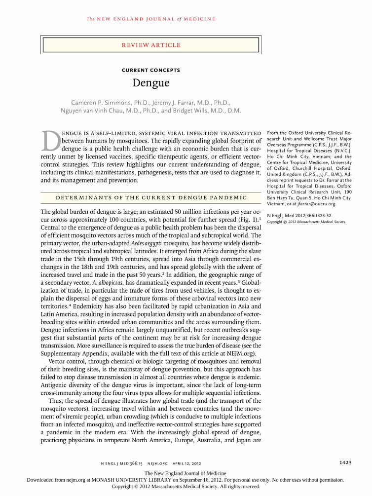
The new england journal of medicine n engl j med 366;15 nejm.org april 12, 2012 1423 Review article Current Concepts Dengue Cameron P. Simmons, Ph.D., Jeremy J. Farrar, M.D., Ph.D., Nguyen van Vinh Chau, M.D., Ph.D., and Bridget Wills, M.D., D.M. From the Oxford University Clinical Re- search Unit and Wellcome Trust Major Overseas Programme (C.P.S., J.J.F., B.W.), Hospital for Tropical Diseases (N.V.C.), Ho Chi Minh City, Vietnam; and the Centre for Tropical Medicine, University of Oxford, Churchill Hospital, Oxford, United Kingdom (C.P.S., J.J.F., B.W.). Ad- dress reprint requests to Dr. Farrar at the Hospital for Tropical Diseases, Oxford University Clinical Research Unit, 190 Ben Ham Tu, Quan 5, Ho Chi Minh City, Vietnam, or at [email protected]. N Engl J Med 2012;366:1423-32. Copyright © 2012 Massachusetts Medical Society. D engue is a self-limited, systemic viral infection transmitted between humans by mosquitoes. The rapidly expanding global footprint of dengue is a public health challenge with an economic burden that is cur- rently unmet by licensed vaccines, specific therapeutic agents, or efficient vector- control strategies. This review highlights our current understanding of dengue, including its clinical manifestations, pathogenesis, tests that are used to diagnose it, and its management and prevention. Determinants of the Current Dengue Pandemic The global burden of dengue is large; an estimated 50 million infections per year oc- cur across approximately 100 countries, with potential for further spread (Fig. 1). 1 Central to the emergence of dengue as a public health problem has been the dispersal of efficient mosquito vectors across much of the tropical and subtropical world. The primary vector, the urban-adapted Aedes aegypti mosquito, has become widely distrib- uted across tropical and subtropical latitudes. It emerged from Africa during the slave trade in the 15th through 19th centuries, spread into Asia through commercial ex- changes in the 18th and 19th centuries, and has spread globally with the advent of increased travel and trade in the past 50 years. 2 In addition, the geographic range of a secondary vector, A. albopictus, has dramatically expanded in recent years. 3 Global- ization of trade, in particular the trade of tires from used vehicles, is thought to ex- plain the dispersal of eggs and immature forms of these arboviral vectors into new territories. 4 Endemicity has also been facilitated by rapid urbanization in Asia and Latin America, resulting in increased population density with an abundance of vector- breeding sites within crowded urban communities and the areas surrounding them. Dengue infections in Africa remain largely unquantified, but recent outbreaks sug- gest that substantial parts of the continent may be at risk for increasing dengue transmission. More surveillance is required to assess the true burden of disease (see the Supplementary Appendix, available with the full text of this article at NEJM.org). Vector control, through chemical or biologic targeting of mosquitoes and removal of their breeding sites, is the mainstay of dengue prevention, but this approach has failed to stop disease transmission in almost all countries where dengue is endemic. Antigenic diversity of the dengue virus is important, since the lack of long-term cross-immunity among the four virus types allows for multiple sequential infections. Thus, the spread of dengue illustrates how global trade (and the transport of the mosquito vectors), increasing travel within and between countries (and the move- ment of viremic people), urban crowding (which is conducive to multiple infections from an infected mosquito), and ineffective vector-control strategies have supported a pandemic in the modern era. With the increasingly global spread of dengue, practicing physicians in temperate North America, Europe, Australia, and Japan are The New England Journal of Medicine Downloaded from nejm.org at MONASH UNIVERSITY LIBRARY on September 16, 2012. For personal use only. No other uses without permission. Copyright © 2012 Massachusetts Medical Society. All rights reserved.
Transcript of Eliminate Dengue
T h e n e w e ngl a nd j o u r na l o f m e dic i n e
n engl j med 366;15 nejm.org april 12, 2012 1423
Review article
Current Concepts
Dengue
Cameron P. Simmons, Ph.D., Jeremy J. Farrar, M.D., Ph.D., Nguyen van Vinh Chau, M.D., Ph.D., and Bridget Wills, M.D., D.M.
From the Oxford University Clinical Re-search Unit and Wellcome Trust Major Overseas Programme (C.P.S., J.J.F., B.W.), Hospital for Tropical Diseases (N.V.C.), Ho Chi Minh City, Vietnam; and the Centre for Tropical Medicine, University of Oxford, Churchill Hospital, Oxford, United Kingdom (C.P.S., J.J.F., B.W.). Ad-dress reprint requests to Dr. Farrar at the Hospital for Tropical Diseases, Oxford University Clinical Research Unit, 190 Ben Ham Tu, Quan 5, Ho Chi Minh City, Vietnam, or at [email protected].
N Engl J Med 2012;366:1423-32.Copyright © 2012 Massachusetts Medical Society.
Dengue is a self-limited, systemic viral infection transmitted between humans by mosquitoes. The rapidly expanding global footprint of dengue is a public health challenge with an economic burden that is cur-
rently unmet by licensed vaccines, specific therapeutic agents, or efficient vector-control strategies. This review highlights our current understanding of dengue, including its clinical manifestations, pathogenesis, tests that are used to diagnose it, and its management and prevention.
De ter mina n t s of the Cur r en t Dengue Pa ndemic
The global burden of dengue is large; an estimated 50 million infections per year oc-cur across approximately 100 countries, with potential for further spread (Fig. 1).1 Central to the emergence of dengue as a public health problem has been the dispersal of efficient mosquito vectors across much of the tropical and subtropical world. The primary vector, the urban-adapted Aedes aegypti mosquito, has become widely distrib-uted across tropical and subtropical latitudes. It emerged from Africa during the slave trade in the 15th through 19th centuries, spread into Asia through commercial ex-changes in the 18th and 19th centuries, and has spread globally with the advent of increased travel and trade in the past 50 years.2 In addition, the geographic range of a secondary vector, A. albopictus, has dramatically expanded in recent years.3 Global-ization of trade, in particular the trade of tires from used vehicles, is thought to ex-plain the dispersal of eggs and immature forms of these arboviral vectors into new territories.4 Endemicity has also been facilitated by rapid urbanization in Asia and Latin America, resulting in increased population density with an abundance of vector-breeding sites within crowded urban communities and the areas surrounding them. Dengue infections in Africa remain largely unquantified, but recent outbreaks sug-gest that substantial parts of the continent may be at risk for increasing dengue transmission. More surveillance is required to assess the true burden of disease (see the Supplementary Appendix, available with the full text of this article at NEJM.org).
Vector control, through chemical or biologic targeting of mosquitoes and removal of their breeding sites, is the mainstay of dengue prevention, but this approach has failed to stop disease transmission in almost all countries where dengue is endemic. Antigenic diversity of the dengue virus is important, since the lack of long-term cross-immunity among the four virus types allows for multiple sequential infections.
Thus, the spread of dengue illustrates how global trade (and the transport of the mosquito vectors), increasing travel within and between countries (and the move-ment of viremic people), urban crowding (which is conducive to multiple infections from an infected mosquito), and ineffective vector-control strategies have supported a pandemic in the modern era. With the increasingly global spread of dengue, practicing physicians in temperate North America, Europe, Australia, and Japan are
The New England Journal of Medicine Downloaded from nejm.org at MONASH UNIVERSITY LIBRARY on September 16, 2012. For personal use only. No other uses without permission.
Copyright © 2012 Massachusetts Medical Society. All rights reserved.

T h e n e w e ngl a nd j o u r na l o f m e dic i n e
n engl j med 366;15 nejm.org april 12, 20121424
more likely than ever to see returning travelers with dengue infection. The diagnosis should be considered in any patient presenting with fever that has developed within 14 days after even a brief trip to the tropics or subtropics, including those regions where dengue has not traditionally been considered an endemic disease.5,6
V irol o gic Fe at ur es
Dengue is caused by one of four single-stranded, positive-sense RNA viruses (dengue virus type 1 through dengue virus type 4), also referred to as serotypes) of the genus flavivirus (family Flavi-viridae). Infectious virus and the virus-encoded NS1 are present in blood during the acute phase, and high-level early viremia and NS1 antigenemia have been associated with more severe clinical presentations.7-9 The detection of NS1 is also the basis for commercial diagnostic assays.10
Dengue viruses exist in two environments: the urban or endemic setting, where humans and mosquitoes are the only known hosts, and forested areas, where transmission of mosquito-borne viruses occurs between nonhuman primates and, rarely, from these primates to humans.11
Within each dengue virus serotype, multiple geno-types comprise phylogenetically related sequences. Subtle antigenic differences exist between geno-types of the same serotype,12,13 but these may not be clinically relevant, since human infection with one serotype is believed to confer long-lived sero-type-specific immunity, but only short-lived cross-immunity between serotypes.
The dynamics of dengue viruses within urban and endemic populations are complex, involving the birth and death of viral lineages.14,16 Although dengue has emerged in multiple new territories over the past 40 years, the viruses themselves are paradoxically “local” in their evolutionary histo-ries, suggesting that the global dispersal of den-gue virus has occurred in relatively infrequent “jumps,” most likely by the movement of viremic humans to new geographic settings with a suit-able vector and a susceptible population.
Immunopatho genesis
Insights into the pathogenesis of severe dengue are hampered by the lack of an animal model that accurately recreates the transient capillary perme-ability syndrome accompanied by a decreasing
Suitability for DengueTransmission
High suitability
Low suitability
Unsuitable or nonendemic
Figure 1. Global Dengue Risk.
Determination of the risk status was based on combined reports from the World Health Organization, the Centers for Disease Control and Prevention, Gideon online, ProMED, DengueMap, Eurosurveillance, and published literature. Risk exclusions were made on the basis of a biologic model of temperature suitability and areas of excessive aridity defined according to the GlobCover “bare areas” land- cover classification. Within areas at risk, environmental suitability for dengue transmission was modeled with the use of a boosted regression-tree algorithm that took into account 8342 confirmed point-occurrence records, random background pseudo-absences (arti-ficially generated absences), and a suite of 18 environmental and climatic covariates and sources of data (Bhatt S, Gething PW, Brady O, and Hay S: personal communication). (For additional details on the method and sources of data, see the Supplementary Appendix, available with the full text of this article at NEJM.org.)
The New England Journal of Medicine Downloaded from nejm.org at MONASH UNIVERSITY LIBRARY on September 16, 2012. For personal use only. No other uses without permission.
Copyright © 2012 Massachusetts Medical Society. All rights reserved.
Current Concepts
n engl j med 366;15 nejm.org april 12, 2012 1425
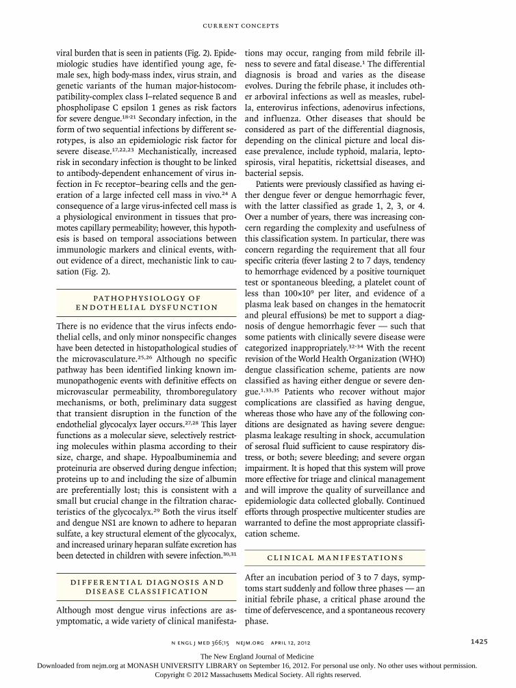
viral burden that is seen in patients (Fig. 2). Epide-miologic studies have identified young age, fe-male sex, high body-mass index, virus strain, and genetic variants of the human major-histocom-patibility-complex class I–related sequence B and phospholipase C epsilon 1 genes as risk factors for severe dengue.18-21 Secondary infection, in the form of two sequential infections by different se-rotypes, is also an epidemiologic risk factor for severe disease.17,22,23 Mechanistically, increased risk in secondary infection is thought to be linked to antibody-dependent enhancement of virus in-fection in Fc receptor–bearing cells and the gen-eration of a large infected cell mass in vivo.24 A consequence of a large virus-infected cell mass is a physiological environment in tissues that pro-motes capillary permeability; however, this hypoth-esis is based on temporal associations between immunologic markers and clinical events, with-out evidence of a direct, mechanistic link to cau-sation (Fig. 2).
Pathoph ysiol o gy of End o theli a l Dysfunc tion
There is no evidence that the virus infects endo-thelial cells, and only minor nonspecific changes have been detected in histopathological studies of the microvasculature.25,26 Although no specific pathway has been identified linking known im-munopathogenic events with definitive effects on microvascular permeability, thromboregulatory mechanisms, or both, preliminary data suggest that transient disruption in the function of the endothelial glycocalyx layer occurs.27,28 This layer functions as a molecular sieve, selectively restrict-ing molecules within plasma according to their size, charge, and shape. Hypoalbuminemia and proteinuria are observed during dengue infection; proteins up to and including the size of albumin are preferentially lost; this is consistent with a small but crucial change in the filtration charac-teristics of the glycocalyx.29 Both the virus itself and dengue NS1 are known to adhere to heparan sulfate, a key structural element of the glycocalyx, and increased urinary heparan sulfate excretion has been detected in children with severe infection.30,31
Differ en ti a l Di agnosis a nd Dise a se Cl a ssific ation
Although most dengue virus infections are as-ymptomatic, a wide variety of clinical manifesta-
tions may occur, ranging from mild febrile ill-ness to severe and fatal disease.1 The differential diagnosis is broad and varies as the disease evolves. During the febrile phase, it includes oth-er arboviral infections as well as measles, rubel-la, enterovirus infections, adenovirus infections, and influenza. Other diseases that should be considered as part of the differential diagnosis, depending on the clinical picture and local dis-ease prevalence, include typhoid, malaria, lepto-spirosis, viral hepatitis, rickettsial diseases, and bacterial sepsis.
Patients were previously classified as having ei-ther dengue fever or dengue hemorrhagic fever, with the latter classified as grade 1, 2, 3, or 4. Over a number of years, there was increasing con-cern regarding the complexity and usefulness of this classification system. In particular, there was concern regarding the requirement that all four specific criteria (fever lasting 2 to 7 days, tendency to hemorrhage evidenced by a positive tourniquet test or spontaneous bleeding, a platelet count of less than 100×109 per liter, and evidence of a plasma leak based on changes in the hematocrit and pleural effusions) be met to support a diag-nosis of dengue hemorrhagic fever — such that some patients with clinically severe disease were categorized inappropriately.32-34 With the recent revision of the World Health Organization (WHO) dengue classification scheme, patients are now classified as having either dengue or severe den-gue.1,33,35 Patients who recover without major complications are classified as having dengue, whereas those who have any of the following con-ditions are designated as having severe dengue: plasma leakage resulting in shock, accumulation of serosal fluid sufficient to cause respiratory dis-tress, or both; severe bleeding; and severe organ impairment. It is hoped that this system will prove more effective for triage and clinical management and will improve the quality of surveillance and epidemiologic data collected globally. Continued efforts through prospective multicenter studies are warranted to define the most appropriate classifi-cation scheme.
Clinic a l M a nifes tations
After an incubation period of 3 to 7 days, symp-toms start suddenly and follow three phases — an initial febrile phase, a critical phase around the time of defervescence, and a spontaneous recovery phase.
The New England Journal of Medicine Downloaded from nejm.org at MONASH UNIVERSITY LIBRARY on September 16, 2012. For personal use only. No other uses without permission.
Copyright © 2012 Massachusetts Medical Society. All rights reserved.
T h e n e w e ngl a nd j o u r na l o f m e dic i n e
n engl j med 366;15 nejm.org april 12, 20121426
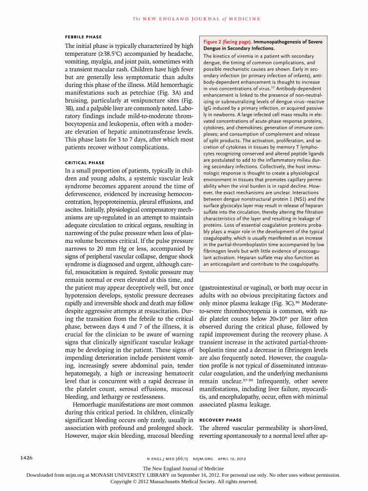
Febrile Phase
The initial phase is typically characterized by high temperature (≥38.5°C) accompanied by headache, vomiting, myalgia, and joint pain, sometimes with a transient macular rash. Children have high fever but are generally less symptomatic than adults during this phase of the illness. Mild hemorrhagic manifestations such as petechiae (Fig. 3A) and bruising, particularly at venipuncture sites (Fig. 3B), and a palpable liver are commonly noted. Labo-ratory findings include mild-to-moderate throm-bocytopenia and leukopenia, often with a moder-ate elevation of hepatic aminotransferase levels. This phase lasts for 3 to 7 days, after which most patients recover without complications.
Critical Phase
In a small proportion of patients, typically in chil-dren and young adults, a systemic vascular leak syndrome becomes apparent around the time of defervescence, evidenced by increasing hemocon-centration, hypoproteinemia, pleural effusions, and ascites. Initially, physiological compensatory mech-anisms are up-regulated in an attempt to maintain adequate circulation to critical organs, resulting in narrowing of the pulse pressure when loss of plas-ma volume becomes critical. If the pulse pressure narrows to 20 mm Hg or less, accompanied by signs of peripheral vascular collapse, dengue shock syndrome is diagnosed and urgent, although care-ful, resuscitation is required. Systolic pressure may remain normal or even elevated at this time, and the patient may appear deceptively well, but once hypotension develops, systolic pressure decreases rapidly and irreversible shock and death may follow despite aggressive attempts at resuscitation. Dur-ing the transition from the febrile to the critical phase, between days 4 and 7 of the illness, it is crucial for the clinician to be aware of warning signs that clinically significant vascular leakage may be developing in the patient. These signs of impending deterioration include persistent vomit-ing, increasingly severe abdominal pain, tender hepatomegaly, a high or increasing hematocrit level that is concurrent with a rapid decrease in the platelet count, serosal effusions, mucosal bleeding, and lethargy or restlessness.
Hemorrhagic manifestations are most common during this critical period. In children, clinically significant bleeding occurs only rarely, usually in association with profound and prolonged shock. However, major skin bleeding, mucosal bleeding
(gastrointestinal or vaginal), or both may occur in adults with no obvious precipitating factors and only minor plasma leakage (Fig. 3C).36 Moderate-to-severe thrombocytopenia is common, with na-dir platelet counts below 20×109 per liter often observed during the critical phase, followed by rapid improvement during the recovery phase. A transient increase in the activated partial-throm-boplastin time and a decrease in fibrinogen levels are also frequently noted. However, the coagula-tion profile is not typical of disseminated intravas-cular coagulation, and the underlying mechanisms remain unclear.37-39 Infrequently, other severe manifestations, including liver failure, myocardi-tis, and encephalopathy, occur, often with minimal associated plasma leakage.
Recovery Phase
The altered vascular permeability is short-lived, reverting spontaneously to a normal level after ap-
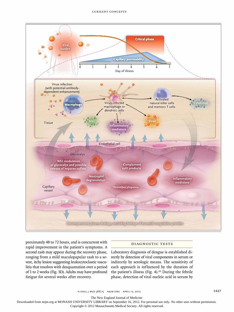
Figure 2 (facing page). Immunopathogenesis of Severe Dengue in Secondary Infections.
The kinetics of viremia in a patient with secondary dengue, the timing of common complications, and possible mechanistic causes are shown. Early in sec-ondary infection (or primary infection of infants), anti-body-dependent enhancement is thought to increase in vivo concentrations of virus.17 Antibody-dependent enhancement is linked to the presence of non-neutral-izing or subneutralizing levels of dengue virus–reactive IgG induced by a primary infection, or acquired passive-ly in newborns. A large infected cell mass results in ele-vated concentrations of acute-phase response proteins, cytokines, and chemokines; generation of immune com-plexes; and consumption of complement and release of split products. The activation, proliferation, and se-cretion of cytokines in tissues by memory T lympho-cytes recognizing conserved and altered peptide ligands are postulated to add to the inflammatory milieu dur-ing secondary infections. Collectively, the host immu-nologic response is thought to create a physiological environment in tissues that promotes capillary perme-ability when the viral burden is in rapid decline. How-ever, the exact mechanisms are unclear. Interactions between dengue nonstructural protein 1 (NS1) and the surface glycocalyx layer may result in release of heparan sulfate into the circulation, thereby altering the filtration characteristics of the layer and resulting in leakage of proteins. Loss of essential coagulation proteins proba-bly plays a major role in the development of the typical coagulopathy, which is usually manifested as an increase in the partial-thromboplastin time accompanied by low fibrinogen levels but with little evidence of procoagu-lant activation. Heparan sulfate may also function as an anticoagulant and contribute to the coagulopathy.
The New England Journal of Medicine Downloaded from nejm.org at MONASH UNIVERSITY LIBRARY on September 16, 2012. For personal use only. No other uses without permission.
Copyright © 2012 Massachusetts Medical Society. All rights reserved.
Current Concepts
n engl j med 366;15 nejm.org april 12, 2012 1427
proximately 48 to 72 hours, and is concurrent with rapid improvement in the patient’s symptoms. A second rash may appear during the recovery phase, ranging from a mild maculopapular rash to a se-vere, itchy lesion suggesting leukocytoclastic vascu-litis that resolves with desquamation over a period of 1 to 2 weeks (Fig. 3D). Adults may have profound fatigue for several weeks after recovery.
Di agnos tic Tes t s
Laboratory diagnosis of dengue is established di-rectly by detection of viral components in serum or indirectly by serologic means. The sensitivity of each approach is influenced by the duration of the patient’s illness (Fig. 4).10 During the febrile phase, detection of viral nucleic acid in serum by
Inflammatorymediators
Decompensated plasma leakage, including leakage of essential coagulation proteins
Tissue
Macrophage
Endothelial cell
Activatednatural killer cells
and memory T cells Cytokines
and memory T cells
VirusNS1
NS1 modulationof glycocalyx and possiblerelease of heparan sulfate
Virus infection(with potential antibody-
dependent enhancement)
Viralburden
Capillary permeability
0 1 2 3 4 5 6 7Day of illness
Critical phase
Capillaryvessel
Glycocalyx
Inflammatorymediators
Neutrophildegranulation
Thrombocytopenia
Complementsplit products
Virus-infectedmacrophage ordendritic cells
GlycocalyxGlycocalyxGlycocalyxGlycocalyxGlycocalyxGlycocalyxGlycocalyxGlycocalyxGlycocalyxGlycocalyxGlycocalyxGlycocalyxGlycocalyxGlycocalyx
03/26/2012
AUTHOR PLEASE NOTE:Figure has been redrawn and type has been reset
Please check carefully
AuthorFig #Title
DEMEArtist
COLOR FIGURE
Draft 3
Dengue1
ME’s nameWilliams
Farrar_ra1110265
Campion
The New England Journal of Medicine Downloaded from nejm.org at MONASH UNIVERSITY LIBRARY on September 16, 2012. For personal use only. No other uses without permission.
Copyright © 2012 Massachusetts Medical Society. All rights reserved.
T h e n e w e ngl a nd j o u r na l o f m e dic i n e
n engl j med 366;15 nejm.org april 12, 20121428
means of reverse-transcriptase–polymerase-chain-reaction (RT-PCR) assay or detection of the virus-expressed soluble nonstructural protein 1 (NS1) by means of enzyme-linked immunosorbent assay (ELISA) or the lateral-flow rapid test (not currently available in the United States) is sufficient for a confirmatory diagnosis. For primary infections in persons who have not been infected previously (which is typical in the case of most travelers), the diagnostic sensitivity of NS1 detection in the fe-brile phase can exceed 90%, and antigenemia may persist for several days after the resolution of fe-ver.40-42 The sensitivity of NS1 detection in the fe-brile phase is lower in secondary infections (60 to 80%), reflecting an anamnestic serologic response
due to a previous dengue virus or related flavivi-rus infection.43
Serologic diagnosis of dengue relies on the de-tection of high levels of serum IgM that bind den-gue virus antigens in an ELISA or a lateral-flow rapid test; IgM can be detected as early as 4 days after the onset of fever. IgM seroconversion be-tween paired samples is considered a confirma-tory finding, whereas detection of IgM in a single specimen obtained from a patient with a clinical syndrome that is consistent with dengue is widely used to establish a presumptive diagnosis. Com-mercially available IgM tests with acceptable per-formance characteristics have recently been identi-fied.44 Serologic diagnosis of dengue can be
A B
DC
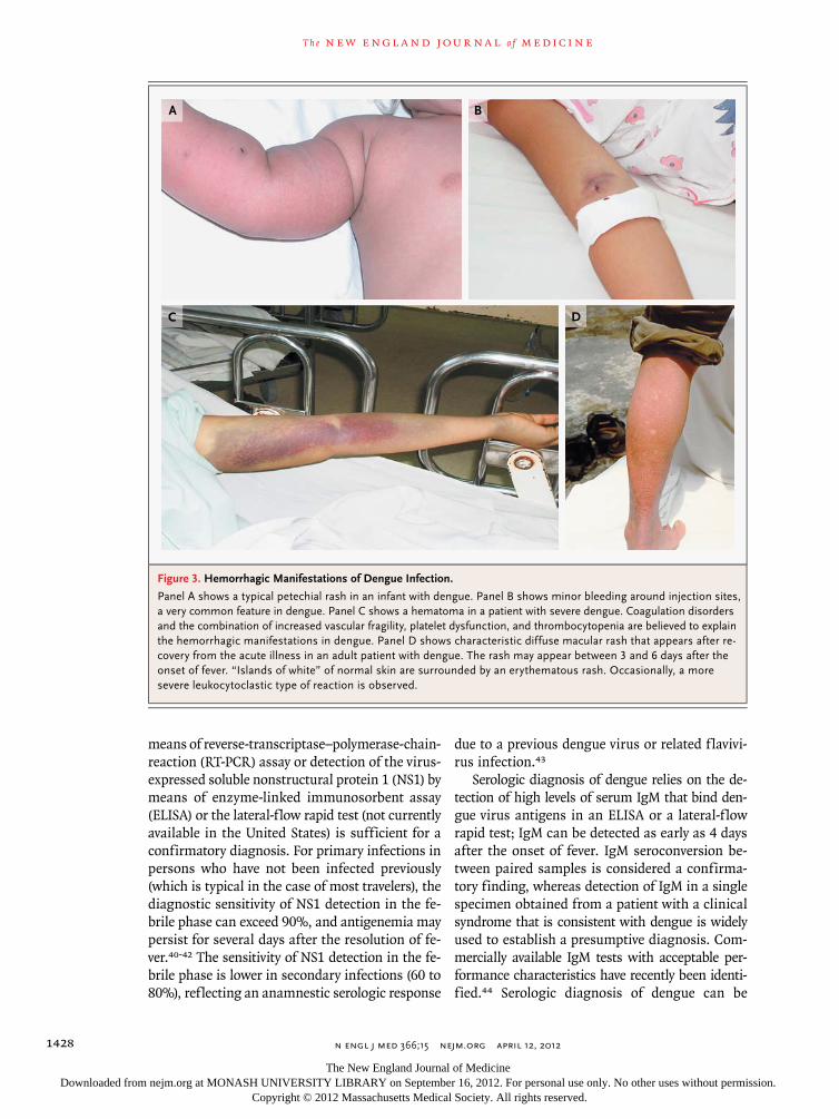
Figure 3. Hemorrhagic Manifestations of Dengue Infection.
Panel A shows a typical petechial rash in an infant with dengue. Panel B shows minor bleeding around injection sites, a very common feature in dengue. Panel C shows a hematoma in a patient with severe dengue. Coagulation disorders and the combination of increased vascular fragility, platelet dysfunction, and thrombocytopenia are believed to explain the hemorrhagic manifestations in dengue. Panel D shows characteristic diffuse macular rash that appears after re-covery from the acute illness in an adult patient with dengue. The rash may appear between 3 and 6 days after the onset of fever. “Islands of white” of normal skin are surrounded by an erythematous rash. Occasionally, a more severe leukocytoclastic type of reaction is observed.
The New England Journal of Medicine Downloaded from nejm.org at MONASH UNIVERSITY LIBRARY on September 16, 2012. For personal use only. No other uses without permission.
Copyright © 2012 Massachusetts Medical Society. All rights reserved.
Current Concepts
n engl j med 366;15 nejm.org april 12, 2012 1429
confounded if the patient has very recently been infected or vaccinated with an antigenically related flavivirus (e.g., a virus associated with yellow fever or Japanese encephalitis). In addition, patients with secondary infections mount rapid anamnestic antibody responses in which dengue virus–reactive IgG may predominate over IgM. In clinical settings where methods of molecular detection (e.g., RT-PCR) are not available, investigation for elevated levels of dengue virus–reactive IgM or soluble NS1 in serum is a pragmatic diagnostic approach in a patient in whom dengue is suspected.43,45
M a nagemen t
Currently, no effective antiviral agents to treat den-gue infection are available, and treatment remains supportive, with particular emphasis on careful fluid management.1 Patients who have no com-plications and are able to tolerate oral fluids may remain at home with instructions to return to the hospital immediately if bleeding or warning signs suggestive of vascular leakage develop. How-ever, our practice is to evaluate these patients daily in a medical clinic with a complete blood count to monitor hematocrit and platelet values.
Development of any warning sign indicates the need for hospitalization and close observation, with judicious use of parenteral fluids in patients with inadequate oral intake or a rapidly increasing hematocrit. If the condition progresses to the den-gue shock syndrome, prompt fluid resuscitation to restore plasma volume is imperative, followed by ongoing fluid therapy to support the circulation at a level just sufficient to maintain critical organ perfusion. Isotonic crystalloid solutions should be used, and isotonic colloid solutions should be re-served for patients presenting with profound shock or those who do not have a response to initial crystalloid therapy.46 To limit the risk of the de-velopment of fluid overload, parenteral fluid thera-py should be kept to the minimum required to maintain cardiovascular stability until permeabil-ity reverts to a normal level.
Blood transfusion can be lifesaving for patients with severe bleeding that compromises cardiovas-cular function, but it should be undertaken with care because of the risk of fluid overload. Platelet concentrates, fresh-frozen plasma, and cryopre-cipitate may also be needed depending on the co-agulation profile. However, at present, there is no evidence that prophylactic platelet transfusions are
of any value in patients who do not have clinically significant bleeding, even when thrombocytopenia is profound.47,48 The use of prophylactic platelet transfusions is increasing in countries where den-gue is endemic, but given the associated clinical risks and the financial costs, controlled trials need to be performed before this becomes established as the standard of care. In patients with severe dengue infection, adjuvant therapy, including vaso-pressor and inotropic therapies, renal-replacement therapy, and further treatment of organ impair-ment, may be necessary.
The establishment of a therapeutic pipeline and the design of randomized, controlled trials of drugs targeting the virus or the immune response are recent developments. Recent trials have as-sessed chloroquine,49 oral prednisolone (A Ran-domized, Placebo-Controlled, Partially Blinded [Drug versus Placebo] Trial of Early Corticosteroid Therapy in Vietnamese Children and Young Adults with suspected Dengue Infection; Current Con-trolled Trials number, ISRCTN39575233), and balapiravir (A Randomized, Double-Blind, Placebo-Controlled Study to Evaluate the Safety and Ef-ficacy of the Dengue Virus Polymerase Inhibitor [Balapiravir] in Male Patients with Confirmed Dengue Virus Infection; ClinicalTrials.gov num-
1 3 5 7 92 4 6 8 10
Day of Illness
Viremia (serum, RT-PCRassay)
NS1 (serum, ELISA, or rapid test)
IgM
IgG (secondary infection)
IgG (primary infection)
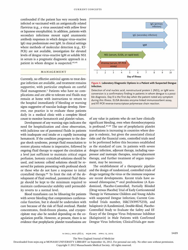
Figure 4. Laboratory Diagnostic Options in a Patient with Suspected Dengue Infection.
Detection of viral nucleic acid, nonstructural protein 1 (NS1), or IgM sero-conversion is a confirmatory finding in patients in whom dengue is a possi-ble diagnosis. Day 0 is the first day when the patient noted any symptom during this illness. ELISA denotes enzyme-linked immunosorbent assay, and RT-PCR reverse-transcriptase polymerase chain reaction.
The New England Journal of Medicine Downloaded from nejm.org at MONASH UNIVERSITY LIBRARY on September 16, 2012. For personal use only. No other uses without permission.
Copyright © 2012 Massachusetts Medical Society. All rights reserved.
T h e n e w e ngl a nd j o u r na l o f m e dic i n e
n engl j med 366;15 nejm.org april 12, 20121430
ber, NCT01096576), and further trials of statins and other antiviral drugs are planned. Currently, there is no evidence in favor of the use of any spe-cific therapeutic agent for dengue.
Effec t s on He a lth C a r e S ys tems
Dengue imposes major demands on health care systems. Although severe dengue occurs in only a small proportion of dengue infections, early iden-tification of high-risk patients is difficult and patients with uncomplicated infections are fre-quently hospitalized for observation. Rapid and effective triage by experienced personnel at the primary health care level, efficient and affordable transportation systems to facilitate daily clinical assessment, and public education campaigns to increase awareness of the disease all help to reduce unnecessary admissions. Among hospitalized pa-tients, meticulous attention to detail is necessary to limit iatrogenic complications, including fluid over-load. Ideally, patients with severe dengue infection should be treated in dedicated high-dependency units where frequent clinical observations by ex-perienced staff with immediate access to repeated hematocrit measurements can ensure that fluid therapy is carefully titrated as needed. In such circumstances, mortality of less than 1% is achiev-able among patients with shock, and the need for ventilatory support and intensive care is mini-mized. Improvements in the early diagnosis and risk prediction of severe disease are urgently need-ed, especially in areas with a high case burden, where appropriate allocation of limited resources is crucial to the outcome. Ongoing research aims to refine the WHO 2009 classification scheme, particularly with regard to warning signs for the development of severe disease.
Ne w A pproaches t o Ta rge ting the V ec t or
New vector-control approaches include the release of genetically modified male mosquitoes that ster-ilize the wild-type female population, thereby re-ducing egg output and the population size of the next generation that would be available for po-tential transmission of the dengue virus.50 An alternative strategy involves embryonic introduc-tion of strains of the obligate intracellular bacte-rium wolbachia into A. aegypti. Strikingly, wolba-
chia-infected A. aegypti are partially resistant to dengue virus infection51,52 and can invade natural A. aegypti populations,51,53 suggesting the possi-bility of induction of widespread biologic resis-tance to dengue viruses in A. aegypti populations.
Vaccines
The leading dengue vaccine candidate, ChimeriVax (Sanofi Pasteur), is a tetravalent formulation of attenuated yellow fever 17D vaccine strains express-ing the dengue virus prM and E proteins.54 It has been difficult to develop a vaccine for dengue that is safe and elicits balanced neutralizing antibody responses to all four serotypes. However, in the past 5 years, remarkable progress has been made, and multicenter phase 2–3 clinical trials that are designed to determine the efficacy of this three-dose vaccine are under way. Data on immunologic correlates of immunity are lacking. Long-term follow-up of vaccinees will be essential to under-stand whether waning vaccine-elicited immunity predisposes recipients to more severe outcomes on subsequent natural infection. Other candidates in early phases of clinical development include vac-cines containing live attenuated dengue viruses and recombinant subunit vaccines.55
Fu t ur e Dir ec tions
The field of dengue research has been invigorated over the past decade, fueled by the growing rec-ognition of the burden of disease coupled with the prospect of a dengue vaccine. However, no vaccine can be an immediate global panacea, and efforts to improve treatment through application of exist-ing best practices in triage and fluid management, along with efforts to develop new antiviral or oth-er therapeutic drugs, must continue. Similarly, in-novative approaches to preventing transmission of the virus, such as through modification of mos-quito populations, should be fostered. An improved understanding of the current epidemiology of the disease and the potential for its future spread would also assist policymakers in allocating resources to combat this global public health challenge.
Dr. Simmons reports that his institution receives consulting fees on his behalf from Unither Virology and Tibotec and grant support on his behalf from Hoffmann–La Roche. No other po-tential conflict of interest relevant to this article was reported.
Disclosure forms provided by the authors are available with the full text of this article at NEJM.org.
The New England Journal of Medicine Downloaded from nejm.org at MONASH UNIVERSITY LIBRARY on September 16, 2012. For personal use only. No other uses without permission.
Copyright © 2012 Massachusetts Medical Society. All rights reserved.
Current Concepts
n engl j med 366;15 nejm.org april 12, 2012 1431
References
1. Dengue: Guidelines for treatment, pre-vention and control. Geneva: World Health Organization, 2009.2. Mousson L, Dauga C, Garrigues T, Schaffner F, Vazeille M, Failloux AB. Phy-logeography of Aedes (Stegomyia) aegypti (L.) and Aedes (Stegomyia) albopictus (Skuse) (Diptera: Culicidae) based on mi-tochondrial DNA variations. Genet Res 2005;86:1-11.3. Lambrechts L, Scott TW, Gubler DJ. Consequences of the expanding global distribution of Aedes albopictus for den-gue virus transmission. PLoS Negl Trop Dis 2010;4(5):e646.4. Reiter P. Aedes albopictus and the world trade in used tires, 1988-1995: the shape of things to come? J Am Mosq Con-trol Assoc 1998;14:83-94.5. Schwartz E, Weld LH, Wilder-Smith A, et al. Seasonality, annual trends, and characteristics of dengue among ill re-turned travelers, 1997-2006. Emerg Infect Dis 2008;14:1081-8.6. Streit JA, Yang M, Cavanaugh JE, Pol-green PM. Upward trend in dengue inci-dence among hospitalized patients, Unit-ed States. Emerg Infect Dis 2011;17:914-6.7. Libraty DH, Young PR, Pickering D, et al. High circulating levels of the dengue virus nonstructural protein NS1 early in dengue illness correlate with the develop-ment of dengue hemorrhagic fever. J In-fect Dis 2002;186:1165-8.8. Libraty DH, Endy TP, Houng HS, et al. Differing influences of virus burden and immune activation on disease severity in secondary dengue-3 virus infections. J In-fect Dis 2002;185:1213-21.9. Vaughn DW, Green S, Kalayanarooj S, et al. Dengue viremia titer, antibody re-sponse pattern, and virus serotype corre-late with disease severity. J Infect Dis 2000;181:2-9.10. Peeling RW, Artsob H, Pelegrino JL, et al. Evaluation of diagnostic tests: dengue. Nat Rev Microbiol 2010;8:Suppl:S30-S38.11. Cardosa J, Ooi MH, Tio PH, et al. Den-gue virus serotype 2 from a sylvatic lin-eage isolated from a patient with dengue hemorrhagic fever. PLoS Negl Trop Dis 2009;3(4):e423.12. Brien JD, Austin SK, Sukupolvi-Petty S, et al. Genotype-specific neutralization and protection by antibodies against den-gue virus type 3. J Virol 2010;84:10630-43.13. Wahala WM, Donaldson EF, de Alwis R, Accavitti-Loper MA, Baric RS, de Silva AM. Natural strain variation and antibody neutralization of dengue serotype 3 vi-ruses. PLoS Pathog 2010;6(3):e1000821.14. Hang VTT, Holmes EC, Veasna D, et al. Emergence of the Asian 1 genotype of dengue virus serotype 2 in Viet Nam: in vivo fitness advantage and lineage re-placement in South-East Asia. PLoS Negl Trop Dis 2010;4(7):e757.
15. Holmes EC, Twiddy SS. The origin, emergence and evolutionary genetics of dengue virus. Infect Genet Evol 2003;3:19-28.16. Messer WB, Gubler DJ, Harris E, Siva-nanthan K, de Silva AM. Emergence and global spread of a dengue serotype 3, sub-type III virus. Emerg Infect Dis 2003; 9:800-9.17. Sangkawibha N, Rojanasuphot S, Ah-andrik S, et al. Risk factors in dengue shock syndrome: a prospective epidemio-logic study in Rayong, Thailand. I. The 1980 outbreak. Am J Epidemiol 1984;120: 653-69.18. Anders KL, Nguyet NM, Chau NV, et al. Epidemiological factors associated with dengue shock syndrome and mortal-ity in hospitalized dengue patients in Ho Chi Minh City, Vietnam. Am J Trop Med Hyg 2011;84:127-34.19. Khor CC, Chau TN, Pang J, et al. Ge-nome-wide association study identifies susceptibility loci for dengue shock syn-drome at MICB and PLCE1. Nat Genet 2011;43:1139-41.20. Nguyen TH, Nguyen TL, Lei HY, et al. Association between sex, nutritional sta-tus, severity of dengue hemorrhagic fever, and immune status in infants with den-gue hemorrhagic fever. Am J Trop Med Hyg 2005;72:370-4.21. Rico-Hesse R, Harrison LM, Salas RA, et al. Origins of dengue type 2 viruses as-sociated with increased pathogenicity in the Americas. Virology 1997;230:244-51.22. Burke DS, Nisalak A, Johnson DE, Scott RM. A prospective study of dengue infections in Bangkok. Am J Trop Med Hyg 1988;38:172-80.23. Kouri GP, Guzmán MG, Bravo JR, Tri-ana C. Dengue haemorrhagic fever/den-gue shock syndrome: lessons from the Cuban epidemic, 1981. Bull World Health Organ 1989;67:375-80.24. Halstead SB. Antibody, macrophages, dengue virus infection, shock, and hem-orrhage: a pathogenetic cascade. Rev In-fect Dis 1989;11:Suppl 4:S830-S839.25. Jessie K, Fong MY, Devi S, Lam SK, Wong KT. Localization of dengue virus in naturally infected human tissues, by im-munohistochemistry and in situ hybrid-ization. J Infect Dis 2004;189:1411-8.26. Leong AS, Wong KT, Leong TY, Tan PH, Wannakrairot P. The pathology of dengue hemorrhagic fever. Semin Diagn Pathol 2007;24:227-36.27. Michel CC, Curry FE. Microvascular permeability. Physiol Rev 1999;79:703-61.28. Levick JR, Michel CC. Microvascular fluid exchange and the revised Starling principle. Cardiovasc Res 2010;87:198-210.29. Wills BA, Oragui EE, Dung NM, et al. Size and charge characteristics of the pro-tein leak in dengue shock syndrome. J Infect Dis 2004;190:810-8.
30. Avirutnan P, Zhang L, Punyadee N, et al. Secreted NS1 of dengue virus attaches to the surface of cells via interactions with heparan sulfate and chondroitin sulfate E. PLoS Pathog 2007;3(11):e183.31. Chen Y, Maguire T, Hileman RE, et al. Dengue virus infectivity depends on enve-lope protein binding to target cell hepa-ran sulfate. Nat Med 1997;3:866-71.32. Phuong CX, Nhan NT, Kneen R, et al. Clinical diagnosis and assessment of se-verity of confirmed dengue infections in Vietnamese children: is the World Health Organization classification system help-ful? Am J Trop Med Hyg 2004;70:172-9. [Erratum, Am J Trop Hyg 2004;70:459.]33. Deen JL, Harris E, Wills B, et al. The WHO dengue classification and case defi-nitions: time for a reassessment. Lancet 2006;368:170-3.34. Rigau-Pérez JG. Severe dengue: the need for new case definitions. Lancet In-fect Dis 2006;6:297-302.35. Alexander N, Balmaseda A, Coelho IC, et al. Multicentre prospective study on dengue classification in four Southeast Asian and three Latin American coun-tries. Trop Med Int Health 2011 May 30 (Epub ahead of print).36. Wichmann O, Hongsiriwon S, Bo-wonwatanuwong C, Chotivanich K, Suk-thana Y, Pukrittayakamee S. Risk factors and clinical features associated with se-vere dengue infection in adults and chil-dren during the 2001 epidemic in Chon-buri, Thailand. Trop Med Int Health 2004;9:1022-9.37. Wills BA, Oragui EE, Stephens AC, et al. Coagulation abnormalities in dengue hemorrhagic fever: serial investigations in 167 Vietnamese children with dengue shock syndrome. Clin Infect Dis 2002;35:277-85.38. Wills B, Tran VN, Nguyen TH, et al. Hemostatic changes in Vietnamese chil-dren with mild dengue correlate with the severity of vascular leakage rather than bleeding. Am J Trop Med Hyg 2009;81:638-44.39. Mairuhu AT, Mac Gillavry MR, Setiati TE, et al. Is clinical outcome of dengue-virus infections influenced by coagulation and fibrinolysis? A critical review of the evidence. Lancet Infect Dis 2003;3:33-41.40. Tricou V, Minh NN, Farrar J, Tran HT, Simmons CP. Kinetics of viremia and NS1 antigenemia are shaped by immune status and virus serotype in adults with dengue. PLoS Negl Trop Dis 2011;5(9):e1309.41. Chaterji S, Allen JC Jr, Chow A, Leo YS, Ooi EE. Evaluation of the NS1 rapid test and the WHO dengue classification schemes for use as bedside diagnosis of acute dengue fever in adults. Am J Trop Med Hyg 2011;84:224-8.42. Dussart P, Petit L, Labeau B, et al. Evaluation of two new commercial tests
The New England Journal of Medicine Downloaded from nejm.org at MONASH UNIVERSITY LIBRARY on September 16, 2012. For personal use only. No other uses without permission.
Copyright © 2012 Massachusetts Medical Society. All rights reserved.
n engl j med 366;15 nejm.org april 12, 20121432
Current Concepts
for the diagnosis of acute dengue virus infection using NS1 antigen detection in human serum. PLoS Negl Trop Dis 2008; 2(8):e280.43. Guzman MG, Jaenisch T, Gaczkowski R, et al. Multi-country evaluation of the sensitivity and specificity of two commer-cially-available NS1 ELISA assays for den-gue diagnosis. PLoS Negl Trop Dis 2010; 4(8):e811.44. Hunsperger EA, Yoksan S, Buchy P, et al. Evaluation of commercially available anti-dengue virus immunoglobulin M tests. Emerg Infect Dis 2009;15:436-40.45. Fry SR, Meyer M, Semple MG, et al. The diagnostic sensitivity of dengue rapid test assays is significantly enhanced by using a combined antigen and antibody testing approach. PLoS Negl Trop Dis 2011;5(6):e1199.46. Wills BA, Dung NM, Loan HT, et al. Comparison of three fluid solutions for
resuscitation in dengue shock syndrome. N Engl J Med 2005;353:877-89.47. Thomas L, Kaidomar S, Kerob-Bauchet B, et al. Prospective observational study of low thresholds for platelet trans-fusion in adult dengue patients. Transfu-sion 2009;49:1400-11.48. Lye DC, Lee VJ, Sun Y, Leo YS. Lack of efficacy of prophylactic platelet transfu-sion for severe thrombocytopenia in adults with acute uncomplicated dengue infection. Clin Infect Dis 2009;48:1262-5.49. Tricou V, Minh NN, Van TP, et al. A randomized controlled trial of chloro-quine for the treatment of dengue in Viet-namese adults. PLoS Negl Trop Dis 2010; 4(8):e785.50. Wise de Valdez MR, Nimmo D, Betz J, et al. Genetic elimination of dengue vec-tor mosquitoes. Proc Natl Acad Sci U S A 2011;108:4772-5.
51. Moreira LA, Iturbe-Ormaetxe I, Jef-fery JA, et al. A Wolbachia symbiont in Aedes aegypti limits infection with den-gue, Chikungunya, and Plasmodium. Cell 2009;139:1268-78.52. Walker T, Johnson PH, Moreira LA, et al. The wMel Wolbachia strain blocks dengue and invades caged Aedes aegypti populations. Nature 2011;476:450-3.53. Hoffmann AA, Montgomery BL, Popovici J, et al. Successful establishment of Wolbachia in Aedes populations to suppress dengue transmission. Nature 2011;476:454-7.54. Guy B, Saville M, Lang J. Development of Sanofi Pasteur tetravalent dengue vac-cine. Hum Vaccin 2010;6:9.55. Durbin AP, Whitehead SS. Dengue vaccine candidates in development. Curr Top Microbiol Immunol 2010;338:129-43.Copyright © 2012 Massachusetts Medical Society.
nejm 200th anniversary and social media
Follow NEJMTeam on Twitter and click “Like” on the New England Journal of Medicine page on Facebook for links to the latest articles, stories, and multimedia available
at the NEJM 200th Anniversary website, http://NEJM200.NEJM.org. Tweets incorporating the hashtag #NEJM200 also appear
in a Twitter feed at the anniversary website.
The New England Journal of Medicine Downloaded from nejm.org at MONASH UNIVERSITY LIBRARY on September 16, 2012. For personal use only. No other uses without permission.
Copyright © 2012 Massachusetts Medical Society. All rights reserved.


![Dengue Fever/Severe Dengue Fever/Chikungunya Fever · Dengue fever and severe dengue (dengue hemorrhagic fever [DHF] and dengue shock syndrome [DSS]) are caused by any of four closely](https://static.fdocuments.net/doc/165x107/5e87bf3e7a86e85d3b149cd7/dengue-feversevere-dengue-feverchikungunya-dengue-fever-and-severe-dengue-dengue.jpg)